EP0310026B1 - Cardiac and pulmonary physiological analysis via intracardiac measurements with a single sensor - Google Patents
Cardiac and pulmonary physiological analysis via intracardiac measurements with a single sensor Download PDFInfo
- Publication number
- EP0310026B1 EP0310026B1 EP88115989A EP88115989A EP0310026B1 EP 0310026 B1 EP0310026 B1 EP 0310026B1 EP 88115989 A EP88115989 A EP 88115989A EP 88115989 A EP88115989 A EP 88115989A EP 0310026 B1 EP0310026 B1 EP 0310026B1
- Authority
- EP
- European Patent Office
- Prior art keywords
- signal
- patient
- heart
- activity
- cardiac
- Prior art date
- Legal status (The legal status is an assumption and is not a legal conclusion. Google has not performed a legal analysis and makes no representation as to the accuracy of the status listed.)
- Expired - Lifetime
Links
Images


Classifications
-
- A—HUMAN NECESSITIES
- A61—MEDICAL OR VETERINARY SCIENCE; HYGIENE
- A61N—ELECTROTHERAPY; MAGNETOTHERAPY; RADIATION THERAPY; ULTRASOUND THERAPY
- A61N1/00—Electrotherapy; Circuits therefor
- A61N1/18—Applying electric currents by contact electrodes
- A61N1/32—Applying electric currents by contact electrodes alternating or intermittent currents
- A61N1/36—Applying electric currents by contact electrodes alternating or intermittent currents for stimulation
- A61N1/362—Heart stimulators
- A61N1/365—Heart stimulators controlled by a physiological parameter, e.g. heart potential
- A61N1/36514—Heart stimulators controlled by a physiological parameter, e.g. heart potential controlled by a physiological quantity other than heart potential, e.g. blood pressure
- A61N1/36521—Heart stimulators controlled by a physiological parameter, e.g. heart potential controlled by a physiological quantity other than heart potential, e.g. blood pressure the parameter being derived from measurement of an electrical impedance
-
- A—HUMAN NECESSITIES
- A61—MEDICAL OR VETERINARY SCIENCE; HYGIENE
- A61N—ELECTROTHERAPY; MAGNETOTHERAPY; RADIATION THERAPY; ULTRASOUND THERAPY
- A61N1/00—Electrotherapy; Circuits therefor
- A61N1/18—Applying electric currents by contact electrodes
- A61N1/32—Applying electric currents by contact electrodes alternating or intermittent currents
- A61N1/36—Applying electric currents by contact electrodes alternating or intermittent currents for stimulation
- A61N1/362—Heart stimulators
- A61N1/365—Heart stimulators controlled by a physiological parameter, e.g. heart potential
- A61N1/36514—Heart stimulators controlled by a physiological parameter, e.g. heart potential controlled by a physiological quantity other than heart potential, e.g. blood pressure
- A61N1/36571—Heart stimulators controlled by a physiological parameter, e.g. heart potential controlled by a physiological quantity other than heart potential, e.g. blood pressure controlled by blood flow rate, e.g. blood velocity or cardiac output
Definitions
- This invention relates to the analysis of instantaneous physiological parameters affecting the heart and lungs and more particularly it relates to means and methods of measuring with a single sensor both cardiac and pulmonary physiological parameters of a patient to derive and analyze data useful in treating the patient for cardiac and pulmonary conditions such as the control of a cardiac pacemaker in accordance with dynamic changes incurred in physiological activity of the patient.
- cardiac and respiratory activity as physiological functional parameters is essential. They are usually monitored using a plurality of measuring elements such as cardiac catheters, special breathing sensors, etc. For this monitoring, not only is the display and evaluation of the functional parameters difficult to coordinate, but also the placement of the detection instruments within the heart and lungs of a patient is difficult.
- pacing rate is controlled for stable long-term control from the temperature of the venous blood within the heart and from an activity sensor for short-term exercise related activity.
- the temperature signals can be modulated by the activity signals for an optimal adaptation of the pacing rate to the particular exercise of the patient.
- Different sensors may be used to check the two functional parameters.
- the pacemaker control is based on the finding that essentially only absolute parameters such as the blood temperature and activity should be used as absolute values for determining a relationship between these parameters and the pacing rate, whereas other physiological functional parameters are merely relative parameters, which at least impede stable long-term control of the pacemaker.
- a heart pacemaker for supplying stimulation pulses to a heart via a pacemaker electrode.
- the stimulation current and the stimulation voltage are measured.
- the ratio of these values is built to form an impedance signal corresponding to the electrode impedance. From this signal a signal corresponding to the respiration rate of the patient is filtered out and serves to control the pulse rate of the pacemaker.
- the impedance is therefore measured roughly five times.
- respiration rate of the patient is determined and employed for controlling the pulse rate of the pulse generator of the heart pacemaker in accordance therewith. It is not intended and not possible to determine other physiological parameters of a patient which vary faster than the respiration rate, and especially it is not possible to determine the cardiac activity of the patient.
- a biomedical apparatus especially a heart pacer is disclosed which is capable of sensing changes in the heart's ventricular volume or stroke volume.
- a relatively low frequency signal under 5 kHz, is applied on a bipolar electrode disposed in the heart, whereby the beating action of the heart serves to modulate this signal due to changes in impedance.
- the modulated signal is then processed to remove R-wave and other electrical artifacts and then demodulated to remove the carrier frequency component leaving the envelope which is proportional to the instantaneous ventricular volume.
- This envelope signal contains stroke volume and ventricular volume information and the stroke volume may be used as parameter to control the frequency of a pulse generator in the heart pacer. Therefore, according to this patent all "electrical" components of the single variable signal are removed and are not used to control the frequency of the pacemaker.
- Another problem with prior art sensors is the ability to dynamically respond closely enough to real time to those physiological parameters of a patient that produce proper control signals for heartbeat stimulus provided by a pacemaker.
- temperature measurements slowly respond and pressure measurements are subject to outside noise influences such as coughing or sneezing that should not affect the heartbeat rate.
- the simplicity of prior art detectors was primarily due to the use of a single variable such as respiratory or blood pressure fluctuations. However, this does not give enough data for successful diagnosis, therapy or pacing rate control. Thus, the problem of simplicity in system and installation of detectors has not been resolved in such a way to produce the desired physiological parameters for analysis and control.
- the present invention as defined in independent claim 1 thus affords a single sensor within the heart operable to measure a single intracardiac functional parameter, and means to derive from that measurement both pulmonary activity and cardiac activity.
- This detector in the case of a patient having a pacemaker is the already implanted stimulus electrode, preferably on the right side of the heart and to use that electrode both as an intracardiac detector and as a heart pacer.
- both intracardiac pressure fluctuations correlating with the patient's breathing and physiological signals coming from the heart itself can be detected using only one measuring element located within the heart and detecting only one integral intracardiac functional parameter.
- the invention also comprises a method of monitoring physiological functioning of the lungs and the heart as defined in independent claim 16.
- a single sensor in the right ventricle can determine from changes of blood parameters, preferably impedance, the necessary functional parameters for proper heartbeat rate control of a pacemaker, namely the rate and depth of respiration, contractility of the myocardium, stroke volume, etc.
- An essential advantage of the invention is that the sole intracardiac measurement, preferably impedance, allows for respiratory and cardiac functional parameters to be clearly distinguished from each other by appropriate filtering with respective high pass and low pass filters. This is not possible with prior art measurements such as the impedance measurement of breathing with a thoracic secondary electrode, in part because the respiratory effects on the signals detected are such that the overall information signal has little indicative value and is superimposed with high interference. Such mutual interference of respiratory or pulmonary and cardiac functional parameters is reliably eliminated by this invention.
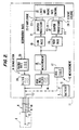
- FIG. 1 shows a cardiac pacemaker 2 implanted in a patient 1, comprising a can 3 from which a probe 4 leads via a vein into heart 5.
- Probe 4 is designed at its front end located in the ventricle as a single sensor 6, which in this embodiment has a first electrode pole 7 located at the tip of the probe 4 and a second electrode pole 8 located more proximally.
- electrical lines 9 and 10 within probe 4 connect the two spaced poles 7 and 8 located within the right ventricle with a control circuit 11 disposed in can 3.
- the block diagram control circuit 11 has functionally defined control circuit elements which can be embodied into an integrated circuit along with associated microprocessing means and appropriate software.
- the control circuit comprises generally a measurement section 12, a logic section 13 and a stimulation section 14. Energy is supplied by battery 15.
- One line 10 from detector electrode pole 8 is connected to ground potential.
- the other detector signal line 9 is coupled to the signal measurement section 12 by high pass filter 16.
- An oscillator 17 provides alternating current of low amplitude unable to pace the heart for impedance measurement with a frequency between approximately 1 kHz and 100 kHz that does not interfere with pacer stimulation.
- the current signal of oscillator 17 is also applied in addition to the stimulus pacer signal 25 to the measuring-stimulus electrode 6 by way of the dotted lead.
- Physiological activity detection or measuring means 18 then processes the single variable signal or subsignal derived from it such as the lower and higher frequency components of the dynamic signals sensed at electrode 6 and modulated on the high frequency oscillations from oscillator 17 supplied via filter 16.
- This single variable signal responds to the changes in physiological parameters such as changes in volume, flow or pressure in the patient's vascular system preferably within the right heart.
- the preferred embodiment of a detection and measurement system for this purpose responds to the variations of blood impedance indirectly indicating the changes in volume.
- the resistance (or impedance) within the heart between the single sensor 6 electrode poles 7 and 8, as defined by Ohm's law, is determined in the impedance measuring means 18 following high-pass filter 16 which transmits the carrier signals from the single signal reproduced by sensor 6 varying with cardiac and pulmonary activity modulated upon the oscillator 17 signal frequency, and rejects the pacing signals 25 passed through low-pass filter 26.
- the resulting raw signal varying dynamically in impedance is fed on one hand to a low-pass filter circuit 19 and on the other hand to a high-pass filter circuit 20, which splits the impedance modulated signal into lower and higher frequency subsignal portions.
- low-pass circuit 19 passes physiological activity signals associated with the patient's lower rate respiratory activity
- high-pass circuit 20 passes physiological activity signals associated with the patient's cardiac activity. Since the heart rate is generally four to five times greater than the respiratory rate, these respiratory or pulmonary and cardiac signals can be separated by filters within the known state of the art.
- the output signals of low-pass and high-pass circuits 19, 20 are each fed to a respective signal shaping circuits 21, 22 for pre-evaluation, e. g. averaging, determination of the derivative in time, evaluation of amplitude and frequency and subsequent integration, or the like.
- An output line 23-1 of low-pass circuit 21 then provides a signal associated with the respiratory rate and corresponding to the periodic frequency of the low-frequency signal.
- the further output line 23-2 provides a signal associated with the depth of respiration and corresponding to the amplitude of the low-frequency signal.
- An output line 24-1 of high-pass circuit 22 provides a signal associated with the contractility of the heart and corresponding to the derivative in time (dV/dt) of the high-pass filtered impedance signal, i. e. the rate of change in time of the systolic stroke volume.
- a further output line 24-2 provides a signal associated with the stroke volume of the heart and corresponding to the amplitude of the high-passed impedance signal.
- All output lines 23, 24 are connected to logic circuits 13, which calculate on the basis of available signals, an optimum pacing rate related to the exercise of the pacemaker wearer.
- This pacing rate is fed to a pulse generator 25 in stimulation signal section 14, which provides corresponding stimulation pulses to the single sensor electrode 6 via a filter 26.
- the frequencies of measuring channel 12 and stimulation channel 14 are separated by filters 16 and 26 so that the signals in one channel do not interfere and impair the functioning of the other channel. In this manner the single sensor electrode 6 can be used both as a measuring electrode and as a stimulation electrode.
- FIG. 3 in the lower frequency curve denoted by the square coordinate markers the filter characteristic of low-pass filter 19 are typified. It is seen that the degree of transmission stated in % has dropped virtually to zero at a frequency of one hertz. Signals correlating with cardiac activity have a frequency higher than this value, so that there is no interference with the low-passed signals representative of breathing rate.
- the low-passed breathing signals are processed in signal shaping circuit 21, and if necessary can be amplitude corrected to allow for filter characteristics, to provide signals for evaluation of the amplitude of respiration.
- the filter characteristics of the high-pass circuit 20, as shown by the curve with the circular coordinate points permits the signals based on cardiac activity to pass without interference with or distortion by the respiratory signals.
- the reference waveform is the electrocardiogram ECG, with the indirect volume measurement taken by impedance measurement shown as oscillation signals occurring during the ventilation period and superimposed on the caridac activity waveform.
- the pressure (P RA ) within the right atrium during a series of heartbeats is prominently shown to be influenced by intervening pulmonary activity.
- the pressure waveform in the right ventricle (P RV ) with both higher and lower frequencies is shown to correlate with the corresponding impedance (Imp) waveform taken from the intracardiac impedance measurements. Therefore, the intracardiac impedance measurements and the intracardiac pressure measurements within the right ventricle provide periodic and amplitude signal data from both the cardiac and the pulmonary activity of the patient.
- Figure 7 shows the impedance in the upper line, the directly measured exhalation (EX) in terms of time and volume in the middle line and the ECG in the lower line.
- the inhalation phases are clearly recognizable in the upper line by a decrease of impedance due to inhalation.
- the intrathoracic pressure drop during inhalation has a strong blood suction effect and thus leads to a greater filling of the right ventricle.
- This increase in right ventricular volume is expressed in a corresponding drop in impedance, since more blood of lower impedance is found in the vicinity of the electrode.
- the increase in pulmonary impedance following an increased amount of air in the lungs due to inhalation plays a negligible part in the inventive intraventricular measurement.
- the principle of simultaneous detection of cardiac and pulmonary parameters in the heart by means of the stated principle of measurement not only opens up possibilities for rate adaptive pacemaker therapy, but also offers a good possibility of detecting and monitoring essential physiological parameters for other diagnostic or therapeutic purposes.
- An example is care of patients in an intensive unit, where therapeutic consequences depend on knowledge of vital data such as cardiorespiratory activity. Since many of these patients are supplied for a short time with pacemaker electrodes anyway, the data can be obtained at the same time without any additional intervention.
- the preferred object of our investigations was the change in intracardiac impedance due to the influence of cardiac and respiratory activity.
- the use of impedance measurements is appropriate because the bipolar electrode already used routinely to stimulate the heart with both poles being located in the heart also can be used for impedance measurement. In this case no additional sensor is required.
Description
- This invention relates to the analysis of instantaneous physiological parameters affecting the heart and lungs and more particularly it relates to means and methods of measuring with a single sensor both cardiac and pulmonary physiological parameters of a patient to derive and analyze data useful in treating the patient for cardiac and pulmonary conditions such as the control of a cardiac pacemaker in accordance with dynamic changes incurred in physiological activity of the patient.
- In modern intensive medicine a successful therapeutic intervention is only possible on the basis of extensive diagnostic information. Knowledge of cardiac and respiratory activity as physiological functional parameters is essential. They are usually monitored using a plurality of measuring elements such as cardiac catheters, special breathing sensors, etc. For this monitoring, not only is the display and evaluation of the functional parameters difficult to coordinate, but also the placement of the detection instruments within the heart and lungs of a patient is difficult.
- Many such functional parameters are also dependent upon a patient's exercise, so that they can also be used to control dynamic variations in the pacing rate of a cardiac pacemaker.
- Some available publications describe pacing rate control of a pacemaker by measured signals based on the detection of one physiological functional parameter. Thus, in U. S. Patent 4,566,456, G. Koning et al., Jan. 28, 1986, the systolic pressure and change in time of the right ventricular pressure is used as the functional parameter. In German Offenlegungsschrift 27 17 659, A. Wirtzfeld, et al., published Oct. 26, 1978, the central venous oxygen saturation parameter is used. In U.S. Patent 4,535,774, W. H. Olson, Aug. 20, 1985 and U.S. Patent 4,674,518, R. W. Salo, June 23, 1987, the ventricular stroke volume of the heart is determined by means of an impedance measurement. In U.S. Patent 4,567,892, G. Plicchi, et al., Feb. 4, 1986, the respiratory rate is determined from an implanted secondary electrode by an impedance measurement. In U.S. Patent 4,697,591, A. Lekholm, et al., Oct. 6, 1987, the respiratory rate is determined from impedance across the chest cavity by using the can and heart implant electrodes. In U.S. Patent 4,596,251, G. Plicchi, et al., June 24, 1986, the respiratory minute volume is measured by impedance changes from at least one electrode located in the chest cavity. Other related respiratory rate controls are effected in U.S. Patents: 3,593,718, J. L. Krasner et al., July 20, 1971; 4,721,110, M. S. Lampadius, Jan. 26, 1988 and 4,702,253, T. A. Nappholz et al., Oct. 27, 1987. In U.S. Patent 4,291,699, L. A. Geddes, et al. Sept. 29, 1981 the change of impedance between two electrodes in one ventricle is used to indicate and control fibrillation of the heart. In U.S. Patent 4,576,183 G. Plicchi, et al., Mar. 18, 1986 subcutaneous electrodes in a patient's chest are used to measure impedance for obtaining a respiratory parameter.
- Recently there have also been proposals to control the pacing rate of a cardiac pacemaker from two or more physiological functional parameters. In German Patent P 36 31 155 , published March 24, 1988, pacing rate is controlled for stable long-term control from the temperature of the venous blood within the heart and from an activity sensor for short-term exercise related activity. The temperature signals can be modulated by the activity signals for an optimal adaptation of the pacing rate to the particular exercise of the patient. Different sensors may be used to check the two functional parameters. The pacemaker control is based on the finding that essentially only absolute parameters such as the blood temperature and activity should be used as absolute values for determining a relationship between these parameters and the pacing rate, whereas other physiological functional parameters are merely relative parameters, which at least impede stable long-term control of the pacemaker.
- U.S. Patent 4,722,342, D. Amundson, Feb. 2, 1988 provides a plurality of different body activity sensors to derive variable pacer controls for body activity.
- In EP-A-0218007 a heart pacemaker is disclosed for supplying stimulation pulses to a heart via a pacemaker electrode. During the stimulation pulses the stimulation current and the stimulation voltage are measured. In a divider the ratio of these values is built to form an impedance signal corresponding to the electrode impedance. From this signal a signal corresponding to the respiration rate of the patient is filtered out and serves to control the pulse rate of the pacemaker. During one respiratory interval the impedance is therefore measured roughly five times. After the stimulation, when the heart contracts, no measurement occurs. Therefore, after the contraction of the heart no relevant signals are revealed by the sensor so that only slowly varying parameters can be measured by this method. Consequently, only the respiration rate of the patient is determined and employed for controlling the pulse rate of the pulse generator of the heart pacemaker in accordance therewith. It is not intended and not possible to determine other physiological parameters of a patient which vary faster than the respiration rate, and especially it is not possible to determine the cardiac activity of the patient.
- In US-A-4686987 a biomedical apparatus, especially a heart pacer is disclosed which is capable of sensing changes in the heart's ventricular volume or stroke volume. A relatively low frequency signal, under 5 kHz, is applied on a bipolar electrode disposed in the heart, whereby the beating action of the heart serves to modulate this signal due to changes in impedance. The modulated signal is then processed to remove R-wave and other electrical artifacts and then demodulated to remove the carrier frequency component leaving the envelope which is proportional to the instantaneous ventricular volume. This envelope signal contains stroke volume and ventricular volume information and the stroke volume may be used as parameter to control the frequency of a pulse generator in the heart pacer. Therefore, according to this patent all "electrical" components of the single variable signal are removed and are not used to control the frequency of the pacemaker.
- In this and related prior art instrumentation for analyzing a patient's physiological condition as related to desired and actual cardiac activity there are unresolved problems corrected by this invention. Thus, the prior art does not provide simple easy to install in the patient detectors, nor do the detectors produce adequate physiological functional parameters providing little possibility of error in derived signals for introducing dynamic changes in heartbeat stimulus produced by pacemakers.
- For example, when multiple sensors are used at different body locations, such as chest and heart cavities, not only is there a chance for erroneous control signals, but there is the corresponding necessity to implant special detectors for this purpose. The chances for control error may be typified by the measurement of impedance across a chest cavity to derive ventilatory response signals under different conditions of activity. Thus, the measured impedance can vary with the position of a patient's body or arm, and is not solely restricted to the period or magnitude of inspiration or expiration. Thus, false control signals could adjust the pacing rate in response to sensed respiratory physiological parameters. Such false signals can also come from interference between multiple sensors and complex processing systems for analyzing and merging various detected signals.
- Another problem with prior art sensors is the ability to dynamically respond closely enough to real time to those physiological parameters of a patient that produce proper control signals for heartbeat stimulus provided by a pacemaker. Thus, for example, temperature measurements slowly respond and pressure measurements are subject to outside noise influences such as coughing or sneezing that should not affect the heartbeat rate.
- The simplicity of prior art detectors was primarily due to the use of a single variable such as respiratory or blood pressure fluctuations. However, this does not give enough data for successful diagnosis, therapy or pacing rate control. Thus, the problem of simplicity in system and installation of detectors has not been resolved in such a way to produce the desired physiological parameters for analysis and control. The present invention as defined in
independent claim 1 thus affords a single sensor within the heart operable to measure a single intracardiac functional parameter, and means to derive from that measurement both pulmonary activity and cardiac activity. This detector in the case of a patient having a pacemaker is the already implanted stimulus electrode, preferably on the right side of the heart and to use that electrode both as an intracardiac detector and as a heart pacer. - Accordingly, both intracardiac pressure fluctuations correlating with the patient's breathing and physiological signals coming from the heart itself can be detected using only one measuring element located within the heart and detecting only one integral intracardiac functional parameter.
- The invention also comprises a method of monitoring physiological functioning of the lungs and the heart as defined in
independent claim 16. - Investigations by applicant correlate breathing and intrathoracic pressure fluctuations with physiological parameters of blood measured in the heart. Thus, for example, a single sensor in the right ventricle can determine from changes of blood parameters, preferably impedance, the necessary functional parameters for proper heartbeat rate control of a pacemaker, namely the rate and depth of respiration, contractility of the myocardium, stroke volume, etc.
- Great advantages are obtained in control of a cardiac pacemaker in response to these detected signals. Significant is the simple construction with the measuring element constituting the simultaneously used stimulation electrode, thus necessitating no further implants.
- An essential advantage of the invention is that the sole intracardiac measurement, preferably impedance, allows for respiratory and cardiac functional parameters to be clearly distinguished from each other by appropriate filtering with respective high pass and low pass filters. This is not possible with prior art measurements such as the impedance measurement of breathing with a thoracic secondary electrode, in part because the respiratory effects on the signals detected are such that the overall information signal has little indicative value and is superimposed with high interference. Such mutual interference of respiratory or pulmonary and cardiac functional parameters is reliably eliminated by this invention.
- The invention shall be explained in more detail in an exemplary embodiment in connection with a cardiac pacemaker with reference to the accompanying drawings, in which:
- Figure 1 shows a schematic view of an inventive cardiac pacemaker electrode system implanted in a patient's heart,
- Figure 2 shows in block diagram form the electronic diagnosis system afforded by this invention,
- Figure 3 is a waveform diagram showing the bandwidth characteristics of the respiratory and cardiac signal components for forming low-pass and high-pass filters,
- Figures 4, 5 and 6 are signal waveform diagrams illustrating the impedance curves for intracardiac measurements in animals, with various parameters, and
- Figure 7 shows inter-related human impedance waveforms with the lower frequency respiratory and higher frequency cardiac signal components.
- Figure 1 shows a
cardiac pacemaker 2 implanted in apatient 1, comprising acan 3 from which a probe 4 leads via a vein intoheart 5. Probe 4 is designed at its front end located in the ventricle as asingle sensor 6, which in this embodiment has afirst electrode pole 7 located at the tip of the probe 4 and asecond electrode pole 8 located more proximally. As seen from Figure 2,electrical lines poles control circuit 11 disposed incan 3. - The block
diagram control circuit 11 has functionally defined control circuit elements which can be embodied into an integrated circuit along with associated microprocessing means and appropriate software. The control circuit comprises generally ameasurement section 12, a logic section 13 and astimulation section 14. Energy is supplied bybattery 15. - One
line 10 fromdetector electrode pole 8 is connected to ground potential. The otherdetector signal line 9 is coupled to thesignal measurement section 12 byhigh pass filter 16. Anoscillator 17 provides alternating current of low amplitude unable to pace the heart for impedance measurement with a frequency between approximately 1 kHz and 100 kHz that does not interfere with pacer stimulation. A low current amplitude in the range of a few microamperes, or alternatively single pulses of 0.01 msec duration and amplitude of less than a milliampere, reduces battery load. The current signal ofoscillator 17 is also applied in addition to thestimulus pacer signal 25 to the measuring-stimulus electrode 6 by way of the dotted lead. This is an interrogating electric signal which responds at thesingle sensor 6 to variations of cardiac activity and of pulmonary activity to produce a single variable signal representative of heart and pulmonary activity, and has such low energy that it does not pace or stimulate the heart, nor interfere with the pacer signals 25. - Physiological activity detection or measuring means 18 then processes the single variable signal or subsignal derived from it such as the lower and higher frequency components of the dynamic signals sensed at
electrode 6 and modulated on the high frequency oscillations fromoscillator 17 supplied viafilter 16. This single variable signal responds to the changes in physiological parameters such as changes in volume, flow or pressure in the patient's vascular system preferably within the right heart. The preferred embodiment of a detection and measurement system for this purpose responds to the variations of blood impedance indirectly indicating the changes in volume. - Accordingly the resistance (or impedance) within the heart between the
single sensor 6electrode poles pass filter 16 which transmits the carrier signals from the single signal reproduced bysensor 6 varying with cardiac and pulmonary activity modulated upon theoscillator 17 signal frequency, and rejects the pacing signals 25 passed through low-pass filter 26. The resulting raw signal varying dynamically in impedance is fed on one hand to a low-pass filter circuit 19 and on the other hand to a high-pass filter circuit 20, which splits the impedance modulated signal into lower and higher frequency subsignal portions. Thus, low-pass circuit 19 passes physiological activity signals associated with the patient's lower rate respiratory activity, whereas high-pass circuit 20 passes physiological activity signals associated with the patient's cardiac activity. Since the heart rate is generally four to five times greater than the respiratory rate, these respiratory or pulmonary and cardiac signals can be separated by filters within the known state of the art. - The output signals of low-pass and high-
pass circuits signal shaping circuits pass circuit 21 then provides a signal associated with the respiratory rate and corresponding to the periodic frequency of the low-frequency signal. The further output line 23-2 provides a signal associated with the depth of respiration and corresponding to the amplitude of the low-frequency signal. An output line 24-1 of high-pass circuit 22 provides a signal associated with the contractility of the heart and corresponding to the derivative in time (dV/dt) of the high-pass filtered impedance signal, i. e. the rate of change in time of the systolic stroke volume. A further output line 24-2 provides a signal associated with the stroke volume of the heart and corresponding to the amplitude of the high-passed impedance signal. - All output lines 23, 24 are connected to logic circuits 13, which calculate on the basis of available signals, an optimum pacing rate related to the exercise of the pacemaker wearer. As represented in the above mentioned prior art, it is known in the art how to use physiological signals to control the pacing rate signals 25 for a heart pacer. This pacing rate is fed to a
pulse generator 25 instimulation signal section 14, which provides corresponding stimulation pulses to thesingle sensor electrode 6 via afilter 26. The frequencies of measuringchannel 12 andstimulation channel 14 are separated byfilters single sensor electrode 6 can be used both as a measuring electrode and as a stimulation electrode. - Figure 3 in the lower frequency curve denoted by the square coordinate markers the filter characteristic of low-
pass filter 19 are typified. It is seen that the degree of transmission stated in % has dropped virtually to zero at a frequency of one hertz. Signals correlating with cardiac activity have a frequency higher than this value, so that there is no interference with the low-passed signals representative of breathing rate. The low-passed breathing signals are processed insignal shaping circuit 21, and if necessary can be amplitude corrected to allow for filter characteristics, to provide signals for evaluation of the amplitude of respiration. The filter characteristics of the high-pass circuit 20, as shown by the curve with the circular coordinate points, permits the signals based on cardiac activity to pass without interference with or distortion by the respiratory signals. - As shown in Figure 4 in numerous measurements with animals, and with corresponding tests on more than twenty healthy persons and pacemaker patients, applicant has confirmed the features on which the invention is based. An initial series of tests on dogs shows that changes in intracardiac physiological parameters, i.e., pressure, volume and the corresponding flow through the heart, correspond to cardiac activity, upon which changes due to the respiratory activity are superimposed. More on these changes in flow following respiration has been disclosed in an article by Gerhard A. Brecher, published in Circulation Research, Volume III, March 1955, pp. 210 to 214. Appropriate alternative means to measure flow within the vascular system are known from the publication: Subselective measurement of Coronary Blood Flow Velocity Using a Steerable Doppler Catheter by: D.H. Sibley et al in JACC, vol. 8, no. 6, 1332 - 1340, Dec. 1986.
- In the same series of tests it was also shown that myocardial contractility exerts an influence on the rate of change in time of the pressure and volume change due to heartbeat within one heartbeat in the right ventricle. The reference waveform is the electrocardiogram ECG, with the indirect volume measurement taken by impedance measurement shown as oscillation signals occurring during the ventilation period and superimposed on the caridac activity waveform. The pressure (PRA) within the right atrium during a series of heartbeats is prominently shown to be influenced by intervening pulmonary activity. The pressure waveform in the right ventricle (PRV) with both higher and lower frequencies is shown to correlate with the corresponding impedance (Imp) waveform taken from the intracardiac impedance measurements. Therefore, the intracardiac impedance measurements and the intracardiac pressure measurements within the right ventricle provide periodic and amplitude signal data from both the cardiac and the pulmonary activity of the patient.
- Thus, it is possible to determine the relative contractility of the heart via the systolic rate of change in time of the impedance signal during one heartbeat as seen from Figures 5a, b, c, d. The signal waveform RV dP/dt, representing contractility, taken from the right ventricle is thus compared with the pressure PRV and impedance signal Imp RV for various patient conditions, i.e., at rest and with different medications simulating different hemodynamic conditions as noted in Figure 5 sections a, b, c and d. The various comparison waveforms show the correlation between pressure and impedance in the right ventricle and relates myocardial contractility to the rate of change of the intracardiac pressure as derived in impedance measurements as well. Note that variations in rate and amplitude are available for denoting the corresponding cardiac activity.
- Furthermore, as evident from Figures 6 a, b and c, not only the influence of the respiratory rate is determined from the intracardiac impedance signal, but also the depth of respiration in a dog externally ventilated. In these comparative waveform sections a, b and c, the respiratory minute volume was kept constant and the respiratory rate was increased from 10 to 20 to 30 breaths per minute. It is evident that the intracardiac impedance measurement within the right ventricle accordingly carries the periodic respiration rate information together with the depth or tidal volume information. Note that the basic parameter is a signal related to the periodic cardiac acitivity upon which is superimposed and modulated with signals relating to respiratory activity. Further experiments confirmed this theory in humans.
- As related in Figure 7, in numerous more advanced investigations of patients and test persons we were able to prove the dependence of intracardiac impedance changes also on spontaneous breathing in humans.
- Figure 7 shows the impedance in the upper line, the directly measured exhalation (EX) in terms of time and volume in the middle line and the ECG in the lower line. The inhalation phases are clearly recognizable in the upper line by a decrease of impedance due to inhalation. The intrathoracic pressure drop during inhalation has a strong blood suction effect and thus leads to a greater filling of the right ventricle. This increase in right ventricular volume is expressed in a corresponding drop in impedance, since more blood of lower impedance is found in the vicinity of the electrode. The increase in pulmonary impedance following an increased amount of air in the lungs due to inhalation plays a negligible part in the inventive intraventricular measurement. In the phase of breath holding after inhalation it can further be seen that after initial increased filling due to inhalation the ventricle returns to its normal size again. Subsequent exhalation then leads to a further, but quite small, increase in impedance due to a further decrease of volume of the ventricle. The fine changes of impedance which correlate with the ECG are due to the right side of the heart volume changes caused by cardiac activity.
- The principle of simultaneous detection of cardiac and pulmonary parameters in the heart by means of the stated principle of measurement not only opens up possibilities for rate adaptive pacemaker therapy, but also offers a good possibility of detecting and monitoring essential physiological parameters for other diagnostic or therapeutic purposes. An example is care of patients in an intensive unit, where therapeutic consequences depend on knowledge of vital data such as cardiorespiratory activity. Since many of these patients are supplied for a short time with pacemaker electrodes anyway, the data can be obtained at the same time without any additional intervention.
- The preferred object of our investigations was the change in intracardiac impedance due to the influence of cardiac and respiratory activity. The use of impedance measurements is appropriate because the bipolar electrode already used routinely to stimulate the heart with both poles being located in the heart also can be used for impedance measurement. In this case no additional sensor is required.
- Nevertheless, the statements made on the basis of our results also can also be applied fundamentally to the measurement of intracardiac and intravascular pressure changes with cardiac and respiratory activity. Corresponding measured data are apparent in Figures 5 to 7. The change of pressure behaves as a complement to volume. As a further feature of the invention, the measurement of blood flow in the heart or the surrounding vessels can also be used in the same way to determine cardiac and pulmonary changes under varying conditions, since pressure, volume and flow variations exhibit a defined dependency between each other and react predictably to pulmonary activity and to cardiac activity.
Claims (26)
- Electronic medical instrumentation for determining physiological parameters related to a patient's condition, comprising
a single sensor (6) located within the heart with corresponding means (17) producing at said sensor a single variable intracardiac signal which responds to the changes in physiological parameters such as volume, flow or pressure, in the patient's vascular system, and
physiological activity detection means (12, 13) for processing said single variable signal,
characterized by the combination of the following features:- the detection means (12, 13) is adapted to separate the single variable intracardiac signal into subsignals, namely- a first subsignal associated with the intrathoracic pressure fluctuations acting externally on the heart from the patient's breathing and therefore representing pulmonary activity of the patient, and- a second subsignal associated with the internal cardiac hemodynamic activity;- said detection means (12, 13) comprises pulmonary ventilation and cardiac hemodynamic activity analyzing means (19 to 22) responsive to respective ones of said subsignals for use in monitoring the patient's condition. - The instrumentation of Claim 1 further comprising:
a heart pacemaker having means for generating heart pacing pulses at said single sensor, and
control means for producing heart pacing pulses responsive to changes derived as a function of the patient's cardiac and pulmonary activity derived from said single variable signal. - The instrumentation of Claim 2 further comprising:
means for generating a periodic electrical interrogation signal at said single sensor of low enough energy that the heart is not paced thereby, which interrogation signal responds to and varies with cardiac and pulmonary activity, and
means for processing in said physiological activity detection means the variations of the interrogation signal with cardiac and pulmonary activity to derive signals representative of pulmonary and cardiac activity of the patient. - The instrumentation of Claim 1 wherein the corresponding means producing the single variable signal representative of cardiac and pulmonary activity responds to changes of pressure in the patient's vascular system.
- The instrumentation of Claim 4 wherein the single variable signal responds to the changes in the volume of the patient's blood in the cardiovascular system.
- The instrumentation of Claim 4 wherein the single sensor is located in the patient's right heart.
- The instrumentation of Claim 1 wherein the corresponding means producing the single variable signal representative of heart activity responds to changes in the flow of blood in the patient's cardiovascular system.
- The instrumentation defined in Claim 1 wherein said detection means further comprises impedance measuring means responsive primarily to impedance changes at said single electrode resulting from pulmonary activity and cardiac activity of the patient.
- The instrumentation defined in Claim 1 further comprising means for dividing the single variable signal into said two sub-signals respectively processed by two processing channels comprising a low frequency filter passing signals corresponding to intrathoric pressure fluctuations at the patient's breathing rate and a high frequency filter passing periodic signals of cardiac activity corresponding to the patient's cardiac activity.
- The instrumentation defined in claim 1, incorporated in a cardiac heartbeat pacing system, comprising in combination:
electronic heartbeat stimulating means for inducing a signal in a bipolar electrode implanted within the heart to stimulate a heartbeat,
measuring means comprising the electrode as the single sensor for producing said single variable signal within the heart representative of at least one physiological factor of the patient in response to a periodic pulmonary activity signal of a frequency and amplitude that represents breathing rate and tidal volume and which does not interfere with signals at the bipolar electrode for stimulating the heartbeat of the patient, and
means for modulating the signal from the heartbeat stimulating means as a function of the pulmonary activity measured by the measuring means. - The instrumentation as defined in Claim 10 wherein said measuring means includes impedance sensing means for distinguishing two said physiological factors from periodically recurring impedance changes relating to cardiac and pulmonary activity, and means for isolating the respective periodically recurring changes into two differing frequency ranges by passing the respective two signals through band pass filters thereby to derive separate non-interfering signals for cardiac and pulmonary activity.
- The instrumentation of Claim 1, incorporated in a heart pacemaker as defined in Claim 15 wherein said single sensor and means for deriving the single signal comprises:
a stimulating electrode situated in a patient's right heart,
means for generating an electrical measurement signal of low energy unable to pace the heart to derive said single signal,
means for transmitting the measurement signal and pacing signal to the stimulating electrode over a common communication channel,
means coupled to said channel for receiving, detecting, and analyzing the single signal to derive said sub signals representative of dynamic changes in pulmonary and cardiac activity, and
means for varying the pacing signal rate in response to said dynamic changes. - The instrumentation of Claim 1, incorporated in a heart pacemaker having a pacing electrode positioned in a patient's right heart, wherein said means for deriving a single variable signal comprises:
pressure sensing means situated in the cardiovascular system of a patient for producing said single signal, and
means for detecting said physiological parameters from monitoring the pressure sensed by said pressure sensing means. - The instrumentation of Claim 1, incorporated in a heart pacemaker having a pacing electrode positioned in a patient's right heart, wherein said means for deriving a single variable signal comprises:
blood flow sensing means situated in a patient's cardiovascular system, and
means for detecting said physiological parameters from monitoring blood flow sensed by said blood flow sensing means. - The instrumentation of Claim 1, incorporated in a heart pacemaker system, comprising the single sensor as part of a blood flow sensing means coupled with means for deriving from a patient therefrom a single signal modulated with a multiplicity of physiological functional parameters relating to the patient's condition, detection means responsive to said single signal to provide sub-signal components representative of said physiological parameters for respectively cardiovascular
activity, and control means for changing the rate of the pacing pulses in response to the detected cardiovascular activity sub-signal components. - The method of monitoring physiological functioning of the lungs and the heart during variations of workload exercise in a patient, from a single variable intracardiac signal responding to the changes in physiological parameters such as volume, flow or pressure, in the patient's vascular system and having two periodic signal components respectively signifying the cardiac activity and the pulmonary activity of the patient, the method comprising:
separating the two periodic signal components into processing channels responsive respectively to a lower frequency pulmonary activity and a higher frequency cardiac hemodynamic activity with said channels having a bandwidth sufficient to identify modulations of rate and amplitude of the respective lower and higher frequency signals for monitoring respectively pulmonary and cardiac activity. - The method defined in Claim 16, comprising: monitoring a patient's cardiac activity from a set of only two separated positions within the heart of a patient by means of a single variable impedance signal carrying separable signal components of two differing periodic frequency ranges respectively in a higher heartbeat rate range and a lower respiratory rate range which respective signal components carry information pertaining to pulmonary and cardiac activity as frequency and amplitude variations of the impedance signal.
- The method defined in Claim 16, comprising:
detecting ventilatory parameters of a patient from an analysis of the influence of periodic intrathoracic pressure fluctuations produced by a detector electrode implanted within the heart in a frequency range including the pulmonary rate and excluding the heartbeat rate. - The method defined in Claim 18 of detecting the thoracic pressure changes during ventilation of a patient from said detector electrode implanted within the right ventricle producing a signal varying with the blood suction effect of pulmonary activity.
- The method of Claim 19 for commonly processing a common signal representative of both the instantaneous dynamic pulmonary and cardiac activity of a patient having the characteristic of insignificant reduction of the pulmonary signal component with changes in cardiac activity obtained from monitoring a single cardiac responsive parameter obtained from said electrode.
- The method of Claim 16, incorporated in a method of operating a pacemaker for stimulating a heartbeat with an electronic stimulation signal induced in an electrode implanted in the heart of a patient, the improvement comprising in combination the steps of, deriving a ventilatory signal from a parameter measurement from said electrode that does not interfere with the stimulation signal, establishing a control signal from said ventilatory signal for the patient being monitored, and controlling the stimulation signal as a function of the control signal.
- The method described in Claim 21 further comprising the step of deriving the ventilatory signal by measurement of the impedance characteristics of the electrode in response to intracardiac impedance.
- The method defined in Claim 21, comprising the steps of:
measuring the impedance within the heart by means of the implanted electrode to produce a variable impedance signal indicative of dynamic instantaneous physiological functioning of the lungs and heart, and
modulating the periodic stimulation signals as a function of the variable impedance signal. - The method defined in Claim 21, comprising: pacing the heart at an impulse rate derived in part from a set of cardiac and pulmonary activity signals derived from a common electronic detection device located in a blood flow path indicating flow of blood in the vicinity of the heart for detection of physiological changes of both cardiac and pulmonary activity derived from a single measurable physiological parameter detectable from the flow of blood within the cardiovascular system of the patient.
- The method defined in Claim 22, wherein the electrode implanted in the heart for controlling a heartbeat pulse rate is used in a concurrent mode as a detector of the impedance within the heart, and controlling stimulus to the electrode as a function of the detected impedance.
- The method defined in any of the preceeding claims 16 to 25, wherein the stimulation of the heart is controlled either by pulmonary or cardiac parameters.
Applications Claiming Priority (2)
| Application Number | Priority Date | Filing Date | Title |
|---|---|---|---|
| DE3732640A DE3732640C1 (en) | 1987-09-28 | 1987-09-28 | Medical device for determining physiological functional parameters |
| DE3732640 | 1987-09-28 |
Publications (3)
| Publication Number | Publication Date |
|---|---|
| EP0310026A2 EP0310026A2 (en) | 1989-04-05 |
| EP0310026A3 EP0310026A3 (en) | 1990-04-11 |
| EP0310026B1 true EP0310026B1 (en) | 1995-11-29 |
Family
ID=6337047
Family Applications (3)
| Application Number | Title | Priority Date | Filing Date |
|---|---|---|---|
| EP88115987A Expired - Lifetime EP0310024B1 (en) | 1987-09-28 | 1988-09-28 | Ventilation controlled rate responsive cardiac pacemaker |
| EP88115989A Expired - Lifetime EP0310026B1 (en) | 1987-09-28 | 1988-09-28 | Cardiac and pulmonary physiological analysis via intracardiac measurements with a single sensor |
| EP88115988A Expired - Lifetime EP0310025B1 (en) | 1987-09-28 | 1988-09-28 | Sef adjusting rate responsive cardiac pacemaker |
Family Applications Before (1)
| Application Number | Title | Priority Date | Filing Date |
|---|---|---|---|
| EP88115987A Expired - Lifetime EP0310024B1 (en) | 1987-09-28 | 1988-09-28 | Ventilation controlled rate responsive cardiac pacemaker |
Family Applications After (1)
| Application Number | Title | Priority Date | Filing Date |
|---|---|---|---|
| EP88115988A Expired - Lifetime EP0310025B1 (en) | 1987-09-28 | 1988-09-28 | Sef adjusting rate responsive cardiac pacemaker |
Country Status (3)
| Country | Link |
|---|---|
| US (3) | US4919136A (en) |
| EP (3) | EP0310024B1 (en) |
| DE (4) | DE3732640C1 (en) |
Cited By (1)
| Publication number | Priority date | Publication date | Assignee | Title |
|---|---|---|---|---|
| CN100371940C (en) * | 2004-06-17 | 2008-02-27 | 黄文义 | Peroidic physiological signal treatment method and system |
Families Citing this family (259)
| Publication number | Priority date | Publication date | Assignee | Title |
|---|---|---|---|---|
| US5164898A (en) * | 1988-06-10 | 1992-11-17 | Ricoh Company, Ltd. | System for determining hazardous substance exposure rate from concentration measurement and heart rate data |
| US4928689A (en) * | 1989-05-15 | 1990-05-29 | Cardiac Pacemakers, Inc. | Rate adaptive cardiac pacer system having living cell tissue for sensing physiologic demand |
| EP0399063B1 (en) * | 1989-05-22 | 1994-01-05 | Pacesetter AB | Implantable medical device to stimulate contraction in tissues with an adjustable stimulation intensity, and process for using same |
| US5088488A (en) * | 1989-12-22 | 1992-02-18 | Medtronic, Inc. | Method and apparatus for implementing histogram storage and trend analysis in a medical stimulator |
| US5024222A (en) * | 1990-02-21 | 1991-06-18 | Siemens-Pacesetter, Inc. | Hemodynamically rate responsive pacemaker and method of automatically adjusting the escape and A-V intervals |
| CA2033765C (en) * | 1990-03-08 | 1999-10-19 | Brian D. Pederson | Variation in cardiac chamber volume or pressure as a controlling parameter |
| US5074303A (en) * | 1990-03-08 | 1991-12-24 | Cardiac Pacemakers, Inc. | Rate adaptive cardiac pacer incorporating switched capacitor filter with cutoff frequency determined by heart rate |
| US5284136A (en) * | 1990-04-04 | 1994-02-08 | Cardiac Pacemakers, Inc. | Dual indifferent electrode pacemaker |
| US5036849A (en) * | 1990-04-04 | 1991-08-06 | Cardiac Pacemakers, Inc. | Variable rate cardiac pacer |
| US5076272A (en) * | 1990-06-15 | 1991-12-31 | Telectronics Pacing Systems, Inc. | Autocontrollable pacemaker with alarm |
| US5154170A (en) * | 1990-08-14 | 1992-10-13 | Medtronic, Inc. | Optimization for rate responsive cardiac pacemaker |
| US5134997A (en) * | 1990-08-14 | 1992-08-04 | Medtronic, Inc. | Rate responsive pacemaker and pacing method |
| US5065759A (en) * | 1990-08-30 | 1991-11-19 | Vitatron Medical B.V. | Pacemaker with optimized rate responsiveness and method of rate control |
| EP0474957A3 (en) * | 1990-09-11 | 1992-06-24 | Bozidar Ferek-Petric | Ultrasonic doppler synchronized cardiac electrotherapy device |
| FR2671012B1 (en) * | 1990-12-27 | 1993-03-19 | Ela Medical Sa | METHOD FOR CONTROLLING A HEART STIMULATOR. |
| DE4111505C2 (en) * | 1991-04-09 | 1997-04-17 | Pacesetter Ab | Arrangement for determining a physiological parameter from a cardiac information signal |
| SE9101276D0 (en) * | 1991-04-26 | 1991-04-26 | Siemens Elema Ab | IMPLANT MEDICAL DEVICE |
| US5168869A (en) * | 1991-06-17 | 1992-12-08 | Raul Chirife | Rate responsive pacemaker controlled by isovolumic contraction time |
| US5201808A (en) * | 1992-02-10 | 1993-04-13 | Telectronics Pacing Systems, Inc. | Minute volume rate-responsive pacemaker employing impedance sensing on a unipolar lead |
| US5423867A (en) * | 1992-03-02 | 1995-06-13 | Pacesetter, Inc. | Rate-responsive pacemaker having automatic sensor threshold with programmable offset |
| US5282840A (en) * | 1992-03-26 | 1994-02-01 | Medtronic, Inc. | Multiple frequency impedance measurement system |
| US5271395A (en) * | 1992-04-17 | 1993-12-21 | Medtronic, Inc. | Method and apparatus for rate-responsive cardiac pacing |
| US5312453A (en) * | 1992-05-11 | 1994-05-17 | Medtronic, Inc. | Rate responsive cardiac pacemaker and method for work-modulating pacing rate deceleration |
| US5197467A (en) * | 1992-06-22 | 1993-03-30 | Telectronics Pacing Systems, Inc. | Multiple parameter rate-responsive cardiac stimulation apparatus |
| EP0584388B1 (en) * | 1992-08-26 | 1998-07-08 | Pacesetter AB | Pacemaker for producing a signal corresponding to a patient's minute-volume |
| US5300093A (en) * | 1992-09-14 | 1994-04-05 | Telectronics Pacing Systems, Inc. | Apparatus and method for measuring, formatting and transmitting combined intracardiac impedance data and electrograms |
| SE9202937D0 (en) * | 1992-10-07 | 1992-10-07 | Siemens Elema Ab | FREQUENCY ADAPTIVE HEART STIMULATOR |
| US5383911A (en) * | 1993-01-29 | 1995-01-24 | Siemens Pacesetter, Inc. | Rate-responsive pacemaker having selectable response to arm movement and pedal impacts |
| SE9300282D0 (en) * | 1993-01-29 | 1993-01-29 | Siemens Elema Ab | PROCEDURE AND DEVICE TO MEET THE FLOW OF AN ELECTROLYTIC HYDRAULIC |
| WO1994022367A1 (en) * | 1993-03-30 | 1994-10-13 | Pfizer Inc. | Radiotelemetry impedance plethysmography device |
| US5404877A (en) * | 1993-06-04 | 1995-04-11 | Telectronics Pacing Systems, Inc. | Leadless implantable sensor assembly and a cardiac emergency warning alarm |
| SE9302358D0 (en) * | 1993-07-07 | 1993-07-07 | Siemens-Elema Ab | HJAERTSTIMULATOR |
| US5441524A (en) * | 1993-08-30 | 1995-08-15 | Medtronic, Inc. | Energy efficient multiple sensor cardiac pacemaker |
| US5423870A (en) * | 1993-11-22 | 1995-06-13 | Cardiac Pacemakers, Inc. | Rate responsive cardiac pacemaker |
| US5602342A (en) * | 1994-01-06 | 1997-02-11 | Pacesetter Ab | Method and device for measuring the flow of an electrolytic fluid |
| US5524632A (en) * | 1994-01-07 | 1996-06-11 | Medtronic, Inc. | Method for implanting electromyographic sensing electrodes |
| US5800470A (en) * | 1994-01-07 | 1998-09-01 | Medtronic, Inc. | Respiratory muscle electromyographic rate responsive pacemaker |
| US5824029A (en) * | 1994-04-28 | 1998-10-20 | Medtronic, Inc. | Implantable medical system for performing transthoracic impedance measurements associated with cardiac function |
| US5606976A (en) * | 1994-07-26 | 1997-03-04 | Trustees Of The University Of Pennsylvania | Method and apparatus for unifying the ventilation/perfusion and pressure/flow models |
| US5562711A (en) * | 1994-11-30 | 1996-10-08 | Medtronic, Inc. | Method and apparatus for rate-responsive cardiac pacing |
| US5713933A (en) * | 1994-11-30 | 1998-02-03 | Medtronic, Inc. | Method and apparatus for automatic pacing threshold determination |
| DE4447447C2 (en) * | 1994-12-29 | 2000-07-06 | Pacesetter Ab Jaerfaella | Pacemaker |
| US5503157A (en) * | 1995-03-17 | 1996-04-02 | Sramek; Bohumir | System for detection of electrical bioimpedance signals |
| US5782879A (en) * | 1995-06-02 | 1998-07-21 | Sulzer Intermedics Inc. | Apparatus and method for discriminating flow of blood in a cardiovascular system |
| US6350242B1 (en) | 1995-09-28 | 2002-02-26 | Data Sciences International, Inc. | Respiration monitoring system based on sensed physiological parameters |
| WO1997011637A1 (en) | 1995-09-28 | 1997-04-03 | Data Sciences International, Inc. | Respiration monitoring system based on sensed blood pressure |
| US5743267A (en) * | 1995-10-19 | 1998-04-28 | Telecom Medical, Inc. | System and method to monitor the heart of a patient |
| US5758652A (en) * | 1995-10-19 | 1998-06-02 | Nikolic; Serjan D. | System and method to measure the condition of a patients heart |
| IL148948A0 (en) * | 1996-01-08 | 2002-11-10 | Impulse Dynamics Nv | Electrical muscle controller |
| IL125424A0 (en) | 1998-07-20 | 1999-03-12 | New Technologies Sa Ysy Ltd | Pacing with hemodynamic enhancement |
| US7167748B2 (en) * | 1996-01-08 | 2007-01-23 | Impulse Dynamics Nv | Electrical muscle controller |
| US8321013B2 (en) | 1996-01-08 | 2012-11-27 | Impulse Dynamics, N.V. | Electrical muscle controller and pacing with hemodynamic enhancement |
| US9289618B1 (en) | 1996-01-08 | 2016-03-22 | Impulse Dynamics Nv | Electrical muscle controller |
| US8825152B2 (en) | 1996-01-08 | 2014-09-02 | Impulse Dynamics, N.V. | Modulation of intracellular calcium concentration using non-excitatory electrical signals applied to the tissue |
| JP4175662B2 (en) | 1996-01-08 | 2008-11-05 | インパルス ダイナミクス エヌ.ヴイ. | Electric muscle control device |
| US6415178B1 (en) * | 1996-09-16 | 2002-07-02 | Impulse Dynamics N.V. | Fencing of cardiac muscles |
| US9713723B2 (en) | 1996-01-11 | 2017-07-25 | Impulse Dynamics Nv | Signal delivery through the right ventricular septum |
| DE19609409C2 (en) * | 1996-03-04 | 2000-01-20 | Biotronik Mess & Therapieg | Therapy device |
| US6208900B1 (en) | 1996-03-28 | 2001-03-27 | Medtronic, Inc. | Method and apparatus for rate-responsive cardiac pacing using header mounted pressure wave transducer |
| US5626623A (en) * | 1996-04-30 | 1997-05-06 | Medtronic, Inc. | Method and apparatus for optimizing pacemaker AV delay |
| US7840264B1 (en) | 1996-08-19 | 2010-11-23 | Mr3 Medical, Llc | System and method for breaking reentry circuits by cooling cardiac tissue |
| US7908003B1 (en) | 1996-08-19 | 2011-03-15 | Mr3 Medical Llc | System and method for treating ischemia by improving cardiac efficiency |
| US5794623A (en) * | 1996-09-27 | 1998-08-18 | Hewlett-Packard Company | Intramyocardial Wenckebach activity detector |
| US5876353A (en) * | 1997-01-31 | 1999-03-02 | Medtronic, Inc. | Impedance monitor for discerning edema through evaluation of respiratory rate |
| US6002952A (en) | 1997-04-14 | 1999-12-14 | Masimo Corporation | Signal processing apparatus and method |
| DE19747172C2 (en) * | 1997-10-24 | 2000-04-13 | Pulsion Verwaltungs Gmbh & Co | Device for determining a pericardial effusion |
| GB2333161B (en) * | 1997-12-24 | 2002-06-12 | Abb Kent Taylor Ltd | Electrode integrity checking |
| US20030036746A1 (en) | 2001-08-16 | 2003-02-20 | Avi Penner | Devices for intrabody delivery of molecules and systems and methods utilizing same |
| US5978710A (en) | 1998-01-23 | 1999-11-02 | Sulzer Intermedics Inc. | Implantable cardiac stimulator with safe noise mode |
| US6314322B1 (en) | 1998-03-02 | 2001-11-06 | Abiomed, Inc. | System and method for treating dilated cardiomyopathy using end diastolic volume (EDV) sensing |
| DE69811865D1 (en) * | 1998-06-17 | 2003-04-10 | Correction Pes S R L | Device for the non-invasive correction of changes in the arch of the foot |
| EP1102560A4 (en) | 1998-08-07 | 2003-03-12 | Infinite Biomedical Technologi | Implantable myocardial ischemia detection, indication and action technology |
| US6725093B1 (en) * | 1998-11-06 | 2004-04-20 | Impulse Dynamics N.V. | Regulation of excitable tissue control of the heart based on physiological input |
| WO2000046349A1 (en) * | 1999-02-04 | 2000-08-10 | Technion Research & Development Foundation Ltd. | Method and apparatus for maintenance and expansion of hemopoietic stem cells and/or progenitor cells |
| US9101765B2 (en) | 1999-03-05 | 2015-08-11 | Metacure Limited | Non-immediate effects of therapy |
| US8346363B2 (en) * | 1999-03-05 | 2013-01-01 | Metacure Limited | Blood glucose level control |
| US8666495B2 (en) | 1999-03-05 | 2014-03-04 | Metacure Limited | Gastrointestinal methods and apparatus for use in treating disorders and controlling blood sugar |
| US8700161B2 (en) | 1999-03-05 | 2014-04-15 | Metacure Limited | Blood glucose level control |
| US8019421B2 (en) * | 1999-03-05 | 2011-09-13 | Metacure Limited | Blood glucose level control |
| AU3732000A (en) * | 1999-03-12 | 2000-09-28 | Cardiac Pacemakers, Inc. | Cardiac rhythm management system with time-dependent frequency response |
| US6263242B1 (en) | 1999-03-25 | 2001-07-17 | Impulse Dynamics N.V. | Apparatus and method for timing the delivery of non-excitatory ETC signals to a heart |
| US6411843B1 (en) | 1999-05-28 | 2002-06-25 | Respironics, Inc. | Method and apparatus for producing a model EMG signal from a measured EMG signal |
| US6233487B1 (en) | 1999-06-08 | 2001-05-15 | Impulse Dynamics N.V. | Apparatus and method for setting the parameters of an alert window used for timing the delivery of ETC signals to a heart under varying cardiac conditions |
| US6223072B1 (en) | 1999-06-08 | 2001-04-24 | Impulse Dynamics N.V. | Apparatus and method for collecting data useful for determining the parameters of an alert window for timing delivery of ETC signals to a heart under varying cardiac conditions |
| DE19940952A1 (en) | 1999-08-20 | 2001-02-22 | Biotronik Mess & Therapieg | Rate-adaptive pacemaker |
| US6360123B1 (en) * | 1999-08-24 | 2002-03-19 | Impulse Dynamics N.V. | Apparatus and method for determining a mechanical property of an organ or body cavity by impedance determination |
| US7127290B2 (en) * | 1999-10-01 | 2006-10-24 | Cardiac Pacemakers, Inc. | Cardiac rhythm management systems and methods predicting congestive heart failure status |
| US6490485B1 (en) | 1999-10-06 | 2002-12-03 | Cardiac Pacemakers, Inc. | Automatic rate-adaptive pacing with auto-lifestyle |
| US6273856B1 (en) | 1999-10-19 | 2001-08-14 | Cardiac Pacemakers, Inc. | Apparatus and methods for METS measurement by accelerometer and minute ventilation sensors |
| AU1049901A (en) | 1999-10-25 | 2001-05-08 | Impulse Dynamics N.V. | Cardiac contractility modulation device having anti-arrhythmic capabilities and a method of operating thereof |
| US7027863B1 (en) | 1999-10-25 | 2006-04-11 | Impulse Dynamics N.V. | Device for cardiac therapy |
| US6993385B1 (en) * | 1999-10-25 | 2006-01-31 | Impulse Dynamics N.V. | Cardiac contractility modulation device having anti-arrhythmic capabilities and a method of operating thereof |
| US6408208B1 (en) | 1999-10-28 | 2002-06-18 | Cardiac Pacemakers, Inc. | Fully automatic and physiologic rate-adaptive pacing |
| US7483743B2 (en) * | 2000-01-11 | 2009-01-27 | Cedars-Sinai Medical Center | System for detecting, diagnosing, and treating cardiovascular disease |
| US8298150B2 (en) * | 2000-01-11 | 2012-10-30 | Cedars-Sinai Medical Center | Hemodynamic waveform-based diagnosis and treatment |
| US6328699B1 (en) * | 2000-01-11 | 2001-12-11 | Cedars-Sinai Medical Center | Permanently implantable system and method for detecting, diagnosing and treating congestive heart failure |
| EP1142608A2 (en) | 2000-04-05 | 2001-10-10 | Pacesetter, Inc. | System and method for prevention of recurrent vasovagal syncope using cardiac pacing |
| US7024248B2 (en) | 2000-10-16 | 2006-04-04 | Remon Medical Technologies Ltd | Systems and methods for communicating with implantable devices |
| US6629931B1 (en) | 2000-11-06 | 2003-10-07 | Medtronic, Inc. | Method and system for measuring a source impedance of at least one cardiac electrical signal in a mammalian heart |
| US6907288B2 (en) * | 2001-04-10 | 2005-06-14 | Cardiac Pacemakers, Inc. | Cardiac rhythm management system adjusting rate response factor for treating hypotension |
| US6912420B2 (en) * | 2001-04-10 | 2005-06-28 | Cardiac Pacemakers, Inc. | Cardiac rhythm management system for hypotension |
| US6625487B2 (en) | 2001-07-17 | 2003-09-23 | Koninklijke Philips Electronics N.V. | Bioelectrical impedance ECG measurement and defibrillator implementing same |
| US6748271B2 (en) * | 2001-07-27 | 2004-06-08 | Cardiac Pacemakers, Inc. | Method and system for treatment of neurocardiogenic syncope |
| US7191000B2 (en) * | 2001-07-31 | 2007-03-13 | Cardiac Pacemakers, Inc. | Cardiac rhythm management system for edema |
| US8777851B2 (en) * | 2001-10-01 | 2014-07-15 | Medtronic, Inc. | Congestive heart failure monitor and ventilation measuring implant |
| US8457743B2 (en) * | 2001-10-01 | 2013-06-04 | Medtronic, Inc. | Method of vagal stimulation to treat patients suffering from congestive heart failure |
| US8359097B2 (en) | 2001-10-01 | 2013-01-22 | Eckhard Alt | Method of detecting sleep apnea and treatment thereof |
| US7778709B2 (en) * | 2001-10-01 | 2010-08-17 | Medtronic, Inc. | Method and device for using impedance measurements based on electrical energy of the heart |
| US8781587B2 (en) * | 2001-10-01 | 2014-07-15 | Eckhard Alt | Detecting and treatment of sleep apnea |
| US20050137480A1 (en) * | 2001-10-01 | 2005-06-23 | Eckhard Alt | Remote control of implantable device through medical implant communication service band |
| US6728576B2 (en) | 2001-10-31 | 2004-04-27 | Medtronic, Inc. | Non-contact EKG |
| SE0200624D0 (en) * | 2002-02-28 | 2002-02-28 | St Jude Medical | Medical device |
| US7013178B2 (en) * | 2002-09-25 | 2006-03-14 | Medtronic, Inc. | Implantable medical device communication system |
| US7139613B2 (en) * | 2002-09-25 | 2006-11-21 | Medtronic, Inc. | Implantable medical device communication system with pulsed power biasing |
| US7226422B2 (en) | 2002-10-09 | 2007-06-05 | Cardiac Pacemakers, Inc. | Detection of congestion from monitoring patient response to a recumbent position |
| AU2003293197A1 (en) * | 2002-12-02 | 2004-06-23 | Scott Laboratories, Inc. | Respiratory monitoring systems and methods |
| US7101339B2 (en) * | 2002-12-13 | 2006-09-05 | Cardiac Pacemakers, Inc. | Respiration signal measurement apparatus, systems, and methods |
| WO2004070396A2 (en) | 2003-02-10 | 2004-08-19 | N-Trig Ltd. | Touch detection for a digitizer |
| US11439815B2 (en) | 2003-03-10 | 2022-09-13 | Impulse Dynamics Nv | Protein activity modification |
| CN1787850B (en) * | 2003-03-10 | 2015-12-16 | 脉冲动力公司 | For transmitting the signal of telecommunication to revise the apparatus and method of gene expression in heart tissue |
| US7186220B2 (en) * | 2003-07-02 | 2007-03-06 | Cardiac Pacemakers, Inc. | Implantable devices and methods using frequency-domain analysis of thoracic signal |
| US7200440B2 (en) | 2003-07-02 | 2007-04-03 | Cardiac Pacemakers, Inc. | Cardiac cycle synchronized sampling of impedance signal |
| US8792985B2 (en) | 2003-07-21 | 2014-07-29 | Metacure Limited | Gastrointestinal methods and apparatus for use in treating disorders and controlling blood sugar |
| US7967756B2 (en) * | 2003-09-18 | 2011-06-28 | Cardiac Pacemakers, Inc. | Respiratory therapy control based on cardiac cycle |
| EP1510173B1 (en) * | 2003-09-01 | 2017-04-05 | BIOTRONIK SE & Co. KG | Intracardial impedance measuring device |
| US7184817B2 (en) * | 2003-12-19 | 2007-02-27 | Cardiac Pacemakers, Inc. | System and method for acquiring breathing pattern signals from intracardiac electrograms and its use for heart failure therapy decision making and disease monitoring |
| US7286884B2 (en) | 2004-01-16 | 2007-10-23 | Medtronic, Inc. | Implantable lead including sensor |
| US7488290B1 (en) * | 2004-02-19 | 2009-02-10 | Cardiac Pacemakers, Inc. | System and method for assessing cardiac performance through transcardiac impedance monitoring |
| US8025624B2 (en) | 2004-02-19 | 2011-09-27 | Cardiac Pacemakers, Inc. | System and method for assessing cardiac performance through cardiac vibration monitoring |
| US8352031B2 (en) | 2004-03-10 | 2013-01-08 | Impulse Dynamics Nv | Protein activity modification |
| US11779768B2 (en) | 2004-03-10 | 2023-10-10 | Impulse Dynamics Nv | Protein activity modification |
| US7627366B1 (en) | 2004-05-17 | 2009-12-01 | Pacesetter, Inc. | Analysis of polarization information |
| DE202004009224U1 (en) * | 2004-06-14 | 2004-08-12 | Isra Vision Systems Ag | Sensor for measuring the surface of an object |
| US7329226B1 (en) | 2004-07-06 | 2008-02-12 | Cardiac Pacemakers, Inc. | System and method for assessing pulmonary performance through transthoracic impedance monitoring |
| US7480528B2 (en) * | 2004-07-23 | 2009-01-20 | Cardiac Pacemakers, Inc. | Method and apparatus for monitoring heart failure patients with cardiopulmonary comorbidities |
| FR2874331B1 (en) * | 2004-08-18 | 2006-10-27 | Ela Medical Sa | ACTIVE IMPLANTABLE MEDICAL DEVICE COMPRISING MEANS OF INTRACARDIAL VOLUME |
| US7387610B2 (en) | 2004-08-19 | 2008-06-17 | Cardiac Pacemakers, Inc. | Thoracic impedance detection with blood resistivity compensation |
| US8271093B2 (en) | 2004-09-17 | 2012-09-18 | Cardiac Pacemakers, Inc. | Systems and methods for deriving relative physiologic measurements using a backend computing system |
| US7813808B1 (en) | 2004-11-24 | 2010-10-12 | Remon Medical Technologies Ltd | Implanted sensor system with optimized operational and sensing parameters |
| CA2594673A1 (en) * | 2004-12-09 | 2006-07-13 | Impulse Dynamics Nv | Protein activity modification |
| US8209009B2 (en) * | 2004-12-17 | 2012-06-26 | Medtronic, Inc. | System and method for segmenting a cardiac signal based on brain stimulation |
| US8209019B2 (en) * | 2004-12-17 | 2012-06-26 | Medtronic, Inc. | System and method for utilizing brain state information to modulate cardiac therapy |
| US8112153B2 (en) * | 2004-12-17 | 2012-02-07 | Medtronic, Inc. | System and method for monitoring or treating nervous system disorders |
| US8485979B2 (en) * | 2004-12-17 | 2013-07-16 | Medtronic, Inc. | System and method for monitoring or treating nervous system disorders |
| US8041419B2 (en) | 2004-12-17 | 2011-10-18 | Medtronic, Inc. | System and method for monitoring or treating nervous system disorders |
| US8112148B2 (en) * | 2004-12-17 | 2012-02-07 | Medtronic, Inc. | System and method for monitoring cardiac signal activity in patients with nervous system disorders |
| US8214035B2 (en) | 2004-12-17 | 2012-07-03 | Medtronic, Inc. | System and method for utilizing brain state information to modulate cardiac therapy |
| US20070239230A1 (en) * | 2004-12-17 | 2007-10-11 | Medtronic, Inc. | System and method for regulating cardiac triggered therapy to the brain |
| US8108038B2 (en) * | 2004-12-17 | 2012-01-31 | Medtronic, Inc. | System and method for segmenting a cardiac signal based on brain activity |
| US20070239060A1 (en) * | 2004-12-17 | 2007-10-11 | Medtronic, Inc. | System and method for regulating cardiac triggered therapy to the brain |
| US8108046B2 (en) * | 2004-12-17 | 2012-01-31 | Medtronic, Inc. | System and method for using cardiac events to trigger therapy for treating nervous system disorders |
| US7775966B2 (en) | 2005-02-24 | 2010-08-17 | Ethicon Endo-Surgery, Inc. | Non-invasive pressure measurement in a fluid adjustable restrictive device |
| US9821158B2 (en) | 2005-02-17 | 2017-11-21 | Metacure Limited | Non-immediate effects of therapy |
| US7658196B2 (en) | 2005-02-24 | 2010-02-09 | Ethicon Endo-Surgery, Inc. | System and method for determining implanted device orientation |
| US7927270B2 (en) | 2005-02-24 | 2011-04-19 | Ethicon Endo-Surgery, Inc. | External mechanical pressure sensor for gastric band pressure measurements |
| US7699770B2 (en) | 2005-02-24 | 2010-04-20 | Ethicon Endo-Surgery, Inc. | Device for non-invasive measurement of fluid pressure in an adjustable restriction device |
| US8016744B2 (en) | 2005-02-24 | 2011-09-13 | Ethicon Endo-Surgery, Inc. | External pressure-based gastric band adjustment system and method |
| US8066629B2 (en) | 2005-02-24 | 2011-11-29 | Ethicon Endo-Surgery, Inc. | Apparatus for adjustment and sensing of gastric band pressure |
| US7775215B2 (en) | 2005-02-24 | 2010-08-17 | Ethicon Endo-Surgery, Inc. | System and method for determining implanted device positioning and obtaining pressure data |
| WO2006097934A2 (en) | 2005-03-18 | 2006-09-21 | Metacure Limited | Pancreas lead |
| US7404799B1 (en) * | 2005-04-05 | 2008-07-29 | Pacesetter, Inc. | System and method for detection of respiration patterns via integration of intracardiac electrogram signals |
| US7630763B2 (en) | 2005-04-20 | 2009-12-08 | Cardiac Pacemakers, Inc. | Thoracic or intracardiac impedance detection with automatic vector selection |
| US7603170B2 (en) * | 2005-04-26 | 2009-10-13 | Cardiac Pacemakers, Inc. | Calibration of impedance monitoring of respiratory volumes using thoracic D.C. impedance |
| US20100063840A1 (en) * | 2005-05-03 | 2010-03-11 | Hoyme Kenneth P | System and method for managing coordination of collected patient data in an automated patient management system |
| US20060253300A1 (en) * | 2005-05-03 | 2006-11-09 | Somberg Benjamin L | System and method for managing patient triage in an automated patient management system |
| US8781847B2 (en) * | 2005-05-03 | 2014-07-15 | Cardiac Pacemakers, Inc. | System and method for managing alert notifications in an automated patient management system |
| EP1898991B1 (en) | 2005-05-04 | 2016-06-29 | Impulse Dynamics NV | Protein activity modification |
| US9089275B2 (en) * | 2005-05-11 | 2015-07-28 | Cardiac Pacemakers, Inc. | Sensitivity and specificity of pulmonary edema detection when using transthoracic impedance |
| US7907997B2 (en) * | 2005-05-11 | 2011-03-15 | Cardiac Pacemakers, Inc. | Enhancements to the detection of pulmonary edema when using transthoracic impedance |
| US7340296B2 (en) | 2005-05-18 | 2008-03-04 | Cardiac Pacemakers, Inc. | Detection of pleural effusion using transthoracic impedance |
| US8900154B2 (en) | 2005-05-24 | 2014-12-02 | Cardiac Pacemakers, Inc. | Prediction of thoracic fluid accumulation |
| US7644714B2 (en) * | 2005-05-27 | 2010-01-12 | Apnex Medical, Inc. | Devices and methods for treating sleep disorders |
| US9839781B2 (en) | 2005-08-22 | 2017-12-12 | Cardiac Pacemakers, Inc. | Intracardiac impedance and its applications |
| US7742815B2 (en) * | 2005-09-09 | 2010-06-22 | Cardiac Pacemakers, Inc. | Using implanted sensors for feedback control of implanted medical devices |
| US7869878B1 (en) * | 2005-10-06 | 2011-01-11 | Pacesetter, Inc. | Implantable cardiac stimulation device providing IEGM with reduced respiration modulation effect and method |
| US7566308B2 (en) * | 2005-10-13 | 2009-07-28 | Cardiac Pacemakers, Inc. | Method and apparatus for pulmonary artery pressure signal isolation |
| US20070129641A1 (en) * | 2005-12-01 | 2007-06-07 | Sweeney Robert J | Posture estimation at transitions between states |
| US7580746B2 (en) * | 2005-12-07 | 2009-08-25 | Cardiac Pacemakers, Inc. | Implantable medical device for generating cardiac pressure-volume loop and optimizing therapy |
| US7844331B2 (en) | 2005-12-20 | 2010-11-30 | Cardiac Pacemakers, Inc. | Method and apparatus for controlling anti-tachyarrhythmia pacing using hemodynamic sensor |
| US7794404B1 (en) | 2006-03-31 | 2010-09-14 | Pacesetter, Inc | System and method for estimating cardiac pressure using parameters derived from impedance signals detected by an implantable medical device |
| US8600497B1 (en) | 2006-03-31 | 2013-12-03 | Pacesetter, Inc. | Systems and methods to monitor and treat heart failure conditions |
| US8712519B1 (en) * | 2006-03-31 | 2014-04-29 | Pacesetter, Inc. | Closed-loop adaptive adjustment of pacing therapy based on cardiogenic impedance signals detected by an implantable medical device |
| US8152710B2 (en) | 2006-04-06 | 2012-04-10 | Ethicon Endo-Surgery, Inc. | Physiological parameter analysis for an implantable restriction device and a data logger |
| US8870742B2 (en) | 2006-04-06 | 2014-10-28 | Ethicon Endo-Surgery, Inc. | GUI for an implantable restriction device and a data logger |
| US20070255154A1 (en) * | 2006-04-28 | 2007-11-01 | Medtronic, Inc. | Activity level feedback for managing obesity |
| US7599741B2 (en) * | 2006-06-29 | 2009-10-06 | Cardiac Pacemakers, Inc. | Systems and methods for improving heart rate kinetics in heart failure patients |
| US7955268B2 (en) | 2006-07-21 | 2011-06-07 | Cardiac Pacemakers, Inc. | Multiple sensor deployment |
| US8343049B2 (en) | 2006-08-24 | 2013-01-01 | Cardiac Pacemakers, Inc. | Physiological response to posture change |
| WO2008039111A1 (en) * | 2006-09-25 | 2008-04-03 | St. Jude Medical Ab | Medical device comprising an impedance measurement means to measure visceral fat. |
| US20080077440A1 (en) * | 2006-09-26 | 2008-03-27 | Remon Medical Technologies, Ltd | Drug dispenser responsive to physiological parameters |
| US9744354B2 (en) | 2008-12-31 | 2017-08-29 | Cyberonics, Inc. | Obstructive sleep apnea treatment devices, systems and methods |
| US8855771B2 (en) | 2011-01-28 | 2014-10-07 | Cyberonics, Inc. | Screening devices and methods for obstructive sleep apnea therapy |
| US9205262B2 (en) | 2011-05-12 | 2015-12-08 | Cyberonics, Inc. | Devices and methods for sleep apnea treatment |
| EP2116274B1 (en) | 2006-10-13 | 2012-07-11 | Apnex Medical, Inc. | Obstructive sleep apnea treatment device |
| US9186511B2 (en) | 2006-10-13 | 2015-11-17 | Cyberonics, Inc. | Obstructive sleep apnea treatment devices, systems and methods |
| US9913982B2 (en) | 2011-01-28 | 2018-03-13 | Cyberonics, Inc. | Obstructive sleep apnea treatment devices, systems and methods |
| US8202224B2 (en) * | 2006-11-13 | 2012-06-19 | Pacesetter, Inc. | System and method for calibrating cardiac pressure measurements derived from signals detected by an implantable medical device |
| US8406879B2 (en) * | 2006-12-20 | 2013-03-26 | Cardiac Pacemakers, Inc. | Rate adaptive cardiac pacing systems and methods |
| US20080195167A1 (en) * | 2006-12-29 | 2008-08-14 | Ryan Timothy J | Cardiac pacemakers and systems and methods for using them |
| US8208999B2 (en) | 2007-04-04 | 2012-06-26 | Pacesetter, Inc. | System and method for estimating electrical conduction delays from immittance values measured using an implantable medical device |
| US8504153B2 (en) * | 2007-04-04 | 2013-08-06 | Pacesetter, Inc. | System and method for estimating cardiac pressure based on cardiac electrical conduction delays using an implantable medical device |
| US8000788B2 (en) * | 2007-04-27 | 2011-08-16 | Medtronic, Inc. | Implantable medical device for treating neurological conditions including ECG sensing |
| US7930022B2 (en) | 2007-05-07 | 2011-04-19 | Cardiac Pacemakers, Inc. | System and method to determine hemodynamic tolerability |
| US20080294060A1 (en) * | 2007-05-21 | 2008-11-27 | Cardiac Pacemakers, Inc. | Devices and methods for disease detection, monitoring and/or management |
| US7970462B2 (en) * | 2007-05-29 | 2011-06-28 | Biotronik Crm Patent Ag | Implantable medical devices evaluating thorax impedance |
| US9743859B2 (en) * | 2007-06-15 | 2017-08-29 | Cardiac Pacemakers, Inc. | Daytime/nighttime respiration rate monitoring |
| US8131352B2 (en) * | 2007-06-20 | 2012-03-06 | Neuropace, Inc. | System and method for automatically adjusting detection thresholds in a feedback-controlled neurological event detector |
| US8070686B2 (en) * | 2007-07-02 | 2011-12-06 | Cardiac Pacemakers, Inc. | Monitoring lung fluid status using the cardiac component of a thoracic impedance-indicating signal |
| US8679026B2 (en) * | 2007-09-24 | 2014-03-25 | St. Jude Medical, AB | Medical device for detecting pulmonary artery pressure |
| US7835797B2 (en) * | 2007-12-04 | 2010-11-16 | Cvrx, Inc. | Method and system for implantable pressure transducer for regulating blood pressure |
| US8187163B2 (en) | 2007-12-10 | 2012-05-29 | Ethicon Endo-Surgery, Inc. | Methods for implanting a gastric restriction device |
| US8100870B2 (en) | 2007-12-14 | 2012-01-24 | Ethicon Endo-Surgery, Inc. | Adjustable height gastric restriction devices and methods |
| US8377079B2 (en) | 2007-12-27 | 2013-02-19 | Ethicon Endo-Surgery, Inc. | Constant force mechanisms for regulating restriction devices |
| US8142452B2 (en) | 2007-12-27 | 2012-03-27 | Ethicon Endo-Surgery, Inc. | Controlling pressure in adjustable restriction devices |
| US8192350B2 (en) | 2008-01-28 | 2012-06-05 | Ethicon Endo-Surgery, Inc. | Methods and devices for measuring impedance in a gastric restriction system |
| US8337389B2 (en) | 2008-01-28 | 2012-12-25 | Ethicon Endo-Surgery, Inc. | Methods and devices for diagnosing performance of a gastric restriction system |
| US8591395B2 (en) | 2008-01-28 | 2013-11-26 | Ethicon Endo-Surgery, Inc. | Gastric restriction device data handling devices and methods |
| US7844342B2 (en) | 2008-02-07 | 2010-11-30 | Ethicon Endo-Surgery, Inc. | Powering implantable restriction systems using light |
| US8221439B2 (en) | 2008-02-07 | 2012-07-17 | Ethicon Endo-Surgery, Inc. | Powering implantable restriction systems using kinetic motion |
| US8114345B2 (en) | 2008-02-08 | 2012-02-14 | Ethicon Endo-Surgery, Inc. | System and method of sterilizing an implantable medical device |
| EP2242538B1 (en) * | 2008-02-11 | 2016-04-06 | Cardiac Pacemakers, Inc. | Methods of monitoring hemodynamic status for ryhthm discrimination within the heart |
| WO2009102640A1 (en) * | 2008-02-12 | 2009-08-20 | Cardiac Pacemakers, Inc. | Systems and methods for controlling wireless signal transfers between ultrasound-enabled medical devices |
| US8591532B2 (en) | 2008-02-12 | 2013-11-26 | Ethicon Endo-Sugery, Inc. | Automatically adjusting band system |
| US8057492B2 (en) | 2008-02-12 | 2011-11-15 | Ethicon Endo-Surgery, Inc. | Automatically adjusting band system with MEMS pump |
| DE102008010651B4 (en) | 2008-02-22 | 2019-04-25 | Biotronik Se & Co. Kg | System and method for evaluating an impedance curve |
| US8034065B2 (en) | 2008-02-26 | 2011-10-11 | Ethicon Endo-Surgery, Inc. | Controlling pressure in adjustable restriction devices |
| US8187162B2 (en) | 2008-03-06 | 2012-05-29 | Ethicon Endo-Surgery, Inc. | Reorientation port |
| US8233995B2 (en) | 2008-03-06 | 2012-07-31 | Ethicon Endo-Surgery, Inc. | System and method of aligning an implantable antenna |
| US8744565B2 (en) * | 2008-04-30 | 2014-06-03 | Medtronic, Inc. | Multi-frequency impedance monitoring system |
| US20090275854A1 (en) * | 2008-04-30 | 2009-11-05 | Zielinski Todd M | System and method of monitoring physiologic parameters based on complex impedance waveform morphology |
| EP2303402A4 (en) * | 2008-06-18 | 2013-03-27 | St Jude Medical | Implantable heart stimulator determining left ventricular systolic pressure |
| WO2009154758A2 (en) | 2008-06-19 | 2009-12-23 | Cardiac Pacemakers, Inc. | Cardiac rhythm management system with hemodynamic tolerability analyzer |
| US8241222B2 (en) * | 2008-07-31 | 2012-08-14 | Medtronic, Inc. | Monitoring hemodynamic status based on intracardiac or vascular impedance |
| US20100076323A1 (en) * | 2008-09-19 | 2010-03-25 | Maneesh Shrivastav | Method and apparatus for determining a respiration parameter in a medical device |
| WO2010039063A1 (en) * | 2008-09-30 | 2010-04-08 | St. Jude Medical Ab | Heart failure detector |
| JP5465252B2 (en) | 2008-10-10 | 2014-04-09 | カーディアック ペースメイカーズ, インコーポレイテッド | System and method for determining cardiac output using pulmonary artery pressure measurements |
| US8632470B2 (en) * | 2008-11-19 | 2014-01-21 | Cardiac Pacemakers, Inc. | Assessment of pulmonary vascular resistance via pulmonary artery pressure |
| US8790295B1 (en) | 2009-01-05 | 2014-07-29 | Medtronic, Inc. | Pressure monitoring to control delivery of therapeutic agent |
| US8672917B2 (en) | 2009-01-05 | 2014-03-18 | Medtronic, Inc. | Pressure monitoring to control delivery of therapeutic agent |
| US8241221B2 (en) * | 2009-02-05 | 2012-08-14 | Pacesetter, Inc. | Systems and methods for use with an implantable medical device for detecting stroke based on electrocardiac signals |
| WO2010140039A2 (en) * | 2009-05-30 | 2010-12-09 | Lange Daniel H | Electro-biometric methods and apparatus |
| US20100324378A1 (en) * | 2009-06-17 | 2010-12-23 | Tran Binh C | Physiologic signal monitoring using ultrasound signals from implanted devices |
| US20110040194A1 (en) * | 2009-08-12 | 2011-02-17 | Rajendra Prasad Jadiyappa | Method and system for determining cardiac parameters |
| US9462959B2 (en) * | 2009-11-20 | 2016-10-11 | Pacesetter, Inc. | Methods and systems that use implanted posture sensor to monitor left atrial pressure and/or inter-thoracic fluid volume |
| US8396563B2 (en) * | 2010-01-29 | 2013-03-12 | Medtronic, Inc. | Clock synchronization in an implantable medical device system |
| WO2011092710A2 (en) | 2010-02-01 | 2011-08-04 | Metacure Limited | Gastrointestinal electrical therapy |
| US8467864B2 (en) * | 2010-03-11 | 2013-06-18 | Pacesetter, Inc. | Systems and methods for use by an implantable medical device for detecting and discriminating stroke and cardiac ischemia using electrocardiac signals and hemodynamic parameters |
| US8965495B2 (en) * | 2010-08-30 | 2015-02-24 | Biotronik Se & Co. Kg | Implantable electronic therapy device |
| US8483833B2 (en) * | 2011-05-09 | 2013-07-09 | Medtronic, Inc. | Techniques for modifying breathing rate using cardiac pacing |
| US8989852B2 (en) | 2011-08-10 | 2015-03-24 | Pacesetter, Inc. | Systems and methods for use by implantable medical devices for detecting and discriminating stroke and cardiac ischemia using electrocardiac signals |
| US8768461B2 (en) | 2011-09-06 | 2014-07-01 | Pacesetter, Inc. | Systems and methods for controlling paired pacing interpulse intervals to reduce contractility disequilibrium using an implantable medical device |
| WO2013148182A1 (en) | 2012-03-27 | 2013-10-03 | The University Of Vermont And State Agricultural College | Cardiac pacemaker and uses thereof |
| US11383083B2 (en) | 2014-02-11 | 2022-07-12 | Livanova Usa, Inc. | Systems and methods of detecting and treating obstructive sleep apnea |
| US9504834B1 (en) | 2015-06-01 | 2016-11-29 | Vladimir Fridman | Pacemaker threshold testing based on pulse oximeter output curve |
| US10391317B2 (en) | 2015-08-28 | 2019-08-27 | Cardiac Pacemakers, Inc. | Systems and methods for cardio-respiratory pacing |
| US10987516B2 (en) | 2015-09-30 | 2021-04-27 | University Of Iowa Research Foundation | Systems and methods for cardiovascular conditioning |
| WO2018017900A1 (en) * | 2016-07-20 | 2018-01-25 | CardioFlow Technologies, LLC | Apparatus and methods for optimizing intra-cardiac pressures for improved exercise capacity |
| US11318314B2 (en) * | 2018-06-14 | 2022-05-03 | Medtronic, Inc. | Delivery of cardiac pacing therapy for cardiac remodeling |
| US11707628B2 (en) | 2019-05-30 | 2023-07-25 | Medtronic, Inc. | Rate responsive pacing |
Citations (2)
| Publication number | Priority date | Publication date | Assignee | Title |
|---|---|---|---|---|
| US4566456A (en) * | 1984-10-18 | 1986-01-28 | Cordis Corporation | Apparatus and method for adjusting heart/pacer rate relative to right ventricular systolic pressure to obtain a required cardiac output |
| EP0288630A1 (en) * | 1987-05-01 | 1988-11-02 | Cardiac Pacemakers, Inc. (a Minnesota corporation) | Biomedical method and apparatus for controlling the administration of therapy to a patient in response to changes in physiologic demand |
Family Cites Families (33)
| Publication number | Priority date | Publication date | Assignee | Title |
|---|---|---|---|---|
| US3593718A (en) * | 1967-07-13 | 1971-07-20 | Biocybernetics Inc | Physiologically controlled cardiac pacer |
| US3587562A (en) * | 1968-02-01 | 1971-06-28 | Becton Dickinson Co | Physiological monitoring system |
| US3608542A (en) * | 1970-06-12 | 1971-09-28 | Beckman Instruments Inc | Physiological monitoring system |
| DE2717659C2 (en) * | 1977-04-21 | 1985-11-14 | Wirtzfeld, Alexander, Prof. Dr.med., 8195 Egling | Pacemaker |
| US4291699A (en) * | 1978-09-21 | 1981-09-29 | Purdue Research Foundation | Method of and apparatus for automatically detecting and treating ventricular fibrillation |
| DE3107128C2 (en) * | 1981-02-26 | 1984-07-05 | Heinze, Roland, Dipl.-Ing., 8000 München | Control circuit for adapting the stimulation frequency of a cardiac pacemaker to the load on a patient |
| US4686987A (en) * | 1981-06-18 | 1987-08-18 | Cardiac Pacemakers, Inc. | Biomedical method and apparatus for controlling the administration of therapy to a patient in response to changes in physiologic demand |
| IT1156564B (en) * | 1982-03-16 | 1987-02-04 | Gianni Plicchi | IMPLANTABLE CARDIAC ELECTROSTIMULATOR, OF A PHYSIOLOGICAL TYPE, IN WHICH THE STIMULATION FREQUENCY IS REGULATED BY THE PATIENT'S RESPIRATORY FREQUENCY |
| US4535774A (en) * | 1983-06-30 | 1985-08-20 | Medtronic, Inc. | Stroke volume controlled pacer |
| US4576183A (en) * | 1983-09-21 | 1986-03-18 | Gianni Plicchi | Electronic circuit for monitoring respiratory parameter for controlling operation of implantable medical device |
| DE3483882D1 (en) * | 1984-02-07 | 1991-02-07 | Schiapparelli Medtronic S P A | HEART PACEMAKER, DEPENDING ON THE BREATHABILITY PER TIME UNIT. |
| DE3419439C1 (en) * | 1984-05-24 | 1985-11-21 | Eckhard Dr. 8000 München Alt | Frequency-dependent pacemaker depending on the load |
| DE3428975A1 (en) * | 1984-08-06 | 1986-02-13 | Michael S. 8113 Kochel Lampadius | BREATH-CONTROLLED HEART PACEMAKER |
| US4671296A (en) * | 1984-12-27 | 1987-06-09 | Aitken Louis F | Racehorse athletic condition measuring instrument |
| US4716887A (en) * | 1985-04-11 | 1988-01-05 | Telectronics N.V. | Apparatus and method for adjusting heart/pacer rate relative to cardiac pCO2 to obtain a required cardiac output |
| US4719921A (en) * | 1985-08-28 | 1988-01-19 | Raul Chirife | Cardiac pacemaker adaptive to physiological requirements |
| US4674518A (en) * | 1985-09-06 | 1987-06-23 | Cardiac Pacemakers, Inc. | Method and apparatus for measuring ventricular volume |
| DE3535534A1 (en) * | 1985-10-04 | 1987-04-09 | Siemens Ag | HEART PACEMAKER |
| US4702253A (en) * | 1985-10-15 | 1987-10-27 | Telectronics N.V. | Metabolic-demand pacemaker and method of using the same to determine minute volume |
| FR2589713A1 (en) * | 1985-11-08 | 1987-05-15 | Gradient | APPARATUS FOR DETECTING CARDIO-RESPIRATORY INSUFFICIENCY |
| DE3541598A1 (en) * | 1985-11-25 | 1987-11-19 | Alt Eckhard | LOAD-RELATED FREQUENCY VARIABLE HEART PACEMAKER |
| DE3545359A1 (en) * | 1985-12-20 | 1987-06-25 | Siemens Ag | HEART PACEMAKER |
| EP0249823B1 (en) * | 1986-06-16 | 1991-12-18 | Pacesetter AB | Device for the control of a heart pacer using impedance measurement at body tissues |
| EP0249825B1 (en) * | 1986-06-16 | 1991-11-06 | Pacesetter AB | Device for impedance measurement at body tissues |
| US4790318A (en) * | 1986-06-16 | 1988-12-13 | Siemens Aktiengesellschaft | Cardiac pacer for pacing a human heart |
| US4697591A (en) * | 1986-06-16 | 1987-10-06 | Siemens Aktiengesellschaft | Cardiac pacer for pacing a human heart and pacing method |
| US4730618A (en) * | 1986-06-16 | 1988-03-15 | Siemens Aktiengesellschaft | Cardiac pacer for pacing a human heart and pacing method |
| US4722342A (en) * | 1986-06-16 | 1988-02-02 | Siemens Aktiengesellschaft | Cardiac pacer for pacing a human heart and pacing method |
| US4733667A (en) * | 1986-08-11 | 1988-03-29 | Cardiac Pacemakers, Inc. | Closed loop control of cardiac stimulator utilizing rate of change of impedance |
| US4791935A (en) * | 1986-08-15 | 1988-12-20 | Medtronic, Inc. | Oxygen sensing pacemaker |
| DE3631155A1 (en) * | 1986-09-12 | 1988-03-24 | Alt Eckhard | FREQUENCY VARIABLE HEART PACEMAKER WITH STRESS-ADEQUATE FREQUENCY BEHAVIOR |
| DE8713037U1 (en) * | 1987-09-28 | 1988-04-14 | Alt, Eckhard, Dr., 8000 Muenchen, De | |
| US4901725A (en) * | 1988-01-29 | 1990-02-20 | Telectronics N.V. | Minute volume rate-responsive pacemaker |
-
1987
- 1987-09-28 DE DE3732640A patent/DE3732640C1/en not_active Expired
-
1988
- 1988-09-28 DE DE3854732T patent/DE3854732T2/en not_active Expired - Fee Related
- 1988-09-28 EP EP88115987A patent/EP0310024B1/en not_active Expired - Lifetime
- 1988-09-28 DE DE3856192T patent/DE3856192T2/en not_active Expired - Fee Related
- 1988-09-28 DE DE88115987T patent/DE3881137T2/en not_active Expired - Fee Related
- 1988-09-28 US US07/250,060 patent/US4919136A/en not_active Expired - Lifetime
- 1988-09-28 US US07/250,242 patent/US4884576A/en not_active Expired - Lifetime
- 1988-09-28 EP EP88115989A patent/EP0310026B1/en not_active Expired - Lifetime
- 1988-09-28 US US07/250,155 patent/US5003976A/en not_active Expired - Lifetime
- 1988-09-28 EP EP88115988A patent/EP0310025B1/en not_active Expired - Lifetime
Patent Citations (2)
| Publication number | Priority date | Publication date | Assignee | Title |
|---|---|---|---|---|
| US4566456A (en) * | 1984-10-18 | 1986-01-28 | Cordis Corporation | Apparatus and method for adjusting heart/pacer rate relative to right ventricular systolic pressure to obtain a required cardiac output |
| EP0288630A1 (en) * | 1987-05-01 | 1988-11-02 | Cardiac Pacemakers, Inc. (a Minnesota corporation) | Biomedical method and apparatus for controlling the administration of therapy to a patient in response to changes in physiologic demand |
Cited By (1)
| Publication number | Priority date | Publication date | Assignee | Title |
|---|---|---|---|---|
| CN100371940C (en) * | 2004-06-17 | 2008-02-27 | 黄文义 | Peroidic physiological signal treatment method and system |
Also Published As
| Publication number | Publication date |
|---|---|
| EP0310024A2 (en) | 1989-04-05 |
| EP0310026A2 (en) | 1989-04-05 |
| EP0310025A2 (en) | 1989-04-05 |
| DE3856192D1 (en) | 1998-07-02 |
| US4919136A (en) | 1990-04-24 |
| DE3732640C1 (en) | 1989-05-18 |
| EP0310026A3 (en) | 1990-04-11 |
| EP0310024B1 (en) | 1993-05-19 |
| EP0310025A3 (en) | 1989-08-30 |
| US5003976A (en) | 1991-04-02 |
| EP0310025B1 (en) | 1998-05-27 |
| DE3881137T2 (en) | 1994-01-13 |
| EP0310024A3 (en) | 1990-04-18 |
| DE3854732D1 (en) | 1996-01-11 |
| DE3854732T2 (en) | 1996-07-04 |
| DE3881137D1 (en) | 1993-06-24 |
| US4884576A (en) | 1989-12-05 |
| DE3856192T2 (en) | 1999-01-21 |
Similar Documents
| Publication | Publication Date | Title |
|---|---|---|
| EP0310026B1 (en) | Cardiac and pulmonary physiological analysis via intracardiac measurements with a single sensor | |
| CA2390016C (en) | Implantable cardiac rhythm management device for assessing status of chf patients | |
| US7186220B2 (en) | Implantable devices and methods using frequency-domain analysis of thoracic signal | |
| US7343199B2 (en) | Measurement of respiratory sinus arrhythmia using respiratory and electrogram sensors in an implantable device | |
| EP1768566B1 (en) | Apparatus for individual intraventricular delay optimization | |
| US6275727B1 (en) | Implantable cardiac rhythm management device for assessing status of CHF patients | |
| EP0565909B1 (en) | An implantable device for monitoring and controlling the sympatho-vagal activity | |
| US6022322A (en) | Non-invasive cardiorespiratory monitor with synchronized bioimpedance sensing | |
| JP2551696B2 (en) | Rate-adaptive pacer using breathing-related changes in ventricular volume or pressure as control parameters | |
| EP1446192B1 (en) | Cardiac pacing parameter optimization and monitoring of cardiac dysfunction | |
| DE69828176T2 (en) | ISCHEMIA DETECTOR | |
| US5427112A (en) | Device for analyzing the function of a heart | |
| JP3342902B2 (en) | Treatment equipment | |
| EP0447024B2 (en) | Rate adaptive cardiac pacer incorporating switched capacitor filter with cutoff frequency determined by heart rate | |
| US7874992B2 (en) | Method for continuous baroreflex sensitivity measurement | |
| US20010034540A1 (en) | Measuring a trans-septum bio-impedance in an active implantable medical device, in a particular pacemaker, defibrillator and/or cardiovertor and/or multisite device | |
| JPH10513368A (en) | Method and apparatus for automatic pacing threshold determination | |
| Fröhlig et al. | Atrial sensing performance of AV universal pacemakers during exercise |
Legal Events
| Date | Code | Title | Description |
|---|---|---|---|
| PUAI | Public reference made under article 153(3) epc to a published international application that has entered the european phase |
Free format text: ORIGINAL CODE: 0009012 |
|
| AK | Designated contracting states |
Kind code of ref document: A2 Designated state(s): DE FR GB IT NL |
|
| PUAL | Search report despatched |
Free format text: ORIGINAL CODE: 0009013 |
|
| AK | Designated contracting states |
Kind code of ref document: A3 Designated state(s): DE FR GB IT NL |
|
| 17P | Request for examination filed |
Effective date: 19900813 |
|
| 17Q | First examination report despatched |
Effective date: 19910522 |
|
| GRAA | (expected) grant |
Free format text: ORIGINAL CODE: 0009210 |
|
| AK | Designated contracting states |
Kind code of ref document: B1 Designated state(s): DE FR GB IT NL |
|
| REF | Corresponds to: |
Ref document number: 3854732 Country of ref document: DE Date of ref document: 19960111 |
|
| ITF | It: translation for a ep patent filed |
Owner name: SPADINI MARUSCO |
|
| ET | Fr: translation filed | ||
| PLBE | No opposition filed within time limit |
Free format text: ORIGINAL CODE: 0009261 |
|
| STAA | Information on the status of an ep patent application or granted ep patent |
Free format text: STATUS: NO OPPOSITION FILED WITHIN TIME LIMIT |
|
| 26N | No opposition filed | ||
| GBPC | Gb: european patent ceased through non-payment of renewal fee |
Effective date: 19980928 |
|
| REG | Reference to a national code |
Ref country code: GB Ref legal event code: 728V |
|
| REG | Reference to a national code |
Ref country code: GB Ref legal event code: 728Y |
|
| REG | Reference to a national code |
Ref country code: GB Ref legal event code: IF02 |
|
| PGFP | Annual fee paid to national office [announced via postgrant information from national office to epo] |
Ref country code: GB Payment date: 20030807 Year of fee payment: 16 |
|
| PGFP | Annual fee paid to national office [announced via postgrant information from national office to epo] |
Ref country code: NL Payment date: 20030812 Year of fee payment: 16 |
|
| PGFP | Annual fee paid to national office [announced via postgrant information from national office to epo] |
Ref country code: FR Payment date: 20030902 Year of fee payment: 16 |
|
| PGFP | Annual fee paid to national office [announced via postgrant information from national office to epo] |
Ref country code: DE Payment date: 20030930 Year of fee payment: 16 |
|
| PG25 | Lapsed in a contracting state [announced via postgrant information from national office to epo] |
Ref country code: GB Free format text: LAPSE BECAUSE OF NON-PAYMENT OF DUE FEES Effective date: 20040928 |
|
| PG25 | Lapsed in a contracting state [announced via postgrant information from national office to epo] |
Ref country code: NL Free format text: LAPSE BECAUSE OF NON-PAYMENT OF DUE FEES Effective date: 20050401 Ref country code: DE Free format text: LAPSE BECAUSE OF NON-PAYMENT OF DUE FEES Effective date: 20050401 |
|
| GBPC | Gb: european patent ceased through non-payment of renewal fee |
Effective date: 20040928 |
|
| PG25 | Lapsed in a contracting state [announced via postgrant information from national office to epo] |
Ref country code: FR Free format text: LAPSE BECAUSE OF NON-PAYMENT OF DUE FEES Effective date: 20050531 |
|
| NLV4 | Nl: lapsed or anulled due to non-payment of the annual fee |
Effective date: 20050401 |
|
| REG | Reference to a national code |
Ref country code: FR Ref legal event code: ST |
|
| PG25 | Lapsed in a contracting state [announced via postgrant information from national office to epo] |
Ref country code: IT Free format text: LAPSE BECAUSE OF NON-PAYMENT OF DUE FEES;WARNING: LAPSES OF ITALIAN PATENTS WITH EFFECTIVE DATE BEFORE 2007 MAY HAVE OCCURRED AT ANY TIME BEFORE 2007. THE CORRECT EFFECTIVE DATE MAY BE DIFFERENT FROM THE ONE RECORDED. Effective date: 20050928 |