EP0511262B1 - Genetic assay for cytochrome p450 - Google Patents
Genetic assay for cytochrome p450 Download PDFInfo
- Publication number
- EP0511262B1 EP0511262B1 EP91902320A EP91902320A EP0511262B1 EP 0511262 B1 EP0511262 B1 EP 0511262B1 EP 91902320 A EP91902320 A EP 91902320A EP 91902320 A EP91902320 A EP 91902320A EP 0511262 B1 EP0511262 B1 EP 0511262B1
- Authority
- EP
- European Patent Office
- Prior art keywords
- dna
- sequence
- mutation
- bufuralol
- site
- Prior art date
- Legal status (The legal status is an assumption and is not a legal conclusion. Google has not performed a legal analysis and makes no representation as to the accuracy of the status listed.)
- Expired - Lifetime
Links
Images


Classifications
-
- C—CHEMISTRY; METALLURGY
- C12—BIOCHEMISTRY; BEER; SPIRITS; WINE; VINEGAR; MICROBIOLOGY; ENZYMOLOGY; MUTATION OR GENETIC ENGINEERING
- C12N—MICROORGANISMS OR ENZYMES; COMPOSITIONS THEREOF; PROPAGATING, PRESERVING, OR MAINTAINING MICROORGANISMS; MUTATION OR GENETIC ENGINEERING; CULTURE MEDIA
- C12N9/00—Enzymes; Proenzymes; Compositions thereof; Processes for preparing, activating, inhibiting, separating or purifying enzymes
- C12N9/0004—Oxidoreductases (1.)
- C12N9/0071—Oxidoreductases (1.) acting on paired donors with incorporation of molecular oxygen (1.14)
- C12N9/0077—Oxidoreductases (1.) acting on paired donors with incorporation of molecular oxygen (1.14) with a reduced iron-sulfur protein as one donor (1.14.15)
-
- C—CHEMISTRY; METALLURGY
- C12—BIOCHEMISTRY; BEER; SPIRITS; WINE; VINEGAR; MICROBIOLOGY; ENZYMOLOGY; MUTATION OR GENETIC ENGINEERING
- C12Q—MEASURING OR TESTING PROCESSES INVOLVING ENZYMES, NUCLEIC ACIDS OR MICROORGANISMS; COMPOSITIONS OR TEST PAPERS THEREFOR; PROCESSES OF PREPARING SUCH COMPOSITIONS; CONDITION-RESPONSIVE CONTROL IN MICROBIOLOGICAL OR ENZYMOLOGICAL PROCESSES
- C12Q1/00—Measuring or testing processes involving enzymes, nucleic acids or microorganisms; Compositions therefor; Processes of preparing such compositions
- C12Q1/68—Measuring or testing processes involving enzymes, nucleic acids or microorganisms; Compositions therefor; Processes of preparing such compositions involving nucleic acids
- C12Q1/6813—Hybridisation assays
- C12Q1/6827—Hybridisation assays for detection of mutation or polymorphism
- C12Q1/683—Hybridisation assays for detection of mutation or polymorphism involving restriction enzymes, e.g. restriction fragment length polymorphism [RFLP]
-
- C—CHEMISTRY; METALLURGY
- C12—BIOCHEMISTRY; BEER; SPIRITS; WINE; VINEGAR; MICROBIOLOGY; ENZYMOLOGY; MUTATION OR GENETIC ENGINEERING
- C12Q—MEASURING OR TESTING PROCESSES INVOLVING ENZYMES, NUCLEIC ACIDS OR MICROORGANISMS; COMPOSITIONS OR TEST PAPERS THEREFOR; PROCESSES OF PREPARING SUCH COMPOSITIONS; CONDITION-RESPONSIVE CONTROL IN MICROBIOLOGICAL OR ENZYMOLOGICAL PROCESSES
- C12Q1/00—Measuring or testing processes involving enzymes, nucleic acids or microorganisms; Compositions therefor; Processes of preparing such compositions
- C12Q1/68—Measuring or testing processes involving enzymes, nucleic acids or microorganisms; Compositions therefor; Processes of preparing such compositions involving nucleic acids
- C12Q1/6844—Nucleic acid amplification reactions
- C12Q1/6858—Allele-specific amplification
-
- C—CHEMISTRY; METALLURGY
- C12—BIOCHEMISTRY; BEER; SPIRITS; WINE; VINEGAR; MICROBIOLOGY; ENZYMOLOGY; MUTATION OR GENETIC ENGINEERING
- C12Q—MEASURING OR TESTING PROCESSES INVOLVING ENZYMES, NUCLEIC ACIDS OR MICROORGANISMS; COMPOSITIONS OR TEST PAPERS THEREFOR; PROCESSES OF PREPARING SUCH COMPOSITIONS; CONDITION-RESPONSIVE CONTROL IN MICROBIOLOGICAL OR ENZYMOLOGICAL PROCESSES
- C12Q1/00—Measuring or testing processes involving enzymes, nucleic acids or microorganisms; Compositions therefor; Processes of preparing such compositions
- C12Q1/68—Measuring or testing processes involving enzymes, nucleic acids or microorganisms; Compositions therefor; Processes of preparing such compositions involving nucleic acids
- C12Q1/6876—Nucleic acid products used in the analysis of nucleic acids, e.g. primers or probes
- C12Q1/6883—Nucleic acid products used in the analysis of nucleic acids, e.g. primers or probes for diseases caused by alterations of genetic material
-
- C—CHEMISTRY; METALLURGY
- C12—BIOCHEMISTRY; BEER; SPIRITS; WINE; VINEGAR; MICROBIOLOGY; ENZYMOLOGY; MUTATION OR GENETIC ENGINEERING
- C12Q—MEASURING OR TESTING PROCESSES INVOLVING ENZYMES, NUCLEIC ACIDS OR MICROORGANISMS; COMPOSITIONS OR TEST PAPERS THEREFOR; PROCESSES OF PREPARING SUCH COMPOSITIONS; CONDITION-RESPONSIVE CONTROL IN MICROBIOLOGICAL OR ENZYMOLOGICAL PROCESSES
- C12Q2600/00—Oligonucleotides characterized by their use
- C12Q2600/156—Polymorphic or mutational markers
Definitions
- the present invention relates to a genetic assay, that is to say an assay which reveals the presence or absence of a genetic characteristic.
- Cytochrome P450-dependent monooxygenases are a supergene family of enzymes that catalyse the oxidation of lipophilic chemicals through the insertion of one atom of molecular oxygen into the substrate. They are involved in the metabolism of xenobiotic compounds, and in particular with the clearance of at least 25 drugs, including debrisoquine, sparteine, bufuralol and dextromethorphan.
- drugs whose metabolism is related to the debrisoquine oxidation polymorphism include (in the cardiovascular area) metoprolol, timolol, propranolol, perhexilene, N-propylamaline, propafenone, encainide, flecainide and mexiletine, (in the psychiatric area) amitriptyline, imipramine, desipramine, nortriptyline, clomipramine, thioridazine, perphenazine, amiflamine and tomoxitene and (in other areas) codeine, methoxyphenamine and phenformin and possibly also chlorpropamine, melatonin and MPTP.
- the P450 system is polymorphic in man, and genetic differences in the P450-mediated metabolism of a wide variety of drugs have been clearly demonstrated.
- the best example of this is the debrisoquine/sparteine polymorphism, (see Ref 1 for a review).
- PM poor metaboliser
- the PM phenotype is characterised by a significantly reduced ability to metabolise the prototype drug debrisoquine to 4-hydroxydebrisoquine, the metabolism being 10-200 times less than in extensive metabolisers (EMs).
- the PM phenotype is inherited as an autosomal recessive trait, and up to 54% of people are carriers of a mutant allele(s).
- the PM phenotype leads to impaired clearance of over twenty other commonly prescribed drugs and may result in serious adverse side effects upon their administration. Thus the ability to predict phenotype is an attractive possibility which would be useful in many clinical situations.
- P450 dbl also called P450 buf 1 or P450 DB
- P450 dbl cytochrome P450 isozyme responsible for the metabolism of debrisoquine, sparteine and other compounds related to the polymorphism
- P450 dbl also called P450 buf 1 or P450 DB
- the cytochrome P450 isozyme responsible for the metabolism of debrisoquine, sparteine and other compounds related to the polymorphism has been purified from human liver. Immunoquantitation of this protein correlates well with the levels of bufuralol-l'-hydroxylase activity in a series of human livers, bufuralol being a prototype substrate for the dbl isozyme.
- no immuno-reactive dbl protein was detected in liver microsomes of PMs suggesting that the complete or almost complete absence of this protein leads to the PM phenotype.
- P450IID cDNA clones from libraries made from the livers of EMs and have shown that they encode active P450dbl by expression in COS-1 cells and measurement of bufuralol-1'-hydroxylase activity (2). Sequence analysis shows that P450 dbl belongs to a distinct P450 subfamily, P450IID (3). P450IID cDNA clones were also obtained from libraries made from the livers of PMs and in these cases they appeared to be derived from aberrantly or incompletely spliced mRNAs, and therefore would not be able to encode an active P450dbl.
- variants Four variants were described: “a” which retains intron 5; “b” which retains intron 6; “b'” which has lost the 3' half of exon 6 in combination with the removal of intron 6; and another cDNA clone from a PM liver, variant "c”, which appears to be normally spliced but has several base substitutions and was not characterised further. It was inferred from these studies that the defective mRNAs (cDNAs) were the products of mutant alleles of the P450dbl gene.
- the gene encoding P450dbl (CYP2D1) has been located on chromosome 22 and Southern blot analysis shows that there is probably more than one gene/pseudogene within the CYP2D locus based on the amount of DNA hybridizing to the dbl cDNA probe.
- the CYP2D locus is highly polymorphic, and two alleles, detected using the restriction enzyme Xba I, have been associated with the PM phenotype (44kb allele and 11.5kb allele; 4). However, at the present time these restriction fragment length polymorphisms (RFLPs) are not informative in predicting phenotype as they do not identify all PM individuals (4).
- One aspect of the invention provides a method of detecting a mutation at positions 100, 271, 281, 294, 408, 506 or 1432 of the DNA sequence of P450IID bufuralol-1'-hydroxylase or a deletion of at least part of exon 9 thereof.
- the enzyme is also called debrisoquine hydroxylase and P450IID6 and is encoded by gene CYPD2D6.
- the mutation is typically one or more base pair substitutions such as C ⁇ T, C ⁇ A, A ⁇ G, C ⁇ G, G ⁇ C or C ⁇ T respectively.
- These alterations are more fully written as cytosine to thymidine, cytosine to adenosine, adenosine to guanine, cytosine to guanine, guanine to cytosine and cytosine to thymidine.
- the mutation is typically a base pair deletion in the spliced product, resulting, apparently, from a G ⁇ A transition in the last nucleotide of intron 3. Since the assay will normally be directed to genomic DNA, it is the G ⁇ A transition which is detected directly.
- the mutations at positions 100, 271, 281 and 294 are either silent or lead to single amino acid substitutions. In themselves, they do not account for the PM phenotype but they are strongly linked with the base pair deletion at 506 and are therefore informative.
- the assay may involve any suitable method for identifying such polymorphisms, such as: sequencing of the DNA at one or more of the relevant positions; differential hybridisation cf an oligonucleotide probe designed to hybridise at the relevant positions of either the wild-type or mutant sequence; denaturing gel electrophoresis following digestion with an appropriate restriction enzyme, preferably following amplification of the relevant DNA regions; S1 nuclease sequence analysis; non-denaturing gel electrophoresis, preferably following amplification of the relevant DNA regions; conventional RFLP (restriction fragment length polymorphism) assays; selective DNA amplification using oligonucleotides which are matched for the EM sequence and unmatched for the PM sequence or vice versa; or the selective introduction of a restriction site using a PCR (or similar) primer matched for the PM or EM genotype, followed by a restriction digest.
- any suitable method for identifying such polymorphisms such as: sequencing of the DNA at one or more of the relevant positions;
- the assay may be indirect, ie capable of detecting a mutation at another position or gene which is known to be linked to one or more of the positions listed above, especially the deletion at position 506.
- Assays directed to the related locus CYP2D7 may be used in this way.
- a number of sites in the "b" variant sequence (Gonzalez et al ) have recently been analysed and also been shown to be informative for the PM phenotype.
- the "b” variant appears to be derived from a gene other than the P450IID6 but its presence is tightly linked to the PM phenotype and the presence of the "a" variant.
- bp 632 G insertion
- bp 637 and 638 TC to CT
- bp 691 C to T
- bp 832 A to G
- bp 1085 T insertion
- 1094 G to A
- bp 1528 T to C
- the probes and primers may be fragments of DNA isolated from nature or may be synthetic.
- a non-denaturing gel may be used to detect differing lengths of fragments resulting from digestion with an appropriate restriction enzyme.
- the DNA is usually amplified before digestion, for example using the polymerase chain reaction (PCR) method disclosed in reference 5 and modifications thereof. Otherwise 10-100 times more DNA would need to be obtained in the first place, and even then the assay would work only if the restriction enzyme cuts DNA infrequently.
- PCR polymerase chain reaction
- Amplification of DNA may be achieved by the established PCR method or by developments thereof or alternatives such as the ligase chain reaction, QB replicase and nucleic acid sequence-based amplification.
- an "appropriate restriction enzyme” is one which will recognise and cut the wild-type sequence and not the mutated sequence or vice versa.
- the sequence which is recognised and cut by the restriction enzyme can be present as a consequence of the mutation or it can be introduced into the normal or mutant allele using mismatched oligonucleotides in the PCR reaction.
- Various enzymes are disclosed below as specific examples, but any enzyme which cuts at the same place (an “isoschizomer”) or which recognises the same sequence and cuts the DNA at a point within or adjacent the sequence will be suitable; more are being discovered all the time. It is convenient if the enzyme cuts DNA only infrequently, in other words if it recognises a sequence which occurs only rarely.
- Restriction enzymes useful in connection with the mutations described above include, for example, Hae III for position 294, SacII for 281, Hha I for 271, EcoR I for 408, BstN I for 506 and ApyI for 100. These enzymes are available commercially from suppliers of biological reagents, such as BRL-Gibco, Paisley, Scotland.
- a pair of PCR primers are used which match (ie hybridise to) either the PM genotype or the EM genotype but not both. Whether amplified DNA is produced will then indicate the PM or EM genotype (and hence phenotype).
- this method relies partly on a negative result (ie the absence of amplified DNA) which could be due to a technical failure. It is therefore less reliable and/or requires additional control experiments.
- a preferable method employs similar PCR primers but, as well as hybridising to only one of the PM or EM sequences, they introduce a restriction site which is not otherwise there in either the PM or EM sequences.
- PCR primers G and H will introduce a Msp I site at the 775 region in the PM sequence. Neither the PM nor EM sequences have a Msp I site at that position.
- the nucleic acid usually genomic DNA rather than RNA, which is assayed may be obtained from any cell of the body (such as hair roots, buccal epithelial cells and blood spots) or ever-urinary deposits. Conveniently, a mouthwash or drop of blood is taken, either of which will contain a few cells.
- the DNA is extracted by known techniques and a specific region of the P450IID sequence is amplified using the PCR. It is then digested with the restriction enzyme and subjected to PAGE (polyacrylamide gel electrophoresis). The gel is stained and photographed to reveal a pattern of fragments indicative of whether the patient is homozygous EM/EM, homozygous PM/PM or heterozygous.
- PAGE polyacrylamide gel electrophoresis
- kits may be provided, according to another aspect of the invention, to perform the assay.
- the kit will typically contain the primer(s) needed for the PCR amplification (if PCR amplification is used) and also control DNA for both homozygotes and the heterozygote, so that the results of the assay can be analysed more readily.
- the kit also comprises the restriction enzyme(s) and, preferably, phenol and SDS (sodium dodecyl sulphate) or similar materials used in the mouthwash.
- the assays of the invention will be extremely valuable in relation to human medicine and may be used prior to treatment with a drug which is toxic if not metabolised or which is effective only if metabolised. They may also be used to identify individuals who are genetically predisposed to be susceptible to or resistant to diseases the etiology of which is linked to the PM/EM phenotype, for example lung and bladder cancer.
- the mutations described above are known to occur in European Caucasians and may or may not be present in other races such as Mongoloids and Negroids.
- the assays of the invention may be used together with assays for such other mutations in order to provide a definitive PM/EM phenotyping assay.
- the assays of the invention may be used as part of the clinical trials of a new drug: by phenotyping the healthy volunteers or patients in the trials and conducting appropriate drug metabolism studies, it can be established whether the drug's metabolism is affected by the PM/EM phenotype.
- P450IID-related cDNA clones Isolation of human P450IID-related cDNA clones .
- the full length P450IID6 (db1) cDNA was used to screen plaques from two human liver lambda gt11 cDNA libraries (Clontech, Palo Alto, CA; and Kwok et al [7]) made from individuals of unknown phenotype.
- Two different cDNA clones, lambda MPA (1.22kb) and lambda MPG (1.56kb) from the Clontech library were subcloned into pUC18 to give pMP32 and pMP33 respectively, and also into M13mp18 for sequence analysis.
- DNA sequence analysis The dideoxy chain termination method was used with [ ⁇ - 35 S] thio dATP (400 Ci mmol -1 ; Amersham International) to sequence DNA cloned in M13 (8, 9). Overlapping sequences were derived using a series of synthetic oligo-nucleotides and both DNA strands were fully sequenced. Sequences were compiled and analysed using Staden Plus software implemented on a DCS286 computer (10). DNA sequences generated during the course of this work have been deposited in the EMBL Data Bank with accession nos. X16865 and X16866.
- Phenotyping Individuals GT, MJ, TR, PJ and ML were phenotyped in vivo by Prof. G. Tucker, Department of Pharmacology and Therapeutics, University of Sheffield, U.K. using debrisoquine; and individuals A, 1.1, 2.2, and 3.3 were phenotyped using sparteine in vivo by us.
- Post-mortem liver samples E4, E6, E8 and E11 were phenotyped in vitro by Dr. U.A. Meyer, Biotechnik, Basel using bufuralol, and LVII showed no cross-reactivity with a monoclonal antibody raised against rat P450db1 (kindly supplied by Dr Meyer).
- DNA amplification Total DNA was isolated from blood lymphocytes or from liver using an Applied Biosystems 340A nucleic acids extractor according to the manufacturer's instructions.
- Target DNA (1 ⁇ g for genomic DNA or 1ng for cloned cDNA) was used in the polymerase chain reaction (PCR; 5) with 600ng of each amplification primer.
- the PCR was carried out using 2.5U Taq DNA polymerase (Cetus Corporation) according to the manufacturer's conditions except that dimethyl sulphoxide was added to 10% (v/v) final concentration.
- the chain reaction was initiated by denaturing DNA at 92°C for 1 min, annealing by cooling to 60°C for 1 min and extending at 72°C for 2 min; twenty cycles were performed using either a Cetus or Techne programmable heating block. Pairs of oligonucleotide primers enabling the amplification of specific exon sequences were used as described in the legend to Figure 2.
- nucleotide sequences derived from pMP32 and pMP33 were compared with the sequences of the db1 cDNA (encoding a functional P450 with bufuralol 1'-hydroxylase activity) as well as the variant "a" cDNA described by Gonzalez et al (2) and amended in the EMBL Data Bank; accession no. Y00300 (Figs. 1 & 2).
- the sequences of pMP32 and pMP33 have an identical sequence over an overlapping region of 445bp. Together these clones constitute a full length cDNA.
- Genomic DNA from individuals phenotyped either as poor or normal metabolizers was amplified by PCR using an oligonucleotide derived from exon 3 and one derived from intron 4 (marked C and D in Fig. 2) using an annealing temperature of 60°C.
- the sequence of oligonucleotide D was AAATCCTGCTCTTCCGAGGC. The use of this oligonucleotide pair and the high annealing temperature assured specificity for the P450IID6 gene.
- the resulting fragment of 334 bp was then digested with the restriction enzyme Bst N1 and the products separated on an 8% polyacrylamide gel. Bands were visualized by ultraviolet irradiation of the gel stained with ethidium bromide.
- Figures 5 and 6 show the rationale and (schematically) the results.
- the dbl1sequence over the intron 3-exon 4 junction suggests two explanations for the base-pair (G) deletion in the cDNA sequences.
- the db1 intron 3-exon 4 junction has the sequence CCCCCAG/GACGCC (the bold letters indicate the start of exon 4) 19 . Therefore, either a base-pair (G) deletion to give CCCCCAG/ACGCC, or a G to A transition to give the sequence CCCCCAAG/ACGCC, which shifts the position of the 3' splice site, will result in the loss of the first base (G) in exon 4. In both cases the Bst NI restriction site is lost. To establish which was the case we sequenced the PCR amplification product from 20 affected individuals.
- an assay based on amplification using the oligonucleotides marked 2 and 3 followed by digestion with Hha I may be used to show the presence or absence of the G to A transition at bp 1094.
- all other IID sequences will cut apart from the "b" variant.
- This is informative for the PM phenotype.
- the region of DNA between 1049 and 1173 is amplified, exposed to Hha I and submitted to gel electrophoresis followed by labelling with suitable probes.
- two fragments of 45 and 79 bases will be produced, whereas for EM phenotypes a single fragment of 124 bases is produced.
Abstract
Description
- The present invention relates to a genetic assay, that is to say an assay which reveals the presence or absence of a genetic characteristic.
- It is known that mutations in regions of the nucleic acids of organisms can alter the nature or amount of polypeptides encoded by such regions or encoded by other regions associated with the site of mutation.
- We have now found several sites of mutation in mammalian DNA associated with the cytochrome P450-dependent monooxygenase supergene family of enzymes. The presence or absence of mutation at one or more of these sites has been found to indicate with a high degree of certainty whether the individual is an "extensive metaboliser" or a "poor metaboliser".
- Skoda et al (1988 P.N.A.S. 85, 5240-5243) disclosed an RFLP-based assay which identifies only about 25% of poor metabolisers.
- Cytochrome P450-dependent monooxygenases (P450s) are a supergene family of enzymes that catalyse the oxidation of lipophilic chemicals through the insertion of one atom of molecular oxygen into the substrate. They are involved in the metabolism of xenobiotic compounds, and in particular with the clearance of at least 25 drugs, including debrisoquine, sparteine, bufuralol and dextromethorphan. Other drugs whose metabolism is related to the debrisoquine oxidation polymorphism (as of June 1990) include (in the cardiovascular area) metoprolol, timolol, propranolol, perhexilene, N-propylamaline, propafenone, encainide, flecainide and mexiletine, (in the psychiatric area) amitriptyline, imipramine, desipramine, nortriptyline, clomipramine, thioridazine, perphenazine, amiflamine and tomoxitene and (in other areas) codeine, methoxyphenamine and phenformin and possibly also chlorpropamine, melatonin and MPTP. The P450 system is polymorphic in man, and genetic differences in the P450-mediated metabolism of a wide variety of drugs have been clearly demonstrated. The best example of this is the debrisoquine/sparteine polymorphism, (see
Ref 1 for a review). Up to 10% of the Caucasian population exhibit the poor metaboliser (PM) phenotype. This is characterised by a significantly reduced ability to metabolise the prototype drug debrisoquine to 4-hydroxydebrisoquine, the metabolism being 10-200 times less than in extensive metabolisers (EMs). The PM phenotype is inherited as an autosomal recessive trait, and up to 54% of people are carriers of a mutant allele(s). The PM phenotype leads to impaired clearance of over twenty other commonly prescribed drugs and may result in serious adverse side effects upon their administration. Thus the ability to predict phenotype is an attractive possibility which would be useful in many clinical situations. - Recently the cytochrome P450 isozyme (P450 dbl, also called
P450 buf 1 or P450 DB) responsible for the metabolism of debrisoquine, sparteine and other compounds related to the polymorphism has been purified from human liver. Immunoquantitation of this protein correlates well with the levels of bufuralol-l'-hydroxylase activity in a series of human livers, bufuralol being a prototype substrate for the dbl isozyme. Furthermore no immuno-reactive dbl protein was detected in liver microsomes of PMs suggesting that the complete or almost complete absence of this protein leads to the PM phenotype. Recent work also provides evidence for the presence of allozymes of P450dbl with altered Km and Vmax probably due to amino acid substitutions. Antibodies against human P450dbl have been found in patients with autoimmune hepatitis type II but the relationship between the debrisoquine polymorphism and the appearance of these autoantibodies is not known. - Gonzalez and coworkers have isolated cDNA clones from libraries made from the livers of EMs and have shown that they encode active P450dbl by expression in COS-1 cells and measurement of bufuralol-1'-hydroxylase activity (2). Sequence analysis shows that P450 dbl belongs to a distinct P450 subfamily, P450IID (3). P450IID cDNA clones were also obtained from libraries made from the livers of PMs and in these cases they appeared to be derived from aberrantly or incompletely spliced mRNAs, and therefore would not be able to encode an active P450dbl. Four variants were described: "a" which retains
intron 5; "b" which retainsintron 6; "b'" which has lost the 3' half ofexon 6 in combination with the removal ofintron 6; and another cDNA clone from a PM liver, variant "c", which appears to be normally spliced but has several base substitutions and was not characterised further. It was inferred from these studies that the defective mRNAs (cDNAs) were the products of mutant alleles of the P450dbl gene. - The gene encoding P450dbl (CYP2D1) has been located on chromosome 22 and Southern blot analysis shows that there is probably more than one gene/pseudogene within the CYP2D locus based on the amount of DNA hybridizing to the dbl cDNA probe. The CYP2D locus is highly polymorphic, and two alleles, detected using the restriction enzyme XbaI, have been associated with the PM phenotype (44kb allele and 11.5kb allele; 4). However, at the present time these restriction fragment length polymorphisms (RFLPs) are not informative in predicting phenotype as they do not identify all PM individuals (4).
- We have now cloned and sequenced further novel P450IID cDNAs, none of which we predict would encode an active P450. By comparison with the available P450IID cDNA sequences, we have identified base-pair differences which form the basis of a genotyping assay for the PM phenotype.
- One aspect of the invention provides a method of detecting a mutation at positions 100, 271, 281, 294, 408, 506 or 1432 of the DNA sequence of P450IID bufuralol-1'-hydroxylase or a deletion of at least part of exon 9 thereof.
- The enzyme is also called debrisoquine hydroxylase and P450IID6 and is encoded by gene CYPD2D6.
- In the case of positions 100, 271, 281, 294, 408 and 1432, the mutation is typically one or more base pair substitutions such as C → T, C → A, A → G, C → G, G → C or C → T respectively. (These alterations are more fully written as cytosine to thymidine, cytosine to adenosine, adenosine to guanine, cytosine to guanine, guanine to cytosine and cytosine to thymidine.) In the case of a polymorphism at position 506, the mutation is typically a base pair deletion in the spliced product, resulting, apparently, from a G → A transition in the last nucleotide of
intron 3. Since the assay will normally be directed to genomic DNA, it is the G → A transition which is detected directly. - The mutations at positions 100, 271, 281 and 294 are either silent or lead to single amino acid substitutions. In themselves, they do not account for the PM phenotype but they are strongly linked with the base pair deletion at 506 and are therefore informative.
- The assay may involve any suitable method for identifying such polymorphisms, such as: sequencing of the DNA at one or more of the relevant positions; differential hybridisation cf an oligonucleotide probe designed to hybridise at the relevant positions of either the wild-type or mutant sequence; denaturing gel electrophoresis following digestion with an appropriate restriction enzyme, preferably following amplification of the relevant DNA regions; S1 nuclease sequence analysis; non-denaturing gel electrophoresis, preferably following amplification of the relevant DNA regions; conventional RFLP (restriction fragment length polymorphism) assays; selective DNA amplification using oligonucleotides which are matched for the EM sequence and unmatched for the PM sequence or vice versa; or the selective introduction of a restriction site using a PCR (or similar) primer matched for the PM or EM genotype, followed by a restriction digest. The assay may be indirect, ie capable of detecting a mutation at another position or gene which is known to be linked to one or more of the positions listed above, especially the deletion at position 506. Assays directed to the related locus CYP2D7 may be used in this way. A number of sites in the "b" variant sequence (Gonzalez et al) have recently been analysed and also been shown to be informative for the PM phenotype. The "b" variant appears to be derived from a gene other than the P450IID6 but its presence is tightly linked to the PM phenotype and the presence of the "a" variant. Base pair differences between the "b" variant and other genes in this cluster which may be used as a basis for a genotyping assay inciude for example, bp 632 (G insertion); bp 637 and 638 (TC to CT); bp 691 (C to T); bp 832 (A to G); bp 1085 (T insertion); 1094 (G to A); bp 1528 (T to C). The probes and primers may be fragments of DNA isolated from nature or may be synthetic.
- A non-denaturing gel may be used to detect differing lengths of fragments resulting from digestion with an appropriate restriction enzyme. The DNA is usually amplified before digestion, for example using the polymerase chain reaction (PCR) method disclosed in
reference 5 and modifications thereof. Otherwise 10-100 times more DNA would need to be obtained in the first place, and even then the assay would work only if the restriction enzyme cuts DNA infrequently. - Amplification of DNA may be achieved by the established PCR method or by developments thereof or alternatives such as the ligase chain reaction, QB replicase and nucleic acid sequence-based amplification.
- An "appropriate restriction enzyme" is one which will recognise and cut the wild-type sequence and not the mutated sequence or vice versa. The sequence which is recognised and cut by the restriction enzyme (or not, as the case may be) can be present as a consequence of the mutation or it can be introduced into the normal or mutant allele using mismatched oligonucleotides in the PCR reaction. Various enzymes are disclosed below as specific examples, but any enzyme which cuts at the same place (an "isoschizomer") or which recognises the same sequence and cuts the DNA at a point within or adjacent the sequence will be suitable; more are being discovered all the time. It is convenient if the enzyme cuts DNA only infrequently, in other words if it recognises a sequence which occurs only rarely.
- Restriction enzymes useful in connection with the mutations described above include, for example, HaeIII for position 294, SacII for 281, HhaI for 271, EcoRI for 408, BstNI for 506 and ApyI for 100. These enzymes are available commercially from suppliers of biological reagents, such as BRL-Gibco, Paisley, Scotland.
- In another method, a pair of PCR primers are used which match (ie hybridise to) either the PM genotype or the EM genotype but not both. Whether amplified DNA is produced will then indicate the PM or EM genotype (and hence phenotype). However , this method relies partly on a negative result (ie the absence of amplified DNA) which could be due to a technical failure. It is therefore less reliable and/or requires additional control experiments.
- A preferable method employs similar PCR primers but, as well as hybridising to only one of the PM or EM sequences, they introduce a restriction site which is not otherwise there in either the PM or EM sequences. For example, PCR primers G and H:will introduce a MspI site at the 775 region in the PM sequence. Neither the PM nor EM sequences have a MspI site at that position. Thus, in a single two-step process of PCR amplification with primers D, C, G and H followed by a restriction digest with BstNI and MspI, both mutations can be detected.

- The nucleic acid, usually genomic DNA rather than RNA, which is assayed may be obtained from any cell of the body (such as hair roots, buccal epithelial cells and blood spots) or ever-urinary deposits. Conveniently, a mouthwash or drop of blood is taken, either of which will contain a few cells. Preferably, the DNA is extracted by known techniques and a specific region of the P450IID sequence is amplified using the PCR. It is then digested with the restriction enzyme and subjected to PAGE (polyacrylamide gel electrophoresis). The gel is stained and photographed to reveal a pattern of fragments indicative of whether the patient is homozygous EM/EM, homozygous PM/PM or heterozygous. The whole procedure, using current technology, takes about 5 hours whereas existing methods in which drug metabolism is monitored take up to 3 days and are much more difficult to perform.
- A kit may be provided, according to another aspect of the invention, to perform the assay. The kit will typically contain the primer(s) needed for the PCR amplification (if PCR amplification is used) and also control DNA for both homozygotes and the heterozygote, so that the results of the assay can be analysed more readily. Conveniently, the kit also comprises the restriction enzyme(s) and, preferably, phenol and SDS (sodium dodecyl sulphate) or similar materials used in the mouthwash.
- The assays of the invention will be extremely valuable in relation to human medicine and may be used prior to treatment with a drug which is toxic if not metabolised or which is effective only if metabolised. They may also be used to identify individuals who are genetically predisposed to be susceptible to or resistant to diseases the etiology of which is linked to the PM/EM phenotype, for example lung and bladder cancer. The mutations described above are known to occur in European Caucasians and may or may not be present in other races such as Mongoloids and Negroids.
- It is entirely possible that other mutations will be found which indicate the PM phenotype. If so, the assays of the invention may be used together with assays for such other mutations in order to provide a definitive PM/EM phenotyping assay.
- The assays of the invention may be used as part of the clinical trials of a new drug: by phenotyping the healthy volunteers or patients in the trials and conducting appropriate drug metabolism studies, it can be established whether the drug's metabolism is affected by the PM/EM phenotype.
- Examples of the invention will now be described with reference to the accompanying drawings, the legends for which are as follows:
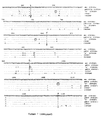
- Figure 1: This is a diagrammatic representation of the db1-related cDNA clones aligned with dbl. Clones pMP32 and pMP33 are from this work, and dbl and "a" taken from Gonzalez et al (2). The numbering refers to that for dbl starting at the initiation codon. The positions of the introns, inferred from comparison with other P450II genes, are indicated on db1 by vertical lines, and the retention of part of intron 1 (pMP33) and intron 5 ("a") is indicated by triangles. Base pair substitutions and other differences between the cDNA sequences are given in full in Fig. 2. The dashes represent sequence not present in the variant cDNAs compared to dbl.
- Figure 2 (on two sheets: Figure 2A and Figure 2B): This shows a comparison of nucleotide sequences of DNA encoding functional debrisoquine hydroxylase (db1) and related cDNAs. The sequences for dbl and variant "b" are taken from Gonzalez et al (2). Numbering starts at the initiation codon for dbl. The variant "a" cDNA sequence was compiled from two cDNAs, pMP32 and pMP33 which contained an identical sequence over an overlapping region of 462 bp. This sequence contained only one base-pair difference to the partial sequence described as variant "a" by Gonzalez et al (G to C at position 383) which covers the region 299-1567 bp. The base-pair deletion at position 506 is marked *. The position of introns (vertical lines) is obtained from the sequence of P45011D6 gene (11). Triangles represent the positions of intron sequences in the isolated variant cDNAs:
1. The first 64-bp ofintron 1 in pMP33; 5. Insertion ofintron 5 in the variant "a" cDNA described by Gonzalez et al but which was absent from pMP32 and pMP33; 6. Insertion of part ofintron 6 in both pMP34 and variant "b". The position of the bp insertions (G) at position 631 and at 983 (T) in the "b" variant are shown **. The positions of oligonucleotide primers used to amplify specific regions of the genes are marked by capital letters and horizontal arrows. The oligonucleotide marked D was taken from the sequence of intron 4 (see Fig 5). The diagnostic restriction sites used to differentiate between either dbl (variant "a") and variant "b" or dbl and variant "a" are marked. - Figure 3: This is a schematic illustration of HaeIII cleavage sites in the PCR fragment generated from the db1 gene using oligonucleotides A and B as primers (see Fig 2). The location of the determinative HaeIII site when position 294 is mutated is shown. The fragment generated is 172bp in length and incorporates the sequences of
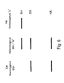
intron 1 andexon 2. - Figure 4: This is a schematic illustration of the DNA fragmentation pattern obtained following polyacrylamide gel electrophoresis of the HaeIII-cleaved
exon 2 DNA shown in Figure 3 for both homozygote EM, homozygote PM and heterozygote individuals. - Figures 5 & 6: Analysis of the mutation site leading to the base-pair deletion in dbl cDNA at position 506. Diagrammatic representation of the method used and (Figure 6) predicted banding pattern for individuals containing the db1 gene, variant "a" or both. Amplification of DNA from
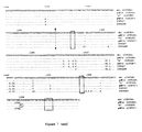
exon 3 intointron 4 produced a 334-bp fragment which in individuals containing the db1 sequence digests into fragments of 105 bp and 229 bp with BstNl. The variant "a" sequence (PM's) is resistant to digestion with this enzyme. - Figure 7: This shows the "b" variant sequence and others alongside the db1 sequence, with the region around
exon 7 shown in more detail in Figure 8. Sites cut by restriction enzymes HaeIII, DraIII and HhaI are shown as A, B and C respectively. HaeIII cuts dbl only. DraIII cuts 2D7 (pMP34) and 2d8 sequences only at 1083. A combination of HaeIII and DraIII is useful but inconvenient. A HhaI site is absent in the "b" variant at 1094 but present in all other variants so far found.Primer pair 1+2 is used for HaeIII/DraIII analysis and 3+2 for HhaI/DraIII analysis. - Preparation of radioactive probes . The human P450 db1 (P450IID1; 2) cDNA probe was kindly provided by Drs F.J. Gonzalez and U.A. Meyer. Restriction fragments for use as probes were isolated from low gelling temperature agarose and radioactively labelled with [α-32P]dCTP (3000 Ci mmol-1; Amersham International) by random primer extension (6). Oligonucleotides were made on an Applied Biosystems 380A machine and labelled with [α-32P]ATP (3000 Ci mmol-1; Amersham International) and T4 polynucleotide kinase.
- Isolation of human P450IID-related cDNA clones. The full length P450IID6 (db1) cDNA was used to screen plaques from two human liver lambda gt11 cDNA libraries (Clontech, Palo Alto, CA; and Kwok et al [7]) made from individuals of unknown phenotype. Two different cDNA clones, lambda MPA (1.22kb) and lambda MPG (1.56kb) from the Clontech library were subcloned into pUC18 to give pMP32 and pMP33 respectively, and also into M13mp18 for sequence analysis.
- DNA sequence analysis. The dideoxy chain termination method was used with [α-35S] thio dATP (400 Ci mmol-1; Amersham International) to sequence DNA cloned in M13 (8, 9). Overlapping sequences were derived using a series of synthetic oligo-nucleotides and both DNA strands were fully sequenced. Sequences were compiled and analysed using Staden Plus software implemented on a DCS286 computer (10). DNA sequences generated during the course of this work have been deposited in the EMBL Data Bank with accession nos. X16865 and X16866.
- Phenotyping. Individuals GT, MJ, TR, PJ and ML were phenotyped in vivo by Prof. G. Tucker, Department of Pharmacology and Therapeutics, University of Sheffield, U.K. using debrisoquine; and individuals A, 1.1, 2.2, and 3.3 were phenotyped using sparteine in vivo by us. Post-mortem liver samples E4, E6, E8 and E11 were phenotyped in vitro by Dr. U.A. Meyer, Biozentrum, Basel using bufuralol, and LVII showed no cross-reactivity with a monoclonal antibody raised against rat P450db1 (kindly supplied by Dr Meyer).
- DNA amplification. Total DNA was isolated from blood lymphocytes or from liver using an Applied Biosystems 340A nucleic acids extractor according to the manufacturer's instructions. Target DNA (1µg for genomic DNA or 1ng for cloned cDNA) was used in the polymerase chain reaction (PCR; 5) with 600ng of each amplification primer. The PCR was carried out using 2.5U Taq DNA polymerase (Cetus Corporation) according to the manufacturer's conditions except that dimethyl sulphoxide was added to 10% (v/v) final concentration. The chain reaction was initiated by denaturing DNA at 92°C for 1 min, annealing by cooling to 60°C for 1 min and extending at 72°C for 2 min; twenty cycles were performed using either a Cetus or Techne programmable heating block. Pairs of oligonucleotide primers enabling the amplification of specific exon sequences were used as described in the legend to Figure 2.
- Analysis of amplified DNA. The products of DNA amplification (between 1/20 - 1/10 of the total) were either left uncut or digested with diagnostic restriction enzymes and separated electrophoretically on 6% polyacrylamide gels. In some cases the DNA was analysed by Southern blotting. Briefly, the DNA was transferred to Hybond N by alkali transfer (1.5M NaCl, 0.25M NaOH) and baked at 80°C for 2h. Membranes to be probed with cDNAs were pre-hybridised at 65°C in 5XSSC, 4X Denhardt's, 10% SDS, 0.1% NaPPi, 20µg ml-1 salmon sperm DNA. Hybridisation was overnight in the same buffer except no salmon sperm DNA was present. Filters were washed to a final stringency of 0.2X SSC, 0.1% SDS, 0.1% NaPPi at 65°C. Oligonucleotide probes covering the region of interest were hybridised to the membranes at 37°C in 6X SSC, 0.1% NaPPi and washed in the same buffer at a temperature dependent on their Tm. Membranes were exposed to Kodak XAR-5 film for between 2h and three days at -80°C.
- The nucleotide sequences derived from pMP32 and pMP33 were compared with the sequences of the db1 cDNA (encoding a functional P450 with bufuralol 1'-hydroxylase activity) as well as the variant "a" cDNA described by Gonzalez et al (2) and amended in the EMBL Data Bank; accession no. Y00300 (Figs. 1 & 2). The sequences of pMP32 and pMP33 have an identical sequence over an overlapping region of 445bp. Together these clones constitute a full length cDNA. However, we did not expect the sequence generated by pMP32/pMP33 to encode a functional protein as it contains part of
intron 1 and perhaps more importantly a single base deletion at position 506 which would lead to a frame-shift (Fig. 2). Variant "a" of Gonzalez et al (2) also contains this same frame-shift and so with or without the retention ofintron 5 orintron 1 this variant would not encode a functional protein (Fig 2). Analysis of the genomic P450II2D sequence showed that the bp deletion at position 506 is due to a G to A transition at the junction ofintron 3 andexon 4. This mutation removes a BstN1 site in the gene compared to db1. - There are a significant number of base pair differences between the sequence of pMP32/pMP33, representing a full length variant, non-functional dbl sequence, and the normal db1 sequence. All of these differences may serve as markers for the PM phenotype and may therefore be of use in a genotyping assay.
- An analysis at position 294 is shown as an example in Figs 3 and 4. DNA covering the region of interest was amplified by PCR from genomic DNA using the oligonucleotides A and B (Fig 2). This generates a DNA fragment of 172bp in length (Fig 3). In PM individuals the 83bp fragment generated by digestion with the restriction enzyme HaeIII will cut into two pieces (39 and 44bp) due to the HaeIII site generated by the mutation at position 294. In homozygous EM's this site is absent. 100% agreement between phenotype, assessed by measuring the rate of metabolism and marker drugs, and genotype was observed in the 16 individuals tested, with the gels corresponding to the expected appearance shown in Figure 4.
- Analysis of a region of exon 9 using oligonucleotide primers G and F (see Fig 2) showed that in some PM individuals nc db1 related band could be observed, indicating a deletion of this region of the gene. This also is informative for the PM phenotype in some individuals.
- Genomic DNA from individuals phenotyped either as poor or normal metabolizers was amplified by PCR using an oligonucleotide derived from
exon 3 and one derived from intron 4 (marked C and D in Fig. 2) using an annealing temperature of 60°C. The sequence of oligonucleotide D was AAATCCTGCTCTTCCGAGGC. The use of this oligonucleotide pair and the high annealing temperature assured specificity for the P450IID6 gene. The resulting fragment of 334 bp was then digested with the restriction enzyme BstN1 and the products separated on an 8% polyacrylamide gel. Bands were visualized by ultraviolet irradiation of the gel stained with ethidium bromide. - Figures 5 and 6 show the rationale and (schematically) the results.
- Examination of the dbl1sequence over the intron 3-
exon 4 junction suggests two explanations for the base-pair (G) deletion in the cDNA sequences. The db1 intron 3-exon 4 junction has the sequence CCCCCAG/GACGCC (the bold letters indicate the start of exon 4)19. Therefore, either a base-pair (G) deletion to give CCCCCAG/ACGCC, or a G to A transition to give the sequence CCCCCAAG/ACGCC, which shifts the position of the 3' splice site, will result in the loss of the first base (G) inexon 4. In both cases the BstNI restriction site is lost. To establish which was the case we sequenced the PCR amplification product from 20 affected individuals. In all of these the G to A transition was shown to be the mutation responsible for the poor metabolizer phenotype. This transition appears to be the primary defect responsible for the poor metabolizer phenotype. Over 80% of individuals tested were homozygous for the G to A transition. Interestingly, individuals with this mutation were the same as those with the mutations at position 100 and inexon 2. - Referring to Figure 7, an assay based on amplification using the oligonucleotides marked 2 and 3 followed by digestion with HhaI may be used to show the presence or absence of the G to A transition at bp 1094. In this case all other IID sequences will cut apart from the "b" variant. This is informative for the PM phenotype. Thus, the region of DNA between 1049 and 1173 is amplified, exposed to HhaI and submitted to gel electrophoresis followed by labelling with suitable probes. For the "b" variant, two fragments of 45 and 79 bases will be produced, whereas for EM phenotypes a single fragment of 124 bases is produced. This is an example of an indirect assay for the bp deletion at 506 in the CYPD2D6 gene.
-
- 1. Eichelbaum, M (1988) Atlas of Science: Pharmacology, pp. 243-251.
- 2. Gonzalez, F.J., Skoda, R.C., Kimura, S., Umeno, M., Zanger, U.M., Nebert, D.W., Gelboin, H.V., Hardwick, J.P. & Meyer, U.A. (1988) Nature, 331, 442-446.
- 3. Nebert, D.W., Nelson, D.R., Adesnik, M., Coon, M.J., Estabrook, R.W., Gonzalez, F.J., Guengerich, F.P., Gunsalus, I.C., Johnson, E.F., Kemper, B., Levin, W., Phillips, I.R., Sato, R. & Waterman, M.R. (1989)
DNA 8,1-14. - 4. Skoda, R.C., Gonzalez, F.J., Demierre, A., & Meyer, U.A. (1988) Proc. Natl. Acad. Sci. U.S.A. , 85 5240-5243.
- 5. Saiki, R.K., et al (1988), Science 239, 487-91.
- 6. Feinberg, D.P. & Vogelstein, B. (1983) Anal. Biochem. 136 , 6-13.
- 7. Kwok, S.C.M., Ledley, F.D., DiLella, A.G., Robson, K.J.H., & Woo, S.L.C. (1985) Biochem 24, 556-561.
- 8. Sanger, F., Coulson, A.R., Barrell, B.G., Smith, A.J.H., & Roe, B.A. (1980) J. Mol. Biol. 143, 161-178.
- 9. Biggin, M.D., Gibson, T.J., & Hong, G.F. (1983) Proc. Natl. Acad. Sci. U.S.A. 80 , 3963-3965.
- 10. Staden, R. (1986) Nucl. Acids. Res. 14 , 217-231.
- 11. Kimura, S., Urmeno, M., Skoda, R.C., Meyer, U.A. and Gonzalez, F.J. (1989) Am. J. Hu. Genet. 45, 889-904
Claims (16)
- A method of identifying whether a sample containing DNA is associated with the PM (poor metaboliser) or EM (extensive metaboliser) phenotype, the method comprising the steps of (1) providing a sample containing DNA ; and (2) identifying the presence or absence of a mutation at one or more of positions 100, 271, 281, 294, 408, 506 or 1432 of the DNA sequence of the P450 IID bufuralol-1'-hydroxylase or a deletion of at least part of exon 9 thereof using a reagent capable of distinguishing one nucleotide from another or the presence or absence of a nucleotide at a given site in the DNA.
- A method according to Claim 1 wherein a mutation at position 271, 281, 294 or 506 is detected.
- A method according to Claim 2 wherein a base-pair deletion at position 506 is detected.
- A method according to any one of Claims 1 to 3 comprising the step of digesting DNA encoding at least part of the bufuralol-1'-hydroxylase amino acid sequence with a restriction enzyme which will cut, or will not cut, at or adjacent to one of the said positions according to whether the mutation is present.
- A method according to Claim 4 comprising the step of amplifying the amount of a selected region of DNA before digesting the DNA as said.
- A method according to claim 4 or 5, when dependent on Claim 3, wherein the restriction enzyme is BstNI or an isoschizomer thereof and the DNA is obtained from a cell of the body.
- A method according to any one of Claims 1 to 3 wherein sample DNA is subjected to the polymerase chain reaction using oligonucleotide primers which are capable of hybridising selectively either to the wild-type or to the mutant sequence at the location being analysed, such that the generation of amplified DNA will indicate whether the said mutation is present.
- A method of identifying a mutation In the DNA sequence of P450IlD bufuralol-1'-hydroxylase at one or more of positions 100, 271, 281, 294, 408, 506, 775 or 1432 comprising (1) subjecting the sample DNA to the polymerase chain reaction using oligonucleotide primers which are capable of hybridising selectively either to the wild-type or to the mutant sequence at the location being analysed, the primers being such as to introduce, upon hybridisation to the said location, a restriction site which is not present in the wild-type of mutated location and (2) subjecting amplified DNA derived from step (1) to a restriction digest with an enzyme which cleaves at the said restriction site.
- A method according to Claim 8 wherein two pairs of primers are used in step (1), one pair hybridising on respective sides of position 506 and the other pair hybridising on respective sides at position 775.
- A method of making a single-stranded DNA compound suitable for use as a primer in a polymerase chain reaction, the compound being adapted to hybridise to a region of wild-type or mutant bufuralol-1'-hydroxylase-encoding DNA flanking positions 100, 271, 281, 294, 408, 506 or 1432 such that, in the polymerase chain reaction, DNA synthesis will or will not proceed from the primer towards the said site according to whether there is a mutation at the relevant said position comprising the step of joining nucleoside derivatives to form the DNA.
- A method of making a compound according to Claim 10 selected from the group consisting of:and analogues and fragments thereof.

- A kit for performing the method of Claim 1 comprising a compound according to Claim 10 or 11.
- A kit according to Claim 12 further comprising at least one restriction enzyme useful in distinguishing one or more of the said polymorphisms.
- A kit according to Claim 12 further comprising a specific oligonucleotide probe useful in distinguishing one or more of the said polymorphisms.
- A kit according to Claim 12, 13 or 14 further comprising at least one control sample of DNA containing the polymorphic site or sites and corresponding to DNA from one or more of the following: a homozygous extensive metaboliser, a homozygous poor metaboliser and a heterozygous individual.
- A kit according to any one of Claims 12 to 15 further comprising means to facilitate obtaining cells from a patient, from which DNA may be extracted for analysis using the said method.
Applications Claiming Priority (3)
| Application Number | Priority Date | Filing Date | Title |
|---|---|---|---|
| GB9001181 | 1990-01-18 | ||
| GB909001181A GB9001181D0 (en) | 1990-01-18 | 1990-01-18 | Genetic assay |
| PCT/GB1991/000066 WO1991010745A1 (en) | 1990-01-18 | 1991-01-17 | Genetic assay for cytochrome p450 |
Publications (2)
| Publication Number | Publication Date |
|---|---|
| EP0511262A1 EP0511262A1 (en) | 1992-11-04 |
| EP0511262B1 true EP0511262B1 (en) | 1996-07-17 |
Family
ID=10669529
Family Applications (1)
| Application Number | Title | Priority Date | Filing Date |
|---|---|---|---|
| EP91902320A Expired - Lifetime EP0511262B1 (en) | 1990-01-18 | 1991-01-17 | Genetic assay for cytochrome p450 |
Country Status (9)
| Country | Link |
|---|---|
| US (1) | US5981174A (en) |
| EP (1) | EP0511262B1 (en) |
| JP (1) | JP3272353B2 (en) |
| AT (1) | ATE140488T1 (en) |
| AU (1) | AU642705C (en) |
| CA (1) | CA2071636C (en) |
| DE (1) | DE69120936T2 (en) |
| GB (1) | GB9001181D0 (en) |
| WO (1) | WO1991010745A1 (en) |
Families Citing this family (27)
| Publication number | Priority date | Publication date | Assignee | Title |
|---|---|---|---|---|
| US7115364B1 (en) | 1993-10-26 | 2006-10-03 | Affymetrix, Inc. | Arrays of nucleic acid probes on biological chips |
| US20060229824A1 (en) | 1993-10-26 | 2006-10-12 | Affymetrix, Inc. | Arrays of nucleic acid probes for analyzing biotransformation genes |
| EP0733112B1 (en) * | 1993-12-08 | 2007-01-10 | Royal Veterinary & Agricultural University | Cytochrome p-450 monooxygenases |
| JP3564579B2 (en) * | 1994-05-06 | 2004-09-15 | 大塚製薬株式会社 | Method for detecting polymorphism in human cytochrome P4502D6 gene |
| CN1151188A (en) * | 1994-07-06 | 1997-06-04 | 大制药株式会社 | Method of detecting human cytochrome P4501A2 gene polymorphism |
| US6391550B1 (en) | 1996-09-19 | 2002-05-21 | Affymetrix, Inc. | Identification of molecular sequence signatures and methods involving the same |
| US6183963B1 (en) * | 1998-10-23 | 2001-02-06 | Signalgene | Detection of CYP1A1, CYP3A4, CYP2D6 and NAT2 variants by PCR-allele-specific oligonucleotide (ASO) assay |
| EP1088900A1 (en) * | 1999-09-10 | 2001-04-04 | Epidauros Biotechnologie AG | Polymorphisms in the human CYP3A4, CYP3A7 and hPXR genes and their use in diagnostic and therapeutic applications |
| JP4721603B2 (en) * | 1999-11-08 | 2011-07-13 | 栄研化学株式会社 | Mutation and / or polymorphism detection method |
| ATE317023T1 (en) * | 1999-11-08 | 2006-02-15 | Eiken Chemical | METHOD FOR DETECTING VARIATIONS OR POLYMORPHISMS |
| WO2001077317A1 (en) * | 2000-04-07 | 2001-10-18 | Eiken Kagaku Kabushiki Kaisha | Method of amplifying nucleic acid by using double-stranded nucleic acid as template |
| WO2001079561A2 (en) * | 2000-04-17 | 2001-10-25 | Liggett Stephen B | Alpha-2 adrenergic receptor polymorphisms |
| US7229756B1 (en) | 2000-10-19 | 2007-06-12 | University Of Cincinnati | Alpha-2B-adrenergic receptor polymorphisms |
| WO2001096604A2 (en) * | 2000-06-12 | 2001-12-20 | Genicon Sciences Corporation | Assay for genetic polymorphisms using scattered light detectable labels |
| US7211386B2 (en) * | 2000-08-10 | 2007-05-01 | University Of Cincinnati | Alpha-2A-adrenergic receptor polymorphisms |
| US20030166182A1 (en) * | 2001-03-30 | 2003-09-04 | Wei Shao | Isolated human drug-metabolizing proteins, nucleic acid molecules encoding human drug-metabolizing proteins, and uses thereof |
| WO2002099118A2 (en) * | 2001-06-05 | 2002-12-12 | Genaissance Pharmaceuticals, Inc. | Method of identifying a polymorphism in cyp2d6 |
| US20040096874A1 (en) * | 2002-04-11 | 2004-05-20 | Third Wave Technologies, Inc. | Characterization of CYP 2D6 genotypes |
| US20040091909A1 (en) * | 2002-07-05 | 2004-05-13 | Huang Doug Hui | High throughput cytochrome P450 genotyping |
| JP2005328758A (en) * | 2004-05-20 | 2005-12-02 | Hitachi Ltd | Method for nucleic acid amplification and method for analyzing single nucleotide polymorphism utilizing the same |
| CA2571823A1 (en) * | 2004-06-30 | 2006-01-12 | Tm Bioscience Pgx, Inc. | Method of detecting mutations in the gene encoding cytochrome p450-2d6 |
| WO2006014869A1 (en) * | 2004-07-26 | 2006-02-09 | Parallele Bioscience, Inc. | Simultaneous analysis of multiple genomes |
| ATE469245T1 (en) * | 2004-09-14 | 2010-06-15 | Univ Colorado | GENETIC TARGETING-BASED METHOD FOR TREATMENT WITH BUCINDOLOL |
| US20100063093A1 (en) * | 2007-03-28 | 2010-03-11 | Curt Wolfgang | Methods for the administration of iloperidone |
| US8586610B2 (en) | 2004-09-30 | 2013-11-19 | Vanda Pharmaceuticals, Inc. | Methods for the administration of iloperidone |
| US7368242B2 (en) * | 2005-06-14 | 2008-05-06 | Affymetrix, Inc. | Method and kits for multiplex hybridization assays |
| ME02347B (en) | 2009-10-19 | 2016-06-20 | Rostaquo S P A | Rostafuroxine for pharmacogenomic treatment of cardiovascular conditions |
Family Cites Families (2)
| Publication number | Priority date | Publication date | Assignee | Title |
|---|---|---|---|---|
| IE61148B1 (en) * | 1988-03-10 | 1994-10-05 | Ici Plc | Method of detecting nucleotide sequences |
| DE69126064T2 (en) * | 1990-06-22 | 1997-08-28 | Hoffmann La Roche | Detection of weak metabolizing agents of drugs |
-
1990
- 1990-01-18 GB GB909001181A patent/GB9001181D0/en active Pending
-
1991
- 1991-01-17 EP EP91902320A patent/EP0511262B1/en not_active Expired - Lifetime
- 1991-01-17 AU AU71791/91A patent/AU642705C/en not_active Expired
- 1991-01-17 AT AT91902320T patent/ATE140488T1/en active
- 1991-01-17 JP JP50231291A patent/JP3272353B2/en not_active Expired - Lifetime
- 1991-01-17 DE DE69120936T patent/DE69120936T2/en not_active Expired - Lifetime
- 1991-01-17 CA CA002071636A patent/CA2071636C/en not_active Expired - Lifetime
- 1991-01-17 WO PCT/GB1991/000066 patent/WO1991010745A1/en active IP Right Grant
-
1993
- 1993-11-04 US US08/145,658 patent/US5981174A/en not_active Expired - Lifetime
Also Published As
| Publication number | Publication date |
|---|---|
| CA2071636A1 (en) | 1991-07-19 |
| US5981174A (en) | 1999-11-09 |
| JPH05503845A (en) | 1993-06-24 |
| AU7179191A (en) | 1991-08-05 |
| JP3272353B2 (en) | 2002-04-08 |
| EP0511262A1 (en) | 1992-11-04 |
| ATE140488T1 (en) | 1996-08-15 |
| DE69120936T2 (en) | 1997-02-20 |
| AU642705C (en) | 2001-07-19 |
| GB9001181D0 (en) | 1990-03-21 |
| AU642705B2 (en) | 1993-10-28 |
| DE69120936D1 (en) | 1996-08-22 |
| CA2071636C (en) | 2001-12-25 |
| WO1991010745A1 (en) | 1991-07-25 |
Similar Documents
| Publication | Publication Date | Title |
|---|---|---|
| EP0511262B1 (en) | Genetic assay for cytochrome p450 | |
| Hassett et al. | Human microsomal epoxide hydrolase: genetic poloymorphism and functional expression in vitro of amino acid variants | |
| EP0463395B1 (en) | Detection of poor metabolizers of drugs | |
| Steen et al. | Detection of the poor metabolizer-associated CYP2D6 (D) gene deletion allele by long-PCR technology | |
| Theophilus et al. | Gaucher disease: molecular heterogeneity and phenotype-genotype correlations. | |
| Hanioka et al. | The human CYP2D locus associated with a common genetic defect in drug oxidation: a G1934----A base change in intron 3 of a mutant CYP2D6 allele results in an aberrant 3'splice recognition site. | |
| Xu et al. | Genotyping of human alcohol dehydrogenases at the ADH2 and ADH3 loci following DNA sequence amplification | |
| Shimkets et al. | Liddle's syndrome: heritable human hypertension caused by mutations in the β subunit of the epithelial sodium channel | |
| Nhamburo et al. | Identification of a new P450 expressed in human lung: complete cDNA sequence, cDNA-directed expression, and chromosome mapping | |
| Gouya et al. | Modulation of the phenotype in dominant erythropoietic protoporphyria by a low expression of the normal ferrochelatase allele. | |
| Labuda et al. | Rapid detection of CYP1A1, CYP2D6, and NAT variants by multiplex polymerase chain reaction and allele-specific oligonucleotide assay | |
| Giebel et al. | A frequent tyrosinase gene mutation in classic, tyrosinase-negative (type IA) oculocutaneous albinism. | |
| Wigderson et al. | Characterization of mutations in Gaucher patients by cDNA cloning. | |
| CA2420322A1 (en) | Detection of cyp2d6 polymorphisms | |
| EP1012340A1 (en) | Genotyping of human cyp3a4 | |
| US9540693B2 (en) | Mutations of the parkin gene, compositions, methods and uses | |
| Heim et al. | [17] Genetic polymorphism of debrisoquine oxidation: restriction fragment analysis and allele-specific amplification of mutant alleles of CYP2D6 | |
| WAN et al. | Genetic polymorphism of CYP2E1, ADH2, and ALDH2 in Mexican-Americans | |
| Panserat et al. | An unequal cross‐over event within the CYP2D gene cluster generates a chimeric CYP2D7/CYP2D6 gene which is associated with the poor metabolizer phenotype. | |
| US5844108A (en) | Primers targeted to NAT2 gene for detection of poor metabolizers of drugs | |
| US5856095A (en) | Identification of two novel mutant alleles of human thiopurine S-methyltransferase, and diagnostic uses thereof | |
| Lee et al. | Genetic heterogeneity of the porphobilinogen deaminase gene in Swedish families with acute intermittent porphyria | |
| IToH et al. | A rapid and simple detection of genetic defects responsible for the phenotypic polymorphism of cytochrome P450 2C19 | |
| CA2398104C (en) | Polymorphisms in the human cyp2d6 gene promoter region and their use in diagnostic and therapeutic applications | |
| CA2091925A1 (en) | Human arylamine n-acetyltransferaase genes |
Legal Events
| Date | Code | Title | Description |
|---|---|---|---|
| PUAI | Public reference made under article 153(3) epc to a published international application that has entered the european phase |
Free format text: ORIGINAL CODE: 0009012 |
|
| 17P | Request for examination filed |
Effective date: 19920706 |
|
| AK | Designated contracting states |
Kind code of ref document: A1 Designated state(s): AT BE CH DE DK ES FR GB GR IT LI LU NL SE |
|
| 17Q | First examination report despatched |
Effective date: 19941128 |
|
| GRAH | Despatch of communication of intention to grant a patent |
Free format text: ORIGINAL CODE: EPIDOS IGRA |
|
| GRAH | Despatch of communication of intention to grant a patent |
Free format text: ORIGINAL CODE: EPIDOS IGRA |
|
| RBV | Designated contracting states (corrected) |
Designated state(s): AT BE CH DE DK ES FR GR IT LI LU NL SE |
|
| GRAA | (expected) grant |
Free format text: ORIGINAL CODE: 0009210 |
|
| AK | Designated contracting states |
Kind code of ref document: B1 Designated state(s): AT BE CH DE DK ES FR GR IT LI LU NL SE |
|
| PG25 | Lapsed in a contracting state [announced via postgrant information from national office to epo] |
Ref country code: DK Effective date: 19960717 Ref country code: BE Effective date: 19960717 Ref country code: AT Effective date: 19960717 Ref country code: GR Free format text: LAPSE BECAUSE OF FAILURE TO SUBMIT A TRANSLATION OF THE DESCRIPTION OR TO PAY THE FEE WITHIN THE PRESCRIBED TIME-LIMIT Effective date: 19960717 Ref country code: ES Free format text: THE PATENT HAS BEEN ANNULLED BY A DECISION OF A NATIONAL AUTHORITY Effective date: 19960717 |
|
| REF | Corresponds to: |
Ref document number: 140488 Country of ref document: AT Date of ref document: 19960815 Kind code of ref document: T |
|
| REF | Corresponds to: |
Ref document number: 69120936 Country of ref document: DE Date of ref document: 19960822 |
|
| ITF | It: translation for a ep patent filed |
Owner name: SAMA PATENTS |
|
| ET | Fr: translation filed | ||
| REG | Reference to a national code |
Ref country code: CH Ref legal event code: NV Representative=s name: KIRKER & CIE SA |
|
| PG25 | Lapsed in a contracting state [announced via postgrant information from national office to epo] |
Ref country code: LU Free format text: LAPSE BECAUSE OF NON-PAYMENT OF DUE FEES Effective date: 19970131 |
|
| PLBQ | Unpublished change to opponent data |
Free format text: ORIGINAL CODE: EPIDOS OPPO |
|
| PLBI | Opposition filed |
Free format text: ORIGINAL CODE: 0009260 |
|
| PLBF | Reply of patent proprietor to notice(s) of opposition |
Free format text: ORIGINAL CODE: EPIDOS OBSO |
|
| 26 | Opposition filed |
Opponent name: AFFYMETRIX, INC. Effective date: 19970417 |
|
| NLR1 | Nl: opposition has been filed with the epo |
Opponent name: AFFYMETRIX, INC. |
|
| PLBF | Reply of patent proprietor to notice(s) of opposition |
Free format text: ORIGINAL CODE: EPIDOS OBSO |
|
| PLBL | Opposition procedure terminated |
Free format text: ORIGINAL CODE: EPIDOS OPPC |
|
| PLBM | Termination of opposition procedure: date of legal effect published |
Free format text: ORIGINAL CODE: 0009276 |
|
| STAA | Information on the status of an ep patent application or granted ep patent |
Free format text: STATUS: OPPOSITION PROCEDURE CLOSED |
|
| 27C | Opposition proceedings terminated |
Effective date: 19990509 |
|
| NLR2 | Nl: decision of opposition | ||
| REG | Reference to a national code |
Ref country code: CH Ref legal event code: PUE Owner name: CANCER RESEARCH TECHNOLOGY LIMITED Free format text: IMPERIAL CANCER RESEARCH TECHNOLOGY LIMITED#SARDINIA HOUSE, SARDINIA STREET#LONDON WC2A 3NL (GB) -TRANSFER TO- CANCER RESEARCH TECHNOLOGY LIMITED#61 LINCOLN'S INN FIELDS#LONDON WC2A 3PX (GB) |
|
| NLS | Nl: assignments of ep-patents |
Owner name: CANCER RESEARCH TECHNOLOGY LIMITED |
|
| REG | Reference to a national code |
Ref country code: FR Ref legal event code: TP |
|
| PGFP | Annual fee paid to national office [announced via postgrant information from national office to epo] |
Ref country code: SE Payment date: 20091218 Year of fee payment: 20 Ref country code: CH Payment date: 20091218 Year of fee payment: 20 |
|
| PGFP | Annual fee paid to national office [announced via postgrant information from national office to epo] |
Ref country code: FR Payment date: 20100106 Year of fee payment: 20 |
|
| PGFP | Annual fee paid to national office [announced via postgrant information from national office to epo] |
Ref country code: IT Payment date: 20091230 Year of fee payment: 20 |
|
| PGFP | Annual fee paid to national office [announced via postgrant information from national office to epo] |
Ref country code: DE Payment date: 20091221 Year of fee payment: 20 |
|
| PGFP | Annual fee paid to national office [announced via postgrant information from national office to epo] |
Ref country code: NL Payment date: 20091224 Year of fee payment: 20 |
|
| REG | Reference to a national code |
Ref country code: CH Ref legal event code: PL |
|
| REG | Reference to a national code |
Ref country code: NL Ref legal event code: V4 Effective date: 20110117 |
|
| PG25 | Lapsed in a contracting state [announced via postgrant information from national office to epo] |
Ref country code: DE Free format text: LAPSE BECAUSE OF EXPIRATION OF PROTECTION Effective date: 20110117 |