EP0534858B2 - Selective restriction fragment amplification : a general method for DNA fingerprinting - Google Patents
Selective restriction fragment amplification : a general method for DNA fingerprinting Download PDFInfo
- Publication number
- EP0534858B2 EP0534858B2 EP92402629A EP92402629A EP0534858B2 EP 0534858 B2 EP0534858 B2 EP 0534858B2 EP 92402629 A EP92402629 A EP 92402629A EP 92402629 A EP92402629 A EP 92402629A EP 0534858 B2 EP0534858 B2 EP 0534858B2
- Authority
- EP
- European Patent Office
- Prior art keywords
- dna
- restriction
- fragments
- primers
- anyone
- Prior art date
- Legal status (The legal status is an assumption and is not a legal conclusion. Google has not performed a legal analysis and makes no representation as to the accuracy of the status listed.)
- Expired - Lifetime
Links
- 230000003321 amplification Effects 0.000 title claims abstract description 61
- 238000003199 nucleic acid amplification method Methods 0.000 title claims abstract description 61
- 239000012634 fragment Substances 0.000 title claims description 303
- 238000007429 general method Methods 0.000 title description 4
- 108020004414 DNA Proteins 0.000 claims abstract description 295
- 238000000034 method Methods 0.000 claims abstract description 126
- 108091008146 restriction endonucleases Proteins 0.000 claims abstract description 87
- 230000008569 process Effects 0.000 claims abstract description 45
- 241001465754 Metazoa Species 0.000 claims abstract description 17
- 241000282414 Homo sapiens Species 0.000 claims abstract description 5
- 150000007523 nucleic acids Chemical class 0.000 claims abstract description 4
- 125000003729 nucleotide group Chemical group 0.000 claims description 107
- 239000002773 nucleotide Substances 0.000 claims description 104
- 238000003752 polymerase chain reaction Methods 0.000 claims description 52
- 108091034117 Oligonucleotide Proteins 0.000 claims description 38
- 108091028043 Nucleic acid sequence Proteins 0.000 claims description 32
- 241000196324 Embryophyta Species 0.000 claims description 26
- 239000000203 mixture Substances 0.000 claims description 26
- 102000054765 polymorphisms of proteins Human genes 0.000 claims description 26
- 230000002068 genetic effect Effects 0.000 claims description 21
- JLCPHMBAVCMARE-UHFFFAOYSA-N [3-[[3-[[3-[[3-[[3-[[3-[[3-[[3-[[3-[[3-[[3-[[5-(2-amino-6-oxo-1H-purin-9-yl)-3-[[3-[[3-[[3-[[3-[[3-[[5-(2-amino-6-oxo-1H-purin-9-yl)-3-[[5-(2-amino-6-oxo-1H-purin-9-yl)-3-hydroxyoxolan-2-yl]methoxy-hydroxyphosphoryl]oxyoxolan-2-yl]methoxy-hydroxyphosphoryl]oxy-5-(5-methyl-2,4-dioxopyrimidin-1-yl)oxolan-2-yl]methoxy-hydroxyphosphoryl]oxy-5-(6-aminopurin-9-yl)oxolan-2-yl]methoxy-hydroxyphosphoryl]oxy-5-(6-aminopurin-9-yl)oxolan-2-yl]methoxy-hydroxyphosphoryl]oxy-5-(6-aminopurin-9-yl)oxolan-2-yl]methoxy-hydroxyphosphoryl]oxy-5-(6-aminopurin-9-yl)oxolan-2-yl]methoxy-hydroxyphosphoryl]oxyoxolan-2-yl]methoxy-hydroxyphosphoryl]oxy-5-(5-methyl-2,4-dioxopyrimidin-1-yl)oxolan-2-yl]methoxy-hydroxyphosphoryl]oxy-5-(4-amino-2-oxopyrimidin-1-yl)oxolan-2-yl]methoxy-hydroxyphosphoryl]oxy-5-(5-methyl-2,4-dioxopyrimidin-1-yl)oxolan-2-yl]methoxy-hydroxyphosphoryl]oxy-5-(5-methyl-2,4-dioxopyrimidin-1-yl)oxolan-2-yl]methoxy-hydroxyphosphoryl]oxy-5-(6-aminopurin-9-yl)oxolan-2-yl]methoxy-hydroxyphosphoryl]oxy-5-(6-aminopurin-9-yl)oxolan-2-yl]methoxy-hydroxyphosphoryl]oxy-5-(4-amino-2-oxopyrimidin-1-yl)oxolan-2-yl]methoxy-hydroxyphosphoryl]oxy-5-(4-amino-2-oxopyrimidin-1-yl)oxolan-2-yl]methoxy-hydroxyphosphoryl]oxy-5-(4-amino-2-oxopyrimidin-1-yl)oxolan-2-yl]methoxy-hydroxyphosphoryl]oxy-5-(6-aminopurin-9-yl)oxolan-2-yl]methoxy-hydroxyphosphoryl]oxy-5-(4-amino-2-oxopyrimidin-1-yl)oxolan-2-yl]methyl [5-(6-aminopurin-9-yl)-2-(hydroxymethyl)oxolan-3-yl] hydrogen phosphate Polymers Cc1cn(C2CC(OP(O)(=O)OCC3OC(CC3OP(O)(=O)OCC3OC(CC3O)n3cnc4c3nc(N)[nH]c4=O)n3cnc4c3nc(N)[nH]c4=O)C(COP(O)(=O)OC3CC(OC3COP(O)(=O)OC3CC(OC3COP(O)(=O)OC3CC(OC3COP(O)(=O)OC3CC(OC3COP(O)(=O)OC3CC(OC3COP(O)(=O)OC3CC(OC3COP(O)(=O)OC3CC(OC3COP(O)(=O)OC3CC(OC3COP(O)(=O)OC3CC(OC3COP(O)(=O)OC3CC(OC3COP(O)(=O)OC3CC(OC3COP(O)(=O)OC3CC(OC3COP(O)(=O)OC3CC(OC3COP(O)(=O)OC3CC(OC3COP(O)(=O)OC3CC(OC3COP(O)(=O)OC3CC(OC3COP(O)(=O)OC3CC(OC3CO)n3cnc4c(N)ncnc34)n3ccc(N)nc3=O)n3cnc4c(N)ncnc34)n3ccc(N)nc3=O)n3ccc(N)nc3=O)n3ccc(N)nc3=O)n3cnc4c(N)ncnc34)n3cnc4c(N)ncnc34)n3cc(C)c(=O)[nH]c3=O)n3cc(C)c(=O)[nH]c3=O)n3ccc(N)nc3=O)n3cc(C)c(=O)[nH]c3=O)n3cnc4c3nc(N)[nH]c4=O)n3cnc4c(N)ncnc34)n3cnc4c(N)ncnc34)n3cnc4c(N)ncnc34)n3cnc4c(N)ncnc34)O2)c(=O)[nH]c1=O JLCPHMBAVCMARE-UHFFFAOYSA-N 0.000 claims description 19
- 108010042407 Endonucleases Proteins 0.000 claims description 14
- 102000004533 Endonucleases Human genes 0.000 claims description 14
- 230000015572 biosynthetic process Effects 0.000 claims description 13
- 241000894007 species Species 0.000 claims description 13
- 239000000523 sample Substances 0.000 claims description 12
- 108010014303 DNA-directed DNA polymerase Proteins 0.000 claims description 8
- 102000016928 DNA-directed DNA polymerase Human genes 0.000 claims description 8
- 241000282412 Homo Species 0.000 claims description 7
- 244000005700 microbiome Species 0.000 claims description 7
- 238000002360 preparation method Methods 0.000 claims description 5
- 239000013615 primer Substances 0.000 claims 20
- 239000002987 primer (paints) Substances 0.000 claims 8
- 239000003155 DNA primer Substances 0.000 claims 1
- 239000012472 biological sample Substances 0.000 claims 1
- 239000008280 blood Substances 0.000 claims 1
- 210000004369 blood Anatomy 0.000 claims 1
- 238000007894 restriction fragment length polymorphism technique Methods 0.000 abstract description 12
- 108020005120 Plant DNA Proteins 0.000 abstract description 3
- 102000039446 nucleic acids Human genes 0.000 abstract description 3
- 108020004707 nucleic acids Proteins 0.000 abstract description 3
- 239000000499 gel Substances 0.000 description 51
- 238000006243 chemical reaction Methods 0.000 description 38
- 240000003768 Solanum lycopersicum Species 0.000 description 33
- 235000007688 Lycopersicon esculentum Nutrition 0.000 description 30
- 238000012408 PCR amplification Methods 0.000 description 27
- 102000004190 Enzymes Human genes 0.000 description 21
- 239000000047 product Substances 0.000 description 21
- 102000053602 DNA Human genes 0.000 description 20
- 108090000790 Enzymes Proteins 0.000 description 20
- 230000000295 complement effect Effects 0.000 description 19
- 239000011543 agarose gel Substances 0.000 description 16
- 238000004458 analytical method Methods 0.000 description 15
- 229920002401 polyacrylamide Polymers 0.000 description 15
- 238000002105 Southern blotting Methods 0.000 description 12
- 239000011324 bead Substances 0.000 description 12
- 241000208822 Lactuca Species 0.000 description 11
- 240000008042 Zea mays Species 0.000 description 11
- 235000002017 Zea mays subsp mays Nutrition 0.000 description 10
- 238000001514 detection method Methods 0.000 description 10
- 239000011541 reaction mixture Substances 0.000 description 10
- 239000000243 solution Substances 0.000 description 10
- 235000005824 Zea mays ssp. parviglumis Nutrition 0.000 description 9
- 235000005822 corn Nutrition 0.000 description 9
- 238000012986 modification Methods 0.000 description 9
- 230000004048 modification Effects 0.000 description 9
- KCXVZYZYPLLWCC-UHFFFAOYSA-N EDTA Chemical compound OC(=O)CN(CC(O)=O)CCN(CC(O)=O)CC(O)=O KCXVZYZYPLLWCC-UHFFFAOYSA-N 0.000 description 8
- 238000001502 gel electrophoresis Methods 0.000 description 8
- 208000005652 acute fatty liver of pregnancy Diseases 0.000 description 7
- 239000000872 buffer Substances 0.000 description 7
- 238000002372 labelling Methods 0.000 description 7
- 238000007403 mPCR Methods 0.000 description 7
- 239000012528 membrane Substances 0.000 description 7
- 239000000758 substrate Substances 0.000 description 7
- 238000003786 synthesis reaction Methods 0.000 description 7
- QKNYBSVHEMOAJP-UHFFFAOYSA-N 2-amino-2-(hydroxymethyl)propane-1,3-diol;hydron;chloride Chemical compound Cl.OCC(N)(CO)CO QKNYBSVHEMOAJP-UHFFFAOYSA-N 0.000 description 6
- 239000003298 DNA probe Substances 0.000 description 6
- 230000006820 DNA synthesis Effects 0.000 description 6
- 208000028782 Hereditary disease Diseases 0.000 description 6
- 241001293495 Lactuca virosa Species 0.000 description 6
- TWRXJAOTZQYOKJ-UHFFFAOYSA-L Magnesium chloride Chemical compound [Mg+2].[Cl-].[Cl-] TWRXJAOTZQYOKJ-UHFFFAOYSA-L 0.000 description 6
- 239000007983 Tris buffer Substances 0.000 description 6
- 238000005194 fractionation Methods 0.000 description 6
- 238000009396 hybridization Methods 0.000 description 6
- 238000002955 isolation Methods 0.000 description 6
- 108090000623 proteins and genes Proteins 0.000 description 6
- LENZDBCJOHFCAS-UHFFFAOYSA-N tris Chemical compound OCC(N)(CO)CO LENZDBCJOHFCAS-UHFFFAOYSA-N 0.000 description 6
- 238000000137 annealing Methods 0.000 description 5
- 239000007795 chemical reaction product Substances 0.000 description 5
- 230000029087 digestion Effects 0.000 description 5
- 201000010099 disease Diseases 0.000 description 5
- 208000037265 diseases, disorders, signs and symptoms Diseases 0.000 description 5
- 238000012216 screening Methods 0.000 description 5
- 108020000946 Bacterial DNA Proteins 0.000 description 4
- 241000283690 Bos taurus Species 0.000 description 4
- HEDRZPFGACZZDS-UHFFFAOYSA-N Chloroform Chemical compound ClC(Cl)Cl HEDRZPFGACZZDS-UHFFFAOYSA-N 0.000 description 4
- 241000283073 Equus caballus Species 0.000 description 4
- LFQSCWFLJHTTHZ-UHFFFAOYSA-N Ethanol Chemical compound CCO LFQSCWFLJHTTHZ-UHFFFAOYSA-N 0.000 description 4
- 241000287828 Gallus gallus Species 0.000 description 4
- 240000005993 Lactuca saligna Species 0.000 description 4
- 238000003975 animal breeding Methods 0.000 description 4
- 238000003776 cleavage reaction Methods 0.000 description 4
- 238000013461 design Methods 0.000 description 4
- 238000002474 experimental method Methods 0.000 description 4
- 239000003550 marker Substances 0.000 description 4
- 238000003976 plant breeding Methods 0.000 description 4
- 230000007017 scission Effects 0.000 description 4
- HRPVXLWXLXDGHG-UHFFFAOYSA-N Acrylamide Chemical compound NC(=O)C=C HRPVXLWXLXDGHG-UHFFFAOYSA-N 0.000 description 3
- 229920000936 Agarose Polymers 0.000 description 3
- 101000836337 Homo sapiens Probable helicase senataxin Proteins 0.000 description 3
- 235000003228 Lactuca sativa Nutrition 0.000 description 3
- 239000000020 Nitrocellulose Substances 0.000 description 3
- 239000004677 Nylon Substances 0.000 description 3
- 102100027178 Probable helicase senataxin Human genes 0.000 description 3
- 241000282898 Sus scrofa Species 0.000 description 3
- 241000589636 Xanthomonas campestris Species 0.000 description 3
- KGBXLFKZBHKPEV-UHFFFAOYSA-N boric acid Chemical compound OB(O)O KGBXLFKZBHKPEV-UHFFFAOYSA-N 0.000 description 3
- 239000004327 boric acid Substances 0.000 description 3
- 238000012217 deletion Methods 0.000 description 3
- 230000037430 deletion Effects 0.000 description 3
- 238000004925 denaturation Methods 0.000 description 3
- 230000036425 denaturation Effects 0.000 description 3
- 238000000338 in vitro Methods 0.000 description 3
- 238000003780 insertion Methods 0.000 description 3
- 230000037431 insertion Effects 0.000 description 3
- 229910001629 magnesium chloride Inorganic materials 0.000 description 3
- 239000000463 material Substances 0.000 description 3
- 230000000813 microbial effect Effects 0.000 description 3
- 238000012544 monitoring process Methods 0.000 description 3
- 229920001220 nitrocellulos Polymers 0.000 description 3
- 229920001778 nylon Polymers 0.000 description 3
- 239000013612 plasmid Substances 0.000 description 3
- 238000012360 testing method Methods 0.000 description 3
- 108020004635 Complementary DNA Proteins 0.000 description 2
- 102000012410 DNA Ligases Human genes 0.000 description 2
- 108010061982 DNA Ligases Proteins 0.000 description 2
- 108020003215 DNA Probes Proteins 0.000 description 2
- 230000004544 DNA amplification Effects 0.000 description 2
- 241000588724 Escherichia coli Species 0.000 description 2
- ZHNUHDYFZUAESO-UHFFFAOYSA-N Formamide Chemical compound NC=O ZHNUHDYFZUAESO-UHFFFAOYSA-N 0.000 description 2
- 240000008415 Lactuca sativa Species 0.000 description 2
- 102000003960 Ligases Human genes 0.000 description 2
- 108090000364 Ligases Proteins 0.000 description 2
- 108020004682 Single-Stranded DNA Proteins 0.000 description 2
- FAPWRFPIFSIZLT-UHFFFAOYSA-M Sodium chloride Chemical compound [Na+].[Cl-] FAPWRFPIFSIZLT-UHFFFAOYSA-M 0.000 description 2
- 108010006785 Taq Polymerase Proteins 0.000 description 2
- 238000000246 agarose gel electrophoresis Methods 0.000 description 2
- 230000009418 agronomic effect Effects 0.000 description 2
- ROOXNKNUYICQNP-UHFFFAOYSA-N ammonium persulfate Chemical compound [NH4+].[NH4+].[O-]S(=O)(=O)OOS([O-])(=O)=O ROOXNKNUYICQNP-UHFFFAOYSA-N 0.000 description 2
- 230000001580 bacterial effect Effects 0.000 description 2
- 230000008901 benefit Effects 0.000 description 2
- 239000003795 chemical substances by application Substances 0.000 description 2
- 238000005520 cutting process Methods 0.000 description 2
- 238000002405 diagnostic procedure Methods 0.000 description 2
- 238000001962 electrophoresis Methods 0.000 description 2
- 238000006911 enzymatic reaction Methods 0.000 description 2
- 230000001747 exhibiting effect Effects 0.000 description 2
- 238000013467 fragmentation Methods 0.000 description 2
- 238000006062 fragmentation reaction Methods 0.000 description 2
- 238000011068 loading method Methods 0.000 description 2
- 238000010369 molecular cloning Methods 0.000 description 2
- 230000035772 mutation Effects 0.000 description 2
- ZIUHHBKFKCYYJD-UHFFFAOYSA-N n,n'-methylenebisacrylamide Chemical compound C=CC(=O)NCNC(=O)C=C ZIUHHBKFKCYYJD-UHFFFAOYSA-N 0.000 description 2
- 238000007899 nucleic acid hybridization Methods 0.000 description 2
- 230000005298 paramagnetic effect Effects 0.000 description 2
- 238000001556 precipitation Methods 0.000 description 2
- 102000004169 proteins and genes Human genes 0.000 description 2
- 238000000926 separation method Methods 0.000 description 2
- 238000010186 staining Methods 0.000 description 2
- 238000012546 transfer Methods 0.000 description 2
- VHJLVAABSRFDPM-UHFFFAOYSA-N 1,4-dithiothreitol Chemical compound SCC(O)C(O)CS VHJLVAABSRFDPM-UHFFFAOYSA-N 0.000 description 1
- NKDFYOWSKOHCCO-YPVLXUMRSA-N 20-hydroxyecdysone Chemical compound C1[C@@H](O)[C@@H](O)C[C@]2(C)[C@@H](CC[C@@]3([C@@H]([C@@](C)(O)[C@H](O)CCC(C)(O)C)CC[C@]33O)C)C3=CC(=O)[C@@H]21 NKDFYOWSKOHCCO-YPVLXUMRSA-N 0.000 description 1
- MPVDXIMFBOLMNW-ISLYRVAYSA-N 7-hydroxy-8-[(E)-phenyldiazenyl]naphthalene-1,3-disulfonic acid Chemical compound OC1=CC=C2C=C(S(O)(=O)=O)C=C(S(O)(=O)=O)C2=C1\N=N\C1=CC=CC=C1 MPVDXIMFBOLMNW-ISLYRVAYSA-N 0.000 description 1
- 108700028369 Alleles Proteins 0.000 description 1
- 241000894006 Bacteria Species 0.000 description 1
- 208000035473 Communicable disease Diseases 0.000 description 1
- 230000007023 DNA restriction-modification system Effects 0.000 description 1
- 238000001712 DNA sequencing Methods 0.000 description 1
- 101001077660 Homo sapiens Serine protease inhibitor Kazal-type 1 Proteins 0.000 description 1
- 241000243785 Meloidogyne javanica Species 0.000 description 1
- KWYHDKDOAIKMQN-UHFFFAOYSA-N N,N,N',N'-tetramethylethylenediamine Chemical compound CN(C)CCN(C)C KWYHDKDOAIKMQN-UHFFFAOYSA-N 0.000 description 1
- 108010021757 Polynucleotide 5'-Hydroxyl-Kinase Proteins 0.000 description 1
- 102000008422 Polynucleotide 5'-hydroxyl-kinase Human genes 0.000 description 1
- 108091081062 Repeated sequence (DNA) Proteins 0.000 description 1
- 238000012300 Sequence Analysis Methods 0.000 description 1
- 102100025144 Serine protease inhibitor Kazal-type 1 Human genes 0.000 description 1
- 241000208122 Solanum peruvianum Species 0.000 description 1
- 235000002558 Solanum peruvianum Nutrition 0.000 description 1
- CZMRCDWAGMRECN-UGDNZRGBSA-N Sucrose Chemical compound O[C@H]1[C@H](O)[C@@H](CO)O[C@@]1(CO)O[C@@H]1[C@H](O)[C@@H](O)[C@H](O)[C@@H](CO)O1 CZMRCDWAGMRECN-UGDNZRGBSA-N 0.000 description 1
- 229930006000 Sucrose Natural products 0.000 description 1
- 241000282894 Sus scrofa domesticus Species 0.000 description 1
- 229920004890 Triton X-100 Polymers 0.000 description 1
- 239000013504 Triton X-100 Substances 0.000 description 1
- XSQUKJJJFZCRTK-UHFFFAOYSA-N Urea Chemical compound NC(N)=O XSQUKJJJFZCRTK-UHFFFAOYSA-N 0.000 description 1
- 241000082085 Verticillium <Phyllachorales> Species 0.000 description 1
- 241000700605 Viruses Species 0.000 description 1
- 235000016383 Zea mays subsp huehuetenangensis Nutrition 0.000 description 1
- 238000010521 absorption reaction Methods 0.000 description 1
- 230000009471 action Effects 0.000 description 1
- 239000012082 adaptor molecule Substances 0.000 description 1
- 238000013019 agitation Methods 0.000 description 1
- 230000004075 alteration Effects 0.000 description 1
- 229910001870 ammonium persulfate Inorganic materials 0.000 description 1
- 230000003466 anti-cipated effect Effects 0.000 description 1
- UDSAIICHUKSCKT-UHFFFAOYSA-N bromophenol blue Chemical compound C1=C(Br)C(O)=C(Br)C=C1C1(C=2C=C(Br)C(O)=C(Br)C=2)C2=CC=CC=C2S(=O)(=O)O1 UDSAIICHUKSCKT-UHFFFAOYSA-N 0.000 description 1
- 239000004202 carbamide Substances 0.000 description 1
- 238000005266 casting Methods 0.000 description 1
- 238000005119 centrifugation Methods 0.000 description 1
- 238000012512 characterization method Methods 0.000 description 1
- 239000013611 chromosomal DNA Substances 0.000 description 1
- 238000001816 cooling Methods 0.000 description 1
- SUYVUBYJARFZHO-RRKCRQDMSA-N dATP Chemical compound C1=NC=2C(N)=NC=NC=2N1[C@H]1C[C@H](O)[C@@H](COP(O)(=O)OP(O)(=O)OP(O)(O)=O)O1 SUYVUBYJARFZHO-RRKCRQDMSA-N 0.000 description 1
- SUYVUBYJARFZHO-UHFFFAOYSA-N dATP Natural products C1=NC=2C(N)=NC=NC=2N1C1CC(O)C(COP(O)(=O)OP(O)(=O)OP(O)(O)=O)O1 SUYVUBYJARFZHO-UHFFFAOYSA-N 0.000 description 1
- 230000003297 denaturating effect Effects 0.000 description 1
- 238000011161 development Methods 0.000 description 1
- 238000003745 diagnosis Methods 0.000 description 1
- 238000009792 diffusion process Methods 0.000 description 1
- 239000003814 drug Substances 0.000 description 1
- 230000005684 electric field Effects 0.000 description 1
- 230000008030 elimination Effects 0.000 description 1
- 238000003379 elimination reaction Methods 0.000 description 1
- 238000010828 elution Methods 0.000 description 1
- 230000009144 enzymatic modification Effects 0.000 description 1
- 238000001704 evaporation Methods 0.000 description 1
- 230000008020 evaporation Effects 0.000 description 1
- 238000004374 forensic analysis Methods 0.000 description 1
- 244000053095 fungal pathogen Species 0.000 description 1
- 238000003205 genotyping method Methods 0.000 description 1
- 239000011521 glass Substances 0.000 description 1
- 230000036541 health Effects 0.000 description 1
- 238000011534 incubation Methods 0.000 description 1
- 208000015181 infectious disease Diseases 0.000 description 1
- 230000000977 initiatory effect Effects 0.000 description 1
- 238000005304 joining Methods 0.000 description 1
- 229940059904 light mineral oil Drugs 0.000 description 1
- 230000004807 localization Effects 0.000 description 1
- 235000009973 maize Nutrition 0.000 description 1
- 238000004519 manufacturing process Methods 0.000 description 1
- 238000002844 melting Methods 0.000 description 1
- 230000008018 melting Effects 0.000 description 1
- 238000001531 micro-dissection Methods 0.000 description 1
- 238000013508 migration Methods 0.000 description 1
- 230000005012 migration Effects 0.000 description 1
- 239000002480 mineral oil Substances 0.000 description 1
- 235000010446 mineral oil Nutrition 0.000 description 1
- 238000002156 mixing Methods 0.000 description 1
- 230000000869 mutational effect Effects 0.000 description 1
- 239000008188 pellet Substances 0.000 description 1
- 239000013600 plasmid vector Substances 0.000 description 1
- 230000002035 prolonged effect Effects 0.000 description 1
- 230000002285 radioactive effect Effects 0.000 description 1
- 238000012163 sequencing technique Methods 0.000 description 1
- 239000011780 sodium chloride Substances 0.000 description 1
- 125000006850 spacer group Chemical group 0.000 description 1
- 238000010561 standard procedure Methods 0.000 description 1
- 239000000126 substance Substances 0.000 description 1
- 239000005720 sucrose Substances 0.000 description 1
- 239000006228 supernatant Substances 0.000 description 1
- 230000001360 synchronised effect Effects 0.000 description 1
- 230000002194 synthesizing effect Effects 0.000 description 1
- 239000013598 vector Substances 0.000 description 1
- NLIVDORGVGAOOJ-MAHBNPEESA-M xylene cyanol Chemical compound [Na+].C1=C(C)C(NCC)=CC=C1C(\C=1C(=CC(OS([O-])=O)=CC=1)OS([O-])=O)=C\1C=C(C)\C(=[NH+]/CC)\C=C/1 NLIVDORGVGAOOJ-MAHBNPEESA-M 0.000 description 1
Images


Classifications
-
- C—CHEMISTRY; METALLURGY
- C12—BIOCHEMISTRY; BEER; SPIRITS; WINE; VINEGAR; MICROBIOLOGY; ENZYMOLOGY; MUTATION OR GENETIC ENGINEERING
- C12Q—MEASURING OR TESTING PROCESSES INVOLVING ENZYMES, NUCLEIC ACIDS OR MICROORGANISMS; COMPOSITIONS OR TEST PAPERS THEREFOR; PROCESSES OF PREPARING SUCH COMPOSITIONS; CONDITION-RESPONSIVE CONTROL IN MICROBIOLOGICAL OR ENZYMOLOGICAL PROCESSES
- C12Q1/00—Measuring or testing processes involving enzymes, nucleic acids or microorganisms; Compositions therefor; Processes of preparing such compositions
- C12Q1/68—Measuring or testing processes involving enzymes, nucleic acids or microorganisms; Compositions therefor; Processes of preparing such compositions involving nucleic acids
- C12Q1/6813—Hybridisation assays
- C12Q1/6827—Hybridisation assays for detection of mutation or polymorphism
- C12Q1/683—Hybridisation assays for detection of mutation or polymorphism involving restriction enzymes, e.g. restriction fragment length polymorphism [RFLP]
-
- C—CHEMISTRY; METALLURGY
- C12—BIOCHEMISTRY; BEER; SPIRITS; WINE; VINEGAR; MICROBIOLOGY; ENZYMOLOGY; MUTATION OR GENETIC ENGINEERING
- C12Q—MEASURING OR TESTING PROCESSES INVOLVING ENZYMES, NUCLEIC ACIDS OR MICROORGANISMS; COMPOSITIONS OR TEST PAPERS THEREFOR; PROCESSES OF PREPARING SUCH COMPOSITIONS; CONDITION-RESPONSIVE CONTROL IN MICROBIOLOGICAL OR ENZYMOLOGICAL PROCESSES
- C12Q1/00—Measuring or testing processes involving enzymes, nucleic acids or microorganisms; Compositions therefor; Processes of preparing such compositions
- C12Q1/68—Measuring or testing processes involving enzymes, nucleic acids or microorganisms; Compositions therefor; Processes of preparing such compositions involving nucleic acids
- C12Q1/6844—Nucleic acid amplification reactions
- C12Q1/6858—Allele-specific amplification
Definitions
- This invention relates to applications of DNA fingerprinting and the use of DNA markers in a number of different fields including, but not limited to, plant and animal breeding, variety or cultivar identification, diagnostic medicine, disease diagnosis in animals and plants, identification of genetically inherited diseases in humans, family relationship analysis, forensic analysis, and microbial typing.
- this invention relates to methods for DNA fingerprinting and for detecting specific DNA markers in genomes ranging from microorganisms to higher plants, animals and humans.
- the invention also relates to synthetic DNA molecules and products based thereon which are used in the methods of the invention in the different fields of application.
- DNA fingerprinting or DNA typing refers to the characterization of either similarities or one or more distinctive features in the genetic make up or genome of an individual, a variety or race, or a species.
- the general rule is that the closer the genetic relationship is, the greater the identity or more appropriate the similarity of genomes, and consequently distinctive features in the genome will be rarer.
- Restriction endonucleases are enzymes which recognize short nucleotide sequences, usually 4 to 8 bases in length and cleave the two DNA strands, thereby producing fragments of DNA of discrete length.
- DNA fragments can be fractionated according to their length on porous matrices, or gels, yielding typical banding patterns, which constitutes a DNA fingerprint of the organism's genetic makeup.
- DNA polymorphisms are new DNA fragments which appear in a fingerprint.
- the DNA is said to be polymorphic at that position and the novel DNA fragment can be used as a DNA marker.
- DNA polymorphisms detected in DNA fingerprints obtained by restriction enzyme cleavage can result from any of the following alterations in the DNA sequence: mutations abolishing the restriction endonuclease target site, mutations creating new target sites, insertions, deletions or inversions between the two restriction sites.
- DNA polymorphisms are generally referred to as RFLP, Restriction Fragment Length Polymorphisms. Such mutational changes will behave as bona fide genetic markers when they are inherited in a mendelian fashion. Consequently, DNA polymorphisms can be used as genetic markers in much the same way as other genetic markers: in parentage analysis, in genetic studies on the inheritance of traits, in the identification of individuals.
- the most widely utilized method involves digesting the DNA of the organism with restriction endonucleases, fractionating the restriction fragments by gel electrophoresis, transferring and binding the fractionated DNA fragments onto membranes and hybridizing the membrane with a specific DNA fragment ("probe").
- the DNA fragment will form double-stranded DNA molecules with the DNA fragment (or fragments) on the membrane which has (have) complementary nucleotide sequences.
- the probe is tagged with a visualizable marker, the DNA fragment to which the probe is attached can be visualized. This procedure is generally referred to as "Southern hybridization".
- restriction fragment length differences correspond to the different allelic forms of the genetic locus recognized by the DNA probe.
- the method has a low resolution and can thus only be used to score single loci or a few loci at most in a single reaction.
- the Polymerase Chain Reaction (PCR) technique is a method for synthesizing specific DNA fragments in vitro.
- the method relies on the use of specific oligonucleotides which will attach to unique sequences on a DNA molecule and a thermostable DNA polymerase.
- the oligonucleotides are designed in such a way that they can anneal to the opposite strands of the DNA and serve as primers in a DNA synthesis reaction in such a way that each will direct the synthesis of new DNA strands.
- PCR Polymerase Chain Reaction
- WO 90/11372 discloses detecting a ⁇ test nudeotide ⁇ in a known DNA sequence. The detection involves using primers defined starting from the DNA sequence adjacent to the ⁇ test nucleotide ⁇ .
- WO 90/08821 relates to microdissection of chromosomal DNA and further digestion of the obtained DNA. The digests are then at both ends ligated to oligonucleotide duplexes which are used to anchor primers corresponding to one of their strands.
- the invention is based on the use of a novel application of polymerase chain reaction technique (PCR) for amplifying one or more restriction fragments from complex mixtures of DNA fragments obtained by digesting genomic DNA molecules with restriction endonucleases.
- PCR polymerase chain reaction technique
- One particular advantage of the invention is to enable the amplification of DNA restriction fragments in situations where the nucleotidic sequence of the 5' and 3' ends of the restriction fragments are not determined. In such cases the usual sequence specific primers hybridizing to each strand of a restriction fragment to be amplified can not be defined and therefore one cannot use the methods known in the art for amplification purposes.
- the method of the invention can be used for instance in two different ways, leading to two different types of applications:
- the invention thus relates to a Process for the amplification by Polymerase Chain Reaction of at least one restriction fragment obtained from a starting DNA which contains a plurality of restriction sites for at least one specific determined Restriction endonuclease, which process comprises :
- This invention relates more particularly to a process and means which enable the polymerase chain reaction (PCR) be applicable to the detection of restriction fragment polymorphisms (RFPs) including length polymorphisms.
- PCR polymerase chain reaction
- RFPs restriction fragment polymorphisms
- This invention comprises methods for detecting RFPs, synthetic oligonucleotides for use in the methods of the invention, kits comprising means for detecting RFPs, and applications of the methods and procedures of the invention for plant and animal breeding, diagnostics of genetically inherited diseases, identification of organisms, and forensic typing, etc...
- this invention provides means for the identification of either individual genomic restriction fragments or of sets of genomic restriction fragments from any organism, microorganism, plant, animal or human, which are either individually genetically linked to one or more particular traits or that collectively provide a fingerprint of the genome that can be used to identify an organism, a variety or an individual.
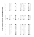
- the general method of the invention for production and for identification of restriction fragments involves the use of restriction endonucleases, ligation of synthetic oligonucleotides to the restriction fragments, and PCR amplification of restriction fragments (figure 1). Restriction endonucleases cleave genomic DNA molecules at specific sites, target sites, thereby generating restriction fragments.
- PCR amplification of restriction fragments no matter whether one knows the nucleotidic sequence of the ends of the restriction fragments or not, can be achieved according to the invention, by first ligating synthetic oligonucleotides (adaptors) to the ends of restriction fragments, thus providing each restriction fragment with two common tags which will serve as a anchor base for,the primers used in PCR amplification.
- synthetic oligonucleotides adaptors
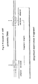
- restriction enzymes produce either flush ends, in which the terminal nucieotides of both strands are base paired,or staggered ends in which one of the two strands protrudes to give a short single strand extension (figure 2).
- adaptors are used with one flush end.
- adaptors which have a single stranded extension which is complementary to the single stranded extension of the restriction fragment. Consequently, for each type of restriction fragment specific adaptors are used, which differ only in one of the ends so as to allow the adaptor to be ligated to the restriction fragment.
- the adaptors used are composed of two synthetic oligonudeotides which are in part complementary to each other, and which are usually approximately 10 to 30 nucleotides long, preferably 12 to 22 nucleotides long and which form double-stranded structures when mixed together in solution.
- the enzyme ligase the adaptors are ligated to the mixture of restriction fragments.
- Restriction fragments prepared with this method will be referred to as tagged restriction fragments and the method will be further referred to as restriction fragment tagging.
- the adaptors can now serve as templates for the primers having the characteristics hereabove defined, used in the subsequent PCR amplification reaction.
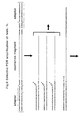
- the restriction fragment carries the same adaptor at both of its ends and a single PCR primer can be used to amplify the restriction fragment as illustrated in figure 3. Since in such a case all restriction fragments are tagged in the same way, it is obvious that PCR amplification of a mixture of tagged restriction fragments will amplify all restriction fragments in a synchronous fashion.
- two different adaptors are ligated to the ends of the restriction fragments. In this case two different PCR primers can be used to amplify such restriction fragments.
- the adaptor for one of the enzyme ends is biotinylated. This allows one to select out of the complex mixture of restriction fragments those restriction fragments that carry at least one end for this restriction enzyme, using usual methods for isolating biotinylated molecules. This step reduces the complexity of the starting mixture of restriction fragments and constitutes an enrichment step prior to the PCR amplification, thereby reducing in certain instances the background.
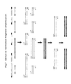
- the simultaneous amplification of several different fragments is often referred to as multiplex PCR.
- the principle of multiplex restriction fragment amplification is illustrated in figure 4.
- the present invention is further based on the definition of specifically designed primers and specific methods to direct the PCR amplification reaction in such a way that a controlled amplification is possible and in a particular embodiment of the invention, in such a way that only a small subset of tagged restriction fragments is amplified.
- restriction endonuclease digests of genomic DNA and in particular of animal, plant or human genomic DNA, yields very large numbers of restriction fragments.
- the number of restriction fragments depends upon the size of the genome and of the frequency of occurrence of the target site of the restriction endonuclease in the genome, which in turn is primarily determined by the number of nucleotides in the target site.
- the number of nucleotides in the target sites of commonly used restriction endonucleases ranges from 4 to 8.
- the genome sizes of organisms vary widely from a few million base pairs in the case of microorganisms to several billion base pairs for animals and plants.
- the number of restriction fragments obtained after cleaving genomic DNA molecules with a restriction enzyme can vary from a few hundred to several million. Generally, the number of restriction fragments is so large that it is not possible to identify individual restriction fragments in genomic DNA digests fractionated by gel electrophoresis. Such digests usually produce a smear of bands.
- PCR amplification of tagged restriction fragments should thus also produce a smear of bands since all fragments should coamplify synchronously in the PCR reaction.
- a general principle to limit the number of restriction fragments which are to be amplified. This is done by preselecting a subset of tagged restriction fragments so that only a relatively small number of tagged restriction fragments will be amplified during the PCR amplification reaction.
- the selective principle defined in this embodiment of the invention resides in the design of the oligonucleotides which are used as primers for the PCR amplification, as is illustrated in figure 5.
- Tagged restriction fragments have the following general structure: a variable DNA sequence (corresponding to the restriction fragment before tagging), flanked on both sides by an inverted DNA sequence (constant sequence).
- the inverted DNA sequence (constant DNA sequence) is composed of part of the target sequence of the restriction endonuclease and of the sequence of the adaptor attached to both ends of the restriction fragment.
- the variable sequences of the restriction fragments comprised between the constant DNA sequences are usually unknown, and will thus have a random sequence composition. Consequently, the nucleotide sequences flanking the constant DNA sequence will be totally random in a large mixture of restriction fragments.
- the present invention therefore also provides specific PCR primers which comprise a constant nucleotide sequence part and in the embodiment of the invention relying to the amplification of a restricted subset of the restriction fragments obtained, a variable sequence part.
- the nucleotide sequence is designed so that the primer will perfectly base pair with the constant DNA sequence of one of the DNA strands at the end of the restriction fragment.
- the variable sequence part comprises a randomly chosen nucleotide sequence ranging from 1 to 10 bases chosen.
- variable sequence more exactly designates a sequence consisting of selected nucleotides forming a sequence which will then remain constant for the purpose of amplifying a subset of restriction fragments.
- sequences of selected bases can be used, in order to define several distinguished primers.
- primers can have the same constant sequence and variable sequences made of selected bases which are different among the primers thus formed.
- variable (selected) sequences to the '3 end of the primers which will direct the preselection of tagged restriction fragments which will be amplified in the PCR step: when the PCR reaction is performed under appropriate conditions the primers will only initiate DNA synthesis on those tagged restriction fragments in which the variable DNA sequence can perfectly base pair with the template strand of the tagged restriction fragment, as illustrated in figure 5.
- the selection is determined by the number of nucleotides residing in the variable sequence part of the primer: the selectivity of the primers increases with the number of nucleotides in the variable (selected) sequence part.
- selectivity of the primers increases with the number of nucleotides in the variable (selected) sequence part.
- selective bases we will also use the term selective bases to denote the nucleotides in the variable sequence part thus showing that the selection of these bases renders the primer selective. It must be realized that a tagged restriction fragment will only be amplified when the selective bases of the primers used recognize both complementary sequences at the ends of the fragment. When the primer matches with only one end, the amplification will be linear rather than exponential, and the product will remain undetected.
- n the number of selective bases: using 1 selective base, 1 out of 16 tagged fragments will be amplified, using 2 selective bases, 1 out of 256, using 3 selective bases, 1 out of 4.096, using 4 selective bases, 1 out of 65.536, and so on, will be amplified.
- One preferred embodiment of the present invention thus allows one to selectively amplify a random subset of tagged restriction fragments from any genomic DNA digest regardless of the number of fragments produced by the restriction enzyme used.
- the number of selective nucleotides is chosen so that the number of restriction fragments which will be amplified is limited to 5 to 200. Although this number can be calculated by dividing the number of fragments by 4 2n , a precise prediction is not possible because not all restriction fragments can be amplified with equal efficiency. Hence, in practice, one finds less fragments of the amplification than theoretically expected. It should also be pointed out that mixtures of two (or more) primers can be used. This will allow the amplification of the fragments recognized by each primer and in addition, the fragments recognized by the two primers.
- the PCR products obtained in accordance with the invention can be identified using standard fractionation methods for separating DNA molecules according to size followed by staining of the DNA molecules with appropriate agents.
- the primers used for the PCR amplification can be tagged with a suitable radio-active label or fluorescent chromophore thus allowing the identification of the reaction products after size fractionation.
- the PCR products are fractionated by gel electrophoresis using standard gel matrices such as, but not limited to, agarose, polyacrylamide or mixed agarose/polyacrylamide.
- the PCR products obtained according to the invention will be denoted further by the term Amplified Restriction Fragments (ARF).
- the means and method of the present invention can be used to generate sets of ARF from restriction digests of any complex genome.
- the invention permits the number of restriction fragments obtained to be tuned in accordance with the resolution of the gel fractionation system used to separate the ARFs.
- the selective primers are designed to produce 5 to 10 ARFs which are then separated by agarose gel electrophoresis.
- Another particular embodiment involves the use of selective primers which are designed to produce 20 to 50 ARFs which are then separated on a high resolution gel electrophoresis system such as, but not limited to, polyacrylamide gels or mixed polyacrylamide-agarose gels.
- restriction enzyme or enzymes are chosen to yield restriction fragments in the size range of 20 to 1000 base pairs, because as is generally known for PCR amplification, this fragment size range is amplified most effectively. Although much fragments can be fractionated on various standard gel matrices, best results are obtained by fractionation on denaturating polyacrylamide gel systems as are currently used for DNA sequencing.
- different sets of ARFs are obtained with each different selective primer in the PCR amplification reaction.
- the patterns of ARFs identified after separation constitute unique and perfectly reproducible fingerprints of the genomic DNA.
- fingerprints can have several applications such as, but not limited to, forensic typing, the diagnostic identification of organisms,and the identification of species, races, varieties or individuals.
- the level of identification will be determined by the degree of similarity (the degree of variability) exhibited by different members of a specific group.
- the variability or similarity is determined by the degree of variation in the nucleotide composition of the related genomes.
- the underlying principle of the invention is that in each Amplified Restriction fragment two nucleotide sequences are detected which are separated from each other by a given distance, as is illustrated in figure 9.
- Each of the two nucleotide sequences is composed of two parts: (a) the target site for the restriction endonuclease and (b) the nucleotide sequence adjacent to the target site which is included in the selective primer.
- these sequence elements and their relative distances will be conserved to a greater or lesser degree.
- the fingerprints constitute a basis for determining the degree of sequence relationships between genomes.
- differences in the ARF patterns can be used to distinguish genomes from each other.
- the particular advantages of the present invention over other methods for fingerprinting genomes is the high resolution that can be obtained with the method: several tens or even hundreds of ARFs can be compared simultaneously.
- Another particular application of the present invention involves the screening and identification of restriction fragment polymorphisms (RFP).
- Changes in the nucleotide composition of genomic DNA often result in polymorphisms of restriction fragments: insertions or deletions affect the size of the restriction fragments containing them (figure 10), nucleotide changes can result in the elimination of restriction endonuclease target sites or the creation of new restriction endonuclease target sites.
- the most commonly used techniques for identifying such changes are Southern blotting experiments using cloned DNA probes, a technique usually referred to as restriction fragment length polymorphism (RFLP) detection. This technique involves the extensive screening of randomly cloned DNA fragments in Southern blotting experiments for associated RFLPs among different genomes.
- RFLP restriction fragment length polymorphism
- RFPs can be identified directly by comparing the ARFs obtained from different genomes.
- the method of the present invention is more sensitive for detecting RFPs because not only differences in the target sites of the restriction endonuclease are detected, but also differences in the adjacent nucleotide sequences comprised in the selective PCR primers. Consequently, the method of the present invention constitutes a far superior method for detecting RFLPs.
- RFLPs are now currently used for several applications including forensic typing, monitoring of genetically inherited diseases in humans and monitoring the inheritance of agronomic traits in plant and animal breeding.
- the underlying principle is that certain DNA polymorphisms which are closely linked with specific genetic traits can be used to monitor the presence or absence of specific genetic traits.
- the analysis of ARF patterns can be used to define the genetic linkage of polymorphic ARFs with specific genetic traits.
- polymorphic ARFs will be further referred to as Amplified Fragment Length Polymorphisms (AFLP®) to distinguish them from RFLP type DNA polymorphisms detected in Southern blotting experiments using cloned DNA probes.
- AFLP® Amplified Fragment Length Polymorphisms
- One particular application of the present invention involves the detection of AFLPs linked to specific genetic traits.
- the application involves the analysis of ARF patterns obtained with different selective primers in restriction digests of genomic DNA of closely related individuals exhibiting differences in the specific genetic trait and the use of analysis techniques that can find correlations between the inheritance of one or more AFLP® and the phenotype exhibited by the specific genetic traits.
- a second preferred embodiment of the present invention involves the use of the method of the invention to identify one or more specific restriction fragments.
- One specific restriction fragment can be amplified from a complex mixture of tagged restriction fragments by first determining the nucleotide sequence of the first 8-12 bases at each end of the restriction fragment. Based on these sequences one can design two primers with each 5 to 10 selective nucleotides exhibiting a sequence complementary to that of the sequence flanking the restriction site of the complementary strand of the restriction fragment. Using such sets of primers one can obtain, after PCR amplification, a single amplified fragment.
- the restriction fragment used in this method can be either a cloned restriction fragment or an amplified restriction fragment.
- the preferred method of the invention for identifying polymorphic DNA markers involves first amplifying randomly chosen set of fragments and identifying AFLPs which yield strong bands after PCR amplification.
- AFLP® can be characterized by sequencing to develop restriction fragment specific primers.
- the AFLP® will be isolated by cutting out the corresponding DNA band from the gel, and determining the nucleotide sequences at both ends to establish the sequence of the first 5 to 10 nucleotides adjacent to the restriction endonuclease target sites. Once these nucleotide sequences are known, restriction fragment specific primers can be designed which will only amplify a single restriction fragment from a genomic DNA digest.
- one set of two different selective primers can be used for detecting a specific restriction fragment.
- the selective bases are chosen such that they are complementary to the nucleotide sequence adjacent to the restriction endonuclease target site, as is illustrated in figure 8.
- the number of selective bases to be included in each primer depends upon the complexity of the restriction endonuclease fragment mixture.
- the PCR technique has developed tremendously over the past few years and is rapidly becoming one of the most widely used diagnostic methods in human health care. Its application includes amongst others detection of infectious diseases and detection of genetically inherited diseases.
- Each diagnostic test is based on the use of two specific synthetic oligonucleotides which are used as primers in the PCR reaction to obtain one or more DNA fragments of specific lengths. In disease detection the test will detect the presence of as little as one DNA molecule per sample, giving the characteristic DNA fragment.
- the primers are designed such that their products can discriminate between normal and disease alleles. The distinction either relies on sequence differences in the DNA segment in the genome which is complementary to the primer or, on distance differences between the two primers.
- multiplex PCR a method often referred to as multiplex PCR.
- the multiplex PCR method suffers from the limitation that generally only few, 5 to 8, different traits can be monitored simultaneously.
- the scientific basis for this limitation is that the optimal conditions for PCR amplification (annealing temperature, Mg + concentration, primer concentration) vary considerably depending on the pair of primers used.
- annealing temperature, Mg + concentration, primer concentration vary considerably depending on the pair of primers used.
- multiplex PCR compromise conditions have to be established under which all primer pairs yield detectable products.
- superimposed upon this phenomenon there is the phenomenon of strong differences in the efficiency of amplification of different fragments. Consequently, one often has encountered the problem that products of certain primer pairs are not detectable in multiplex PCR reactions.
- the methods of the present invention in essence overcomes these limitations of multiplex PCR, because all the primers used in the present invention have a substantial part of their nucleotide sequence in common. Furthermore, by selecting AFLP®, we select DNA markers that are amplified with equal efficiency. Hence, the optima of the PCR amplification conditions for the different selective primers exhibit much less variation than is observed with commonly used sequence specific primers. In essence, ideal compromise between the number of bases in the synthetic oligonucleotide which are necessary to obtain the required specificity of detecting a single DNA fragment of a given size in a complex genome, which is calculated above, and the length and composition of the oligonucleotide which is optimal for efficient PCR amplification. The method of the invention thus provides a far superior method for multiplex PCR.
- the present invention provides a general method for isolating DNA markers from any genome and for using such DNA markers in all possible applications of DNA fingerprinting.
- EXAMPLE 1 SELECTIVE RESTRICTION FRAGMENT AMPLIFICATION OF TOMATO DNA USING PSTI
- Total Tomato DNA ( Lycopersicon esculentum c.v. Moneymaker) was isolated from young leaves as described by Bernatzski and Tanksley (Theor. Appl. Genet. 72, 314-321). The typical yield was 50 - 100 ⁇ g DNA per gram of fresh leaf material.
- the DNA was restricted with PstI (Pharmacia) and double-stranded (ds) PstI -adapters were ligated to the restriction fragments following the procedure described below. These adapters had the following structure:
- the 3'TGCA-overhang in these adapters anneals to the staggered ends created by Pst I.
- the Pst I recognition sequence CTGCAG is not restored upon ligation of this adapter, because the 5' C-residue is replaced by A.
- the ligation reaction was designed in such a way that the end result is almost exclusively DNA fragment- to-adapter molecules. This was achieved by: 1. using nonphosphorylated adapters, which excludes adapter- to-adapter ligation, 2. Performing the ligation and restriction reaction at the same time. The latter procedure results in restriction of any fragment-to-fragment ligation product, thereby eliminating these products almost completely. Adapter-to-fragment ligation products cannot be restricted by the restriction enzyme, because the PstI recognition sequence is not restored in these products.
- the reaction conditions used for the adapter ligation were:
- the ligation reaction was performed in a reaction volume of 20 ⁇ l for 3 hours at 37°C. After the adapter ligation, non-ligated adapters were removed by selective precipitation. For this purpose the reaction mixture was increased to 100 ⁇ l and NH4Ac was added to a final concentration of 2.5 M. 100 ⁇ l ethanol of -20°C was added and the mixture was incubated for 5 minutes at room temperature. The DNA was collected by centrifugation for 10 minutes at 14000 rpm in a cooled eppendorf centrifuge at 4°C.
- the DNA pellet was washed once with 0.5 ml of 70% ethanol at room temperature, and dissolved in 40 ⁇ l of T0.1E (10 mM Tris.HCl pH 8.0, 0.1 mM EDTA). The DNA was stored at -20°C. The selective precipitation procedure described here removes the non-ligated adapters efficiently from the reaction mixture, but small DNA-fragments ( ⁇ 200 bp) are also lost.
- T0.1E 10 mM Tris.HCl pH 8.0, 0.1 mM EDTA
- the DNA prepared above was used as template for amplification of the PstI -fragments.
- the reaction mixture for the PCR contained:
- the reaction mixture was covered with 20 ⁇ l of light mineral oil to prevent evaporation during the amplification reaction.
- the PCR was performed on a Perkin Elmer DNA Thermal Cycler using the following cycle profile: 1 minute at 94°C, 1 minute at 60°C, a temperature increase from 60°C to 72°C at a rate of 1 °C/5 seconds, and 21 ⁇ 2 minute at 72°C. A total of 33 cycles were performed.
- 20 ⁇ l chloroform was added, and 10 ⁇ l of loading dye, in this case 50% sucrose with 0.1% w/v of the dye Orange G (Merck).
- Tomato DNA restricted with Pstl and tagged with the Pstl- adapter was amplified using the conditions specified above. Four different primers were selected with the sequences:
- Primer 1 is part of the the top strand of the adapter used to modify the DNA, and therefore should amplify all PstI - fragments.
- Primer 2 contains part of the adapter sequence, the PstI-recognition sequence (lower case letters) and one selective nucleotide (bold) and should amplify theoretically about 1/16 part of all PstI -fragments.
- Primers 3 and 4 are similar to primer 2, but contain 2 and 3 selective nucleotides respectively, and therefore are expected to amplify about 1/256 and 1/4096 of the PstI -fragments. Part of the reaction mixtures were analysed on a 1.0 % agarose gel, which is shown in Figure 11.
- Lanes 1 and 6 of this figure contain DNA markers, of which the sizes are indicated at the left. Lanes 2, 3, 4 and 5 contain the PCR's obtained with primers 1, 2, 3 and 4 respectively. The results indicate that only in case of the primer with 3 selective nucleotides, the number of amplified fragments was such that a clear band pattern was obtained. The other 3 primers gave band patterns, which could not be resolved on agarose gels, because to many PCR products were generated. Within these many PCR products always some fragments predominate, and are seen as bands on a background smear of the other PCR products. Probably these stronger products are present in higher copy numbers on the Tomato genome, or amplify more efficient than the other products. It was anticipated that primers with 3 selective nucleotides had to be used to generate a clear band pattern on agarose gels, because of the total number of PstI -fragments of Tomato genomic DNA (20.000 to 100.000).
- the amplified fragments were tested on Southern blots to verify that these fragments corresponded to bonafide restriction fragments of the same size.
- four individual fragments obtained with primer 4 were cut out of the agarose gel.
- the DNA was purified from these gel slices by means of absorption to glass beads (Gene Clean, manufacturer Bio 101), and part of the purified DNA was reamplified to obtain about 1 ⁇ g of each of the four DNA fragments.
- the reamplification reactions were subsequently electrophoresed on a 1.0% preparative agarose gel, and the desired DNA fragments were purified. 200 ng of each fragment was labeled with ( ⁇ -32P)dATP using a random hexamer labelling kit according to procedures advised by the manufacturer (Boehringer Mannheim).
- Total Tomato DNA was restricted with PstI , and electrophoresed on a 1.0% agarose gel. Four clearly separated lanes each containing about 3 ⁇ g of restricted DNA were used. Next, the agarose gel was blotted to a Genescreen+ hybridisation membrane as indicated by the manufacturer (New England Nuclear). After blotting the gel was cut in four slices, each containing one lane of the Tomato DNA restricted with PstI . These four slices were each hybridised to one of the four DNA probes following the procedure described by Klein-Lankhorst et al. (Theor. Apll. Genet. 81, 661-667). The hybridised blots were autoradiographed for 40 hours using Kodak XAR5 films. The results obtained showed that all genomic DNA fragments recognised by the four DNA probes, had the same length as these probes. This demonstrated that the amplified fragments, used as probes, originated from the fragments detected on the blots.
- Three sets of primers were designed for 3 corresponding random Pstl -fraqments from Tomato genomic DNA, of which the sequence next to the PstI -recognition sequence was known.
- Sets of primers with 5 selective nucleotides were made as shown below.
- Tomato DNA was digested with Pstl and adapters were ligated to the ends of the restriction fragments as described above. This DNA was used as template in PCR's with Primer sets 1 or 2 or 3, using the conditions as described in one of the previous sections. The reaction products of each PCR were analysed on a 1.0% agarose gel. This gel is shown in Figure 12.
- Figure 12 shows 13 lanes, of which lanes 1, 2, 12 and 13 are DNA markers. The sizes in kilobases of these markers are indicated at both sides of the gel.
- Lanes 3, 6 and 9 show plasmid DNA with each of the three Pstl -fragments restricted with Pstl , which yields the vector fragment, pUC18 (Yanisch-Perron et al., Gene 33, 103-119), and the inserted Pstl -fragment.
- Lanes 4 and 5 show amplification with primer set 1 of 5 fg of the corresponding plasmid DNA and 1 ng of total genomic DNA respectively.
- Lanes 7 and 8 show amplification with primer set 2 of plasmid DNA and total genomic DNA, and lanes 10 and 11 show amplification with primer set 3.
- DNA was isolated from the two Tomato lines (line 83M-71392, Mi-sensitive, and line 83M-71398, Mi-resistant , obtained from De Ruiter Seeds, Bleiswijk, The Netherlands) and subsequently restricted with PstI and provided with adapters as described above.
- a large number of amplification reactions were performed using primers, which differed in their extension of selective nucleotides. Three selective nucleotides were used, and apart from single primers also combinations of two different primers were used. The reactions were analysed on mixed polyacrylamide/agarose gels: 2.5% polyacrylamide and 1.0% agarose was used, with a ratio acrylamide to bisacrylamide of 20 : 1.
- the polymorphic bands in lanes 9 and 11 were expected to be the same, because the same primer was present in both reactions (the difference is the presence of a second primer in lane 11).
- the two polymorphic fragments of lanes 11 and 12 were cut out of the gel, the gel slices were crushed by forcing them through a 18 gauge needle and the DNA was eluted from the gel slices by elution through diffusion in 200 ⁇ l of 100 mM Tris.HCl pH 8.0, 10 mM EDTA. 2 ⁇ l was used for reamplification of these fragments as described above.
- EXAMPLE 2 SELECTIVE RESTRICTION FRAGMENT AMPLIFICATION OF TOMATO DNA WITH TWO RESTRIC TION ENZYMES
- SRDA selective restriction fragment amplification
- Total Tomato DNA was isolated from young leaves as described in example 1.
- Two pairs of so called isogenic lines were used as source of the DNA, named Gem R and Gem S , and GCR26 and GCR151 respectively (These lines are described in the following references: Denby and Williams, [1962], Can. J. Plant Sci. 42 , 681-685, Smith and Ritchie, [1983], Plant Mol. Biol. Rep. 1 , 41-45).
- the two individuals of each pair of isogenic lines are genetically very similar, but differ in the presence of a trait confering resistance to the fungal pathogen Verticillium albo-atratum .
- the first step of the modification of the DNAs comprised the restriction of the DNAs with the two enzymes PstI and MseI .
- the restriction of the DNA, and also the subsequent ligation of the adapters to the DNA-fragments was carried out in the same buffer, which was named RL-buffer (restriction-ligation buffer), and which contained: 10 mM Tris.HAc/10 mM MgAc/50 mM KAc/5 mM DTT, pH 7.5.
- the next step in the modification of the DNAs was the ligation of adapter molecules to the ends of the DNA fragments. First appropiate double-stranded adapter molecules had to be prepared.
- the resulting reaction mix of 60 ⁇ l was incubated for 3 hours at 37°C.
- the adapters were designed in such a way that the restriction sites were not restored after ligation. In this way fragment-to-fragment ligation was prevented, since fragment concatamers are restricted, because the restriction enzymes were still active during the ligation reaction. Adapter-to-adapter ligation was not possible because the adapters were not phosphorylated (see also example 1).
- Preparation of the template-DNAs for SRFA using two restriction enzymes generally involved an extra step not used when using SRFA with a single enzyme. In this step the DNA-fragments to which a biotinylated adapter was ligated were separated from all other fragments.
- Biotinylated fragments were separated from non-biotinylated fragments MseI - MseI -fragments) in this step, by binding to paramagnetic streptavidine beads (Dynal). 10 ⁇ l beads were washed once in 100 ⁇ l STEX (100 mM NaCl/ 10 mM Tris.HCl/1 mM EDTA/ 0.1 % Triton X-100 pH 8.0), and resuspended in 140 ⁇ l STEX. The beads were subsequently added to the ligation mixture, to give a final volume of 200 ⁇ l .
- DNAs restricted with the restriction enzymes provided with adapters, attached to the paramagnetic streptavidine beads and purified from the MseI - MseI fragments prepared as described above will be referred to as template-DNAs in the following steps.
- the template-DNAs prepared as decribed above should contain all PstI - MseI fragments from the mentioned Tomato lines, and in addition a small amount of PstI - PstI -fragments with no internal Msel -fragments.
- a number of these PstI - MseI fragments were visualised by amplification, essentially as described in example 1.
- Gel analyses of the amplification products was performed on denaturing acrylamide gels (Maxam and Gilbert, Proc. Natl. Acad. Sci. U.S.A. 74 , 560-564), because the kind of fragments obtained by the procedure described in this example were much smaller than the ones described in example 1.
- the primer selected for labeling was the 19-mer 5-GATGAGTCCTGAGTAAgaa-3 which was named Msel -primer-1, and in which the selective nucleotides are indicated with lower case letters.
- the labeling was performed in the following way:
- a total of 28 PCRs were performed, in which each of the 4 template-DNAs were amplified with 7 primer combinations.
- Each primer combination had the same MseI -primer ( MseI -primer-1, described above), but varied in the choice of the PstI -primer.
- a total of 7 different primers were chosen (As with the MseI -primer the selective nucleotides are indicated with lower case letters):
- the PCR-mixture consisted of:
- the amplifications were performed on a Perkin Elmer 9600 thermal cycler.
- the cycle profile was as follows: 1 cycle denaturation : 30 sec at 94°C annealing : 30 sec at 65°C extension : 60 sec at 72°C 11 cycles denaturation : 30 sec at 94°C lower annealing temperature 0.7°C each cycle, 64.3°C, 63.6°C, 62.9°C, 62.2°C, 61.5°C, 60.8°C, 60.1°C, 59.4°C, 58.7°C, 58.0°C, . 57.3°C.
- the reaction products were analyzed on 4.5% denaturing polyacrylamide gels.
- 50 x 38 cm gels were used, of which the gel cassettes to prepare these gels were purchased from Biorad.
- 100 ml of gel solution was used containing 4.5% w/v acrylamide/0.225% w/v bisacrylamide/7.5 M Urea/50 mM Tris/50 mM Boric acid/1 mM EDTA, pH 8.3.
- 100 ml gel solution was mixed with 500 ⁇ l 10% Ammonium persulfate and 100 ⁇ l TEMED immediately before casting the gel.
- Tris/Boric acid/EDTA-buffer was used as electrophoresis buffer and contained: 100 mM Tris/100 mM Boric acid/2 mM EDTA, pH 8.3.
- the reaction mixtures were mixed with an equal volume (20 ⁇ l) of 98% formamide/10 mM EDTA/0.01% w / v bromo phenol blue/0.01% w/v xylene cyanol.
- the resulting mixtures were heated for 3 minutes at 95°C, and then quickly cooled on ice. 2 ⁇ l of each sample was loaded on the gel. Gels were run at constant power of 110 Watts to give a constant heat development during electrophoresis. Under these conditions the field strength of the gels corresponded to 40 to 50 Volt/cm.
- the results of the SRFA reactions are shown in figure 14.
- the lanes are numbered from 1 to 28, and contain each time the four Tomato lines with one of the 7 primer combinations.
- the order of the Tomato lines on the gel is: 1. GCR26, 2. GCR151, 3. Gem R , 4. Gem S .
- Lanes 1 to 4 contain these DNAs amplified with Msel -primer-1 and Pstl -primer-1
- lanes 5 to 8 contain these DNAs amplified with Msel -primer-1 and Pstl -primer-2
- lanes 9 to 12 contain these DNAs amplified with Msel -primer-1 and Pstl -primer-3
- lanes 13 to 16 contain these DNAs amplified with Msel -primer-1 and Pstl -primer-4
- lanes 17 to 20 contain these DNAs amplified with Msel -primer-1 and Pstl -primer-5
- lanes 21 to 24 contain these DNAs amplified with Msel -primer-1 and Pstl -primer-6
- lanes 25 to 28 contain these DNAs amplified with Msel- primer-1 and Pstl -primer-7.
- the gel contains no size markers but the DNA fragments visualised correspond with ⁇
- EXAMPLE 3 SELECTIVE RESTRICTION FRAGMENT AMPLIFICATION OF DNA OF VARIOUS LACTUCA SPECIES WITH TWO RESTRICTION ENZYMES
- example 2 the principle of selective restriction fragment (SRFA) amplification using two restriction enzymes is exemplified for Tomato DNA.
- SRFA selective restriction fragment
- DNAs were isolated as described in example 1 using young leaf material of various Lactuca species. As indicated below these plants include a commercial lettuce ( L.sativa ) variety, and several individuals of two wild Lactuca species, Lsatigna and L.virosa . The plants were arbitrarily designated the following names:
- the DNAs prepared as decribed above were used as templates for SRFA reactions.
- Two primer combinations were used employing a single MseI -primer and two different PstI -primers. These primers (selective nucleotides depicted in lower case letters) were:
- EXAMPLE 4 SELECTIVE RESTRICTION FRAGMENT AMPLIFICATION OF CORN INBRED LINES WITH A VARIETY OF RESTRICTION ENZYME COMBINATIONS
- example 2 and 3 the principle of selective restriction fragment (SRFA) amplification using two restriction enzymes is exemplified using Tomato DNA and Lettuce (Lactuca species) DNAs respectively.
- Tomato DNA and Lettuce (Lactuca species) DNAs respectively.
- Corn (Zea perennial) lines respectively.
- a variety of restriction enzyme combinations can be used to obtain DNA fingerprints of in this case Corn lines.
- DNA of these lines was isolated from young leaf material as described by Saghai-Mahoof et al. (1984), Proc. Natl. Acad. Sci. U.S.A. 81 , 8014-8018).
- the following restriction enzyme combinations (EKs) were used to make the template-DNAs: Pstl / Taql , EcoRl / Taql , Asel / Taql , Sse8387-I / Taql . All enzymes were purchased from Pharmacia, except Asel which was purchased from New England Biolabs, and Sse8387-I which was purchased from Amersham. Template DNAs were prepared essentially as described in examples 2 and 3, with the following exceptions:
- the primers selected for labeling of the amplification products were the following Taql -primers having 3 selective nucleotides (indicated by lower case letters):
- primers were used for detection of amplification products with all four enzyme combinations. For each enzyme combination 4 primers for the other enzyme were selected to give a total of 16 combinations for each enzyme. These primers are indicated below (selective nucleotides shown in lower case letters). For EcoRI and Asel primers with 3 selective nucleotides were selected, for PstI primers with 2 selective nucleotides were chosen, and for Ssel primers with a single selected nucleotide were chosen. For enzymes cutting less frequently in the Corn genomic DNA, primers were selected containing extensions with fewer selective nucleotides.
- Lanes 1 to 8 show DNA-fingerprints of the two Corn DNAs obtained by SRFA with Tagl -primer-3 and Pstl -primers-1, -2, -3 and -4 respectively
- lanes 9 to 16 show DNA-fingerprints of the two Corn DNAs obtained by SRFA with Taql -primer-4 and Pstl -primers-1 , -2, -3 and -4 respectively
- lane 17 shows the size marker lambda-DNA restricted with Pstl , of which the sizes of some of the fragments in nucleotides are indicated at the right
- lanes 18 to 25 show DNA-fingerprints of the two Corn DNAs obtained by SRFA with Taql -primer-1 and EcoRI -primers-1 , -2, -3 and -4 respectively.
- SRFA selective restriction fragment
- campestris citri 8657 13. campestris citri 8654 14. campestris citri 8650 15. campestris citri 682 16. campestris citri 681 17. campestris citri 9325 18. campestris citri 9176 20. campestris citri 9671 21. campestris citri 9665 22. campestris citri 9182 23. campestris citri 568 24. campestris citri 9167 25. campestris citri 9175 26. campestris citri 9160
- Amplification of restriction fragments was performed as described in example 2.
- the primers selected for SRFA were the Taql -primer 5-CGATGAGTCCTGACCGAg-3 (having one selective nucleotide indicated in lower case letter), and the Apal -primer 5-GACTGCGTACAGGCCCg-3 (having one selective nucleotide indicated in lower case letter).
- the Apal -primer was labeled at the 5'end for detection of the amplified fragments as described in example 2.
- Each of the 26 DNAs was amplified using the primer set described above. Amplification conditions were as described in example 2, except that the last 9 cycles of the PCR were omitted, because of the lower complexity of the DNAs compared to the plant DNA in examples 2, 3 and 4.
- Lanes 1 to 26 represent bacterial DNAs 1 to 26.
- the sizes of marker DNAs (not visible on the gel) in nucleotides are indicated to the right of the gel. This figures shows clea ⁇ ly that the relatedness of the bacterial strains is reflected by the similarity of the band patterns.
- EXAMPLE 6 SELECTIVE RESTRICTION FRAGMENT AMPLIFICATIONS OF DNA OF VARIOUS ANIMALS WITH TWO RESTRICTION ENZYMES
- SRFA restriction fragment amplification
- DNAs were isolated from bloodsamples following procedures described by Maniatis et al.,(1982).
- DNA samples 1 to 3 (chicken), 4 to 7 (pig), 8 to 11 (cow) and 12 to 15 (horse) were digested by restriction enzymes Sse8387l and Msel .
- the DNA fragments were ligated to adapters as described in example 2. Since the restriction enzymes Sse8387l and Pstl generate compatible 3' overhangs we could use the Pstl - and Msel -adapter described in example 2.
- Template DNAs named above and prepared as described in example 2 served as templates in SRFA reactions.
- the primer combinations used consisted of a single Msel -primer and different SseI -primers:
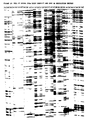
- Amplification of Sse83871 - Msel fragments using primer pairs described above was carried out using the protocol described in example 2. Reaction products were run on denaturing polyacrylamide gels also described in example 2. An autoradiograph showing fingerprints of the above samples is shown in Figure 18. Lanes 1 through 15 show fingerprints of DNAs 1 to 15 amplified with the Msel -primer paired with Sse83871 -primer-1, lanes 16 through 30 show similar patterns obtained with the Msel -primer combined with Sse8387l -primer-2. Differences in fingerprints between animals of one species reflect heterogeneity in animal populations; overall-patterns are characteristic for a specific species.
- the invention relates to a Process for the amplification by Polymerase Chain Reaction of at least one restriction fragment obtained from a starting DNA which contains a plurality of restriction sites for at least one specific determined Restriction endonuclease, which process comprises :
- the terminal nucleotide of at least one of said primers in the direction of the elongation sought corresponds to the last of the nucleotides involved in the restriction site for said specific endonuclease, and which process comprises identifying or recovering the restriction fragments of said starting DNA which have been amplified.
- At least one of said primers includes a selected sequence comprising a determined number (one or several nucleotides) extending beyond the last of the nucleotides involved in the restriction site for said specific endonuclease in the direction of its own elongation within the corresponding restriction fragments during the amplification step.
- double-stranded DNA-linker contains several sites for different specific endonucleases which are all distinct from one another, which processes comprise repeating, on a same starting DNA the steps of the process defined above with one of these restriction endonucleases yet with another of said distinct specific endonucleases and upon using primers whose nucleotide sequences are selected as defined in the above description, yet with respect to said other specific endonuclease.
- the process described above or of the oligonucleotide of the invention is appropriate, for the identification of polymorphisms in determined DNAs originating from the same live species, e.g. genomic DNAs of a microbial, plant or animal, including humans, or of fragments thereof, either among themselves or relative to a corresponding determined DNA standard, which use comprises subjecting the DNAs under study to the process or to the contact of the oligonucleotide in conditions allowing an amplification or elongation reaction, comparing the restriction patterns obtained starting from each of said DNAs and, optionally, of said standard DNA and relating the existence and, where appropriate, the localization of that DNA polymorphism to the differences observed between the sizes of the restriction fragments of the different DNAs.
- the invention also relates to a fragmented DNA whose different fragments have sequences which all correspond to initial digests of the unfragmented starting DNA from which they are produced with a same determined specific endonuclease, characterized in that all of said fragments were tagged at their 5' and 3' ends respectively by determined 3' and 5' adaptors corresponding to the cleaved part of a same starting DNA linker which initially included a single restriction site for said specific endonuclease, and optionally prolonged with determined constant sequences.
- the fragmented DNA can be in the form of a pattern of migration bands on a suitable support, e.g. gel support, in which its fragments had initially been caused to migrate under the influence of an electric field.
- the fragmented DNA can also comprise end portions including oligonucleotide characterized by the following composition, starting from the 5' end :
- the invention further relates to a kit for the fragmentation of determined DNAs by at least one specific restriction endonuclease into fragments and analysis of these fragments which comprises:
- a particular embodiment of this kit is such that said oligonucleotide segments for the elongation of both said 5' and 3' adaptors or 5' and 3' ends of the tagged DNA fragments, have identical nucleotide sequences.
- the linker of the kit contains several respective unique sites for specific endonucleases all different from one another, said kit further including primers corresponding to each of the 3' and 5' adaptors formed by cleavage of said linker with said different specific endonucleases respectively, wherein said primers are respectively as defined in claim 8, in respect of the 3' and 5' adaptors which are produced in said linker by cleavage thereof by each of said specific endonucleases.
- the kit can comprise fragmented DNA standards as defined above in respect of the corresponding specific restriction endonucleases, wherein each of said fragmented DNA standards is in respect of each of the determined specific restriction enzymes.
Abstract
Description
- This invention relates to applications of DNA fingerprinting and the use of DNA markers in a number of different fields including, but not limited to, plant and animal breeding, variety or cultivar identification, diagnostic medicine, disease diagnosis in animals and plants, identification of genetically inherited diseases in humans, family relationship analysis, forensic analysis, and microbial typing.
- More specifically, this invention relates to methods for DNA fingerprinting and for detecting specific DNA markers in genomes ranging from microorganisms to higher plants, animals and humans. The invention also relates to synthetic DNA molecules and products based thereon which are used in the methods of the invention in the different fields of application.
- DNA fingerprinting or DNA typing, as well as other methods of genotyping, profiling and DNA identification analysis, refer to the characterization of either similarities or one or more distinctive features in the genetic make up or genome of an individual, a variety or race, or a species. The general rule is that the closer the genetic relationship is, the greater the identity or more appropriate the similarity of genomes, and consequently distinctive features in the genome will be rarer. These similar or distinctive features can be revealed by analyzing the DNA of an organism after clearing the DNA with a restriction endonuclease. Restriction endonucleases are enzymes which recognize short nucleotide sequences, usually 4 to 8 bases in length and cleave the two DNA strands, thereby producing fragments of DNA of discrete length. Because of their high degree of sequence specificity, restriction endonucleases will cleave DNA molecules in a very specific fashion. The result is that a reproducible set of DNA fragments will be produced. DNA fragments can be fractionated according to their length on porous matrices, or gels, yielding typical banding patterns, which constitutes a DNA fingerprint of the organism's genetic makeup.
- When the fingerprints of very closely related species, varieties or races are compared, the DNA fingerprints can be identical or very similar. When differences are observed within otherwise identicalDNA fingerprints, such differences are referred to as DNA polymorphisms: these are new DNA fragments which appear in a fingerprint. The DNA is said to be polymorphic at that position and the novel DNA fragment can be used as a DNA marker. DNA polymorphisms detected in DNA fingerprints obtained by restriction enzyme cleavage can result from any of the following alterations in the DNA sequence: mutations abolishing the restriction endonuclease target site, mutations creating new target sites, insertions, deletions or inversions between the two restriction sites.
- Such DNA polymorphisms are generally referred to as RFLP, Restriction Fragment Length Polymorphisms. Such mutational changes will behave as bona fide genetic markers when they are inherited in a mendelian fashion. Consequently, DNA polymorphisms can be used as genetic markers in much the same way as other genetic markers: in parentage analysis, in genetic studies on the inheritance of traits, in the identification of individuals.
- For almost all living organisms, except viruses, restriction digests of the total genomic DNA of the organisms yield so many bands that it is not possible to score individual bands. Therefore, all methods for DNA fingerpinting are based on the principle that only a small fraction of the DNA fragments are visualized so as to yield a simple banding pattern which constitutes the DNA fingerprint.
- The most widely utilized method involves digesting the DNA of the organism with restriction endonucleases, fractionating the restriction fragments by gel electrophoresis, transferring and binding the fractionated DNA fragments onto membranes and hybridizing the membrane with a specific DNA fragment ("probe"). The DNA fragment will form double-stranded DNA molecules with the DNA fragment (or fragments) on the membrane which has (have) complementary nucleotide sequences. When the probe is tagged with a visualizable marker, the DNA fragment to which the probe is attached can be visualized. This procedure is generally referred to as "Southern hybridization". When differences are observed in the sizes of the corresponding restriction fragments to which the probe attaches in closely related genomic DNA molecules, these differences are referred to as DNA polymorphisms, more specifically restriction fragment length polymorphisms. The restriction fragment length differences correspond to the different allelic forms of the genetic locus recognized by the DNA probe. Although the Southern hybridization method for DNA fingerprinting has been widely used, the method is laborious and time consuming.
- Furthermore, the method has a low resolution and can thus only be used to score single loci or a few loci at most in a single reaction.
- The Polymerase Chain Reaction (PCR) technique is a method for synthesizing specific DNA fragments in vitro. The method relies on the use of specific oligonucleotides which will attach to unique sequences on a DNA molecule and a thermostable DNA polymerase. The oligonucleotides are designed in such a way that they can anneal to the opposite strands of the DNA and serve as primers in a DNA synthesis reaction in such a way that each will direct the synthesis of new DNA strands. Hence, in one round of synthesis a complete copy of the DNA molecule between the primers will be made, so that the DNA between the primers is duplicated. Each round of DNA synthesis results in the doubling of the amount of DNA, hence leading to the amplification of the DNA comprised between the two primers. Consequently, the PCR technique allows one to synthesize a precise DNA segment using a small amount of "substrate DNA". WO 90/11372 discloses detecting a 〈〈 test nudeotide 〉〉 in a known DNA sequence. The detection involves using primers defined starting from the DNA sequence adjacent to the 〈〈 test nucleotide 〉〉.
WO 90/08821 relates to microdissection of chromosomal DNA and further digestion of the obtained DNA. The digests are then at both ends ligated to oligonucleotide duplexes which are used to anchor primers corresponding to one of their strands. - In the present invention we have devised a new method to amplify, with the PCR method, restriction fragments obtained after cleaving the DNA of an organism with at least one restriction enzyme. In this novel application of the PCR method the oligonucleotides used are not directed against a known DNA sequence but are designed such that they recognize the ends of the restriction fragments. To this end it is necessary to modify the ends of the restriction fragments by adding oligonucleotide linkers (or adaptors) to the ends. The reason for this is that the ends of restriction enzymes have only usually few nucleotides in common, i.e. 2 to 8 nucleotides, too short to be used to design primers for PCR amplification.
- The invention is based on the use of a novel application of polymerase chain reaction technique (PCR) for amplifying one or more restriction fragments from complex mixtures of DNA fragments obtained by digesting genomic DNA molecules with restriction endonucleases. One particular advantage of the invention is to enable the amplification of DNA restriction fragments in situations where the nucleotidic sequence of the 5' and 3' ends of the restriction fragments are not determined. In such cases the usual sequence specific primers hybridizing to each strand of a restriction fragment to be amplified can not be defined and therefore one cannot use the methods known in the art for amplification purposes.
- The method of the invention can be used for instance in two different ways, leading to two different types of applications:
- (1) Methods for DNA fingerprinting of genomes by randomly selecting subsets of one or more restriction fragments to be amplified by the PCR technique. The invention also covers synthetic oligonucleotides for use in said methods and some applications of said methods can be forensic typing, microbial identification, varietal identification, pedigree analysis and screening of DNA markers linked to genetic traits;
- (2) Methods for identifying one or more preselected DNA fragments which can be polymorphic, by PCR amplification. The invention also covers specific synthetic oligonucleotides for use in said methods and some applications of said methods can be the screening of genetically inherited diseases in humans, monitoring the inheritance of agronomic traits in plant and animal breeding and the detection of infections agents in diseases.
-
-
- Figure 1 provides a graphic outline for the general method of PCR amplification of tagged restriction fragments obtained by digesting genomic DNA molecules with a restriction enzyme.
- Figure 2 depicts the ligation of adaptors to different ends of restriction fragments: flush ends and staggered ends.
- Figure 3 depicts the PCR amplification of tagged restriction fragments. The boxed areas depict the adaptors which are ligated to the restriction fragment, and the primers which are used in the PCR amplification. The arrows indicate the direction of DNA synthesis.
- Figure 4 provides a graphic outline for the PCR amplification of tagged restriction fragments.
- Figure 5 shows the general design of the selective primers used in the PCR amplification of tagged restriction fragments. The boxes denote the inverted repeat sequences at the ends of the restriction fragments. The selectivity of the primers is illustrated in two examples where there is respectively a perfect match and a total mismatch between the selective base sequence and that of the restriction fragment template DNA.
- Figure 6 shows the principle of selective PCR amplification using a PCR primer which selects template DNA molecules having a trinucleotide sequence adjacent to the adaptor sequence.
- Figure 7 depicts the selective PCR amplification of tagged restriction fragments.
- Figure 8 shows the principle of fragment specific amplification using a combination of two PCR primers each comprising 6 selective bases. Each primer forms a double-stranded structure in the different strand of the restriction fragment and thereby forms a primer/template complex from which DNA synthesis can be initiated (represented by the arrows).
- Figure 9 depicts the general sequence elements which are recognized with the method of selective restriction fragment amplification, including the two nucleotide sequences which are recognized and the distance separating the two sequences.
- Figure 10 depicts the types of nucleotide sequence variations which are detected in the method of identifying amplified fragment length polymorphisms.
- Figure 11 shows a 1,0% agarose gel with the analysis of the results of the amplification of Tomato DNA restricted with PstI, using primers of increasing selectivity.
- Figure 12 shows a 1,0% agarose gel with the analysis of the results of specific amplification of 3 different PstI fragments of Tomato DNA using fragment specific primers.
- Figure 13 shows a 2,5% polyacrylamide/1% agarose gel with DNA fingerprints obtained by Selective Restriction Fragment amplification of two Tomato lines.
- Figure 14 shows part of a 4,5% denaturing polyacrylamide gel with DNA fingerprints of 4 Tomato lines using SRFA with the enzyme combination PstI/MseI.
- Figure 15 shows part of a 4,5% denaturing polyacrylamide gel with DNA fingerprints of 10 Lactuca lines using SRFA with the enzyme combination PstI/MseI.
- Figure 16 shows part of a 4,5% denaturing polyacrylamide gel with DNA fingerprints of 2 Corn lines using SRFA with the enzyme combinations PstI/TaqI and EcoRI/TaqI.
- Figure 17 shows part of a 4,5% denaturing polyacrylamide gel with DNA fingerprints of 26 Xanthomonas campestris strains using SRFA with the enzyme combination ApaI/TaqI.
- Figure 18 shows part of a 4,5% denaturing polyacrylamide gel with DNA fingerprints of different individuals of 4 domestic animals Chicken, Pig, Cow and Horse using SRFA with the enzyme combination SseI/MseI.
-
- In the description and examples that follow, a number of terms are used herein. In order to provide a clear and consistent understanding of the specification and claims, including the scope to be given such terms, the following definitions are provided.
- Restriction Endonuclease: a restriction endonuclease or restriction enzyme is an enzyme that recognizes a specific base sequence (target site) in a double-stranded DNA molecule, and will cleave both strands of the DNA molecule at every target site.
- Restriction Fragments: the DNA molecules produced by digestion with a restriction endonuclease are referred to as restriction fragments. Any given genome will be digested by a particular restriction endonuclease into a discrete set of restriction fragments. The DNA fragments that result from restriction endonuclease cleavage are separated and detected by gel electrophoresis.
- Restriction Fragment Length Polymorphism (RFLP): the genomic DNA of two closely related organisms, for example, will exhibit differences in their nucleotide sequence composition at many sites. When these differences occur in the target site for a restriction endonuclease, the modified target site will not be cleaved at that point. Likewise, a nucleotide sequence variation may introduce a novel target site where non exists in the other organism, causing the DNA to be cut by the restriction enzyme at that point. Alternatively, insertions or deletions of nucleotides occurring in one organism between two target sites for a restriction endonuclease will modify the distance between those target sites. Because of this, digestion of the two organism's DNA will produce restriction fragments having different lengths. A polymorphism in the length of restriction fragments produced by digestion of the DNA of the two organ-isms will result.
- Gel Electrophoresis: To detect restriction fragments, an analytical method for fractioning double-stranded DNA molecules on the basis of size is required. The most commonly used technique for achieving such fractionation is gel electrophoresis. The rate at which DNA fragments move in such gels depends on their size; thus, the distances travelled decrease as the fragment lengths increase. The DNA fragments fractionated by gel electrophoresis can be visualized directly by a staining procedure if the number of fragments included in the pattern is small.
- Synthetic oligonucleotides: the single-stranded DNA molecules having preferably from almost 10 to almost 50 bases, which can be synthesized chemically are referred to as synthetic oligonucleotides. In general, these synthetic DNA molecules are designed to have a unique nucleotide sequence, although it is possible to synthesize families of molecules having related sequences and which have different nucleotide compositions at specific positions within the nucleotide sequence. The term synthetic oligonucleotide will be used to refer to DNA molecules having a unique nucleotide sequence. The term mixed synthetic oligonucleotides will be used to refer to families of related synthetic oligonucleotides.
- Ligation: the enzymatic reaction catalyzed by the enzyme ligase in which two double-stranded DNA molecules are covalently joined together is referred to as ligation. In general, both DNA strands are covalently joined together, but it is also possible to prevent the ligation of one of the two strands, through chemical or enzymatic modification of one of the ends. In that case the covalent joining will occur in only one of the two DNA strands.
- Adaptors: short double-stranded DNA molecules, with a limited number of base pairs, e.g. 10 to 30 base pairs long, which are designed in such a way that they can be ligated to the ends of restriction fragments. Adaptors are composed of two synthetic oligonucleotides which have nucleotide sequences which are in part complementary to each other. When mixing the two synthetic oligonucleotides, they will form a double-stranded structure in solution under appropriate conditions. One of the ends of the adaptor molecule is designed so that it can be ligated to the end of a restriction fragment, the other end is designed so that it cannot be ligated.
- Polymerase Chain Reaction (PCR): the enzymatic reaction in which DNA fragments are synthesized from a substrate DNA in vitro is referred to as PCR. The reaction involves the use of two synthetic oligonucleotides, which are complementary to nucleotide sequences in DNA molecules which are separated by a short distance of a few hundred to a few thousand base pairs, and the use of a thermostable DNA polymerase. The chain reaction consists for example of a series of 10 to 30 cycles. In each cycle the substrate DNA is first denaturated at high temperature. After cooling down the synthetic oligonucleotides which are present in vast excess will form double-stranded structures with the substrate DNA molecules in solution at specific sites on the substrate DNA molecule that have complementary nucleotide sequences. The oligonucleotide-substrate DNA complexes will then serve as initiation sites for the DNA synthesis reaction catalyzed by the DNA polymerase, resulting in the synthesis of a new DNA strand complementary to the substrate DNA strand.
- DNA amplification: the term DNA amplification will be used to denote the synthesis of double-stranded DNA molecules in vitro using Polymerase Chain Reaction (PCR). The products of the PCR reaction will be referred to as amplified DNA fragments.
- Primers: in general, the term primer refers to a DNA strand which can prime the synthesis of DNA. DNA polymerase cannot synthesize DNA de novo without primers: they can only extend an existing DNA strand in a reaction in which the complementary strand is used as a template to direct the order of nucleotides to be assembled. We will refer to the synthetic oligonucleotide molecules which are used in the PCR reaction as primers.
- Southern Hybridization Procedure: the purpose of the Southern hybridization procedure, also referred to as Southern blotting, is to transfer physically DNA fractionated by agarose gel electrophoresis onto a support such as nylon membrane or nitrocellulose filter paper while retaining the relative positions of DNA fragments resulting from the fractionation procedure. The methodology used to accomplish the transfer from agarose gel to the support is to draw the DNA from the gel into the support by capillary action.
- Nucleic Acid Hybridization: Nucleic acid hybridization is used to detect related DNA sequences by hybridization of single-stranded DNA on supports such as nylon membrane or nitrocellulose filter papers. Nucleic acid molecules that have complementary base sequences will reform the double-stranded structure if mixed in solution under the proper conditions. The double-stranded structure will be formed between two complementary single-stranded nucleic acids even if one is immobilized on a support. In the Southern hybridization procedure, the latter situation occurs.
- Hybridization Probe: to detect a particular DNA sequence in the Southern hybridization procedure, a labelled DNA molecule or hybridization probe is reacted to the fractionated DNA bound to a support such as nylon membrane or nitrocellulose filter paper. The areas on the filter that carry DNA sequences complementary to the labelled DNA probe become labelled themselves as a consequence of the reannealing reaction. The areas of the filter that exhibit such labelling can then be detected according to the type of label used. The hybridization probe is generally produced by molecular cloning of a specific DNA sequence from the maize genome.
- The invention thus relates to a Process for the amplification by Polymerase Chain Reaction of at least one restriction fragment obtained from a starting DNA which contains a plurality of restriction sites for at least one specific determined Restriction endonuclease, which process comprises :
- (a) digesting said starting DNA with at least one restriction endonuclease, to fragment it into restriction fragments;
- (b) ligating the restriction fragments obtained with at least one double-stranded synthetic oligonucleotide adaptor having one end which is compatible to be ligated to one or both of the ends of the restriction fragments to thereby produce tagged restriction fragments;
- (c) contacting said tagged restriction fragments under hybridizing
conditions with at least one oligonucleotide primer;
wherein said primer or primers include a nucleotide sequence having the same nucleotide sequence as the terminal parts of the strands at the ends of said tagged restriction fragments, including the nucleotides involved in the formation of the target sequence for said restriction endonuclease and including at least part of the nucleotides present in the ligated adaptors, wherein at least one of said primers includes at its 3' end, a selected sequence comprising at least one nucleotide located immediately adjacent to the nucleotides involved in the formation of the target sequence for said restriction endonuclease - (d) amplifying said tagged restriction fragments hybridized with said primer or primers in the presence of the required nucleotides and DNA polymerase or elongating the hybridized primers along those tagged restriction fragments, and
- (e) identifying or recovering amplified or elongated DNA fragments as produced in step (d).
-
- This invention relates more particularly to a process and means which enable the polymerase chain reaction (PCR) be applicable to the detection of restriction fragment polymorphisms (RFPs) including length polymorphisms. This invention comprises methods for detecting RFPs, synthetic oligonucleotides for use in the methods of the invention, kits comprising means for detecting RFPs, and applications of the methods and procedures of the invention for plant and animal breeding, diagnostics of genetically inherited diseases, identification of organisms, and forensic typing, etc...
- Specifically, this invention provides means for the identification of either individual genomic restriction fragments or of sets of genomic restriction fragments from any organism, microorganism, plant, animal or human, which are either individually genetically linked to one or more particular traits or that collectively provide a fingerprint of the genome that can be used to identify an organism, a variety or an individual.
- The general method of the invention for production and for identification of restriction fragments involves the use of restriction endonucleases, ligation of synthetic oligonucleotides to the restriction fragments, and PCR amplification of restriction fragments (figure 1). Restriction endonucleases cleave genomic DNA molecules at specific sites, target sites, thereby generating restriction fragments.
- PCR amplification of restriction fragments no matter whether one knows the nucleotidic sequence of the ends of the restriction fragments or not, can be achieved according to the invention, by first ligating synthetic oligonucleotides (adaptors) to the ends of restriction fragments, thus providing each restriction fragment with two common tags which will serve as a anchor base for,the primers used in PCR amplification.
- Typically, restriction enzymes produce either flush ends, in which the terminal nucieotides of both strands are base paired,or staggered ends in which one of the two strands protrudes to give a short single strand extension (figure 2). In the case of restriction fragments with flush ends, adaptors are used with one flush end. In the case of restriction fragments with staggered ends adaptors are used which have a single stranded extension which is complementary to the single stranded extension of the restriction fragment. Consequently, for each type of restriction fragment specific adaptors are used, which differ only in one of the ends so as to allow the adaptor to be ligated to the restriction fragment. Typically, the adaptors used are composed of two synthetic oligonudeotides which are in part complementary to each other, and which are usually approximately 10 to 30 nucleotides long, preferably 12 to 22 nucleotides long and which form double-stranded structures when mixed together in solution. Using the enzyme ligase the adaptors are ligated to the mixture of restriction fragments. When using a large molar excess of adaptors over restriction fragments one ensures that all restriction fragments will end up carrying adaptors at both ends. Restriction fragments prepared with this method will be referred to as tagged restriction fragments and the method will be further referred to as restriction fragment tagging.
- The adaptors can now serve as templates for the primers having the characteristics hereabove defined, used in the subsequent PCR amplification reaction. In a preferred embodiment of the invention, the restriction fragment carries the same adaptor at both of its ends and a single PCR primer can be used to amplify the restriction fragment as illustrated in figure 3. Since in such a case all restriction fragments are tagged in the same way, it is obvious that PCR amplification of a mixture of tagged restriction fragments will amplify all restriction fragments in a synchronous fashion. In another embodiment using two different restriction enzymes to cleave the DNA, two different adaptors are ligated to the ends of the restriction fragments. In this case two different PCR primers can be used to amplify such restriction fragments. In another preferred embodiment using two restriction enzymes the adaptor for one of the enzyme ends is biotinylated. This allows one to select out of the complex mixture of restriction fragments those restriction fragments that carry at least one end for this restriction enzyme, using usual methods for isolating biotinylated molecules. This step reduces the complexity of the starting mixture of restriction fragments and constitutes an enrichment step prior to the PCR amplification, thereby reducing in certain instances the background. The simultaneous amplification of several different fragments is often referred to as multiplex PCR. The principle of multiplex restriction fragment amplification is illustrated in figure 4.
- The present invention is further based on the definition of specifically designed primers and specific methods to direct the PCR amplification reaction in such a way that a controlled amplification is possible and in a particular embodiment of the invention, in such a way that only a small subset of tagged restriction fragments is amplified.
- In general, restriction endonuclease digests of genomic DNA, and in particular of animal, plant or human genomic DNA, yields very large numbers of restriction fragments. The number of restriction fragments depends upon the size of the genome and of the frequency of occurrence of the target site of the restriction endonuclease in the genome, which in turn is primarily determined by the number of nucleotides in the target site. The number of nucleotides in the target sites of commonly used restriction endonucleases ranges from 4 to 8. The genome sizes of organisms vary widely from a few million base pairs in the case of microorganisms to several billion base pairs for animals and plants. Hence, the number of restriction fragments obtained after cleaving genomic DNA molecules with a restriction enzyme can vary from a few hundred to several million. Generally, the number of restriction fragments is so large that it is not possible to identify individual restriction fragments in genomic DNA digests fractionated by gel electrophoresis. Such digests usually produce a smear of bands.
- PCR amplification of tagged restriction fragments should thus also produce a smear of bands since all fragments should coamplify synchronously in the PCR reaction. In a preferred embodiment of the invention applicable to genomic DNAs of large sizes, we have used a general principle to limit the number of restriction fragments which are to be amplified. This is done by preselecting a subset of tagged restriction fragments so that only a relatively small number of tagged restriction fragments will be amplified during the PCR amplification reaction.
- The selective principle defined in this embodiment of the invention resides in the design of the oligonucleotides which are used as primers for the PCR amplification, as is illustrated in figure 5.
- Tagged restriction fragments have the following general structure: a variable DNA sequence (corresponding to the restriction fragment before tagging), flanked on both sides by an inverted DNA sequence (constant sequence). The inverted DNA sequence (constant DNA sequence) is composed of part of the target sequence of the restriction endonuclease and of the sequence of the adaptor attached to both ends of the restriction fragment. The variable sequences of the restriction fragments comprised between the constant DNA sequences are usually unknown, and will thus have a random sequence composition. Consequently, the nucleotide sequences flanking the constant DNA sequence will be totally random in a large mixture of restriction fragments.
- The present invention therefore also provides specific PCR primers which comprise a constant nucleotide sequence part and in the embodiment of the invention relying to the amplification of a restricted subset of the restriction fragments obtained, a variable sequence part. In the constant sequence part the nucleotide sequence is designed so that the primer will perfectly base pair with the constant DNA sequence of one of the DNA strands at the end of the restriction fragment. The variable sequence part comprises a randomly chosen nucleotide sequence ranging from 1 to 10 bases chosen.
- The expression "variable sequence" more exactly designates a sequence consisting of selected nucleotides forming a sequence which will then remain constant for the purpose of amplifying a subset of restriction fragments. In a particular embodiment of the invention, several sequences of selected bases can be used, in order to define several distinguished primers. In such a case, primers can have the same constant sequence and variable sequences made of selected bases which are different among the primers thus formed.
- It is the addition of these variable (selected) sequences to the '3 end of the primers which will direct the preselection of tagged restriction fragments which will be amplified in the PCR step: when the PCR reaction is performed under appropriate conditions the primers will only initiate DNA synthesis on those tagged restriction fragments in which the variable DNA sequence can perfectly base pair with the template strand of the tagged restriction fragment, as illustrated in figure 5.
- The selection is determined by the number of nucleotides residing in the variable sequence part of the primer: the selectivity of the primers increases with the number of nucleotides in the variable (selected) sequence part. We will also use the term selective bases to denote the nucleotides in the variable sequence part thus showing that the selection of these bases renders the primer selective. It must be realized that a tagged restriction fragment will only be amplified when the selective bases of the primers used recognize both complementary sequences at the ends of the fragment. When the primer matches with only one end, the amplification will be linear rather than exponential, and the product will remain undetected.
- It is possible to estimate beforehand the degree of selectivity obtained with variable sequences with different numbers of selective bases, using the
general formula 42n, where n equals the number of selective bases: using 1 selective base, 1 out of 16 tagged fragments will be amplified, using 2 selective bases, 1 out of 256, using 3 selective bases, 1 out of 4.096, using 4 selective bases, 1 out of 65.536, and so on, will be amplified. One preferred embodiment of the present invention thus allows one to selectively amplify a random subset of tagged restriction fragments from any genomic DNA digest regardless of the number of fragments produced by the restriction enzyme used. - In a preferred embodiment, the number of selective nucleotides is chosen so that the number of restriction fragments which will be amplified is limited to 5 to 200. Although this number can be calculated by dividing the number of fragments by 42n, a precise prediction is not possible because not all restriction fragments can be amplified with equal efficiency. Hence, in practice, one finds less fragments of the amplification than theoretically expected. It should also be pointed out that mixtures of two (or more) primers can be used. This will allow the amplification of the fragments recognized by each primer and in addition, the fragments recognized by the two primers. Finally, it should be pointed out that the selection based on the base pairing between the selective nucleotides of the primer and the complementary template is strongly influenced by the temperature chosen for the annealing step in the PCR reaction when this temperature is below or too close to the melting temperature of the primer/template complex, primers will anneal the imperfectly matching template sequences allowing a mismatch to occur in the complex. This should be avoided because it will lead to the amplification of many more fragments then predicted, producing more variable results. 7'
- The PCR products obtained in accordance with the invention can be identified using standard fractionation methods for separating DNA molecules according to size followed by staining of the DNA molecules with appropriate agents. Alternatively, the primers used for the PCR amplification can be tagged with a suitable radio-active label or fluorescent chromophore thus allowing the identification of the reaction products after size fractionation. In a preferred embodiment of the invention the PCR products are fractionated by gel electrophoresis using standard gel matrices such as, but not limited to, agarose, polyacrylamide or mixed agarose/polyacrylamide. The PCR products obtained according to the invention will be denoted further by the term Amplified Restriction Fragments (ARF).
- The means and method of the present invention can be used to generate sets of ARF from restriction digests of any complex genome. The invention permits the number of restriction fragments obtained to be tuned in accordance with the resolution of the gel fractionation system used to separate the ARFs. In one particular embodiment the selective primers are designed to produce 5 to 10 ARFs which are then separated by agarose gel electrophoresis. Another particular embodiment involves the use of selective primers which are designed to produce 20 to 50 ARFs which are then separated on a high resolution gel electrophoresis system such as, but not limited to, polyacrylamide gels or mixed polyacrylamide-agarose gels.
- In one preferred embodiment the restriction enzyme or enzymes are chosen to yield restriction fragments in the size range of 20 to 1000 base pairs, because as is generally known for PCR amplification, this fragment size range is amplified most effectively. Although much fragments can be fractionated on various standard gel matrices, best results are obtained by fractionation on denaturating polyacrylamide gel systems as are currently used for DNA sequencing.
- In accordance with the invention, different sets of ARFs are obtained with each different selective primer in the PCR amplification reaction. The patterns of ARFs identified after separation constitute unique and perfectly reproducible fingerprints of the genomic DNA. Such fingerprints can have several applications such as, but not limited to, forensic typing, the diagnostic identification of organisms,and the identification of species, races, varieties or individuals. The level of identification will be determined by the degree of similarity (the degree of variability) exhibited by different members of a specific group. The variability or similarity is determined by the degree of variation in the nucleotide composition of the related genomes. The underlying principle of the invention is that in each Amplified Restriction fragment two nucleotide sequences are detected which are separated from each other by a given distance, as is illustrated in figure 9. Each of the two nucleotide sequences is composed of two parts: (a) the target site for the restriction endonuclease and (b) the nucleotide sequence adjacent to the target site which is included in the selective primer. In related organisms, species, varieties, races or individuals these sequence elements and their relative distances will be conserved to a greater or lesser degree. Hence, the fingerprints constitute a basis for determining the degree of sequence relationships between genomes. On the other hand, differences in the ARF patterns can be used to distinguish genomes from each other. The particular advantages of the present invention over other methods for fingerprinting genomes is the high resolution that can be obtained with the method: several tens or even hundreds of ARFs can be compared simultaneously.
- Another particular application of the present invention involves the screening and identification of restriction fragment polymorphisms (RFP). Changes in the nucleotide composition of genomic DNA often result in polymorphisms of restriction fragments: insertions or deletions affect the size of the restriction fragments containing them (figure 10), nucleotide changes can result in the elimination of restriction endonuclease target sites or the creation of new restriction endonuclease target sites. The most commonly used techniques for identifying such changes are Southern blotting experiments using cloned DNA probes, a technique usually referred to as restriction fragment length polymorphism (RFLP) detection. This technique involves the extensive screening of randomly cloned DNA fragments in Southern blotting experiments for associated RFLPs among different genomes. In accordance with the method of the present invention, RFPs can be identified directly by comparing the ARFs obtained from different genomes. In principle, the method of the present invention is more sensitive for detecting RFPs because not only differences in the target sites of the restriction endonuclease are detected, but also differences in the adjacent nucleotide sequences comprised in the selective PCR primers. Consequently, the method of the present invention constitutes a far superior method for detecting RFLPs.
- RFLPs are now currently used for several applications including forensic typing, monitoring of genetically inherited diseases in humans and monitoring the inheritance of agronomic traits in plant and animal breeding. The underlying principle is that certain DNA polymorphisms which are closely linked with specific genetic traits can be used to monitor the presence or absence of specific genetic traits.
- According to the method of the present invention, the analysis of ARF patterns can be used to define the genetic linkage of polymorphic ARFs with specific genetic traits. Such polymorphic ARFs will be further referred to as Amplified Fragment Length Polymorphisms (AFLP®) to distinguish them from RFLP type DNA polymorphisms detected in Southern blotting experiments using cloned DNA probes.
- One particular application of the present invention involves the detection of AFLPs linked to specific genetic traits. The application involves the analysis of ARF patterns obtained with different selective primers in restriction digests of genomic DNA of closely related individuals exhibiting differences in the specific genetic trait and the use of analysis techniques that can find correlations between the inheritance of one or more AFLP® and the phenotype exhibited by the specific genetic traits.
- A second preferred embodiment of the present invention involves the use of the method of the invention to identify one or more specific restriction fragments. One specific restriction fragment can be amplified from a complex mixture of tagged restriction fragments by first determining the nucleotide sequence of the first 8-12 bases at each end of the restriction fragment. Based on these sequences one can design two primers with each 5 to 10 selective nucleotides exhibiting a sequence complementary to that of the sequence flanking the restriction site of the complementary strand of the restriction fragment. Using such sets of primers one can obtain, after PCR amplification, a single amplified fragment. The restriction fragment used in this method can be either a cloned restriction fragment or an amplified restriction fragment. Since not many restriction fragments cannot be amplified very efficiently, the preferred method of the invention for identifying polymorphic DNA markers involves first amplifying randomly chosen set of fragments and identifying AFLPs which yield strong bands after PCR amplification. These AFLP® can be characterized by sequencing to develop restriction fragment specific primers. Typically, the AFLP® will be isolated by cutting out the corresponding DNA band from the gel, and determining the nucleotide sequences at both ends to establish the sequence of the first 5 to 10 nucleotides adjacent to the restriction endonuclease target sites. Once these nucleotide sequences are known, restriction fragment specific primers can be designed which will only amplify a single restriction fragment from a genomic DNA digest. In this particular embodiment of the invention, one set of two different selective primers can be used for detecting a specific restriction fragment. In each of the two selective primers of one set the selective bases are chosen such that they are complementary to the nucleotide sequence adjacent to the restriction endonuclease target site, as is illustrated in figure 8. The number of selective bases to be included in each primer depends upon the complexity of the restriction endonuclease fragment mixture.
- The PCR technique has developed tremendously over the past few years and is rapidly becoming one of the most widely used diagnostic methods in human health care. Its application includes amongst others detection of infectious diseases and detection of genetically inherited diseases. Each diagnostic test is based on the use of two specific synthetic oligonucleotides which are used as primers in the PCR reaction to obtain one or more DNA fragments of specific lengths. In disease detection the test will detect the presence of as little as one DNA molecule per sample, giving the characteristic DNA fragment. In the case of genetically inherited diseases the primers are designed such that their products can discriminate between normal and disease alleles. The distinction either relies on sequence differences in the DNA segment in the genome which is complementary to the primer or, on distance differences between the two primers.
- Because the primers exhibit an extremely high degree of specificity, it is possible to monitor different diseases simultaneously, a method often referred to as multiplex PCR. The multiplex PCR method, however, suffers from the limitation that generally only few, 5 to 8, different traits can be monitored simultaneously. The scientific basis for this limitation is that the optimal conditions for PCR amplification (annealing temperature, Mg+ concentration, primer concentration) vary considerably depending on the pair of primers used. In multiplex PCR compromise conditions have to be established under which all primer pairs yield detectable products. In addition, superimposed upon this phenomenon there is the phenomenon of strong differences in the efficiency of amplification of different fragments. Consequently, one often has encountered the problem that products of certain primer pairs are not detectable in multiplex PCR reactions.
- The methods of the present invention in essence overcomes these limitations of multiplex PCR, because all the primers used in the present invention have a substantial part of their nucleotide sequence in common. Furthermore, by selecting AFLP®, we select DNA markers that are amplified with equal efficiency. Hence, the optima of the PCR amplification conditions for the different selective primers exhibit much less variation than is observed with commonly used sequence specific primers. In essence, ideal compromise between the number of bases in the synthetic oligonucleotide which are necessary to obtain the required specificity of detecting a single DNA fragment of a given size in a complex genome, which is calculated above, and the length and composition of the oligonucleotide which is optimal for efficient PCR amplification. The method of the invention thus provides a far superior method for multiplex PCR.
- The present invention provides a general method for isolating DNA markers from any genome and for using such DNA markers in all possible applications of DNA fingerprinting.
- The following examples and figures provide an illustration of the invention which is nevertheless not limited to these examples.
- Total Tomato DNA (Lycopersicon esculentum c.v. Moneymaker) was isolated from young leaves as described by Bernatzski and Tanksley (Theor. Appl. Genet. 72, 314-321). The typical yield was 50 - 100 µg DNA per gram of fresh leaf material. The DNA was restricted with PstI (Pharmacia) and double-stranded (ds) PstI-adapters were ligated to the restriction fragments following the procedure described below. These adapters had the following structure:

- The 3'TGCA-overhang in these adapters anneals to the staggered ends created by PstI. The PstI recognition sequence CTGCAG is not restored upon ligation of this adapter, because the 5' C-residue is replaced by A. The ligation reaction was designed in such a way that the end result is almost exclusively DNA fragment- to-adapter molecules. This was achieved by: 1. using nonphosphorylated adapters, which excludes adapter- to-adapter ligation, 2. Performing the ligation and restriction reaction at the same time. The latter procedure results in restriction of any fragment-to-fragment ligation product, thereby eliminating these products almost completely. Adapter-to-fragment ligation products cannot be restricted by the restriction enzyme, because the PstI recognition sequence is not restored in these products. The reaction conditions used for the adapter ligation were:
- 2 µg Tomato DNA
- 0.2 µg adapters
- 20 units PstI
- 1 unit T4 DNA-ligase
- 10 mM Tris.HAc pH 7.5, 10 mM MgAc, 50 mM KAc,
- 2 mM dithiotreitol, 0.5 mM ATP
-
- The ligation reaction was performed in a reaction volume of 20 µl for 3 hours at 37°C. After the adapter ligation, non-ligated adapters were removed by selective precipitation. For this purpose the reaction mixture was increased to 100 µl and NH4Ac was added to a final concentration of 2.5 M. 100 µl ethanol of -20°C was added and the mixture was incubated for 5 minutes at room temperature. The DNA was collected by centrifugation for 10 minutes at 14000 rpm in a cooled eppendorf centrifuge at 4°C. The DNA pellet was washed once with 0.5 ml of 70% ethanol at room temperature, and dissolved in 40 µl of T0.1E (10 mM Tris.HCl pH 8.0, 0.1 mM EDTA). The DNA was stored at -20°C. The selective precipitation procedure described here removes the non-ligated adapters efficiently from the reaction mixture, but small DNA-fragments (≤ 200 bp) are also lost.
- The DNA prepared above was used as template for amplification of the PstI-fragments. The reaction mixture for the PCR contained:
- 1 ng template DNA
- 150 ng primer
- 1 unit Taq DNA polymerase (Perkin Elmer)
- 200 µM of all 4 dNTP's
- 10 mM Tris.HCl pH 8.5, 1.5 mM MgCl2, 50 mM KCl
- H2O to a total volume of 50 µl
-
- The reaction mixture was covered with 20 µl of light mineral oil to prevent evaporation during the amplification reaction. The PCR was performed on a Perkin Elmer DNA Thermal Cycler using the following cycle profile: 1 minute at 94°C, 1 minute at 60°C, a temperature increase from 60°C to 72°C at a rate of 1 °C/5 seconds, and 2½ minute at 72°C. A total of 33 cycles were performed. After the
reaction 20 µl chloroform was added, and 10 µl of loading dye, in this case 50% sucrose with 0.1% w/v of the dye Orange G (Merck). This was then mixed well with the reaction mixture and briefly centrifuged to separate the organic fase (mineral oil and chloroform) from the reaction mixture supplemented with the loading dye. 20 µl of this reaction mixture was analysed on a 1.0% agarose gel. - Tomato DNA restricted with Pstl and tagged with the Pstl- adapter was amplified using the conditions specified above. Four different primers were selected with the sequences:


-
Primer 1 is part of the the top strand of the adapter used to modify the DNA, and therefore should amplify all PstI -fragments.Primer 2 contains part of the adapter sequence, the PstI-recognition sequence (lower case letters) and one selective nucleotide (bold) and should amplify theoretically about 1/16 part of all PstI-fragments.Primers primer 2, but contain 2 and 3 selective nucleotides respectively, and therefore are expected to amplify about 1/256 and 1/4096 of the PstI-fragments. Part of the reaction mixtures were analysed on a 1.0 % agarose gel, which is shown in Figure 11.Lanes Lanes primers - The amplified fragments were tested on Southern blots to verify that these fragments corresponded to bonafide restriction fragments of the same size. For this purpose four individual fragments obtained with
primer 4, were cut out of the agarose gel. The DNA was purified from these gel slices by means of absorption to glass beads (Gene Clean, manufacturer Bio 101), and part of the purified DNA was reamplified to obtain about 1 µg of each of the four DNA fragments. The reamplification reactions were subsequently electrophoresed on a 1.0% preparative agarose gel, and the desired DNA fragments were purified. 200 ng of each fragment was labeled with (α-32P)dATP using a random hexamer labelling kit according to procedures advised by the manufacturer (Boehringer Mannheim). Total Tomato DNA was restricted with PstI, and electrophoresed on a 1.0% agarose gel. Four clearly separated lanes each containing about 3 µg of restricted DNA were used. Next, the agarose gel was blotted to a Genescreen+ hybridisation membrane as indicated by the manufacturer (New England Nuclear). After blotting the gel was cut in four slices, each containing one lane of the Tomato DNA restricted with PstI. These four slices were each hybridised to one of the four DNA probes following the procedure described by Klein-Lankhorst et al. (Theor. Apll. Genet. 81, 661-667). The hybridised blots were autoradiographed for 40 hours using Kodak XAR5 films. The results obtained showed that all genomic DNA fragments recognised by the four DNA probes, had the same length as these probes. This demonstrated that the amplified fragments, used as probes, originated from the fragments detected on the blots. - Three sets of primers were designed for 3 corresponding random Pstl-fraqments from Tomato genomic DNA, of which the sequence next to the PstI-recognition sequence was known. Sets of primers with 5 selective nucleotides were made as shown below.
- Primer set 1:

- Primer set 2:

- Primer set 3:

-
- Tomato DNA was digested with Pstl and adapters were ligated to the ends of the restriction fragments as described above. This DNA was used as template in PCR's with Primer sets 1 or 2 or 3, using the conditions as described in one of the previous sections. The reaction products of each PCR were analysed on a 1.0% agarose gel. This gel is shown in Figure 12. Figure 12 shows 13 lanes, of which
lanes Lanes Lanes lanes primer set 3. These results demonstrate that it is possible to amplify a single Pstl-fragment out of a mixture of at least 20.000 fragments using the selective restriction fragment amplification technique with primers having 5 selective nucleotides. - In the previous sections it was clearly demonstrated that with the selective restriction fragment amplification technique it is possible to amplify restriction fragments, either at random, or specific fragments, when sequence information is available. Hence, it should be possible to search for restriction site polymorphisms between two individuals of the same species. This is described below for two Tomato lines, which are very related but differ in the presence of the root knot nematode resistance gene, Mi, in one of the lines. This Mi-gene originates from Lycopersicon peruvianum, a species distantly related to the edible Tomato L.esculentum. It has been introduced into the L.esculentum line by crossing, and subsequent back crossing 12 times to the L.esculentum parent, and selecting the offspring for presence of the Mi-gene. Therefore, the two Tomato lines differ only in a small portion of their genetic material, i.e. the Mi-gene and surrounding region. The Mi-region was calculated to constitute < 1% of the genome of this line, using classical genetic methods.
- DNA was isolated from the two Tomato lines (line 83M-71392, Mi-sensitive, and line 83M-71398, Mi-resistant , obtained from De Ruiter Seeds, Bleiswijk, The Netherlands) and subsequently restricted with PstI and provided with adapters as described above. A large number of amplification reactions were performed using primers, which differed in their extension of selective nucleotides. Three selective nucleotides were used, and apart from single primers also combinations of two different primers were used. The reactions were analysed on mixed polyacrylamide/agarose gels: 2.5% polyacrylamide and 1.0% agarose was used, with a ratio acrylamide to bisacrylamide of 20 : 1. Gels were run on a Protean II gel unit (Biorad), using spacers of 1.5 mm. A total of 16 different primers was used giving 16 reactions with a single primer, and 120 reactions with all possible combinations of two primers. A typical example of a gel with six of these combinations is shown in Figure 13.
Lanes 1 and14 of this gel contain DNA markers, of which the sizes in kilobases are indicated at the right side of the gel.Lanes lanes lanes lanes - Two sets of primers with 6 selective nucleotides were synthesised based on the sequences of the two fragments as described above. We were able to amplify each fragment specifically using these primer sets. Fragments were only amplified from the Tomato line, from which they originated. Hence, these primer sets exhibited the same polymorphism, initially found with the primers with 3 selective nucleotides used to find this polymorphism.
- In example 1 the principle of selective restriction fragment amplification (SRDA) is exemplified using Tomato DNA and the restriction enzyme PstI. In this example SRFA using two different restriction enzymes, PstI and MseI, will be illustrated.
- Total Tomato DNA was isolated from young leaves as described in example 1. Two pairs of so called isogenic lines were used as source of the DNA, named GemR and GemS, and GCR26 and GCR151 respectively (These lines are described in the following references: Denby and Williams, [1962], Can. J. Plant Sci. 42, 681-685, Smith and Ritchie, [1983], Plant Mol. Biol. Rep. 1, 41-45). The two individuals of each pair of isogenic lines are genetically very similar, but differ in the presence of a trait confering resistance to the fungal pathogen Verticillium albo-atratum.
- The first step of the modification of the DNAs comprised the restriction of the DNAs with the two enzymes PstI and MseI. The restriction of the DNA, and also the subsequent ligation of the adapters to the DNA-fragments was carried out in the same buffer, which was named RL-buffer (restriction-ligation buffer), and which contained: 10 mM Tris.HAc/10 mM MgAc/50 mM KAc/5 mM DTT, pH 7.5.
-
- 2.5 µg DNA
- 12.5 units PstI (Pharmacia, 10 units/µl)
- 12.5 units Msel (N.E. Biolabs, 4 units/µl)
- 5 µl 10 x RL-buffer
- H2O to 50 µl
-
- Incubation was carried out at 37°C for 1 hr.
- The next step in the modification of the DNAs was the ligation of adapter molecules to the ends of the DNA fragments. First appropiate double-stranded adapter molecules had to be prepared.
-

- For preparation of a solution of 50 pMoles/µl of this adapter 8 µg (1430 pMoles) of the 16-mer 5-GACGAT-GAGTCCTGAG-3 was mixed with 7 µg (1430 pMoles) of the 14-mer 5-TACTCAGGACTCAT-3 in a total volume of 28.6 µl of H2O.

- For preparation of a solution of 5 pMoles/µl of this adapter 5.25 µg (715 pMoles) of the biotinylated 21-mer 5-bio-CTCGTAGACTGCGTACATGCA-3 was mixed with 3.5 µg (715 pMoles) of the 14-mer 5-TGTACGCAGTCTAC-3 in a total volume of 143 µl of H2O.
- To the restricted DNA a mix of 10 µl was added containing:
- 1 µl Pstl bio-adapter (= 5 pMol)
- 1 µl Msel adapter (= 50 pMol)
- 1.2 µl 10 mM ATP
- 1 µl 10 x RL-buffer
- 1 µnit T4 DNA ligase (Pharmacia, 5 units/µl)
- H2O to 10 µl
-
- The resulting reaction mix of 60 µl was incubated for 3 hours at 37°C.
- The adapters were designed in such a way that the restriction sites were not restored after ligation. In this way fragment-to-fragment ligation was prevented, since fragment concatamers are restricted, because the restriction enzymes were still active during the ligation reaction. Adapter-to-adapter ligation was not possible because the adapters were not phosphorylated (see also example 1).
- Preparation of the template-DNAs for SRFA using two restriction enzymes generally involved an extra step not used when using SRFA with a single enzyme. In this step the DNA-fragments to which a biotinylated adapter was ligated were separated from all other fragments.
- Biotinylated fragments were separated from non-biotinylated fragments MseI-MseI-fragments) in this step, by binding to paramagnetic streptavidine beads (Dynal). 10 µl beads were washed once in 100 µl STEX (100 mM NaCl/ 10 mM Tris.HCl/1 mM EDTA/ 0.1 % Triton X-100 pH 8.0), and resuspended in 140 µl STEX. The beads were subsequently added to the ligation mixture, to give a final volume of 200 µl . This was incubated for 30 minutes with gentle agitation at room temperature, to ensure proper binding of the biotinylated DNA-fragments to the beads. The beads were collected by holding the tubes containing the beads close to a magnet. This prevented the beads from being pipetted when the supernatant was transferred to another tube. The beads were washed once, and subsequently transferred to a fresh tube. Then the beads were washed 3 times with 200 µl STEX. Finally the beads were resuspended in 200 µl T01.E (10 mM Tris/0.1 mM EDTA, pH 8.0), and transferred to a fresh tube. The DNA was kept a 4°C.
- The DNAs restricted with the restriction enzymes, provided with adapters, attached to the paramagnetic streptavidine beads and purified from the MseI-MseI fragments prepared as described above will be referred to as template-DNAs in the following steps.
- The template-DNAs prepared as decribed above should contain all PstI-MseI fragments from the mentioned Tomato lines, and in addition a small amount of PstI-PstI-fragments with no internal Msel-fragments. In this experiment a number of these PstI-MseI fragments were visualised by amplification, essentially as described in example 1. Gel analyses of the amplification products was performed on denaturing acrylamide gels (Maxam and Gilbert, Proc. Natl. Acad. Sci. U.S.A. 74, 560-564), because the kind of fragments obtained by the procedure described in this example were much smaller than the ones described in example 1. In addition these types of gels allowed the separation of up to 100 bands per lane, which was about ten times more than the agarose gels described in example 1. The fragments were visualised by labeling one of the PCR-primers at the 5'end with (τ-32P)ATP and polynucleotide kinase.
- The primer selected for labeling was the 19-mer 5-GATGAGTCCTGAGTAAgaa-3 which was named Msel-primer-1, and in which the selective nucleotides are indicated with lower case letters. The labeling was performed in the following way:
- 3.0 µl 18-mer (from solution of 50 ng/µl = 150 ng)
- 5.0 µl (τ-32P)-ATP (from solution of 10 µCi/µl = 50 µCi)
- 3.0
µl 250 mM Tris.HCl/100 mM MgCl2/50 mM DTT, pH 7.5 - 0.5 µl T4-kinase (
Pharmacia 10 units/µl) - 18.5 µl H2O
-
- This gave a total volume of 30 µl, which was incubated at 37°C for 30 minutes. For each
PCR 1 µl of this 5'labeled primer was added. - A total of 28 PCRs were performed, in which each of the 4 template-DNAs were amplified with 7 primer combinations. Each primer combination had the same MseI-primer (MseI-primer-1, described above), but varied in the choice of the PstI-primer. A total of 7 different primers were chosen (As with the MseI-primer the selective nucleotides are indicated with lower case letters):


- All PCR-primers were dissolved in H2O at a concentration of 50 ng/µl.
- The PCR-mixture consisted of:
- 2.0 µl of template-DNA
- 1.0 µl of 5'labeled MseI-primer (5 ng)
- 0.5 µl unlabeled MseI-primer (25 ng)
- 0.6 µl PstI-primer (30 ng)
- 2.0 µl of 100 mM Tris.HCl/15 mM MgCl2/500 mM KCl, pH 8.5
- 0.8 µl of 5 mM dNTPs
- 0.1 µl of Taq polymerase (Cetus Perkin Elmer, 5 units/µl)
- 13.0 µl of H2O
-
- All components of the reaction were added and mixed well, an essential component of the PCR, generally the enzyme, was added last. Subsequently the reaction was started as soon as possible.
- The amplifications were performed on a Perkin Elmer 9600 thermal cycler. The cycle profile was as follows:
1 cycle denaturation : 30 sec at 94°C annealing : 30 sec at 65°C extension : 60 sec at 72° C 11 cycles denaturation : 30 sec at 94°C lower annealing temperature 0.7°C each cycle, 64.3°C, 63.6°C, 62.9°C, 62.2°C, 61.5°C, 60.8°C, 60.1°C, 59.4°C, 58.7°C, 58.0°C, . 57.3°C. Incubate for 30 seconds at each temperature extension : 60 sec at 72° C 23 cycles denaturation : 30 sec at 94°C annealing : 30 sec at 56°C extension : 60 sec at 72°C - The reaction products were analyzed on 4.5% denaturing polyacrylamide gels. 50 x 38 cm gels were used, of which the gel cassettes to prepare these gels were purchased from Biorad. 100 ml of gel solution was used containing 4.5% w/v acrylamide/0.225% w/v bisacrylamide/7.5 M Urea/50 mM Tris/50 mM Boric acid/1 mM EDTA, pH 8.3. 100 ml gel solution was mixed with 500
µl 10% Ammonium persulfate and 100 µl TEMED immediately before casting the gel. A Tris/Boric acid/EDTA-buffer was used as electrophoresis buffer and contained: 100 mM Tris/100 mM Boric acid/2 mM EDTA, pH 8.3. The reaction mixtures were mixed with an equal volume (20 µl) of 98% formamide/10 mM EDTA/0.01% w/v bromo phenol blue/0.01% w/v xylene cyanol. The resulting mixtures were heated for 3 minutes at 95°C, and then quickly cooled on ice. 2 µl of each sample was loaded on the gel. Gels were run at constant power of 110 Watts to give a constant heat development during electrophoresis. Under these conditions the field strength of the gels corresponded to 40 to 50 Volt/cm. - The results of the SRFA reactions are shown in figure 14. The lanes are numbered from 1 to 28, and contain each time the four Tomato lines with one of the 7 primer combinations. The order of the Tomato lines on the gel is: 1. GCR26, 2. GCR151, 3. GemR, 4. GemS.
Lanes 1 to 4 contain these DNAs amplified with Msel-primer-1 and Pstl-primer-1,lanes 5 to 8 contain these DNAs amplified with Msel-primer-1 and Pstl-primer-2,lanes 9 to 12 contain these DNAs amplified with Msel-primer-1 and Pstl-primer-3,lanes 13 to 16 contain these DNAs amplified with Msel-primer-1 and Pstl-primer-4,lanes 17 to 20 contain these DNAs amplified with Msel-primer-1 and Pstl-primer-5,lanes 21 to 24 contain these DNAs amplified with Msel-primer-1 and Pstl-primer-6, andlanes 25 to 28 contain these DNAs amplified with Msel-primer-1 and Pstl-primer-7. The gel contains no size markers but the DNA fragments visualised correspond with ± 200 nucleotides at the bottom of the Figure to ± 500 nucleotides at the top. - In example 2 the principle of selective restriction fragment (SRFA) amplification using two restriction enzymes is exemplified for Tomato DNA. In this example we will illustrate that similar results are obtained using DNAs of various Lactuca species using the same two restriction enzymes PstI and Msel.
- DNAs were isolated as described in example 1 using young leaf material of various Lactuca species. As indicated below these plants include a commercial lettuce (L.sativa) variety, and several individuals of two wild Lactuca species, Lsatigna and L.virosa. The plants were arbitrarily designated the following names:
- 1. L.saligna, nr. 21,
plant 1 - 2. L.saligna, nr. 21,
plant 2 - 3. L.saligna, nr. 22,
plant 1 - 4. L.saligna, nr. 22,
plant 2 - 5. L.virosa, nr. 01,
plant 1 - 6. L.virosa, nr. 01,
plant 2 - 7. L.virosa, nr. 02,
- 8. L.virosa, nr. 03,
plant 1 - 9. L.virosa, nr. 03,
plant 2 - 10. L.sativa, a commercial butterhead variety The genetic material analysed thus represented 6 different plant types, including two different individuals of 4 of these plants.
-
- Modification of the Lactuca DNAs to generate the templates for the SRFA was performed identical to the procedure described in example 2.
- The DNAs prepared as decribed above were used as templates for SRFA reactions. Two primer combinations were used employing a single MseI-primer and two different PstI-primers. These primers (selective nucleotides depicted in lower case letters) were:


- Amplification of Pstl-Msel fragments using the primers depicted above was carried out exactly as described in example 2, and the generated fragments were visualised on denaturing polyacrylamide gels as described in example 2. The band patterns obtained are shown in Figure 15.
Lanes 1 to 10show DNAs 1 to 10 amplified with the Msel-primer in combination with PstI-primer-I,lanes 11 to 20show DNAs 1 to 10 amplified with the MseI-primer in combination with the PstI-primer 2. Size markers (not visible in this Figure) in nucleotides are indicated to the right of the gel. The differences in band patterns reflects the differences in relatedness of the various plants. - In example 2 and 3 the principle of selective restriction fragment (SRFA) amplification using two restriction enzymes is exemplified using Tomato DNA and Lettuce (Lactuca species) DNAs respectively. In this example it will be illustrated that similar results are obtained with Corn (Zea mais) lines. In addition it will be illustrated that a variety of restriction enzyme combinations can be used to obtain DNA fingerprints of in this case Corn lines.
- Two corn inbred lines were used, named 1 and 2. The source of these lines is irrelevant, because in our experience any selected line gave good DNA fingerprints using SRFA. DNA of these lines was isolated from young leaf material as described by Saghai-Mahoof et al. (1984), Proc. Natl. Acad. Sci. U.S.A. 81, 8014-8018). The following restriction enzyme combinations (EKs) were used to make the template-DNAs: Pstl/Taql, EcoRl/Taql, Asel/Taql, Sse8387-I/Taql. All enzymes were purchased from Pharmacia, except Asel which was purchased from New England Biolabs, and Sse8387-I which was purchased from Amersham. Template DNAs were prepared essentially as described in examples 2 and 3, with the following exceptions:
- Restriction of the DNA was performed by first incubating with Taql at 65°C for one hour, and subsequently incubating with the second enzyme, Pstl, Asel, EcoRl or Sse8387-I, for an additional hour at 37°C. Ligation of adapters was as described in example 2 using the following adapters:


- Amplification of restriction fragments was performed as described in example 2. The primers selected for labeling of the amplification products were the following Taql-primers having 3 selective nucleotides (indicated by lower case letters):


- These 4 primers were used for detection of amplification products with all four enzyme combinations. For each
enzyme combination 4 primers for the other enzyme were selected to give a total of 16 combinations for each enzyme. These primers are indicated below (selective nucleotides shown in lower case letters). For EcoRI and Asel primers with 3 selective nucleotides were selected, for PstI primers with 2 selective nucleotides were chosen, and for Ssel primers with a single selected nucleotide were chosen. For enzymes cutting less frequently in the Corn genomic DNA, primers were selected containing extensions with fewer selective nucleotides.


- A total of 128 PCRs were performed (2 DNAs x 4 enzyme combinations x 16 primer combinations), following the protocol described in example 2. The reaction products of these PCRs were analysed on 3 gels (containing 48 lanes/gel) as described in example 2. All primer combination gave DNA fingerprints of 50 to 100 bands per lane, except for the combination SseI/Tagl, which gave only 10 to 15 bands per lane. An example of one of the gels is shown in Figure 16. This Figure shows part of the gel with the analysis of DNA fingerprints obtained with the enzyme combinations Pstl/Taql and EcoRl/Taql.
Lanes 1 to 8 show DNA-fingerprints of the two Corn DNAs obtained by SRFA with Tagl-primer-3 and Pstl-primers-1, -2, -3 and -4 respectively,lanes 9 to 16 show DNA-fingerprints of the two Corn DNAs obtained by SRFA with Taql-primer-4 and Pstl-primers-1 , -2, -3 and -4 respectively,lane 17 shows the size marker lambda-DNA restricted with Pstl, of which the sizes of some of the fragments in nucleotides are indicated at the right, andlanes 18 to 25 show DNA-fingerprints of the two Corn DNAs obtained by SRFA with Taql-primer-1 and EcoRI-primers-1 , -2, -3 and -4 respectively. - In example 2, 3 and 4 the principle of selective restriction fragment (SRFA) amplification using two restriction enzymes is exemplified for Tomato, Lettuce (Lactuca species) and Corn DNAs respectively. In this example it will be illustrated that this technique can also be used to characterise bacterial DNAs. A number of Xanthomonas campestris strains were obtained from the Laboratory of Micobiology in Gent, Belgium, to illustrate the usability of the technique in bacteria.
- All DNAs were prepared from Xanthomonas campestris strains isolated from a variety of origins, mostly from infected plants. These strains, numbered 1 to 26 are listed below, and may be obtained from the Laboratory of Microbiology in Ghent, Belgium.
DNA subspecies pathovar isolate 1. albilineans 494 2. fragariae 708 3. oryzae oryzae 5047 4. oryzae populi 5743 5. maltophilia 958 6. campestris campestris 568 7. campestris alfalfae 497 8. campestris coracanae 686 9. campestris citri 8655 10. campestris citri 9658 11. campestris citri 9181 12. campestris citri 8657 13. campestris citri 8654 14. campestris citri 8650 15. campestris citri 682 16. campestris citri 681 17. campestris citri 9325 18. campestris citri 9321 19. campestris citri 9176 20. campestris citri 9671 21. campestris citri 9665 22. campestris citri 9182 23. campestris citri 568 24. campestris citri 9167 25. campestris citri 9175 26. campestris citri 9160 - DNA of these bacterial strains was isolated as described by Marmur (J. Mol. Biol. 3, 208-218). The DNAs were restricted essentially as described in example 4, with the exception that TaqI and ApaI were chosen as restriction enzymes. Ligation of adapters was as described in example 4 using the following adapters:


- Amplification of restriction fragments was performed as described in example 2. The primers selected for SRFA were the Taql-primer 5-CGATGAGTCCTGACCGAg-3 (having one selective nucleotide indicated in lower case letter), and the Apal-primer 5-GACTGCGTACAGGCCCg-3 (having one selective nucleotide indicated in lower case letter). The Apal-primer was labeled at the 5'end for detection of the amplified fragments as described in example 2.
- Each of the 26 DNAs was amplified using the primer set described above. Amplification conditions were as described in example 2, except that the last 9 cycles of the PCR were omitted, because of the lower complexity of the DNAs compared to the plant DNA in examples 2, 3 and 4.
- The DNA fingerprints obtained with the bacterial DNAs as described in this example are shown in Figure 17.
Lanes 1 to 26 representbacterial DNAs 1 to 26. The sizes of marker DNAs (not visible on the gel) in nucleotides are indicated to the right of the gel. This figures shows cleaµly that the relatedness of the bacterial strains is reflected by the similarity of the band patterns. - In the previous examples selective restriction fragment amplification (SRFA) was exemplified for plant DNA of various sources. Here we illustrate the efficacy of the procedure using random samples of DNA obtained from different domestic animals. The animal species tested are : Gallus domesticus (chicken); Susscrofa domestica L. (pig); Bos taurus (cow); Equus caballus (horse). Restriction enzymes used are Sse8387I and MseI.
- DNAs were isolated from bloodsamples following procedures described by Maniatis et al.,(1982).
DNA samples 1 to 3 (chicken), 4 to 7 (pig), 8 to 11 (cow) and 12 to 15 (horse) were digested by restriction enzymes Sse8387l and Msel. The DNA fragments were ligated to adapters as described in example 2. Since the restriction enzymes Sse8387l and Pstl generate compatible 3' overhangs we could use the Pstl- and Msel-adapter described in example 2. - Template DNAs named above and prepared as described in example 2 served as templates in SRFA reactions. The primer combinations used consisted of a single Msel-primer and different SseI-primers:


- Amplification of Sse83871-Msel fragments using primer pairs described above was carried out using the protocol described in example 2. Reaction products were run on denaturing polyacrylamide gels also described in example 2. An autoradiograph showing fingerprints of the above samples is shown in Figure 18.
Lanes 1 through 15 show fingerprints ofDNAs 1 to 15 amplified with the Msel-primer paired with Sse83871-primer-1,lanes 16 through 30 show similar patterns obtained with the Msel-primer combined with Sse8387l-primer-2. Differences in fingerprints between animals of one species reflect heterogeneity in animal populations; overall-patterns are characteristic for a specific species. - The invention relates to a Process for the amplification by Polymerase Chain Reaction of at least one restriction fragment obtained from a starting DNA which contains a plurality of restriction sites for at least one specific determined Restriction endonuclease, which process comprises :
- (a) digesting said starting DNA with at least one restriction endonuclease, to fragment it into restriction fragments;
- (b) ligating the restriction fragments obtained with at least one double-stranded synthetic oligonucleotide adaptor having one end which is compatible to be ligated to one or both of the ends of the restriction fragments to thereby produce tagged restriction fragments;
- (c) contacting said tagged restriction fragments under hybridizing
conditions with at least one oligonucleotide primer;
wherein said primer or primers include a nucleotide sequences having the same nucleotide sequence as the terminal parts of the strands at the ends of said tagged restriction fragments, including the nucleotides involved in the formation of the target sequence for said restriction endonuclease and including at least part of the nucleotides present in the ligated adaptors, wherein at least one of said primers includes at its 3' end, a selected sequence comprising at least one nucleotide located immediately adjacent to the nucleotides involved in the formation of the target sequence for said restriction endonuclease - (d) amplifying said tagged restriction fragments hybridized with said primer or primers in the presence of the required nucleotides and DNA polymerase or elongating the hybridized primers along those tagged restriction fragments, and
- (e) identifying or recovering amplified or elongated DNA fragments as produced in step (d).
-
- In a particular embodiment of this process, the terminal nucleotide of at least one of said primers in the direction of the elongation sought corresponds to the last of the nucleotides involved in the restriction site for said specific endonuclease, and which process comprises identifying or recovering the restriction fragments of said starting DNA which have been amplified.
- In another particular embodiment of this process, at least one of said primers includes a selected sequence comprising a determined number (one or several nucleotides) extending beyond the last of the nucleotides involved in the restriction site for said specific endonuclease in the direction of its own elongation within the corresponding restriction fragments during the amplification step.
- In a specific embodiment of the above-described process, double-stranded DNA-linker contains several sites for different specific endonucleases which are all distinct from one another, which processes comprise repeating, on a same starting DNA the steps of the process defined above with one of these restriction endonucleases yet with another of said distinct specific endonucleases and upon using primers whose nucleotide sequences are selected as defined in the above description, yet with respect to said other specific endonuclease.
- The process described above or of the oligonucleotide of the invention, is appropriate, for the identification of polymorphisms in determined DNAs originating from the same live species, e.g. genomic DNAs of a microbial, plant or animal, including humans, or of fragments thereof, either among themselves or relative to a corresponding determined DNA standard, which use comprises subjecting the DNAs under study to the process or to the contact of the oligonucleotide in conditions allowing an amplification or elongation reaction, comparing the restriction patterns obtained starting from each of said DNAs and, optionally, of said standard DNA and relating the existence and, where appropriate, the localization of that DNA polymorphism to the differences observed between the sizes of the restriction fragments of the different DNAs.
- The invention also relates to a fragmented DNA whose different fragments have sequences which all correspond to initial digests of the unfragmented starting DNA from which they are produced with a same determined specific endonuclease, characterized in that all of said fragments were tagged at their 5' and 3' ends respectively by determined 3' and 5' adaptors corresponding to the cleaved part of a same starting DNA linker which initially included a single restriction site for said specific endonuclease, and optionally prolonged with determined constant sequences. The fragmented DNA can be in the form of a pattern of migration bands on a suitable support, e.g. gel support, in which its fragments had initially been caused to migrate under the influence of an electric field.
- The fragmented DNA can also comprise end portions including oligonucleotide characterized by the following composition, starting from the 5' end :
- (i) a nucleotide sequence (constant sequence) of at least 10 bases,but not longer than 30 bases, complementary to a determined DNA sequence used as adaptor, immediately followed by:
- (ii) a nucleotide sequence complementary to the target site of a specific restriction endonuclease used in step (a), in so far as that nucleotide sequence or part of it, is not comprised in (ii), immediately followed by:
- (iii) a nucleotide sequence of at least one nucleotide, but shorter than 10 nucleotides, selected, e.g which is 1 to 5 nucleotides long.
-
- The invention further relates to a kit for the fragmentation of determined DNAs by at least one specific restriction endonuclease into fragments and analysis of these fragments which comprises:
- the specific restriction endonuclease;
- a double-stranded DNA oligonucleotide linker including itself a single site within its own nucleotidic sequence for said specific endonuclease to thereby cleave said linker in corresponding 5' and 3' adaptors respectively, wherein said double-stranded DNA linker had a sufficient size to provide 5' and 3' parts which may subsequently provide templates for the PCR primers of this kit;
- PCR primers which respectively comprise, on the one hand the same sequences as the strands of the 5' and 3' adaptors complementary to the strands subsequently acting as templates for said primers wherein said primers further include the nucleotides complementary to those which are involved in the formation of the site for said determined specific restriction endonuclease in the template strands;
- if appropriate, oligonucleotide segments of determined (constant) sequences for generating sites of sufficient length for hybridization with said primers, for the elongation of the 5' ends of said 5' adaptors or the 3' ends of said 3' adaptors or both, prior to digestion of said linker by said specific restriction endonuclease to produce said 5' and 3' adaptors respectively, or alternatively for the elongation of the tagged fragments obtained subsequent to the ligation of said 5' and 3' adaptors to the extremities of the fragments of the starting DNA;
- optionally a fragmented DNA standard corresponding to the determined DNA subject to a fragmentation study, whereby the fragments of said DNA standard were obtained by digesting it with said specific endonuclease.
- A particular embodiment of this kit is such that said oligonucleotide segments for the elongation of both said 5' and 3' adaptors or 5' and 3' ends of the tagged DNA fragments, have identical nucleotide sequences.
- In another embodiment, the linker of the kit contains several respective unique sites for specific endonucleases all different from one another, said kit further including primers corresponding to each of the 3' and 5' adaptors formed by cleavage of said linker with said different specific endonucleases respectively, wherein said primers are respectively as defined in claim 8, in respect of the 3' and 5' adaptors which are produced in said linker by cleavage thereof by each of said specific endonucleases.
- Also in a particular embodiment the kit can comprise fragmented DNA standards as defined above in respect of the corresponding specific restriction endonucleases, wherein each of said fragmented DNA standards is in respect of each of the determined specific restriction enzymes.
Claims (25)
- Process for the amplification by Polymerase Chain Reaction of at least one restriction fragment obtained from a starting DNA , which contains a plurality of restriction sites for at least one specific dertermined restriction endonuclease which process comprises :(a) digesting said starting DNA with at least one restriction endonuclease, to fragment it into restriction fragments;(b) ligating the restriction fragments obtained with at least one double-stranded synthetic oligonucleotide adaptor having one end which is compatible to be ligated to one or both of the ends of the restriction fragments to thereby produce tagged restriction fragments;(c) contacting said tagged restriction fragments under hybridizing conditions with at least one oligonucleotide primer;
wherein said primer or primers include a nucleotide sequence having the same nucleotide sequence as the terminal parts of the strands at the ends of said tagged restriction fragments, including the nucleotides involved in the formation of the target sequence for said restriction endonuclease and including at least part of the nucleotides present in the ligated adaptors, wherein at least one of said primers includes at its 3' end, a selected sequence comprising at least one nucleotide located immediately adjacent to the nucleotides involved in the formation of the target sequence for said restriction endonuclease(d) amplifying said tagged restriction fragments hybridized with said primer or primers in the presence of the required nucleotides and DNA polymerase or elongating the hybridized primers along those tagged restriction fragments, and(e) identifying or recovering amplified or elongated DNA fragments as produced in step (d). - Process according to claim 1 wherein in step (c), the primer or primers perfectly base pair with a constant DNA sequence composed of part of the target sequence of the restriction endonuclease and of the sequence of the adaptor.
- A process of claim 1 , wherein at least one primer comprises a selected sequence of 1 to 10 nucleotide(s).
- A process according to anyone of claims 1 to 3, wherein the primers comprise 10 to 50 nucleotides, and wherein the target sequence of the restriction endonuclease comprises 4 to 8 nucleotides.
- A process according to anyone of claims 1 to 4, wherein the nucleotide sequence of the primers base pairing with the adaptor comprises from 10 to 30 nucleotides.
- A process according to anyone of claims 1 to 5, wherein the adaptors are designed in such a way that the corresponding target sequence of the endonuclease is not restored when the adaptor hybridizes with the tagged restriction fragment(s) and, wherein the primer or primers are oligonucleotides comprising a constant sequence perfectly base pairing to at least one end of the sequence of at least one tagged restriction fragment from a mixture, which constant sequence comprises nucleotides base pairing to part of the target sequence for said restriction endonuclease, and at least one of the said primers includes adjacent to the 3' end of the nucleotides involved in the formation of said target sequence, 1 to 10 additional selected nucleotides.
- A process according to anyone of claims 1 to 6 wherein the nucleic acid sequence of the starting DNA is at least in part unknown.
- A process according to anyone of claims 1 to 7, wherein two different restriction endonucleases are used.
- A process according to anyone of claims 1 to 8, wherein all the primers have the same constant nucleotide sequence.
- A process according to anyone of claims 1 to 8, wherein a mixture of different primers is used.
- A process according to anyone of claims 1 to 8, wherein at least one primer contains 1, 2, or 3, selected nucleotides at their 3' end.
- A process according to anyone of claims 1 to 10, wherein 2 different restriction endonucleases are used in step (a) and wherein at least one double-stranded synthetic oligonucleotide adaptor for one of the endonucleases is biotinylated.
- A process according to anyone of claims 1 to 12 wherein at least one primer is labelled.
- A process according to anyone of claims 1 to 13 wherein the number of selective nucleotides is chosen so that the number of restriction fragments which will be amplified is limited to 5 to 200.
- A process for the preparation of a set of amplified restriction fragments comprising:a) amplifying a subset of restriction fragments from a mixture of generated restriction fragments obtained from a starting DNA using a process according to anyone of claims 1 to 14,b) separating the amplified tagged restriction fragments.c) identifying the amplified restriction fragments.
- A process according to anyone of claims 1 to 14 wherein said starting DNA to be amplified is genomic DNA from a human or animal biological sample, especially a tissue or a blood sample.
- A process according to anyone of claims 1 to 14 wherein said . starting DNA to be amplified is genomic DNA from plant, or plant tissue.
- A process according to anyone of claims 1 to 14 wherein said starting DNA to be amplified is DNA of a microorganism.
- Process according to anyone of claims 1 to 15 for the identification of polymorphisms between different starting DNAs originating from the same live species, which comprises identifying differences observed between the amplified restriction fragments of the different starting DNAs.
- Process of anyone of claims 1 to 14 for the identification of polymorphisms in genomic DNAs of a microorganism, plant or animal, including humans, or of fragments thereof, either among themselves or relative to a corresponding determined DNA standard.
- Process as claimed in anyone of claims 1 to 14, for identifying DNA polymorphisms associated with genetically inherited traits in humans, DNA polymorphisms associated with genetically inherited traits in animals, or for identifying DNA polymorphisms associated with genetically inherited traits in plants, and for identifying DNA markers based on such DNA polymorphisms.
- Process according to anyone of claims 1 to 14, further comprising comparing two or more amplified or elongated DNA fragments identified or recovered in step (e) of said process for detecting similarities between plant or animal varieties, species, cultivars, microorganisms, or for evaluating genetic distances and characterizing such plant or animal varieties, species, cultivars, microorganisms.
- Process according to anyone of claims 1 to 14, wherein the amplified or elongated DNA fragments as produced in step (e) are identified or recovered as DNA fingerprints.
- A process for identification of DNA markers linked to a genetic trait, said process comprising identifying polymorphisms according to claim 22, between starting DNAs originating from the same live species which exhibit differences in said genetic trait and correlating said polymorphisms with the phenotype exhibited by said genetic trait.
- The process according to claims 1 to 14 further comprising the steps of:(f) isolating at least one DNA fragment identified or recovered in step (e);(g) determining the nucleotide sequence of the first 8-10 nucleotide residues internally adjacent to the target sequence of the restriction endonuclease at both ends of said DNA fragments;(h) designing oligonucleotide primers having a nucleotide sequence according to the primers of step (c) of claim 1 wherein the selected nucleotide sequence comprises 5 to 10 nucleotide residues which correspond to the first 8-12 nucleotide residues internally adjacent to the restriction sites at both ends of said DNA fragment.
Priority Applications (3)
| Application Number | Priority Date | Filing Date | Title |
|---|---|---|---|
| EP99115309A EP0969102B1 (en) | 1991-09-24 | 1992-09-24 | Primers, kits and sets of restriction fragments used in selective restriction fragment amplification |
| EP92402629A EP0534858B2 (en) | 1991-09-24 | 1992-09-24 | Selective restriction fragment amplification : a general method for DNA fingerprinting |
| GR20000401580T GR3033895T3 (en) | 1991-09-24 | 2000-07-05 | Selective restriction fragment amplification : a general method for DNA fingerprinting. |
Applications Claiming Priority (3)
| Application Number | Priority Date | Filing Date | Title |
|---|---|---|---|
| EP91402542 | 1991-09-24 | ||
| EP91402542 | 1991-09-24 | ||
| EP92402629A EP0534858B2 (en) | 1991-09-24 | 1992-09-24 | Selective restriction fragment amplification : a general method for DNA fingerprinting |
Related Child Applications (2)
| Application Number | Title | Priority Date | Filing Date |
|---|---|---|---|
| EP99115309A Division-Into EP0969102B1 (en) | 1991-09-24 | 1992-09-24 | Primers, kits and sets of restriction fragments used in selective restriction fragment amplification |
| EP99115309A Division EP0969102B1 (en) | 1991-09-24 | 1992-09-24 | Primers, kits and sets of restriction fragments used in selective restriction fragment amplification |
Publications (3)
| Publication Number | Publication Date |
|---|---|
| EP0534858A1 EP0534858A1 (en) | 1993-03-31 |
| EP0534858B1 EP0534858B1 (en) | 2000-04-05 |
| EP0534858B2 true EP0534858B2 (en) | 2005-04-27 |
Family
ID=8208616
Family Applications (2)
| Application Number | Title | Priority Date | Filing Date |
|---|---|---|---|
| EP99115309A Expired - Lifetime EP0969102B1 (en) | 1991-09-24 | 1992-09-24 | Primers, kits and sets of restriction fragments used in selective restriction fragment amplification |
| EP92402629A Expired - Lifetime EP0534858B2 (en) | 1991-09-24 | 1992-09-24 | Selective restriction fragment amplification : a general method for DNA fingerprinting |
Family Applications Before (1)
| Application Number | Title | Priority Date | Filing Date |
|---|---|---|---|
| EP99115309A Expired - Lifetime EP0969102B1 (en) | 1991-09-24 | 1992-09-24 | Primers, kits and sets of restriction fragments used in selective restriction fragment amplification |
Country Status (20)
| Country | Link |
|---|---|
| US (1) | US6045994A (en) |
| EP (2) | EP0969102B1 (en) |
| JP (3) | JP3236295B2 (en) |
| KR (1) | KR100321510B1 (en) |
| AT (2) | ATE382094T1 (en) |
| AU (1) | AU672760B2 (en) |
| CA (1) | CA2119557C (en) |
| CZ (2) | CZ298187B6 (en) |
| DE (2) | DE69230873T3 (en) |
| DK (2) | DK0534858T4 (en) |
| ES (2) | ES2299229T3 (en) |
| FI (2) | FI113184B (en) |
| GR (1) | GR3033895T3 (en) |
| HU (1) | HU223760B1 (en) |
| IL (1) | IL103267A (en) |
| NO (1) | NO318016B1 (en) |
| PT (2) | PT969102E (en) |
| RU (1) | RU2182176C2 (en) |
| WO (1) | WO1993006239A1 (en) |
| ZA (1) | ZA927323B (en) |
Cited By (3)
| Publication number | Priority date | Publication date | Assignee | Title |
|---|---|---|---|---|
| US7935488B2 (en) | 1991-09-24 | 2011-05-03 | Keygene N.V. | Selective restriction fragment amplification: fingerprinting |
| US8945884B2 (en) | 2000-12-11 | 2015-02-03 | Life Technologies Corporation | Methods and compositions for synthesis of nucleic acid molecules using multiplerecognition sites |
| US11277983B2 (en) | 2015-11-10 | 2022-03-22 | Pop Vriend Seeds Bv | Peronospora resistance in spinacia sp |
Families Citing this family (277)
| Publication number | Priority date | Publication date | Assignee | Title |
|---|---|---|---|---|
| US6074818A (en) * | 1990-08-24 | 2000-06-13 | The University Of Tennessee Research Corporation | Fingerprinting of nucleic acids, products and methods |
| US7026115B1 (en) | 1991-09-24 | 2006-04-11 | Keygene N.V. | Selective restriction fragment amplification: fingerprinting |
| ZA929319B (en) * | 1991-12-11 | 1993-05-24 | Igen Inc | Method for exponential amplification of nucleic acid by a single unpaired primer. |
| EP1382386A3 (en) * | 1992-02-19 | 2004-12-01 | The Public Health Research Institute Of The City Of New York, Inc. | Novel oligonucleotide arrays and their use for sorting, isolating, sequencing, and manipulating nucleic acids |
| GB9301776D0 (en) * | 1993-01-29 | 1993-03-17 | Univ Alberta | Method for detecting genomic variation |
| US5422252A (en) * | 1993-06-04 | 1995-06-06 | Becton, Dickinson And Company | Simultaneous amplification of multiple targets |
| US5470723A (en) * | 1993-05-05 | 1995-11-28 | Becton, Dickinson And Company | Detection of mycobacteria by multiplex nucleic acid amplification |
| US5650274A (en) * | 1993-06-25 | 1997-07-22 | Hitachi, Ltd. | DNA analyzing method |
| GB2283568B (en) * | 1993-10-13 | 1997-12-24 | Mini Agriculture & Fisheries | Identification of fruit juice components |
| RU2111254C1 (en) * | 1994-07-11 | 1998-05-20 | Институт молекулярной биологии им.В.А.Энгельгардта РАН | Method of detection of differentially expressing template rnas and cloning the corresponding cdna fragments |
| US5710000A (en) * | 1994-09-16 | 1998-01-20 | Affymetrix, Inc. | Capturing sequences adjacent to Type-IIs restriction sites for genomic library mapping |
| GB2295011A (en) * | 1994-11-10 | 1996-05-15 | Unilever Plc | Selection of DNA markers using adaptor molecules |
| GB2295228A (en) * | 1994-11-10 | 1996-05-22 | Unilever Plc | Selective amplification of DNA having a plurality of sites for a specific endonuclease |
| EP0804618B1 (en) * | 1994-11-28 | 1999-01-27 | E.I. Du Pont De Nemours And Company | Compound microsatellite primers for the detection of genetic polymorphisms |
| EP0718409A3 (en) * | 1994-12-22 | 1999-02-03 | Hitachi, Ltd. | DNA preparation method |
| EP0721987A1 (en) * | 1995-01-16 | 1996-07-17 | Keygene N.V. | Amplification of simple sequence repeats |
| US6031153A (en) * | 1995-01-23 | 2000-02-29 | Novartis Ag | Method for protecting plants |
| US5565340A (en) * | 1995-01-27 | 1996-10-15 | Clontech Laboratories, Inc. | Method for suppressing DNA fragment amplification during PCR |
| US5707807A (en) * | 1995-03-28 | 1998-01-13 | Research Development Corporation Of Japan | Molecular indexing for expressed gene analysis |
| US6395887B1 (en) | 1995-08-01 | 2002-05-28 | Yale University | Analysis of gene expression by display of 3'-end fragments of CDNAS |
| US5712126A (en) * | 1995-08-01 | 1998-01-27 | Yale University | Analysis of gene expression by display of 3-end restriction fragments of CDNA |
| IL122973A0 (en) * | 1995-08-07 | 1998-08-16 | Keygene Nv | Nucleic acid and its use in conferring resistance against wilt inducing fungi |
| US6418382B2 (en) | 1995-10-24 | 2002-07-09 | Curagen Corporation | Method and apparatus for identifying, classifying, or quantifying DNA sequences in a sample without sequencing |
| US5871697A (en) | 1995-10-24 | 1999-02-16 | Curagen Corporation | Method and apparatus for identifying, classifying, or quantifying DNA sequences in a sample without sequencing |
| US5972693A (en) * | 1995-10-24 | 1999-10-26 | Curagen Corporation | Apparatus for identifying, classifying, or quantifying DNA sequences in a sample without sequencing |
| US6218119B1 (en) | 1996-01-16 | 2001-04-17 | Keygene, N. V. | Amplification of simple sequence repeats |
| WO1997029211A1 (en) * | 1996-02-09 | 1997-08-14 | The Government Of The United States Of America, Represented By The Secretary, Department Of Health And Human Services | RESTRICTION DISPLAY (RD-PCR) OF DIFFERENTIALLY EXPRESSED mRNAs |
| US6091004A (en) * | 1996-06-21 | 2000-07-18 | Novartis Finance Corporation | Gene encoding a protein involved in the signal transduction cascade leading to systemic acquired resistance in plants |
| EP0937155B1 (en) | 1996-08-09 | 2004-10-13 | Keygene N.V. | Resistance against nematodes and/or aphids |
| EP0823481A1 (en) | 1996-08-09 | 1998-02-11 | Keygene N.V. | Resistance against nematodes |
| AU4065397A (en) | 1996-08-14 | 1998-03-06 | Life Technologies, Inc. | Stable compositions for nucleic acid amplification and sequencing |
| AU4174397A (en) * | 1996-08-30 | 1998-03-19 | Life Technologies, Inc. | Methods for identification and isolation of specific nucleotide sequences in cdna and genomic dna |
| US20040142327A1 (en) * | 1996-11-22 | 2004-07-22 | Jhy-Jhu Lin | Methods for identifying and isolating DNA polymorphisms or genetic markers |
| US5986082A (en) * | 1996-12-13 | 1999-11-16 | Novartis Ag | Altered forms of the NIM1 gene conferring disease resistance in plants |
| US5861295A (en) | 1997-01-02 | 1999-01-19 | Life Technologies, Inc. | Nucleic acid-free thermostable enzymes and methods of production thereof |
| CA2286864A1 (en) * | 1997-01-10 | 1998-07-16 | Pioneer Hi-Bred International, Inc. | Hybridization-based genetic amplification and analysis |
| US6306588B1 (en) | 1997-02-07 | 2001-10-23 | Invitrogen Corporation | Polymerases for analyzing or typing polymorphic nucleic acid fragments and uses thereof |
| EP3034626A1 (en) | 1997-04-01 | 2016-06-22 | Illumina Cambridge Limited | Method of nucleic acid sequencing |
| US6090556A (en) * | 1997-04-07 | 2000-07-18 | Japan Science & Technology Corporation | Method for quantitatively determining the expression of a gene |
| ATE445630T1 (en) | 1997-04-22 | 2009-10-15 | Life Technologies Corp | METHOD FOR PRODUCING ASLV REVERSER TRANSCRIPTASE COMPOSED OF MULTIPLE SUBUNITS |
| US6673577B1 (en) * | 1997-08-07 | 2004-01-06 | Curagen Corporation | Detection and confirmation of nucleic acid sequences by use of poisoning oligonucleotides |
| US6190868B1 (en) | 1997-08-07 | 2001-02-20 | Curagen Corporation | Method for identifying a nucleic acid sequence |
| US6833242B2 (en) * | 1997-09-23 | 2004-12-21 | California Institute Of Technology | Methods for detecting and sorting polynucleotides based on size |
| ATE537270T1 (en) * | 1997-10-30 | 2011-12-15 | Cold Spring Harbor Lab | PROBE ARRANGEMENTS AND THEIR USES FOR DNA DISTINCTION PROCEDURES |
| JP2002501760A (en) * | 1998-02-02 | 2002-01-22 | アマーシャム・ファルマシア・バイオテック・アクチボラグ | Nucleic acid analysis method |
| WO1999053100A2 (en) | 1998-04-16 | 1999-10-21 | Case Western Reserve University | Method for finding genetic markers of somaclonal variation |
| US6324479B1 (en) | 1998-05-08 | 2001-11-27 | Rosetta Impharmatics, Inc. | Methods of determining protein activity levels using gene expression profiles |
| US6218122B1 (en) | 1998-06-19 | 2001-04-17 | Rosetta Inpharmatics, Inc. | Methods of monitoring disease states and therapies using gene expression profiles |
| DE69813203T2 (en) * | 1998-07-21 | 2004-02-12 | Keygene N.V. | Improved primers for AFLP amplification |
| DE69816286T2 (en) * | 1998-07-29 | 2004-05-27 | Keygene N.V. | Method for the detection of nucleic acid methylations by AFLP |
| US6306593B1 (en) * | 1998-09-17 | 2001-10-23 | Union Camp Corporation | Selective AFLP primers for reduction of complexity of maker genotyping and DNA markers for loblolly pine identified using the AFLP primers |
| US6146830A (en) * | 1998-09-23 | 2000-11-14 | Rosetta Inpharmatics, Inc. | Method for determining the presence of a number of primary targets of a drug |
| US6703228B1 (en) * | 1998-09-25 | 2004-03-09 | Massachusetts Institute Of Technology | Methods and products related to genotyping and DNA analysis |
| JP3855492B2 (en) * | 1998-10-08 | 2006-12-13 | 株式会社日立製作所 | Mixed nucleic acid fragment analysis |
| WO2000021358A1 (en) | 1998-10-14 | 2000-04-20 | Agriculture And Agri-Food Canada | Hybrid alfalfa (medicago sativa) |
| US6361947B1 (en) * | 1998-10-27 | 2002-03-26 | Affymetrix, Inc. | Complexity management and analysis of genomic DNA |
| US6950752B1 (en) | 1998-10-27 | 2005-09-27 | Rosetta Inpharmatics Llc | Methods for removing artifact from biological profiles |
| US6203987B1 (en) | 1998-10-27 | 2001-03-20 | Rosetta Inpharmatics, Inc. | Methods for using co-regulated genesets to enhance detection and classification of gene expression patterns |
| US6468476B1 (en) | 1998-10-27 | 2002-10-22 | Rosetta Inpharmatics, Inc. | Methods for using-co-regulated genesets to enhance detection and classification of gene expression patterns |
| ATE274069T1 (en) * | 1998-11-09 | 2004-09-15 | Methexis Genomics N V | ANALYSIS OF BAMPLICONS OBTAINED BY RESTRICTION ENDONUCLEASE |
| US6801859B1 (en) | 1998-12-23 | 2004-10-05 | Rosetta Inpharmatics Llc | Methods of characterizing drug activities using consensus profiles |
| US6370478B1 (en) | 1998-12-28 | 2002-04-09 | Rosetta Inpharmatics, Inc. | Methods for drug interaction prediction using biological response profiles |
| US6222093B1 (en) | 1998-12-28 | 2001-04-24 | Rosetta Inpharmatics, Inc. | Methods for determining therapeutic index from gene expression profiles |
| US6605432B1 (en) * | 1999-02-05 | 2003-08-12 | Curators Of The University Of Missouri | High-throughput methods for detecting DNA methylation |
| US6630333B1 (en) | 1999-03-23 | 2003-10-07 | Invitrogen Corporation | Substantially pure reverse transriptases and methods of production thereof |
| DE60038109T2 (en) * | 1999-04-09 | 2008-11-27 | Keygene N.V. | Method for analyzing AFLP reaction mixtures using primer extension techniques |
| US6994969B1 (en) | 1999-04-30 | 2006-02-07 | Methexis Genomics, N.V. | Diagnostic sequencing by a combination of specific cleavage and mass spectrometry |
| US6808633B1 (en) | 1999-06-23 | 2004-10-26 | Hitachi, Ltd. | Centrifugal separator and sample preparation device using the separator |
| US20020119448A1 (en) * | 1999-06-23 | 2002-08-29 | Joseph A. Sorge | Methods of enriching for and identifying polymorphisms |
| US6420117B1 (en) | 1999-09-14 | 2002-07-16 | The University Of Georgia Research Foundation, Inc. | Miniature inverted repeat transposable elements and methods of use |
| US6221600B1 (en) | 1999-10-08 | 2001-04-24 | Board Of Regents, The University Of Texas System | Combinatorial oligonucleotide PCR: a method for rapid, global expression analysis |
| US6958225B2 (en) | 1999-10-27 | 2005-10-25 | Affymetrix, Inc. | Complexity management of genomic DNA |
| AUPQ439699A0 (en) * | 1999-12-01 | 1999-12-23 | University Of Sydney, The | Detection of nucleic acid fragments |
| AU3246601A (en) * | 1999-12-29 | 2001-07-16 | Keygene N.V. | Method for generating oligonucleotides, in particular for the detection of amplified restriction fragments obtained using AFLP |
| AU2001254670A1 (en) * | 2000-03-02 | 2001-09-12 | Akzo Nobel N.V. | Detection of hepatitis b virus rna |
| NZ521626A (en) | 2000-03-29 | 2005-09-30 | Cambia | Methods for genotyping by hybridization analysis |
| US7217516B2 (en) * | 2000-05-15 | 2007-05-15 | Keygene N.V. | Methods and kits comprising AFLP primers, and ramp primers with a part complementary to a compound microsatellite repeat and an anchor part complementary to nucleotides adjacent to the repeat |
| CN1430670A (en) | 2000-05-26 | 2003-07-16 | 茵维特罗根公司 | Thermostable reverse transcriptases and uses thereof |
| US7078208B2 (en) | 2000-05-26 | 2006-07-18 | Invitrogen Corporation | Thermostable reverse transcriptases and uses thereof |
| AU2001271720A1 (en) | 2000-07-05 | 2002-01-14 | Rosetta Inpharmatics, Inc. | Methods and compositions for determining gene function |
| US6984522B2 (en) | 2000-08-03 | 2006-01-10 | Regents Of The University Of Michigan | Isolation and use of solid tumor stem cells |
| AR031640A1 (en) * | 2000-12-08 | 2003-09-24 | Applied Research Systems | ISOTHERMAL AMPLIFICATION OF NUCLEIC ACIDS IN A SOLID SUPPORT |
| US20030143554A1 (en) * | 2001-03-31 | 2003-07-31 | Berres Mark E. | Method of genotyping by determination of allele copy number |
| EP1384022A4 (en) | 2001-04-06 | 2004-08-04 | California Inst Of Techn | Nucleic acid amplification utilizing microfluidic devices |
| JP2004526453A (en) * | 2001-04-16 | 2004-09-02 | アプレラ コーポレイション | Methods and compositions for nucleotide analysis |
| WO2002086169A1 (en) * | 2001-04-23 | 2002-10-31 | Dana-Farber Cancer Institute, Inc. | Methods for rapid screening of polymorphisms, mutations and methylation |
| US20030044785A1 (en) * | 2001-05-01 | 2003-03-06 | American Type Culture Collection | Method for identifying and genotyping microorganisms |
| BR0209700A (en) | 2001-06-07 | 2004-10-13 | Pioneer Hi Bred Int | Resistance to sclerotinia production by qtl control in soybean |
| GB0114853D0 (en) * | 2001-06-18 | 2001-08-08 | Medical Res Council | Happier Mapping |
| DE60229958D1 (en) | 2001-06-28 | 2009-01-02 | Mountain View Pharmaceuticals | POLYMER-STABILIZED PROTEINASES |
| WO2003010516A1 (en) * | 2001-07-23 | 2003-02-06 | Curagen Corporation | Method for identifying a nucleic acid sequence |
| US6872529B2 (en) * | 2001-07-25 | 2005-03-29 | Affymetrix, Inc. | Complexity management of genomic DNA |
| US20110151438A9 (en) | 2001-11-19 | 2011-06-23 | Affymetrix, Inc. | Methods of Analysis of Methylation |
| US7510829B2 (en) | 2001-11-19 | 2009-03-31 | Affymetrix, Inc. | Multiplex PCR |
| AU2002358110A1 (en) * | 2001-12-10 | 2003-07-09 | Novartis Ag | Methods of treating psychosis and schizophrenia based on a polymorphism in the ctf gene |
| US7418351B2 (en) | 2002-01-31 | 2008-08-26 | Rosetta Inpharmatics Llc | Methods for analysis of measurement errors in measured signals |
| EP1483720A1 (en) | 2002-02-01 | 2004-12-08 | Rosetta Inpharmactis LLC. | Computer systems and methods for identifying genes and determining pathways associated with traits |
| WO2003074734A2 (en) * | 2002-03-05 | 2003-09-12 | Solexa Ltd. | Methods for detecting genome-wide sequence variations associated with a phenotype |
| US20040110166A1 (en) * | 2002-03-07 | 2004-06-10 | Macevicz Stephen C. | Genome-wide scanning of genetic polymorphisms |
| WO2003085379A2 (en) | 2002-04-01 | 2003-10-16 | Fluidigm Corporation | Microfluidic particle-analysis systems |
| AU2003224978A1 (en) | 2002-04-12 | 2003-10-27 | Curators Of The University Of Missouri | ECIST microarrays for dual screening of DNA hypermethylation and gene silencing |
| US7115370B2 (en) * | 2002-06-05 | 2006-10-03 | Capital Genomix, Inc. | Combinatorial oligonucleotide PCR |
| US20040072217A1 (en) * | 2002-06-17 | 2004-04-15 | Affymetrix, Inc. | Methods of analysis of linkage disequilibrium |
| US9388459B2 (en) | 2002-06-17 | 2016-07-12 | Affymetrix, Inc. | Methods for genotyping |
| US20060099591A1 (en) | 2002-08-22 | 2006-05-11 | Andreas Krause | Diagnosis of chronic rejection |
| JP4634799B2 (en) | 2002-09-13 | 2011-02-16 | ライフ テクノロジーズ コーポレーション | Thermostable reverse transcriptase and use thereof |
| WO2004028955A2 (en) | 2002-09-25 | 2004-04-08 | California Institute Of Technology | Microfluidic large scale integration |
| US8871446B2 (en) | 2002-10-02 | 2014-10-28 | California Institute Of Technology | Microfluidic nucleic acid analysis |
| US7459273B2 (en) * | 2002-10-04 | 2008-12-02 | Affymetrix, Inc. | Methods for genotyping selected polymorphism |
| US20040219565A1 (en) | 2002-10-21 | 2004-11-04 | Sakari Kauppinen | Oligonucleotides useful for detecting and analyzing nucleic acids of interest |
| EP1581661B1 (en) * | 2003-01-10 | 2012-09-12 | Keygene N.V. | Aflp-based method for integrating physical and genetic maps |
| US7996155B2 (en) | 2003-01-22 | 2011-08-09 | Microsoft Corporation | ANOVA method for data analysis |
| ES2342665T3 (en) * | 2003-01-29 | 2010-07-12 | 454 Corporation | SEQUENCING FROM TWO EXTREME. |
| EP2365095A1 (en) | 2003-02-26 | 2011-09-14 | Callida Genomics, Inc. | Random array DNA analysis by hybridization |
| US8206913B1 (en) | 2003-03-07 | 2012-06-26 | Rubicon Genomics, Inc. | Amplification and analysis of whole genome and whole transcriptome libraries generated by a DNA polymerization process |
| US7718403B2 (en) * | 2003-03-07 | 2010-05-18 | Rubicon Genomics, Inc. | Amplification and analysis of whole genome and whole transcriptome libraries generated by a DNA polymerization process |
| US8609830B2 (en) | 2003-05-16 | 2013-12-17 | Merck Sharp & Dohme Corp. | Methods and compositions for RNA interference |
| BRPI0410636A (en) | 2003-05-23 | 2006-07-18 | Cold Spring Harbor Lab | virtual representation of nucleotide sequences |
| US20050053980A1 (en) * | 2003-06-20 | 2005-03-10 | Illumina, Inc. | Methods and compositions for whole genome amplification and genotyping |
| US20040259100A1 (en) * | 2003-06-20 | 2004-12-23 | Illumina, Inc. | Methods and compositions for whole genome amplification and genotyping |
| US20050181394A1 (en) * | 2003-06-20 | 2005-08-18 | Illumina, Inc. | Methods and compositions for whole genome amplification and genotyping |
| US20060246457A1 (en) * | 2003-07-02 | 2006-11-02 | Keygene N.V. | Splice site aflp |
| US20050032102A1 (en) * | 2003-07-22 | 2005-02-10 | Affymetrix, Inc. | Mapping genomic rearrangements |
| US8114978B2 (en) | 2003-08-05 | 2012-02-14 | Affymetrix, Inc. | Methods for genotyping selected polymorphism |
| WO2005024068A2 (en) | 2003-09-05 | 2005-03-17 | Sequenom, Inc. | Allele-specific sequence variation analysis |
| US7269517B2 (en) | 2003-09-05 | 2007-09-11 | Rosetta Inpharmatics Llc | Computer systems and methods for analyzing experiment design |
| KR20060120063A (en) | 2003-09-29 | 2006-11-24 | 패스워크 인포메틱스 아이엔씨 | Systems and methods for detecting biological features |
| CA2902980A1 (en) | 2004-03-08 | 2005-09-29 | Rubicon Genomics, Inc. | Methods and compositions for generating and amplifying dna libraries for sensitive detection and analysis of dna methylation |
| EP1574585A1 (en) * | 2004-03-12 | 2005-09-14 | Plant Research International B.V. | Method for selective amplification of DNA fragments for genetic fingerprinting |
| CA2561381C (en) | 2004-03-26 | 2015-05-12 | Sequenom, Inc. | Base specific cleavage of methylation-specific amplification products in combination with mass analysis |
| US8192937B2 (en) * | 2004-04-07 | 2012-06-05 | Exiqon A/S | Methods for quantification of microRNAs and small interfering RNAs |
| US8135595B2 (en) | 2004-05-14 | 2012-03-13 | H. Lee Moffitt Cancer Center And Research Institute, Inc. | Computer systems and methods for providing health care |
| US7618778B2 (en) | 2004-06-02 | 2009-11-17 | Kaufman Joseph C | Producing, cataloging and classifying sequence tags |
| EP1623996A1 (en) * | 2004-08-06 | 2006-02-08 | Deutsches Krebsforschungszentrum Stiftung des öffentlichen Rechts | Improved method of selecting a desired protein from a library |
| FR2876378B1 (en) * | 2004-10-08 | 2007-01-05 | Univ Joseph Fourier Etablissem | UNIVERSAL PRIMERS AND THEIR USE FOR THE DETECTION AND IDENTIFICATION OF PLANTS IN COMPLEX MIXTURES |
| US7335760B2 (en) | 2004-12-22 | 2008-02-26 | Ceres, Inc. | Nucleic acid sequences encoding zinc finger proteins |
| US9505846B2 (en) | 2005-01-28 | 2016-11-29 | Life Technologies Corporation | Multi-component inhibitors of nucleic acid polymerases |
| JP5238264B2 (en) | 2005-03-03 | 2013-07-17 | ライク・ズワーン・ザードテールト・アン・ザードハンデル・ベスローテン・フェンノートシャップ | Near-reverse breeding methods and organisms |
| AU2006222151B2 (en) | 2005-03-03 | 2011-05-19 | Rijk Zwaan Zaadteelt En Zaadhandel B.V. | Reverse progeny mapping |
| US20060223066A1 (en) * | 2005-03-29 | 2006-10-05 | Applera Corporation | Methods for normalizing and for identifying small nucleic acids |
| US7452671B2 (en) | 2005-04-29 | 2008-11-18 | Affymetrix, Inc. | Methods for genotyping with selective adaptor ligation |
| US9365893B2 (en) * | 2005-05-10 | 2016-06-14 | State Of Oregon Acting By And Through The State Board Of Higher Education On Behalf Of The University Of Oregon | Methods of mapping polymorphisms and polymorphism microarrays |
| CA2611671C (en) | 2005-06-15 | 2013-10-08 | Callida Genomics, Inc. | Single molecule arrays for genetic and chemical analysis |
| DE602006018744D1 (en) | 2005-06-23 | 2011-01-20 | Keygene Nv | STRATEGIES WITH HIGH THROUGHPUT TO IDENTIFY AND DETECT POLYMORPHISMS |
| JP2008546405A (en) * | 2005-06-23 | 2008-12-25 | キージーン ナムローゼ フェンノートシャップ | Improved strategy for sequencing complex genomes using high-throughput sequencing techniques |
| GB0514910D0 (en) | 2005-07-20 | 2005-08-24 | Solexa Ltd | Method for sequencing a polynucleotide template |
| GB0514935D0 (en) | 2005-07-20 | 2005-08-24 | Solexa Ltd | Methods for sequencing a polynucleotide template |
| DK1924704T3 (en) | 2005-08-02 | 2011-09-05 | Rubicon Genomics Inc | Compositions and Methods for Processing and Multiplying DNA, including Using Multiple Enzymes in a Single Reaction |
| WO2007018602A1 (en) | 2005-08-02 | 2007-02-15 | Rubicon Genomics, Inc. | Isolation of cpg islands by thermal segregation and enzymatic selection-amplification method |
| US7622121B2 (en) | 2005-09-21 | 2009-11-24 | New York University | Heat shock proteins from Mycobacterium leprae and uses thereof |
| CA2623539C (en) | 2005-09-29 | 2015-12-15 | Keygene N.V. | High throughput screening of mutagenized populations |
| US10316364B2 (en) | 2005-09-29 | 2019-06-11 | Keygene N.V. | Method for identifying the source of an amplicon |
| US7960104B2 (en) | 2005-10-07 | 2011-06-14 | Callida Genomics, Inc. | Self-assembled single molecule arrays and uses thereof |
| EP2546360A1 (en) | 2005-10-07 | 2013-01-16 | Callida Genomics, Inc. | Self-assembled single molecule arrays and uses thereof |
| GB0522310D0 (en) | 2005-11-01 | 2005-12-07 | Solexa Ltd | Methods of preparing libraries of template polynucleotides |
| AU2006314528A1 (en) | 2005-11-16 | 2007-05-24 | Novartis Ag | Biomarkers for anti-nogo-A antibody treatment in spinal cord injury |
| GB0524069D0 (en) * | 2005-11-25 | 2006-01-04 | Solexa Ltd | Preparation of templates for solid phase amplification |
| US8137936B2 (en) * | 2005-11-29 | 2012-03-20 | Macevicz Stephen C | Selected amplification of polynucleotides |
| US11306351B2 (en) | 2005-12-21 | 2022-04-19 | Affymetrix, Inc. | Methods for genotyping |
| DK3404114T3 (en) | 2005-12-22 | 2021-06-28 | Keygene Nv | Method for detecting high throughput AFLP-based polymorphism |
| US8222482B2 (en) | 2006-01-26 | 2012-07-17 | Ceres, Inc. | Modulating plant oil levels |
| EP1987159B2 (en) | 2006-02-08 | 2020-08-12 | Illumina Cambridge Limited | Method for sequencing a polynucleotide template |
| JP5180845B2 (en) | 2006-02-24 | 2013-04-10 | カリダ・ジェノミックス・インコーポレイテッド | High-throughput genomic sequencing on DNA arrays |
| SG10201405158QA (en) * | 2006-02-24 | 2014-10-30 | Callida Genomics Inc | High throughput genome sequencing on dna arrays |
| ES2301342B1 (en) * | 2006-03-14 | 2009-05-01 | Oryzon Genomics, S.A. | "METHOD OF ANALYSIS OF NUCLEIC ACIDS". |
| EP2021503A1 (en) * | 2006-03-17 | 2009-02-11 | Solexa Ltd. | Isothermal methods for creating clonal single molecule arrays |
| DK3239304T3 (en) * | 2006-04-04 | 2020-10-26 | Keygene Nv | High-throughput detection of molecular markers based on AFLP and high-throughput sequencing |
| US20130191941A1 (en) | 2006-07-05 | 2013-07-25 | Shing Kwok | Modulating light response pathways in plants, increasing light-related tolerances in plants, and increasing biomass in plants |
| US8178300B2 (en) | 2006-07-12 | 2012-05-15 | Keygene N.V. | Method for the identification of the clonal source of a restriction fragment |
| US9328378B2 (en) * | 2006-07-31 | 2016-05-03 | Illumina Cambridge Limited | Method of library preparation avoiding the formation of adaptor dimers |
| US7754429B2 (en) | 2006-10-06 | 2010-07-13 | Illumina Cambridge Limited | Method for pair-wise sequencing a plurity of target polynucleotides |
| AR063496A1 (en) | 2006-10-18 | 2009-01-28 | Seminis Vegetable Seeds Inc | CARROTS WITH MORE LICOPEN CONTENT |
| WO2008070352A2 (en) | 2006-10-27 | 2008-06-12 | Complete Genomics, Inc. | Efficient arrays of amplified polynucleotides |
| WO2008070375A2 (en) * | 2006-11-09 | 2008-06-12 | Complete Genomics, Inc. | Selection of dna adaptor orientation |
| US20090105961A1 (en) * | 2006-11-09 | 2009-04-23 | Complete Genomics, Inc. | Methods of nucleic acid identification in large-scale sequencing |
| WO2008093098A2 (en) | 2007-02-02 | 2008-08-07 | Illumina Cambridge Limited | Methods for indexing samples and sequencing multiple nucleotide templates |
| WO2008111453A1 (en) * | 2007-03-07 | 2008-09-18 | The University Of Tokyo | Method for amplification of dna fragment |
| US20080293589A1 (en) * | 2007-05-24 | 2008-11-27 | Affymetrix, Inc. | Multiplex locus specific amplification |
| AR066922A1 (en) | 2007-06-08 | 2009-09-23 | Monsanto Technology Llc | METHODS OF MOLECULAR IMPROVEMENT OF THE GERMOPLASMA OF A PLANT BY DIRECTED SEQUENCING |
| DK2167667T3 (en) | 2007-07-09 | 2015-07-27 | Bayer Cropscience Nv | Brassica plant comprising the mutant fatty acyl-ACP thioesterase alleles |
| US9388457B2 (en) | 2007-09-14 | 2016-07-12 | Affymetrix, Inc. | Locus specific amplification using array probes |
| US8951731B2 (en) * | 2007-10-15 | 2015-02-10 | Complete Genomics, Inc. | Sequence analysis using decorated nucleic acids |
| US7897344B2 (en) * | 2007-11-06 | 2011-03-01 | Complete Genomics, Inc. | Methods and oligonucleotide designs for insertion of multiple adaptors into library constructs |
| US8415099B2 (en) * | 2007-11-05 | 2013-04-09 | Complete Genomics, Inc. | Efficient base determination in sequencing reactions |
| US20090263872A1 (en) * | 2008-01-23 | 2009-10-22 | Complete Genomics Inc. | Methods and compositions for preventing bias in amplification and sequencing reactions |
| US8298768B2 (en) * | 2007-11-29 | 2012-10-30 | Complete Genomics, Inc. | Efficient shotgun sequencing methods |
| US8518640B2 (en) * | 2007-10-29 | 2013-08-27 | Complete Genomics, Inc. | Nucleic acid sequencing and process |
| US8489340B2 (en) | 2007-11-02 | 2013-07-16 | Ceres, Inc. | Method for predicting the amount of accessible carbohydrate in a feedstock sample using a near-infrared model |
| WO2009061840A1 (en) * | 2007-11-05 | 2009-05-14 | Complete Genomics, Inc. | Methods and oligonucleotide designs for insertion of multiple adaptors employing selective methylation |
| NL2000992C2 (en) | 2007-11-09 | 2009-05-12 | Nunhems Bv | New cucumber plants with compact growth. |
| PL2220239T3 (en) | 2007-11-28 | 2015-10-30 | Bayer Cropscience Nv | Brassica plant comprising a mutant indehiscent allele |
| US8592150B2 (en) | 2007-12-05 | 2013-11-26 | Complete Genomics, Inc. | Methods and compositions for long fragment read sequencing |
| WO2009097368A2 (en) | 2008-01-28 | 2009-08-06 | Complete Genomics, Inc. | Methods and compositions for efficient base calling in sequencing reactions |
| ES2535011T3 (en) | 2008-02-15 | 2015-05-04 | Ceres Inc. | Drought and heat tolerance in plants |
| US9074244B2 (en) | 2008-03-11 | 2015-07-07 | Affymetrix, Inc. | Array-based translocation and rearrangement assays |
| WO2009132028A1 (en) * | 2008-04-21 | 2009-10-29 | Complete Genomics, Inc. | Array structures for nucleic acid detection |
| EP2350288A1 (en) | 2008-05-26 | 2011-08-03 | Bayer BioScience N.V. | Methods and means to modify fiber strength in fiber-producing plants |
| DK3156488T3 (en) | 2008-07-17 | 2019-11-25 | BASF Agricultural Solutions Seed US LLC | BRASSICA PLANT INCLUDING A MUTANT-IN-ALLEL (INDEHISCENT - NON-ORIGINAL) |
| EP2172565A1 (en) * | 2008-09-24 | 2010-04-07 | Cyano Biotech GmbH | Method of identifying and/or differentiating cyanophyta |
| US20110287492A1 (en) * | 2008-12-04 | 2011-11-24 | Keygene N.V. | Method for the reduction of repetitive sequences in adapter-ligated restriction fragments |
| CN102272334B (en) | 2009-01-13 | 2014-08-20 | 关键基因股份有限公司 | Novel genome sequencing strategies |
| EP2406402B1 (en) | 2009-03-13 | 2018-05-16 | Cornell University | Method to assess human allograft status from microrna expression levels |
| US8071864B2 (en) | 2009-04-15 | 2011-12-06 | Monsanto Technology Llc | Plants and seeds of corn variety CV897903 |
| US8362332B2 (en) | 2009-04-15 | 2013-01-29 | Monsanto Technology Llc | Plants and seeds of corn variety CV165560 |
| US8071865B2 (en) | 2009-04-15 | 2011-12-06 | Monsanto Technology Llc | Plants and seeds of corn variety CV589782 |
| EP2248914A1 (en) | 2009-05-05 | 2010-11-10 | Max-Planck-Gesellschaft zur Förderung der Wissenschaften e.V. | The use of class IIB restriction endonucleases in 2nd generation sequencing applications |
| US9524369B2 (en) | 2009-06-15 | 2016-12-20 | Complete Genomics, Inc. | Processing and analysis of complex nucleic acid sequence data |
| CN102770018A (en) | 2009-07-07 | 2012-11-07 | 安莎种子公司 | Cucumber vein yellowing virus (CVYV) resistant cucumber plants (Cucumis sativus L.) |
| PL2659771T3 (en) | 2009-07-20 | 2019-05-31 | Ceres Inc | Transgenic plants having increased biomass |
| US20110059453A1 (en) * | 2009-08-23 | 2011-03-10 | Affymetrix, Inc. | Poly(A) Tail Length Measurement by PCR |
| CN102597256B (en) | 2009-08-25 | 2014-12-03 | 伊鲁米那股份有限公司 | Methods for selecting and amplifying polynucleotides |
| WO2011071382A1 (en) * | 2009-12-10 | 2011-06-16 | Keygene N.V. | Polymorfphic whole genome profiling |
| EP2333104A1 (en) | 2009-12-11 | 2011-06-15 | Lexogen GmbH | RNA analytics method |
| US8835358B2 (en) | 2009-12-15 | 2014-09-16 | Cellular Research, Inc. | Digital counting of individual molecules by stochastic attachment of diverse labels |
| US8932812B2 (en) | 2009-12-17 | 2015-01-13 | Keygene N.V. | Restriction enzyme based whole genome sequencing |
| EP2354243A1 (en) | 2010-02-03 | 2011-08-10 | Lexogen GmbH | Complexity reduction method |
| US9746479B2 (en) | 2010-03-09 | 2017-08-29 | Cornell University | Methods and compositions to predict and detect acute rejection |
| NL2004412C2 (en) | 2010-03-16 | 2011-09-20 | Nickerson Zwaan B V | Male sterile leek plants. |
| US9313961B2 (en) | 2010-04-20 | 2016-04-19 | Seminis Vegetable Seeds, Inc. | White-stem trait containing plants |
| WO2011140329A1 (en) | 2010-05-06 | 2011-11-10 | Ceres, Inc. | Transgenic plants having increased biomass |
| EP2580351B1 (en) | 2010-06-09 | 2018-08-29 | Keygene N.V. | Combinatorial sequence barcodes for high throughput screening |
| WO2012058223A1 (en) | 2010-10-27 | 2012-05-03 | Ceres, Inc. | Transgenic plants having altered biomass composition |
| US8766054B2 (en) | 2010-12-08 | 2014-07-01 | Hm.Clause | Phytophthora resistance in sweet peppers |
| EP2655629B1 (en) | 2010-12-24 | 2024-04-24 | BASF Agricultural Solutions Seed US LLC | Brassica plant comprising a mutant alcatraz allele |
| EP2663655B1 (en) | 2011-01-14 | 2015-09-02 | Keygene N.V. | Paired end random sequence based genotyping |
| TWI465572B (en) * | 2011-05-19 | 2014-12-21 | Univ Chang Gung | Method, composition and system of amplification and detection of target microbial DNA |
| AU2012281242B2 (en) | 2011-07-08 | 2016-12-22 | Keygene N.V. | Sequence based genotyping based on oligonucleotide ligation assays |
| AU2012304328B2 (en) | 2011-09-09 | 2017-07-20 | The Board Of Trustees Of The Leland Stanford Junior University | Methods for obtaining a sequence |
| US9938536B2 (en) | 2011-11-02 | 2018-04-10 | Ceres, Inc. | Transgenic plants having increased tolerance to aluminum |
| WO2013086499A2 (en) | 2011-12-09 | 2013-06-13 | Ceres, Inc. | Transgenic plants having altered biomass composition |
| CN104334739A (en) | 2012-01-13 | 2015-02-04 | Data生物有限公司 | Genotyping by next-generation sequencing |
| US10011871B2 (en) | 2012-02-17 | 2018-07-03 | Fred Hutchinson Cancer Research Center | Compositions and methods for accurately identifying mutations |
| US11177020B2 (en) | 2012-02-27 | 2021-11-16 | The University Of North Carolina At Chapel Hill | Methods and uses for molecular tags |
| BR112015000058A2 (en) | 2012-07-06 | 2017-08-08 | Bayer Cropscience Nv | soybean rod1 gene sequences and uses thereof |
| EP2870248A1 (en) | 2012-07-06 | 2015-05-13 | Bayer CropScience NV | Brassica rod1 gene sequences and uses thereof |
| EP2877583A1 (en) | 2012-07-06 | 2015-06-03 | Bayer CropScience NV | Brassica plants with modified seed oil composition |
| EP2728013A1 (en) * | 2012-10-31 | 2014-05-07 | Universitätsspital Basel | Method for the simultaneous amplification of a plurality of different nucleic acid target sequences |
| US9758828B2 (en) | 2013-01-31 | 2017-09-12 | Cornell University | Methods to detect, treat and prevent acute cellular rejection in kidney allografts |
| SI2986727T1 (en) | 2013-04-19 | 2019-04-30 | BASF Agricultural Solutions Seed US LLC | Hybrid brassica plants and methods for producing same |
| RU2549453C2 (en) * | 2013-04-26 | 2015-04-27 | Федеральное государственное бюджетное образовательное учреждение высшего профессионального образования "Алтайский государственный университет" | SET OF SYNTHETIC OLIGONUCLEOTIDES FOR IDENTIFICATION OF SPECIES AFFILIATION OF COMMON HEATHER (Calluna vulgaris (L.) Hull.) |
| CN103233072B (en) * | 2013-05-06 | 2014-07-02 | 中国海洋大学 | High-flux mythelation detection technology for DNA (deoxyribonucleic acid) of complete genome |
| RU2535063C1 (en) * | 2013-05-30 | 2014-12-10 | Федеральное государственное бюджетное образовательное учреждение высшего профессионального образования "Алтайский государственный университет" | KIT OF SYNTHETIC OLIGONUCLEOTIDES FOR IDENTIFYING SPECIES OF COTTON WEED (Filaginella uliginosa (L) Opiz) |
| RU2535064C1 (en) * | 2013-05-30 | 2014-12-10 | Федеральное государственное бюджетное образовательное учреждение высшего профессионального образования "Алтайский государственный университет" | SET OF SYNTHETIC OLIGONUCLEOTIDES FOR SPECIES IDENTIFICATION OF SPINY ELEUTEROCOCUS, ELEUTHEROCOCCUS (Eleutherococcus senticocus (Rupr. et Maxim.) Maxim.) |
| RU2535060C1 (en) * | 2013-05-30 | 2014-12-10 | Федеральное государственное бюджетное образовательное учреждение высшего профессионального образования "Алтайский государственный университет" | SET OF SYNTHETIC OLIGONUCLEOTIDES FOR SPECIES IDENTIFICATION OF JAPANESE ANGELICA TREE (Aralia elata (Miq.) Seem.) |
| US9982301B2 (en) | 2014-01-07 | 2018-05-29 | Cornell University | Urine mRNA profile and acute dysfunction of kidney allograft |
| JP2017503521A (en) | 2014-01-22 | 2017-02-02 | ライフ テクノロジーズ コーポレーション | Novel reverse transcriptase for use in high temperature nucleic acid synthesis |
| US9107356B2 (en) | 2014-05-02 | 2015-08-18 | Nunhems B.V. | Hybrid carrot variety NUN 85931 |
| US20140245471A1 (en) | 2014-05-02 | 2014-08-28 | Nunhems B.V. | Carrot plants with a high anthocyanin level |
| CA3226788A1 (en) | 2014-07-15 | 2016-01-21 | Ceres, Inc. | Methods of increasing crop yield under abiotic stress |
| RU2573937C1 (en) * | 2014-10-20 | 2016-01-27 | Федеральное государственное бюджетное образовательное учреждение высшего профессионального образования "Алтайский государственный университет" | SET OF SYNTHETIC OLIGONUCLEOTIDES FOR SPECIES IDENTIFICATION OF BUPLEURUM MULTINERVE (Bupleurum multinerve DC) |
| AU2015340590B2 (en) | 2014-10-30 | 2021-10-21 | Nunhems B.V. | Lettuce plants comprising resistance against Nasonovia ribisnigri biotype 1 |
| EP3288370A1 (en) | 2015-04-28 | 2018-03-07 | Bayer CropScience NV | Brassica plants with modified seed oil composition |
| US11236346B2 (en) | 2015-09-04 | 2022-02-01 | Keygene N.V. | Diplospory gene |
| US10226015B2 (en) | 2015-11-10 | 2019-03-12 | Pop Vriend Research B.V. | Peronospora resistance in Spinacia sp |
| WO2018091438A1 (en) | 2016-11-17 | 2018-05-24 | Nunhems B.V. | Multibranching artichoke plant |
| US11584958B2 (en) | 2017-03-31 | 2023-02-21 | Grail, Llc | Library preparation and use thereof for sequencing based error correction and/or variant identification |
| WO2019068647A1 (en) | 2017-10-04 | 2019-04-11 | Hild Samen Gmbh | Complete resistance to downy mildew in basil |
| BR112020014168A2 (en) | 2018-01-12 | 2020-12-08 | Basf Se | PROTEIN, ISOLATED NUCLEIC ACID, RECOMBINANT GENE, VECTOR, HOSTING CELL, PLANT, PART OF PLANT OR WHEAT SEED, METHODS OF PRODUCTION, WHEAT PRODUCT, FLOUR, WHOLE MUSHROOM, STARCH, MUSHROOMS AND MUSHROOMS. AND / OR SELECTING A WHEAT PLANT |
| EP3742892B1 (en) | 2018-01-26 | 2023-10-18 | Nunhems B.V. | Spinach plants resistant to at least peronospora farinosa races 8, 9, 11, 13 and 16 |
| CA3117768A1 (en) | 2018-11-28 | 2020-06-04 | Keygene N.V. | Targeted enrichment by endonuclease protection |
| AU2019398268A1 (en) | 2018-12-13 | 2021-06-10 | Nunhems B.V. | Solanaceous plant capable of stenospermocarpic fruit formation |
| EP3927840A1 (en) | 2019-02-21 | 2021-12-29 | Keygene N.V. | Genotyping of polyploids |
| EP3976633A1 (en) | 2019-05-29 | 2022-04-06 | Keygene N.V. | Gene for parthenogenesis |
| EP3982717A1 (en) | 2019-06-13 | 2022-04-20 | Nunhems B.V. | Tomato plant producing fruit having improved ripening characteristics |
| WO2021105296A2 (en) | 2019-11-26 | 2021-06-03 | Nunhems B.V. | Molecular markers for reduced pyruvate level trait in allium cepa |
| JP2023505712A (en) | 2019-12-12 | 2023-02-10 | キージーン ナムローゼ フェンノートシャップ | Nucleic acid manipulation in semi-solid state |
| JP2023506631A (en) | 2019-12-20 | 2023-02-17 | キージーン ナムローゼ フェンノートシャップ | NGS library preparation using covalently closed nucleic acid molecule ends |
| MX2022013271A (en) | 2020-04-23 | 2022-11-14 | Nunhems Bv | Solanum lycopersicum plants having improved tobamovirus resistance. |
| IL298023A (en) | 2020-05-13 | 2023-01-01 | Nunhems Bv | Tomato plants having suppressed meiotic recombination |
| IL298817A (en) | 2020-06-10 | 2023-02-01 | Nunhems Bv | Capsicum annuum plants having improved thrips resistance |
| EP4225914A1 (en) | 2020-10-06 | 2023-08-16 | Keygene N.V. | Targeted sequence addition |
| KR20230088741A (en) | 2020-10-13 | 2023-06-20 | 키진 엔.브이. | Modified promoters of parthenogenetic genes |
| EP4251761A1 (en) | 2020-11-24 | 2023-10-04 | Keygene N.V. | Targeted enrichment using nanopore selective sequencing |
| EP4262358A1 (en) | 2020-12-21 | 2023-10-25 | Basf Agricultural Solutions Seed Us Llc | Brassica napus plants comprising an improved fertility restorer |
| CN116888276A (en) * | 2020-12-31 | 2023-10-13 | 北京美康基因科学股份有限公司 | Multiple PCR library construction method for high-throughput targeted sequencing |
| WO2022265965A1 (en) | 2021-06-14 | 2022-12-22 | 10X Genomics, Inc. | Reverse transcriptase variants for improved performance |
| AU2022313321A1 (en) | 2021-07-23 | 2024-02-01 | BASF Agricultural Solutions Seed US LLC | Blackleg resistant plants and methods for the identification of blackleg resistant plants |
Family Cites Families (9)
| Publication number | Priority date | Publication date | Assignee | Title |
|---|---|---|---|---|
| US4321365A (en) * | 1977-10-19 | 1982-03-23 | Research Corporation | Oligonucleotides useful as adaptors in DNA cloning, adapted DNA molecules, and methods of preparing adaptors and adapted molecules |
| US5093245A (en) * | 1988-01-26 | 1992-03-03 | Applied Biosystems | Labeling by simultaneous ligation and restriction |
| JPH04504356A (en) * | 1989-01-31 | 1992-08-06 | ユニバーシティ オブ マイアミ | Microdissection and amplification of chromosomal DNA |
| WO1990011372A1 (en) * | 1989-03-21 | 1990-10-04 | Collaborative Research, Inc. | Multiplex dna diagnostic test |
| EP0495911A1 (en) * | 1989-10-16 | 1992-07-29 | Genelabs Technologies, Inc. | Non-specific dna amplification |
| WO1991014003A2 (en) * | 1990-03-09 | 1991-09-19 | Ig Laboratories, Inc. | Characterization and analysis of polymorphic vntr loci |
| US5126239A (en) * | 1990-03-14 | 1992-06-30 | E. I. Du Pont De Nemours And Company | Process for detecting polymorphisms on the basis of nucleotide differences |
| WO1992007095A1 (en) * | 1990-10-15 | 1992-04-30 | Stratagene | Arbitrarily primed polymerase chain reaction method for fingerprinting genomes |
| WO1992013104A1 (en) * | 1991-01-22 | 1992-08-06 | Board Of Regents, The University Of Texas System | 5' and 3' polymerase chain reaction walking from known dna sequences |
-
1992
- 1992-09-24 EP EP99115309A patent/EP0969102B1/en not_active Expired - Lifetime
- 1992-09-24 WO PCT/EP1992/002216 patent/WO1993006239A1/en active IP Right Grant
- 1992-09-24 PT PT99115309T patent/PT969102E/en unknown
- 1992-09-24 ZA ZA927323A patent/ZA927323B/en unknown
- 1992-09-24 DE DE69230873T patent/DE69230873T3/en not_active Expired - Lifetime
- 1992-09-24 AT AT99115309T patent/ATE382094T1/en active
- 1992-09-24 HU HU9400809A patent/HU223760B1/en active IP Right Grant
- 1992-09-24 DE DE69233719T patent/DE69233719T2/en not_active Expired - Lifetime
- 1992-09-24 CZ CZ0321397A patent/CZ298187B6/en not_active IP Right Cessation
- 1992-09-24 RU RU94033149/13A patent/RU2182176C2/en active
- 1992-09-24 AU AU26629/92A patent/AU672760B2/en not_active Expired
- 1992-09-24 CA CA002119557A patent/CA2119557C/en not_active Expired - Lifetime
- 1992-09-24 JP JP50580793A patent/JP3236295B2/en not_active Expired - Lifetime
- 1992-09-24 DK DK92402629T patent/DK0534858T4/en active
- 1992-09-24 ES ES99115309T patent/ES2299229T3/en not_active Expired - Lifetime
- 1992-09-24 DK DK99115309T patent/DK0969102T3/en active
- 1992-09-24 IL IL10326792A patent/IL103267A/en not_active IP Right Cessation
- 1992-09-24 ES ES92402629T patent/ES2147550T5/en not_active Expired - Lifetime
- 1992-09-24 KR KR1019940700959A patent/KR100321510B1/en not_active IP Right Cessation
- 1992-09-24 EP EP92402629A patent/EP0534858B2/en not_active Expired - Lifetime
- 1992-09-24 PT PT92402629T patent/PT534858E/en unknown
- 1992-09-24 AT AT92402629T patent/ATE191510T1/en active
- 1992-09-24 CZ CZ1994669A patent/CZ291877B6/en not_active IP Right Cessation
-
1994
- 1994-01-12 US US08/180,470 patent/US6045994A/en not_active Expired - Lifetime
- 1994-03-23 NO NO19941064A patent/NO318016B1/en not_active IP Right Cessation
- 1994-03-24 FI FI941360A patent/FI113184B/en not_active IP Right Cessation
-
2000
- 2000-07-05 GR GR20000401580T patent/GR3033895T3/en unknown
- 2000-07-25 JP JP2000224187A patent/JP4088403B2/en not_active Expired - Lifetime
-
2003
- 2003-10-17 FI FI20031526A patent/FI115402B/en not_active IP Right Cessation
-
2007
- 2007-12-21 JP JP2007329833A patent/JP4385072B2/en not_active Expired - Lifetime
Non-Patent Citations (3)
| Title |
|---|
| Fugger et al., J.Immunol. Methods, Vol. 129, pp. 175-185 (1990) † |
| Ko et al., Nuc.Acids Res., Vol. 18, pp. 493-494 (1990) † |
| Schwartz et al., Proc.Natl.Acad.Sci. USA, Vol. 74, pp. 994-998 (1977) † |
Cited By (4)
| Publication number | Priority date | Publication date | Assignee | Title |
|---|---|---|---|---|
| US7935488B2 (en) | 1991-09-24 | 2011-05-03 | Keygene N.V. | Selective restriction fragment amplification: fingerprinting |
| US9309520B2 (en) | 2000-08-21 | 2016-04-12 | Life Technologies Corporation | Methods and compositions for synthesis of nucleic acid molecules using multiple recognition sites |
| US8945884B2 (en) | 2000-12-11 | 2015-02-03 | Life Technologies Corporation | Methods and compositions for synthesis of nucleic acid molecules using multiplerecognition sites |
| US11277983B2 (en) | 2015-11-10 | 2022-03-22 | Pop Vriend Seeds Bv | Peronospora resistance in spinacia sp |
Also Published As
Similar Documents
| Publication | Publication Date | Title |
|---|---|---|
| EP0534858B2 (en) | Selective restriction fragment amplification : a general method for DNA fingerprinting | |
| US7935488B2 (en) | Selective restriction fragment amplification: fingerprinting | |
| US5391480A (en) | Method for detecting a nucleotide at a specific location within a nucleic acid using exonuclease activity | |
| AU704660B2 (en) | Compound microsatellite primers for the detection of genetic polymorphisms | |
| EP0777747B1 (en) | Nucleotide sequencing method | |
| US5861245A (en) | Arbitrarily primed polymerase chain reaction method for fingerprinting genomes | |
| CA2366107C (en) | Compositions and methods for genetic analysis | |
| US7572583B2 (en) | Selective restriction fragment amplification: fingerprinting | |
| US20030044785A1 (en) | Method for identifying and genotyping microorganisms | |
| CA2203143A1 (en) | Compound microsatellite primers for the detection of genetic polymorphisms |
Legal Events
| Date | Code | Title | Description |
|---|---|---|---|
| PUAI | Public reference made under article 153(3) epc to a published international application that has entered the european phase |
Free format text: ORIGINAL CODE: 0009012 |
|
| 17P | Request for examination filed |
Effective date: 19920925 |
|
| AK | Designated contracting states |
Kind code of ref document: A1 Designated state(s): AT BE CH DE DK ES FR GB GR IE IT LI LU MC NL PT SE |
|
| 17Q | First examination report despatched |
Effective date: 19951013 |
|
| GRAG | Despatch of communication of intention to grant |
Free format text: ORIGINAL CODE: EPIDOS AGRA |
|
| GRAG | Despatch of communication of intention to grant |
Free format text: ORIGINAL CODE: EPIDOS AGRA |
|
| GRAG | Despatch of communication of intention to grant |
Free format text: ORIGINAL CODE: EPIDOS AGRA |
|
| GRAG | Despatch of communication of intention to grant |
Free format text: ORIGINAL CODE: EPIDOS AGRA |
|
| GRAG | Despatch of communication of intention to grant |
Free format text: ORIGINAL CODE: EPIDOS AGRA |
|
| GRAH | Despatch of communication of intention to grant a patent |
Free format text: ORIGINAL CODE: EPIDOS IGRA |
|
| GRAH | Despatch of communication of intention to grant a patent |
Free format text: ORIGINAL CODE: EPIDOS IGRA |
|
| GRAA | (expected) grant |
Free format text: ORIGINAL CODE: 0009210 |
|
| AK | Designated contracting states |
Kind code of ref document: B1 Designated state(s): AT BE CH DE DK ES FR GB GR IE IT LI LU MC NL PT SE |
|
| REF | Corresponds to: |
Ref document number: 191510 Country of ref document: AT Date of ref document: 20000415 Kind code of ref document: T |
|
| REG | Reference to a national code |
Ref country code: CH Ref legal event code: EP |
|
| REF | Corresponds to: |
Ref document number: 69230873 Country of ref document: DE Date of ref document: 20000511 |
|
| REG | Reference to a national code |
Ref country code: IE Ref legal event code: FG4D |
|
| ITF | It: translation for a ep patent filed |
Owner name: BIANCHETTI - BRACCO - MINOJA S.R.L. |
|
| REG | Reference to a national code |
Ref country code: CH Ref legal event code: NV Representative=s name: KELLER & PARTNER PATENTANWAELTE AG |
|
| ET | Fr: translation filed | ||
| PGFP | Annual fee paid to national office [announced via postgrant information from national office to epo] |
Ref country code: MC Payment date: 20000817 Year of fee payment: 9 |
|
| REG | Reference to a national code |
Ref country code: DK Ref legal event code: T3 |
|
| REG | Reference to a national code |
Ref country code: ES Ref legal event code: FG2A Ref document number: 2147550 Country of ref document: ES Kind code of ref document: T3 |
|
| PG25 | Lapsed in a contracting state [announced via postgrant information from national office to epo] |
Ref country code: LU Free format text: LAPSE BECAUSE OF NON-PAYMENT OF DUE FEES Effective date: 20000924 |
|
| REG | Reference to a national code |
Ref country code: PT Ref legal event code: SC4A Free format text: AVAILABILITY OF NATIONAL TRANSLATION Effective date: 20000703 |
|
| PLBQ | Unpublished change to opponent data |
Free format text: ORIGINAL CODE: EPIDOS OPPO |
|
| PLBI | Opposition filed |
Free format text: ORIGINAL CODE: 0009260 |
|
| PLBF | Reply of patent proprietor to notice(s) of opposition |
Free format text: ORIGINAL CODE: EPIDOS OBSO |
|
| 26 | Opposition filed |
Opponent name: ROSE, MARKUS Effective date: 20010105 |
|
| NLR1 | Nl: opposition has been filed with the epo |
Opponent name: ROSE, MARKUS |
|
| PLBF | Reply of patent proprietor to notice(s) of opposition |
Free format text: ORIGINAL CODE: EPIDOS OBSO |
|
| PLBF | Reply of patent proprietor to notice(s) of opposition |
Free format text: ORIGINAL CODE: EPIDOS OBSO |
|
| PG25 | Lapsed in a contracting state [announced via postgrant information from national office to epo] |
Ref country code: MC Free format text: LAPSE BECAUSE OF NON-PAYMENT OF DUE FEES Effective date: 20010924 |
|
| REG | Reference to a national code |
Ref country code: GB Ref legal event code: IF02 |
|
| PLAB | Opposition data, opponent's data or that of the opponent's representative modified |
Free format text: ORIGINAL CODE: 0009299OPPO |
|
| PLBQ | Unpublished change to opponent data |
Free format text: ORIGINAL CODE: EPIDOS OPPO |
|
| R26 | Opposition filed (corrected) |
Opponent name: ROSE, MARKUS Effective date: 20010105 |
|
| APBP | Date of receipt of notice of appeal recorded |
Free format text: ORIGINAL CODE: EPIDOSNNOA2O |
|
| NLR1 | Nl: opposition has been filed with the epo |
Opponent name: ROSE, MARKUS |
|
| APBU | Appeal procedure closed |
Free format text: ORIGINAL CODE: EPIDOSNNOA9O |
|
| APAA | Appeal reference recorded |
Free format text: ORIGINAL CODE: EPIDOS REFN |
|
| PUAH | Patent maintained in amended form |
Free format text: ORIGINAL CODE: 0009272 |
|
| STAA | Information on the status of an ep patent application or granted ep patent |
Free format text: STATUS: PATENT MAINTAINED AS AMENDED |
|
| 27A | Patent maintained in amended form |
Effective date: 20050427 |
|
| AK | Designated contracting states |
Kind code of ref document: B2 Designated state(s): AT BE CH DE DK ES FR GB GR IE IT LI LU MC NL PT SE |
|
| REG | Reference to a national code |
Ref country code: CH Ref legal event code: AEN Free format text: MAINTIEN DU BREVET DONT L'ETENDUE A ETE MODIFIEE |
|
| NLR2 | Nl: decision of opposition |
Effective date: 20050427 |
|
| REG | Reference to a national code |
Ref country code: DK Ref legal event code: T4 |
|
| REG | Reference to a national code |
Ref country code: SE Ref legal event code: RPEO |
|
| REG | Reference to a national code |
Ref country code: GR Ref legal event code: EP Ref document number: 20050402109 Country of ref document: GR |
|
| NLR3 | Nl: receipt of modified translations in the netherlands language after an opposition procedure | ||
| APAH | Appeal reference modified |
Free format text: ORIGINAL CODE: EPIDOSCREFNO |
|
| REG | Reference to a national code |
Ref country code: ES Ref legal event code: DC2A Date of ref document: 20050727 Kind code of ref document: T5 |
|
| ET3 | Fr: translation filed ** decision concerning opposition | ||
| PGFP | Annual fee paid to national office [announced via postgrant information from national office to epo] |
Ref country code: DE Payment date: 20100910 Year of fee payment: 19 |
|
| PGFP | Annual fee paid to national office [announced via postgrant information from national office to epo] |
Ref country code: DK Payment date: 20110817 Year of fee payment: 20 Ref country code: IE Payment date: 20110819 Year of fee payment: 20 Ref country code: CH Payment date: 20110915 Year of fee payment: 20 |
|
| PGFP | Annual fee paid to national office [announced via postgrant information from national office to epo] |
Ref country code: AT Payment date: 20110819 Year of fee payment: 20 Ref country code: GR Payment date: 20110823 Year of fee payment: 20 Ref country code: GB Payment date: 20110824 Year of fee payment: 20 Ref country code: SE Payment date: 20110829 Year of fee payment: 20 Ref country code: PT Payment date: 20110816 Year of fee payment: 20 Ref country code: ES Payment date: 20110926 Year of fee payment: 20 |
|
| PGFP | Annual fee paid to national office [announced via postgrant information from national office to epo] |
Ref country code: NL Payment date: 20110822 Year of fee payment: 20 Ref country code: IT Payment date: 20110926 Year of fee payment: 20 |
|
| PGFP | Annual fee paid to national office [announced via postgrant information from national office to epo] |
Ref country code: FR Payment date: 20111012 Year of fee payment: 20 Ref country code: BE Payment date: 20110923 Year of fee payment: 20 |
|
| REG | Reference to a national code |
Ref country code: DE Ref legal event code: R071 Ref document number: 69230873 Country of ref document: DE |
|
| REG | Reference to a national code |
Ref country code: DE Ref legal event code: R071 Ref document number: 69230873 Country of ref document: DE |
|
| BE20 | Be: patent expired |
Owner name: *KEYGENE N.V. Effective date: 20120924 |
|
| REG | Reference to a national code |
Ref country code: PT Ref legal event code: MM4A Free format text: MAXIMUM VALIDITY LIMIT REACHED Effective date: 20120924 Ref country code: DK Ref legal event code: EUP |
|
| REG | Reference to a national code |
Ref country code: NL Ref legal event code: V4 Effective date: 20120924 |
|
| REG | Reference to a national code |
Ref country code: CH Ref legal event code: PL |
|
| REG | Reference to a national code |
Ref country code: GB Ref legal event code: PE20 Expiry date: 20120923 |
|
| REG | Reference to a national code |
Ref country code: SE Ref legal event code: EUG |
|
| PG25 | Lapsed in a contracting state [announced via postgrant information from national office to epo] |
Ref country code: GB Free format text: LAPSE BECAUSE OF EXPIRATION OF PROTECTION Effective date: 20120923 Ref country code: DE Free format text: LAPSE BECAUSE OF EXPIRATION OF PROTECTION Effective date: 20120925 |
|
| REG | Reference to a national code |
Ref country code: GR Ref legal event code: MA Ref document number: 20050402109 Country of ref document: GR Effective date: 20120925 |
|
| REG | Reference to a national code |
Ref country code: AT Ref legal event code: MK07 Ref document number: 191510 Country of ref document: AT Kind code of ref document: T Effective date: 20120924 |
|
| PG25 | Lapsed in a contracting state [announced via postgrant information from national office to epo] |
Ref country code: PT Free format text: LAPSE BECAUSE OF EXPIRATION OF PROTECTION Effective date: 20121001 |
|
| REG | Reference to a national code |
Ref country code: IE Ref legal event code: MM4A |
|
| REG | Reference to a national code |
Ref country code: ES Ref legal event code: FD2A Effective date: 20130711 |
|
| PG25 | Lapsed in a contracting state [announced via postgrant information from national office to epo] |
Ref country code: ES Free format text: LAPSE BECAUSE OF EXPIRATION OF PROTECTION Effective date: 20120925 Ref country code: IE Free format text: LAPSE BECAUSE OF EXPIRATION OF PROTECTION Effective date: 20120924 |