EP0585415B1 - Dna encoding a human 5-ht 1e receptor and uses thereof - Google Patents
Dna encoding a human 5-ht 1e receptor and uses thereof Download PDFInfo
- Publication number
- EP0585415B1 EP0585415B1 EP93900769A EP93900769A EP0585415B1 EP 0585415 B1 EP0585415 B1 EP 0585415B1 EP 93900769 A EP93900769 A EP 93900769A EP 93900769 A EP93900769 A EP 93900769A EP 0585415 B1 EP0585415 B1 EP 0585415B1
- Authority
- EP
- European Patent Office
- Prior art keywords
- receptor
- human
- cell
- chemical compound
- nucleic acid
- Prior art date
- Legal status (The legal status is an assumption and is not a legal conclusion. Google has not performed a legal analysis and makes no representation as to the accuracy of the status listed.)
- Expired - Lifetime
Links
Images


Classifications
-
- A—HUMAN NECESSITIES
- A61—MEDICAL OR VETERINARY SCIENCE; HYGIENE
- A61K—PREPARATIONS FOR MEDICAL, DENTAL OR TOILETRY PURPOSES
- A61K49/00—Preparations for testing in vivo
- A61K49/0004—Screening or testing of compounds for diagnosis of disorders, assessment of conditions, e.g. renal clearance, gastric emptying, testing for diabetes, allergy, rheuma, pancreas functions
-
- A—HUMAN NECESSITIES
- A61—MEDICAL OR VETERINARY SCIENCE; HYGIENE
- A61K—PREPARATIONS FOR MEDICAL, DENTAL OR TOILETRY PURPOSES
- A61K47/00—Medicinal preparations characterised by the non-active ingredients used, e.g. carriers or inert additives; Targeting or modifying agents chemically bound to the active ingredient
- A61K47/50—Medicinal preparations characterised by the non-active ingredients used, e.g. carriers or inert additives; Targeting or modifying agents chemically bound to the active ingredient the non-active ingredient being chemically bound to the active ingredient, e.g. polymer-drug conjugates
- A61K47/51—Medicinal preparations characterised by the non-active ingredients used, e.g. carriers or inert additives; Targeting or modifying agents chemically bound to the active ingredient the non-active ingredient being chemically bound to the active ingredient, e.g. polymer-drug conjugates the non-active ingredient being a modifying agent
- A61K47/62—Medicinal preparations characterised by the non-active ingredients used, e.g. carriers or inert additives; Targeting or modifying agents chemically bound to the active ingredient the non-active ingredient being chemically bound to the active ingredient, e.g. polymer-drug conjugates the non-active ingredient being a modifying agent the modifying agent being a protein, peptide or polyamino acid
- A61K47/64—Drug-peptide, drug-protein or drug-polyamino acid conjugates, i.e. the modifying agent being a peptide, protein or polyamino acid which is covalently bonded or complexed to a therapeutically active agent
- A61K47/6425—Drug-peptide, drug-protein or drug-polyamino acid conjugates, i.e. the modifying agent being a peptide, protein or polyamino acid which is covalently bonded or complexed to a therapeutically active agent the peptide or protein in the drug conjugate being a receptor, e.g. CD4, a cell surface antigen, i.e. not a peptide ligand targeting the antigen, or a cell surface determinant, i.e. a part of the surface of a cell
-
- C—CHEMISTRY; METALLURGY
- C07—ORGANIC CHEMISTRY
- C07K—PEPTIDES
- C07K14/00—Peptides having more than 20 amino acids; Gastrins; Somatostatins; Melanotropins; Derivatives thereof
- C07K14/435—Peptides having more than 20 amino acids; Gastrins; Somatostatins; Melanotropins; Derivatives thereof from animals; from humans
- C07K14/705—Receptors; Cell surface antigens; Cell surface determinants
- C07K14/70571—Receptors; Cell surface antigens; Cell surface determinants for neuromediators, e.g. serotonin receptor, dopamine receptor
-
- A—HUMAN NECESSITIES
- A61—MEDICAL OR VETERINARY SCIENCE; HYGIENE
- A61K—PREPARATIONS FOR MEDICAL, DENTAL OR TOILETRY PURPOSES
- A61K38/00—Medicinal preparations containing peptides
Definitions
- 5-HT 1 serotonin receptor 1
- 5-HT 2 5-HT 3
- 5-HT 4 Peroutka, 1991
- 5-HT1 sites have been subclassified as: 5-HT 1A , 5-HT 1B , 5-HT 1C , 5-HT 1D (Hamon et al., 1990) and 5-HT 1E (Leonhardt et al., 1989).
- 5-HT 1E Longhardt et al., 1989
- the 5-HT 1E binding site was first observed in human cortical tissue using [ 3 H]5-HT as the radioligand probe in the presence of 5-carboxyamidotryptamine and mesulergine to mask other members of the 5-HT 1 receptor class.
- the affinity constants of the nine drugs tested indicated a unique pharmacological profile.
- the low affinity of 5-CT and ergotamine seemed to clearly discriminate the pharmacologically defined 5-HT 10 site from that of this novel serotonergic site.
- 5-HT 1E sites are saturable and exist in a density consistent with other known neurotransmitter receptors.
- this site appeared to interact with a GTP-binding protein.
- the data provided a framework suggesting that the 5-HT 1E binding site may represent a functional receptor.
- This invention provides an isolated nucleic acid molecule encoding a human 5-HT 1E receptor.
- This invention also provides an isolated protein which is a human 5-HT 1E receptor.
- This invention provides a vector comprising an isolated nucleic acid molecule encoding a human 5-HT 1E receptor.
- This invention also provides vectors such as plasmids comprising a DNA molecule encoding a human 5-HT 1E receptor, adapted for expression in a bacterial cell, a yeast cell, or a mammalian cell which additionally comprise the regulatory elements necessary for expression of the DNA in the bacterial, yeast, or mammalian cells so located relative to the DNA encoding the 5-HT 1E receptor as to permit expression thereof.
- vectors such as plasmids comprising a DNA molecule encoding a human 5-HT 1E receptor, adapted for expression in a bacterial cell, a yeast cell, or a mammalian cell which additionally comprise the regulatory elements necessary for expression of the DNA in the bacterial, yeast, or mammalian cells so located relative to the DNA encoding the 5-HT 1E receptor as to permit expression thereof.
- This invention provides a mammalian cell comprising a DNA molecule encoding a human 5-HT 1E receptor.
- This invention provides a method for determining whether a ligand not known to be capable of binding to a human 5-HT 1E receptor can bind to a human 5-HT 1E receptor which comprises contacting a mammalian cell comprising an isolated DNA molecule encoding a human 5-HT 1E receptor with the ligand under conditions permitting binding of ligands known to bind to a 5-HT 1E receptor, detecting the presence of any of the ligand bound to a human 5-HT 1E receptor, and thereby determining whether the ligand binds to a human 5-HT 1E receptor.
- This invention also provides a method for determining whether a ligand not known to be capable of binding to the human 5-HT 1E receptor can functionally activate its activity or prevent the action of a ligand which does so. This comprises contacting a mammalian cell comprising an isolated DNA molecule which encodes a human 5-HT 1E receptor with the ligand under conditions permitting the activation or blockade of a functional response, detected by means of a bioassay from the mammalian cell such as a second messenger response, and thereby determining whether the ligand activates or prevents the activation of the human 5-HT 1E receptor functional output.
- This invention further provides a method of screening drugs to identify drugs which specifically interact with, and bind to, the human 5-HT 1E receptor on the surface of a cell which comprises contacting a mammalian cell comprising an isolated DNA molecule encoding a human 5-HT 1E receptor with a plurality of drugs, determining those drugs which bind to the mammalian cell, and thereby identifying drugs which specifically interact with, and bind to, a human 5-HT 1E receptor.
- This invention also provides a method of screening drugs to identify drugs which interact with, and activate or block the activation of, the human 5-HT 1E receptor on the surface of a cell which comprises contacting the mammalian cell comprising an isolated DNA molecule encoding and expressing a human 5-HT 1E receptor with a plurality of drugs, determining those drugs which activate or block the activation of the receptor in the mammalian cell using a bioassay such as a second messenger assays, and thereby identifying drugs which specifically interact with, and activate or block the activation of, a human 5-HT 1E receptor.
- a bioassay such as a second messenger assays
- This invention provides an antibody directed to a human 5-HT 1E receptor.
- This invention provides a transgenic nonhuman mammal expressing DNA encoding a human 5-HT 1E receptor.
- This invention further provides a transgenic nonhuman mammal whose genome comprises antisense DNA complementary to DNA encoding a human 5-HT 1E receptor so placed as to be transcribed into antisense mRNA which is complementary to mRNA encoding a 5-HT 1E receptor and which hybridizes to mRNA encoding a 5-HT 1E receptor thereby reducing ist translation.
- This invention provides a method of determining the physiological effects of expressing varying levels of human 5-HT 1E receptors which comprises producing a transgenic nonhuman animal whose levels of human 5-HT 1E receptor expresssion are varied by use of an inducible promoter which regulates human 5-HT 1E receptor expression.
- This invention also provides a method of determining the physiological effects of expressing varying levels of human 5-HT 1E receptors which comprises producing a panel of transgenic nonhuman animals each expressing a different amount of human 5-HT 1E receptor.
- This invention provides a method of preparing the isolated 5-HT 1E receptor which comprises inducing cells to express 5-HT 1E receptor, recovering the receptor from the resulting cells and purifying the receptor so recovered.
- This invention also provides a method of preparing the isolated 5-HT 1E receptor which comprises inserting nucleic acid encoding 5-HT 1E receptor in a suitable vector, inserting the resulting vector in a suitable host cell, recovering the receptor produced by the resulting cell, and purifying the receptor so recovered.
- This invention also provides a transgenic nonhuman mammal expressing DNA encoding a receptor.
- This invention also provides a method of determining the physiological effects of expressing varying levels of a receptor which comprises producing a transgenic nonhuman animal whose levels of receptor expression are varied by use of an inducible promoter which regulates receptor expression.
- This invention also provides a method of determining the physiological effects of expressing varying levels of a receptor which comprises producing a panel of transgenic nonhuman animals each expressing a different amount of the receptor.
- This invention further provides a transgenic nonhuman mammal whose genome comprises antisense DNA complementary to DNA encoding a receptor so placed as to be transcribed into antisense mRNA which is complementary to mRNA encoding the receptor and which hybridizes to mRNA encoding the receptor thereby preventing its translation.
- This invention provides a method for determining whether a ligand not known to be capable of binding to a receptor can bind to a receptor which comprises contacting a mammalian cell comprising an isolated DNA molecule encoding the receptor with the ligand under conditions permitting binding of ligands known to bind to receptor, detecting the presence of any of the ligand bound to the receptor, and thereby determining whether the ligand binds to the receptor.
- the 5-HT receptor family is defined as the group of mammalian proteins that function as receptors for serotonin.
- a 5-HT receptor subfamily is defined as a subset of proteins belonging to the 5-HT receptor family which are encoded by genes which exhibit homology of greater than 72% or higher with each other in their deduced amino acid sequences within presumed transmembrane regions (linearly contiguous stretches of hydrophobic amino acids, bordered by charged or polar amino acids, that are long enough to form secondary protein structures that span a lipid bilayer).
- Four human 5-HT receptor subfamilies can be distinguished based on the information presently available: 5-HT 1 , 5-HT 2 , 5-HT 3 , and 5-HT 4 (Peroutka, 1991).
- the 5-HT 2 receptor subfamily contains the human 5-HT 2 receptor. Although no other human members of this family have been described, the rat 5-HT 2 receptor (Pritchett, et al. 1988; Julius, et al. Proc. Natl. Acad. Sci. USA 87:928-932, 1990) and the rat 5HT 1C receptor (Julius, et al. 1988) constitute a rat 5-HT receptor subfamily.
- the 5-HT 1 subfamily has been subdivided further as: 5-HT 1A , 5-HT 1B , 5-HT 1C , 5-HT 1D (Hamon et al., 1990) and 5-HT 1E (Leonhardt et al., 1989).
- the 5-HT 1A subfamily contains the human 5-HT 1A receptor, also known as G-21 (Fargin, et al. 1988)
- the 5-HT 1D receptor subfamily contains two members, the 5-HT 1D-1 receptor (also termed 5-H ) and the 5-HT 1D-2 receptor (also termed 5-H ).
- the 5-HT 1E subfamily contains the human 5-HT 1E receptor (also termed clone hp75d). Although this definition differs from the pharmacological definition used earlier, there is significant overlap between the present definition and the pharmacological definition.
- This invention provides an isolated nucleic acid molecule encoding a human 5-HT 1E receptor.
- isolated nucleic acid molecule means a nucleic acid molecule that is, a molecule in a form which does not occur in nature. Such a receptor is by definition a member of the 5-HT 1E receptor subfamily. Therefore, any receptor which meets the defining criteria given above is a human 5-HT 1E receptor.
- One means of isolating a human 5-HT 1E receptor is to probe a human genomic library with a natural or artificially designed DNA probe, using methods well known in the art. DNA probes derived from the human receptor gene 5-HT 1E are particularly useful probes for this purpose.
- DNA and cDNA molecules which encode human 5-HT 1E receptors may be used to obtain complementary genomic DNA, cDNA or RNA from human, mammalian or other animal sources, or to isolate related cDNA or genomic clones by the screening of cDNA or genomic libraries, by methods described in more detail below. Transcriptional regulatory elements from the 5' untranslated region of the isolated clones, and other stability, processing, transcription, translation, and tissue specificity-determining regions from the 3' and 5' untranslated regions of the isolated genes are thereby obtained.
- Examples of a nucleic acid molecule are an RNA, cDNA, or isolated genomic DNA molecule encoding a human 5-HT 1E receptor. Such molecules may have coding sequences substantially the same as the coding sequence shown in Figure 1.
- the DNA molecule of Figure 1 encodes the sequence of the human 5-HT 1E receptor gene.
- This invention further provides a cDNA molecule of encoding a human 5-HT 1E receptor having a coding sequence substantially the same as the coding sequence shown in Figure 1. This molecule is obtained by the means described above.
- This invention also provides an isolated protein which is a human 5-HT 1E receptor.
- isolated protein means a protein molecule free of other cellular components.
- An example of such protein is an isolated protein having substantially the same amino acid sequence as the amino acid sequence shown in Figure 1 which is a human 5-HT 1E receptor.
- One means for obtaining isolated 5-HT 1E receptor is to express DNA encoding the receptor in a suitable host, such as a bacterial, yeast, or mammalian cell, using methods well known in the art, and recovering the receptor protein after it has been expressed in such a host, again using methods well known in the art.
- the receptor may also be isolated from cells which express it, in particular from cells which have been transfected with the expression vectors described below in more detail.
- This invention provides a vector comprising an isolated nucleic acid molecule such as DNA, RNA, or cDNA encoding a human 5-HT 1E receptor.
- vectors are viruses such as bacteriophages (such as phage lambda), cosmids, plasmids (such as pUC18, available from Pharmacia, Piscataway, NJ), and other recombination vectors.
- Nucleic acid molecules are inserted into vector genomes by methods well known in the art. For example, insert and vector DNA can both be exposed to a restriction enzyme to create complementary ends on both molecules which base pair with each other and are then ligated together with a ligase.
- linkers can be ligated to the insert DNA which correspond to a restriction site in the vector DNA, which is then digested with the restriction enzyme which cuts at that site.
- Other means are also available.
- a specific example of such plasmids is a plasmid comprising cDNA having a coding sequence substantially the same as the coding sequence shown in Figure 1 and designated clone hp75d (Seq. I.D. No. 1).
- This invention also provides vectors comprising a DNA molecule encoding a human 5-HT 1E receptor, adapted for expression in a bacterial cell, a yeast cell, or a mammalian cell which additionally comprise the regulatory elements necessary for expression of the DNA in the bacterial, yeast, or mammalian cells so located relative to the DNA encoding a human 5-HT 1E receptor as to permit expression thereof.
- DNA having coding sequences substantially the same as the coding sequence shown in Figure 1 may usefully be inserted into the vectors to express human 5-HT 1E receptors.
- Regulatory elements required for expression include promoter sequences to bind RNA polymerase and transcription initiation sequences for ribosome binding.
- a bacterial expression vector includes a promoter such as the lac promoter and for transcription initiation the Shine-Dalgarno sequence and the start codon AUG (Maniatis, et al., Molecular Cloning, Cold Spring Harbor Laboratory, 1982).
- a eukaryotic expression vector includes a heterologous or homologous promoter for RNA polymerase II, a downstream polyadenylation signal, the start codon AUG, and a termination codon for detachment of the ribosome.
- Such vectors may be obtained commercially or assembled from the sequences described by methods well known in the art, for example the methods described above for constructing vectors in general. Expression vectors are useful to produce cells that express the receptor. Certain uses for such cells are described in more detail below.
- This invention further provides a plasmid adapted for expression in a bacterial, yeast, or, in particular, a mammalian cell which comprises a DNA molecule encoding a human 5-HT 1E receptor and the regulatory elements necessary for expression of the DNA in the bacterial, yeast, or mammalian cell so located relative to the DNA encoding a human 5-HT 1E receptor as to permit expression thereof.
- plasmids adapted for expression in a mammalian cell are pSVL (available from Pharmacia, Piscataway, NJ) and pcEXV-3 (Miller J. and Germain R.N., J. Exp. Med. 164:1478 (1986)).
- a specific example of such plasmid is a plasmid adapted for expression in a mammalian cell comprising cDNA having coding sequences substantially the same as the coding sequence shown in Figure 1 and the regulatory elements necessary for expression of the DNA in the mammalian cell which is designated pcEXV-hp75d and deposited under ATCC Accession No. 75138.
- plasmids adapted for expression in a mammalian cell which comprise DNA of encoding human 5-HT 1E receptors and the regulatory elements necessary to express such DNA in the mammalian cell may be constructed utilizing existing plasmids and adapted as appropriate to contain the regulatory elements necessary to express the DNA in the mammalian cell.
- the plasmids may be constructed by the methods described above for expression vectors and vectors in general, and by other methods well known in the art.
- This invention provides a mammalian celle comprising a DNA molecule encoding a human 5-HRT 1E receptor, such as a mammalian cell comprising a plasmid adapted for expression in a mammalian cell, which comprises a DNA molecule encoding a human 5-HT 1E receptor, the protein encoded thereby is expressed on the cell surface, and the regulatory elements necessary for expression of the DNA in the mammalian cell so located relative to the DNA encoding a human 5-HT 1E receptor as to permit expression thereof.
- Numerous mammalian cells may be used as hosts, including, for example, the mouse fibroblast cell NIH3T3, CHO cells, HeLa cells, Ltk cells, Y1 cells, etc.
- Ltk cell A particular example of an Ltk cell is a cell designated 5-HT 1E -7 and deposited under ATCC Accession No. CRL 10913 and comprises the plasmid, designated pcEXV-hp75d.
- Another example is the murine adrenal Y1 cell line designated Y-5-HT 1E and deposited under ATCC Accession No. CRL 10914.
- Expression plasmids such as that described supra may be used to transfect mammalian cells by methods well know in the art such as calcium phosphate precipitation, or DNA encoding these 5-HT 1E receptors may be otherwise introduced into mammalian cells, e.g., by microinjection, to obtain mammalian cells which comprise DNA, e.g., cDNA or a plasmid, encoding either human 5-HT 1E receptor.
- This invention provides a method for determining whether a ligand not known to be capable of binding to a human 5-HT 1E receptor can bind to a human 5-HT 1E receptor which comprises contacting a mammalian cell comprising a DNA molecule encoding a human 5-HT 1E receptor, the protein encoded thereby is expressed on the cell surface, with the ligand under conditions permitting binding of ligands known to bind to the 5-HT 1E receptor, detecting the presence of any of the ligand bound to the 5-HT 1E receptor, and thereby determining whether the ligand binds to the 5-HT 1E receptor.
- This invention also provides a method for determining whether a ligand not known to be capable of binding to the human 5-HT 1E receptor can functionally activate its activity or prevent the action of a ligand which does so. This comprises contacting a mammalian cell comprising an isolated DNA molecule which encodes a human 5-HT 1E receptor with the ligand under conditions permitting the activation or blockade of a functional response, detected by means of a bioassay from the mammalian cell such as a second messenger response, and thereby determining whether the ligand activates or prevents the activation of the human 5-HT 1E receptor functional output.
- the DNA in the cell may have a coding sequence substantially the same as the coding sequence shown in Figure 1 preferably, the mammalian cell is nonneuronal in origin.
- An example of a nonneuronal mammalian cell is an Ltk cell, in particular the Ltk cell designated 5-HT 1E -7.
- Another example of a non-neuronal mammalian cell to be used for functional assays is a Y1 murine adrenal cell, specifically the Y1 cell designated Y-5-HT 1E .
- the preferred method for determining whether a ligand is capable of binding to the human 5-HT 1E receptor comprises contacting a transfected nonneuronal mammalian cell (i.e.
- a cell that does not naturally express any type of 5-HT or G-protein coupled receptor thus will only express such a receptor if it is transfected into the cell) expressing a 5-HT 1E receptor on its surface, or contacting a membrane preparation derived from such a transfected cell, with the ligand under conditions which are known to prevail, and thus to be associated with, in vivo binding of the ligands to a 5-HT 1E receptor, detecting the presence of any of the ligand being tested bound to the 5-HT 1E receptor on the surface of the cell, and thereby determining whether the ligand binds to, activates or prevents the activation of the 5-HT 1E receptor.
- This response system is obtained by transfection of isolated DNA into a suitable host cell containing the desired second messenger system such as phosphoinositide hydrolysis, adenylate cyclase, guanylate cyclase or ion channels.
- a suitable host cell containing the desired second messenger system such as phosphoinositide hydrolysis, adenylate cyclase, guanylate cyclase or ion channels.
- Such a transfection system provides a completes response system for investigation or assay of the activity of human 5-HT 1E receptors with ligands as described above. Transfection systems are useful as living cell cultures for competitive binding assays between known or candidate drugs and ligands which bind to the receptor and which are labeled by radioactive spectroscopic or other reagents.
- Membrane preparation containing the receptor isolated from transfected cells are also useful for these competitive binding assays.
- Functional assays of second messenger systems or their sequelae in transfection systems act as assays for binding affinity and efficacy in the activation of receptor function.
- a transfection system constitutes a "drug discovery system" useful for the identification of natural or synthetic compounds with potential for drug development that can be further modified or used directly as therapeutic compounds to activate or inhibit the natural functions of the human 5-HT 1E receptor.
- the transfection system is also useful for determining the affinity an efficacy of known drugs at the human 5-HT 1E receptor sites.
- This invention also provides a method of screening drugs to identify drugs which specifically interact with, and bind to, the human 5-HT 1E receptor on the surface of a cell which comprises contacting a mammalian cell comprising a DNA molecule encoding a human 5-HT 1E receptor on the surface of a cell with a plurality of drugs, determining those drugs which bind to the mammalian cell, and thereby identifying drugs which specifically interact with, and bind to, the human 5-HT 1E receptor.
- This invention also provides a method of screening drugs to identify drugs which interact with, and activate or block the activation of , the human 5-HT 1E receptor on the surface of a cell which comprises contacting the mammalian cell comprising an isolated DNA molecule encoding and expressing a human 5-HT 1E receptor with a plurality of drugs, determining those drugs which activate or block the activation of the receptor in the mammalian cell using a bioassay such as a second messenger assays, and thereby identifying drugs which specifically interact with , and activate or block the activation of, a human 5-HT 1E receptor.
- the DNA in the cell may have a coding sequence substantially the same as the coding sequence shown in Figure 1.
- the mammalian cell is nonneuronal in origin.
- Nonneuronal mammalian cell is an Ltk cell, in particular the Ltk cell designated 5-HT 1E -7.
- Ltk cell in particular the Ltk cell designated 5-HT 1E -7.
- Another example of a non-neuronal mammalian cell to be used for functional assays is a Y1 murine adrenal cell, specifically the Y1 cell designated Y-5-H 1E .
- Drug candidates are identified by choosing chemical compounds which bind with high affinity to the expressed 5-HT 1E receptor protein in transfected cells, using radioligand binding methods well known in the art, examples of which are shown in the binding assays described herein.
- Drug candidates are also screened for selectivity by identifying compounds which bind with high affinity to one particular 5-HT 1E receptor subtype but do not bind with high affinity to any other serotonin receptor subtype or to any other known receptor site. Because selective, high affinity compounds interact primarily with the target 5-HT 1E receptor site after administration to the patient, the chances of producing a drug with unwanted side effects are minimized by this approach.
- a drug identified by the method described above can be used to prepare a pharmaceutical composition comprising the drug and a pharmaceutically acceptable carrier.
- pharmaceutically acceptable carrier encompasses any of the standard pharmaceutical carriers, such as a phosphate buffered saline solution, water, and emulsions, such as an oil/water or water/oil emulsion, and various types of wetting agents.
- the candidate drug may be administered to patients by that route of administration determined to make the drug bio-available, in an appropriate solid or solution formulation, to gain the desired therapeutic benefit.
- This invention provides an antibody directed to the human 5-HT 1E receptor, for example a monoclonal antibody directed to an epitope of a human 5-HT 1E receptor present on the surface of a cell and having an amino acid sequence substantially the same as an amino acid sequence for a cell surface epitope of the human 5-HT 1E receptor included in the amino acid sequence shown in Figure 1 (Seq. I.D. No. 2).
- Amino acid sequences may be analyze by methods well known in the art to determine whether they produce hydrophobic or hydrophilic regions in the proteins which they build.
- hydrophobic regions are well known to form the part of the protein that is inserted into the lipid bilayer which forms the cell membrane, while hydrophilic regions are located on the cell surface, in an aqueous environment. Therefore antibodies to the hydrophilic amino acid sequences shown in Figure 1 will bind to a surface epitope of a human 5-HT 1E receptor, as described.
- Antibodies directed to human 5-HT 1E receptors may be serum-derived or monoclonal and are prepared using methods well known in the art. For example, monoclonal antibodies are prepared using hybridoma technology by fusing antibody producing B cells from immunised animals with myeloma cells and selecting the resulting hybridoma cell line producing the desired antibody.
- Cells such as NIH3T3 cells or Ltk cells may be used as immunogens to raise such an antibody.
- synthetic peptides may be prepared using commercially available machines and the amino acid sequence shown in Figure 1 (Seq. I.D. No. 2).
- DNA such as a cDNA or a fragment thereof, may be cloned and expressed and the resulting polypeptide recovered and used as an immunogen.
- These antibodies are useful to detect the presence of human 5-HT 1E receptors encoded by the isolated DNA, or to inhibit the function of the receptors in living animals, in humans, or in biological tissues or fluids isolated from animals or humans.
- This invention provides a pharmaceutical composition which comprises an amount of an antibody directed to the human 5-Ht 1E receptor effective to block binding of naturally occurring ligands to the 5-HT 1E receptor, and a pharmaceutically acceptable carrier.
- a monoclonal antibody directed to an epitope of a human 5-HT 1E receptor present on the surface of a cell and having an amino acid sequence substantially the same as an amino acid sequence for a cell surface epitope of the human 5-HT 1E receptor included in the amino acid sequence shown in Figure 1 is useful for this purposes.
- a method is useful for determining whether a given cell is defective in expression of 5-HT 1E receptors on the surface of the cell.
- Bound antibodies are detected by methods well known in the art, for example by binding fluorescent markers to the antibodies and examining the cell sample under a fluorescence microscope to detect fluorescence on a cell indicative of antibody binding.
- the monoclonal antibodies described above are useful for this purpose.
- This invention also provides a transgenic nonhuman mammal whose genome comprises antisense DNA complementary to DNA encoding a human 5-HT 1E receptor so placed as to be transcribed into antisense mRNA which is complementary to mRNA encoding a 5-HT 1E receptor and which hybridizes to mRNA encoding a 5-HT 1E receptor thereby reducing its translation.
- the DNA may additionally comprise an inducible promoter or additionally comprise tissue specific regulatory elements, so that expression can be induced, or restricted to specific cell types.
- Examples of DNA are DNA or cDNA molecules having a coding sequence substantially the same as the coding sequence shown in Figure 1.
- An example of a transgenic animal is a transgenic mouse.
- tissue specificity-determining regions examples are the metallothionein promotor (Low, M.J., Lechan. R.M., Hammer, R.E. et al. Science 231:1002-1004 (1986)) and the L7 promotor (Oberdick, J., Smeyne, R.J., Mann, J.R., Jackson, S. and Morgan, J.I. Science 248:223-226 (1990)).
- Animal model systems which elucidate the physiological and behavioral roles of human 5-HT 1E receptors are produced by creating transgenic animals in which the expression of a 5-HT 1E receptor is either increased or decreased, or the amino acid sequence of the expressed 5-HT 1E receptor protein is altered, by a variety of techniques. Examples of these techniques include: 1) Insertion of normal or mutant versions of DNA encoding a human 5-HT 1E receptor or homologous animal versions of these genes, by microinjection, retroviral infection or other means well known to those skilled in the art, into appropriate fertilized embryos in order to produce a transgenic animal (Hogan B. et al. Manipulating the Mouse Embryo, A Laboratory Manual, Cold Spring Harbor Laboratory (1986)).
- Microinjection adds genes to the genome, but does not remove them, and so is useful for producing an animal which expresses its own and added receptors, resulting in overexpression of the receptor.
- One means available for producing a transgenic animal is as follows: Female mice are mated and the resulting fertilized eggs are dissected out of their oviducts. The eggs are stored in an appropriate medium such as M2 medium (Hogan B. et al. Manipulating the Mouse Embryo, A Laboratory Manual, Cold Spring Harbor Laboratory (1986). DNA or cDNA encoding a human 5-HT 1E receptor is purified from a vector (such as plasmid pCEXV-hp75d described above) by methods well known in the art.
- a vector such as plasmid pCEXV-hp75d described above
- Inducible promoters may be fused with the coding region of the DNA to provide an experimental means to regulate expression of the trans-gene.
- tissue specific regulatory elements may be fused with the coding region to permit tissue-specific expression of the trans-gene.
- the DNA in an appropriately buffered solution, is put into a microinjection needle (which may be made from capillary tubing using a pipet puller) and the egg to be injected is put in a depression slide. The needle is inserted into the pronucleus of the egg, and the DNA solution is injected.
- the injected egg is then transferred into the oviduct of a pseudopregnant mouse (a mouse stimulated by the appropriate hormones to maintain pregnancy but which is not actually pregnant), where it proceeds to the uterus, implants, and develops to term.
- pseudopregnant mouse a mouse stimulated by the appropriate hormones to maintain pregnancy but which is not actually pregnant
- microinjection is not the only method for inserting DNA into the egg cell, and is used here only for exemplary purposes.
- the transgenic animal model systems described above are useful for testing the biological activity of drugs directed against these 5-HT 1E receptors even before such drugs become available.
- These animal model systems are useful for predicting or evaluating possible therapeutic applications of drugs which activate or inhibit these 5-HT 1E receptors by inducing or inhibiting expression of the native or trans-gene and thus increasing or decreasing expression of normal or mutant 5-HT 1E receptors in the living animal.
- a model system is produced in which the biological activity of drugs directed against these 5-HT 1E receptors are evaluated before such drugs become available.
- the transgenic animals which over or under produce the 5-HT 1E receptor indicate by their physiological state whether over or under production of the 5-HT 1E receptor is therapeutically useful.
- a drug such as an antidepressant acts by blocking neurotransmitter uptake, and thereby increases the amount of neurotransmitter in the synaptic cleft.
- the physiological result of this action is to stimulate the production of less receptor by the affected cells, leading eventually to underexpression. Therefore, an animal which underexpresses receptor is useful as a test system to investigate whether the actions of such drugs which result in under expression are in fact therapeutic.
- This invention provides a method of determining the physiological effects of expressing varying levels of human 5-HT 1E receptors which comprises producing a transgenic nonhuman animal whose levels of human 5-HT 1E receptor expression are varied by use of an inducible promoter which regulates human 5-HT 1E receptor expression.
- This invention also provides a method of determining the physiological effects of expressing varying levels of human 5-HT 1E receptors which comprises producing a panel of transgenic nonhunan animals each expressing a different amount of human 5-HT 1E receptor. Such animals may be produced by introducing different amounts of DNA encoding a human 5-HT 1E receptor into the oocytes from which the transgenic animals are developed.
- This invention provides a method of preparing the isolated 5-HT 1E receptor which comprises inducing cells to express 5-HT 1E receptor, recovering the receptor from the resulting cells, and purifying the receptor so recovered.
- An example of an isolated 5-HT 1E receptor is an isolated protein having substantially the same amino acid sequence as the amino acid sequence shown in Figure 1.
- cells can be induced to express receptors by exposure to substances such as hormones.
- the cells can then be homogenized and the receptor isolated from the homogenate using an affinity column comprising, for example, serotonin or another substance which is known to bind to the receptor.
- the resulting fractions can then be purified by contacting them with an ion exchange column, and determining which fraction contains receptor activity or binds anti-receptor antibodies.
- This invention provides a method of preparing the isolated 5-HT 1E receptor which comprises inserting nucleic acide encoding 5-HT 1E receptor in a suitable vector, inserting the resulting vector in a suitable host cell, recovering the receptor produced by the resulting cell, and purifying the receptor so recovered.
- An example of an isolated 5-HT 1E receptor is an isolated protein having substantially the same amino acid sequence as the amino acid sequence shown in Figure 1.
- This method for preparing 5-HT 1E receptor uses recombinant DNA technology methods well known in the art. For example, isolated nucleic acid encoding 5-HT 1E receptor is inserted in a suitable vector, such as an expression vector.
- a suitable host cell such as a bacterial cell, or a eukaryotic cell such as a yeast cell, is transfected with the vector.
- 5-HT 1E receptor is isolated from the culture medium by affinity purification or by chromatography or by other methods well known in the art.
- This invention also provides a transgenic nonhuman mammal expressing DNA encoding a receptor.
- This invention provides a method of determining the physiological effects of expressing varying levels of a receptor which comprises producing a transgenic nonhuman animal whose levels of receptor expression are varied by use of an inducible promoter which regulates receptor expression.
- This invention also provides a method of determining the physiological effects of expressing varying levels of a receptor which comprises producing a panel of transgenic nonhuman animals each expressing a different amount of the receptor.
- This invention further provides transgenic nonhuman mammal whose genome comprises antisense DNA complementary to DNA encoding a receptor so placed as to be transcribed into antisense mRNA which is complementary to mRNA encoding the receptor and which hybridizes to mRNA encoding the receptor thereby preventing its translation.
- This invention provides a method for determinig whether a ligand not known to be capable of binding to a receptor can bind to a receptor which comprises contacting a mammalian cell comprising an isolated DNA molecule encoding the receptor with the ligand under conditions permitting binding of ligands known to bind to a receptor, detecting the presence of any of the ligand bound to the receptor, and thereby determing whether the ligand binds to the receptor.
- Applicants have identified individual receptor subtype proteins and have described methods for the identification of pharmacological compounds for therapeutic treatments. Pharmacological compounds which are directed against specific receptor subtypes provide effective new therapies with minimal side effects.
- This invention identifies for the first time a new receptor protein, its amino acid sequence, and its human gene. Furthermore, this invention describes a previously unrecognized group of receptors within the definition of a 5-HT 1E receptor.
- the information and experimental tools provided by this discovery are useful to generate new therapeutic agents, and new therapeutic or diagnostic assays for this new receptor protein, its associated mRNA molecule or its associated genomic DNA.
- the information and experimental tools provided by this discovery will be useful to generate new therapeutic agents, and new therapeutic or diagnostic assays for this new receptor protein, its associated mRNA molecule, or its associated genomic DNA.
- this invention relates to the first isolation of a human cDNA and genomic clone encoding a 5-HT 1E receptor.
- a new human gene for the receptor identified herein as 5-HT 1E has been identified and characterized, and a series of related cDNA and genomic clones have been isolated.
- the human 5-HT 1E receptor has been expressed in Ltk cells and Y1 cells by transfecting the cells with the plasmid pcEXV-hp75d. The pharmacological binding properties of the protein encoded have been determined, and these binding properties classify this protein as a serotonin 5-HT 1E receptor.
- Mammalian cell lines expressing this human 5-HT 1E receptor at the cell surface have been constructed, thus establishing the first well-defined, cultured cell lines with which to study this 5-HT 1E receptor.
- a human placental genomic library (Stratagene) was screened using oligonucleotides derived from the human 5-HT 1DB receptor gene (U.S. Serial No. 520,716), as a probe. Overlapping oligomers complementary to the 5-HT 1DB sequence in transmembrane domains III, V and VI were labeled with 32 P-dATP and 32 P-dCTP by synthesis with the large fragment of DNA Polymerase (Maniatis et al., 1982).
- Hybridization was performed at 40°C in a solution containing 25% formamide, 10% dextran sulfate, 5X SSC (1X SSC is 0.15 M sodium chloride, 0.015 M sodium citrate), 1X Denhardt's (0.02% polyvinylpyrrolidone, 0.02% Ficoll, and 0.02% bovine serum albumin), and 200 ⁇ g/ml of sonicated salmon sperm DNA.
- the filters were washed at 40°C in 0.1X SSC containing 0.1% sodium dodecyl sulfate (SDS) and exposed at -70°C to Kodak XAR film in the presence of an intensifying screen.
- SDS sodium dodecyl sulfate
- Lambda phage hybridizing to the probe were plaque purified and DNA was prepared for Southern blot analysis (Southern, 1975; Maniatis et al., 1982). For subcloning and further Southern blot analysis DNA was inserted into pUC18 (Pharmacia, Piscataway, N.J.) Nucleotide sequence analysis was done by the Sanger dideoxy nucleotide chain-termination method (Sanger 1977) on denatured double-stranded plasmid templates using Sequenase (U.S. Biochemical Corp., Cleveland, Ohio).
- clone hp75d The entire coding region of clone hp75d was cloned into the eukaryotic expression vector pcEXV-3 (Miller, 1986). Stable cell lines were obtained by cotransfection with the plasmid pcEXV-3 (containing the 5-HT 1E receptor gene) and the plasmid pGCcos3neo (containing the aminoglycoside transferase gene) into Ltk cells and Y1 cells using calcium phosphate (reagents obtained from Specialty Media, Lavellette, NJ).
- the cells were grown in a controlled environment (37° C, 5% CO 2 ) as monolayers in Dulbecco's modified Eagle medium (Gibco, Grand Island, N.Y.) or Flo Medium Specialty Media, Inc., Lavallette, NJ) containing 25 mM glucose and supplemented with 10% bovine calf serum, 100 U/ml penicillin G and 100 ⁇ g/ml streptomycin sulfate. Stable clones were then selected for resistance to the antibiotic G-418 and harvested membranes were screened for their ability to bind [ 3 H] serotonin.
- Dulbecco's modified Eagle medium Gibco, Grand Island, N.Y.
- Flo Medium Specialty Media, Inc. Lavallette, NJ
- RNA from human brain tissue was extracted using the RNasol protocol as described by the manufacturer.
- cDNA was prepared from 5 ⁇ g of total RNA with random hexanucleotide primers (500 pmoles) using Superscript reverse transcriptase (BRL, MD.) in 50 mM Tris-HCL pH8.3 buffer containing 40u RNasin, 2.5 mM MgCl 2 , 50 mM KCL and 1mM dNTPs, at 42°C for 1 hr. The reaction was stopped by heating at 95°C for 5 min. and chilled on ice.
- oligonucleotides derived from the third cytoplasmic loop region were carried out on a Perkin Elmer Cetus thermal cycler by first a 5 min. incubation at 95°C followed by 30 rounds of the following cycle: 2 min. at 94°C, 2 min. at 42°C, 3 min. at 72°C, with a 3" extension, followed at the end by a 15 min. incubation at 72°C.
- control PCR reactions were run in parallel with RNA diluted in the same manner as the cDNA samples. If necessary, RNA samples were pretreated with RNase-free DNase to eliminate any contaminating DNA.
- Hybridization vas performed at 40°C in a solution containing 50% formamide, 10% dextran sulfate, 5X SSC (1X SSC is 0.15M sodium chloride, 0.015M sodium citratre), 1X Denhardt's (0.02% polyvinylpyrrolidone, 0.02% Ficoll, and 0.02% bovine serum albumin), and 200 ⁇ g/ml of sonicated salmon sperm DNA.
- the filters were washed at 50°C in 0.1X SSC containing 0.1% sodium dodecyl sulfate and exposed at -70°C to Kodak XAR film in the presence of an intensifying screen.
- Membrane Peparation Membranes were prepared from transfected Ltk cells which were grown to 100% confluency. The cells were washed twice with phosphate-buffered saline, scraped from the culture dishes into 5 ml of ice-cold phosphate-buffered saline, and centrifuged at 200 x g for 5 min at 4°C. The pellet was resuspended in 2.5 ml of ice-cold Tris buffer (20 mM Tris -HCl, pH 7.4 at 23°C, 5 mM EDTA) and homogenized by a Wheaton tissue grinder.
- Tris buffer 20 mM Tris -HCl, pH 7.4 at 23°C, 5 mM EDTA
- the lysate was subsequently centrifuged at 200 x g for 5 min at 4°C to pellet large fragments which were discarded. The supernatant was collected and centrifuged at 40,000 x g for 20 min at 4°C. The pellet resulting from this centrifugation was washed once in ice-cold Tris buffer and finally resuspended in a final buffer containing 50 mM Tris-HCl and 0.5 mM EDTA, pH 7.4 at 23°C. Membrane preparations were kept on ice and utilized within two hours for the radioligand binding assays. Protein concentrations were determined by the method of Bradford (1976) using bovine serum albumin as the standard.
- Radioligand Binding [ 3 H]5HT binding was performed using slight modifications of the 5-HT 1E assay conditions reported by Leonhardt et al. (1989) with the omission of masking ligands. Radioligand binding studies were achieved at 37° C in a total volumes of 250 ⁇ l of buffer (50 mM Tris, 10 mM MgCl 2 , 0.2 mM EDTA, 10 ⁇ M pargyline, 0.1% ascorbate, pH 7.4. at 37° C) in 96 well microtiter plates. Saturation studies were conducted using [ 3 H]5-HT at 12 different concentrations ranging from 0.5 nM to 100 nM.
- Displacement studies were performed using 4.5-5.5 nM [ 3 H] 5-HT.
- the binding profile of drugs in competition experiments was accomplished using 10-12 concentrations of compound. Incubation times were 30 min for both saturation and displacement studies based upon initial investigations which determined equilibrium binding conditions. Nonspecific binding was defined in the presence of 10 ⁇ M 5-HT. Binding was initiated by the addition of 50 ⁇ l membrane homogenates (10-20 ⁇ g) . The reaction was terminated by rapid filtration through presoaked (0.5% polyethyleneimine) filters using 48R Cell Brandel Harvester (Caithersburg, MD).
- a 5-HT dose-effect curve was then conducted by adding 12 different final concentrations of drug ranging from 10uM to 0.1 nM, followed immediately by the addition of forskolin (10 ⁇ M). Subsequently, the cells were incubated for an additional 10 minutes at 37°C, 5% CO 2 . The media was aspirated and the reaction terminated by the addition of 100mM HCl.
- cAMP Radioimmunoassay kit Advanced Magnetics, Cambridge, MA.
- the 5-HT 1E receptor oligo probe (45 mer sequence: GATGGTACACTGGCTGGGGGGTGGGCTGAGTTGACGGTGGCT) (Seq. I.D). No. 12) was synthesized in Molecular Biology Department of Neurogenetic Corp.
- the 3' end tailing reaction was as follows: To a sterile 1.5 ml Eppendorf tube add (1) 8 ⁇ l of 5-HT 1E receptor oligo probe (520 ng, denatured at 95°C, then chilled on ice); (2) 8 ⁇ l of 5X tailing buffer (BRL); (3) 5 ⁇ l of digoxigenin-11-dUTP (Boehringer) ; (4) 4 ⁇ l of dATP at 1/50 dilution; (5) 7 ⁇ l of terminal transferase (TdT, BRL); (6) 7 ⁇ l of distilled water. The above reaction mixture was incubated at 37°C for 5 min. The tailed oligo probe was purified by ethahol precipitation, vacuum dried and reconstituted is 40 ⁇ l of distilled water.
- Tissue Preparation Guinea pig brains were dissected and frozen, first in methylbutane, then on dry ice. Brain sections were cut at 11 ⁇ m on a cryostat, thaw-mounted on gelatin-coated slides, and stored at -80°C. On the day of the experiment, the brain sections were quickly brought to room temperature using a cool air stream, and fixed in 3% paraformaldehyde made up in 0.1 M PBS containing 0.02% diethylpyrocarbonate (DEPC). After rinsed in 0.1M PBS and 2X SSC, the tissue sections were dehydrated in graded ethanol and air dried.
- DEPC diethylpyrocarbonate
- Prehybridization All the tissue sections were preincubated with hybridization buffer in a humid chamber at 25°C for 1 hr. Twenty ml of the hybridization buffer contained (1). 10 ml of 100% formamide; (2) 4 ml of 20xSSC; (3) 0.4 ml of 50X Denhardts; 14) 1.0 ml of 10 mg/ml salmon sperm DNA; (5) 0.5 ml of 10 mg/ml yeast tRNA; (6) 4 ml of Dextran sultate.
- Hybridization The digoxigenin labeled 5-HT 1E oligo probe (40 ⁇ l) was diluted in 1 ml of hybridization buffer. Each tissue section was then covered with 0.1 ml of the hybridization buffer with probe and incubated at 40°C for 82 hr. After incubation all the sections were washed in 2X SSC (25°C, 1 hr), in 1X SSC (25°C, 1 hr), finally in 0.5X SSC, first 37°C 30 min, then 25°C 30 min.
- a human genomic placental library was screened with oligonucleotide probes derived From transmembrane domains III, V and VI of the 5-HT 1D ⁇ receptor gene. The hybridization of these probes with the library was performed at low stringency and the result was the appearance of several hundred positive signals. Subsequently, approximately 350 of these clones were purified and categorized into various groups, based upon which of the three probes were responsible for the hybridization signal associated with a given clone. One group of clones exhibited hybridization signals with both transmembrane domain probes III and V. A number of these clones were subject to Southern blot analysis and determined to be identical or overlapping clones. A representative of this group, hp75d, was further characterized by nucleic acid sequence analysis and encoded what appeared to be a new serotonin receptor based upon its deduced amino acid sequence.
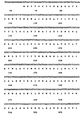
- DNA sequence information obtained from clone hp75d is shown in Figure 1.
- An open reading frame extending from an ATG start codon at position 1 to a stop codon at position 1095 can encode a protein 365 amino acids in length, having a relative molecular mass (M r ) of 41,633.
- M r relative molecular mass
- a comparison of this protein sequence with previously characterized neurotransmitter receptors indicates that hp75d encodes a receptor which is a new member of a family of molecules which span the lipid bilayer seven times and couple to guanine nucleotide regulatory proteins (the G protein-coupled receptor family).
- 5-CT possessed low affinity and, thus, discriminates this receptor from that of the 5-HT 1D receptor as well as other members of this class.
- rauwolfia alkaloids and serotonergic agents that possess high affinity for various subtypes of receptor within the serotonin family including ketanserin ( 5-HT 2 ) , 8-OH-DPAT (5-HT 1A ), DOI (5-HT 1C /5-HT 2 ), spiperone (5-HT 1A /5-HT 2 ), pindolol (5-HT 1A /5-HT 1B ) and zacopride (5-HT 3 ) had very poor affinity. In all cases, the Hill Coefficients did not differ significantly from unity. Taken together, the pharmacological profile of the 5-HT 1E receptor is unique and contrasts to that of other known serotonin receptors.
- 5HT 1E receptor mRNA The tissue distribution of 5HT 1E receptor mRNA was detected using PCR technology on cDNA from tissue-derived total RNA.
- the mRNA localization in human brain tissues demonstrates the presence of 5HT 1E in: frontal cortex, cerebellar cortex, temporal cortex, choroid plexus, hippocampus, brain stem and cortex.
- frontal cortex cerebellar cortex
- temporal cortex temporal cortex
- choroid plexus hippocampus
- hippocampus hippocampus
- brain stem and cortex we have not identified an area of the brain which does not contain 5HT 1E .
- 5HT 1E mRNA is abundant in human brain.
- 5HT 1E receptor mRNA 5HT 1E receptor mRNA
- 5HT 1E receptor mRNA 5HT 1E receptors
- the abundance of 5HT 1E receptors (mRNA) in the hippocampus may affect mood, behavior and hallucinogenesis.
- a greater understanding of possible physiological roles of this receptor subtype may be realized by the development of more specific 5HT 1E receptor drugs as well as physiological manipulations of 5HT 1E mRNAs/receptors.
- the cellular localization of 5-HT 1E receptor mRNA was detected with digoxigenin-11-dUTP labeled oligo probes employing in situ hybridization(ISHH) technology.
- Digoxigenin-11-dUTP labeled oxytocin(OT) oligo probes were used as a positive control for the experiment since the distribution of OT neurons in the central nervous system is well known.
- OT cells were intensely stained in the Guinea Pig's hypothalamus. Tissue sections pre-treated with RNase A were used as a negative control. Guinea pig brain sections were examined under a light microscope.
- 5-HT 1E receptor mRNA was found in cells located in the frontal cortex, piriform cortex, hippocampus (CA1, CA2 and CA3), lateral septal nucleus, triangular septal nucleus, septofimbrial nucleus and the basal ganglia (caudate-putamen and globus pallidus). It was detected in the amygdaloid complex, the bed nucleus stria terminalis and the hypothalamic area including anterior hypothalamus, periventricular nucleus, paraventricular nucleus (magnocellular and parvocellular populations), supraoptic nucleus which seemed to include both vasopressin and oxytocin cell populations, and the lateral, hypothalamus.
- the 1E receptor mRNA was also defected in the thalamic area including anteroventral thalamic anterodorsal thalamic, mediodorsal thalamic, ventrolateral thalamic, reticular thalamic paracentral thalamic, paratenial thalamic nuclei and the nucleus stria medullaris. Control sections pre-treated with RNase A did not exhibit any staining pattern.
- subtypes generally share a common transmitter and also have similar pharmacological profiles and physiological roles (for example, 5-HT 2 and 5-HT 1C or 5-HT 1D ⁇ and 5-HT 1D ⁇ ). Such “subtypes” display an amino acid identity of approximately 75-80% in their transmembrane domains. Serotonin receptors which are not members of the same "subfamily", but are members of the serotonin "family” (in which the receptors use the same neurotransmitter; i.e. 5-HT 2 and 5-H ) generally show much lower transmembrane homology (approximately 45%). Such transmembrane amino acid homologies can, therefore, give insight into the relationship between receptors and be used as predictors of receptor pharmacology. According to this type of analysis, although the newly cloned receptor appears to be more related to the 5-HT 1D subfamily, it is likely to be in a subfamily distinct from all the other serotonin receptors.
- the radioligand concentration should have been extended to at least 50 nM or 10 times the estimated Kd (Yamamura et al., 1985).
- the initial study was limited in tissue supply and lacked a comprehensive pharmacological characterization.
- the cloning of the 5-HT 1E site will now allow more extensive investigations into the nature of this unique receptor.
- the application of the human 5-HT 1E receptor clone to pharmaceutical research can lead to new drug design and development.
- the localization of this receptor in the cerebral cortex (Leonhardt, et al., 1989) and parts of the basal ganglia such as the putamen and globus pallidus (Lowther et al., 1991) suggests a putative link to limbic, cognitive and/or motor function (Nieuwenhuys et al., 1988; Kandel and Schwartz, 1985) and, thus, may be involved in such abnormal conditions as dementia, Parkinson's disease, feeding disorders, anxiety and schizophrenia.
- serotonin uptake blockers are effective in treating neuropsychiatric disorders such as depression and obsessive-compulsive illness (Blier et al., 1987; Asberg et al., 1986; Fin et al., 1985).
- these agents have side effects and, in fact, the mechanism of action for these compounds are not linked to any particular serotonergic receptor.
- ergoline derivatives have had clinical usefulness as drugs capable of relieving migraines and, thus, the involvement of the 5-HT 1E receptor in this disorder deserves future attention.
- in depth investigations into the localization of the 5-HT 1E receptor in brain and peripheral tissue will target new sites that can lead to specific functional roles for this serotonergic receptor.
- Another consideration for therapeutic application of this site may be related to the treatment of feeding disorders such as obesity, bulimia nervosa and/or anorexia nervosa.
- feeding disorders such as obesity, bulimia nervosa and/or anorexia nervosa.
- serotonin and feeding behavior has received much attention during the last decade.
- serotonin uptake blockers which have been used to treat feeding disorders act nonselectively and as such have side-effect potential (Jimerson et al., 1990).
- serotonergic receptors Although many different serotonergic receptors are involved in feeding, the search for the one site that can be exploited for selective drug development has yet to be found. There is no doubt that interest exists in finding drugs that interact with the serotonin system for the treatment of feeding disorders (Cooper, 1989).
- the pharmacological profile of the cloned human 5-HT 1E receptor is unique and contrasts to other known serotonergic receptors.
- the utility of this site expressed in a cellular system and, thus, isolated for study will create excellent opportunities in drug development directed towards a novel serotonergic receptor that may have wide-range implications for drug therapy.
- the potential therapeutic applications may extend to neuropsychiatric disorders including depression, anxiety, schizophrenia, dementia and obsessive-compulsive illness as well as obesity and migraine.
- the localization of 5-HT 1E receptor mRNA by in situ hybridization makes it possible to predict its physiological and pathological functions.
- 5-HT 1E receptor mRNA is detected in the limbic structures, such as the hippocampus, septal nuclei, piriform cortex (olfactory system), amygdaloid complex and the bed nucleus stria terminalis.
- the olfactory system sends afferent fibers to the hippocampus through the subiculum, and to the amygdaloid complex (Kupfermann,1985).
- the outputs of the hippocampus project to the septal area and hypothalamus while the amygdaloid complex projects to the hypothalamus via the stria terminalis (Kupfermann, 1985).
- the hypothalamus regulates the body adjustments to the external and internal environments. It can control hunger (the ventromedial nucleus and the lateral hypothalamus), endocrine functions (e.g. the supraoptic nucleus, the paraventricular nucleus and the periventricular nucleus), affective (emotional) behavior (the ventromedial and dorsomedial nuclei) and the activity of the visceral nervous system (the anterior hypothalamus) (Diamond et al., 1985).
- the discovery of 5-HT 1E receptor mRNA in these nuclei indicates physiological and pathological roles of this receptor subtype in cardiovascular, gastrointestinal, endocrine, neurological and psychiatric systems.
- the thalamus is a relay station where the sensory and motor-related pathways passing up the brain stem synapse before proceeding on to the cerebral cortex for more elaborate integration and analysis.
- the 5-HT 1E receptor mRNA was found in the anterior thalamic nucleus which receives the input from the mammillothalamic tract and sends fibers to the cingulate gyrus. Thus, this nucleus is an important part of the circuit connecting the hypothalamus, the thalamus, and the limbic lobe (Diamond et al., 1985).
- the 5-HT 1E receptor mRNA was also found in the mediodorsal thalamic nucleus which is involved in emotional behavior and in the ventral lateral thalamic nucleus which connects the basal ganglia and premotor area. Furthermore, 5-NT 1E receptor mRNA was found in the nucleus reticularis thalami through which pass most of the thalamocortical and corticothalamic fibers.
- the axons of corticothalamic and thalamocortical neurons provide collaterals for synapse with reticular cells, exerting a facilitatory effect, while the axons of the GABA-rich reticularis cells project back on the specific thalamic neurons, continuously modulating (by inhibition) the ascending flow of thalamocortical impulses (Diamond et al., 1985).
- the reticularis neurons play a significant role in the interactions between the thalamus and the frontal cortex.
- the 5-HT 1E receptor mRNA was also visualized in the basal ganglia including the caudate-putamen and globus pallidus both of which control motor activity (Alheid et al., 1990).
Landscapes
- Health & Medical Sciences (AREA)
- Life Sciences & Earth Sciences (AREA)
- Engineering & Computer Science (AREA)
- Chemical & Material Sciences (AREA)
- Bioinformatics & Cheminformatics (AREA)
- Proteomics, Peptides & Aminoacids (AREA)
- General Health & Medical Sciences (AREA)
- Organic Chemistry (AREA)
- Animal Behavior & Ethology (AREA)
- Gastroenterology & Hepatology (AREA)
- Toxicology (AREA)
- Veterinary Medicine (AREA)
- Public Health (AREA)
- Immunology (AREA)
- Epidemiology (AREA)
- Medicinal Chemistry (AREA)
- Molecular Biology (AREA)
- Cell Biology (AREA)
- Biomedical Technology (AREA)
- Pathology (AREA)
- Biophysics (AREA)
- Neurology (AREA)
- Zoology (AREA)
- Rheumatology (AREA)
- Biochemistry (AREA)
- Diabetes (AREA)
- Urology & Nephrology (AREA)
- Genetics & Genomics (AREA)
- Pharmacology & Pharmacy (AREA)
- Endocrinology (AREA)
- Peptides Or Proteins (AREA)
- Micro-Organisms Or Cultivation Processes Thereof (AREA)
- Measuring Or Testing Involving Enzymes Or Micro-Organisms (AREA)
- Preparation Of Compounds By Using Micro-Organisms (AREA)
- Medicines That Contain Protein Lipid Enzymes And Other Medicines (AREA)
- Saccharide Compounds (AREA)
Abstract
Description
- Since the purification of a pressor substance in blood serum termed serotonin (Rapport et al., 1947) and later identified as 5-hydroxytryptamine (5-HT)(Rapport, 1949), there has been a plethora of reports demonstrating that this indoleamine not only plays a role in the functioning of peripheral tissues but, indeed, performs a key role in the brain as a neurotransmitter. Certainly, the anatomical localization of serotonin and serotonergic neurons in both the peripheral and central nervous systems supports its role in such diverse physiologic and behavioral functions as pain perception, sleep, aggression, sexual activity, hormone secretion, thermoregulation, motor activity, cardiovascular function, food intake and renal regulation (For review see Green, 1985; Osborne and Hamon, 1988; Sanders-Bush, 1988; Peroutka, 1991). Taken together, it appears that serotonin plays an important role in homeostasis and in modulating responsiveness to environmental stimuli. Accordingly, studies demonstrating that abnormalities in the serotonergic system may be associated with disease states has created a drug development effort towards agents which may selectively modulate the function of serotonin (Glennon, 1990).
- In relation to the characterization of physiologic or biochemical responses resulting from the release of serotonin are simultaneous investigations examining the receptor sites responsible for the actions elicited by the indoleamine transmitter. Following early in vitro pharmacological assays describing the existence of two different serotonin receptors, designated as D and M, in the guinea pig ileum (Gaddum and Picarelli, 1957), the advent of receptor binding technique in the 1970's has brought to light during the last decade the diversity of 5-HT receptors existing in both the brain and peripheral tissues. Thus, although the concept of D and M receptors has not been invalidated, serotonin receptors not fitting either category have been identified using radioligand methods. To date using this technique, there appears to be four classes of serotonin receptors found in the brain: 5-HT1, 5-HT2, 5-HT3 and, 5-HT4 (Peroutka, 1991). Furthermore, 5-HT1 sites have been subclassified as: 5-HT1A, 5-HT1B, 5-HT1C, 5-HT1D (Hamon et al., 1990) and 5-HT1E (Leonhardt et al., 1989). Although a detailed characterization of the 5-HT1E binding site is lacking, extensive pharmacologic, biochemical and functional properties have clearly shown that the other four subtypes of 5-HT1 sites are receptors according to classical criteria. Interestingly, the 5-HT1E binding site was first observed in human cortical tissue using [3H]5-HT as the radioligand probe in the presence of 5-carboxyamidotryptamine and mesulergine to mask other members of the 5-HT1 receptor class. The affinity constants of the nine drugs tested indicated a unique pharmacological profile. In particular, the low affinity of 5-CT and ergotamine seemed to clearly discriminate the pharmacologically defined 5-HT10 site from that of this novel serotonergic site. Importantly, it was demonstrated that 5-HT1E sites are saturable and exist in a density consistent with other known neurotransmitter receptors. Furthermore, this site appeared to interact with a GTP-binding protein. Overall, the data provided a framework suggesting that the 5-HT1E binding site may represent a functional receptor.
- During the last few years, the field of molecular biology has provided an important facet to receptor research by cloning these proteins and allowing more precise characterizations in isolated systems (Hartig et al., 1990). This has been accomplished for the 5-HT1A (Fargin et al., 1988), 5-HT1C (Julius et al., 1988), 5-HT1D (Branchek et al., 1990) and 5-HT2 receptors (Pritchett et al., 1988). Thus, there is no doubt that these binding sites represent "true" functional receptors. Indeed, the pharmacological characterization of serotonin receptors involved in various physiological or biochemical functions is a key component of drug development for the serotonergic system. As one can deduce from the diversity of serotonin binding sites, many targets are available for advancement in selective drug design. The coupling of molecular biological methods to pharmacological characterization particularly for cloned human receptors will open new avenues for pharmaceutical development which have not been previously explored.
- This invention provides an isolated nucleic acid molecule encoding a human 5-HT1E receptor.
- This invention also provides an isolated protein which is a human 5-HT1E receptor.
- This invention provides a vector comprising an isolated nucleic acid molecule encoding a human 5-HT1E receptor.
- This invention also provides vectors such as plasmids comprising a DNA molecule encoding a human 5-HT1E receptor, adapted for expression in a bacterial cell, a yeast cell, or a mammalian cell which additionally comprise the regulatory elements necessary for expression of the DNA in the bacterial, yeast, or mammalian cells so located relative to the DNA encoding the 5-HT1E receptor as to permit expression thereof.
- This invention provides a mammalian cell comprising a DNA molecule encoding a human 5-HT1E receptor.
- This invention provides a method for determining whether a ligand not known to be capable of binding to a human 5-HT1E receptor can bind to a human 5-HT1E receptor which comprises contacting a mammalian cell comprising an isolated DNA molecule encoding a human 5-HT1E receptor with the ligand under conditions permitting binding of ligands known to bind to a 5-HT1E receptor, detecting the presence of any of the ligand bound to a human 5-HT1E receptor, and thereby determining whether the ligand binds to a human 5-HT1E receptor.
- This invention also provides a method for determining whether a ligand not known to be capable of binding to the human 5-HT1E receptor can functionally activate its activity or prevent the action of a ligand which does so. This comprises contacting a mammalian cell comprising an isolated DNA molecule which encodes a human 5-HT1E receptor with the ligand under conditions permitting the activation or blockade of a functional response, detected by means of a bioassay from the mammalian cell such as a second messenger response, and thereby determining whether the ligand activates or prevents the activation of the human 5-HT1E receptor functional output.
- This invention further provides a method of screening drugs to identify drugs which specifically interact with, and bind to, the human 5-HT1E receptor on the surface of a cell which comprises contacting a mammalian cell comprising an isolated DNA molecule encoding a human 5-HT1E receptor with a plurality of drugs, determining those drugs which bind to the mammalian cell, and thereby identifying drugs which specifically interact with, and bind to, a human 5-HT1E receptor.
- This invention also provides a method of screening drugs to identify drugs which interact with, and activate or block the activation of, the human 5-HT1E receptor on the surface of a cell which comprises contacting the mammalian cell comprising an isolated DNA molecule encoding and expressing a human 5-HT1E receptor with a plurality of drugs, determining those drugs which activate or block the activation of the receptor in the mammalian cell using a bioassay such as a second messenger assays, and thereby identifying drugs which specifically interact with, and activate or block the activation of, a human 5-HT1E receptor.
- This invention provides an antibody directed to a human 5-HT1E receptor.
- This invention provides a transgenic nonhuman mammal expressing DNA encoding a human 5-HT1E receptor.
- This invention further provides a transgenic nonhuman mammal whose genome comprises antisense DNA complementary to DNA encoding a human 5-HT1E receptor so placed as to be transcribed into antisense mRNA which is complementary to mRNA encoding a 5-HT1E receptor and which hybridizes to mRNA encoding a 5-HT1E receptor thereby reducing ist translation.
- This invention provides a method of determining the physiological effects of expressing varying levels of human 5-HT1E receptors which comprises producing a transgenic nonhuman animal whose levels of human 5-HT1E receptor expresssion are varied by use of an inducible promoter which regulates human 5-HT1E receptor expression.
- This invention also provides a method of determining the physiological effects of expressing varying levels of human 5-HT1E receptors which comprises producing a panel of transgenic nonhuman animals each expressing a different amount of human 5-HT1E receptor.
- This invention provides a method of preparing the isolated 5-HT1E receptor which comprises inducing cells to express 5-HT1E receptor, recovering the receptor from the resulting cells and purifying the receptor so recovered.
- This invention also provides a method of preparing the isolated 5-HT1E receptor which comprises inserting nucleic acid encoding 5-HT1E receptor in a suitable vector, inserting the resulting vector in a suitable host cell, recovering the receptor produced by the resulting cell, and purifying the receptor so recovered.
- This invention also provides a transgenic nonhuman mammal expressing DNA encoding a receptor.
- This invention also provides a method of determining the physiological effects of expressing varying levels of a receptor which comprises producing a transgenic nonhuman animal whose levels of receptor expression are varied by use of an inducible promoter which regulates receptor expression.
- This invention also provides a method of determining the physiological effects of expressing varying levels of a receptor which comprises producing a panel of transgenic nonhuman animals each expressing a different amount of the receptor.
- This invention further provides a transgenic nonhuman mammal whose genome comprises antisense DNA complementary to DNA encoding a receptor so placed as to be transcribed into antisense mRNA which is complementary to mRNA encoding the receptor and which hybridizes to mRNA encoding the receptor thereby preventing its translation.
- This invention provides a method for determining whether a ligand not known to be capable of binding to a receptor can bind to a receptor which comprises contacting a mammalian cell comprising an isolated DNA molecule encoding the receptor with the ligand under conditions permitting binding of ligands known to bind to receptor, detecting the presence of any of the ligand bound to the receptor, and thereby determining whether the ligand binds to the receptor.
-
- Figure 1. (Figures 1A-1I) Nucleotide and deduced amino
acid sequence of gene 5-HT1E. (Seq. I.D. Nos. 1 and 2).
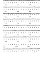
Numbers above the nucleotide sequence indicate nucleotide position. DNA sequence was determined by the chain termination method of Sanger, et al., on denatured double-stranded plasmid templates using the enzyme Sequenase. Deduced amino acid sequence (single letter code) of a long open reading frame is shown. - Figure 2. Comparison of the human 5-HT1E receptor primary
structures with other serotonin receptors. (Seq. I.D.
Nos. 3 - 5HT1A; 4 - 5HT1C; 5 - 5HT1Dα; 6 - 5HT1Dβ;7 -
5HT2)
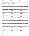
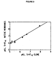
Amino acid sequences (single letter code) are aligned to optimize homology. The putative transmembrane spanning domains are indicated by stars and identified by Roman numerals (TM I-VII). - Figure 3. Respective pKi values of various drugs for the
inhibition of [3H]5-HT specific binding to the cloned 5-HT1E
receptor and human cortical membrane preparations
containing the native 5-HT1E receptor.
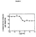
Values for the native membrane preparation are taken from Leonhardt et al., 1989. "r" is the correlation coefficient between pKi values calculated for the two receptor preparations and clearly indicates the similarity in binding profile. - Figure 4. 5-HT concentration-effect curve for the
inhibition of forskolin-stimulated CAMP formation in Y1
cells transfected with the 5-HT1E receptor.
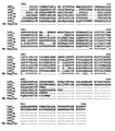
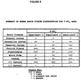
Values are expressed as a percentage of forskolin(Fsk)-stimulated cAMP production. - Figure 5. 5-HT1E mRNA localization in the human brain.
cDNA was reverse transcribed from total RNA (5 µg) using random hexamers (500 pmoles). One microgram of cDNA was subjected to 30 cycles of PCR amplification using 5'- and 3'-primers directed to the third cytoplasmic loop of the 5HT1E receptor gene. Amplified fragments were subjected to Southern blot analysis using an end-labeled oligonucleotide probe which was internal to the PCR primers. (+) control consisted of 5HT1E recombinant plasmid; (-) control consisted of all cDNA and PCR reagents without the addition of cDNA template. Intensity of signal is depicted as the number of plus signs as defined in the figure key. -
-
- +++ = dark
- ++ = light
- + = present but faint
- - = absent
-
- As used herein, the 5-HT receptor family is defined as the group of mammalian proteins that function as receptors for serotonin. A 5-HT receptor subfamily is defined as a subset of proteins belonging to the 5-HT receptor family which are encoded by genes which exhibit homology of greater than 72% or higher with each other in their deduced amino acid sequences within presumed transmembrane regions (linearly contiguous stretches of hydrophobic amino acids, bordered by charged or polar amino acids, that are long enough to form secondary protein structures that span a lipid bilayer). Four human 5-HT receptor subfamilies can be distinguished based on the information presently available: 5-HT1, 5-HT2, 5-HT3, and 5-HT4 (Peroutka, 1991). The 5-HT2 receptor subfamily contains the human 5-HT2 receptor. Although no other human members of this family have been described, the rat 5-HT2 receptor (Pritchett, et al. 1988; Julius, et al. Proc. Natl. Acad. Sci. USA 87:928-932, 1990) and the rat 5HT1C receptor (Julius, et al. 1988) constitute a rat 5-HT receptor subfamily. The 5-HT1 subfamily has been subdivided further as: 5-HT1A, 5-HT1B, 5-HT1C, 5-HT1D (Hamon et al., 1990) and 5-HT1E (Leonhardt et al., 1989). The 5-HT1A subfamily contains the human 5-HT1A receptor, also known as G-21 (Fargin, et al. 1988) The 5-HT1D receptor subfamily contains two members, the 5-HT1D-1 receptor (also termed 5-H) and the 5-HT1D-2 receptor (also termed 5-H
 ). The 5-HT1E subfamily contains the human 5-HT1E receptor (also termed clone hp75d). Although this definition differs from the pharmacological definition used earlier, there is significant overlap between the present definition and the pharmacological definition. Members of the 5-HT1E receptor subfamily so described include the 5-HT1E receptor and any other receptors which have a greater than 72% homology to the DNA and amino acid sequence shown in Figure 1 according to the definition of "subfamily". This invention relates to the discovery of the first member of the human 5-HT1E receptor subfamily.
- This invention provides an isolated nucleic acid molecule encoding a human 5-HT1E receptor. As used herein, the term "isolated nucleic acid molecule" means a nucleic acid molecule that is, a molecule in a form which does not occur in nature. Such a receptor is by definition a member of the 5-HT1E receptor subfamily. Therefore, any receptor which meets the defining criteria given above is a human 5-HT1E receptor. One means of isolating a human 5-HT1E receptor is to probe a human genomic library with a natural or artificially designed DNA probe, using methods well known in the art. DNA probes derived from the human receptor gene 5-HT1E are particularly useful probes for this purpose. DNA and cDNA molecules which encode human 5-HT1E receptors may be used to obtain complementary genomic DNA, cDNA or RNA from human, mammalian or other animal sources, or to isolate related cDNA or genomic clones by the screening of cDNA or genomic libraries, by methods described in more detail below. Transcriptional regulatory elements from the 5' untranslated region of the isolated clones, and other stability, processing, transcription, translation, and tissue specificity-determining regions from the 3' and 5' untranslated regions of the isolated genes are thereby obtained. Examples of a nucleic acid molecule are an RNA, cDNA, or isolated genomic DNA molecule encoding a human 5-HT1E receptor. Such molecules may have coding sequences substantially the same as the coding sequence shown in Figure 1. The DNA molecule of Figure 1 encodes the sequence of the human 5-HT1E receptor gene.
- This invention further provides a cDNA molecule of encoding a human 5-HT1E receptor having a coding sequence substantially the same as the coding sequence shown in Figure 1. This molecule is obtained by the means described above.
- This invention also provides an isolated protein which is a human 5-HT1E receptor. As used herein, the term "isolated protein means a protein molecule free of other cellular components. An example of such protein is an isolated protein having substantially the same amino acid sequence as the amino acid sequence shown in Figure 1 which is a human 5-HT1E receptor. One means for obtaining isolated 5-HT1E receptor is to express DNA encoding the receptor in a suitable host, such as a bacterial, yeast, or mammalian cell, using methods well known in the art, and recovering the receptor protein after it has been expressed in such a host, again using methods well known in the art. The receptor may also be isolated from cells which express it, in particular from cells which have been transfected with the expression vectors described below in more detail.
- This invention provides a vector comprising an isolated nucleic acid molecule such as DNA, RNA, or cDNA encoding a human 5-HT1E receptor. Examples of vectors are viruses such as bacteriophages (such as phage lambda), cosmids, plasmids (such as pUC18, available from Pharmacia, Piscataway, NJ), and other recombination vectors. Nucleic acid molecules are inserted into vector genomes by methods well known in the art. For example, insert and vector DNA can both be exposed to a restriction enzyme to create complementary ends on both molecules which base pair with each other and are then ligated together with a ligase. Alternatively, linkers can be ligated to the insert DNA which correspond to a restriction site in the vector DNA, which is then digested with the restriction enzyme which cuts at that site. Other means are also available. A specific example of such plasmids is a plasmid comprising cDNA having a coding sequence substantially the same as the coding sequence shown in Figure 1 and designated clone hp75d (Seq. I.D. No. 1).
- This invention also provides vectors comprising a DNA molecule encoding a human 5-HT1E receptor, adapted for expression in a bacterial cell, a yeast cell, or a mammalian cell which additionally comprise the regulatory elements necessary for expression of the DNA in the bacterial, yeast, or mammalian cells so located relative to the DNA encoding a human 5-HT1E receptor as to permit expression thereof. DNA having coding sequences substantially the same as the coding sequence shown in Figure 1 may usefully be inserted into the vectors to express human 5-HT1E receptors. Regulatory elements required for expression include promoter sequences to bind RNA polymerase and transcription initiation sequences for ribosome binding. For example, a bacterial expression vector includes a promoter such as the lac promoter and for transcription initiation the Shine-Dalgarno sequence and the start codon AUG (Maniatis, et al., Molecular Cloning, Cold Spring Harbor Laboratory, 1982). Similarly, a eukaryotic expression vector includes a heterologous or homologous promoter for RNA polymerase II, a downstream polyadenylation signal, the start codon AUG, and a termination codon for detachment of the ribosome. Such vectors may be obtained commercially or assembled from the sequences described by methods well known in the art, for example the methods described above for constructing vectors in general. Expression vectors are useful to produce cells that express the receptor. Certain uses for such cells are described in more detail below.
- This invention further provides a plasmid adapted for expression in a bacterial, yeast, or, in particular, a mammalian cell which comprises a DNA molecule encoding a human 5-HT1E receptor and the regulatory elements necessary for expression of the DNA in the bacterial, yeast, or mammalian cell so located relative to the DNA encoding a human 5-HT1E receptor as to permit expression thereof. Some plasmids adapted for expression in a mammalian cell are pSVL (available from Pharmacia, Piscataway, NJ) and pcEXV-3 (Miller J. and Germain R.N., J. Exp. Med. 164:1478 (1986)). A specific example of such plasmid is a plasmid adapted for expression in a mammalian cell comprising cDNA having coding sequences substantially the same as the coding sequence shown in Figure 1 and the regulatory elements necessary for expression of the DNA in the mammalian cell which is designated pcEXV-hp75d and deposited under ATCC Accession No. 75138. Those skilled in the art will readily appreciate that numerous plasmids adapted for expression in a mammalian cell which comprise DNA of encoding human 5-HT1E receptors and the regulatory elements necessary to express such DNA in the mammalian cell may be constructed utilizing existing plasmids and adapted as appropriate to contain the regulatory elements necessary to express the DNA in the mammalian cell. The plasmids may be constructed by the methods described above for expression vectors and vectors in general, and by other methods well known in the art.
- The deposit discussed supra, and the other deposits discussed herein, were made pursuant to, and in satisfaction of, the Budapest Treaty on the International Recognition of the Deposit of Microorganisms for the Purpose of Patent Procedure with the American Type Culture Collection (ATCC), 12301, Parklawn Drive, Rockville, Maryland 20852.
- This invention provides a mammalian celle comprising a DNA molecule encoding a human 5-HRT1E receptor, such as a mammalian cell comprising a plasmid adapted for expression in a mammalian cell, which comprises a DNA molecule encoding a human 5-HT1E receptor, the protein encoded thereby is expressed on the cell surface, and the regulatory elements necessary for expression of the DNA in the mammalian cell so located relative to the DNA encoding a human 5-HT1E receptor as to permit expression thereof. Numerous mammalian cells may be used as hosts, including, for example, the mouse fibroblast cell NIH3T3, CHO cells, HeLa cells, Ltk cells, Y1 cells, etc. A particular example of an Ltk cell is a cell designated 5-HT1E-7 and deposited under ATCC Accession No. CRL 10913 and comprises the plasmid, designated pcEXV-hp75d. Another example is the murine adrenal Y1 cell line designated Y-5-HT1E and deposited under ATCC Accession No. CRL 10914. Expression plasmids such as that described supra may be used to transfect mammalian cells by methods well know in the art such as calcium phosphate precipitation, or DNA encoding these 5-HT1E receptors may be otherwise introduced into mammalian cells, e.g., by microinjection, to obtain mammalian cells which comprise DNA, e.g., cDNA or a plasmid, encoding either human 5-HT1E receptor.
- This invention provides a method for determining whether a ligand not known to be capable of binding to a human 5-HT1E receptor can bind to a human 5-HT1E receptor which comprises contacting a mammalian cell comprising a DNA molecule encoding a human 5-HT1E receptor, the protein encoded thereby is expressed on the cell surface, with the ligand under conditions permitting binding of ligands known to bind to the 5-HT1E receptor, detecting the presence of any of the ligand bound to the 5-HT1E receptor, and thereby determining whether the ligand binds to the 5-HT1E receptor. This invention also provides a method for determining whether a ligand not known to be capable of binding to the human 5-HT1E receptor can functionally activate its activity or prevent the action of a ligand which does so. This comprises contacting a mammalian cell comprising an isolated DNA molecule which encodes a human 5-HT1E receptor with the ligand under conditions permitting the activation or blockade of a functional response, detected by means of a bioassay from the mammalian cell such as a second messenger response, and thereby determining whether the ligand activates or prevents the activation of the human 5-HT1E receptor functional output. The DNA in the cell may have a coding sequence substantially the same as the coding sequence shown in Figure 1 preferably, the mammalian cell is nonneuronal in origin. An example of a nonneuronal mammalian cell is an Ltk cell, in particular the Ltk cell designated 5-HT1E-7. Another example of a non-neuronal mammalian cell to be used for functional assays is a Y1 murine adrenal cell, specifically the Y1 cell designated Y-5-HT1E. The preferred method for determining whether a ligand is capable of binding to the human 5-HT1E receptor comprises contacting a transfected nonneuronal mammalian cell (i.e. a cell that does not naturally express any type of 5-HT or G-protein coupled receptor, thus will only express such a receptor if it is transfected into the cell) expressing a 5-HT1E receptor on its surface, or contacting a membrane preparation derived from such a transfected cell, with the ligand under conditions which are known to prevail, and thus to be associated with, in vivo binding of the ligands to a 5-HT1E receptor, detecting the presence of any of the ligand being tested bound to the 5-HT1E receptor on the surface of the cell, and thereby determining whether the ligand binds to, activates or prevents the activation of the 5-HT1E receptor. This response system is obtained by transfection of isolated DNA into a suitable host cell containing the desired second messenger system such as phosphoinositide hydrolysis, adenylate cyclase, guanylate cyclase or ion channels. Such a host system is isolated from pre-existing cell lines, or can be generated by inserting appropriate components of second messenger systems into existing cell lines. Such a transfection system provides a completes response system for investigation or assay of the activity of human 5-HT1E receptors with ligands as described above. Transfection systems are useful as living cell cultures for competitive binding assays between known or candidate drugs and ligands which bind to the receptor and which are labeled by radioactive spectroscopic or other reagents. Membrane preparation containing the receptor isolated from transfected cells are also useful for these competitive binding assays. Functional assays of second messenger systems or their sequelae in transfection systems act as assays for binding affinity and efficacy in the activation of receptor function. A transfection system constitutes a "drug discovery system" useful for the identification of natural or synthetic compounds with potential for drug development that can be further modified or used directly as therapeutic compounds to activate or inhibit the natural functions of the human 5-HT1E receptor. The transfection system is also useful for determining the affinity an efficacy of known drugs at the human 5-HT1E receptor sites.
- This invention also provides a method of screening drugs to identify drugs which specifically interact with, and bind to, the human 5-HT1E receptor on the surface of a cell which comprises contacting a mammalian cell comprising a DNA molecule encoding a human 5-HT1E receptor on the surface of a cell with a plurality of drugs, determining those drugs which bind to the mammalian cell, and thereby identifying drugs which specifically interact with, and bind to, the human 5-HT1E receptor. This invention also provides a method of screening drugs to identify drugs which interact with, and activate or block the activation of , the human 5-HT1E receptor on the surface of a cell which comprises contacting the mammalian cell comprising an isolated DNA molecule encoding and expressing a human 5-HT1E receptor with a plurality of drugs, determining those drugs which activate or block the activation of the receptor in the mammalian cell using a bioassay such as a second messenger assays, and thereby identifying drugs which specifically interact with , and activate or block the activation of, a human 5-HT1E receptor. The DNA in the cell may have a coding sequence substantially the same as the coding sequence shown in Figure 1. Preferably, the mammalian cell is nonneuronal in origin. An example of a nonneuronal mammalian cell is an Ltk cell, in particular the Ltk cell designated 5-HT1E-7. Another example of a non-neuronal mammalian cell to be used for functional assays is a Y1 murine adrenal cell, specifically the Y1 cell designated Y-5-H1E. Drug candidates are identified by choosing chemical compounds which bind with high affinity to the expressed 5-HT1E receptor protein in transfected cells, using radioligand binding methods well known in the art, examples of which are shown in the binding assays described herein. Drug candidates are also screened for selectivity by identifying compounds which bind with high affinity to one particular 5-HT1E receptor subtype but do not bind with high affinity to any other serotonin receptor subtype or to any other known receptor site. Because selective, high affinity compounds interact primarily with the target 5-HT1E receptor site after administration to the patient, the chances of producing a drug with unwanted side effects are minimized by this approach.
- A drug identified by the method described above can be used to prepare a pharmaceutical composition comprising the drug and a pharmaceutically acceptable carrier. As used herein, the term "pharmaceutically acceptable carrier" encompasses any of the standard pharmaceutical carriers, such as a phosphate buffered saline solution, water, and emulsions, such as an oil/water or water/oil emulsion, and various types of wetting agents. Once the candidate drug has been shown to be adequately bio-available following a particular route of administration, for example orally or by infection (adequate therapeutic concentrations must be maintained at the site of action for an adequate period to gain the desired therapeutic benefit), and has been shown to be non-toxic and therapeutically effective in appropriate disease models, the drug may be administered to patients by that route of administration determined to make the drug bio-available, in an appropriate solid or solution formulation, to gain the desired therapeutic benefit.
- This invention provides an antibody directed to the human 5-HT1E receptor, for example a monoclonal antibody directed to an epitope of a human 5-HT1E receptor present on the surface of a cell and having an amino acid sequence substantially the same as an amino acid sequence for a cell surface epitope of the human 5-HT1E receptor included in the amino acid sequence shown in Figure 1 (Seq. I.D. No. 2). Amino acid sequences may be analyze by methods well known in the art to determine whether they produce hydrophobic or hydrophilic regions in the proteins which they build. In the case of cell membrane proteins, hydrophobic regions are well known to form the part of the protein that is inserted into the lipid bilayer which forms the cell membrane, while hydrophilic regions are located on the cell surface, in an aqueous environment. Therefore antibodies to the hydrophilic amino acid sequences shown in Figure 1 will bind to a surface epitope of a human 5-HT1E receptor, as described. Antibodies directed to human 5-HT1E receptors may be serum-derived or monoclonal and are prepared using methods well known in the art. For example, monoclonal antibodies are prepared using hybridoma technology by fusing antibody producing B cells from immunised animals with myeloma cells and selecting the resulting hybridoma cell line producing the desired antibody. Cells such as NIH3T3 cells or Ltk cells may be used as immunogens to raise such an antibody. Alternatively, synthetic peptides may be prepared using commercially available machines and the amino acid sequence shown in Figure 1 (Seq. I.D. No. 2). As a still further alternative, DNA, such as a cDNA or a fragment thereof, may be cloned and expressed and the resulting polypeptide recovered and used as an immunogen. These antibodies are useful to detect the presence of human 5-HT1E receptors encoded by the isolated DNA, or to inhibit the function of the receptors in living animals, in humans, or in biological tissues or fluids isolated from animals or humans.
- This invention provides a pharmaceutical composition which comprises an amount of an antibody directed to the human 5-Ht1E receptor effective to block binding of naturally occurring ligands to the 5-HT1E receptor, and a pharmaceutically acceptable carrier. A monoclonal antibody directed to an epitope of a human 5-HT1E receptor present on the surface of a cell and having an amino acid sequence substantially the same as an amino acid sequence for a cell surface epitope of the human 5-HT1E receptor included in the amino acid sequence shown in Figure 1 is useful for this purposes.
- This invention provides a method of detecting the presence of a 5-HT1E receptor on the surface of a cell which comprises contacting the cell with an antibody directed to the human 5-HT1E receptor, under conditions permitting binding of the antibody to the receptor detecting the presence of the antibody bound to the cell, and thereby the presence of the human 5=HT1E receptor on the surface of the cell. Such a method is useful for determining whether a given cell is defective in expression of 5-HT1E receptors on the surface of the cell. Bound antibodies are detected by methods well known in the art, for example by binding fluorescent markers to the antibodies and examining the cell sample under a fluorescence microscope to detect fluorescence on a cell indicative of antibody binding. The monoclonal antibodies described above are useful for this purpose.
- This invention also provides a transgenic nonhuman mammal whose genome comprises antisense DNA complementary to DNA encoding a human 5-HT1E receptor so placed as to be transcribed into antisense mRNA which is complementary to mRNA encoding a 5-HT1E receptor and which hybridizes to mRNA encoding a 5-HT1E receptor thereby reducing its translation. The DNA may additionally comprise an inducible promoter or additionally comprise tissue specific regulatory elements, so that expression can be induced, or restricted to specific cell types. Examples of DNA are DNA or cDNA molecules having a coding sequence substantially the same as the coding sequence shown in Figure 1. An example of a transgenic animal is a transgenic mouse. Examples as of tissue specificity-determining regions are the metallothionein promotor (Low, M.J., Lechan. R.M., Hammer, R.E. et al. Science 231:1002-1004 (1986)) and the L7 promotor (Oberdick, J., Smeyne, R.J., Mann, J.R., Jackson, S. and Morgan, J.I. Science 248:223-226 (1990)).
- Animal model systems which elucidate the physiological and behavioral roles of human 5-HT1E receptors are produced by creating transgenic animals in which the expression of a 5-HT1E receptor is either increased or decreased, or the amino acid sequence of the expressed 5-HT1E receptor protein is altered, by a variety of techniques. Examples of these techniques include: 1) Insertion of normal or mutant versions of DNA encoding a human 5-HT1E receptor or homologous animal versions of these genes, by microinjection, retroviral infection or other means well known to those skilled in the art, into appropriate fertilized embryos in order to produce a transgenic animal (Hogan B. et al. Manipulating the Mouse Embryo, A Laboratory Manual, Cold Spring Harbor Laboratory (1986)). 2) Homologous recombination (Capecchi M.R. Science 244:1288-1292 (1989); Zimmer, A. and Gruss, P. Nature 338:150-153 (1989)) of mutant or normal, human or animal versions of these genes with the native gene locus in transgenic animals to alter the regulation of expression or the structure of these 5=HT1E receptors. The technique of homologous recombination is well known in the art. It replaces the native gene with the inserted gene and so is useful for producing an animal that cannot express native receptor but does express, for example, an inserted mutant receptor, which has replaced the native receptor in the animal's genome by recombination, resulting in underexpression of the receptor. Microinjection adds genes to the genome, but does not remove them, and so is useful for producing an animal which expresses its own and added receptors, resulting in overexpression of the receptor. One means available for producing a transgenic animal, with a mouse as an example, is as follows: Female mice are mated and the resulting fertilized eggs are dissected out of their oviducts. The eggs are stored in an appropriate medium such as M2 medium (Hogan B. et al. Manipulating the Mouse Embryo, A Laboratory Manual, Cold Spring Harbor Laboratory (1986). DNA or cDNA encoding a human 5-HT1E receptor is purified from a vector (such as plasmid pCEXV-hp75d described above) by methods well known in the art. Inducible promoters may be fused with the coding region of the DNA to provide an experimental means to regulate expression of the trans-gene. Alternatively or in addition, tissue specific regulatory elements may be fused with the coding region to permit tissue-specific expression of the trans-gene. The DNA, in an appropriately buffered solution, is put into a microinjection needle (which may be made from capillary tubing using a pipet puller) and the egg to be injected is put in a depression slide. The needle is inserted into the pronucleus of the egg, and the DNA solution is injected. The injected egg is then transferred into the oviduct of a pseudopregnant mouse (a mouse stimulated by the appropriate hormones to maintain pregnancy but which is not actually pregnant), where it proceeds to the uterus, implants, and develops to term. As noted above, microinjection is not the only method for inserting DNA into the egg cell, and is used here only for exemplary purposes.
- Since the normal action of receptor-specific drugs is to activate or to inhibit the receptor, the transgenic animal model systems described above are useful for testing the biological activity of drugs directed against these 5-HT1E receptors even before such drugs become available. These animal model systems are useful for predicting or evaluating possible therapeutic applications of drugs which activate or inhibit these 5-HT1E receptors by inducing or inhibiting expression of the native or trans-gene and thus increasing or decreasing expression of normal or mutant 5-HT1E receptors in the living animal. Thus, a model system is produced in which the biological activity of drugs directed against these 5-HT1E receptors are evaluated before such drugs become available. The transgenic animals which over or under produce the 5-HT1E receptor indicate by their physiological state whether over or under production of the 5-HT1E receptor is therapeutically useful. It is therefore useful to evaluate drug action based on the transgenic model system. One use is based on the fact that it is well known in the art that a drug such as an antidepressant acts by blocking neurotransmitter uptake, and thereby increases the amount of neurotransmitter in the synaptic cleft. The physiological result of this action is to stimulate the production of less receptor by the affected cells, leading eventually to underexpression. Therefore, an animal which underexpresses receptor is useful as a test system to investigate whether the actions of such drugs which result in under expression are in fact therapeutic. Another use is that if overexpression is found to lead to abnormalities, then a drug which down-regulates or acts as an antagonist to 5-HT1E receptor is indicated as worth developing, and if a promising therapeutic application is uncovered by these animal model systems, activation or inhibition of the 5-HT1E receptor is achieved therapeutically either by producing agonist or antagonist drugs directed against these 5-HT1E receptors or by any method which increases or decreases the expression of these 5-HT1E receptors in man.
- This invention provides a method of determining the physiological effects of expressing varying levels of human 5-HT1E receptors which comprises producing a transgenic nonhuman animal whose levels of human 5-HT1E receptor expression are varied by use of an inducible promoter which regulates human 5-HT1E receptor expression. This invention also provides a method of determining the physiological effects of expressing varying levels of human 5-HT1E receptors which comprises producing a panel of transgenic nonhunan animals each expressing a different amount of human 5-HT1E receptor. Such animals may be produced by introducing different amounts of DNA encoding a human 5-HT1E receptor into the oocytes from which the transgenic animals are developed.
- This invention provides a method of preparing the isolated 5-HT1E receptor which comprises inducing cells to express 5-HT1E receptor, recovering the receptor from the resulting cells, and purifying the receptor so recovered. An example of an isolated 5-HT1E receptor is an isolated protein having substantially the same amino acid sequence as the amino acid sequence shown in Figure 1. For example, cells can be induced to express receptors by exposure to substances such as hormones. The cells can then be homogenized and the receptor isolated from the homogenate using an affinity column comprising, for example, serotonin or another substance which is known to bind to the receptor. The resulting fractions can then be purified by contacting them with an ion exchange column, and determining which fraction contains receptor activity or binds anti-receptor antibodies.
- This invention provides a method of preparing the isolated 5-HT1E receptor which comprises inserting nucleic acide encoding 5-HT1E receptor in a suitable vector, inserting the resulting vector in a suitable host cell, recovering the receptor produced by the resulting cell, and purifying the receptor so recovered. An example of an isolated 5-HT1E receptor is an isolated protein having substantially the same amino acid sequence as the amino acid sequence shown in Figure 1. This method for preparing 5-HT1E receptor uses recombinant DNA technology methods well known in the art. For example, isolated nucleic acid encoding 5-HT1E receptor is inserted in a suitable vector, such as an expression vector. A suitable host cell, such as a bacterial cell, or a eukaryotic cell such as a yeast cell, is transfected with the vector. 5-HT1E receptor is isolated from the culture medium by affinity purification or by chromatography or by other methods well known in the art.
- This invention also provides a transgenic nonhuman mammal expressing DNA encoding a receptor.
- This invention provides a method of determining the physiological effects of expressing varying levels of a receptor which comprises producing a transgenic nonhuman animal whose levels of receptor expression are varied by use of an inducible promoter which regulates receptor expression.
- This invention also provides a method of determining the physiological effects of expressing varying levels of a receptor which comprises producing a panel of transgenic nonhuman animals each expressing a different amount of the receptor.
- This invention further provides transgenic nonhuman mammal whose genome comprises antisense DNA complementary to DNA encoding a receptor so placed as to be transcribed into antisense mRNA which is complementary to mRNA encoding the receptor and which hybridizes to mRNA encoding the receptor thereby preventing its translation.
- This invention provides a method for determinig whether a ligand not known to be capable of binding to a receptor can bind to a receptor which comprises contacting a mammalian cell comprising an isolated DNA molecule encoding the receptor with the ligand under conditions permitting binding of ligands known to bind to a receptor, detecting the presence of any of the ligand bound to the receptor, and thereby determing whether the ligand binds to the receptor.
- Applicants have identified individual receptor subtype proteins and have described methods for the identification of pharmacological compounds for therapeutic treatments. Pharmacological compounds which are directed against specific receptor subtypes provide effective new therapies with minimal side effects.
- This invention identifies for the first time a new receptor protein, its amino acid sequence, and its human gene. Furthermore, this invention describes a previously unrecognized group of receptors within the definition of a 5-HT1E receptor. The information and experimental tools provided by this discovery are useful to generate new therapeutic agents, and new therapeutic or diagnostic assays for this new receptor protein, its associated mRNA molecule or its associated genomic DNA. The information and experimental tools provided by this discovery will be useful to generate new therapeutic agents, and new therapeutic or diagnostic assays for this new receptor protein, its associated mRNA molecule, or its associated genomic DNA.
- Specifically, this invention relates to the first isolation of a human cDNA and genomic clone encoding a 5-HT1E receptor. A new human gene for the receptor identified herein as 5-HT1E has been identified and characterized, and a series of related cDNA and genomic clones have been isolated. In addition, the human 5-HT1E receptor has been expressed in Ltk cells and Y1 cells by transfecting the cells with the plasmid pcEXV-hp75d. The pharmacological binding properties of the protein encoded have been determined, and these binding properties classify this protein as a serotonin 5-HT1E receptor. Mammalian cell lines expressing this human 5-HT1E receptor at the cell surface have been constructed, thus establishing the first well-defined, cultured cell lines with which to study this 5-HT1E receptor.
- The invention will be better understood by reference to the Experimental Details which follow, but those skilled in the art will readily appreciate that the specific experiments detailed are only illustrative of the invention as described more fully in the claims which follow thereafter.
- Cloning and Sequencing: A human placental genomic library (Stratagene) was screened using oligonucleotides derived from the human 5-HT1DB receptor gene (U.S. Serial No. 520,716), as a probe. Overlapping oligomers complementary to the 5-HT1DB sequence in transmembrane domains III, V and VI were labeled with 32P-dATP and 32P-dCTP by synthesis with the large fragment of DNA Polymerase (Maniatis et al., 1982). Hybridization was performed at 40°C in a solution containing 25% formamide, 10% dextran sulfate, 5X SSC (1X SSC is 0.15 M sodium chloride, 0.015 M sodium citrate), 1X Denhardt's (0.02% polyvinylpyrrolidone, 0.02% Ficoll, and 0.02% bovine serum albumin), and 200 µg/ml of sonicated salmon sperm DNA. The filters were washed at 40°C in 0.1X SSC containing 0.1% sodium dodecyl sulfate (SDS) and exposed at -70°C to Kodak XAR film in the presence of an intensifying screen. Lambda phage hybridizing to the probe were plaque purified and DNA was prepared for Southern blot analysis (Southern, 1975; Maniatis et al., 1982). For subcloning and further Southern blot analysis DNA was inserted into pUC18 (Pharmacia, Piscataway, N.J.) Nucleotide sequence analysis was done by the Sanger dideoxy nucleotide chain-termination method (Sanger 1977) on denatured double-stranded plasmid templates using Sequenase (U.S. Biochemical Corp., Cleveland, Ohio).
- Expression: The entire coding region of clone hp75d was cloned into the eukaryotic expression vector pcEXV-3 (Miller, 1986). Stable cell lines were obtained by cotransfection with the plasmid pcEXV-3 (containing the 5-HT1E receptor gene) and the plasmid pGCcos3neo (containing the aminoglycoside transferase gene) into Ltk cells and Y1 cells using calcium phosphate (reagents obtained from Specialty Media, Lavellette, NJ). The cells were grown in a controlled environment (37° C, 5% CO2) as monolayers in Dulbecco's modified Eagle medium (Gibco, Grand Island, N.Y.) or Flo Medium Specialty Media, Inc., Lavallette, NJ) containing 25 mM glucose and supplemented with 10% bovine calf serum, 100 U/ml penicillin G and 100 µg/ml streptomycin sulfate. Stable clones were then selected for resistance to the antibiotic G-418 and harvested membranes were screened for their ability to bind [3H] serotonin.
- Detection of 5-HT1EReceptor mRNA in Brain Using PCR: Total RNA from human brain tissue was extracted using the RNasol protocol as described by the manufacturer. cDNA was prepared from 5 µg of total RNA with random hexanucleotide primers (500 pmoles) using Superscript reverse transcriptase (BRL, MD.) in 50 mM Tris-HCL pH8.3 buffer containing 40u RNasin, 2.5 mM MgCl2, 50 mM KCL and 1mM dNTPs, at 42°C for 1 hr. The reaction was stopped by heating at 95°C for 5 min. and chilled on ice. After terminating the reaction, RNase H (2u) was added and incubated at 37°C for 20 min. An aliquot (1 µg) of the first strand cDNa was diluted (1:5) into a 50 µl PCR reaction mixture (200 µM dNTPs final concentration) containing 1.25 U of Taq polymerase (Cetus Corp., CA) in the buffer supplied by the manufacturer, and 1 µM of 5' primers = 5' TACCACGCGGCCAAGAGCCTTTACCA 3' (Seq. I.D. No. 9) and 3' primer = 5'TGGTGCTAGAGATCTGCTGACGTTC 3' (Seq. I.D. No. 10), oligonucleotides derived from the third cytoplasmic loop region. The PCR amplification reaction was carried out on a Perkin Elmer Cetus thermal cycler by first a 5 min. incubation at 95°C followed by 30 rounds of the following cycle: 2 min. at 94°C, 2 min. at 42°C, 3 min. at 72°C, with a 3" extension, followed at the end by a 15 min. incubation at 72°C. In order to control for the amplification of DNA (carried over during the RNA extraction), control PCR reactions were run in parallel with RNA diluted in the same manner as the cDNA samples. If necessary, RNA samples were pretreated with RNase-free DNase to eliminate any contaminating DNA. Positive controls were included in all experiments, consisting of plasmid containing 5HT1E receptor gene sequences. The products of the PCR amplification were separated by electrophoresis in 1.5% agarose. Amplified fragments were identified by blotting the gel to nitrocellulose and probing with an oligonucleotide internal to the 5'- and 3'-primers. (5' GAGAAGTCAGACACACAGAAAGTCTGTAAGTTTTACAACTTGC 3') (Seq. I.D. No. 11) and end-labeled with [c-32P] ATP using T4 polynucleotide kinase. Hybridization vas performed at 40°C in a solution containing 50% formamide, 10% dextran sulfate, 5X SSC (1X SSC is 0.15M sodium chloride, 0.015M sodium citratre), 1X Denhardt's (0.02% polyvinylpyrrolidone, 0.02% Ficoll, and 0.02% bovine serum albumin), and 200 µg/ml of sonicated salmon sperm DNA. the filters were washed at 50°C in 0.1X SSC containing 0.1% sodium dodecyl sulfate and exposed at -70°C to Kodak XAR film in the presence of an intensifying screen.
- Membrane Peparation: Membranes were prepared from transfected Ltk cells which were grown to 100% confluency. The cells were washed twice with phosphate-buffered saline, scraped from the culture dishes into 5 ml of ice-cold phosphate-buffered saline, and centrifuged at 200 x g for 5 min at 4°C. The pellet was resuspended in 2.5 ml of ice-cold Tris buffer (20 mM Tris -HCl, pH 7.4 at 23°C, 5 mM EDTA) and homogenized by a Wheaton tissue grinder. The lysate was subsequently centrifuged at 200 x g for 5 min at 4°C to pellet large fragments which were discarded. The supernatant was collected and centrifuged at 40,000 x g for 20 min at 4°C. The pellet resulting from this centrifugation was washed once in ice-cold Tris buffer and finally resuspended in a final buffer containing 50 mM Tris-HCl and 0.5 mM EDTA, pH 7.4 at 23°C. Membrane preparations were kept on ice and utilized within two hours for the radioligand binding assays. Protein concentrations were determined by the method of Bradford (1976) using bovine serum albumin as the standard.
- Radioligand Binding: [3H]5HT binding was performed using slight modifications of the 5-HT1E assay conditions reported by Leonhardt et al. (1989) with the omission of masking ligands. Radioligand binding studies were achieved at 37° C in a total volumes of 250 µl of buffer (50 mM Tris, 10 mM MgCl2, 0.2 mM EDTA, 10 µM pargyline, 0.1% ascorbate, pH 7.4. at 37° C) in 96 well microtiter plates. Saturation studies were conducted using [3H]5-HT at 12 different concentrations ranging from 0.5 nM to 100 nM. Displacement studies were performed using 4.5-5.5 nM [3H] 5-HT. The binding profile of drugs in competition experiments was accomplished using 10-12 concentrations of compound. Incubation times were 30 min for both saturation and displacement studies based upon initial investigations which determined equilibrium binding conditions. Nonspecific binding was defined in the presence of 10 µM 5-HT. Binding was initiated by the addition of 50 µl membrane homogenates (10-20 µg) . The reaction was terminated by rapid filtration through presoaked (0.5% polyethyleneimine) filters using 48R Cell Brandel Harvester (Caithersburg, MD). Subsequently, filters were washed for 5 seconds with ice cold buffer (50 mM Tris HCL, pH 7.4 at 4° C), dried, and placed into vials containing 2.5 ml of Readi-Safe (Beckman, Fullerton, CA) and radioactivity was measured using a Beckman LS 5000TA liquid scintillation counter. The efficiency of counting of [3H]5HT averaged between 45-50%. Binding data was analyzed by computer-assisted nonlinear regression analysis (Accufit and Accucomp, Lundon Software, Chagrin Falls, OH). IC50 values were converted to Ki values using the Cheng-Prusoff equation (1973). All experiments were performed in triplicate.
- Transfected Y1 cells (expression level = 688 fmol/ mg of protein) were incubated in DMEM, 5mM theophylline, 10mM Hepes (4-[2-Hydroxyethyl]-1-piperazineethanesulfonic acid), 10µM pargyline, for 20 minutes at 37°C, 5% CO2. A 5-HT dose-effect curve was then conducted by adding 12 different final concentrations of drug ranging from 10uM to 0.1 nM, followed immediately by the addition of forskolin (10 µM). Subsequently, the cells were incubated for an additional 10 minutes at 37°C, 5% CO2. The media was aspirated and the reaction terminated by the addition of 100mM HCl. The plates were stored at 4°C for 15 minutes and centrifuged for 5 minutes (500 x g at 4°C ) to pellet cellular debris. Aliquots of the supernatant fraction were then stored at -20°C prior to assessment of cAMP formation by radioimmunoassay (cAMP Radioimmunoassay kit, Advanced Magnetics, Cambridge, MA).
- Labeling of 5-HT1E receptor oligo probe with digoxigenin-11-dUTP: The 5-HT1E receptor oligo probe (45 mer sequence: GATGGTACACTGGCTGGGGGGTGGGCTGAGTTGACGGTGGCT) (Seq. I.D). No. 12) was synthesized in Molecular Biology Department of Neurogenetic Corp. The 3' end tailing reaction was as follows: To a sterile 1.5 ml Eppendorf tube add (1) 8 µl of 5-HT1E receptor oligo probe (520 ng, denatured at 95°C, then chilled on ice); (2) 8 µl of 5X tailing buffer (BRL); (3) 5 µl of digoxigenin-11-dUTP (Boehringer) ; (4) 4 µl of dATP at 1/50 dilution; (5) 7 µl of terminal transferase (TdT, BRL); (6) 7 µl of distilled water. The above reaction mixture was incubated at 37°C for 5 min. The tailed oligo probe was purified by ethahol precipitation, vacuum dried and reconstituted is 40 µl of distilled water.
- Tissue Preparation: Guinea pig brains were dissected and frozen, first in methylbutane, then on dry ice. Brain sections were cut at 11 µm on a cryostat, thaw-mounted on gelatin-coated slides, and stored at -80°C. On the day of the experiment, the brain sections were quickly brought to room temperature using a cool air stream, and fixed in 3% paraformaldehyde made up in 0.1 M PBS containing 0.02% diethylpyrocarbonate (DEPC). After rinsed in 0.1M PBS and 2X SSC, the tissue sections were dehydrated in graded ethanol and air dried.
- Prehybridization: All the tissue sections were preincubated with hybridization buffer in a humid chamber at 25°C for 1 hr. Twenty ml of the hybridization buffer contained (1). 10 ml of 100% formamide; (2) 4 ml of 20xSSC; (3) 0.4 ml of 50X Denhardts; 14) 1.0 ml of 10 mg/ml salmon sperm DNA; (5) 0.5 ml of 10 mg/ml yeast tRNA; (6) 4 ml of Dextran sultate.
- Hybridization: The digoxigenin labeled 5-HT1E oligo probe (40 µl) was diluted in 1 ml of hybridization buffer. Each tissue section was then covered with 0.1 ml of the hybridization buffer with probe and incubated at 40°C for 82 hr. After incubation all the sections were washed in 2X SSC (25°C, 1 hr), in 1X SSC (25°C, 1 hr), finally in 0.5X SSC, first 37°
C 30 min, then 25°C 30 min. - Immunological Detection: Following the post-hybridization washes slides were rinsed in Buffer #1 (100mH Tris-HCl; 150mM NaCl; pH 7.5) for 1 min, then incubated with 2% normal sheep serum plus 0.3% triton X-100 in
Buffer # 1 for 30 min. After this preparation the sections were incubated with anti-digoxigenin conjugated with alkaline phosphatase at dilution 1:500 inBuffer # 1 containing 1% normal sheep serum and 0.3% triton X-100 at RT for 1.5 hr, then at 4°C overnight. The next day the tissue sections were rinsed inBuffer # 1 for 10 min, then in Buffer #2 (100 mM Tris-HCl; 100 mM NaCl; 50 mM MgCl2; pH9.5) for 10 min. The chromagen solution was prepared immediately before the colorimetric reaction: i.e. to 10 ml ofBuffer # 2, (1) 45 µl of 4-nitro blue tetrazolium chloride (NBT, 75 mg/ml in 70% dimethylformamide); (2) 35 µl of 5-bromo-4-chloro-3-indolyl-phosphate (x-phosphate, 50 mg/ml in 100% dimethylformamide); and (3) 2.5 mg of levamisole were added. In order to carry out the colorimetric reaction sections were incubated with the chromogen solution in a humid, light-tight box overnight. The chromogen reaction was halted by rinsing the slides in Buffer #3 (10 mM Tris-HCl; 1 mM EDTA; pH 8.0). The sections were then rinsed in PBS, covered with Aquamount, and examined under a light microscope. - Drugs: [3H]5-HT (specific activity = 28 Ci/mmole) was obtained from New England Nuclear, Boston, MA. All other chemicals were obtained from commercial sources and were of the highest grade purity available.
- A human genomic placental library was screened with oligonucleotide probes derived From transmembrane domains III, V and VI of the 5-HT1Dβ receptor gene. The hybridization of these probes with the library was performed at low stringency and the result was the appearance of several hundred positive signals. Subsequently, approximately 350 of these clones were purified and categorized into various groups, based upon which of the three probes were responsible for the hybridization signal associated with a given clone. One group of clones exhibited hybridization signals with both transmembrane domain probes III and V. A number of these clones were subject to Southern blot analysis and determined to be identical or overlapping clones. A representative of this group, hp75d, was further characterized by nucleic acid sequence analysis and encoded what appeared to be a new serotonin receptor based upon its deduced amino acid sequence.
- DNA sequence information obtained from clone hp75d is shown in Figure 1. An open reading frame extending from an ATG start codon at
position 1 to a stop codon at position 1095 can encode a protein 365 amino acids in length, having a relative molecular mass (Mr) of 41,633. A comparison of this protein sequence with previously characterized neurotransmitter receptors indicates that hp75d encodes a receptor which is a new member of a family of molecules which span the lipid bilayer seven times and couple to guanine nucleotide regulatory proteins (the G protein-coupled receptor family). A variety of structural features which are invariant in this family were present including the aspartic acid residues of transmembrane regions II and III, the DRY sequence at the end of transmembrane region III, and the conserved proline residues of transmembrane regions IV, V, VI and VII (Hartig et al. and references therein), were present in clone hp75d. A comparison of the transmembrane homology of hp75d to the other cloned serotonin receptors is shown in Figure 2 and exhibits the following order of identity: 5-HT1Dα (65%), 5-HT1Dβ (64%), 5-HT1A (52%), 5-HT1C (40%) and 5-HT2 (39%). - Saturation analysis of membranes prepared from stably transfected Ltk cells demonstrated that the receptor expressed was saturable and of high affinity. Scatchard plot analysis by non-linear regression revealed a Kd of 10.3 ± 1.2 nM (mean ± S.E.M., n=7) and a Bmax consistent with a high level of expression, 10.9 ± 2.6 picomoles / mg of protein (mean ± S.E.M., n=7). The percent specific binding determined at the measured Kd value for [3H]5-HT was greater than 85% of total binding. Furthermore, evidence that the receptor is coupled to a G-protein was demonstrated by the ability of Gpp(MH)p, a non-hydrolyzable analog of GTP, to inhibit the specific binding of [3H]5-HT (IC50 = 1885 ± 556, nN = 0.87 ± 0.04, Imax = 26.4 ± 5.6%).
- Pharmacological analysis of the receptor was accomplished by testing the ability of drugs from different chemical classes to displace [3H]5-HT specific binding (Table 1). Of the compounds investigated, 5-HT and 5-hydroxylated tryptamine derivatives possessed the highest affinity which according to the classification system of Peroutka
Ki (nM) values of various drugs for the inhibition of [3H]5-HT specific binding to clonal 5-HT1E cell membranes. Binding assays were performed with 4.5-5.5 nM of [3H]5-HT and 10-12 different concentrations of each inhibitory drug. Ki values were calculated from the IC50 values using the Cheng-Prusoff equation. Each value is the mean ± S.E.M. of 3-4 independent determinations. COMPOUND Ki (nM) 5-HT 10.9 ±1.0 Lysergol 42.8 ± 5.3 Ergonovine 87.7 ± 7.6 Methylergonovine 89.4 ± 4.2 α-Methyl-5-HT 121 ± 13 Methiothepin 194 ± 4 1-Napthylpiperazine 207 ± 69 Methysergide 228 ± 16 Oxymetazoline 419 ± 49 5-Methoxy-N,N-DMT 528 ± 32 Ergotamine 599 ± 39 2-Methyl-5-HT 817 ± 203 Yohimbine 1270 ± 233 Sumatriptan 2520 ± 135 Tryptamine 2559 ± 827 DOI 2970 ± 592 5-Methoxytryptamine 3151 ± 1041 8-OH-DPAT 3333 ± 310 Rauwolscine 3434 ± 102 Spiperone 5051 ± 689 TFMPP 6293 ± 259 5-CT 7875 ± 284 Ketanserin >10,000 Pindolol >10,000 Zacopride >10,000 LY-165163 (PAPP) >10,000 DP-5-CT >10,000 - The tissue distribution of 5HT1E receptor mRNA was detected using PCR technology on cDNA from tissue-derived total RNA. The mRNA localization in human brain tissues demonstrates the presence of 5HT1E in: frontal cortex, cerebellar cortex, temporal cortex, choroid plexus, hippocampus, brain stem and cortex. we have not identified an area of the brain which does not contain 5HT1E. We conclude that the 5HT1E mRNA is abundant in human brain. These findings suggest that the 5HT1E receptor is not restricted to any one region of the brain and possibly cell type, but is expressed in numerous neuronal cell groups in many distinct regions of the human brain, as has been described for 5HT1E.
- the presence of 5HT1E receptor mRNA in various regions of the human brain suggest that 5HT1E may modulate a number of the central actions attributed to serotonin. The abundance of 5HT1E receptors (mRNA) in the hippocampus may affect mood, behavior and hallucinogenesis. A greater understanding of possible physiological roles of this receptor subtype may be realized by the development of more specific 5HT1E receptor drugs as well as physiological manipulations of 5HT1E mRNAs/receptors.
- The cellular localization of 5-HT1E receptor mRNA was detected with digoxigenin-11-dUTP labeled oligo probes employing in situ hybridization(ISHH) technology. Digoxigenin-11-dUTP labeled oxytocin(OT) oligo probes were used as a positive control for the experiment since the distribution of OT neurons in the central nervous system is well known. OT cells were intensely stained in the Guinea Pig's hypothalamus. Tissue sections pre-treated with RNase A were used as a negative control. Guinea pig brain sections were examined under a light microscope. 5-HT1E receptor mRNA was found in cells located in the frontal cortex, piriform cortex, hippocampus (CA1, CA2 and CA3), lateral septal nucleus, triangular septal nucleus, septofimbrial nucleus and the basal ganglia (caudate-putamen and globus pallidus). It was detected in the amygdaloid complex, the bed nucleus stria terminalis and the hypothalamic area including anterior hypothalamus, periventricular nucleus, paraventricular nucleus (magnocellular and parvocellular populations), supraoptic nucleus which seemed to include both vasopressin and oxytocin cell populations, and the lateral, hypothalamus. The 1E receptor mRNA was also defected in the thalamic area including anteroventral thalamic anterodorsal thalamic, mediodorsal thalamic, ventrolateral thalamic, reticular thalamic paracentral thalamic, paratenial thalamic nuclei and the nucleus stria medullaris. Control sections pre-treated with RNase A did not exhibit any staining pattern.
- The deduced amino acid sequence of hp75d was analyzed to uncover relationships between it and the other cloned serotonin receptor sequences. Although the homology within the membrane spanning domains was greatest with the 5-HT1Dα receptor (Fig. 2), the nature of this newly cloned receptor could not be clearly predicted. The rational for this ambiguity is the interpretation of the transmembrane domain homology (approximately 65%) to the 5-NT1Dα and 5=HT1Dβ receptor subfamily. Closely related members of a "subfamily"of serotonin receptors (i.e. "subtypes") generally share a common transmitter and also have similar pharmacological profiles and physiological roles (for example, 5-HT2 and 5-HT1C or 5-HT1Dα and 5-HT1Dβ). Such "subtypes" display an amino acid identity of approximately 75-80% in their transmembrane domains. Serotonin receptors which are not members of the same "subfamily", but are members of the serotonin "family" (in which the receptors use the same neurotransmitter; i.e. 5-HT2 and 5-H) generally show much lower transmembrane homology (approximately 45%). Such transmembrane amino acid homologies can, therefore, give insight into the relationship between receptors and be used as predictors of receptor pharmacology. According to this type of analysis, although the newly cloned receptor appears to be more related to the 5-HT1D subfamily, it is likely to be in a subfamily distinct from all the other serotonin receptors.

- The present pharmacological evidence substantiates the existence of a novel serotonin receptor, in the human brain as first suggested by Leonhardt, et al. (1989). Comparison of the pharmacological profile observed in native cortical membranes to that revealed for the cloned 5-HT1E receptor yielded a correlation coefficient of 0.987 (Fig. 3) . Indeed, there were some differences in measured affinity constants but the relative values of selected drugs performed in both studies was similar. In this regard, a close examination of the Scatchard plot analysis performed in the study by Leonhardt et al. (1989) reveals that the reported Kd value may have been underestimated since the radioligand concentrations did not exceed 10 nM. In order to have accurately determined the dissociation constant, the radioligand concentration should have been extended to at least 50 nM or 10 times the estimated Kd (Yamamura et al., 1985). The initial study, however, was limited in tissue supply and lacked a comprehensive pharmacological characterization. The cloning of the 5-HT1E site will now allow more extensive investigations into the nature of this unique receptor.
- The Structure-activity relationships observed in the present study suggest that there are important requirements for high affinity binding to the 5-HT1E receptor. Substitution or removal of the 5-hydroxy group on serotonin decreases the
affinity 300 fold for the receptor (egs., tryptamine, 5-methoxytryptamine and 5-carboxyamidotryptamine). Additionally, 2-methylation and α-methylation of 5-HT essentially abolishes its affinity for the 5-HT1E site. In contrast to these substitutions, N, N -dimethylation of the aliphatic side chain of the indole ring increases the affinity approximately 20 fold (unpublished observations). Basic structural requirements of the ergoline derivatives demonstrate that N-methylation of the indole ring decreases affinity as does bulky substitutions. Furthermore, piperazine derivatives are not bound at high affinity. - Notably, the application of the human 5-HT1E receptor clone to pharmaceutical research can lead to new drug design and development. The localization of this receptor in the cerebral cortex (Leonhardt, et al., 1989) and parts of the basal ganglia such as the putamen and globus pallidus (Lowther et al., 1991) suggests a putative link to limbic, cognitive and/or motor function (Nieuwenhuys et al., 1988; Kandel and Schwartz, 1985) and, thus, may be involved in such abnormal conditions as dementia, Parkinson's disease, feeding disorders, anxiety and schizophrenia. Notably, the ergot compounds that possess affinity for this site have been demonstrated to affect these type of behaviors in humane as well as animals (Wilkinson and Dourish, 1991). In relation to this, it appears that the 5-HT1E binding site is also present in rat and bovine brain (Leonhardt et al., 1989) as well as guinea-pig, rabbit and dog where the data was initially interpreted as evidence for subtypes of the 5-HT1D receptor (Middlemiss, 1990). Nonetheless, it must be taken into consideration that this novel site can possibly lead to selective drug therapy devoid of side effects. In regard to this, serotonin uptake blockers are effective in treating neuropsychiatric disorders such as depression and obsessive-compulsive illness (Blier et al., 1987; Asberg et al., 1986; Insel et al., 1985). However, these agents have side effects and, in fact, the mechanism of action for these compounds are not linked to any particular serotonergic receptor. The possibility that agents selective for the 5-HT1E receptor may have clinical utility as antidepressants, for example, without the side effects attributed to current treatment modalities can have significant implications for drug therapy. Furthermore, it should be noted that ergoline derivatives have had clinical usefulness as drugs capable of relieving migraines and, thus, the involvement of the 5-HT1E receptor in this disorder deserves future attention. Ultimately, in depth investigations into the localization of the 5-HT1E receptor in brain and peripheral tissue will target new sites that can lead to specific functional roles for this serotonergic receptor.
- Another consideration for therapeutic application of this site may be related to the treatment of feeding disorders such as obesity, bulimia nervosa and/or anorexia nervosa. The involvement of serotonin and feeding behavior has received much attention during the last decade. It is now known that many of the identified and well-characterized serotonergic receptors are capable of modulating feeding (Blundell and Lawton, 1990). Notably, serotonin uptake blockers which have been used to treat feeding disorders act nonselectively and as such have side-effect potential (Jimerson et al., 1990). Although many different serotonergic receptors are involved in feeding, the search for the one site that can be exploited for selective drug development has yet to be found. There is no doubt that interest exists in finding drugs that interact with the serotonin system for the treatment of feeding disorders (Cooper, 1989).
- Thus, the pharmacological profile of the cloned human 5-HT1E receptor is unique and contrasts to other known serotonergic receptors. The utility of this site expressed in a cellular system and, thus, isolated for study will create excellent opportunities in drug development directed towards a novel serotonergic receptor that may have wide-range implications for drug therapy. Indeed, the potential therapeutic applications may extend to neuropsychiatric disorders including depression, anxiety, schizophrenia, dementia and obsessive-compulsive illness as well as obesity and migraine. The localization of 5-HT1E receptor mRNA by in situ hybridization makes it possible to predict its physiological and pathological functions. 5-HT1E receptor mRNA is detected in the limbic structures, such as the hippocampus, septal nuclei, piriform cortex (olfactory system), amygdaloid complex and the bed nucleus stria terminalis. The olfactory system sends afferent fibers to the hippocampus through the subiculum, and to the amygdaloid complex (Kupfermann,1985). In turn the outputs of the hippocampus project to the septal area and hypothalamus while the amygdaloid complex projects to the hypothalamus via the stria terminalis (Kupfermann, 1985). The involvement of the limbic system in emotional behavior (e.g., fear, pleasure, sexual activities) and memory is well known (Kupfermann, 1985). Therefore, the finding of 5-HT1E receptor mRNA in these structures indicates a potential role in neuropsychiatric disorders such as depression, obsessive-compulsive illness, anxiety, schizophrenia and dementia.
- The hypothalamus regulates the body adjustments to the external and internal environments. It can control hunger (the ventromedial nucleus and the lateral hypothalamus), endocrine functions (e.g. the supraoptic nucleus, the paraventricular nucleus and the periventricular nucleus), affective (emotional) behavior (the ventromedial and dorsomedial nuclei) and the activity of the visceral nervous system (the anterior hypothalamus) (Diamond et al., 1985). The discovery of 5-HT1E receptor mRNA in these nuclei indicates physiological and pathological roles of this receptor subtype in cardiovascular, gastrointestinal, endocrine, neurological and psychiatric systems.
- The thalamus is a relay station where the sensory and motor-related pathways passing up the brain stem synapse before proceeding on to the cerebral cortex for more elaborate integration and analysis. The 5-HT1E receptor mRNA was found in the anterior thalamic nucleus which receives the input from the mammillothalamic tract and sends fibers to the cingulate gyrus. Thus, this nucleus is an important part of the circuit connecting the hypothalamus, the thalamus, and the limbic lobe (Diamond et al., 1985). The 5-HT1E receptor mRNA was also found in the mediodorsal thalamic nucleus which is involved in emotional behavior and in the ventral lateral thalamic nucleus which connects the basal ganglia and premotor area. Furthermore, 5-NT1E receptor mRNA was found in the nucleus reticularis thalami through which pass most of the thalamocortical and corticothalamic fibers. The axons of corticothalamic and thalamocortical neurons provide collaterals for synapse with reticular cells, exerting a facilitatory effect, while the axons of the GABA-rich reticularis cells project back on the specific thalamic neurons, continuously modulating (by inhibition) the ascending flow of thalamocortical impulses (Diamond et al., 1985). The reticularis neurons play a significant role in the interactions between the thalamus and the frontal cortex. In addition, the 5-HT1E receptor mRNA was also visualized in the basal ganglia including the caudate-putamen and globus pallidus both of which control motor activity (Alheid et al., 1990). These findings indicate important roles of, the 5-HT1E receptors in emotional behavior, sensation (e.g., pain) and motor activity.
-
- Alheid, G.F.; Heimer, L. & Switzer III, R.C., In: The Human Nervous System. Ed. George Paxinos, Academic Press, Inc., 1990.
- Asberg, M., Eriksson, B., Matensson, B., Traskman-Bendz, L. and Wagner, A.: Therapeutic effects of serotonin uptake inhibitors in depression. J. Clin. Psychiat. 47:23-35, 1986.
- Blier, P., DeMontigny, C. and Chaput, Y.: Modifications of the serotonin system by antidepressant treatments: Implications for the therapeutic response in major depression. J. Clin. Psychpharmacol. 7(6):24s-35s, 1987.
- Blundell, J.E. and Lawton, C.L.: Serotonin receptor subtypes and the organization of feeding behaviour: Experimental models. In: Serotonin: From cell biology to pharmacology and therapeutics, (eds. Paoletti, R., Vanhoutte, P.M., Brunello, N. and Maggi, F.M.) Boston:Kluwer Academic Publishers, pp. 213-219, 1990.
- Bradford, M.: A rapid and sensitive method for the quantification of microgram quantities of protein utilizing the principle of protein-dye binding. Anal. Biochem. 72:248-254, 1976.
- Branchek, T., Weinshank, R.L., Macchi, M.J., Zgombick,
J.M. and Hartig, P.R.: Cloning and expression of a human
5-HT1D receptor. The Second IUPHAR Satellite Meeting on
Serotonin, Basel, Switzerland, July 11-13, 1990,
Abstract # 2. - Cheng, Y.C. and Prusoff, W.H. : Relationship between the inhibition constant (Ki) and the concentration of inhibitor which causes 50% inhibition (IC50) of an enzyme reaction. Biochem. Pharmacol. 22:3099-3108, 1973.
- Cooper, S.J.: Drugs interacting with 5-HT systems show promise for treatment of eating disorders. TIPS 10:56-57, 1989.
- Diamond, M.C.; Scheibel, A.B. and Elson, L.M.: The human brain coloring book. Barnes & Noble Books, 1985
- Fargin, A., Raymond, J.R., Lohse, M.J., Kobilka, B.K. Caron, M.G. and Lefkowitz, R.J.: The genomic clone G-21 which resembles a β-adrenergic receptor sequence encodes the 5-HT1A receptor. Nature 335:358-360, 1988.
- Gaddum, J.H. and Picarelli, Z.P.: Two kinds of tryptamine receptor. Brit. J. Pharmacol. 12:323-328, 1957.
- Glennon, R.A.: Serotonin receptors: Clinical implications. Neurosci. Biobehav. Rev. 14:35-47, 1990.
- Green, A.R.: Neuropharmacology of serotonin. Oxford: Oxford University Press, 1985.
- Hamon, M., Lanfumey, L., El Mestikawy, S., Boni, C., Miquel, M.-C., Bolanos, F., Schechter, L. and Gozlan, H.: The main features of central 5-HT1 receptors. Neuropsychopharmacol. 3(5/6):349-360, 1990.
- Hartig, P.R. , Kao, H.-T. , Macchi, M. , Adham, N., Zgombick, J., Weinshank, R. and Branchek, T.: The molecular biology of serotonin receptors: An overview. Neuropsychopharmacol. 3(5/6):335-347, 1990.
- Insel, T. R. , Mueller, E.A. , Alterman, I., Linnoila, M. and Murphy, D.L. : Obsessive-compulsive disorder and serotonin: Is there a connection? Biol. Psychiat. 20:1174-1188, 1985.
- Jimerson, D.C., Lesem, M.D., Hegg, A.P. and Brewerton, T.D.: Serotonin in human eating disorders. Ann. N.Y. Acad. Sci. 600:532-544, 1990.
- Julius, D., MacDermott, A. B. , Axel, R. and Jessell, T.M. :
Molecular characterization of a functional cDNA encoding
the
serotonin 1C receptor. Science 241:558-564, 1988. - Kandel, E.R. and Schwartz, J.H.: Principles of neuroscience. New York: Elsevier Publishing Co. , 1985.
- Kupfermann, I.: Hypothalamus and limbic system. In: Principles of neuroscience. Eds: Kandel, E.R. and Schwartz, J.H., New York: Elsevier Publishing Co., 1985
- Leonhardt, S., Herrick-Davis, K. and Titeler, M.: Detection of a novel serotonin receptor subtype (5-HT1E) in human brain: Interaction with a GTP-binding protein. J. Neurochem. 53(2):465-471, 1989.
- Lowther, S. , De Paermentier, F., Crompton, M.R., Katona, C.L.E. and Horton, R.W.: 5HT1D and 5HT1E binding sites in depression: A post-mortem study in suicide victims. Serotonin 1991 - 5-Hydroxytryptamine -CNS Receptors and Brain Function, Birmingham, UK, July 14-17, 1991, p. 175, Abstract # P. 145.
- Maniatis, T., Fritsch, E. F., and Sambrook, J. Molecular Cloning: A Laboratory Manual, Cold Spring Harbor Laboratory, Cold Spring Harbor, New York, 1982.
- Kiddlemiss, D.N., Suman-Chauhan, N., Smith, S.M. , Picton, C., Shaw, D. and Bevan, Y.: Subpopulations of 5-HT1D recognition sites in guinea-pig, rabbit, dog and human cortex. The Second IUPHAR Satellite Meeting on Serotonin, Basel, Switzerland, July 11-13, 1990, Abstract # P-30.
- Miller, J., and R.N. Germain. Efficient cell surface expression of class II MHC molecules in the absence of associated invariant chain. J.Exp.Med. 164:1478-1489, 1986.
- Nieuwenhuys, R., Voogd, J. and van Huijzen, C.: The human nervous system: A synopsis and atlas. New. York: Springer verlag, 1988.
- Osborne, N.N. and Hamon, M.: Neuronal; serotonin. Chichester: John Wiley and Sons, Inc., 1988.
- Peroutka, S.J.: Serotonin receptor subtypes: Basic and clinical aspects. New York: Wiley-Liss, Inc., 1991.
- Peroutka, S.J. and Snyder, S.H.: Multiple serotonin receptors, differential binding, of [3H]5-hydroxytryptamine, [3H] lysergic acid diethylamide and [3H] spiroperidol. Mol. Pharmacol. 16:687-699, 1979.
- Pritchett, D.B., Bach, A.W.J., Wozny, M., Taleb, O., Dal Toso, R. , Shih, J. and Seeburg, P.H.: Structure and functional expression of cloned rat serotonin 5-HT2 receptor. EMBO J. 7:4135-4140. 1988.
- Rapport, M.M., Green, A.A. and Page, I.H.: Purification of the substance which is responsible for vasoconstrictor activity of serum. Fed. Proc. 6:184, 1947.
- Rapport. M.M.: Serum vasoconstrictor (serotonin) V. Presence of creatinine in the complex. A proposed structure of the vasoconstrictor principle. J. Biol. Chem. 180:961-969, 1949.
- Sanger, S. DNA sequencing with chain-terminating inhibitors. Proc.Natl.Acad.Sci.USA 74:5463-5467, 1977.
- Sanders-Bush, E.: the Serotonin Receptors. Clifton, New Jersey: Humana Press, 1988.
- Southern, E.M. Detection of specific sequences among DMA fragments separated by gel electrophoresis. J.Mol.Biol. 98:503-505, 1975.
- wilkinson, L.O. and Dourish, C.T.: Serotonin and animal behavior. In: Serotonin receptor subtypes: Basic and clinical aspects. (ed. Peroutka, S.J.) New York:Wiley-Liss. Inc., pgs. 147-210, 1991.
- Yamamura, H.I., Enna, S.J. and Kuhar, M.J.: Neurotransmitter receptor binding. New York: Raven Press, 1985.
-
-
- (1) GENERAL INFORMATION:
- (i) APPLICANT: Weinshank, Richard L.
Branchek, Theresa
Hartig, Paul R. - (ii) TITLE OF INVENTION: DNA ENCODING A HUMAN 5-HT1E RECEPTOR AND USES THEREOF
- (iii) NUMBER OF SEQUENCES: 12
- (iv) CORRESPONDENCE ADDRESS:
- (A) ADDRESSEE: Cooper & Dunham
- (B) STREET: 30 Rockefeller Plaza
- (C) CITY: New York
- (D) STATE: New York
- (E) COUNTRY: U.S.A.
- (F) ZIP: 10112
- (v) COMPUTER READABLE FORM:
- (A) MEDIUM TYPE: Floppy disk
- (B) COMPUTER: IBM PC compatible
- (C) OPERATING SYSTEM: PC-DOS/MS-DOS
- (D) SOFTWARE: Patent In Release #1.0, Version #1.25
- (vi) CURRENT APPLICATION DATA:
- (A) APPLICATION NUMBER:
- (B) FILING DATE:
- (C) CLASSIFICATION:
- (viii) ATTORNEY/AGENT INFORMATION:
- (A) NAME: White, John P.
- (B) REGISTRATION NUMBER: 28,678
- (C) REFERENCE/DOCKET NUMBER: 1795/39317
- (ix) TELECOMMUNICATION INFORMATION:
- (A) TELEPHONE: 212-977-9550
- (B) TELEFAX: 212-664-0525
- (C) TELEX: 422523 COOP UI
- (2) INFORMATION FOR SEQ ID NO:1:
- (i) SEQUENCE CHARACTERISTICS:
- (A) LENGTH: 2463 base pairs
- (B) TYPE: nucleic acid
- (C) STRANDEDNESS: both
- (D) TOPOLOGY: linear
- (ii) MOLECULE TYPE: cDNA
- (iii) HYPOTHETICAL: NO
- (iv) ANTI-SENSE: NO
- (v) FRAGMENT TYPE: N-terminal
- (vii) IMMEDIATE SOURCE:
- (A) LIBRARY: human placental genomic
- (B) CLONE: hp75d
- (viii) POSITION IN GENOME:
- (C) UNITS: bp
- (ix) FEATURE:
- (A) NAME/KEY: CDS
- (B) LOCATION: 736..1830
- (ix) FEATURE:
- (A) NAME/KEY: mat_peptide
- (B) LOCATION: 736..1830
- (xi) SEQUENCE DESCRIPTION: SEQ ID NO:1:


- (2) INFORMATION FOR SEQ ID NO:2:
- (i) SEQUENCE CHARACTERISTICS:
- (A) LENGTH: 365 amino acids
- (B) TYPE: amino acid
- (D) TOPOLOGY: linear
- (ii) MOLECULE TYPE: protein
- (xi) SEQUENCE DESCRIPTION: SEQ ID NO:2:


- (2) INFORMATION FOR SEQ ID NO:3:
- (i) SEQUENCE CHARACTERISTICS:
- (A) LENGTH: 422 amino acids
- (B) TYPE: amino acid
- (C) STRANDEDNESS: unknown
- (D) TOPOLOGY: unknown
- (ii) MOLECULE TYPE: protein
- (iii) HYPOTHETICAL: NO
- (iv) ANTI-SENSE: NO
- (v) FRAGMENT TYPE: N-terminal
- (xi) SEQUENCE DESCRIPTION: SEQ ID NO:3:


- (2) INFORMATION FOR SEQ ID NO:4:
- (i) SEQUENCE CHARACTERISTICS:
- (A) LENGTH: 460 amino acids
- (B) TYPE: amino acid
- (C) STRANDEDNESS: unknown
- (D) TOPOLOGY: unknown
- (ii) MOLECULE TYPE: protein
- (iii) HYPOTHETICAL: NO
- (iv) ANTI-SENSE: NO
- (v) FRAGMENT TYPE: N-terminal
- (xi) SEQUENCE DESCRIPTION: SEQ ID NO:4:


- (2) INFORMATION FOR SEQ ID NO:5:
- (i) SEQUENCE CHARACTERISTICS:
- (A) LENGTH: 375 amino acids
- (B) TYPE: amino acid
- (C) STRANDEDNESS: unknown
- (D) TOPOLOGY: unknown
- (ii) MOLECULE TYPE: protein
- (iii) HYPOTHETICAL: NO
- (iv) ANTI-SENSE: NO
- (v) FRAGMENT TYPE: N-terminal
- (xi) SEQUENCE DESCRIPTION: SEQ ID NO:5:


- (2) INFORMATION FOR SEQ ID NO: 6:
- (i) SEQUENCE CHARACTERISTICS:
- (A) LENGTH: 398 amino acids
- (B) TYPE: amino acid
- (C) STRANDEDNESS : unknown
- (D) TOPOLOGY: unknown
- (ii) MOLECULE TYPE: protein
- (iii) HYPOTHETICAL: NO
- (iv) ANTI-SENSE: NO
- (v) FRAGMENT TYPE: N-terminal
- (xi) SEQUENCE DESCRIPTION: SEQ ID NO:6:


- (2) INFORMATION FOR SEQ ID NO:7:
- (i) SEQUENCE CHARACTERISTICS:
- (A) LENGTH: 471 amino acids
- (B) TYPE: amino acid
- (C) STRANDEDNESS: unknown
- (D) TOPOLOGY: unknown
- (ii) MOLECULE TYPE: protein
- (iii) HYPOTHETICAL: NO
- (iv) ANTI-SENSE: NO
- (v) FRAGMENT TYPE: N-terminal
- (xi) SEQUENCE DESCRIPTION: SEQ ID NO:7:


- (2) INFORMATION FOR SEQ ID NO:8:
- (i) SEQUENCE CHARACTERISTICS:
- (A) LENGTH: 42 base pairs
- (B) TYPE: nucleic acid
- (C) STRANDEDNESS: single
- (D) TOPOLOGY: linear
- (ii) MOLECULE TYPE: cDNA
- (iii) HYPOTHETICAL: NO
- (iv) ANTI-SENSE: NO
- (v) FRAGMENT TYPE: N-terminal
- (vi) ORIGINAL SOURCE:
- (A) ORGANISM: oligonucleotide
- (xi) SEQUENCE DESCRIPTION: SEQ ID NO:8:

- (2) INFORMATION FOR SEQ ID NO:9:
- (i) SEQUENCE CHARACTERISTICS:
- (A) LENGTH: 26 base pairs
- (B) TYPE: nucleic acid
- (C) STRANDEDNESS: single
- (D) TOPOLOGY: linear
- (ii) MOLECULE TYPE: cDNA
- (iii) HYPOTHETICAL: NO
- (iv) ANTI-SENSE: NO
- (vi) ORIGINAL SOURCE:
- (A) ORGANISM: oligonucleotide
- (xi) SEQUENCE DESCRIPTION: SEQ ID NO:9:

- (2) INFORMATION FOR SEQ ID NO:10:
- (i) SEQUENCE CHARACTERISTICS:
- (A) LENGTH: 25 base pairs
- (B) TYPE: nucleic acid
- (C) STRANDEDNESS: single
- (D) TOPOLOGY: linear
- (ii) MOLECULE TYPE: cDNA
- (iii) HYPOTHETICAL: NO
- (iv) ANTI-SENSE: NO
- (vi) ORIGINAL SOURCE:
- (A) ORGANISM: oligonucleotide
- (xi) SEQUENCE DESCRIPTION: SEQ ID NO: 10:

- (2) INFORMATION FOR SEQ ID NO:11:
- (i) SEQUENCE CHARACTERISTICS:
- (A) LENGTH: 45 base pairs
- (B) TYPE: nucleic acid
- (C) STRANDEDNESS: single
- (D) TOPOLOGY: linear
- (ii) MOLECULE TYPE: cDNA
- (iii) HYPOTHETICAL: NO
- (iv) ANTI-SENSE: NO
- (vi) ORIGINAL SOURCE:
- (A) ORGANISM: oligonucleotide
- (xi) SEQUENCE DESCRIPTION: SEQ ID NO:11:

- (2) INFORMATION FOR SEQ ID NO:12:
- (i) SEQUENCE CHARACTERISTICS:
- (A) LENGTH: 42 base pairs
- (B) TYPE: nucleic acid
- (C) STRANDEDNESS: single
- (D) TOPOLOGY: linear
- (ii) MOLECULE TYPE: cDNA
- (iii) HYPOTHETICAL: NO
- (iv) ANTI-SENSE: NO
- (vi) ORIGINAL SOURCE:
- (A) ORGANISM: OLIGONUCLEOTIDE
- (xi) SEQUENCE DESCRIPTION: SEQ ID NO:12:

-
Claims (41)
- A nucleic acid molecule encoding a protein of the 5-HT1E receptor subfamily selected from the group consisting of:(a) nucleic acid molecules containing the deoxyribonucleotide sequence given in SEQ ID No: 1 or a corresponding ribonucleotide sequence;(b) nucleic acid molecules encoding the amino acid sequence given in SEQ ID NO: 2; and(c) nucleic acid molecules which hybridize to a nucleic acid molecule of (a) or (b) and that encode a protein which has the pharmacological profile characteristic of a 5-HT1E receptor, as shown in Table 1.
- The nucleic acid molecule of claim 1, which is a DNA molecule.
- The DNA molecule of claim 2, which is a cDNA molecule.
- A vector comprising the nucleic acid molecule of any one of claim 1 to 3.
- The vector of claim 4, which is a plasmid.
- The vector of claim 4 or 5 adapted for expression in a cell which comprises the regulatory elements necessary for expression of the nucleic acid molecule in a desired host cell so located relative to the nucleic acid molecule encoding the 5-HT1E receptor as to permit expression thereof in the host cell.
- The vector of claim 6, wherein said host cell is a bacterial, yeast, or mammalian cell.
- The vector of claim 6, which is plasmid pcEXV-hp75d (ATCC Accession No. 75138).
- A host cell transfected with a vector of any one of claims 4 to 8.
- The host cell of claim 9, which is a bacterial, a yeast, or a mammalian cell.
- The mammalian cell of claim 10, which is a Ltk-cell or a Y1 cell.
- The host cell of claim 11, which is an Ltk- cell comprising the plasmid of claim 8, designated 5-HT1E-7 (ATCC Accession No. CRL 10913), or which is a Y1 cell comprising the plasmid of claim 8, designated Y-5-HT1E (ATCC Accession No. CRL 10914).
- A membrane preparation obtainable from the host cell of any one of claims 9 to 12, wherein the host cell does not naturally express a human 5-HT1E receptor and only expresses such receptor if nucleic acid encoding the receptor is transfected into the host cell.
- A protein encoded by a nucleic acid molecule of any one of claim 1 to 3.
- A method of preparing a human 5HT1E receptor of claim 14 which comprises:(a) inducing cells to express 5-HT1E receptor;(b) recovering the receptor from the resulting cells; and(c) purifying the receptor so recovered.
- A method of preparing a human 5-HT1E receptor of claim 14 which comprises:(a) inserting a nucleic acid molecule encoding a 5-HT1E receptor of any one of claims 1 to 3 in a suitable vector which comprises the regulatory elements necessary for expression of the nucleic acid operatively linked to the nucleic acid so as to permit expression thereof;(b) inserting the resulting vector in a suitable host cell so as to obtain a cell which expresses the nucleic acid and produces the receptor;(c) recovering the receptor produced by the resulting cell; and(d) purifying the receptor so recovered.
- An antibody directed to a protein of claim 14.
- A monoclonal antibody directed to an epitope of a human 5-HT1E receptor of claim 14 present on the surface of a cell and having an amino acid sequence the same as an amino acid sequence for a cell surface epitope of the human 5-HT1E receptor.
- A transgenic nonhuman mammal expressing a nucleic acid molecule of any one of claim 1 to 3.
- A transgenic nonhuman mammal whose genome comprises antisense DNA complementary to DNA encoding a human 5-HT1E receptor encoded by a nucleic acid molecule of any of claim 1-3, so placed as to be transcribed into antisense mRNA which is complementary to mRNA encoding a human 5-HT1E receptor and which hybridizes to mRNA encoding a human 5-HT1E receptor thereby reducing its translation.
- The transgenic nonhuman mammal of any one of claim 19 to 20, wherein the DNA encoding a human 5-HT1E receptor additionally comprises an inducible promoter or tissue specific regulatory elements.
- The transgenic nonhuman mammal of any one of claim 19 to 21, which is a mouse.
- A method for determining whether a chemical compound not known to be capable of binding to a human 5-HT1E receptor binds to the human 5-HT1E receptor, which comprises:(a) contactingwith the compound under conditions permitting binding of compounds known to bind to a 5-HT1E receptor, and(i) a plurality of mammalian cells of any one of claim 10 to 12, or(ii) a membrane fraction isolated from a cell extract prepared therefrom;(b) detecting the presence of any of the compound bound to the human 5-HT1E receptor, thereby determining whether the compound binds to the human 5-HT1E receptor.
- A method for determining whether a chemical compound not known to be capable of binding to a human 5-HT1E receptor can bind to, and activate or prevent the activation of, the human 5-HT1E receptor, which comprises(a) contactingwith the compound under conditions permitting activation or blockade of a functional response and(i) a plurality of mammalian cells of any one of claim 10 to 12, or(ii) a membrane fraction isolated from a cell extract prepared therefrom;(b) detecting the functional response by means of a second messenger assay, thereby determining whether the compound binds to and activates or prevents the activation of the human 5-HT1E receptor.
- A method of screening chemical compounds to identify drug candidates which specifically bind to a human 5-HT1E receptor on the surface of a cell which comprises(a) contactingwith a plurality of compounds, and(i) a plurality of mammalian cells of any one of claim 10 to 12, or(ii) a membrane fraction isolated from a cell extract prepared therefrom;(b) determining those compounds which bind to the human 5-HT1E receptor expressed on the surface of any such cell, or membrane fraction, thereby identifying drug candidates which specifically bind to the human 5-HT1E receptor.
- A method of screening chemical compounds to identify drug candidates which specifically bind to and activate, or block the activation of, a human 5-HT1E receptor on the surface of a cell which comprises(a) contactingwith a plurality of compounds, and(i) a plurality of mammalian cells of any one of claim 10 to 12, or(ii) a membrane fraction isolated from a cell extract prepared therefrom;(b) determining those compounds which bind to and activate, or block the activation of, the human 5-HT1E receptor in the mammalian cell using a bioassay, such as a second messenger assay, thereby identifying drug candidates which bind to and activate, or block the activation of, the human 5-HT1E receptor.
- A method of screening chemical compounds to identify drug candidates which bind to a human 5-HT1E receptor on the surface of a cell which comprises contacting a ligand known to bind to the human 5-HT1E receptor withunder conditions permitting binding of ligands to the 5-HT1E receptor, sequentially contacting chemical compounds not known to bind to the human 5-HT1E receptor with the bound ligand, and determining those compounds which displace the bound ligand, so as to thereby identify drug candidates which bind to the human 5-HT1E receptor.(a) a plurality of mammalian cells of any one of claim 10 to 12, or(b) a membrane fraction isolated from a cell extract prepared therefrom;
- The method of any one of claim 23, 24, 25, 26, or 27, wherein the mammalian cell is nonneuronal in origin.
- The method of claim 28, wherein the mammalian cell nonneuronal in origin is an Ltk- cell or a Y1 cell.
- A method of detecting the presence of a human 5-HT1E receptor on the surface of a cell which comprises contacting the cell with the antibody of claim 17 or 18 under conditions permitting binding of the antibody to the receptor, detecting the presence of the antibody bound to the cell, and thereby detecting the presence of a human 5-HT1E receptor on the surface of the cell.
- A method of determining the physiological effects of expressing varying levels of human 5-HT1E receptors which comprises producing a transgenic nonhuman mammal of any one of claims 19 to 22 whose levels of human 5-HT1E receptor expression are varied by use of an inducible promoter which regulates human 5-HT1E receptor expression.
- A method of determining the physiological effects of expressing varying levels of 5-HT1E receptors which comprises producing a panel of transgenic nonhuman mammals of any one of claim 19 to 22 each expressing a different amount of human 5-HT1E receptor.
- A pharmaceutical composition comprising an amount of the antibody of claim 17 or 18 effective to block binding of naturally occurring ligands to the human 5-HT1E receptor and a pharmaceutically acceptable carrier.
- A process for identifying a chemical compound which specifically binds to a human 5-HT1E receptor encoded by a nucleic acid molecule of any of claim 1-3, which comprises contacting nonneuronal cells expressing on their cell surface the human 5-HT1E receptor or a membrane preparation of such cells, with the chemical compound under conditions suitable for binding, and detecting specific binding of the chemical compound to the human 5-HT1E receptor.
- A process involving competitive binding for identifying a chemical compound which specifically binds to a human 5-HT1E receptor encoded by a nucleic acid molecule of any of claim 1-3, which comprises separately contacting nonneuronal cells expressing on their cell surface the human 5-HT1E receptor or a membrane preparation of such cells, with both the chemical compound and a second chemical compound known to bind to the receptor, and with only the second chemical compound, under conditions suitable for binding, and detecting specific binding of the chemical compound to the human 5-HT1E receptor, a decrease in the binding of the second chemical compound to the human 5-HT1E receptor in the presence o.f the chemical compound indicating that the chemical compound binds to the human 5-HT1E receptor.
- A process for determining whether a chemical compound specifically binds to and activates a human 5-HT1E receptor encoded by a nucleic acid molecule of any of claim 1-3, which comprises contacting nonneuronal cells producing a second messenger response and expressing on their cell surface the human 5-HT1E receptor or a membrane preparation of such cells, with the chemical compound under conditions suitable for activation of the human 5-HT1E receptor, and measuring the second messenger response in the presence and in the absence of the chemical compound, a change in the second messenger response in the presence of the chemical compound indicating that the compound activates the human 5-HT1E receptor.
- A process for determining whether a chemical compound specifically binds to and inhibits activation of a human 5-HT1E receptor encoded by a nucleic acid molecule of any of claim 1-3, which comprises separately contacting nonneuronal cells producing a second messenger response and expressing on their cell surface the human 5-HT1E receptor or a membrane preparation of such cells, with both the chemical compound and a second chemical compound known to activate the 5-HT1E receptor, and with only the second chemical compound, under conditions suitable for activation of the human 5-HT1E receptor, and measuring the second messenger response in the presence of only the second chemical compound and in the presence of both the second chemical compound and the chemical compound, a smaller change in the second messenger response in the presence of both the chemical compound and the second chemical compound than in the presence of only the second chemical compound indicating that the chemical compound inhibits activation of the human 5-HT1E receptor.
- The process of claim 36, wherein the second messenger response comprises adenylate cyclase activity and the change in second messenger response is a decrease in adenylate cyclase activity.
- The process of claim 37, wherein the second messenger response comprises adenylate cyclase activity and the change in second messenger response is a smaller decrease in the level of adenylate cyclase activity in the presence of both the chemical compound and the second chemical compound than in the presence of only the second chemical compound.
- The process of any one of claim 34 to 37, wherein the nonneuronal cell is a mammalian cell.
- The process of claim 40, wherein the mammalian cell is an Ltk-cell or a Y1 cell.
Applications Claiming Priority (3)
| Application Number | Priority Date | Filing Date | Title |
|---|---|---|---|
| US80362691A | 1991-12-02 | 1991-12-02 | |
| US803626 | 1991-12-02 | ||
| PCT/US1992/010377 WO1993011147A1 (en) | 1991-12-02 | 1992-12-02 | Dna encoding a human 5-ht1e receptor and uses thereof |
Publications (3)
| Publication Number | Publication Date |
|---|---|
| EP0585415A1 EP0585415A1 (en) | 1994-03-09 |
| EP0585415A4 EP0585415A4 (en) | 1994-04-13 |
| EP0585415B1 true EP0585415B1 (en) | 2005-03-30 |
Family
ID=25187041
Family Applications (1)
| Application Number | Title | Priority Date | Filing Date |
|---|---|---|---|
| EP93900769A Expired - Lifetime EP0585415B1 (en) | 1991-12-02 | 1992-12-02 | Dna encoding a human 5-ht 1e receptor and uses thereof |
Country Status (10)
| Country | Link |
|---|---|
| US (3) | US5476782A (en) |
| EP (1) | EP0585415B1 (en) |
| JP (1) | JPH06508764A (en) |
| AT (1) | ATE292175T1 (en) |
| AU (1) | AU672768B2 (en) |
| CA (1) | CA2101162A1 (en) |
| DE (2) | DE69233496D1 (en) |
| ES (1) | ES2085246T1 (en) |
| GR (1) | GR960300015T1 (en) |
| WO (1) | WO1993011147A1 (en) |
Families Citing this family (18)
| Publication number | Priority date | Publication date | Assignee | Title |
|---|---|---|---|---|
| US5661024A (en) * | 1989-10-31 | 1997-08-26 | Synaptic Pharmaceutical Corporation | DNA encoding a human serotonic (5-HT2) receptor and uses thereof |
| US5155218A (en) * | 1990-05-08 | 1992-10-13 | Neurogenetic Corporation | Dna encoding human 5-ht1d receptors |
| US5935925A (en) * | 1990-05-08 | 1999-08-10 | Synaptic Pharmaceutical Corporation | Methods of treating migraine and compounds useful for such methods |
| WO1993011147A1 (en) * | 1991-12-02 | 1993-06-10 | Synaptic Pharmaceutical Corporation | Dna encoding a human 5-ht1e receptor and uses thereof |
| US5360735A (en) * | 1992-01-08 | 1994-11-01 | Synaptic Pharmaceutical Corporation | DNA encoding a human 5-HT1F receptor, vectors, and host cells |
| US6406859B1 (en) | 1992-01-08 | 2002-06-18 | Synaptic Pharmaceutical Corporation | DNA encoding a 5-HT 1F receptor and uses thereof |
| US6300087B1 (en) | 1992-11-03 | 2001-10-09 | Synaptic Pharmaceutical Corporation | DNA encoding a human serotonin receptor (5-HT4B) and uses thereof |
| WO1994009828A1 (en) * | 1992-11-03 | 1994-05-11 | Synaptic Pharmaceutical Corporation | Dna encoding a human serotonin receptor (5-ht4b) and uses thereof |
| US5472866A (en) | 1992-12-24 | 1995-12-05 | Synaptic Pharmaceutical Corporation | DNA encoding 5-HT4A serotonin receptors |
| US20020115149A1 (en) * | 1993-01-08 | 2002-08-22 | Synaptic Pharmaceutical Corporation | DNA encoding human 5-HT1D receptors and uses thereof |
| EP0665887B1 (en) * | 1993-08-20 | 1998-10-14 | Smithkline Beecham Plc | Human 5-ht 2? receptor |
| US5698766A (en) * | 1995-04-05 | 1997-12-16 | The Regents Of The University Of California | Transgenic animal model for testing drugs for treating eating disorders and epilepsy |
| US6221660B1 (en) | 1999-02-22 | 2001-04-24 | Synaptic Pharmaceutical Corporation | DNA encoding SNORF25 receptor |
| WO2004087882A2 (en) * | 2003-03-26 | 2004-10-14 | Washington University | Neuregulin protein regulation of synaptic proteins |
| US20070270339A1 (en) * | 2004-04-23 | 2007-11-22 | Serono, Inc. | Use of GPCR54 Ligands for the Treatment of Infertility |
| DE102004026744A1 (en) * | 2004-05-28 | 2005-12-29 | Philipps-Universität Marburg | Invention relating to cDNA production from cells after laser microdissection |
| US20080014172A1 (en) * | 2004-05-28 | 2008-01-17 | Applied Research Systems Ars Holding N.V. | Use of Il-17 in the Treatment of Fertility-Related Disorders |
| JP4286877B2 (en) * | 2007-03-13 | 2009-07-01 | Okiセミコンダクタ株式会社 | Silicon carbide semiconductor device and manufacturing method thereof |
Family Cites Families (9)
| Publication number | Priority date | Publication date | Assignee | Title |
|---|---|---|---|---|
| US4675285A (en) * | 1984-09-19 | 1987-06-23 | Genetics Institute, Inc. | Method for identification and isolation of DNA encoding a desired protein |
| US4904582A (en) * | 1987-06-11 | 1990-02-27 | Synthetic Genetics | Novel amphiphilic nucleic acid conjugates |
| US4985352A (en) * | 1988-02-29 | 1991-01-15 | The Trustees Of Columbia University In The City Of New York | DNA encoding serotonin 1C (5HT1c) receptor, isolated 5HT1c receptor, mammalian cells expressing same and uses thereof |
| FR2628750B1 (en) * | 1988-03-21 | 1991-11-08 | Pasteur Institut | SEQUENCE MODIFIED VECTOR ENCODING AMINO ACID SEQUENCE CONTAINED IN MAMMALIAN PROTEIN HAVING BIOLOGICAL MEMBRANE RECEPTOR ACTIVITY |
| US5155218A (en) * | 1990-05-08 | 1992-10-13 | Neurogenetic Corporation | Dna encoding human 5-ht1d receptors |
| WO1993011147A1 (en) * | 1991-12-02 | 1993-06-10 | Synaptic Pharmaceutical Corporation | Dna encoding a human 5-ht1e receptor and uses thereof |
| US5360735A (en) * | 1992-01-08 | 1994-11-01 | Synaptic Pharmaceutical Corporation | DNA encoding a human 5-HT1F receptor, vectors, and host cells |
| GB2267906A (en) * | 1992-06-11 | 1993-12-22 | Merck Sharp & Dohme | Human brain serotonin receptor |
| US5472866A (en) * | 1992-12-24 | 1995-12-05 | Synaptic Pharmaceutical Corporation | DNA encoding 5-HT4A serotonin receptors |
-
1992
- 1992-12-02 WO PCT/US1992/010377 patent/WO1993011147A1/en active IP Right Grant
- 1992-12-02 AU AU32338/93A patent/AU672768B2/en not_active Expired
- 1992-12-02 EP EP93900769A patent/EP0585415B1/en not_active Expired - Lifetime
- 1992-12-02 JP JP5510317A patent/JPH06508764A/en active Pending
- 1992-12-02 DE DE69233496T patent/DE69233496D1/en not_active Expired - Lifetime
- 1992-12-02 CA CA002101162A patent/CA2101162A1/en not_active Abandoned
- 1992-12-02 AT AT93900769T patent/ATE292175T1/en not_active IP Right Cessation
- 1992-12-02 DE DE0585415T patent/DE585415T1/en active Pending
- 1992-12-02 ES ES93900769T patent/ES2085246T1/en active Pending
-
1995
- 1995-01-09 US US08/370,542 patent/US5476782A/en not_active Expired - Lifetime
- 1995-10-12 US US08/542,358 patent/US5786155A/en not_active Expired - Lifetime
-
1996
- 1996-03-31 GR GR960300015T patent/GR960300015T1/el unknown
-
1998
- 1998-02-04 US US09/018,351 patent/US6096507A/en not_active Expired - Fee Related
Also Published As
| Publication number | Publication date |
|---|---|
| AU3233893A (en) | 1993-06-28 |
| DE585415T1 (en) | 1996-10-10 |
| ES2085246T1 (en) | 1996-06-01 |
| EP0585415A1 (en) | 1994-03-09 |
| GR960300015T1 (en) | 1996-03-31 |
| EP0585415A4 (en) | 1994-04-13 |
| ATE292175T1 (en) | 2005-04-15 |
| WO1993011147A1 (en) | 1993-06-10 |
| JPH06508764A (en) | 1994-10-06 |
| DE69233496D1 (en) | 2005-05-04 |
| US5786155A (en) | 1998-07-28 |
| CA2101162A1 (en) | 1993-06-03 |
| AU672768B2 (en) | 1996-10-17 |
| US6096507A (en) | 2000-08-01 |
| US5476782A (en) | 1995-12-19 |
Similar Documents
| Publication | Publication Date | Title |
|---|---|---|
| EP0574579B1 (en) | Dna encoding a human 5-ht 1f receptor and uses thereof | |
| EP0585415B1 (en) | Dna encoding a human 5-ht 1e receptor and uses thereof | |
| US5472866A (en) | DNA encoding 5-HT4A serotonin receptors | |
| JP4216326B2 (en) | DNA encoding human neuropeptide Y / peptide YY / pancreatic polypeptide receptor (Y4) and use of the DNA | |
| AU691469B2 (en) | DNA encoding taurine and gaba transporters and uses thereof | |
| JPH10510709A (en) | Method for altering feeding behavior, compounds useful for the method, and DNA encoding atypical neuropeptide Y / peptide YY receptor (Y5) in the hypothalamus | |
| US20030027254A1 (en) | Processes for preparing compositions involving GALR3 receptor specific compounds | |
| JPH09509314A (en) | Nucleic acid encoding neuropeptide Y / peptide YY (Y2) receptor, and use of the nucleic acid | |
| US5766848A (en) | Methods for identifying compounds which specifically bind a human betaine/GABA transporter | |
| US6432655B1 (en) | Method of obtaining compositions | |
| JP2003501029A (en) | DNA coding for SNORF33 receptor | |
| US6406859B1 (en) | DNA encoding a 5-HT 1F receptor and uses thereof | |
| US6300087B1 (en) | DNA encoding a human serotonin receptor (5-HT4B) and uses thereof | |
| AU667510C (en) | DNA encoding a human 5-HT-1F receptor and uses thereof | |
| AU703697B2 (en) | DNA encoding a human 5-HT1F receptor and uses thereof | |
| US20030166066A1 (en) | DNA encoding a human serotonin receptor (5-HT4B) and uses thereof | |
| Gerald et al. | DNA encoding 5-HT4A serotonin receptors | |
| US20030196212A1 (en) | Novel pheromone receptor |
Legal Events
| Date | Code | Title | Description |
|---|---|---|---|
| PUAI | Public reference made under article 153(3) epc to a published international application that has entered the european phase |
Free format text: ORIGINAL CODE: 0009012 |
|
| 17P | Request for examination filed |
Effective date: 19931006 |
|
| AK | Designated contracting states |
Kind code of ref document: A1 Designated state(s): AT BE CH DE DK ES FR GB GR IE IT LI LU MC NL PT SE |
|
| RHK1 | Main classification (correction) |
Ipc: C12N 15/12 |
|
| A4 | Supplementary search report drawn up and despatched |
Effective date: 19940223 |
|
| AK | Designated contracting states |
Kind code of ref document: A4 Designated state(s): AT BE CH DE DK ES FR GB GR IE IT LI LU MC NL PT SE |
|
| ITCL | It: translation for ep claims filed |
Representative=s name: JACOBACCI CASETTA & PERANI S.P.A. |
|
| TCNL | Nl: translation of patent claims filed | ||
| TCAT | At: translation of patent claims filed | ||
| EL | Fr: translation of claims filed | ||
| REG | Reference to a national code |
Ref country code: ES Ref legal event code: BA2A Ref document number: 2085246 Country of ref document: ES Kind code of ref document: T1 |
|
| DET | De: translation of patent claims | ||
| 17Q | First examination report despatched |
Effective date: 19990719 |
|
| RIC1 | Information provided on ipc code assigned before grant |
Ipc: 7C 12N 15/00 B Ipc: 7A 01K 67/027 B Ipc: 7G 01N 33/68 B Ipc: 7A 61K 49/00 B Ipc: 7A 61K 39/395 B Ipc: 7C 12P 21/08 B Ipc: 7C 12Q 1/68 B Ipc: 7C 12N 5/10 B Ipc: 7C 07K 14/705 B Ipc: 7C 12N 15/12 A |
|
| RTI1 | Title (correction) |
Free format text: DNA ENCODING A HUMAN 5-HT 1E RECEPTOR AND USES THEREOF |
|
| GRAP | Despatch of communication of intention to grant a patent |
Free format text: ORIGINAL CODE: EPIDOSNIGR1 |
|
| GRAS | Grant fee paid |
Free format text: ORIGINAL CODE: EPIDOSNIGR3 |
|
| GRAA | (expected) grant |
Free format text: ORIGINAL CODE: 0009210 |
|
| AK | Designated contracting states |
Kind code of ref document: B1 Designated state(s): AT BE CH DE DK ES FR GB GR IE IT LI LU MC NL PT SE |
|
| PG25 | Lapsed in a contracting state [announced via postgrant information from national office to epo] |
Ref country code: NL Free format text: LAPSE BECAUSE OF FAILURE TO SUBMIT A TRANSLATION OF THE DESCRIPTION OR TO PAY THE FEE WITHIN THE PRESCRIBED TIME-LIMIT Effective date: 20050330 Ref country code: LI Free format text: LAPSE BECAUSE OF FAILURE TO SUBMIT A TRANSLATION OF THE DESCRIPTION OR TO PAY THE FEE WITHIN THE PRESCRIBED TIME-LIMIT Effective date: 20050330 Ref country code: IT Free format text: LAPSE BECAUSE OF FAILURE TO SUBMIT A TRANSLATION OF THE DESCRIPTION OR TO PAY THE FEE WITHIN THE PRE;WARNING: LAPSES OF ITALIAN PATENTS WITH EFFECTIVE DATE BEFORE 2007 MAY HAVE OCCURRED AT ANY TIME BEFORE 2007. THE CORRECT EFFECTIVE DATE MAY BE DIFFERENT FROM THE ONE RECORDED.SCRIBED TIME-LIMIT Effective date: 20050330 Ref country code: CH Free format text: LAPSE BECAUSE OF FAILURE TO SUBMIT A TRANSLATION OF THE DESCRIPTION OR TO PAY THE FEE WITHIN THE PRESCRIBED TIME-LIMIT Effective date: 20050330 Ref country code: BE Free format text: LAPSE BECAUSE OF FAILURE TO SUBMIT A TRANSLATION OF THE DESCRIPTION OR TO PAY THE FEE WITHIN THE PRESCRIBED TIME-LIMIT Effective date: 20050330 Ref country code: AT Free format text: LAPSE BECAUSE OF FAILURE TO SUBMIT A TRANSLATION OF THE DESCRIPTION OR TO PAY THE FEE WITHIN THE PRESCRIBED TIME-LIMIT Effective date: 20050330 |
|
| REG | Reference to a national code |
Ref country code: GB Ref legal event code: FG4D |
|
| REG | Reference to a national code |
Ref country code: CH Ref legal event code: EP |
|
| REF | Corresponds to: |
Ref document number: 69233496 Country of ref document: DE Date of ref document: 20050504 Kind code of ref document: P |
|
| REG | Reference to a national code |
Ref country code: IE Ref legal event code: FG4D |
|
| RAP2 | Party data changed (patent owner data changed or rights of a patent transferred) |
Owner name: LUNDBECK RESEARCH USA INC. |
|
| PG25 | Lapsed in a contracting state [announced via postgrant information from national office to epo] |
Ref country code: GR Free format text: LAPSE BECAUSE OF FAILURE TO SUBMIT A TRANSLATION OF THE DESCRIPTION OR TO PAY THE FEE WITHIN THE PRESCRIBED TIME-LIMIT Effective date: 20050630 Ref country code: DK Free format text: LAPSE BECAUSE OF FAILURE TO SUBMIT A TRANSLATION OF THE DESCRIPTION OR TO PAY THE FEE WITHIN THE PRESCRIBED TIME-LIMIT Effective date: 20050630 |
|
| PG25 | Lapsed in a contracting state [announced via postgrant information from national office to epo] |
Ref country code: DE Free format text: LAPSE BECAUSE OF FAILURE TO SUBMIT A TRANSLATION OF THE DESCRIPTION OR TO PAY THE FEE WITHIN THE PRESCRIBED TIME-LIMIT Effective date: 20050701 |
|
| PG25 | Lapsed in a contracting state [announced via postgrant information from national office to epo] |
Ref country code: ES Free format text: LAPSE BECAUSE OF FAILURE TO SUBMIT A TRANSLATION OF THE DESCRIPTION OR TO PAY THE FEE WITHIN THE PRESCRIBED TIME-LIMIT Effective date: 20050711 |
|
| NLT2 | Nl: modifications (of names), taken from the european patent patent bulletin |
Owner name: LUNDBECK RESEARCH USA INC. Effective date: 20050629 |
|
| PG25 | Lapsed in a contracting state [announced via postgrant information from national office to epo] |
Ref country code: PT Free format text: LAPSE BECAUSE OF FAILURE TO SUBMIT A TRANSLATION OF THE DESCRIPTION OR TO PAY THE FEE WITHIN THE PRESCRIBED TIME-LIMIT Effective date: 20050907 |
|
| REG | Reference to a national code |
Ref country code: CH Ref legal event code: PL |
|
| NLV1 | Nl: lapsed or annulled due to failure to fulfill the requirements of art. 29p and 29m of the patents act | ||
| PG25 | Lapsed in a contracting state [announced via postgrant information from national office to epo] |
Ref country code: MC Free format text: LAPSE BECAUSE OF NON-PAYMENT OF DUE FEES Effective date: 20051231 Ref country code: LU Free format text: LAPSE BECAUSE OF NON-PAYMENT OF DUE FEES Effective date: 20051231 |
|
| PLBE | No opposition filed within time limit |
Free format text: ORIGINAL CODE: 0009261 |
|
| STAA | Information on the status of an ep patent application or granted ep patent |
Free format text: STATUS: NO OPPOSITION FILED WITHIN TIME LIMIT |
|
| 26N | No opposition filed |
Effective date: 20060102 |
|
| EN | Fr: translation not filed | ||
| PG25 | Lapsed in a contracting state [announced via postgrant information from national office to epo] |
Ref country code: SE Free format text: LAPSE BECAUSE OF FAILURE TO SUBMIT A TRANSLATION OF THE DESCRIPTION OR TO PAY THE FEE WITHIN THE PRESCRIBED TIME-LIMIT Effective date: 20050630 |
|
| PG25 | Lapsed in a contracting state [announced via postgrant information from national office to epo] |
Ref country code: FR Free format text: LAPSE BECAUSE OF NON-PAYMENT OF DUE FEES Effective date: 20051231 |
|
| PG25 | Lapsed in a contracting state [announced via postgrant information from national office to epo] |
Ref country code: FR Free format text: LAPSE BECAUSE OF NON-PAYMENT OF DUE FEES Effective date: 20050330 |
|
| PGFP | Annual fee paid to national office [announced via postgrant information from national office to epo] |
Ref country code: GB Payment date: 20101201 Year of fee payment: 19 |
|
| PGFP | Annual fee paid to national office [announced via postgrant information from national office to epo] |
Ref country code: IE Payment date: 20111212 Year of fee payment: 20 |
|
| REG | Reference to a national code |
Ref country code: GB Ref legal event code: PE20 Expiry date: 20121201 |
|
| PG25 | Lapsed in a contracting state [announced via postgrant information from national office to epo] |
Ref country code: GB Free format text: LAPSE BECAUSE OF EXPIRATION OF PROTECTION Effective date: 20121201 |
|
| REG | Reference to a national code |
Ref country code: IE Ref legal event code: MM4A |
|
| PG25 | Lapsed in a contracting state [announced via postgrant information from national office to epo] |
Ref country code: IE Free format text: LAPSE BECAUSE OF EXPIRATION OF PROTECTION Effective date: 20121202 |