EP0682256A1 - Methods of detecting collagen degradation in vivo - Google Patents
Methods of detecting collagen degradation in vivo Download PDFInfo
- Publication number
- EP0682256A1 EP0682256A1 EP95201148A EP95201148A EP0682256A1 EP 0682256 A1 EP0682256 A1 EP 0682256A1 EP 95201148 A EP95201148 A EP 95201148A EP 95201148 A EP95201148 A EP 95201148A EP 0682256 A1 EP0682256 A1 EP 0682256A1
- Authority
- EP
- European Patent Office
- Prior art keywords
- cross
- peptide
- type
- collagen
- link
- Prior art date
- Legal status (The legal status is an assumption and is not a legal conclusion. Google has not performed a legal analysis and makes no representation as to the accuracy of the status listed.)
- Granted
Links
Images


Classifications
-
- G—PHYSICS
- G01—MEASURING; TESTING
- G01N—INVESTIGATING OR ANALYSING MATERIALS BY DETERMINING THEIR CHEMICAL OR PHYSICAL PROPERTIES
- G01N33/00—Investigating or analysing materials by specific methods not covered by groups G01N1/00 - G01N31/00
- G01N33/48—Biological material, e.g. blood, urine; Haemocytometers
- G01N33/50—Chemical analysis of biological material, e.g. blood, urine; Testing involving biospecific ligand binding methods; Immunological testing
- G01N33/68—Chemical analysis of biological material, e.g. blood, urine; Testing involving biospecific ligand binding methods; Immunological testing involving proteins, peptides or amino acids
- G01N33/6881—Chemical analysis of biological material, e.g. blood, urine; Testing involving biospecific ligand binding methods; Immunological testing involving proteins, peptides or amino acids from skin
-
- C—CHEMISTRY; METALLURGY
- C07—ORGANIC CHEMISTRY
- C07K—PEPTIDES
- C07K14/00—Peptides having more than 20 amino acids; Gastrins; Somatostatins; Melanotropins; Derivatives thereof
- C07K14/435—Peptides having more than 20 amino acids; Gastrins; Somatostatins; Melanotropins; Derivatives thereof from animals; from humans
- C07K14/78—Connective tissue peptides, e.g. collagen, elastin, laminin, fibronectin, vitronectin, cold insoluble globulin [CIG]
-
- C—CHEMISTRY; METALLURGY
- C07—ORGANIC CHEMISTRY
- C07K—PEPTIDES
- C07K16/00—Immunoglobulins [IGs], e.g. monoclonal or polyclonal antibodies
- C07K16/18—Immunoglobulins [IGs], e.g. monoclonal or polyclonal antibodies against material from animals or humans
-
- C—CHEMISTRY; METALLURGY
- C07—ORGANIC CHEMISTRY
- C07K—PEPTIDES
- C07K7/00—Peptides having 5 to 20 amino acids in a fully defined sequence; Derivatives thereof
- C07K7/02—Linear peptides containing at least one abnormal peptide link
-
- G—PHYSICS
- G01—MEASURING; TESTING
- G01N—INVESTIGATING OR ANALYSING MATERIALS BY DETERMINING THEIR CHEMICAL OR PHYSICAL PROPERTIES
- G01N33/00—Investigating or analysing materials by specific methods not covered by groups G01N1/00 - G01N31/00
- G01N33/48—Biological material, e.g. blood, urine; Haemocytometers
- G01N33/50—Chemical analysis of biological material, e.g. blood, urine; Testing involving biospecific ligand binding methods; Immunological testing
- G01N33/68—Chemical analysis of biological material, e.g. blood, urine; Testing involving biospecific ligand binding methods; Immunological testing involving proteins, peptides or amino acids
- G01N33/6887—Chemical analysis of biological material, e.g. blood, urine; Testing involving biospecific ligand binding methods; Immunological testing involving proteins, peptides or amino acids from muscle, cartilage or connective tissue
-
- G—PHYSICS
- G01—MEASURING; TESTING
- G01N—INVESTIGATING OR ANALYSING MATERIALS BY DETERMINING THEIR CHEMICAL OR PHYSICAL PROPERTIES
- G01N2333/00—Assays involving biological materials from specific organisms or of a specific nature
- G01N2333/435—Assays involving biological materials from specific organisms or of a specific nature from animals; from humans
- G01N2333/46—Assays involving biological materials from specific organisms or of a specific nature from animals; from humans from vertebrates
- G01N2333/47—Assays involving proteins of known structure or function as defined in the subgroups
- G01N2333/4701—Details
- G01N2333/4712—Muscle proteins, e.g. myosin, actin, protein
-
- G—PHYSICS
- G01—MEASURING; TESTING
- G01N—INVESTIGATING OR ANALYSING MATERIALS BY DETERMINING THEIR CHEMICAL OR PHYSICAL PROPERTIES
- G01N2333/00—Assays involving biological materials from specific organisms or of a specific nature
- G01N2333/435—Assays involving biological materials from specific organisms or of a specific nature from animals; from humans
- G01N2333/78—Connective tissue peptides, e.g. collagen, elastin, laminin, fibronectin, vitronectin, cold insoluble globulin [CIG]
-
- G—PHYSICS
- G01—MEASURING; TESTING
- G01N—INVESTIGATING OR ANALYSING MATERIALS BY DETERMINING THEIR CHEMICAL OR PHYSICAL PROPERTIES
- G01N2800/00—Detection or diagnosis of diseases
- G01N2800/10—Musculoskeletal or connective tissue disorders
- G01N2800/101—Diffuse connective tissue disease, e.g. Sjögren, Wegener's granulomatosis
- G01N2800/102—Arthritis; Rheumatoid arthritis, i.e. inflammation of peripheral joints
-
- G—PHYSICS
- G01—MEASURING; TESTING
- G01N—INVESTIGATING OR ANALYSING MATERIALS BY DETERMINING THEIR CHEMICAL OR PHYSICAL PROPERTIES
- G01N2800/00—Detection or diagnosis of diseases
- G01N2800/10—Musculoskeletal or connective tissue disorders
- G01N2800/105—Osteoarthritis, e.g. cartilage alteration, hypertrophy of bone
-
- G—PHYSICS
- G01—MEASURING; TESTING
- G01N—INVESTIGATING OR ANALYSING MATERIALS BY DETERMINING THEIR CHEMICAL OR PHYSICAL PROPERTIES
- G01N2800/00—Detection or diagnosis of diseases
- G01N2800/10—Musculoskeletal or connective tissue disorders
- G01N2800/108—Osteoporosis
Definitions
- the present invention relates to methods for detecting and monitoring collagen degradation in vivo. More specifically, it relates to methods for quantitating cross-linked telopeptides produced in vivo upon degradation of collagen type II or type III.
- This application is divided from WO 91/08478 (EPA 91 900109.9), which, as published, described and claimed methods for detecting and monitoring collagen degradation in terms of telopeptides of collagen types I, II and III.
- the Class 1 collagens subdivided into types I, II, III, V, and XI, are known to form fibrils. These collagens are all synthesized as procollagen molecules, made up of N-terminal and C-terminal propeptides, which are attached to the core collagen molecule. After removal of the propeptides, which occurs naturally in vivo during collagen synthesis, the remaining core of the collagen molecule consists largely of a triple helical domain having terminal telopeptide sequences which are nontriple-helical. These telopeptide sequences have an important function as sites of intermolecular cross-linking of collagen fibrils extracellularly.
- the present invention relates to methods of detecting collagen degradation based on assaying for particular cross-linked telopeptides produced in vivo upon collagen degradation.
- assays have been developed for monitoring degradation of collagen in vivo by measuring various biochemical markers, some of which have been degradation products of collagen.
- bone turnover associated with Paget's disease has been monitored by measuring small peptides containing hydroxyproline, which are excreted in the urine following degradation of bone collagen.
- UK Patent application GB 2,205,643 reports that the degradation of type III collagen in the body is quantitatively determined by measuring the concentration of an N-terminal telopeptide from type III collagen in a body fluid. In this reference, it is reported that cross-linked telopeptide regions are not desirable. In fact, this reference reports that it is necessary to use a non-cross-linked source of collagen to obtain the telopeptide.
- the peptides of the present invention are all cross-linked.
- Collagen cross-links are discussed in greater detail below, under the heading "Collagen Cross-Linking”.
- telopeptides rather than propeptides, the two being distinguished by their location in the collagen molecule and the timing of their cleavage in vivo .
- US Patent 4,778,768 relates to a method of determining changes occurring in articular cartilage involving quantifying proteoglycan monomer or antigenic fragments thereof in a synovial fluid sample. This patent does not relate to detecting cross-linked telopeptides derived from degraded collagen.
- Assays for type I collagen degradation described in WO-A-85/04491, can be utilized to detect and assess bone resorption in vivo .
- hydroxyproline an amino acid largely restricted to collagen, and the principal structural protein in bone and all other connective tissues, is excreted in urine. Its excretion rate is known to be increased in certain conditions, notably Paget's disease, a metabolic bone disorder in which bone turnover is greatly increased, as pointed out above. For this reason, urinary hydroxyproline has been used extensively as an amino acid marker for collagen degradation. Singer, F.R., et al. (1978), cited hereinabove.
- US Patent No 3,600,132 discloses a process for determination of hydroxyproline in body fluids such as serum, urine, lumbar fluid and other intercellular fluids in order to monitor deviations in collagen metabolism.
- body fluids such as serum, urine, lumbar fluid and other intercellular fluids
- this inventor notes that in pathologic conditions such as Paget's disease, Marfan's syndrome, osteogenesis imperfecta, neoplastic growth in collagen tissues and in various forms of dwarfism, increased collagen anabolism or catabolism as measured by hydroxyproline content in biological fluids can be determined.
- This inventor measures hydroxyproline by oxidising it to a pyrrole compound with hydrogen peroxide and N -chloro- p -toluenesulphonamide followed by colorimetric determination in p -dimethyl-amino-benzaldehyde.
- hydroxyproline In the case of Paget's disease, the increased urinary hydroxyproline probably comes largely from bone degradation; hydroxyproline, however, generally cannot be used as a specific index. Much of the hydroxyproline in urine may come from new collagen synthesis (considerable amounts of the newly made protein are degraded and excreted without ever becoming incorporated into tissue fabric), and from turnover of certain blood proteins as well as other proteins that contain hydroxyproline. Furthermore, about 80% of the free hydroxyproline derived from protein degradation is metabolized in the liver and never appears in the urine. Kiviriko, K.I. Int . Rev . Connect . Tissue Res . 5:93 (1970), and Weiss, P.H., and Klein, L., J. Clin. Invest. 48:1 (1969).
- Hydroxylysine and its glycoside derivatives both peculiar to collagenous proteins, have been considered to be more accurate than hydroxyproline as markers of collagen degradation.
- hydroxyproline hydroxylysine and its glycosides are probably equally non-specific markers of bone resorption.
- Two basic pathways of cross-linking can be differentiated for the banded (67nm repeat) fibrillar collagens, one based on lysine aldehydes, the other on hydroxylysine aldehydes.
- the lysine aldehyde pathway dominates in adult skin, cornea, sclera, and rat tail tendon and also frequently occurs in other soft connective tissues.
- the hydroxylysine aldehyde pathway dominates in bone, cartilage, ligament, most tendons and most internal connective tissues of the body, Eyre, D.R. et al. (1974) vida supra .
- the operating pathway is governed by whether lysine residues are hydroxylated in the telopeptide sites where aldehyde residues will later be formed by lysyl oxidase (Barnes, M.J. et al., Biochem . J ., 139:461 (1974)).
- the present invention involves quantitating particular peptides rather than amino acids.
- these mature cross-links may be concentrated more in an unmineralized fraction of bone collagen than in the mineralized collagen (Banes, A.J., et al., Biochem . Biophys . Res . Commun ., 113:1975 (1983).
- Eyre, D.R. In: The Chemistry and Biology of Mineralized Tissues (Butler, W.T. ed.)p. 105 (1985), Ebsco Media Inc., Birmingham, Alabama.
- Robins reports that lysyl pyridinoline is unreactive toward antiserum to pyridinoline covalently linked to bovine serum albumin (Robins et al., Ann . Rheum . Diseases , 45:969-973 (1986)).
- Robins' urinary index for cartilage destruction is based on the discovery that hydroxylysyl pyridinoline, derived primarily from cartilage, is found in urine at concentrations proportional to the rate of joint cartilage resorption (i.e. degradation). In principle, this index could be used to measure whole body cartilage loss; however, no information on bone resorption would be available.
- the most useful such method would be one that could be applied to body fluids, especially urine.
- the method should be sensitive, i.e. quantifiably down to 1 picomole and rapidly measure 24-hour bone resorption rates so that the progress of various therapies can be assessed.
- telopeptides are produced in vivo during collagen degradation and remodeling.
- the term "telopeptides" is used herein to mean cross-linked peptides having sequences that are associated with the telopeptide region of type II or type III collagen.
- the telopeptides disclosed herein will have fewer amino acid residues than the entire telopeptide domains of type II or type III collagen.
- the telopeptides of the present invention will comprise two alpha one (a1) peptides linked by a pyridinium cross-link and further linked by the pyridinium cross-link to a residue or peptide of the collagen triple-helical domain.
- a1 alpha one
- peptides will generally be provided in purified form e.g., substantially free of impurities, particularly other peptides.
- the present invention also relates to methods for determining in vivo degradation of type II or type III collagen.
- the methods involve quantitating in a fluid the concentration of particular telopeptides that have a 3-hydroxypyridinium cross-link and that are derived from collagen degradation.
- the methods disclosed in the present invention are analogous to those disclosed in WO-A-85/04491 for determining the absolute rate of bone resorption in vivo.
- Those methods involved quantitating in a body fluid the concentration of telopeptides having a 3-hydroxypyridinium cross-link derived from type I collagen resorption.
- the patient's body fluid is contacted with an immunological binding partner specific to a telopeptide having a 3-hydroxypyridinium cross-link derived from type II or type III collagen.
- the body fluid may be used as is or purified prior to the contacting step. This purification step may be accomplished using a number of standard procedures, including cartridge adsorption and elution, molecular sieve chromatography, dialysis, ion exchange, alumina chromatography, hydroxyapatite chromatography, and combinations thereof.
- Suitable embodiments of quantitating the concentration of peptide fragments having a 3-hydroxypyridinium cross-link in a body fluid include electrochemical titration, natural fluorescence spectroscopy, and ultraviolet absorbance. Electrochemical titration may be conducted directly upon a body fluid without further purification. However, when this is not possible due to excessive quantities of contaminating substances, the body fluid is first purified prior to the electrochemical titration step. Suitable methods for purification prior to electrochemical detection include dialysis, ion exchange chromatography, alumina chromatography, molecular sieve chromatography, hydroxyapatite chromatography and ion exchange absorption and elution.
- Fluorometric measurement of a body fluid containing a 3-hydroxypyridinium cross-link is an alternative way of quantitating collagen degradation.
- the fluorometric assay can be conducted directly on a body fluid without further purification. However, for certain body fluids, particularly urine, it is preferred that purification of the body fluid be conducted prior to the fluorometric assay.
- This purification step consists of dialyzing an aliquot of a body fluid such as urine against an aqueous solution thereby producing partially purified peptide fragments retained within the nondiffusate (retentate).
- the nondiffusate is then lyophilized, dissolved in an ion pairing solution and absorbed onto an affinity chromatography column.
- the chromatography column is washed with a volume of ion pairing solution and, thereafter, the peptide fragments are eluted from the column with an eluting solution.
- These purified peptide fragments may then be hydrolyzed and the hydrolysate resolved chromatographically. Chromatographic resolution may be conducted by either high-performance liquid chromatography or microbore high performance liquid chromatography.
- the invention includes peptides having structures identical to peptides derived from collagen degradation, substantially free from other human peptides, which may be obtained from a body fluid.
- the peptides contain at least one 3-hydroxypyridinium cross-link, in particular, a lysyl pyridinoline cross-link or a hydroxylysyl pyridinoline cross-link, and are derived from the telopeptide region of type II or type III collagen linked to one or more residues from a triple-helical domain, typically by the action of endogenous proteases and/or peptidases.
- Another aspect of the present invention involves assays for the peptides described herein in which the pyridinium rings are intact and cleaved. Since it is suspected that some cleavage of pyridinium rings occurs in vivo , assays that detect both intact and cleaved pyridinium rings may lead to more accurate assessments of collagen degradation.
- specific binding partners to the individual peptides containing intact or cleaved pyridinium rings may be employed in the assays. Individual specific binding partners that recognize both types of peptides (both intact and cleaved pyridinium ring containing peptides) may be employed. Alternatively, specific binding partners that discriminate between peptides containing the intact pyridinium ring and those in which the pyridinium ring is cleaved, could also be used.
- the invention generally relates to all specific binding partners to the peptides described herein.
- Specific binding partners are molecules that are capable of binding to the peptides of the present invention. Included within this term are immunological binding partners, such as antibodies (monoclonal and polyclonal), antigen-binding fragments of antibodies (e.g., Fab and F(ab')2 fragments), single-chain antigen-binding molecules, and the like, whether by hybridoma or rDNA technologies.
- the invention includes fused cell hybrids (hybridomas) that produce monoclonal antibodies specific for the above-described collagen peptides having 3-hydroxypyridinium cross-links (both with an intact pyridinium ring and one that has been cleaved).
- the invention further includes monoclonal antibodies produced by the fused cell hybrids, and those antibodies (as well as binding fragments thereof, e.g., Fab) coupled to a detectable marker.
- detectable markers include enzymes, chromophores, fluorophores, coenzymes, enzyme inhibitors, chemiluminescent materials, paramagnetic metals, spin labels, and radioisotopes.
- detectable markers include enzymes, chromophores, fluorophores, coenzymes, enzyme inhibitors, chemiluminescent materials, paramagnetic metals, spin labels, and radioisotopes.
- Such specific binding partners may alternatively be coupled to one member of a ligand-binding partner complex (e.g., avidin-biotin), in which case the detectable marker can be supplied bound to the complementary member of the complex.
- the invention also includes test kits useful for quantitating the amount of peptides having 3-hydroxypyridinium cross-links derived from collagen degradation in a body fluid.
- the kits may include a specific binding partner to a peptide derived from degraded collagen as disclosed herein.
- the specific binding partners of the test kits may be coupled to a detectable marker or a member of a ligand-binding partner complex, as described above.
- FIGURE 1 is a depiction of type II collagen and a proposal for the source of telopeptides. It is not established whether the two telopeptides shown come from one collagen molecule as depicted in FIGURE 1 or from two collagen molecules.
- FIGURE 2 shows relative fluorescence (297 nm excitation; 390 nm emission) versus fraction number (4 ml), obtained during molecular sieve chromatographic purification of cross-linked telopeptides.
- Cross-linked type II collagen telopeptides are contained in the fractions designated II.
- FIGURE 3A shows relative fluorescence (330 nm excitation, 390 nm emission) versus elution time of fractions during ion exchange HPLC (DEAE-5PW).
- Cross-linked type II collagen telopeptides are contained in the fraction designated IV.
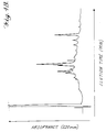
- FIGURE 3B shows absorbance (220 nm) versus elution time in minutes for the same chromatogram.
- FIGURE 4A shows relative fluorescence (297 nm excitation, 390 nm emission) versus elution time of fractions during reverse phase HPLC.
- Cross-linked type II collagen telopeptides are eluted as indicated.
- the fractions indicated by the bar (-) show evidence by sequence and composition analysis of the peptides indicated that retain or have lost the gly (G) and pro (P) residues.
- FIGURE 4B shows absorbance (220 nm) as a function of elution time during reverse phase HPLC.
- a specific telopeptide having a hydroxylysyl pyridinoline cross-link derived from the C-terminal telopeptide domain of type II collagen has the following amino acid sequence (referred to hereinbelow as the core peptide structure): wherein the cross-linking residue depicted as Hyl-Hyl-Hyl is hydroxylysyl pyridinoline (HP), a natural 3-hydroxypyridinium residue present in mature collagen fibrils of various tissues.
- the core peptide structure of the type II collagen peptides may be found in body fluids as a component of larger peptides that bear additional amino acids or amino acid sequences on one or more ends of the three peptide sequences joined by the HP residue.
- FIGURE 1 shows how type II collagen telopeptides, which are linked to a triple-helical sequence, may be produced in vivo from a human source using the proteolytic enzymes pepsin and trypsin. Smaller fragments that have lost amino acids from the core peptide structure, particularly from the helical sequence, may also occur in body fluids. Generally, additions or deletions of amino acids from the core peptide structure will involve from 1 to about 3 amino acids.
- Additional amino acids will generally be determined by the type II collagen telopeptide sequence that occurs naturally in vivo .
- peptides having the following structure can be isolated chromatographically from urine, and another of structure: may be isolated.
- glycosylated variants of the core structure and its larger and smaller variants may occur in which a galactose residue or a glucosyl galactose residue are attached to the side chain hydroxyl group of the HP cross-linking residue.
- Each peak in the graph shown in Figures 4A and 4B may correspond to a cross-linked fragment of particular structure that may be quantitated for purposes of the present invention.
- the isolated peptide fragments represent the products of proteolytic degradation of type II collagen fibrils within the body.
- the core structure containing the HP residue is relatively resistant to further proteolysis and provides a quantitative measure of the amount of type II collagen degraded.
- Collagen type II is present in hyaline cartilage of joints in the adult skeleton. Quantitation of the collagen type II telopeptides in a body fluid, for example by way of a monoclonal antibody that recognizes an epitope in the peptide structure, would provide a quantitative measure of whole-body cartilage destruction or remodeling.
- the present invention involves an assay for cartilage tissue degradation in humans based on quantifying the urinary excretion rate of at least one member of this family of telopeptides. Such an assay could be used, for example, to:
- Osteoarthritis is a degenerative disease of the articulating cartilages of joints. In its early stages it is largely non-inflammatory (i.e. distinct from rheumatoid arthritis). It is not a single disease but represents the later stages of joint failure that may result from various factors (e.g. genetic predisposition, mechanical over usage, joint malformation or a prior injury, etc.). Destruction of joint articular cartilage is the central progressive feature of osteoarthritis. The incidence of osteoarthritis, based on radiographic surveys, ranges from 4% in the 18-24 year age group to 85% in the 75-79 year age group. At present the disease can only be diagnosed by pain and radiographic or other imaging signs of advanced cartilage erosion.
- the assays disclosed above may be used to detect early evidence of accelerated cartilage degradation in mildly symptomatic patients, to monitor disease progress in more advanced patients, and as a means of monitoring the effects of drugs or other therapies.
- rheumatoid arthritis juvenile rheumatoid arthritis, ankylosing spondylitis, psoriatic arthritis, Reiter's syndrome, relapsing polychondritis, the low back pain syndrome, and other infectious forms of arthritis.
- the type II collagen-specific assays described herein could be used to diagnose and monitor these diseases and evaluate their response to therapy, as disclosed above in connection with osteoarthritis.
- Cross-linked peptides that are derived from proteolysis of human type III collagen may be present in body fluids. These peptides have a core structure embodied in the following parent structures: where Hyl Hyl Hyl is hydroxylysyl pryidinoline, and Gln is glutamine or pyrrolidine carboxylic acid.
- a likely cross-linked peptide derived from type III collagen in body fluids has the core structure: that is derived from a two al(III)N-telopeptide domains linked to an hydroxylysyl pyridinoline residue (Hyl-Hyl-Hyl).
- a second possible fragment of the C-telopeptide cross-linking domain based on the collagen types I and II peptides observed in urine, has the core structure:
- a specific assay for type III collagen degradation by quantitating cross-linked type III collagen peptides as disclosed above can be used for detecting, diagnosing, and monitoring various inflammatory disorders, possibly with particular application to the vascuiitis syndromes.
- an assay could be used as a differential diagnostic tool for patients with various degenerative and inflammatory disorders that result in connective tissue destruction or pathological processes.
- Urine is collected from a normal adolescent during a rapid phase of skeletal growth.
- sequence of chromatographic steps that include but are not limited to, adsorption on selective cartridges of a hydrophobic interaction support and an ion-exchange support and molecular sieve, ion-exchange and reverse-phase HPLC column chromatography steps, individual peptides are isolated.
- the cross-linked peptides containing HP (and LP) residues are detected during column chromatography by their natural fluorescence (Ex max 297 nm ⁇ pH 4, Ex max 330 nm, > pH 6; Em max 390 nm).
- An exemplary isolation procedure is provided in the Example below.
- the first step was molecular sieve chromatography on a column of Bio-Gel P-10 (Bio Rad Labs, 2.5 cm X 90 cm) eluted by 10% (v/v) acetic acid, monitoring the effluent for HP fluorescence as shown in FIGURE 2.
- the Y-axis is the relative fluorescence emission at 390 nm (297 nm excitation), and the X-axis is the fraction number.
- the fraction size was 4 ml.
- the fractions indicated as II are enriched in the cross-linked collagen type II telopeptides.
- the cross-linked collagen type I telopeptides are contained in the fractions indicated as III and IV.
- Fractions spanning pool II (enriched in the type II collagen cross-linked peptides) were combined, freeze-dried and fractionated by ion-exchange column chromatography on a DEAE-HPLC column (TSK-DEAE-5PW, 7.5mm X 7.5mm, Bio-Rad Labs), equilibrated with 0.02 M Tris/HC1, 10% (v/v) acetonitrile, pH 7.5 and eluted with a gradient of 0.05M NaC1 in the same buffer as shown in FIGURE 2.
- FIGURE 3A plots relative fluorescence emission at 390 nm (330 nm excitation) versus elution time.
- the cross-linked collagen type II telopeptides are found primarily in the segment indicated as IV.
- FIGURE 3B plots absorbance at 220 nm as a function of elution time in minutes.
- Pool IV contains the type II collagen cross-linked peptides.
- Individual peptides were then resolved from pool IV by reverse phase HPLC on a C-18 column (Aquapore RP-300, 25cm X 4.6mm, Brownlee Labs), eluting with a gradient of 0-30% (v/v) acetonitrile in 0.1% (v/v) trifluoroacetic acid.
- FIGURE 4A shows a plot of relative fluorescence intensity at 390 nm (297 nm excitation) as a function of elution time. The peaks associated with particular peptides are indicated in FIGURE 4A.
- FIGURE 4B shows the relative absorbance at 220 nm as a function of time.
- Cross-linked peptide fragments of type III collagen containing HP cross-linking residues may be isolated by a similar combination of steps from the urine of normal growing subjects or, for example, from the urine of patients with inflammatory disorders of the vasculature.
- telopeptides by “quantitating” is meant measuring by any suitable means, including but not limited to spectrophotometric, gravimetric, volumetric, coulometric, immunometric, potentiometric, or amperometric means the concentration of peptide fragments containing 3-hydroxypyridinium cross-links in an aliquot of a body fluid.
- suitable body fluids include urine, serum, and synovial fluid.
- the preferred body fluid is urine.
- the aliquot assayed be from a combined pool of urine collected over a fixed period of time, for example, 24 hours. In this way, the absolute rate of collagen degradation is calculated for a 24 hour period.
- urinary peptides may be measured as a ratio relative to a marker substance found in urine such as creatinine. In this way the urinary index of collagen degradation would remain independent of urine volume.
- monoclonal or polyclonal antibodies are produced which are specific to the peptide fragments containing pyridinoline cross-links found in a body fluid such as urine.
- Type II or type III telopeptides may be isolated from a body fluid of any patient but may be more easily obtained from patients suffering from diseases involving type II or type III collagen degradation or from rapidly growing adolescents.
- Immunological binding partners capable of specifically binding to peptide fragments derived from bone collagen obtained from a physiological fluid can be prepared by methods well known in the art. The preferred method for isolating these peptide fragments is described above.
- immunological binding partners as used herein is meant antibodies and antibody fragments capable of binding to a telopeptide.
- Suitable carrier molecules include, but are not limited to, bovine serum albumin, ovalbumin, thyroglobulin, and keyhold limpet hemocyanin (KLH). Preferred carriers are thryoglobulin and KLH.
- hapten-protein conjugates are equally successful immunogens.
- the selection of a protocol for binding the particular hapten to the carrier protein therefore depends on the amino acid sequence of the ordinary peptide fragments selected. For example, a protol could involve coupling this hapten to keyhold limpet hemocyanin (KLH), or other suitable carrier, with glutaraldehyde.
- KLH keyhold limpet hemocyanin
- An alternative protocol is to couple the peptides to KLH with a carbodiimide.
- binding agents include, but are not limited to, carbodiimides, glutaraldehyde, mixed anhydrides, as well as both homobifunctional and heterobifunctional reagents (see for example the Pierce 1986-87 catalog, Pierce Chemical Co., Rockford, IL).
- Preferred binding agents include carbodiimides and heterobifunctional reagents such as m-Maleimidobenzyl-N-hydroxysuccinimide ester (MBS).
- Either monoclonal or polyclonal antibodies to the hapten-carrier molecule immunogen can be produced. However, it is preferred that monoclonal antibodies (Mab) be prepared. For this reason it is preferred that immunization be carried out in the mouse. Immunization protocols for the mouse usually include an adjuvant. Examples of suitable protocols are described by Chard, T. (1987) vida supra. Spleen cells from the immunized mouse are harvested and homogenized and thereafter fused with cancer cells in the presence of polyethylene glycol to produce a fused cell hybrid which produces monoclonal antibodies specific to peptide fragments derived from collagen. Examples of such peptides are represented by the formulas given above.
- Suitable cancer cells include myeloma, hepatoma, carconima and sarcoma cells. Detailed descriptions of this procedure, including screening protocols, protocols for growing selected hybrid cells and harvesting monoclonal antibodies produced by the selected hybrid cells are provided in Galfre, G. and Milstein, C., Meth. Enzymol., 73:1 (1981).
- a preferred preliminary screening protocol involves the use of peptide fragments derived from type II or type III collagen resorption and containing 3-hydroxypyridinium cross-links in a solid phase radioimmunoassay.
- a specific example describing a preferred monoclonal antibody is provided below.
- Methods utilizing recombinant DNA techniques may also be adapted to construct monoclonal antibodies. These methods may be accomplished by the construction of expression libraries and primers by one of ordinary skill in the art (see William D. Huse et al., "Generation of a Large Combinational Library of the Immunoglobulin Repertoire in Phage Lambda," Science 246:1275-1281, December 1989, see also L. Sastry et al., "Cloning of the Immunological Repertoire in Escherichia coli for Generation of Monoclonal Catalytic Antibodies: Construction of a Heavy Chain Variable Region-Specific cDNA Library," Proc. Natl. Acad. Sci.
- mRNA is isolated from a B cell population, and utilized to create heavy and light chain immunoglobulin cDNA expression libraries in the ⁇ ImmunoZap(H) and ⁇ ImmunoZap(L) vectors.
- vectors may be screened individually or co-expressed to form Fab fragments or antibodies (see Huse et al., supra: see also Sastry et al., supra). Positive plaques may subsequently be converted to a non-lytic plasmid which allows high level expression of monoclonal antibody fragments from E. coli.
- the monoclonal antibodies or other immunological binding partners used in connection with the present invention are preferably specific for a particular type of collagen telopeptide.
- Assays for the type II or type III collagen degradation telopeptides should preferably be able to distinguish between the type I, type II, and type III peptides. However, in some cases, such selectivity will not be necessary, for example, if it is known that a patient is not suffering degradation of one type of collagen but is suspected of suffering degradation from the assayed type of collagen. Because of the differences in amino acid sequences between the type I, type II and type III families of telopeptides, cross-reactivity should not occur to a significant degree.
- hybridomas can be selected for during the screening of splenocyte fusion clones that produce monoclonal antibodies specific for the cross-linked telopeptide of interest (and lack affinity for those of the other two collagen types). Based on the differences in sequence of the isolated peptide structures, such specificity is entirely feasible.
- Peptide fragments of the parent types I, II and III collagens, suitable for such hybridoma screening can be prepared from human bone, cartilage and other tissues and used to screen clones from mice immunized appropriately with the individual cross-linked peptide antigens isolated from body fluid.
- Immunological binding partners are employed in various immunometric assays to quantitate the concentration of the peptides having 3-hydroxypyridinium cross-links described above.
- These immunometric assays preferably comprise a monoclonal antibody or antibody fragment coupled to a detectable marker.
- suitable detectable markers include but are not limited to: enzymes, coenzymes, enzyme inhibitors, chromophores, fluorophores, chemiluminescent materials, paramagnetic metals, spin labels, and radionuclides.
- telopeptides examples include, but are not limited to, enzyme linked immunosorbent assay (ELISA) (Ingvall, E., Meth. Enzymol., 70 (1981)), radio-immunoassay (RIA), and "sandwich” immunoradiometric assay (IRMA).
- ELISA enzyme linked immunosorbent assay
- RIA radio-immunoassay
- IRMA immunoradiometric assay
- these immunometric methods can be used to determine the absolute rate of bone resorption or collagen degradation by simply contacting a body fluid with the immunological binding partner specific to a collagen telopeptide having a 3-hydroxypyridinium cross-link.
- immunometric assays described above be conducted directly on untreated body fluids (e.g. urine, blood, serum, or synovial fluid). Occasionally, however, contaminating substances may interfere with the assay necessitating partial purification of the body fluid. Partial purification procedures include, but are not limited to, cartridge adsorption and elution, molecular sieve chromatography, dialysis, ion exchange, alumina chromatography, hydroxyapatite chromatography and combinations thereof.
- Test kits suitable for use in accordance with the present invention, contain specific binding partners such as monoclonal antibodies prepared as described above, that specifically bind to peptide fragments derived from collagen degradation found in a body fluid. It is preferred that the specific binding partners of this test kit be coupled to a detectable marker of the type described above. Test kits containing a panel of two or more specific binding partners, particularly immunological binding partners, are also contemplated. Each immunological binding partner in such a test kit will preferably not cross-react substantially with a telopeptide derived from another type of collagen. For example, an immunological binding partner that binds specifically with a type II collagen telopeptide should preferably not cross-react with either a type I or type III collagen telopeptide.
- test kits may contain a first specific binding partner to a collagen-derived telopeptide having a cross-link containing a pyridinium ring (which may be OH-substituted), and a second specific binding partner to a telopeptide having the same structure as the first telopeptide except that the pyridinium ring has been cleaved, such as photolytically.
- the following is an example of preparation of a monoclonal antibody against a telopeptide immunogen.
- a fraction enriched in the peptide is prepared from adolescent human urine using reverse phase and molecular sieve chromatography.
- the peptide is conjugated to keyhole limpet hemocyanin (KLH) with glutaraldehyde using standard procedures.
- Mice (Balb/c) are immunized subcutaneously with this conjugate (50-70) g), first in complete Freund's adjuvant, then boosted (25 )g) at 3 weekly intervals in incomplete Freund's adjuvant intraperitoneally.
- mice After test bleeds shows a high titer against the peptide conjugated to bovine serum albumin (BSA) using an ELISA format, selected mice are boosted with a low dose (5g) of the immunogen in sterile PBS intravenously. Three days later, cells from the spleens of individual mice are fused with mouse myeloma cells using standard hybridoma technology. The supernatants of hybridoma clones growing in individual wells of 96-well plates are screened for reactive monoclonal antibodies, initially using a crude telopeptide preparation conjugated to BSA.
- BSA bovine serum albumin
- the antibodies produced by individual hybridomas are characterized against a panel of screening antigens conjugated to BSA using ELISA analysis.
- An inhibition assay is used in which telopeptide conjugated to BSA is plated out in the plastic wells, and antibody is pre-incubated with a solution of the potential antigen.
- a secondary antibody goat anti-mouse IgG conjugated to horseradish peroxidase, HRP is used for color development using an appropriate substrate.
- a quantitative assay of the subject peptide(s) having intact pyridinium rings will underestimate the amount of collagen degradation. Accordingly, an assay based on two possible approaches is expected to be comparatively more accurate.
- the two envisaged embodiments are: a single specific binding partner is employed that recognizes both closed and open-ringed embodiments of the targeted peptide(s); or two specific binding partners are employed, which differentiate between the closed- and open-ringed epitopes, respectively.
- Specific binding partners that discriminate between open and closed ring forms of the targeted peptides may be obtained by incorporating an appropriate screening step into the standard procedures for obtaining such specific binding partners.
- a library of candidate monoclonal antibodies can be screened for their ability to bind to the peptide having an opened pyridinoline ring (e.g., by ultraviolet light irradiation) and their ability to bind to the peptide having an intact pyridinoline ring.
- the open-ring and intact-ring peptides useful in such a screening step can be obtained by known purification techniques such as RP-HPLC, as described above.
- An alternative procedure for assaying for the above-described peptides consists of measuring a physical property of the peptides having 3-hydroxypyridinium cross-links.
- One such physical property relies upon electrochemical detection.
- This method consists of injecting an aliquot of a body fluid, such as urine, into an electrochemical detector poised at a redox potential suitable for detection of peptides containing the 3-hydroxypyridinium ring.
- the 3-hydroxypyridinium ring being aphenol, is subject to reversible oxidation and therefore the electrochemical detector (e.g., Model 5100A Coulochem sold by Esa 45 Wiggins Ave., Bedford, MA) is a highly desirable instrument suitable for quantitating the concentration of the present peptides.
- amperometric e.g., BioAnalytical Systems
- ESA electrochemical detector
- Bedford, MA 01730 Two basic forms of electrochemical detector are currently commercially available: amperometric (e.g., BioAnalytical Systems) and coulometric (ESA, Inc., Bedford, MA 01730). Both are suitable for use in accordance with the present invention, however, the latter system is inherently more sensitive and therefore preferred since complete oxidation or reduction of the analyzed molecule in the column effluent is achieved.
- screening or guard electrodes can be placed "upstream" from the analytical electrode to selectively oxidize or reduce interfering substances thereby greatly improving selectivity.
- the voltage of the analytical electrode is tuned to the redox potential of the sample molecule, and one or more pretreatment cells are set to destroy interferents in the sample.
- a standard current/voltage curve is established for standard peptides containing lysyl pyridinoline or hydroxylysyl pyridinoline in order to determine the proper voltage to set for optimal sensitivity. This voltage is then modified depending upon the body fluid, to minimize interference from contaminants and optimize sensitivity. Electrochemical detectors, and the optimum conditions for their use are known to those skilled in the art. Complex mixtures of body fluids can often be directly analyzed with the electrochemical detector without interference. Accordingly, for most patients no pretreatment of the body fluid is necessary. In some cases however, interfering compounds may reduce the reliability of the measurements. In such cases, pretreatment of the body fluid (e.g., urine) may be necessary.
- the body fluid e.g., urine
- a body fluid is first purified prior to electrochemically titrating the purified peptide fragments.
- the purification step may be conducted in a variety of ways including but not limited to dialysis, ion exchange chromatography, alumina chromatography, hydroxyapatite chromatography, molecular sieve chromatography, or combinations thereof.
- a measured aliquot (25 ml) of a 24 hour urine sample is dialyzed in reduced porosity dialysis tubing to remove the bulk of contaminating fluorescent solutes.
- the non-diffusate is then lyophilized, redissolved in 1% heptafluorobutyric acid (HFBA), an ion pairing solution, and the peptides adsorbed on a Waters Sep-Pak C-18 cartridge.
- HFBA heptafluorobutyric acid
- This cartridge is then washed with 5 ml of 1% HFBA, and then eluted with 3 ml of 50% methanol in 1% HFBA.
- Another preferred method of purification consists of adsorbing a measured aliquot of urine onto an ion-exchange adsorption filter and eluting the adsorption filter with a buffered eluting solution. The eluate fractions containing peptide fragments having 3-hydroxypyridinium cross-links are then collected to be assayed.
- Still another preferred method of purification employs molecular sieve chromatography.
- an aliquot of urine is applied to a Bio-Gel P2 or Sephadex G-20 column and the fraction eluting in the 1000-5000 Dalton range is collected.
- a combination of the above methods may be used to purify or partially purify urine or other body fluids in order to isolate the peptide fragments having 3-hydroxypyridinium cross-links.
- the purified or partially purified peptide fragments obtained by the above procedures may be subjected to additional purification procedures, further processed or assayed directly in the partially purified state. Additional purification procedures include resolving partially purified peptide fragments employing high performance liquid chromatography (HPLC) or microbore HPLC when increased sensitivity is desired. These peptides may then be quantitated by electrochemical titration.
- HPLC high performance liquid chromatography
- microbore HPLC microbore HPLC
- a preferred electrochemical titration protocol consists of tuning the redox potential of the detecting cell of the electrochemical detector (Coulochem Model 5100A) for maximum signal with pure HP. The detector is then used to monitor the effluent from a C-18 HPLC column used to resolve the partially purified peptides.
- An alternative preferred method for quantitating the concentration of peptides having 3-hydroxypyridinium cross-links as described herein is to measure the characteristic natural fluorescence of these peptides.
- fluorometric assay may be conducted directly without further purification of the body fluid.
- the peptides are resolved by HPLC and the natural fluorescence of the HP and LP amino acid residues is measured at 395 nm upon excitation at 297 nm, essentially as described by Eyre, D.R., et al., Analyte . Biochem . 137:380 (1984), herein incorporated by reference.
- the fluorometric assay be conducted on urine.
- Urine usually contains substantial amounts of naturally occurring fluorescent contaminants that must be removed prior to conducting the fluorometric assay. Accordingly, urine samples are first partially purified as described above for electrochemical detection. This partially purified urine sample can then be fluorometrically assayed as described above.
- the HP and LP cross-linked peptides in the partially purified urine samples or other body fluids can be hydrolyzed in 6M HC1 at about 108°C for approximately 24 hours as described by Eyre, et al. (1984) vida supra .
- the body fluid preferably urine
- the body fluid is passed directly through a C-18 reverse phase affinity cartridge after adding acetonitrile/methanol 5 to 10% V/V.
- the non-retentate is adjusted to 0.05-0.10M with a cationic ion-pairing agent such as tetrabutyl ammonium hydroxide and passed through a second C-18 reverse phase cartridge.
- the washed retentate, containing fluorescent peptides, from this second cartridge is eluted with acetonitrile:water (or methanol:water), dried and fluorescent peptides are analyzed by reverse phase HPLC or microbore HPLC using an anionic ion-pairing agent such as 0.01M trifluoracetic acid in the eluant.
- the ratio of HP:LP found in normal human urine and urine from patients having Paget's disease are both approximately 4.5:1. This is slightly higher than the 4:1 ratio found in bone itself (Eyre, et al., 1984).
- the higher ratio found in urine indicates that a portion of the HP fraction in urine may come from sources other than bone, such as the diet, or other sources of collagen degradation, i.e., cartilage catabolism.
Abstract
Description
- The present invention relates to methods for detecting and monitoring collagen degradation in vivo. More specifically, it relates to methods for quantitating cross-linked telopeptides produced in vivo upon degradation of collagen type II or type III. This application is divided from WO 91/08478 (EPA 91 900109.9), which, as published, described and claimed methods for detecting and monitoring collagen degradation in terms of telopeptides of collagen types I, II and III.
- Three known classes of collagens have been described to date. The
Class 1 collagens, subdivided into types I, II, III, V, and XI, are known to form fibrils. These collagens are all synthesized as procollagen molecules, made up of N-terminal and C-terminal propeptides, which are attached to the core collagen molecule. After removal of the propeptides, which occurs naturally in vivo during collagen synthesis, the remaining core of the collagen molecule consists largely of a triple helical domain having terminal telopeptide sequences which are nontriple-helical. These telopeptide sequences have an important function as sites of intermolecular cross-linking of collagen fibrils extracellularly. - The present invention relates to methods of detecting collagen degradation based on assaying for particular cross-linked telopeptides produced in vivo upon collagen degradation. In the past, assays have been developed for monitoring degradation of collagen in vivo by measuring various biochemical markers, some of which have been degradation products of collagen. For example, bone turnover associated with Paget's disease has been monitored by measuring small peptides containing hydroxyproline, which are excreted in the urine following degradation of bone collagen. Russell et al., Metab, Bone Dis. and Rel. Res. 4 and 5, 255-262 (1981); and Singer, F.R., et al., Metabolic Bone Disease,Vol. II (eds Avioli, L.V. and Kane, S.M.), 489-575 (1978), Academic Press, New York.
- Other researchers have measured the cross-linking compound pyridinoline in urine as an index of collagen degradation in joint disease. See, for background and for example, Wu and Eyre, Biochemistry,23:1850 (1984); Black et al., Annals of the Rheumatic Diseases, 48:641-644 (1989); Robins et al., Annals of the Rheumatic Diseases, 45:969-973 (1986); and Seibel et al., The Journal of Rheumatology, 16:964 (1989). In contrast to the present invention, some prior researchers have hydrolyzed peptides from body fluids and then looked for the presence of individual hydroxypyridinium residues. None of these researchers has reported measuring a telopeptide containing a cross-link that is naturally produced in vivo upon collagen degradation, as in the present invention.
- UK Patent application GB 2,205,643 reports that the degradation of type III collagen in the body is quantitatively determined by measuring the concentration of an N-terminal telopeptide from type III collagen in a body fluid. In this reference, it is reported that cross-linked telopeptide regions are not desirable. In fact, this reference reports that it is necessary to use a non-cross-linked source of collagen to obtain the telopeptide. The peptides of the present invention are all cross-linked.
- Collagen cross-links are discussed in greater detail below, under the heading "Collagen Cross-Linking".
- There are a number of reports indicating that collagen degradation can be measured by quantitating certain procollagen peptides. The present invention involves telopeptides rather than propeptides, the two being distinguished by their location in the collagen molecule and the timing of their cleavage in vivo. See US Patent 4,504,587; US Patent 4,312,853; Pierard et al., Analytical Biochemistry 141:127-136 (1984); Niemela, Clin. Chem., 31/8:1301-1304 (1985); and Rohde et al., European Journal of Clinical Investigation, 9:451-459 (1979).
- US Patent 4,778,768 relates to a method of determining changes occurring in articular cartilage involving quantifying proteoglycan monomer or antigenic fragments thereof in a synovial fluid sample. This patent does not relate to detecting cross-linked telopeptides derived from degraded collagen.
- Dodge, J. Clin. Invest., 83:647-661 (1981) discloses methods for analyzing type II collagen degradation utilizing a polyclonal antiserum that specifically reacts with unwound alpha-chains and cyanagen bromide-derived peptides of human and bovine type II collagens. The peptides involved are not cross-linked telopeptides as in the present invention.
- Amino acid sequences of human type III collagen, human proa1(II) collagen, and the entire preproa1(III) chain of human type III collagen and corresponding cDNA clones have been investigated and determined by several groups of researchers. See Loidl et al., Nucleic Acids Research 12:9383-9394 (1984); Sangiorgi et al., Nucleic Acids Research, 13:2207-2225 (1985); Baldwin et al., Biochem. J., 262:521-528 (1989); and Ala-Kokko et al., Biochem. J., 260:509-516 (1989). None of these references specifies the structures of particular telopeptide degradation products that could be measured to determine the amount of degraded fibrillar collagen in vivo.
- In spite of the above-described background information, there remains a need for effective and simple assays for determining collagen degradation in vivo. Such assays could be used to detect and monitor disease states in humans, such as osteoarthritis (type II collagen degradation), and various inflammatory disorders, such as vasculitis syndrome (type III collagen degradation).
- Assays for type I collagen degradation, described in WO-A-85/04491, can be utilized to detect and assess bone resorption in vivo.
- Several potential organic indices have been tested. For example, hydroxyproline, an amino acid largely restricted to collagen, and the principal structural protein in bone and all other connective tissues, is excreted in urine. Its excretion rate is known to be increased in certain conditions, notably Paget's disease, a metabolic bone disorder in which bone turnover is greatly increased, as pointed out above. For this reason, urinary hydroxyproline has been used extensively as an amino acid marker for collagen degradation. Singer, F.R., et al. (1978), cited hereinabove.
- US Patent No 3,600,132 discloses a process for determination of hydroxyproline in body fluids such as serum, urine, lumbar fluid and other intercellular fluids in order to monitor deviations in collagen metabolism. In particular, this inventor notes that in pathologic conditions such as Paget's disease, Marfan's syndrome, osteogenesis imperfecta, neoplastic growth in collagen tissues and in various forms of dwarfism, increased collagen anabolism or catabolism as measured by hydroxyproline content in biological fluids can be determined. This inventor measures hydroxyproline by oxidising it to a pyrrole compound with hydrogen peroxide and N-chloro-p-toluenesulphonamide followed by colorimetric determination in p-dimethyl-amino-benzaldehyde.
- In the case of Paget's disease, the increased urinary hydroxyproline probably comes largely from bone degradation; hydroxyproline, however, generally cannot be used as a specific index. Much of the hydroxyproline in urine may come from new collagen synthesis (considerable amounts of the newly made protein are degraded and excreted without ever becoming incorporated into tissue fabric), and from turnover of certain blood proteins as well as other proteins that contain hydroxyproline. Furthermore, about 80% of the free hydroxyproline derived from protein degradation is metabolized in the liver and never appears in the urine. Kiviriko, K.I. Int. Rev. Connect. Tissue Res. 5:93 (1970), and Weiss, P.H., and Klein, L., J. Clin. Invest. 48:1 (1969).
- Hydroxylysine and its glycoside derivatives, both peculiar to collagenous proteins, have been considered to be more accurate than hydroxyproline as markers of collagen degradation. However, for the same reasons described above for hydroxyproline, hydroxylysine and its glycosides are probably equally non-specific markers of bone resorption.
- Krane, S.M. and Simon, L.S. Develop. Biochem., 22:185 (1981).
- The polymers of most genetic types of vertebrate collagen require the formation of aldehyde-mediated cross-links for normal function. Collagen aldehydes are derived from a few specific lysine or hydroxylysine side-chains by the action of lysyl oxidase. Various di-, tri- and tetrafunctional cross-linking amino acids are formed by the spontaneous intra- and intermolecular reactions of these aldehydes within the newly formed collagen polymers; the type of cross-linking residue varies specifically with tissue type (see Eyre, D.R. et al., Ann. Rev. Biochem., 53:717-748 (1984)).
- Two basic pathways of cross-linking can be differentiated for the banded (67nm repeat) fibrillar collagens, one based on lysine aldehydes, the other on hydroxylysine aldehydes. The lysine aldehyde pathway dominates in adult skin, cornea, sclera, and rat tail tendon and also frequently occurs in other soft connective tissues. The hydroxylysine aldehyde pathway dominates in bone, cartilage, ligament, most tendons and most internal connective tissues of the body, Eyre, D.R. et al. (1974) vida supra. The operating pathway is governed by whether lysine residues are hydroxylated in the telopeptide sites where aldehyde residues will later be formed by lysyl oxidase (Barnes, M.J. et al., Biochem. J., 139:461 (1974)).
- The chemical structure(s) of the mature cross-linking amino acids on the lysine aldehyde pathway are unknown, but hydroxypyridinium residues have been identified as mature products on the hydroxylysine aldehyde route. On both pathways and in most tissues the intermediate, borohydride-reducible cross-linking residues disappear as the newly formed collagen matures, suggesting that they are relatively short-lived intermediates (Bailey, A.J. et al., FEBS Lett, 16:86 (1971). Exceptions are bone and dentin, where the reducible residues persist in appreciable concentration throughout life, in part apparently because the rapid mineralization of the newly made collagen fibrils inhibits future spontaneous cross-linking interactions (Eyre, D.R., In: The Chemistry and Biology of Mineralized Connective Tissues,(Veis, A. ed.) pp. 51-55 (1981), Elsevier, New York, and Walters, C. et al., Calc. Tiss. Intl. 35:401-405 (1983)).
- Two chemical forms of 3-hydroxypyridinium cross-link have been identified (Formula I and II). Both compounds are naturally fluorescent, with the same characteristic excitation and emission spectra (Fujimoto, D. et al. Biochem. Biophys. Res. Commun., 76:1124 (1977), and Eyre, D.R., Develop. Biochem. 22:50 (1981)). These amino acids can be resolved and assayed directly in tissue hydrolysates with good sensitivity using reverse phase HPLC and fluorescence detection. Eyre, D.R. et al., Analyte. Biochem., 137:380-388 (1984). It should be noted that the present invention involves quantitating particular peptides rather than amino acids.

In growing animals, it has been reported that these mature cross-links may be concentrated more in an unmineralized fraction of bone collagen than in the mineralized collagen (Banes, A.J., et al., Biochem. Biophys. Res. Commun., 113:1975 (1983). However, other studies on young bovine or adult human bone do not support this concept, Eyre, D.R., In: The Chemistry and Biology of Mineralized Tissues (Butler, W.T. ed.)p. 105 (1985), Ebsco Media Inc., Birmingham, Alabama. - The presence of collagen hydroxypyridinium cross-links in human urine was first reported by Gunja-Smith and Boucek (Gunja-Smith, Z. and Boucek, R.J., Biochem J., 197:759-762 (1981)) using lengthy isolation procedures for peptides and conventional amino acid analysis. At that time, they were aware only of the HP forms of the cross-link. Robins (Robins, S.P., Biochem J., 207:617-620 (1982) has reported an enzyme-linked immunoassay to measure HP in urine, having raised polyclonal antibodies to the free amino acid conjugated to bovine serum albumin. This assay is intended to provide an index for monitoring increased joint destruction that occurs with arthritic diseases and is based, according to Robins, on the finding that pyridinoline is much more prevalent in cartilage than in bone collagen.
- In more recent work involving enzyme-linked immunoassay, Robins reports that lysyl pyridinoline is unreactive toward antiserum to pyridinoline covalently linked to bovine serum albumin (Robins et al., Ann. Rheum. Diseases, 45:969-973 (1986)). Robins' urinary index for cartilage destruction is based on the discovery that hydroxylysyl pyridinoline, derived primarily from cartilage, is found in urine at concentrations proportional to the rate of joint cartilage resorption (i.e. degradation). In principle, this index could be used to measure whole body cartilage loss; however, no information on bone resorption would be available.
- Also of interest is the international publication No. WO 89/12824, by The Rowett Research Institute. This later publication describes a method of monitoring collagen degradation, comprising assaying a biological fluid sample which contains a fragment of collagen including lysyl pryidinoline or hydroxylysyl pyridinoline or a substituted form thereof, and a method of determining the tissue origin of degraded collagen thereby.
- A need therefore exists for a method that allows the measurement of whole-body cartilage or collagenous connective tissue degradation rates in humans. The most useful such method would be one that could be applied to body fluids, especially urine. The method should be sensitive, i.e. quantifiably down to 1 picomole and rapidly measure 24-hour bone resorption rates so that the progress of various therapies can be assessed.
- The present invention is based on the discovery of the presence of particular cross-linked telopeptides in body fluids of patients and normal human subjects. These telopeptides are produced in vivo during collagen degradation and remodeling. The term "telopeptides" is used herein to mean cross-linked peptides having sequences that are associated with the telopeptide region of type II or type III collagen. Generally, the telopeptides disclosed herein will have fewer amino acid residues than the entire telopeptide domains of type II or type III collagen. Typically, the telopeptides of the present invention will comprise two alpha one (a1) peptides linked by a pyridinium cross-link and further linked by the pyridinium cross-link to a residue or peptide of the collagen triple-helical domain. Having disclosed the structures of these telopeptides herein, it will be appreciated by one of ordinary skill in the art that they may also be produced other than in vivo e.g., synthetically. These peptides will generally be provided in purified form e.g., substantially free of impurities, particularly other peptides.
- The present invention also relates to methods for determining in vivo degradation of type II or type III collagen. The methods involve quantitating in a fluid the concentration of particular telopeptides that have a 3-hydroxypyridinium cross-link and that are derived from collagen degradation. The methods disclosed in the present invention are analogous to those disclosed in WO-A-85/04491 for determining the absolute rate of bone resorption in vivo. Those methods involved quantitating in a body fluid the concentration of telopeptides having a 3-hydroxypyridinium cross-link derived from type I collagen resorption.
- In a representative assay, the patient's body fluid is contacted with an immunological binding partner specific to a telopeptide having a 3-hydroxypyridinium cross-link derived from type II or type III collagen. The body fluid may be used as is or purified prior to the contacting step. This purification step may be accomplished using a number of standard procedures, including cartridge adsorption and elution, molecular sieve chromatography, dialysis, ion exchange, alumina chromatography, hydroxyapatite chromatography, and combinations thereof.
- Other representative embodiments of quantitating the concentration of peptide fragments having a 3-hydroxypyridinium cross-link in a body fluid include electrochemical titration, natural fluorescence spectroscopy, and ultraviolet absorbance. Electrochemical titration may be conducted directly upon a body fluid without further purification. However, when this is not possible due to excessive quantities of contaminating substances, the body fluid is first purified prior to the electrochemical titration step. Suitable methods for purification prior to electrochemical detection include dialysis, ion exchange chromatography, alumina chromatography, molecular sieve chromatography, hydroxyapatite chromatography and ion exchange absorption and elution.
- Fluorometric measurement of a body fluid containing a 3-hydroxypyridinium cross-link is an alternative way of quantitating collagen degradation.
- The fluorometric assay can be conducted directly on a body fluid without further purification. However, for certain body fluids, particularly urine, it is preferred that purification of the body fluid be conducted prior to the fluorometric assay. This purification step consists of dialyzing an aliquot of a body fluid such as urine against an aqueous solution thereby producing partially purified peptide fragments retained within the nondiffusate (retentate). The nondiffusate is then lyophilized, dissolved in an ion pairing solution and absorbed onto an affinity chromatography column. The chromatography column is washed with a volume of ion pairing solution and, thereafter, the peptide fragments are eluted from the column with an eluting solution. These purified peptide fragments may then be hydrolyzed and the hydrolysate resolved chromatographically. Chromatographic resolution may be conducted by either high-performance liquid chromatography or microbore high performance liquid chromatography.
- The invention includes peptides having structures identical to peptides derived from collagen degradation, substantially free from other human peptides, which may be obtained from a body fluid. The peptides contain at least one 3-hydroxypyridinium cross-link, in particular, a lysyl pyridinoline cross-link or a hydroxylysyl pyridinoline cross-link, and are derived from the telopeptide region of type II or type III collagen linked to one or more residues from a triple-helical domain, typically by the action of endogenous proteases and/or peptidases.
- The structures of the type II and type III telopeptides are disclosed below.
- Another aspect of the present invention involves assays for the peptides described herein in which the pyridinium rings are intact and cleaved. Since it is suspected that some cleavage of pyridinium rings occurs in vivo, assays that detect both intact and cleaved pyridinium rings may lead to more accurate assessments of collagen degradation. In connection with this aspect of the present invention, specific binding partners to the individual peptides containing intact or cleaved pyridinium rings, may be employed in the assays. Individual specific binding partners that recognize both types of peptides (both intact and cleaved pyridinium ring containing peptides) may be employed. Alternatively, specific binding partners that discriminate between peptides containing the intact pyridinium ring and those in which the pyridinium ring is cleaved, could also be used.
- The invention generally relates to all specific binding partners to the peptides described herein. "Specific binding partners" are molecules that are capable of binding to the peptides of the present invention. Included within this term are immunological binding partners, such as antibodies (monoclonal and polyclonal), antigen-binding fragments of antibodies (e.g., Fab and F(ab')₂ fragments), single-chain antigen-binding molecules, and the like, whether by hybridoma or rDNA technologies.
- The invention includes fused cell hybrids (hybridomas) that produce monoclonal antibodies specific for the above-described collagen peptides having 3-hydroxypyridinium cross-links (both with an intact pyridinium ring and one that has been cleaved).
- The invention further includes monoclonal antibodies produced by the fused cell hybrids, and those antibodies (as well as binding fragments thereof, e.g., Fab) coupled to a detectable marker. Examples of detectable markers include enzymes, chromophores, fluorophores, coenzymes, enzyme inhibitors, chemiluminescent materials, paramagnetic metals, spin labels, and radioisotopes. Such specific binding partners may alternatively be coupled to one member of a ligand-binding partner complex (e.g., avidin-biotin), in which case the detectable marker can be supplied bound to the complementary member of the complex.
- The invention also includes test kits useful for quantitating the amount of peptides having 3-hydroxypyridinium cross-links derived from collagen degradation in a body fluid. The kits may include a specific binding partner to a peptide derived from degraded collagen as disclosed herein. The specific binding partners of the test kits may be coupled to a detectable marker or a member of a ligand-binding partner complex, as described above.
- FIGURE 1 is a depiction of type II collagen and a proposal for the source of telopeptides. It is not established whether the two telopeptides shown come from one collagen molecule as depicted in FIGURE 1 or from two collagen molecules.
- FIGURE 2 shows relative fluorescence (297 nm excitation; 390 nm emission) versus fraction number (4 ml), obtained during molecular sieve chromatographic purification of cross-linked telopeptides. Cross-linked type II collagen telopeptides are contained in the fractions designated II.
- FIGURE 3A shows relative fluorescence (330 nm excitation, 390 nm emission) versus elution time of fractions during ion exchange HPLC (DEAE-5PW). Cross-linked type II collagen telopeptides are contained in the fraction designated IV.
- FIGURE 3B shows absorbance (220 nm) versus elution time in minutes for the same chromatogram.
- FIGURE 4A shows relative fluorescence (297 nm excitation, 390 nm emission) versus elution time of fractions during reverse phase HPLC. Cross-linked type II collagen telopeptides are eluted as indicated. The fractions indicated by the bar (-) show evidence by sequence and composition analysis of the peptides indicated that retain or have lost the gly (G) and pro (P) residues.
- FIGURE 4B shows absorbance (220 nm) as a function of elution time during reverse phase HPLC.
- A specific telopeptide having a hydroxylysyl pyridinoline cross-link derived from the C-terminal telopeptide domain of type II collagen has the following amino acid sequence (referred to hereinbelow as the core peptide structure):

wherein the cross-linking residue depicted as Hyl-Hyl-Hyl is hydroxylysyl pyridinoline (HP), a natural 3-hydroxypyridinium residue present in mature collagen fibrils of various tissues. - The core peptide structure of the type II collagen peptides may be found in body fluids as a component of larger peptides that bear additional amino acids or amino acid sequences on one or more ends of the three peptide sequences joined by the HP residue. FIGURE 1 shows how type II collagen telopeptides, which are linked to a triple-helical sequence, may be produced in vivo from a human source using the proteolytic enzymes pepsin and trypsin. Smaller fragments that have lost amino acids from the core peptide structure, particularly from the helical sequence, may also occur in body fluids. Generally, additions or deletions of amino acids from the core peptide structure will involve from 1 to about 3 amino acids. Additional amino acids will generally be determined by the type II collagen telopeptide sequence that occurs naturally in vivo. As examples, peptides having the following structure:

can be isolated chromatographically from urine, and another of structure:
may be isolated. In addition, glycosylated variants of the core structure and its larger and smaller variants may occur in which a galactose residue or a glucosyl galactose residue are attached to the side chain hydroxyl group of the HP cross-linking residue. Each peak in the graph shown in Figures 4A and 4B may correspond to a cross-linked fragment of particular structure that may be quantitated for purposes of the present invention. - These structures are consistent with their site of origin in human type II collagen fibrils at a molecular cross-linking site formed between two a1(II) C-telopeptides and
residue 87 of a triple-helical domain, the known sequences about which are:
The isolated peptide fragments represent the products of proteolytic degradation of type II collagen fibrils within the body. The core structure containing the HP residue is relatively resistant to further proteolysis and provides a quantitative measure of the amount of type II collagen degraded. - Collagen type II is present in hyaline cartilage of joints in the adult skeleton. Quantitation of the collagen type II telopeptides in a body fluid, for example by way of a monoclonal antibody that recognizes an epitope in the peptide structure, would provide a quantitative measure of whole-body cartilage destruction or remodeling. In a preferred embodiment, the present invention involves an assay for cartilage tissue degradation in humans based on quantifying the urinary excretion rate of at least one member of this family of telopeptides. Such an assay could be used, for example, to:
- (1) screen adult human subjects for those individuals having abnormally high rates of cartilage destruction as an early diagnostic indicator of osteoarthritis;
- (2) monitor the effects of potential antiarthritic drugs on cartilage metabolism in osteoarthritic and rheumatoid arthritic patients; or
- (3) monitor the progress of degenerative joint disease in patients with osteoarthritis and rheumatoid arthritis and their responses to various therapeutic interventions.
- Osteoarthritis is a degenerative disease of the articulating cartilages of joints. In its early stages it is largely non-inflammatory (i.e. distinct from rheumatoid arthritis). It is not a single disease but represents the later stages of joint failure that may result from various factors (e.g. genetic predisposition, mechanical over usage, joint malformation or a prior injury, etc.). Destruction of joint articular cartilage is the central progressive feature of osteoarthritis. The incidence of osteoarthritis, based on radiographic surveys, ranges from 4% in the 18-24 year age group to 85% in the 75-79 year age group. At present the disease can only be diagnosed by pain and radiographic or other imaging signs of advanced cartilage erosion.
- The assays disclosed above may be used to detect early evidence of accelerated cartilage degradation in mildly symptomatic patients, to monitor disease progress in more advanced patients, and as a means of monitoring the effects of drugs or other therapies.
- In normal young adults (with mature skeletons) there is probably very little degradation of cartilage collagen. A test that could measure fragments of cartilage collagen in the urine (and in the blood and joint fluid) would be very useful for judging the "health" of cartilage in the whole body and in individual joints. The type II collagen-specific peptide assays described above will accomplish this. In the long term, such an assay could become a routine diagnostic screen for spotting those individuals whose joints are wearing away. They could be targeted early on for preventative therapy, for example, by the next generation of so-called chondroprotective drugs now being evaluated by the major pharmaceutical companies who are all actively seeking better agents to treat osteoarthritis.
- Other diseases in which joint cartilage is destroyed include: rheumatoid arthritis, juvenile rheumatoid arthritis, ankylosing spondylitis, psoriatic arthritis, Reiter's syndrome, relapsing polychondritis, the low back pain syndrome, and other infectious forms of arthritis. The type II collagen-specific assays described herein could be used to diagnose and monitor these diseases and evaluate their response to therapy, as disclosed above in connection with osteoarthritis.
- Cross-linked peptides that are derived from proteolysis of human type III collagen may be present in body fluids. These peptides have a core structure embodied in the following parent structures:


where
Hyl
Hyl
Hyl
is hydroxylysyl pryidinoline, and
Gln is glutamine or pyrrolidine carboxylic acid. - A likely cross-linked peptide derived from type III collagen in body fluids has the core structure:

that is derived from a two al(III)N-telopeptide domains linked to an hydroxylysyl pyridinoline residue (Hyl-Hyl-Hyl). - A second possible fragment of the C-telopeptide cross-linking domain, based on the collagen types I and II peptides observed in urine, has the core structure:

A specific assay for type III collagen degradation by quantitating cross-linked type III collagen peptides as disclosed above, can be used for detecting, diagnosing, and monitoring various inflammatory disorders, possibly with particular application to the vascuiitis syndromes. In conjunction with assays for measuring bone type I and cartilage type II collagen degradation rates, such an assay could be used as a differential diagnostic tool for patients with various degenerative and inflammatory disorders that result in connective tissue destruction or pathological processes. - Urine is collected from a normal adolescent during a rapid phase of skeletal growth. Using a sequence of chromatographic steps that include but are not limited to, adsorption on selective cartridges of a hydrophobic interaction support and an ion-exchange support and molecular sieve, ion-exchange and reverse-phase HPLC column chromatography steps, individual peptides are isolated. The cross-linked peptides containing HP (and LP) residues are detected during column chromatography by their natural fluorescence (Ex max 297 nm < pH 4, Ex max 330 nm, > pH 6; Em max 390 nm). An exemplary isolation procedure is provided in the Example below.
- Fresh urine (at 4°C) diluted 5 times with water and adjusted to 2% (v/v) trifluoroacetic acid, passed through a C-18 hydrophobic binding cartridge (Waters C-18 Sep-pak prewetted with 80% (v/v) acetonitrile then washed with water). Retained peptides were washed with water then eluted with 3 ml of 20% (v/v) acetonitrile, and this eluent was adjusted to 0.05 M NH₄HCO₃, 10% (v/v) acetonitrile by addition of an equal volume of 0.1 M NH₄HCO₃. This solution was passed through a QMA-Sep-pak (Waters), which was washed with 10 ml of 0.1M NaCl, 20% (v/v) acetonitrile followed by 10 ml of water and the peptides were then eluted with 3 ml of 1% (v/v) trifluoroacetic acid and dried by Speed-Vac (Savant).
- Peptides were fractionated in three chromatographic steps. The first step was molecular sieve chromatography on a column of Bio-Gel P-10 (Bio Rad Labs, 2.5
cm X 90 cm) eluted by 10% (v/v) acetic acid, monitoring the effluent for HP fluorescence as shown in FIGURE 2. In FIGURE 2, the Y-axis is the relative fluorescence emission at 390 nm (297 nm excitation), and the X-axis is the fraction number. The fraction size was 4 ml. The fractions indicated as II are enriched in the cross-linked collagen type II telopeptides. The cross-linked collagen type I telopeptides are contained in the fractions indicated as III and IV. Fractions spanning pool II (enriched in the type II collagen cross-linked peptides) were combined, freeze-dried and fractionated by ion-exchange column chromatography on a DEAE-HPLC column (TSK-DEAE-5PW, 7.5mm X 7.5mm, Bio-Rad Labs), equilibrated with 0.02 M Tris/HC1, 10% (v/v) acetonitrile, pH 7.5 and eluted with a gradient of 0.05M NaC1 in the same buffer as shown in FIGURE 2. - FIGURE 3A plots relative fluorescence emission at 390 nm (330 nm excitation) versus elution time. The cross-linked collagen type II telopeptides are found primarily in the segment indicated as IV. FIGURE 3B plots absorbance at 220 nm as a function of elution time in minutes. Pool IV contains the type II collagen cross-linked peptides. Individual peptides were then resolved from pool IV by reverse phase HPLC on a C-18 column (Aquapore RP-300, 25cm X 4.6mm, Brownlee Labs), eluting with a gradient of 0-30% (v/v) acetonitrile in 0.1% (v/v) trifluoroacetic acid. FIGURE 4A shows a plot of relative fluorescence intensity at 390 nm (297 nm excitation) as a function of elution time. The peaks associated with particular peptides are indicated in FIGURE 4A. FIGURE 4B shows the relative absorbance at 220 nm as a function of time.
- Cross-linked peptide fragments of type III collagen containing HP cross-linking residues may be isolated by a similar combination of steps from the urine of normal growing subjects or, for example, from the urine of patients with inflammatory disorders of the vasculature.
- As used in this description and in the appended claims with respect to type II or type III telopeptides, by "quantitating" is meant measuring by any suitable means, including but not limited to spectrophotometric, gravimetric, volumetric, coulometric, immunometric, potentiometric, or amperometric means the concentration of peptide fragments containing 3-hydroxypyridinium cross-links in an aliquot of a body fluid. Suitable body fluids include urine, serum, and synovial fluid. The preferred body fluid is urine.
- Since the concentration of urinary peptides will decrease as the volume of urine increases, it is further preferred that when urine is the body fluid selected, the aliquot assayed be from a combined pool of urine collected over a fixed period of time, for example, 24 hours. In this way, the absolute rate of collagen degradation is calculated for a 24 hour period. Alternatively, urinary peptides may be measured as a ratio relative to a marker substance found in urine such as creatinine. In this way the urinary index of collagen degradation would remain independent of urine volume.
- In one embodiment of the present invention, monoclonal or polyclonal antibodies are produced which are specific to the peptide fragments containing pyridinoline cross-links found in a body fluid such as urine.
- Type II or type III telopeptides may be isolated from a body fluid of any patient but may be more easily obtained from patients suffering from diseases involving type II or type III collagen degradation or from rapidly growing adolescents.
- Immunological binding partners capable of specifically binding to peptide fragments derived from bone collagen obtained from a physiological fluid can be prepared by methods well known in the art. The preferred method for isolating these peptide fragments is described above. By immunological binding partners as used herein is meant antibodies and antibody fragments capable of binding to a telopeptide.
- Both monoclonal and polyclonal antibodies specifically binding the peptides disclosed herein and their equivalents are prepared by methods known in the art. For example, Campbell, A.M. Laboratory Techniques in Biochemistry and Molecular Biology, Vol. 13 (1986). Elsevier, herein incorporated by reference. It is possible to produce antibodies to the above peptides or their equivalents as isolated. However, because the molecular weights of these peptide fragments are generally less than 5,000, it is preferred that the hapten be conjugated to a carrier molecule. Suitable carrier molecules include, but are not limited to, bovine serum albumin, ovalbumin, thyroglobulin, and keyhold limpet hemocyanin (KLH). Preferred carriers are thryoglobulin and KLH.
- It is well known in the art that the orientation of the hapten, as it is bound to the carrier protein, is of critical importance to the specificity of the anti-serum. Furthermore, not all hapten-protein conjugates are equally successful immunogens. The selection of a protocol for binding the particular hapten to the carrier protein therefore depends on the amino acid sequence of the ordinary peptide fragments selected. For example, a protol could involve coupling this hapten to keyhold limpet hemocyanin (KLH), or other suitable carrier, with glutaraldehyde. An alternative protocol is to couple the peptides to KLH with a carbodiimide.
- Other peptides, depending on the source, may require different binding protocols. Accordingly, a number of binding agents may be suitably employed. These include, but are not limited to, carbodiimides, glutaraldehyde, mixed anhydrides, as well as both homobifunctional and heterobifunctional reagents (see for example the Pierce 1986-87 catalog, Pierce Chemical Co., Rockford, IL). Preferred binding agents include carbodiimides and heterobifunctional reagents such as m-Maleimidobenzyl-N-hydroxysuccinimide ester (MBS).
- Methods for binding the hapten to the carrier molecule are known in the art. See for example, Chard, T., Laboratory Techniques in Biochemistry and Molecular Biology, Vol. 6 (1987) Partz Elsevier, N.Y., herein incorporated by reference.
- Either monoclonal or polyclonal antibodies to the hapten-carrier molecule immunogen can be produced. However, it is preferred that monoclonal antibodies (Mab) be prepared. For this reason it is preferred that immunization be carried out in the mouse. Immunization protocols for the mouse usually include an adjuvant. Examples of suitable protocols are described by Chard, T. (1987) vida supra. Spleen cells from the immunized mouse are harvested and homogenized and thereafter fused with cancer cells in the presence of polyethylene glycol to produce a fused cell hybrid which produces monoclonal antibodies specific to peptide fragments derived from collagen. Examples of such peptides are represented by the formulas given above. Suitable cancer cells include myeloma, hepatoma, carconima and sarcoma cells. Detailed descriptions of this procedure, including screening protocols, protocols for growing selected hybrid cells and harvesting monoclonal antibodies produced by the selected hybrid cells are provided in Galfre, G. and Milstein, C., Meth. Enzymol., 73:1 (1981).
- A preferred preliminary screening protocol involves the use of peptide fragments derived from type II or type III collagen resorption and containing 3-hydroxypyridinium cross-links in a solid phase radioimmunoassay. A specific example describing a preferred monoclonal antibody is provided below.
- Methods utilizing recombinant DNA techniques may also be adapted to construct monoclonal antibodies. These methods may be accomplished by the construction of expression libraries and primers by one of ordinary skill in the art (see William D. Huse et al., "Generation of a Large Combinational Library of the Immunoglobulin Repertoire in Phage Lambda," Science 246:1275-1281, December 1989, see also L. Sastry et al., "Cloning of the Immunological Repertoire in Escherichia coli for Generation of Monoclonal Catalytic Antibodies: Construction of a Heavy Chain Variable Region-Specific cDNA Library," Proc. Natl. Acad. Sci. USA 86:5728-5732, August 1989; see also Michelle Altine-Mees et al., "Monoclonal Antibody Expression Libraries: A Rapid Alternative to Hybridomas," Strategies in Molecular Biology 3:1-9, January 1990), or through the purchase of commercial kits which are available for this purpose (i.e., from Stratacyte, La Jolla, California). Briefly, within this embodiment, mRNA is isolated from a B cell population, and utilized to create heavy and light chain immunoglobulin cDNA expression libraries in the λImmunoZap(H) and λImmunoZap(L) vectors. These vectors may be screened individually or co-expressed to form Fab fragments or antibodies (see Huse et al., supra: see also Sastry et al., supra). Positive plaques may subsequently be converted to a non-lytic plasmid which allows high level expression of monoclonal antibody fragments from E. coli.
- The monoclonal antibodies or other immunological binding partners used in connection with the present invention are preferably specific for a particular type of collagen telopeptide. Assays for the type II or type III collagen degradation telopeptides should preferably be able to distinguish between the type I, type II, and type III peptides. However, in some cases, such selectivity will not be necessary, for example, if it is known that a patient is not suffering degradation of one type of collagen but is suspected of suffering degradation from the assayed type of collagen. Because of the differences in amino acid sequences between the type I, type II and type III families of telopeptides, cross-reactivity should not occur to a significant degree. Indeed, hybridomas can be selected for during the screening of splenocyte fusion clones that produce monoclonal antibodies specific for the cross-linked telopeptide of interest (and lack affinity for those of the other two collagen types). Based on the differences in sequence of the isolated peptide structures, such specificity is entirely feasible. Peptide fragments of the parent types I, II and III collagens, suitable for such hybridoma screening, can be prepared from human bone, cartilage and other tissues and used to screen clones from mice immunized appropriately with the individual cross-linked peptide antigens isolated from body fluid.
- Immunological binding partners, especially monoclonal antibodies, produced by the above procedures, or equivalent procedures, are employed in various immunometric assays to quantitate the concentration of the peptides having 3-hydroxypyridinium cross-links described above. These immunometric assays preferably comprise a monoclonal antibody or antibody fragment coupled to a detectable marker. Examples of suitable detectable markers include but are not limited to: enzymes, coenzymes, enzyme inhibitors, chromophores, fluorophores, chemiluminescent materials, paramagnetic metals, spin labels, and radionuclides. Examples of standard immunometric methods suitable for quantitating the telopeptides include, but are not limited to, enzyme linked immunosorbent assay (ELISA) (Ingvall, E., Meth. Enzymol., 70 (1981)), radio-immunoassay (RIA), and "sandwich" immunoradiometric assay (IRMA).
- In its simplest form, these immunometric methods can be used to determine the absolute rate of bone resorption or collagen degradation by simply contacting a body fluid with the immunological binding partner specific to a collagen telopeptide having a 3-hydroxypyridinium cross-link.
- It is preferred that the immunometric assays described above be conducted directly on untreated body fluids (e.g. urine, blood, serum, or synovial fluid). Occasionally, however, contaminating substances may interfere with the assay necessitating partial purification of the body fluid. Partial purification procedures include, but are not limited to, cartridge adsorption and elution, molecular sieve chromatography, dialysis, ion exchange, alumina chromatography, hydroxyapatite chromatography and combinations thereof.
- Test kits, suitable for use in accordance with the present invention, contain specific binding partners such as monoclonal antibodies prepared as described above, that specifically bind to peptide fragments derived from collagen degradation found in a body fluid. It is preferred that the specific binding partners of this test kit be coupled to a detectable marker of the type described above. Test kits containing a panel of two or more specific binding partners, particularly immunological binding partners, are also contemplated. Each immunological binding partner in such a test kit will preferably not cross-react substantially with a telopeptide derived from another type of collagen. For example, an immunological binding partner that binds specifically with a type II collagen telopeptide should preferably not cross-react with either a type I or type III collagen telopeptide. A small degree (e.g., 5-10%) of cross-reactivity may be tolerable. Other test kits may contain a first specific binding partner to a collagen-derived telopeptide having a cross-link containing a pyridinium ring (which may be OH-substituted), and a second specific binding partner to a telopeptide having the same structure as the first telopeptide except that the pyridinium ring has been cleaved, such as photolytically.
- The following is an example of preparation of a monoclonal antibody against a telopeptide immunogen.
- A fraction enriched in the peptide is prepared from adolescent human urine using reverse phase and molecular sieve chromatography. The peptide is conjugated to keyhole limpet hemocyanin (KLH) with glutaraldehyde using standard procedures. Mice (Balb/c) are immunized subcutaneously with this conjugate (50-70) g), first in complete Freund's adjuvant, then boosted (25 )g) at 3 weekly intervals in incomplete Freund's adjuvant intraperitoneally. After test bleeds shows a high titer against the peptide conjugated to bovine serum albumin (BSA) using an ELISA format, selected mice are boosted with a low dose (5g) of the immunogen in sterile PBS intravenously. Three days later, cells from the spleens of individual mice are fused with mouse myeloma cells using standard hybridoma technology. The supernatants of hybridoma clones growing in individual wells of 96-well plates are screened for reactive monoclonal antibodies, initially using a crude telopeptide preparation conjugated to BSA. After formal cloning by limiting dilution, the antibodies produced by individual hybridomas are characterized against a panel of screening antigens conjugated to BSA using ELISA analysis. An inhibition assay is used in which telopeptide conjugated to BSA is plated out in the plastic wells, and antibody is pre-incubated with a solution of the potential antigen. A secondary antibody (goat anti-mouse IgG conjugated to horseradish peroxidase, HRP) is used for color development using an appropriate substrate.
- If opening of the pyridinium ring in the telopeptides occurs either in vivo or even in vitro under routine handling conditions, then a quantitative assay of the subject peptide(s) having intact pyridinium rings will underestimate the amount of collagen degradation. Accordingly, an assay based on two possible approaches is expected to be comparatively more accurate. The two envisaged embodiments are: a single specific binding partner is employed that recognizes both closed and open-ringed embodiments of the targeted peptide(s); or two specific binding partners are employed, which differentiate between the closed- and open-ringed epitopes, respectively.
- Specific binding partners that discriminate between open and closed ring forms of the targeted peptides may be obtained by incorporating an appropriate screening step into the standard procedures for obtaining such specific binding partners. For example, to obtain a monoclonal antibody that binds specifically to an open ring form of a telopeptide, a library of candidate monoclonal antibodies can be screened for their ability to bind to the peptide having an opened pyridinoline ring (e.g., by ultraviolet light irradiation) and their ability to bind to the peptide having an intact pyridinoline ring. The open-ring and intact-ring peptides useful in such a screening step can be obtained by known purification techniques such as RP-HPLC, as described above.
- An alternative procedure for assaying for the above-described peptides consists of measuring a physical property of the peptides having 3-hydroxypyridinium cross-links. One such physical property relies upon electrochemical detection. This method consists of injecting an aliquot of a body fluid, such as urine, into an electrochemical detector poised at a redox potential suitable for detection of peptides containing the 3-hydroxypyridinium ring. The 3-hydroxypyridinium ring, being aphenol, is subject to reversible oxidation and therefore the electrochemical detector (e.g., Model 5100A Coulochem sold by
Esa 45 Wiggins Ave., Bedford, MA) is a highly desirable instrument suitable for quantitating the concentration of the present peptides. Two basic forms of electrochemical detector are currently commercially available: amperometric (e.g., BioAnalytical Systems) and coulometric (ESA, Inc., Bedford, MA 01730). Both are suitable for use in accordance with the present invention, however, the latter system is inherently more sensitive and therefore preferred since complete oxidation or reduction of the analyzed molecule in the column effluent is achieved. In addition, screening or guard electrodes can be placed "upstream" from the analytical electrode to selectively oxidize or reduce interfering substances thereby greatly improving selectivity. Essentially, the voltage of the analytical electrode is tuned to the redox potential of the sample molecule, and one or more pretreatment cells are set to destroy interferents in the sample. - In a preferred assay method, a standard current/voltage curve is established for standard peptides containing lysyl pyridinoline or hydroxylysyl pyridinoline in order to determine the proper voltage to set for optimal sensitivity. This voltage is then modified depending upon the body fluid, to minimize interference from contaminants and optimize sensitivity. Electrochemical detectors, and the optimum conditions for their use are known to those skilled in the art. Complex mixtures of body fluids can often be directly analyzed with the electrochemical detector without interference. Accordingly, for most patients no pretreatment of the body fluid is necessary. In some cases however, interfering compounds may reduce the reliability of the measurements. In such cases, pretreatment of the body fluid (e.g., urine) may be necessary.
- Accordingly, in an alternative embodiment of the invention, a body fluid is first purified prior to electrochemically titrating the purified peptide fragments. The purification step may be conducted in a variety of ways including but not limited to dialysis, ion exchange chromatography, alumina chromatography, hydroxyapatite chromatography, molecular sieve chromatography, or combinations thereof. In a preferred purification protocol, a measured aliquot (25 ml) of a 24 hour urine sample is dialyzed in reduced porosity dialysis tubing to remove the bulk of contaminating fluorescent solutes. The non-diffusate is then lyophilized, redissolved in 1% heptafluorobutyric acid (HFBA), an ion pairing solution, and the peptides adsorbed on a Waters Sep-Pak C-18 cartridge. This cartridge is then washed with 5 ml of 1% HFBA, and then eluted with 3 ml of 50% methanol in 1% HFBA.
- Another preferred method of purification consists of adsorbing a measured aliquot of urine onto an ion-exchange adsorption filter and eluting the adsorption filter with a buffered eluting solution. The eluate fractions containing peptide fragments having 3-hydroxypyridinium cross-links are then collected to be assayed.
- Still another preferred method of purification employs molecular sieve chromatography. For example, an aliquot of urine is applied to a Bio-Gel P2 or Sephadex G-20 column and the fraction eluting in the 1000-5000 Dalton range is collected. It will be obvious to those skilled in the art that a combination of the above methods may be used to purify or partially purify urine or other body fluids in order to isolate the peptide fragments having 3-hydroxypyridinium cross-links. The purified or partially purified peptide fragments obtained by the above procedures may be subjected to additional purification procedures, further processed or assayed directly in the partially purified state. Additional purification procedures include resolving partially purified peptide fragments employing high performance liquid chromatography (HPLC) or microbore HPLC when increased sensitivity is desired. These peptides may then be quantitated by electrochemical titration.
- A preferred electrochemical titration protocol consists of tuning the redox potential of the detecting cell of the electrochemical detector (Coulochem Model 5100A) for maximum signal with pure HP. The detector is then used to monitor the effluent from a C-18 HPLC column used to resolve the partially purified peptides.
- An alternative preferred method for quantitating the concentration of peptides having 3-hydroxypyridinium cross-links as described herein is to measure the characteristic natural fluorescence of these peptides. For those body fluids containing few naturally occurring fluorescent materials other than the 3-hydroxypyridinium cross-links, fluorometric assay may be conducted directly without further purification of the body fluid. In this case, the peptides are resolved by HPLC and the natural fluorescence of the HP and LP amino acid residues is measured at 395 nm upon excitation at 297 nm, essentially as described by Eyre, D.R., et al., Analyte. Biochem. 137:380 (1984), herein incorporated by reference.
- It is preferred, in accordance with the present invention, that the fluorometric assay be conducted on urine. Urine, however, usually contains substantial amounts of naturally occurring fluorescent contaminants that must be removed prior to conducting the fluorometric assay. Accordingly, urine samples are first partially purified as described above for electrochemical detection. This partially purified urine sample can then be fluorometrically assayed as described above. Alternatively, the HP and LP cross-linked peptides in the partially purified urine samples or other body fluids can be hydrolyzed in 6M HC1 at about 108°C for approximately 24 hours as described by Eyre, et al. (1984) vida supra. This process hydrolyzes the amino acids connected to the lysine precursors of "tripeptide" HP and LP cross-links, producing the free HP and LP amino acids represented by Formulae I and II. These small "tripeptides" are then resolved by the techniques described above, preferably by HPLC, and the natural fluorescence is measured (Ex 297 nm, Ex 390 nm).
- Optionally the body fluid (preferably urine) is passed directly through a C-18 reverse phase affinity cartridge after adding acetonitrile/
methanol 5 to 10% V/V. The non-retentate is adjusted to 0.05-0.10M with a cationic ion-pairing agent such as tetrabutyl ammonium hydroxide and passed through a second C-18 reverse phase cartridge. The washed retentate, containing fluorescent peptides, from this second cartridge is eluted with acetonitrile:water (or methanol:water), dried and fluorescent peptides are analyzed by reverse phase HPLC or microbore HPLC using an anionic ion-pairing agent such as 0.01M trifluoracetic acid in the eluant. - The ratio of HP:LP found in normal human urine and urine from patients having Paget's disease are both approximately 4.5:1. This is slightly higher than the 4:1 ratio found in bone itself (Eyre, et al., 1984). The higher ratio found in urine indicates that a portion of the HP fraction in urine may come from sources other than bone, such as the diet, or other sources of collagen degradation, i.e., cartilage catabolism.
- While the invention has been described in conjunction with preferred embodiments, one of ordinary skill after reading the foregoing specification will be able to effect various changes, substitutions or equivalents, and alterations to the subject matter set forth herein. Hence, the invention can be practiced in ways other than those specifically described herein. It is therefore intended that the protection granted by Letters Patent hereon be limited only by the appended claims and equivalents thereof.
Claims (27)
- A method of determining type II or type III collagen degradation, comprising quantitating in a body fluid, optionally urine, blood, serum or synovial fluid, the concentration of at least a first 3-hydroxypyridinium cross-linked type II or type III collagen telopeptide.
- A method of claim 1 further comprising the step of quantitating in a body fluid the concentration of a second 3-hydroxypyridinium cross-linked type II or type III collagen telopeptide, wherein the 3-hydroxypyridinium cross-link of one of the collagen telopeptides has a cleaved pyridinium ring.
- A method of claim 2, which method comprises contacting the body fluid with at least one specific binding partner to the first and/or the second polypeptide.
- A method of determining cartilage degradation in vivo, comprising quantitating in a body fluid the concentrations of a first peptide comprising a C-terminal type II collagen telopeptide containing a hydroxylysyl pyridinoline cross-link which has an intact pyridinium ring, and a second peptide comprising a C-terminal type II collagen telopeptide containing a hydroxylysyl pyridinoline cross-link which has a cleaved pyridinium ring.
- A method of any one of claims 2 to 4, wherein the first and second peptides have the structure:


- A method of determining collagenous connective tissue degradation in vivo, comprising quantitating in a body fluid the concentrations of a first peptide comprising a type III collagen telopeptide containing a 3-hydroxy-pyridinium cross-link, and a second peptide comprising a type III collagen telopeptide containing a 3-hydroxypyridinium cross-link which has a cleaved pyridinium ring.
- A method of claim 6, wherein the 3-hydroxypyridinium cross-link is hydroxylysyl pyridinoline.
- A method of any one of claims 2, 3, 6 or 7 wherein the type III collagen telopeptide is:


- A method of any one of claims 2 to 8, wherein the body fluid is urine, blood, serum, or synovial fluid.
- A kit for carrying out a method of any one of claims 1 to 9, comprising at least one specific binding partner to said type II or type III collagen telopeptide.
- A method of determining cartilage degradation in vivo, comprising quantitating the concentration of a peptide in a body fluid, optionally urine, blood, serum, or synovial fluid, said peptide comprising a C-terminal type II collagen telopeptide containing a hydroxylysyl pyridinoline cross-link.
- A method of claim 11, wherein the detecting step comprises contacting the body fluid with a specific binding partner to said C-terminal type II collagen telopeptide.
- A method of claim 11 or claim 12, wherein the C-terminal type II collagen telopeptide is:


or


- A method of determining collagenous connective tissue degradation in vivo, comprising quantitating the concentration of a peptide in a body fluid, optionally urine, blood, serum, or synovial fluid, said peptide comprising a type III collagen telopeptide containing a 3-hydroxy-pyridinium cross-link, optionally wherein the 3-hydroxypyridinium cross-link is hydroxylysyl pyridinoline.
- A method of claim 14, wherein the detecting step comprises contacting the body fluid with a specific binding partner to said type III collagen cross-linked telopeptide.
- A method of claim 14 or claim 15, wherein the type III collagen telopeptide is:


- A peptide comprising an amino acid sequence of a C-terminal type II collagen telopeptide containing a 3-hydroxypyridinium cross-link, the peptide having an amino acid composition of 2 Asx, 2 Glx, 9 Gly, 7 Pro, 5 Leu, 3 Arg, 1 His, 1 Val, and 1 hydroxylysyl pyridinoline residue, wherein
each Asx is independently selected form the group consisting of Asp and Asn, and
each Glx is independently selected from the group consisting of Glu and Gln,
or the peptide has an amino acid composition given above less from 1 to 21 amino acids. - A peptide of claim 17, having the amino acid sequence


or


- A peptide comprising an amino acid sequence of a type III collagen telopeptide and having a 3-hydroxypyridinium cross-link, the peptide having an amino acid composition of
2 Glx, 4 Tyr, 4 Ser, 2 Asx, 4 Val, 5 Gly, 2 Ala, 1 Ile, 1 His, 1 Arg, and one hydroxylysyl pyridinoline residue,
or
13 Gly, 2 Ile, 2 Glx, 4 Ala, 3 Phe, 1 Pro, 1 Met, 1 His, 1 Arg, and one hydroxylysyl pyridinoline residue, wherein
each Asx is independently selected from the group consisting of Asp and Asn, and
each Glx is independently selected from the group consisting of Glu and Gln,
or the peptide has an amino acid composition given above less from 1 to 18 amino acids. - A peptide of claim 19, having the amino acid sequence


- A test kit for carrying out a method of any one of claims 1 to 9 or 11 to 16, comprising a specific binding partner to a C-terminal type II collagen telopeptide containing a hydroxylysyl pyridinoline cross-link or to a type III collagen telopeptide containing a 3-hydroxypyridinoline cross-link.
- A test kit comprising one or more immunological binding partners that specifically bind to a C-terminal type II collagen telopeptide containing a hydroxylysyl pyridinoline cross-link or to a type III collagen telopeptide containing a hydroxylysyl pyridinoline cross-link.
- A method of analyzing body fluids for the presence of an analyte indicative of a physiological condition, comprising the steps of contacting the body fluid with a specific binding partner for said analyte, and detecting any binding that occurs between the analyte and the specific binding partner, the improvement comprising contacting the body fluid with at least one specific binding partner to a cross-linked telopeptide having a sequence identical to that of a cross-linked telopeptide produced in vivo upon degradation of type II or type III collagen.
- A cell line that produces a monoclonal antibody that binds to a peptide of any one of claims 17 to 20.
- An antibody obtainable from a cell line of claim 24.
- An antibody that binds to a peptide of any one of claims 17 to 20.
- The use of a specific binding partner for one or more of a 3-hydroxypyridinium cross-linked type II or type III collagen telopeptide or a derivative of such a telopeptide having a 3-hydroxypyridinium cross-link with a cleaved pyridinium ring, in the preparation of an agent for investigating type II or type III collagen degradation.
Priority Applications (1)
| Application Number | Priority Date | Filing Date | Title |
|---|---|---|---|
| EP04104918A EP1560026A1 (en) | 1989-12-01 | 1990-11-30 | Methods of detecting collagen degradation |
Applications Claiming Priority (5)
| Application Number | Priority Date | Filing Date | Title |
|---|---|---|---|
| US444881 | 1989-12-01 | ||
| US07/444,881 US5140103A (en) | 1987-11-06 | 1989-12-01 | Peptide fragments containing HP and LP cross-links |
| US614719 | 1990-11-21 | ||
| US07/614,719 US5300434A (en) | 1987-11-06 | 1990-11-21 | Hybridoma cell line producing an antibody to type-I collagen amino-terminal telopeptide |
| EP91900109A EP0502928B1 (en) | 1989-12-01 | 1990-11-30 | Methods of detecting bone resorption in vivo |
Related Parent Applications (2)
| Application Number | Title | Priority Date | Filing Date |
|---|---|---|---|
| EP91900109A Division EP0502928B1 (en) | 1989-12-01 | 1990-11-30 | Methods of detecting bone resorption in vivo |
| EP91900109.9 Division | 1990-11-30 |
Related Child Applications (1)
| Application Number | Title | Priority Date | Filing Date |
|---|---|---|---|
| EP04104918A Division EP1560026A1 (en) | 1989-12-01 | 1990-11-30 | Methods of detecting collagen degradation |
Publications (2)
| Publication Number | Publication Date |
|---|---|
| EP0682256A1 true EP0682256A1 (en) | 1995-11-15 |
| EP0682256B1 EP0682256B1 (en) | 2004-10-13 |
Family
ID=27034100
Family Applications (6)
| Application Number | Title | Priority Date | Filing Date |
|---|---|---|---|
| EP98202404A Ceased EP0890840A3 (en) | 1989-12-01 | 1990-11-30 | Assaying for collagen degradation in vivo |
| EP95201148A Expired - Lifetime EP0682256B1 (en) | 1989-12-01 | 1990-11-30 | Methods of detecting collagen degradation in vivo |
| EP91900109A Revoked EP0502928B1 (en) | 1989-12-01 | 1990-11-30 | Methods of detecting bone resorption in vivo |
| EP04104918A Ceased EP1560026A1 (en) | 1989-12-01 | 1990-11-30 | Methods of detecting collagen degradation |
| EP05077420A Ceased EP1632502A1 (en) | 1989-12-01 | 1990-11-30 | Methods of detecting collagen degradation in vivo |
| EP95201176A Revoked EP0682257B1 (en) | 1989-12-01 | 1990-11-30 | Methods of detecting collagen degradation in vivo |
Family Applications Before (1)
| Application Number | Title | Priority Date | Filing Date |
|---|---|---|---|
| EP98202404A Ceased EP0890840A3 (en) | 1989-12-01 | 1990-11-30 | Assaying for collagen degradation in vivo |
Family Applications After (4)
| Application Number | Title | Priority Date | Filing Date |
|---|---|---|---|
| EP91900109A Revoked EP0502928B1 (en) | 1989-12-01 | 1990-11-30 | Methods of detecting bone resorption in vivo |
| EP04104918A Ceased EP1560026A1 (en) | 1989-12-01 | 1990-11-30 | Methods of detecting collagen degradation |
| EP05077420A Ceased EP1632502A1 (en) | 1989-12-01 | 1990-11-30 | Methods of detecting collagen degradation in vivo |
| EP95201176A Revoked EP0682257B1 (en) | 1989-12-01 | 1990-11-30 | Methods of detecting collagen degradation in vivo |
Country Status (13)
| Country | Link |
|---|---|
| US (5) | US5300434A (en) |
| EP (6) | EP0890840A3 (en) |
| JP (6) | JP2782017B2 (en) |
| AT (3) | ATE279730T1 (en) |
| AU (1) | AU645049B2 (en) |
| DE (4) | DE69034170T2 (en) |
| DK (3) | DK0682256T3 (en) |
| ES (2) | ES2061422T3 (en) |
| GR (3) | GR940300047T1 (en) |
| IE (1) | IE65280B1 (en) |
| NO (1) | NO309483B1 (en) |
| RU (1) | RU2139541C1 (en) |
| WO (1) | WO1991008478A2 (en) |
Cited By (3)
| Publication number | Priority date | Publication date | Assignee | Title |
|---|---|---|---|---|
| US4628502A (en) * | 1983-09-14 | 1986-12-09 | Compagnie Industrielle Des Telecommunications Cit-Alcatel | Data and signaling time slot transfer and processing system for a set of multiplex lines |
| WO2000079284A1 (en) * | 1999-06-17 | 2000-12-28 | Washington Research Foundation | Cartilage resorption assays |
| US7767652B2 (en) | 2004-07-21 | 2010-08-03 | Medtronic, Inc. | Medical devices and methods for reducing localized fibrosis |
Families Citing this family (49)
| Publication number | Priority date | Publication date | Assignee | Title |
|---|---|---|---|---|
| US6027903A (en) * | 1987-11-06 | 2000-02-22 | Washington Research Foundation | Kit for detecting analyte indicative of type I collagen resorption in vivo |
| US4973666A (en) * | 1987-11-06 | 1990-11-27 | Washington Research Foundation | Peptide fragments containing HP and LP cross-links |
| US5320970A (en) * | 1987-11-06 | 1994-06-14 | Washington Research Foundation | Detection of collagen degradation in vivo |
| US5962639A (en) * | 1987-11-06 | 1999-10-05 | Washington Research Foundation | Synthetic peptides corresponding to telopeptide sequences of cross-linked type I collagen metabolites |
| US6153732A (en) * | 1987-11-06 | 2000-11-28 | Washington Research Foundation | Kit for detecting analyte indicative of type II collagen resorption in vivo |
| US5300434A (en) * | 1987-11-06 | 1994-04-05 | Washington Research Foundation | Hybridoma cell line producing an antibody to type-I collagen amino-terminal telopeptide |
| GB8929366D0 (en) * | 1989-12-30 | 1990-02-28 | Rowett Research Inst | Method to detect connective tissue disorder in humans and animals |
| GB9105893D0 (en) * | 1991-03-20 | 1991-05-08 | Orion Yhtymae Oy | Bone resorption assay based on a peptide liberated during collagen degradation |
| US5350855A (en) * | 1992-09-30 | 1994-09-27 | Metra Biosystems, Inc. | Derivatized D-acyl pyridinium reagent |
| US6132976A (en) * | 1992-12-04 | 2000-10-17 | Shriners Hospitals For Children | Immunoassays for the measurement of collagen denaturation and cleavage in cartilage |
| JP3448295B2 (en) * | 1992-12-17 | 2003-09-22 | メトラ バイオシステムズ,インコーポレイテッド | Serum pyridinium cross-linked assay |
| US5736344A (en) * | 1992-12-17 | 1998-04-07 | Metra Biosystems, Inc. | Serum pyridinium crosslinks assay |
| ATE277946T1 (en) * | 1992-12-28 | 2004-10-15 | David J Baylink | METHOD OF MEASURING BONE RESORPTION |
| DK104093D0 (en) * | 1993-09-17 | 1993-09-17 | Osteometer A S | PROCEDURE FOR DETERMINING COLLAGEN FRAGMENTS IN BODY LIQUIDS, TEST KITS AND MEANS FOR EXERCISING THE PROCEDURE AND USING THE PROCEDURE FOR DIAGNOSTICING THE DISEASES OF TABLET METAL |
| US6110689A (en) | 1994-01-21 | 2000-08-29 | Osteometer A/S | Method of assaying collagen fragments in body fluids, a test kit and means for carrying out the method and use of the method to diagnose the presence of disorders associated with the metabolism of collagen |
| US5516699A (en) * | 1994-02-16 | 1996-05-14 | Institute Of Molecular Biology, Inc. | Pyridinoline crosslinks as markers of periodontal and peri-implant disease activity |
| GB9506050D0 (en) | 1995-03-24 | 1995-05-10 | Osteometer A S | Assaying collagen fragments in body fluids |
| ES2154739T3 (en) | 1994-10-17 | 2001-04-16 | Osteometer Biotech As | ESTIMATION OF THE COLLAGEN FRAGMENTATION MODEL IN BODY FLUIDS AND THE DIAGNOSIS OF DISORDERS ASSOCIATED WITH THE COLLAGEN METABOLISM. |
| US5661039A (en) * | 1995-03-01 | 1997-08-26 | Metra Biosystems, Inc. | Perspiration assay for bone resorption |
| DE19518232A1 (en) * | 1995-05-12 | 1996-11-14 | Ruediger Dr Schade | Anti-connective tissue avian vitellin antibodies |
| US5750647A (en) * | 1995-05-19 | 1998-05-12 | Washington Research Foundation | Synthetic peptide analogs of NTx |
| US6107047A (en) * | 1996-03-21 | 2000-08-22 | Osteometer Biotech A/S | Assaying protein fragments in body fluids |
| DE69734601T2 (en) * | 1996-05-16 | 2006-08-03 | The Texas A & M University System, College Station | COMPOSITION OF COLLAGEN BINDING PROTEIN AND METHODS OF THEIR USES |
| GB9617616D0 (en) | 1996-08-22 | 1996-10-02 | Osteometer Biotech As | Assaying protein fragments in body fluids |
| JP3913787B2 (en) | 1996-12-09 | 2007-05-09 | オステオメーター・ビオテク・エー/エス | Method for sandwich measurement of type I collagen fragments |
| US5834610A (en) * | 1997-05-06 | 1998-11-10 | Johnson; Gary M. | Conversion of pyridinoline to deoxypyridinoline |
| US5989925A (en) * | 1997-06-27 | 1999-11-23 | Serex, Inc. | Antibody to and assay for peptide linked-pyridinoline as indicator of bone resorption level |
| WO1999006840A1 (en) * | 1997-07-31 | 1999-02-11 | Metra Biosystems, Inc. | Collagen-peptide assay method |
| US6117646A (en) * | 1997-09-22 | 2000-09-12 | Osteometer Biotech A/S | Assaying protein fragments in body fluids |
| US6030792A (en) * | 1997-11-13 | 2000-02-29 | Pfizer Inc | Assays for measurement of protein fragments in biological media |
| US6916903B2 (en) * | 1998-06-19 | 2005-07-12 | Washington Research Foundation | Collagen type III synthetic peptides for collagen resorption assays |
| US6348320B1 (en) | 1998-06-19 | 2002-02-19 | Washington Research Foundation | Cartilage resorption assays measuring type II collagen fragments |
| US6602980B1 (en) | 1998-06-19 | 2003-08-05 | Washington Research Foundation | Collagen type III synthetic peptides for collagen resorption assays |
| EP1088228B1 (en) * | 1998-06-19 | 2005-08-24 | Washington Research Foundation | Cartilage resorption assays |
| EP1149111B1 (en) * | 1999-01-06 | 2009-08-12 | University Of Southern California | Method and composition for angiogenesis inhibition |
| AU2001293964B2 (en) * | 2000-10-03 | 2007-06-14 | Rowett Research Institute | Method of assaying pyrrole-containing biological compounds |
| US20020169288A1 (en) * | 2001-03-15 | 2002-11-14 | Magnus Hook | Collagen-binding adhesin from staphylococcus epidermidis and method of use |
| US20060030538A1 (en) * | 2004-07-21 | 2006-02-09 | Medtronic, Inc. | Methods for reducing or preventing localized fibrosis using SiRNA |
| AU2006337105B2 (en) | 2005-11-01 | 2013-05-02 | Abbvie Biotechnology Ltd | Methods and compositions for diagnosing ankylosing spondylitis using biomarkers |
| JP2007162988A (en) * | 2005-12-12 | 2007-06-28 | Sanden Corp | Vapor compression refrigerating cycle |
| CA2690568A1 (en) * | 2007-06-20 | 2008-12-24 | Schering Corporation | Joint destruction biomarkers for anti-il-17a therapy of inflammatory joint disease |
| CN108196072A (en) * | 2007-11-05 | 2018-06-22 | 北欧生物科技公司 | For carrying out the biochemical markers of CVD risk assessment |
| WO2009153783A1 (en) * | 2008-06-16 | 2009-12-23 | Mor Research Applications Ltd. | Monitoring skin metabolism products for evaluating burn injury |
| EP2479190A4 (en) * | 2009-09-16 | 2013-12-04 | Shionogi & Co | Collagen neoepitope antibody |
| US8759535B2 (en) | 2010-02-18 | 2014-06-24 | High Point Pharmaceuticals, Llc | Substituted fused imidazole derivatives, pharmaceutical compositions, and methods of use thereof |
| USRE49477E1 (en) | 2012-04-20 | 2023-03-28 | Thomas Jefferson University | Engineered antibody for inhibition of fibrosis |
| WO2017098321A1 (en) | 2015-12-11 | 2017-06-15 | Spartan Bioscience Inc. | Tube sealing system and methods for nucleic acid amplification |
| WO2023068248A1 (en) * | 2021-10-20 | 2023-04-27 | 積水メディカル株式会社 | Immunoassay method for cross-linked n-telopeptide of type i collagen, immunoassay kit, and antibody or antibody fragment thereof |
| WO2023068249A1 (en) * | 2021-10-20 | 2023-04-27 | 積水メディカル株式会社 | Measuring reagent for cross-linked n-telopeptide of type i collagen, preparation method thereof, and immunoassay method using same |
Citations (3)
| Publication number | Priority date | Publication date | Assignee | Title |
|---|---|---|---|---|
| GB2205643A (en) * | 1987-05-08 | 1988-12-14 | ||
| WO1989004491A1 (en) * | 1987-11-06 | 1989-05-18 | Washington Research Foundation | Urinary assay for measuring bone resorption |
| WO1989012824A1 (en) * | 1988-06-25 | 1989-12-28 | The Rowett Research Institute | Method of monitoring collagen degradation |
Family Cites Families (19)
| Publication number | Priority date | Publication date | Assignee | Title |
|---|---|---|---|---|
| NL6716836A (en) * | 1967-12-11 | 1969-06-13 | ||
| JPS5164788A (en) * | 1974-12-03 | 1976-06-04 | Orion Yakuhin Kogyo Kk | SHODOKUYOMENNO METSUKINHOHO |
| US4094646A (en) * | 1977-06-02 | 1978-06-13 | The Baltimore Spice Company | Rapid method of assaying collagen in meat and meat products |
| LU78457A1 (en) * | 1977-11-04 | 1979-06-13 | Prayon Soc | PHOSPHORIC ACID MANUFACTURING PROCESS |
| DE2816841A1 (en) * | 1978-04-18 | 1979-10-31 | Max Planck Gesellschaft | RADIOIMMUNOLOGICAL DETERMINATION OF PROCOLLAGEN (TYPE III) AND PROCOLLAGEN PEPTIDE (TYPE III) |
| US4298593A (en) * | 1979-08-21 | 1981-11-03 | Abbott Laboratories | Reagents and methods utilizing labeled Fab bound to antigens |
| US4371374A (en) * | 1980-11-17 | 1983-02-01 | The Rockefeller University | Monitoring metabolic control in diabetic patients by measuring glycosylated amino acids and peptides in urine |
| DE3209149A1 (en) * | 1982-03-13 | 1983-10-06 | Hoechst Ag | METHOD FOR THE COMMON IMMUNOLOGICAL DETERMINATION OF PROCOLLAGEN PEPTIDE (TYPE III) AND PROCOLLAGEN PEPTIDE COL 1 (TYPE III) AND METHOD FOR THE PRODUCTION OF ANTI-PROCOLLAGEN PEPTIDE COL 1 (TYPE III) SERUM |
| EP0128041A3 (en) * | 1983-06-06 | 1986-12-03 | David Jeston Baylink | Polypeptides exhibiting skeletal growth factor activity |
| JPS60198156A (en) * | 1984-03-19 | 1985-10-07 | 凸版印刷株式会社 | Sterilization of plastic product |
| JPS60256457A (en) * | 1984-06-01 | 1985-12-18 | 大日本印刷株式会社 | Pasturization of packing material |
| US4731326A (en) * | 1984-06-04 | 1988-03-15 | Ortho Diagnostic Systems Inc. | Disease diagnosis by detection of shed normal tissue antigens |
| US4774227A (en) * | 1986-02-14 | 1988-09-27 | Collagen Corporation | Collagen compositions for bone repair containing autogeneic marrow |
| ATE112684T1 (en) * | 1987-04-28 | 1994-10-15 | Boehringer Mannheim Gmbh | USE OF IGF-II TO TREAT BONE DISEASES. |
| US5320970A (en) * | 1987-11-06 | 1994-06-14 | Washington Research Foundation | Detection of collagen degradation in vivo |
| US5300434A (en) * | 1987-11-06 | 1994-04-05 | Washington Research Foundation | Hybridoma cell line producing an antibody to type-I collagen amino-terminal telopeptide |
| WO1990004417A1 (en) * | 1988-10-24 | 1990-05-03 | Bruce Caterson | Methods of and compositions for diagnosing, monitoring and treating the early stages of osteoarthritis |
| WO1990008195A1 (en) * | 1989-01-13 | 1990-07-26 | President And Fellows Of Harvard College | Monoclonal antibody to human type ix collagen |
| GB9105893D0 (en) * | 1991-03-20 | 1991-05-08 | Orion Yhtymae Oy | Bone resorption assay based on a peptide liberated during collagen degradation |
-
1990
- 1990-11-21 US US07/614,719 patent/US5300434A/en not_active Expired - Lifetime
- 1990-11-30 EP EP98202404A patent/EP0890840A3/en not_active Ceased
- 1990-11-30 AU AU68898/91A patent/AU645049B2/en not_active Ceased
- 1990-11-30 JP JP3500884A patent/JP2782017B2/en not_active Expired - Lifetime
- 1990-11-30 DE DE69034170T patent/DE69034170T2/en not_active Expired - Fee Related
- 1990-11-30 DE DE0502928T patent/DE502928T1/en active Pending
- 1990-11-30 DE DE69025199T patent/DE69025199T2/en not_active Revoked
- 1990-11-30 DK DK95201148T patent/DK0682256T3/en active
- 1990-11-30 DE DE69033082T patent/DE69033082T2/en not_active Expired - Fee Related
- 1990-11-30 EP EP95201148A patent/EP0682256B1/en not_active Expired - Lifetime
- 1990-11-30 AT AT95201148T patent/ATE279730T1/en not_active IP Right Cessation
- 1990-11-30 AT AT95201176T patent/ATE179521T1/en not_active IP Right Cessation
- 1990-11-30 EP EP91900109A patent/EP0502928B1/en not_active Revoked
- 1990-11-30 ES ES91900109T patent/ES2061422T3/en not_active Expired - Lifetime
- 1990-11-30 EP EP04104918A patent/EP1560026A1/en not_active Ceased
- 1990-11-30 WO PCT/US1990/007015 patent/WO1991008478A2/en not_active Application Discontinuation
- 1990-11-30 DK DK91900109.9T patent/DK0502928T3/en active
- 1990-11-30 DK DK95201176T patent/DK0682257T3/en active
- 1990-11-30 AT AT91900109T patent/ATE133790T1/en not_active IP Right Cessation
- 1990-11-30 RU SU5052245A patent/RU2139541C1/en not_active IP Right Cessation
- 1990-11-30 EP EP05077420A patent/EP1632502A1/en not_active Ceased
- 1990-11-30 ES ES95201176T patent/ES2133656T3/en not_active Expired - Lifetime
- 1990-11-30 EP EP95201176A patent/EP0682257B1/en not_active Revoked
- 1990-12-03 IE IE434490A patent/IE65280B1/en not_active IP Right Cessation
-
1992
- 1992-05-29 NO NO922149A patent/NO309483B1/en not_active IP Right Cessation
-
1994
- 1994-04-01 US US08/221,705 patent/US5473052A/en not_active Expired - Lifetime
- 1994-07-29 GR GR940300047T patent/GR940300047T1/en unknown
-
1995
- 1995-06-01 US US08/457,831 patent/US5576189A/en not_active Expired - Lifetime
- 1995-12-04 US US08/567,618 patent/US5656439A/en not_active Expired - Fee Related
-
1996
- 1996-02-01 GR GR960400036T patent/GR3018852T3/en unknown
- 1996-06-13 US US08/664,102 patent/US5677198A/en not_active Expired - Fee Related
- 1996-07-08 JP JP8195242A patent/JP2999416B2/en not_active Expired - Lifetime
-
1998
- 1998-01-07 JP JP10011978A patent/JP2999444B2/en not_active Expired - Lifetime
-
1999
- 1999-06-30 GR GR990401732T patent/GR3030646T3/en unknown
- 1999-07-16 JP JP20334599A patent/JP3299941B2/en not_active Expired - Lifetime
- 1999-09-01 JP JP11247011A patent/JP2000050865A/en active Pending
-
2001
- 2001-10-16 JP JP2001317594A patent/JP2002139492A/en active Pending
Patent Citations (3)
| Publication number | Priority date | Publication date | Assignee | Title |
|---|---|---|---|---|
| GB2205643A (en) * | 1987-05-08 | 1988-12-14 | ||
| WO1989004491A1 (en) * | 1987-11-06 | 1989-05-18 | Washington Research Foundation | Urinary assay for measuring bone resorption |
| WO1989012824A1 (en) * | 1988-06-25 | 1989-12-28 | The Rowett Research Institute | Method of monitoring collagen degradation |
Non-Patent Citations (1)
| Title |
|---|
| D. BLACK ET AL.: "Urinary excretion of the hydroxypyridinium cross links of collagen in patients with rheumatoid arthritis.", ANNALS OFTHE RHEUMATIC DISEASES, vol. 48, no. 8, LONDON, GB, pages 641 - 644 * |
Cited By (3)
| Publication number | Priority date | Publication date | Assignee | Title |
|---|---|---|---|---|
| US4628502A (en) * | 1983-09-14 | 1986-12-09 | Compagnie Industrielle Des Telecommunications Cit-Alcatel | Data and signaling time slot transfer and processing system for a set of multiplex lines |
| WO2000079284A1 (en) * | 1999-06-17 | 2000-12-28 | Washington Research Foundation | Cartilage resorption assays |
| US7767652B2 (en) | 2004-07-21 | 2010-08-03 | Medtronic, Inc. | Medical devices and methods for reducing localized fibrosis |
Also Published As
Similar Documents
| Publication | Publication Date | Title |
|---|---|---|
| EP0682256B1 (en) | Methods of detecting collagen degradation in vivo | |
| US5641837A (en) | Method of detecting collagen degradation in vivo | |
| US6100379A (en) | Synthetic peptides corresponding to telopeptide sequences of cross-linked type II collagen metabolites | |
| US5919634A (en) | Methods of detecting collagen type II degradation in vivo | |
| US6010862A (en) | Methods of detecting collagen type III degradation in vivo | |
| US6027903A (en) | Kit for detecting analyte indicative of type I collagen resorption in vivo | |
| US6153732A (en) | Kit for detecting analyte indicative of type II collagen resorption in vivo | |
| CA2156935C (en) | Methods of detecting collagen degradation in vivo |
Legal Events
| Date | Code | Title | Description |
|---|---|---|---|
| PUAI | Public reference made under article 153(3) epc to a published international application that has entered the european phase |
Free format text: ORIGINAL CODE: 0009012 |
|
| 17P | Request for examination filed |
Effective date: 19950503 |
|
| AC | Divisional application: reference to earlier application |
Ref document number: 502928 Country of ref document: EP |
|
| AK | Designated contracting states |
Kind code of ref document: A1 Designated state(s): AT BE CH DE DK ES FR GB GR IT LI LU NL SE |
|
| RAP1 | Party data changed (applicant data changed or rights of an application transferred) |
Owner name: WASHINGTON RESEARCH FOUNDATION |
|
| 17Q | First examination report despatched |
Effective date: 20010716 |
|
| RTI1 | Title (correction) |
Free format text: METHODS OF DETECTING COLLAGEN DEGRADATION IN VIVO |
|
| RTI1 | Title (correction) |
Free format text: METHODS OF DETECTING COLLAGEN DEGRADATION IN VIVO |
|
| GRAP | Despatch of communication of intention to grant a patent |
Free format text: ORIGINAL CODE: EPIDOSNIGR1 |
|
| GRAS | Grant fee paid |
Free format text: ORIGINAL CODE: EPIDOSNIGR3 |
|
| GRAA | (expected) grant |
Free format text: ORIGINAL CODE: 0009210 |
|
| AC | Divisional application: reference to earlier application |
Ref document number: 0502928 Country of ref document: EP Kind code of ref document: P |
|
| AK | Designated contracting states |
Kind code of ref document: B1 Designated state(s): AT BE CH DE DK ES FR GB GR IT LI LU NL SE |
|
| PG25 | Lapsed in a contracting state [announced via postgrant information from national office to epo] |
Ref country code: IT Free format text: LAPSE BECAUSE OF FAILURE TO SUBMIT A TRANSLATION OF THE DESCRIPTION OR TO PAY THE FEE WITHIN THE PRESCRIBED TIME-LIMIT;WARNING: LAPSES OF ITALIAN PATENTS WITH EFFECTIVE DATE BEFORE 2007 MAY HAVE OCCURRED AT ANY TIME BEFORE 2007. THE CORRECT EFFECTIVE DATE MAY BE DIFFERENT FROM THE ONE RECORDED. Effective date: 20041013 Ref country code: AT Free format text: LAPSE BECAUSE OF FAILURE TO SUBMIT A TRANSLATION OF THE DESCRIPTION OR TO PAY THE FEE WITHIN THE PRESCRIBED TIME-LIMIT Effective date: 20041013 |
|
| REG | Reference to a national code |
Ref country code: GB Ref legal event code: FG4D |
|
| REG | Reference to a national code |
Ref country code: CH Ref legal event code: EP |
|
| REF | Corresponds to: |
Ref document number: 69034170 Country of ref document: DE Date of ref document: 20041118 Kind code of ref document: P |
|
| PGFP | Annual fee paid to national office [announced via postgrant information from national office to epo] |
Ref country code: AT Payment date: 20041126 Year of fee payment: 15 |
|
| PGFP | Annual fee paid to national office [announced via postgrant information from national office to epo] |
Ref country code: ES Payment date: 20041129 Year of fee payment: 15 |
|
| PGFP | Annual fee paid to national office [announced via postgrant information from national office to epo] |
Ref country code: GR Payment date: 20041130 Year of fee payment: 15 |
|
| PG25 | Lapsed in a contracting state [announced via postgrant information from national office to epo] |
Ref country code: GR Free format text: LAPSE BECAUSE OF FAILURE TO SUBMIT A TRANSLATION OF THE DESCRIPTION OR TO PAY THE FEE WITHIN THE PRESCRIBED TIME-LIMIT Effective date: 20050113 |
|
| PG25 | Lapsed in a contracting state [announced via postgrant information from national office to epo] |
Ref country code: ES Free format text: LAPSE BECAUSE OF FAILURE TO SUBMIT A TRANSLATION OF THE DESCRIPTION OR TO PAY THE FEE WITHIN THE PRESCRIBED TIME-LIMIT Effective date: 20050124 |
|
| REG | Reference to a national code |
Ref country code: CH Ref legal event code: NV Representative=s name: KATZAROV S.A. |
|
| REG | Reference to a national code |
Ref country code: SE Ref legal event code: TRGR |
|
| REG | Reference to a national code |
Ref country code: DK Ref legal event code: T3 |
|
| ET | Fr: translation filed | ||
| PLBE | No opposition filed within time limit |
Free format text: ORIGINAL CODE: 0009261 |
|
| STAA | Information on the status of an ep patent application or granted ep patent |
Free format text: STATUS: NO OPPOSITION FILED WITHIN TIME LIMIT |
|
| 26N | No opposition filed |
Effective date: 20050714 |
|
| PGFP | Annual fee paid to national office [announced via postgrant information from national office to epo] |
Ref country code: NL Payment date: 20081103 Year of fee payment: 19 Ref country code: DK Payment date: 20081114 Year of fee payment: 19 Ref country code: DE Payment date: 20081127 Year of fee payment: 19 Ref country code: CH Payment date: 20081130 Year of fee payment: 19 |
|
| PGFP | Annual fee paid to national office [announced via postgrant information from national office to epo] |
Ref country code: SE Payment date: 20081107 Year of fee payment: 19 Ref country code: BE Payment date: 20081110 Year of fee payment: 19 |
|
| PGFP | Annual fee paid to national office [announced via postgrant information from national office to epo] |
Ref country code: FR Payment date: 20081112 Year of fee payment: 19 |
|
| PGFP | Annual fee paid to national office [announced via postgrant information from national office to epo] |
Ref country code: GB Payment date: 20081126 Year of fee payment: 19 |
|
| PGFP | Annual fee paid to national office [announced via postgrant information from national office to epo] |
Ref country code: LU Payment date: 20091118 Year of fee payment: 20 |
|
| BERE | Be: lapsed |
Owner name: *WASHINGTON RESEARCH FOUNDATION Effective date: 20091130 |
|
| REG | Reference to a national code |
Ref country code: NL Ref legal event code: V1 Effective date: 20100601 |
|
| REG | Reference to a national code |
Ref country code: CH Ref legal event code: PL |
|
| REG | Reference to a national code |
Ref country code: DK Ref legal event code: EBP |
|
| EUG | Se: european patent has lapsed | ||
| GBPC | Gb: european patent ceased through non-payment of renewal fee |
Effective date: 20091130 |
|
| REG | Reference to a national code |
Ref country code: FR Ref legal event code: ST Effective date: 20100730 |
|
| PG25 | Lapsed in a contracting state [announced via postgrant information from national office to epo] |
Ref country code: NL Free format text: LAPSE BECAUSE OF NON-PAYMENT OF DUE FEES Effective date: 20100601 Ref country code: LI Free format text: LAPSE BECAUSE OF NON-PAYMENT OF DUE FEES Effective date: 20091130 Ref country code: FR Free format text: LAPSE BECAUSE OF NON-PAYMENT OF DUE FEES Effective date: 20091130 Ref country code: CH Free format text: LAPSE BECAUSE OF NON-PAYMENT OF DUE FEES Effective date: 20091130 Ref country code: BE Free format text: LAPSE BECAUSE OF NON-PAYMENT OF DUE FEES Effective date: 20091130 |
|
| PG25 | Lapsed in a contracting state [announced via postgrant information from national office to epo] |
Ref country code: DE Free format text: LAPSE BECAUSE OF NON-PAYMENT OF DUE FEES Effective date: 20100601 |
|
| PG25 | Lapsed in a contracting state [announced via postgrant information from national office to epo] |
Ref country code: GB Free format text: LAPSE BECAUSE OF NON-PAYMENT OF DUE FEES Effective date: 20091130 |
|
| PG25 | Lapsed in a contracting state [announced via postgrant information from national office to epo] |
Ref country code: DK Free format text: LAPSE BECAUSE OF NON-PAYMENT OF DUE FEES Effective date: 20091130 |
|
| PG25 | Lapsed in a contracting state [announced via postgrant information from national office to epo] |
Ref country code: SE Free format text: LAPSE BECAUSE OF NON-PAYMENT OF DUE FEES Effective date: 20091201 |