EP0870842A2 - Adaptor-tagged competitive PCR - Google Patents
Adaptor-tagged competitive PCR Download PDFInfo
- Publication number
- EP0870842A2 EP0870842A2 EP98302726A EP98302726A EP0870842A2 EP 0870842 A2 EP0870842 A2 EP 0870842A2 EP 98302726 A EP98302726 A EP 98302726A EP 98302726 A EP98302726 A EP 98302726A EP 0870842 A2 EP0870842 A2 EP 0870842A2
- Authority
- EP
- European Patent Office
- Prior art keywords
- cdna
- adaptor
- sample
- samples
- adaptors
- Prior art date
- Legal status (The legal status is an assumption and is not a legal conclusion. Google has not performed a legal analysis and makes no representation as to the accuracy of the status listed.)
- Granted
Links
Images


Classifications
-
- C—CHEMISTRY; METALLURGY
- C12—BIOCHEMISTRY; BEER; SPIRITS; WINE; VINEGAR; MICROBIOLOGY; ENZYMOLOGY; MUTATION OR GENETIC ENGINEERING
- C12Q—MEASURING OR TESTING PROCESSES INVOLVING ENZYMES, NUCLEIC ACIDS OR MICROORGANISMS; COMPOSITIONS OR TEST PAPERS THEREFOR; PROCESSES OF PREPARING SUCH COMPOSITIONS; CONDITION-RESPONSIVE CONTROL IN MICROBIOLOGICAL OR ENZYMOLOGICAL PROCESSES
- C12Q1/00—Measuring or testing processes involving enzymes, nucleic acids or microorganisms; Compositions therefor; Processes of preparing such compositions
- C12Q1/68—Measuring or testing processes involving enzymes, nucleic acids or microorganisms; Compositions therefor; Processes of preparing such compositions involving nucleic acids
- C12Q1/6809—Methods for determination or identification of nucleic acids involving differential detection
-
- C—CHEMISTRY; METALLURGY
- C12—BIOCHEMISTRY; BEER; SPIRITS; WINE; VINEGAR; MICROBIOLOGY; ENZYMOLOGY; MUTATION OR GENETIC ENGINEERING
- C12N—MICROORGANISMS OR ENZYMES; COMPOSITIONS THEREOF; PROPAGATING, PRESERVING, OR MAINTAINING MICROORGANISMS; MUTATION OR GENETIC ENGINEERING; CULTURE MEDIA
- C12N15/00—Mutation or genetic engineering; DNA or RNA concerning genetic engineering, vectors, e.g. plasmids, or their isolation, preparation or purification; Use of hosts therefor
- C12N15/09—Recombinant DNA-technology
- C12N15/10—Processes for the isolation, preparation or purification of DNA or RNA
- C12N15/1096—Processes for the isolation, preparation or purification of DNA or RNA cDNA Synthesis; Subtracted cDNA library construction, e.g. RT, RT-PCR
-
- C—CHEMISTRY; METALLURGY
- C12—BIOCHEMISTRY; BEER; SPIRITS; WINE; VINEGAR; MICROBIOLOGY; ENZYMOLOGY; MUTATION OR GENETIC ENGINEERING
- C12Q—MEASURING OR TESTING PROCESSES INVOLVING ENZYMES, NUCLEIC ACIDS OR MICROORGANISMS; COMPOSITIONS OR TEST PAPERS THEREFOR; PROCESSES OF PREPARING SUCH COMPOSITIONS; CONDITION-RESPONSIVE CONTROL IN MICROBIOLOGICAL OR ENZYMOLOGICAL PROCESSES
- C12Q1/00—Measuring or testing processes involving enzymes, nucleic acids or microorganisms; Compositions therefor; Processes of preparing such compositions
- C12Q1/68—Measuring or testing processes involving enzymes, nucleic acids or microorganisms; Compositions therefor; Processes of preparing such compositions involving nucleic acids
- C12Q1/6844—Nucleic acid amplification reactions
- C12Q1/6853—Nucleic acid amplification reactions using modified primers or templates
- C12Q1/6855—Ligating adaptors
Definitions
- the present invention relates to a method for quantitatively determining the expression of a gene.
- Northern hybridization is carried out. For the routine determination at the laboratory level, the presence of 5 pg (picograms) of RNA is sufficient for detection. However, in cases where the amount of expression of a target gene is extremely small, 0.3-3 ⁇ g of mRNA is required for detection. Thus, it is difficult to apply Northern hybridization to cases where samples of limited amounts are only available (e.g., clinical samples).
- PCR Polymerase chain reaction
- the present inventor has found that a gene expression can be quantitatively determined easily by providing at least two types of samples each containing a cDNA coding for the target gene, adding a different adaptor to each of the cDNAs contained in the samples and amplifying the resultant adaptor-tagged cDNAs in one reaction system.
- the present invention has been achieved.
- the present invention relates to a method for quantitatively determining the expression of a gene, comprising providing at least two types of samples each containing a cDNA coding for the gene, adding a different adaptor to each of the cDNAs contained in the samples, mixing equal amounts of the samples each containing the adaptor-tagged cDNA, amplifying the resultant cDNAs and calculating an amount ratio between the amplified products.
- the adaptors those which comprise nucleotides having different lengths, nucleotides each having at least one restriction site, or nucleotides having different sequences may be used, for example.
- Fig. 1 is a schematic illustration of the method of the invention for quantitatively determining a gene expression.
- Fig. 2 is a schematic illustration of the method of the invention for quantitatively determining a gene expression.
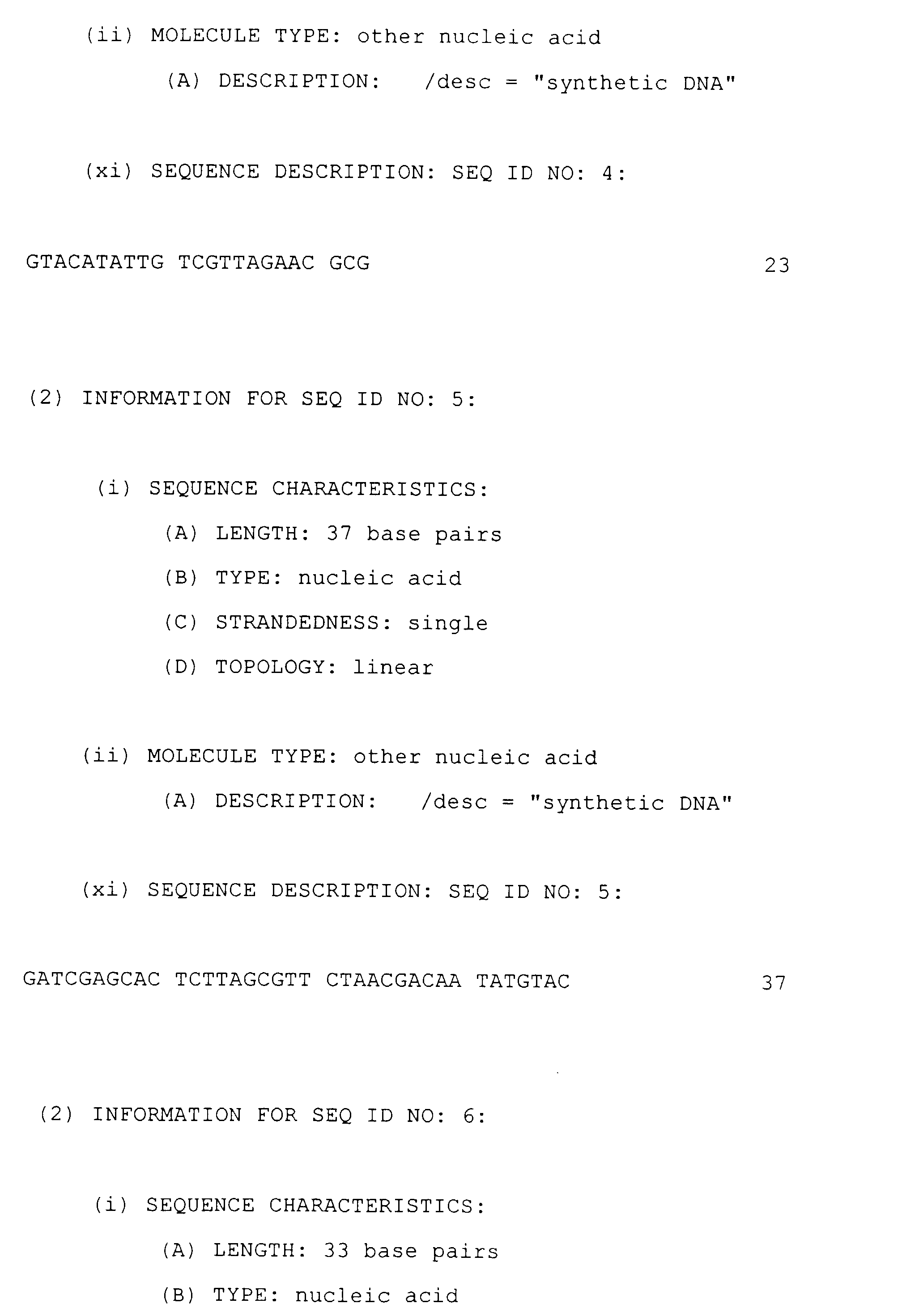
- Fig. 3 presents the results of analysis of PCR products (amplified cDNA fragments) using a sequencer.
- Fig. 4 presents the results of analysis of PCR products (amplified cDNA fragments) using a sequencer.
- the present invention relates to a method for quantitatively determining an identical gene contained in plurality of samples by amplifying the gene in one reaction system comprising the samples.
- the method of the invention is characterized by providing at least two types of samples each containing a cDNA coding for a target gene, adding a different adaptor sequence to each of the cDNAs contained in the samples, mixing equal amounts of the samples each containing the adaptor sequence-tagged cDNA, amplifying the resultant cDNAs and calculating an amount ratio between the amplified cDNAs.
- This method is called an adaptor-tagged competitive PCR (ATAC-PCR).
- At least two types of samples each containing a cDNA to be determined are prepared.
- two types of samples each containing an identical cDNA are taken as an example.
- Sample A One of the samples containing a cDNA is designated Sample A, and the other is designated Sample B.
- Preparation of the cDNA in Sample A and Sample B can be performed by any of the conventional techniques. For example, a technique of preparing poly(A) + RNA from cells of various organs and converting the RNA into cDNA with a reverse transcriptase (Gubler, U. and Hoffman, B.J., Gene, 25, 263-269 (1983); Okayama, H. and Berg, P., Mol. Cell. Biol., 18, 5294 (1982)) may be used.
- Sample A and Sample B each containing a cDNA which is the target of quantitative determination may be derived from different tissues or cells, or may be derived from an identical tissue or cell.
- Sample A may be a liver cell extract and Sample B may be a kidney cell extract.
- As the type of a cDNA to be quantitatively determined those cDNAs derived from various organ RNAs including, but not limited to, a cDNA coding for a liver-derived apolipoprotein and a cDNA coding for a kidney-derived apolipoprotein may be given.
- the amount of a target cDNA in Sample A or Sample B may be known. Alternatively, the amount of a target cDNA may be unknown in both samples.
- an adaptor means an oligonucleotide which is designed so that a cDNA can be discriminated when amplified.
- An adaptor is designed as a double-stranded oligonucleotide so that it can be ligated to the cut site of a cDNA.
- the adaptors for use in the present invention may be designed so that one to be added to a cDNA in Sample A is different from one to be added to a cDNA in Sample B in length. Alternatively, they may be designed so that at least one restriction site is contained in each of the adaptors. Alternatively, they may be designed so that one to be added to a cDNA in Sample A is different from one to be added to a cDNA in Sample B in nucleotide sequence.
- a common sequence for both adaptors (the bold line “-" in the adaptors in Fig. 1) is created. Then, a sequence of 5-15 bases is added to one of the adaptors so that they can be discriminated by length. At this time, the sequence to be added is designed so that it is located between the cohesive end of the cDNA and the binding site of an adaptor primer to be used for the subsequent amplification (see Fig. 2; the dotted line “ ... " in the adaptor for Sample B).
- a substance e.g., an antigen, an enzyme, biotin
- a specific substance an antibody, a substrate, streptavidin
- Fig. 1 illustrates biotin ("-b")-added cDNAs.
- the restriction sites to be introduced into adaptors are designed so that one adaptor contains at least one restriction site. Alternatively, the number of such sites may be determined considering the number of samples each containing a target cDNA.
- the restriction site may be a site for MluI, NotI, SalI, SfiI, XhoI or the like.
- restriction site(s) When cDNAs contained in two samples are to be quantitatively determined, either one or two restriction sites may be introduced into one adaptor.
- the restriction site(s) is(are) designed so that it(they) is(are) located between the binding site of an adaptor primer and the cohesive end of the cDNA.
- the adaptor sequences are designed so that the two adaptors have different recognition sites with each other.
- a SalI site is introduced into an adaptor to be added to the cDNA in Sample A
- a MluI site into an adaptor to be added to the cDNA in Sample B.
- the adaptor is designed so that it is cut at only one of the two restriction sites when treated with a restriction enzyme.
- the adaptors are designed so that one restriction enzyme which cuts one adaptor tagged to one cDNA will not cut the other adaptor tagged to the other cDNA.
- the adaptor tagged to the cDNA in Sample A is designed so that it is recognized by the restriction enzyme MluI alone.
- biotin or the like is added to the cDNA so that the adaptor-tagged sample alone can be recovered.
- adaptors which are different from each other in nucleotide sequence may be discretionally designed, synthesized and used.
- the target cDNA can be detected by preparing oligonucleotides which are complementary to those adaptors and thus hybridizable thereto, and labelling them.
- an adaptor to be added to a cDNA in Sample A is designated adaptor X and an adaptor to be added to a cDNA in Sample B is designated adaptor Y.
- These adaptors are designed so that the sequence of adaptor X is different from the sequence of adaptor Y except the region to which an adaptor primer is added, and so that a nucleotide hybridizing to adaptor X cannot hybridize to at least adaptor Y.
- the nucleotide sequences of adaptor X and adaptor Y can be designed discretionally and chemically synthesized.
- the lengths of the nucleotide sequences of these adaptors may be the same or different, ranging from 15 to 50 bases, preferably from 25 to 30 bases.
- the region to which an adaptor primer for subsequent amplification is to be added is common in sequence.
- the oligonucleotide to be hybridized to the adaptor should be designed and prepared so that it does not hybridize to the above-described region to which an adaptor primer is to be added. Accordingly, as long as this condition is satisfied, the region in the adaptor to which the oligonucleotide is hybridized is not particularly limited.
- Equal amounts of Sample A and Sample B each containing the adaptor-tagged cDNA are mixed. Then, amplification is carried out using the cDNAs in these samples as templates (Figs. 1 and 2). This amplification is performed, for example, by polymerase chain reaction (PCR).
- PCR polymerase chain reaction
- an adaptor primer and a gene specific primer are used as primers.
- An adaptor primer is a primer which can hybridize to the adaptors designed as described above.
- a gene specific primer is a primer which can hybridize to at least a part of the cDNA to be quantitatively determined.
- Each of these primers has a length of 20-25 bases and can be obtained, for example, by chemical synthesis.
- the number of PCR cycles and temperature conditions, etc. may be decided appropriately.
- Each of the cDNA fragments is amplified maintaining its amount ratio. Since each fragment can be discriminated by the adaptor as to whether it is derived from liver mRNA or kidney mRNA, an absolute or relative amount of expression of the cDNA can be determined from an amount ratio between the final products by the detection and quantitative determination described below.
- the amplified products are detected with an autosequencer (from Pharmacia, etc.) or an image scanner (from Molecular Dynamics) when a fluorescent label was used, or with a densitometer or the like when a radioisotope was used.
- an autosequencer from Pharmacia, etc.
- an image scanner from Molecular Dynamics
- the amount ratio between the cDNAs in two samples is calculated using the following formulas.
- the adaptors added to the cDNAs in the two samples are exchanged with each other, and then similar amplification and determination are performed to determine an amount ratio (m 1 /m 2 ) .
- b 1 m 1 /m 2 ((1+e 2 ) n /(1+e 1 ) n )
- each adaptor contains a different restriction site from each other.
- the amplified products are divided into two portions, each of which is treated with one of the relevant restriction enzymes.
- fragments with different lengths can be obtained.
- an adaptor having a SalI site alone is added to the cDNA in Sample A (derived from liver) and an adaptor having a MluI site alone is added to the cDNA in Sample B (derived from kidney) instead of the adaptors used in this Figure.
- the reaction solution is divided into two portions. One is treated with SalI and the other is treated MluI.
- a MluI site-containing fragment i.e., the cDNA derived from kidney
- a SalI site-containing fragment i.e., the cDNA derived from liver
- an amount ratio of expression between the cDNAs in both samples can be determined.
- the liver-derived cDNA is detected by SalI treatment and the kidney-derived cDNA by MluI treatment.
- an amount ratio of expression between the cDNAs is determined similarly. Thereafter, a geometric average of this ratio and the previously obtained ratio can be calculated.
- oligonucleotides are synthesized which are complementary to the oligonucleotide sequences of the individual adaptors prepared as described above, respectively, and thus specifically hybridizable thereto. As explained above, each of these oligonucleotides is designed and synthesized so that it can hybridize to one adaptor but cannot hybridize to the other adaptor. These oligonucleotides have only to hybridize to one of the two strands of the adaptors.
- the oligonucleotide sequences thus prepared are labelled.
- a labelling substance a fluorescent dye or a radioisotope is used, for example.
- the labelled oligonucleotides described above are added to the amplified products to allow hybridization of the labelled oligonucleotides to the sequences of the adaptor regions (excluding the region to which the adaptor primer is added), respectively.
- each of the amplified cDNAs having the adaptor sequence is formed as a double-stranded cDNA.
- the above hybridization is performed after the amplified products have been converted into single-stranded cDNAs.
- the denaturation from double-stranded cDNA to single-stranded cDNA may be carried out by, for example, a denaturing reaction in PCR (e.g., at 94 °C for 30 seconds).
- the hybridization may be carried out by, for example, an annealing reaction in PCR (e.g., at 55°C for 1 minute).
- the adaptor strand to which the above oligonucleotide is to be hybridized must be fixed to a solid phase in order to remove the free, labelled-oligonucleotide after hybridization.
- a combination of biotin-streptavidin or the like is used for the fixation.
- streptavidin is fixed to a solid phase, and biotin is bound to the adaptor strand to which the oligonucleotide is to be hybridized.
- the fluorescence intensity or concentration of the labelling substance is determined after the hybridization.
- an amount ratio between the cDNAs can be calculated in the same manner as described above.
- the method of the invention is applicable to the quantitatively determination of cDNA fragments classified by the so-called molecular indexing.
- the molecular indexing is a method of classifying expressed genes (cDNAs) by using specific 3 restriction enzymes (class IIS restriction enzymes, e.g., FokI, BsmAI and BamFI) and oligonucleotides to be ligated to the cut sites generated by the above restriction enzymes.
- class IIS restriction enzymes e.g., FokI, BsmAI and BamFI
- expressed genes (cDNAs) are classified into 576 groups, and the individual fragments are separated from each other (Kikuya Kato, "Discriminatory Representation Technique for Expressed Genes", Bio Industry, 13 (6), pp.16-23, 1996; Japanese Unexamined Patent Publication No. 8-322598; and U.S. Patent No. 5, 707, 807).
- At least two samples each containing a target cDNA fragment classified by the molecular indexing are mixed in equal amounts and amplified. Thereafter, an amount ratio between the cDNA fragments can be obtained.
- the oligonucleotides can be used as adaptors in the present invention. Then, an amplification reaction similar to that described above can be performed to quantitatively determine the cDNA fragment.
- the molecular indexing is a method in which a large number of fragments are amplified by PCR.
- the application of the method of the invention i.e., adaptor-tagged competitive PCR (ATAC-PCR)) thereto is advantageous, because identification of those fragments exhibiting difference in expression becomes easy even if a non-specific reaction product appears in samples in common.
- an amount ratio between a cDNA coding for mouse liver-derived apolipoprotein and a cDNA coding for mouse kidney-derived apolipoprotein was determined using mouse liver tissues and mouse kidney tissues.
- Mouse liver-derived total RNA and mouse kidney-derived total RNA were prepared separately from mouse frozen tissues (liver and kidney) by the guanidine isothiocyanate method.
- a chemically synthesized biotinylated oligo(dT) 18 primer was added and heated at 70 °C for 2-3 minutes. Then, the resultant solution was placed in a reaction solution of the following composition and kept at 37 °C for 1 hour.
- Reaction Solution 5x Reverse transcriptase buffer 4 ⁇ l 2 mM dNTP 4 ⁇ l 0.1 M DTT 2 ⁇ l 10 pmol/ ⁇ l 5'-biotinylated oligo(dT) 18 primer 1.5 ⁇ l RNase inhibitor (40 U/ ⁇ l) 0.5 ⁇ l 200 U/ ⁇ l M-MLV reverse transcriptase 1 ⁇ l
- a reaction solution of the following composition was added and reacted at 16°C for 1 hour and then at room temperature for 1 hour to synthesize double-stranded cDNA separately.
- Composition of the Reaction Solution 10 mM MgCl 2 70.5 ⁇ l 1 M Tris-HCl (pH 7.5) 10 ⁇ l 1 M (NH 4 ) 2 SO 4 1.5 ⁇ l RNase H (1U/ ⁇ l) 1.5 ⁇ l E. coli DNA polymerase (10 U/ ⁇ l) 4.5 ⁇ l
- reaction solution was heated at 75 °C for 10 minutes, diluted with 9 volumes of distilled water and then used in the adaptor addition reaction described below.
- sample A Adaptor Sample B Adaptor (a) Mouse liver cDNA MA-4 Mouse liver cDNA 3 ⁇ l MA-1 10 ⁇ l + Mouse kidney cDNA 7 ⁇ l (b) Mouse liver cDNA MA-4 Mouse liver cDNA 1 ⁇ l MA-1 10 ⁇ l + Mouse kidney cDNA 9 ⁇ l (c) Mouse liver cDNA MA-1 Mouse liver cDNA 3 ⁇ l MA-4 10 ⁇ l + Mouse kidney cDNA 7 ⁇ l (d) Mouse liver cDNA MA-1 Mouse liver cDNA 1 ⁇ l MA-4 10 ⁇ l + Mouse kidney cDNA 9 ⁇ l
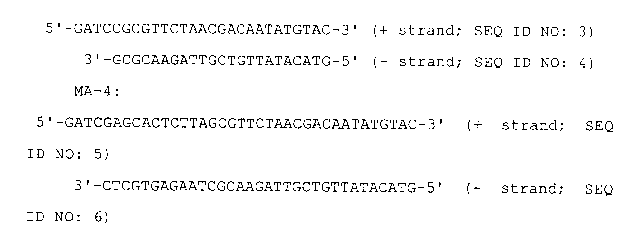
- an apolipoprotein A-1 specific primer As a gene specific primer, an apolipoprotein A-1 specific primer was used. As an adaptor primer, C1S (labelled with Cy5) was used. The sequences for individual primers are as follows: Apolipoprotein A-1 specific primer: The sequences for adaptors are as follows:
- reaction solution of the above-described composition was kept at 16 °C overnight.
- PCR cycle was as follows: 30-35 cycles of denaturation at 94°C for 30 seconds, annealing at 55°C for 1 minute and extension at 72 °C 1 minute; then, final extension at 72°C for 20 minutes.
- cDNA was synthesized in the same manner as in Example 1 using mouse liver-derived and mouse kidney-derived total RNAs.
- the following primers (called "double-anchored oligo-dT primer(s)") were used in a mixture combined in equal amounts.
- As an amount of this mixed primer 1.5 ml of 10 pmol/ ⁇ l mixed primers was used.
- reaction solution of the following composition was kept at 37°C for 50 minutes to 1 hour.
- 10x M buffer (TaKaRa) 5 ⁇ l 0.1% BSA 5 ⁇ l cDNA sample 40 ⁇ l
- Restriction enzyme FokI (10 U/ ⁇ l) 0.5 ⁇ l
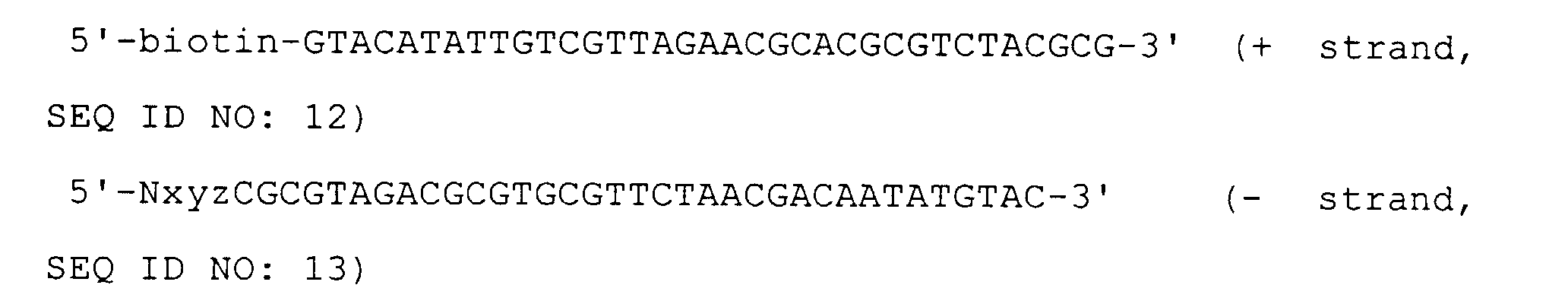
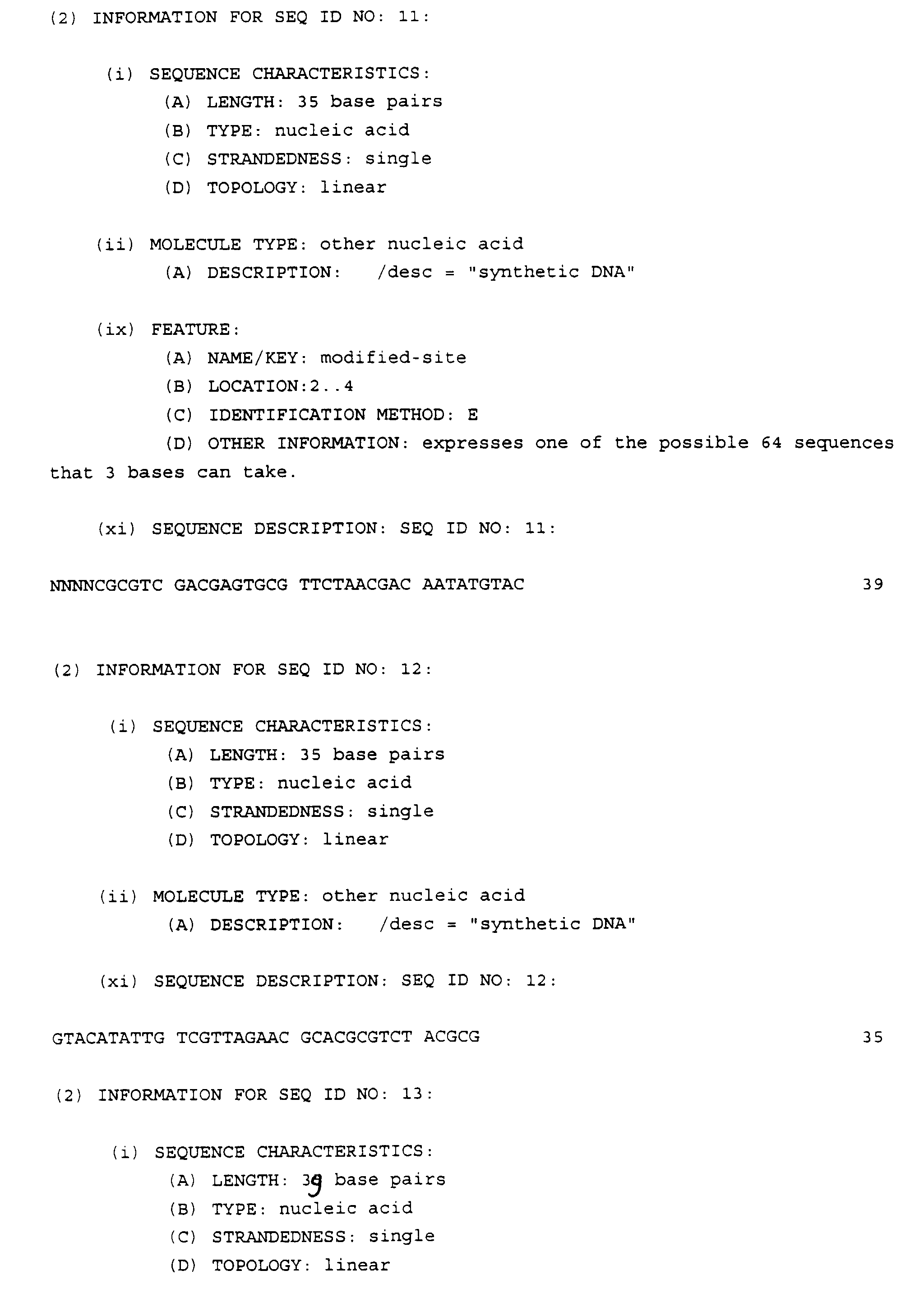
- This biotinylated adaptor has at its cohesive end a base sequence Nxyz [wherein N represents A, G, C or T (a mixture of 4 bases); and xyz represents one of the possible 64 sequences that 3 bases can take] and has a recognition site for the restriction enzyme SalI within its adaptor sequence.
- the sequences of this adaptor are as follows:
- This biotinylated adaptor has at its cohesive end a base sequence Nxyz [wherein N represents A, G, C or T (a mixture of 4 bases); and xyz represents one of the possible 64 sequences that 3 bases can take] and has a recognition site for the restriction enzyme MluI within its adaptor sequence.
- the sequences of this adaptor are as follows:
- a ligation reaction was performed in a reaction solution of the following composition. 10x E. coli ligase buffer 1 ⁇ l 100 mM (NH 4 ) 2 SO 4 1 ⁇ l 0.5 mM NAD 1 ⁇ l 10 pmol/ ⁇ l Adaptor solution 1 ⁇ l cDNA sample described above 1 ⁇ l E. coli DNA ligase 6 units
- Microtubes into which beads were to be dispensed were coated with PBS 0.1% BSA.
- the microtubes were washed with lx B&W buffer (10 mM Tris-HCl, pH 7.5, 1 M NaCl, 1 mM EDTA) twice.
- Streptavidin-coated paramagnetic beads were washed with lx B&W buffer (10 mM Tris-HCl, pH 7.5, 1 M NaCl, 1 mM EDTA) twice and suspended in an equal volume of 1x B&W buffer.
- 0.1 M NaOH was prepared by diluting 5 N NaOH.
- the PCR was performed 20 cycles, one cycle consisting of denaturation at 94°C for 1 minute, annealing at 55°C for 1 minute and extension at 72 °C 1 minute.
- a PCR was performed in a reaction solution of the following composition.
- Enzyme Reaction Solution 10x PCR buffer for Stoffel fragment 1 ⁇ l 2 mM dNTP 1 ⁇ l 25 mM MgCl 2 1.2 ⁇ l Distilled water 3.7 ⁇ l 10 U/ ⁇ l Stoffel fragment 0.13 ⁇ l 10 pmol/ ⁇ l Texas Red-labelled C1S 1 ⁇ l 10 pmol/ ⁇ l double-anchored primer (A, C or G) 2 ⁇ l cDNA sample mixture treated with restriction enzymes in (6) above 1 ⁇ l
- the PCR was performed 20 cycles, one cycle consisting of denaturation at 94°C for 1 minute, annealing at 55°C for 1 minute and extension at 72 °C 1 minute.
- a T4DPase mix of the following composition was added to the reaction solution, which was then kept at 37°C for 1 hour.
- Composition of T4DPase Solution 10x M buffer 0.5 ⁇ l 2 mM dNTP 0.5 ⁇ l Distilled water 4 ⁇ l T4 DNA polymerase 0.5 unit
- the resultant solution was heated at 72°C for 10 minutes and then subjected to dialysis. 2 ⁇ l of the dialysate was thermally denatured and applied to a Hitachi automatic sequencer.
- Fig. 4 The results are shown in Fig. 4, in which 1) is representation on fluorograms and 2) is representation on electropherograms(the vertical axis represents fluorescence intensity and the horizontal axis represents strand length in time).
- A in the Fig. represents the fluorescence intensity of a target fragment of detection.
- B in the Fig. represent the fluorescence intensity of a non-specifically expressed fragment appearing in both samples compared.
- the fluorescence intensity of each of the amplified fragments is shown in Table 3 below.
- the fluorescence intensity of the target fragment was corrected with the fluorescence intensity of the non-specific fragment expressed in both samples to be compared.
- Fluorescence Intensity A Fluorescence Intensity B Corrected Value (A/B) a 500 1896 0.264 b 127 1944 0.0653 c 88 1940 0.0454 d 467 2340 0.200
- the ratio of the expression of a target fragment in liver to the expression thereof in kidney is expressed by a/b and by d/c. According to the corrected values, a/b is 4.04 and d/c is 4.41. Thus, the geometric average of those amount ratios is 4.21. Accordingly, the method of the invention could be successfully applied to molecular indexing to obtain an amount ratio between amplified products.
- a method for quantitatively determining the expression of a gene is provided.
- the method of the present invention is especially useful for handling a large number of samples such as clinical samples, or quantitatively determining a large number of genes.
Abstract
Description
| Composition of the Reaction Solution | |
| 5x Reverse transcriptase buffer | 4 µl |
| 2 mM dNTP | 4 µl |
| 0.1 M DTT | 2 µl |
| 10 pmol/µl 5'-biotinylated oligo(dT)18 primer | 1.5 µl |
| RNase inhibitor (40 U/µl) | 0.5 µl |
| 200 U/µl M-MLV reverse transcriptase | 1 µl |
| Composition of the Reaction Solution | |
| 10 mM MgCl2 | 70.5 µl |
| 1 M Tris-HCl (pH 7.5) | 10 µl |
| 1 M (NH4)2SO4 | 1.5 µl |
| RNase H (1U/µl) | 1.5 µl |
| E. coli DNA polymerase (10 U/µl) | 4.5 µl |
| 10x M buffer | 5 µl |
| cDNA sample obtained in (2) | 45 µl |
| Restriction enzyme MboI | 5 U |
| 10x Ligation buffer | 1.5 µl |
| 10 pmol/µl Adaptor MA-1 or MA-4 | 3 µl |
| Mouse liver-derived and/or kidney-derived cDNA (see Table 1) | 10 µl |
| 350 U/µl T4 DNA ligase | 0.5 µl |
| Sample A | Adaptor | Sample B | Adaptor | |
| (a) | Mouse liver cDNA | MA-4 | Mouse liver cDNA 3 µl | MA-1 |
| 10 µl | + | |||
| Mouse kidney cDNA 7µl | ||||
| (b) | Mouse liver cDNA | MA-4 | Mouse liver cDNA 1 µl | MA-1 |
| 10 µl | + | |||
| Mouse kidney cDNA 9µl | ||||
| (c) | Mouse liver cDNA | MA-1 | Mouse liver cDNA 3 µl | MA-4 |
| 10 µl | + | |||
| Mouse kidney cDNA 7µl | ||||
| (d) | Mouse liver cDNA | MA-1 | Mouse liver cDNA 1 µl | MA-4 |
| 10 µl | + | |||
| Mouse kidney cDNA 9µl |
Apolipoprotein A-1 specific primer:

| 10x Buffer for Stoffel fragment | 1 µl |
| 25 mM MgCl2 | 1.5 µl |
| 2 mM dNTP | 1 µl |
| 10 pmol/µl Cy5-labelled C1S | 0.25 µl |
| 10 pmol/µl Apolipoprotein A-1 specific primer | 0.25 µl |
| Stoffel fragments | 1 unit |
| Distilled water to make 10 µl |
| 10x M buffer (TaKaRa) | 0.5 µl |
| Distilled water | 4.5 µl |
| T4 DNA polymerase | 0.1 unit |
| MA-1-added Fragments | MA-4-added Fragments | Amount Ratio | |
| (a) | 2432.5 | 5535.5 | 0.439 |
| (b) | 542.27 | 4615.5 | 0.117 |
| (c) | 5246.2 | 1459.5 | 0.278 |
| (d) | 6146.2 | 525.02 | 0.085 |

| 10x M buffer (TaKaRa) | 5 µl |
| 0.1% BSA | 5 µl |
| cDNA sample | 40 µl |
| Restriction enzyme FokI (10 U/µl) | 0.5 µl |

| 10x E. coli ligase buffer | 1 µl |
| 100 mM (NH4)2SO4 | 1 µl |
| 0.5 mM NAD | 1 µl |
| 10 pmol/µl Adaptor solution | 1 µl |
| cDNA sample described above | 1 µl |
| E. coli DNA ligase | 6 units |
| Enzyme Reaction Solution | |
| 10x PCR buffer for Stoffel fragment | 1 µl |
| 2 mM dNTP | 1 µl |
| 25 mM MgCl2 | 1.2µl |
| Distilled water | 3.7 µl |
| 10 U/µl Stoffel fragment | 0.13µl |
| 10 pmol/µl unlabelled C1S | 1 µl |
| 10 pmol/µl double-anchored primer (A, C or G) | 2 µl |
| Enzyme Reaction Solution | |
| 10x PCR buffer for Stoffel fragment | 1 µl |
| 2 mM dNTP | 1 µl |
| 25 mM MgCl2 | 1.2µl |
| Distilled water | 3.7µl |
| 10 U/µl Stoffel fragment | 0.13µl |
| 10 pmol/µl Texas Red-labelled C1S | 1 µl |
| 10 pmol/µl double-anchored primer (A, C or G) | 2 µl |
| cDNA sample mixture treated with restriction | |
| enzymes in (6) above | 1 µl |
| Composition of T4DPase Solution (per Sample) | |
| 10x M buffer | 0.5 µl |
| 2 mM dNTP | 0.5 µl |
| Distilled water | 4 µl |
| T4 DNA polymerase | 0.5 unit |
| Fluorescence Intensity A | Fluorescence Intensity B | Corrected Value (A/B) | |
| a | 500 | 1896 | 0.264 |
| b | 127 | 1944 | 0.0653 |
| c | 88 | 1940 | 0.0454 |
| d | 467 | 2340 | 0.200 |


Claims (3)
- A method for quantitatively determining the expression of a gene, comprising providing at least two types of samples each containing a cDNA coding for the gene, adding a different adaptor to each of the cDNAs contained in the samples, mixing equal amounts of the samples each containing the adaptor-tagged cDNA, amplifying the resultant cDNAs and calculating an amount ratio between the amplified products.
- The method of claim 1, wherein said different adaptors comprise nucleotides different from each other in length or nucleotides each having at least one restriction site.
- The method of claim 1, wherein said different adaptors comprise nucleotides different from each other in sequence.
Applications Claiming Priority (3)
| Application Number | Priority Date | Filing Date | Title |
|---|---|---|---|
| JP88495/97 | 1997-04-07 | ||
| JP8849597 | 1997-04-07 | ||
| JP8849597 | 1997-04-07 |
Publications (3)
| Publication Number | Publication Date |
|---|---|
| EP0870842A2 true EP0870842A2 (en) | 1998-10-14 |
| EP0870842A3 EP0870842A3 (en) | 1999-01-20 |
| EP0870842B1 EP0870842B1 (en) | 2004-02-11 |
Family
ID=13944405
Family Applications (1)
| Application Number | Title | Priority Date | Filing Date |
|---|---|---|---|
| EP98302726A Expired - Lifetime EP0870842B1 (en) | 1997-04-07 | 1998-04-07 | Adaptor-tagged competitive PCR |
Country Status (3)
| Country | Link |
|---|---|
| US (1) | US6090556A (en) |
| EP (1) | EP0870842B1 (en) |
| DE (1) | DE69821540T2 (en) |
Cited By (26)
| Publication number | Priority date | Publication date | Assignee | Title |
|---|---|---|---|---|
| GB2365124A (en) * | 2000-07-21 | 2002-02-13 | Karolinska Innovations Ab | Analysis and identification of transcribed genes, and fingerprinting |
| WO2002061145A2 (en) * | 2001-01-31 | 2002-08-08 | Ambion, Inc. | Competitive amplification of fractionated targets from multiple nucleic acid samples |
| WO2002061140A2 (en) * | 2001-01-31 | 2002-08-08 | Ambion, Inc. | Competitive population normalization for comparative analysis of nucleic acid samples |
| WO2002061143A2 (en) * | 2001-01-31 | 2002-08-08 | Ambion, Inc. | Comparative analysis of nucleic acids using population tagging |
| WO2002064835A2 (en) * | 2001-01-31 | 2002-08-22 | Ambion, Inc. | Methods for nucleic acid fingerprint analysis |
| WO2002090564A1 (en) * | 2001-05-07 | 2002-11-14 | Fox Chase Cancer Center | Methods for gene expression profiling |
| WO2002103007A1 (en) * | 2001-06-19 | 2002-12-27 | Eisai C0. Ltd. | Method of uniformizing dna fragment contents and subtraction method |
| WO2004029292A1 (en) * | 2002-09-26 | 2004-04-08 | Aisin Seiki Kabushiki Kaisha | Method of recovering dna |
| WO2004065628A1 (en) * | 2003-01-21 | 2004-08-05 | Guoliang Fu | Quantitative multiplex detection of nucleic acids |
| GB2414797A (en) * | 2003-01-21 | 2005-12-07 | Guoliang Fu | Quantitative multiplex detection of nucleic acids |
| US7253286B2 (en) | 2000-10-20 | 2007-08-07 | Eisai Co., Ltd | Nitrogen-containing aromatic derivatives |
| WO2008137466A2 (en) * | 2007-05-01 | 2008-11-13 | Asuragen, Inc. | Nucleic acid quantitation methods |
| US7994159B2 (en) | 2003-03-10 | 2011-08-09 | Eisai R&D Management Co., Ltd. | c-Kit kinase inhibitor |
| US8058474B2 (en) | 2003-11-11 | 2011-11-15 | Eisai R&D Management Co., Ltd. | Urea derivative and process for preparing the same |
| US8071562B2 (en) * | 2007-12-01 | 2011-12-06 | Mirna Therapeutics, Inc. | MiR-124 regulated genes and pathways as targets for therapeutic intervention |
| US8765709B2 (en) | 2004-11-12 | 2014-07-01 | Asuragen, Inc. | Methods and compositions involving miRNA and miRNA inhibitor molecules |
| US9222085B2 (en) | 2011-02-03 | 2015-12-29 | Mirna Therapeutics, Inc. | Synthetic mimics of MIR-124 |
| US9365852B2 (en) | 2008-05-08 | 2016-06-14 | Mirna Therapeutics, Inc. | Compositions and methods related to miRNA modulation of neovascularization or angiogenesis |
| US9644241B2 (en) | 2011-09-13 | 2017-05-09 | Interpace Diagnostics, Llc | Methods and compositions involving miR-135B for distinguishing pancreatic cancer from benign pancreatic disease |
| US9945862B2 (en) | 2011-06-03 | 2018-04-17 | Eisai R&D Management Co., Ltd. | Biomarkers for predicting and assessing responsiveness of thyroid and kidney cancer subjects to lenvatinib compounds |
| US10047388B2 (en) | 2004-05-28 | 2018-08-14 | Asuragen, Inc. | Methods and compositions involving MicroRNA |
| US10259791B2 (en) | 2014-08-28 | 2019-04-16 | Eisai R&D Management Co., Ltd. | High-purity quinoline derivative and method for manufacturing same |
| US10517861B2 (en) | 2013-05-14 | 2019-12-31 | Eisai R&D Management Co., Ltd. | Biomarkers for predicting and assessing responsiveness of endometrial cancer subjects to lenvatinib compounds |
| US11090386B2 (en) | 2015-02-25 | 2021-08-17 | Eisai R&D Management Co., Ltd. | Method for suppressing bitterness of quinoline derivative |
| US11369623B2 (en) | 2015-06-16 | 2022-06-28 | Prism Pharma Co., Ltd. | Anticancer combination of a CBP/catenin inhibitor and an immune checkpoint inhibitor |
| US11547705B2 (en) | 2015-03-04 | 2023-01-10 | Merck Sharp & Dohme Llc | Combination of a PD-1 antagonist and a VEGF-R/FGFR/RET tyrosine kinase inhibitor for treating cancer |
Families Citing this family (41)
| Publication number | Priority date | Publication date | Assignee | Title |
|---|---|---|---|---|
| US6248534B1 (en) * | 1999-05-25 | 2001-06-19 | Kikuya Kato | Method for determining the detection value of a target RNA |
| EP1334113A4 (en) * | 2000-10-20 | 2007-08-08 | Expression Diagnostics Inc | Leukocyte expression profiling |
| US7026121B1 (en) | 2001-06-08 | 2006-04-11 | Expression Diagnostics, Inc. | Methods and compositions for diagnosing and monitoring transplant rejection |
| US6905827B2 (en) | 2001-06-08 | 2005-06-14 | Expression Diagnostics, Inc. | Methods and compositions for diagnosing or monitoring auto immune and chronic inflammatory diseases |
| US7235358B2 (en) | 2001-06-08 | 2007-06-26 | Expression Diagnostics, Inc. | Methods and compositions for diagnosing and monitoring transplant rejection |
| US20030170700A1 (en) * | 2002-01-09 | 2003-09-11 | Lynx Therapeutics, Inc. | Secreted and cell surface polypeptides affected by cholesterol and uses thereof |
| US20030166026A1 (en) * | 2002-01-09 | 2003-09-04 | Lynx Therapeutics, Inc. | Identification of specific biomarkers for breast cancer cells |
| CN1182256C (en) * | 2002-08-09 | 2004-12-29 | 周国华 | Gene expression amount comparing analysis method |
| US7892745B2 (en) | 2003-04-24 | 2011-02-22 | Xdx, Inc. | Methods and compositions for diagnosing and monitoring transplant rejection |
| US7645575B2 (en) | 2004-09-08 | 2010-01-12 | Xdx, Inc. | Genes useful for diagnosing and monitoring inflammation related disorders |
| AU2005283422C1 (en) | 2004-09-17 | 2017-02-02 | Eisai R & D Management Co., Ltd. | Medicinal composition |
| US20070092886A1 (en) * | 2005-03-22 | 2007-04-26 | Raymond Tabibiazar | Methods and compositions for diagnosis, monitoring and development of therapeutics for treatment of atherosclerotic disease |
| ATE491045T1 (en) | 2005-06-23 | 2010-12-15 | Keygene Nv | HIGH-THROUGHPUT STRATEGIES FOR IDENTIFICATION AND DETECTION OF POLYMORPHISMS |
| EP1925676A4 (en) | 2005-08-02 | 2010-11-10 | Eisai R&D Man Co Ltd | Method for assay on the effect of vascularization inhibitor |
| CN1908189A (en) * | 2005-08-02 | 2007-02-07 | 博奥生物有限公司 | Method of external assistant identifying intestinal-type gastric cancer and differentiation degree thereof and special reagent case |
| US10316364B2 (en) | 2005-09-29 | 2019-06-11 | Keygene N.V. | Method for identifying the source of an amplicon |
| JP5237099B2 (en) | 2005-09-29 | 2013-07-17 | キージーン ナムローゼ フェンノートシャップ | High-throughput screening of mutated populations |
| WO2007063807A1 (en) * | 2005-11-29 | 2007-06-07 | Olympus Corporation | Method of analyzing change in primary structure of nucleic acid |
| WO2007073165A1 (en) | 2005-12-22 | 2007-06-28 | Keygene N.V. | Method for high-throughput aflp-based polymorphism detection |
| ES2545264T3 (en) | 2006-04-04 | 2015-09-09 | Keygene N.V. | High performance detection of molecular markers based on restriction fragments |
| CN101443009A (en) | 2006-05-18 | 2009-05-27 | 卫材R&D管理有限公司 | Antitumor agent for thyroid cancer |
| US7993832B2 (en) | 2006-08-14 | 2011-08-09 | Xdx, Inc. | Methods and compositions for diagnosing and monitoring the status of transplant rejection and immune disorders |
| EP2065372B1 (en) | 2006-08-28 | 2012-11-28 | Eisai R&D Management Co., Ltd. | Antitumor agent for undifferentiated gastric cancer |
| WO2008140484A2 (en) | 2006-11-09 | 2008-11-20 | Xdx, Inc. | Methods for diagnosing and monitoring the status of systemic lupus erythematosus |
| US20080187928A1 (en) * | 2006-12-29 | 2008-08-07 | The Salk Institute For Biological Studies | Methods for enhancing exercise performance |
| WO2008093855A1 (en) | 2007-01-29 | 2008-08-07 | Eisai R & D Management Co., Ltd. | Composition for treatment of undifferentiated-type of gastric cancer |
| US8361714B2 (en) | 2007-09-14 | 2013-01-29 | Asuragen, Inc. | Micrornas differentially expressed in cervical cancer and uses thereof |
| CA2704000C (en) | 2007-11-09 | 2016-12-13 | Eisai R&D Management Co., Ltd. | Combination of anti-angiogenic substance and anti-tumor platinum complex |
| ES2573515T3 (en) | 2010-06-25 | 2016-06-08 | Eisai R&D Management Co., Ltd. | Anti-tumor agent that uses compounds with combined kinase inhibitory effect |
| RU2580609C2 (en) | 2011-04-18 | 2016-04-10 | Эйсай Ар Энд Ди Менеджмент Ко., Лтд. | Anticancer therapeutic agent |
| US10100092B2 (en) | 2011-11-03 | 2018-10-16 | Vca, Inc. | Compositions and methods to detect various infectious organisms |
| EP2800820B1 (en) | 2011-12-19 | 2020-04-08 | Valley Health System | Methods and kits for detecting subjects having pancreatic cancer |
| EP3182126A3 (en) | 2012-06-15 | 2017-08-02 | Wayne State University | Biomarker test for prediction or early detection of preeclampsia and/or hellp syndrome |
| EP3339861B8 (en) | 2012-06-15 | 2023-11-01 | Genesis Theranostix Korlatolt Felelossegu Tarsasag | Biomarker test for prediction or early detection of preeclampsia |
| ES2765573T3 (en) | 2012-08-13 | 2020-06-09 | Univ Rockefeller | Melanoma treatment and diagnosis |
| ES2782825T3 (en) | 2012-10-31 | 2020-09-16 | Univ Rockefeller | Metastatic Colon Cancer Treatment |
| AU2013364953A1 (en) | 2012-12-21 | 2015-04-30 | Eisai R&D Management Co., Ltd. | Amorphous form of quinoline derivative, and method for producing same |
| WO2015187612A1 (en) | 2014-06-02 | 2015-12-10 | Valley Health System | Method and systems for lung cancer diagnosis |
| CA3078981A1 (en) | 2017-11-21 | 2019-05-31 | Rgenix, Inc. | Polymorphs and uses thereof |
| EP3660172A1 (en) | 2018-11-28 | 2020-06-03 | Bioscreening and Diagnostics LLC | Method for detection of traumatic brain injury (tbi) |
| WO2021119397A1 (en) | 2019-12-13 | 2021-06-17 | Rgenix, Inc. | Metal salts and uses thereof |
Citations (3)
| Publication number | Priority date | Publication date | Assignee | Title |
|---|---|---|---|---|
| EP0534858A1 (en) * | 1991-09-24 | 1993-03-31 | Keygene N.V. | Selective restriction fragment amplification : a general method for DNA fingerprinting |
| WO1994023023A1 (en) * | 1993-04-06 | 1994-10-13 | University Of Rochester | Method for quantitative measurement of gene expression using multiplex competitive reverse transcriptase-polymerase chain reaction |
| WO1997005286A1 (en) * | 1995-08-01 | 1997-02-13 | Yale University | ANALYSIS OF GENE EXPRESSION BY DISPLAY OF 3'-END RESTRICTION FRAGMENTS OF cDNAs |
Family Cites Families (2)
| Publication number | Priority date | Publication date | Assignee | Title |
|---|---|---|---|---|
| RU2111254C1 (en) * | 1994-07-11 | 1998-05-20 | Институт молекулярной биологии им.В.А.Энгельгардта РАН | Method of detection of differentially expressing template rnas and cloning the corresponding cdna fragments |
| DE69621507T2 (en) * | 1995-03-28 | 2003-01-09 | Japan Science & Tech Corp | Method for molecular indexing of genes using restriction enzymes |
-
1998
- 1998-04-06 US US09/056,052 patent/US6090556A/en not_active Expired - Fee Related
- 1998-04-07 EP EP98302726A patent/EP0870842B1/en not_active Expired - Lifetime
- 1998-04-07 DE DE69821540T patent/DE69821540T2/en not_active Expired - Fee Related
Patent Citations (3)
| Publication number | Priority date | Publication date | Assignee | Title |
|---|---|---|---|---|
| EP0534858A1 (en) * | 1991-09-24 | 1993-03-31 | Keygene N.V. | Selective restriction fragment amplification : a general method for DNA fingerprinting |
| WO1994023023A1 (en) * | 1993-04-06 | 1994-10-13 | University Of Rochester | Method for quantitative measurement of gene expression using multiplex competitive reverse transcriptase-polymerase chain reaction |
| WO1997005286A1 (en) * | 1995-08-01 | 1997-02-13 | Yale University | ANALYSIS OF GENE EXPRESSION BY DISPLAY OF 3'-END RESTRICTION FRAGMENTS OF cDNAs |
Non-Patent Citations (3)
| Title |
|---|
| K. KATO "Adaptor-tagged competitive PCR: a novel method for measuring relative gene expression", Nucleic Acids Research, November 1997, Vol. 25, No. 22, pages 4694-4696, XP002900285 * |
| KATO K.: 'Description of the entire mRNA population by a 3'-end cDNA fragment generated by class IIS restriction enzymes' NUCLEIC ACIDS RESEARCH vol. 23, 1995, pages 3685 - 3690 * |
| ZENILMAN M.E.: 'Competitive reverse-transcriptase polymerase chain reaction without an artificial internal standard' ANALYTICAL BIOCHEMISTRY vol. 224, 1995, pages 339 - 346 * |
Cited By (45)
| Publication number | Priority date | Publication date | Assignee | Title |
|---|---|---|---|---|
| GB2365124B (en) * | 2000-07-21 | 2002-05-01 | Karolinska Innovations Ab | Methods for analysis and identification of transcribed genes and fingerprinting |
| GB2365124A (en) * | 2000-07-21 | 2002-02-13 | Karolinska Innovations Ab | Analysis and identification of transcribed genes, and fingerprinting |
| US7612092B2 (en) | 2000-10-20 | 2009-11-03 | Eisai R & D Management Co., Ltd. | Nitrogen-containing aromatic derivatives |
| US7973160B2 (en) | 2000-10-20 | 2011-07-05 | Eisai R&D Management Co., Ltd. | Nitrogen-containing aromatic derivatives |
| US7253286B2 (en) | 2000-10-20 | 2007-08-07 | Eisai Co., Ltd | Nitrogen-containing aromatic derivatives |
| US8372981B2 (en) | 2000-10-20 | 2013-02-12 | Eisai R&D Management Co., Ltd. | Nitrogen-containing aromatic derivatives |
| WO2002061143A3 (en) * | 2001-01-31 | 2003-03-27 | Ambion Inc | Comparative analysis of nucleic acids using population tagging |
| WO2002061145A3 (en) * | 2001-01-31 | 2003-07-24 | Ambion Inc | Competitive amplification of fractionated targets from multiple nucleic acid samples |
| WO2002061140A3 (en) * | 2001-01-31 | 2003-10-16 | Ambion Inc | Competitive population normalization for comparative analysis of nucleic acid samples |
| WO2002064835A3 (en) * | 2001-01-31 | 2003-12-11 | Ambion Inc | Methods for nucleic acid fingerprint analysis |
| WO2002064835A2 (en) * | 2001-01-31 | 2002-08-22 | Ambion, Inc. | Methods for nucleic acid fingerprint analysis |
| WO2002061143A2 (en) * | 2001-01-31 | 2002-08-08 | Ambion, Inc. | Comparative analysis of nucleic acids using population tagging |
| WO2002061140A2 (en) * | 2001-01-31 | 2002-08-08 | Ambion, Inc. | Competitive population normalization for comparative analysis of nucleic acid samples |
| WO2002061145A2 (en) * | 2001-01-31 | 2002-08-08 | Ambion, Inc. | Competitive amplification of fractionated targets from multiple nucleic acid samples |
| WO2002090564A1 (en) * | 2001-05-07 | 2002-11-14 | Fox Chase Cancer Center | Methods for gene expression profiling |
| WO2002103007A1 (en) * | 2001-06-19 | 2002-12-27 | Eisai C0. Ltd. | Method of uniformizing dna fragment contents and subtraction method |
| WO2004029292A1 (en) * | 2002-09-26 | 2004-04-08 | Aisin Seiki Kabushiki Kaisha | Method of recovering dna |
| GB2414797A (en) * | 2003-01-21 | 2005-12-07 | Guoliang Fu | Quantitative multiplex detection of nucleic acids |
| WO2004065628A1 (en) * | 2003-01-21 | 2004-08-05 | Guoliang Fu | Quantitative multiplex detection of nucleic acids |
| US7994159B2 (en) | 2003-03-10 | 2011-08-09 | Eisai R&D Management Co., Ltd. | c-Kit kinase inhibitor |
| US8058474B2 (en) | 2003-11-11 | 2011-11-15 | Eisai R&D Management Co., Ltd. | Urea derivative and process for preparing the same |
| US10047388B2 (en) | 2004-05-28 | 2018-08-14 | Asuragen, Inc. | Methods and compositions involving MicroRNA |
| US9447414B2 (en) | 2004-11-12 | 2016-09-20 | Asuragen, Inc. | Methods and compositions involving miRNA and miRNA inhibitor molecules |
| US8765709B2 (en) | 2004-11-12 | 2014-07-01 | Asuragen, Inc. | Methods and compositions involving miRNA and miRNA inhibitor molecules |
| US9051571B2 (en) | 2004-11-12 | 2015-06-09 | Asuragen, Inc. | Methods and compositions involving miRNA and miRNA inhibitor molecules |
| US9068219B2 (en) | 2004-11-12 | 2015-06-30 | Asuragen, Inc. | Methods and compositions involving miRNA and miRNA inhibitor molecules |
| US9506061B2 (en) | 2004-11-12 | 2016-11-29 | Asuragen, Inc. | Methods and compositions involving miRNA and miRNA inhibitor molecules |
| US9382537B2 (en) | 2004-11-12 | 2016-07-05 | Asuragen, Inc. | Methods and compositions involving miRNA and miRNA inhibitor molecules |
| WO2008137466A3 (en) * | 2007-05-01 | 2009-03-12 | Asuragen Inc | Nucleic acid quantitation methods |
| WO2008137466A2 (en) * | 2007-05-01 | 2008-11-13 | Asuragen, Inc. | Nucleic acid quantitation methods |
| US8071562B2 (en) * | 2007-12-01 | 2011-12-06 | Mirna Therapeutics, Inc. | MiR-124 regulated genes and pathways as targets for therapeutic intervention |
| US9365852B2 (en) | 2008-05-08 | 2016-06-14 | Mirna Therapeutics, Inc. | Compositions and methods related to miRNA modulation of neovascularization or angiogenesis |
| US9222085B2 (en) | 2011-02-03 | 2015-12-29 | Mirna Therapeutics, Inc. | Synthetic mimics of MIR-124 |
| US9945862B2 (en) | 2011-06-03 | 2018-04-17 | Eisai R&D Management Co., Ltd. | Biomarkers for predicting and assessing responsiveness of thyroid and kidney cancer subjects to lenvatinib compounds |
| US11598776B2 (en) | 2011-06-03 | 2023-03-07 | Eisai R&D Management Co., Ltd. | Biomarkers for predicting and assessing responsiveness of thyroid and kidney cancer subjects to lenvatinib compounds |
| US9644241B2 (en) | 2011-09-13 | 2017-05-09 | Interpace Diagnostics, Llc | Methods and compositions involving miR-135B for distinguishing pancreatic cancer from benign pancreatic disease |
| US10655184B2 (en) | 2011-09-13 | 2020-05-19 | Interpace Diagnostics, Llc | Methods and compositions involving miR-135b for distinguishing pancreatic cancer from benign pancreatic disease |
| US10517861B2 (en) | 2013-05-14 | 2019-12-31 | Eisai R&D Management Co., Ltd. | Biomarkers for predicting and assessing responsiveness of endometrial cancer subjects to lenvatinib compounds |
| US10407393B2 (en) | 2014-08-28 | 2019-09-10 | Eisai R&D Management Co., Ltd. | High-purity quinoline derivative and method for manufacturing same |
| US10822307B2 (en) | 2014-08-28 | 2020-11-03 | Eisai R&D Management Co., Ltd. | High-purity quinoline derivative and method for manufacturing same |
| US11186547B2 (en) | 2014-08-28 | 2021-11-30 | Eisai R&D Management Co., Ltd. | High-purity quinoline derivative and method for manufacturing same |
| US10259791B2 (en) | 2014-08-28 | 2019-04-16 | Eisai R&D Management Co., Ltd. | High-purity quinoline derivative and method for manufacturing same |
| US11090386B2 (en) | 2015-02-25 | 2021-08-17 | Eisai R&D Management Co., Ltd. | Method for suppressing bitterness of quinoline derivative |
| US11547705B2 (en) | 2015-03-04 | 2023-01-10 | Merck Sharp & Dohme Llc | Combination of a PD-1 antagonist and a VEGF-R/FGFR/RET tyrosine kinase inhibitor for treating cancer |
| US11369623B2 (en) | 2015-06-16 | 2022-06-28 | Prism Pharma Co., Ltd. | Anticancer combination of a CBP/catenin inhibitor and an immune checkpoint inhibitor |
Also Published As
| Publication number | Publication date |
|---|---|
| EP0870842B1 (en) | 2004-02-11 |
| DE69821540D1 (en) | 2004-03-18 |
| DE69821540T2 (en) | 2004-12-30 |
| US6090556A (en) | 2000-07-18 |
| EP0870842A3 (en) | 1999-01-20 |
Similar Documents
| Publication | Publication Date | Title |
|---|---|---|
| US6090556A (en) | Method for quantitatively determining the expression of a gene | |
| EP0777747B1 (en) | Nucleotide sequencing method | |
| EP3559274B1 (en) | Reagents and methods for the analysis of linked nucleic acids | |
| JP5456950B2 (en) | Multiplex amplification of short tandem repeat loci | |
| US6235483B1 (en) | Methods and kits for indirect labeling of nucleic acids | |
| JP5838169B2 (en) | Method for generating uniquely specific nucleic acid probes | |
| EP0791074B1 (en) | Method for the detection of ras oncogenes, in particular the k-ras ancogene | |
| WO2013192292A1 (en) | Massively-parallel multiplex locus-specific nucleic acid sequence analysis | |
| JP2002518060A (en) | Nucleotide detection method | |
| JP2004521634A (en) | Mutation detection using MutS and RecA | |
| EP0849363B1 (en) | Method of identifying a nucleic acid using triple helix formation of adjacently annealed probes | |
| EP3775269A1 (en) | Integrative dna and rna library preparations and uses thereof | |
| JP2021512597A (en) | Preparation of single-stranded circular DNA templates for single molecules | |
| CN110869515A (en) | Sequencing method for genome rearrangement detection | |
| JP2008534011A (en) | Unique sequence hybridization probe (USP) | |
| US20220042096A1 (en) | Flexible and high-throughput sequencing of targeted genomic regions | |
| US20050009047A1 (en) | Global linear non-biased nucleic acid amplification | |
| WO1994017206A1 (en) | Method for amplifying nucleic acid sequences | |
| JP2905192B2 (en) | Gene expression quantification method | |
| JP2003510011A (en) | Coupled polymerase chain reaction-restriction endonuclease digestion-ligase detection reaction | |
| US6017739A (en) | Method and nucleic acid-concentratiing assay kit for concentrating mutant nucleic acid | |
| US20220154268A1 (en) | System and Methods for Detection of Low-Copy Number Nucleic Acids and Protein | |
| EP4012029A1 (en) | Method for capturing nucleic acid molecule, preparation method for nucleic acid library, and a sequencing method | |
| KR20180053679A (en) | A multi-valued probe with a single nucleotide resolution | |
| US20040009483A1 (en) | Method of linear mRNA amplification using total RNA |
Legal Events
| Date | Code | Title | Description |
|---|---|---|---|
| PUAI | Public reference made under article 153(3) epc to a published international application that has entered the european phase |
Free format text: ORIGINAL CODE: 0009012 |
|
| 17P | Request for examination filed |
Effective date: 19980427 |
|
| AK | Designated contracting states |
Kind code of ref document: A2 Designated state(s): DE FR GB |
|
| AX | Request for extension of the european patent |
Free format text: AL;LT;LV;MK;RO;SI |
|
| PUAL | Search report despatched |
Free format text: ORIGINAL CODE: 0009013 |
|
| AK | Designated contracting states |
Kind code of ref document: A3 Designated state(s): AT BE CH CY DE DK ES FI FR GB GR IE IT LI LU MC NL PT SE |
|
| AX | Request for extension of the european patent |
Free format text: AL;LT;LV;MK;RO;SI |
|
| AKX | Designation fees paid |
Free format text: DE FR GB |
|
| 17Q | First examination report despatched |
Effective date: 20011221 |
|
| GRAP | Despatch of communication of intention to grant a patent |
Free format text: ORIGINAL CODE: EPIDOSNIGR1 |
|
| GRAS | Grant fee paid |
Free format text: ORIGINAL CODE: EPIDOSNIGR3 |
|
| GRAA | (expected) grant |
Free format text: ORIGINAL CODE: 0009210 |
|
| AK | Designated contracting states |
Kind code of ref document: B1 Designated state(s): DE FR GB |
|
| REG | Reference to a national code |
Ref country code: GB Ref legal event code: FG4D |
|
| REF | Corresponds to: |
Ref document number: 69821540 Country of ref document: DE Date of ref document: 20040318 Kind code of ref document: P |
|
| ET | Fr: translation filed | ||
| PLBE | No opposition filed within time limit |
Free format text: ORIGINAL CODE: 0009261 |
|
| STAA | Information on the status of an ep patent application or granted ep patent |
Free format text: STATUS: NO OPPOSITION FILED WITHIN TIME LIMIT |
|
| 26N | No opposition filed |
Effective date: 20041112 |
|
| PGFP | Annual fee paid to national office [announced via postgrant information from national office to epo] |
Ref country code: GB Payment date: 20080320 Year of fee payment: 11 |
|
| PGFP | Annual fee paid to national office [announced via postgrant information from national office to epo] |
Ref country code: FR Payment date: 20080325 Year of fee payment: 11 Ref country code: DE Payment date: 20080326 Year of fee payment: 11 |
|
| GBPC | Gb: european patent ceased through non-payment of renewal fee |
Effective date: 20090407 |
|
| REG | Reference to a national code |
Ref country code: FR Ref legal event code: ST Effective date: 20091231 |
|
| PG25 | Lapsed in a contracting state [announced via postgrant information from national office to epo] |
Ref country code: DE Free format text: LAPSE BECAUSE OF NON-PAYMENT OF DUE FEES Effective date: 20091103 |
|
| PG25 | Lapsed in a contracting state [announced via postgrant information from national office to epo] |
Ref country code: GB Free format text: LAPSE BECAUSE OF NON-PAYMENT OF DUE FEES Effective date: 20090407 Ref country code: FR Free format text: LAPSE BECAUSE OF NON-PAYMENT OF DUE FEES Effective date: 20091222 |