EP0876428B1 - SULFO BENZ e]INDOCYANINE FLUORESCENT DYES - Google Patents
SULFO BENZ e]INDOCYANINE FLUORESCENT DYES Download PDFInfo
- Publication number
- EP0876428B1 EP0876428B1 EP96934591A EP96934591A EP0876428B1 EP 0876428 B1 EP0876428 B1 EP 0876428B1 EP 96934591 A EP96934591 A EP 96934591A EP 96934591 A EP96934591 A EP 96934591A EP 0876428 B1 EP0876428 B1 EP 0876428B1
- Authority
- EP
- European Patent Office
- Prior art keywords
- analyte
- sample
- binding
- group
- labelled
- Prior art date
- Legal status (The legal status is an assumption and is not a legal conclusion. Google has not performed a legal analysis and makes no representation as to the accuracy of the status listed.)
- Expired - Lifetime
Links
Images
Classifications
-
- G—PHYSICS
- G01—MEASURING; TESTING
- G01N—INVESTIGATING OR ANALYSING MATERIALS BY DETERMINING THEIR CHEMICAL OR PHYSICAL PROPERTIES
- G01N33/00—Investigating or analysing materials by specific methods not covered by groups G01N1/00 - G01N31/00
- G01N33/48—Biological material, e.g. blood, urine; Haemocytometers
- G01N33/50—Chemical analysis of biological material, e.g. blood, urine; Testing involving biospecific ligand binding methods; Immunological testing
- G01N33/53—Immunoassay; Biospecific binding assay; Materials therefor
- G01N33/531—Production of immunochemical test materials
- G01N33/532—Production of labelled immunochemicals
- G01N33/533—Production of labelled immunochemicals with fluorescent label
-
- C—CHEMISTRY; METALLURGY
- C09—DYES; PAINTS; POLISHES; NATURAL RESINS; ADHESIVES; COMPOSITIONS NOT OTHERWISE PROVIDED FOR; APPLICATIONS OF MATERIALS NOT OTHERWISE PROVIDED FOR
- C09B—ORGANIC DYES OR CLOSELY-RELATED COMPOUNDS FOR PRODUCING DYES, e.g. PIGMENTS; MORDANTS; LAKES
- C09B23/00—Methine or polymethine dyes, e.g. cyanine dyes
- C09B23/0066—Methine or polymethine dyes, e.g. cyanine dyes the polymethine chain being part of a carbocyclic ring,(e.g. benzene, naphtalene, cyclohexene, cyclobutenene-quadratic acid)
-
- C—CHEMISTRY; METALLURGY
- C09—DYES; PAINTS; POLISHES; NATURAL RESINS; ADHESIVES; COMPOSITIONS NOT OTHERWISE PROVIDED FOR; APPLICATIONS OF MATERIALS NOT OTHERWISE PROVIDED FOR
- C09B—ORGANIC DYES OR CLOSELY-RELATED COMPOUNDS FOR PRODUCING DYES, e.g. PIGMENTS; MORDANTS; LAKES
- C09B23/00—Methine or polymethine dyes, e.g. cyanine dyes
- C09B23/02—Methine or polymethine dyes, e.g. cyanine dyes the polymethine chain containing an odd number of >CH- or >C[alkyl]- groups
-
- Y—GENERAL TAGGING OF NEW TECHNOLOGICAL DEVELOPMENTS; GENERAL TAGGING OF CROSS-SECTIONAL TECHNOLOGIES SPANNING OVER SEVERAL SECTIONS OF THE IPC; TECHNICAL SUBJECTS COVERED BY FORMER USPC CROSS-REFERENCE ART COLLECTIONS [XRACs] AND DIGESTS
- Y10—TECHNICAL SUBJECTS COVERED BY FORMER USPC
- Y10S—TECHNICAL SUBJECTS COVERED BY FORMER USPC CROSS-REFERENCE ART COLLECTIONS [XRACs] AND DIGESTS
- Y10S436/00—Chemistry: analytical and immunological testing
- Y10S436/80—Fluorescent dyes, e.g. rhodamine
-
- Y—GENERAL TAGGING OF NEW TECHNOLOGICAL DEVELOPMENTS; GENERAL TAGGING OF CROSS-SECTIONAL TECHNOLOGIES SPANNING OVER SEVERAL SECTIONS OF THE IPC; TECHNICAL SUBJECTS COVERED BY FORMER USPC CROSS-REFERENCE ART COLLECTIONS [XRACs] AND DIGESTS
- Y10—TECHNICAL SUBJECTS COVERED BY FORMER USPC
- Y10S—TECHNICAL SUBJECTS COVERED BY FORMER USPC CROSS-REFERENCE ART COLLECTIONS [XRACs] AND DIGESTS
- Y10S436/00—Chemistry: analytical and immunological testing
- Y10S436/815—Test for named compound or class of compounds
- Y10S436/817—Steroids or hormones
Definitions
- the present invention relates to a new class of fluorescent dyes and their valence tautomers belonging to the sulfo benz[e]indocyanine family.
- the instant invention also relates to the synthesis of the new class of fluorescent dyes belonging to the sulfo benz[e]indocyanine family.
- the new fluorescent dyes can be excited using powerful yet inexpensive light emitting diodes and diode lasers, they exhibit good water solubility, and can be attached or conjugated to a wide variety of molecules or surfaces for labeling purposes.
- the new fluorescent dyes are particularly useful in techniques such as immunoassays, DNA probes, high pressure liquid chromatography (HPLC), capillary electrophoresis, fluorescence polarization, total internal reflection fluorescence (T.I.R.F.), flow cytometry, DNA sequencing, and optical sensors.
- fluorescence technology has become widespread in the areas of clinical chemistry, i.e., laboratory testing and the medical diagnostic areas.
- the technology is particularly effective for making very sensitive and specific test determinations, competing effectively in many areas with radioimmunoassays and enzymatic immunoassays.
- the phenomenon of fluorescence occurs when a molecule or atom is bombarded with light of given wavelengths; namely, the conversion of that light to an emission of light of a different wavelength.
- the conversion is instantaneous, but in real terms the finite time differences between the absorption of the light by the molecule and the time interval during which the emitted light is given off is a measure of the characteristics of the bodies being measured.
- the process of fluorescence starts with the absorption of light photons by atoms or molecules.
- the frequency of light absorption varies with the atom or molecule involved.
- Fluorescent molecules in any specific environment have two characteristic spectra.
- the first, the so-called excitation spectrum, is represented by a series of wavelengths of light which are absorbed by the molecule with differing efficiencies. That is, out of a possible number of existing wavelengths which may be absorbed by the molecule to cause fluorescence, usually one of these will be absorbed at a greater level. Most atoms or molecules that absorb light convert this light energy into heat, but a few emit light or "fluoresce” at a lower light frequency. Photon absorption occurs rapidly in about 10 -15 seconds. If the light excitation is abruptly interrupted, as with a very short pulse of light, photon light emission in the second spectrum will decay rapidly with a time constant that depends on the atom or molecule involved. The range of decay times is usually between 10 -10 to 10 -6 seconds (0.1 to 1000 nanoseconds). The intensity of the emission spectrum is directly proportional to the intensity of the exciting light.
- the analytical value of fluorescence decay time measurement arises from the fact that each atom or molecule has its own distinctive rate of decay. Each atom or molecule is excited at a different frequency and emits light only at a particular emission wavelength.
- Analytical probes having fluorescent labels are valuable reagents for the analysis and separation of molecules and cells. Specific applications include: (1) identification and separation of subpopulations of cells in a mixture of cells by the techniques of fluorescence flow cytometry, fluorescence-activated cell sorting, and fluorescence microscopy; (2) determination of the concentration of a substance that binds to a second species (e.g., antigen-antibody reactions) in the technique of fluorescence immunoassay; (3) localization of substances in gels and other insoluble supports by the techniques of fluorescence staining.
- a second species e.g., antigen-antibody reactions
- fluorescer When using fluorescers for the above purposes, there are many constraints on the choice of the fluorescer.
- One constraint is the absorption and emission characteristics of the fluorescer. Many ligands, receptors, and materials associated with such compounds in the sample in which the compounds are found, e.g., blood, urine, and cerebrospinal fluid, will fluoresce and interfere with an accurate determination of the fluorescence of the fluorescent label.
- Another consideration is the ability to conjugate the fluorescer to ligands and receptors and the effect of such conjugation on the fluorescer. In many situations, conjugation to another molecule may result in a substantial change in the fluorescent characteristics of the fluorescer and in some cases, substantially destroy or reduce the quantum efficiency of the fluorescer.
- a third consideration is the quantum efficiency of the fluorescer. Also of concern is whether the fluorescent molecules will interact with each other when in close proximity, resulting in self-quenching. An additional concern is whether there is non-specific binding of the fluorescer to other compounds or container walls, either by themselves or in conjunction with the compound to which the fluorescer is conjugated.
- US 5 453 505 discloses the usefulness of N,N'-carboxylalkyl-substituted benz(e)indocyanine dyes having a rigid polymethine chain for fluorescent labelling.
- N-phthalimidoalkyl- and N,N'-phtholimidoalkyl-substituted sulfo benz(e)indocyanine fluorescent dyes of the present invention as well as their uses in immunoassays, DNA probes, DNA sequencing, HPLC, capillary clectrophoresis, fluorescence polarization, total internal reflection fluorescence, flow cytometry, and optical sensors.
- the above analytical techniques would enjoy substantial benefits when using the fluorescent dyes of the present invention, which have high quantum efficiency, absorption, and emission characteristics at the red and near infrared region, simple means for conjugation, and are substantially free of non-specific interference.
- a primary object of the present invention is to provide sulfo benz[e]indocyanine fluorescent dyes.
- An additional object of the present invention is to provide sulfo benz[e]indocyanine fluorescent dyes of high hydrophilicity having one or more sulfonic acid groups attached to the benzo ring.
- a further objective of the instant invention is to provide sulfo benz[e]indocyanine fluorescent dyes having high quantum efficiencies.
- Still another object of the present invention is to provide sulfo benz[e]indocyanine fluorescent dyes having reactive sites for conjugation.
- Another object of the present invention is to provide sulfo benz[e]indocyanine fluorescent dyes having a rigidity enhancing central ring in the molecule.
- a still further object of the instant invention is to provide sulfo beaz[e]indocyanine fluorescent dyes having variable polymethine chains for tuning the fluorescent behavior of the dyes.
- Another primary objective of the present invention is to provide biologically active molecules having attached thereon sulfo benz[e]indocyanine fluorescent dyes.
- a further object of the present invention is to provide sulfo benz[e]indocyanine fluorescent dyes useful in the red and near infrared region.
- An additional object of the instant invention is the use of the sulfo benz[e]indocyanine fluorescent dyes in biological assays.
- the instant invention is also directed to the use of the above fluorescent dyes to label biological molecules which are useful in biological assays.
- the leaving group Z is basically any organic leaving group which can be easily displaced when the fluorescent dyes of the present invention are attached or conjugated to a specific molecule, e.g., an antibody.
- a typical leaving group is n-hydroxysuccimide.
- the fluorescent dyes of the present invention can be symmetrical, i.e., R 1 and R 2 are identical, or asymmetrical, i.e., R 1 and R 2 are not identical.
- Valence tautomerism means the shifting of the conjugated bonds as shown below for an asymmetric fluorescent dye:
- the fluorescent dyes of the instant invention have the following important features:
- the compounds of the present invention are synthesized by starting with the new compound 2,3,3-trimethyl benz[e]indolenine-7-sulfonic acid (4).
- the synthesis of compound (4) is outlined in Scheme 1 below: Starting with compound (4) a multiple number of intermediates such as compounds (5) and (6) can be synthesized as shown in Scheme II below:
- the symmetrical or asymmetrical dyes are synthesized starting with intermediates such as compounds (5) and (6) by condensation with triethyl orthoformate or various dialdehyde dianilides.
- Scheme III shows the synthesis of a symmetrical N,N'-phthalimido alkyl sulfobenz[e]indocyanine dye according to the invention (compound (7)).
- the cyanine dyes were synthesized starting from a new intermediate, the 2,3,3-trimethyl benz[e]indolenine-7-sulfonic acid. From this molecule different cyclammonium quaternary salts were obtained, and the latter ware condensed with compounds such as triethyl orthoformate or various dialdehyde dianilides.
- the side chains attached to the 1- and 1'-nitrogen atoms of the benz[e]indolenine may be the same or different, leading to symmetrical or asymmetrical dyes. All these compounds are usually in the salt form and the counter-ion (M + ) depends on reaction and purification conditions.
- the dyes of the present invention may exist as monomers, H-aggregates and J-aggregates, depending on the chemical equilibrium involved.
- the pKa and pH will have a substantial effect as to the form which the dye takes when dissolved in a given solvent.
- an equilibrium also exists between monomers and aggregates consisting of dimers, trimers, or higher polymers. Factors such as ionic strength, dye concentration, temperature and solvent effect the equilibrium.
- the fluorescent dyes of the present invention combine all the optimum properties that are desirable in these types of dyes.
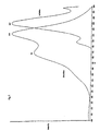
- the desirable properties of these dyes include absorption maxima matching light emitting diodes and laser diodes (see Table I and Figure 1), emission maxima in the low background red and near infrared spectral region; and chemical properties fulfilling labeling requirements (such as presence of reactive sites, hydrosolubility, lack of non-specific binding to proteins and surfaces, including skin, and stability).
- DYE ABSORPTION ( ⁇ MAX ) EMISSION ( ⁇ MAX ) COMMERCIAL LIGHT EMITTING DIODES/LASER DIODES: EMITTING WAVELENGTH (7) 741,810 847 750,810
- the fluorescent dyes of the present invention are particularly useful in clinical chemistry, immunochemistry and analytical chemistry.
- the compounds can be used in fluorometric immunoassays, DNA probes, high pressure liquid chromatography (HPLC), capillary electrophoresis, fluorescence polarization, total internal reflection fluorescence (T.I.R.F.), flow cytometry, DNA sequencing, and optical sensors.
- HPLC high pressure liquid chromatography
- T.I.R.F. total internal reflection fluorescence
- flow cytometry DNA sequencing
- optical sensors optical sensors.
- the fluorescent compounds of the present invention it is typically necessary to conjugate the fluorescent label to one of the binding partners, e.g., an antibody or an antigen or a biologically active protein molecule.
- linking groups may be employed for conjugating the protein to the other molecule.
- the functional group of interest for linking will be carbonyl, either an aldehyde to provide for reductive amination or a carboxyl, which in conjunction with carbodiimide or as an activated ester, e.g., N-hydroxy succinimide, will form a covalent bond with the amino groups present in the protein.
- Another useful functional group is a thio ether or disulfide, where the protein may be modified with an activated olefin and a mercapto group added or activated mercapto groups joined, e.g., Ellman's reagent, isothiocyanate, diazonium, nitrene, or carbene.
- an activated olefin e.g., Ellman's reagent, isothiocyanate, diazonium, nitrene, or carbene.
- antigens or antibodies are labeled with the fluorescent dyes of the invention by ordinary chemical reaction.
- a fluorescent dye can be reacted with an antigen or antibody to form a labeled reaction product by covalent linkage; specifically, the reaction occurs between the functional group (i.e., mercapto, amino, hydroxy, or carboxy) of the dye and an amino, imino, mercapto, carboxy, carboxylic acid amide, or hydroxy group(s) of an antigen or antibody.
- the reaction between the two can be carried out by any of the following procedures:
- Examples of compounds containing groups which react with the functional groups described above further include, e.g., activated esters, activated halogens, aldehydes, activated vinyl esters, activated halogens, aldehydes, activated vinyl compounds, acid anhydrides, acid halides, thioisocyanates, isocyanates, carboxylic acids, amides, alkyl halides, nitrophenyl halides, etc. Accordingly, these functional groups can originally be present in the fluorescent dyes or can be introduced as a result of the reaction of a compound having a bifunctional group and the fluorescent dye.
- Reaction conditions for labeling vary depending upon the kind of the antigen or antibody, the kind of fluorescent dye etc., and conditions are selected so as to not damage the biological activity of the antigen or antibody to be labeled. Accordingly, the reaction temperature is generally chosen from the range of from 40 to 60C, preferably -20 to 40C; and the reaction time from the range of from 10 minutes to 16 hours.
- the reaction pressure is preferably atmospheric pressure, but can suitably be chosen from the range of 1 to 20 atmospheres. It is advantageous that water or a pH buffer solution be used as a solvent for the labeling. Organic solvents such as DMF (dimethylformamide), methylene chloride, etc., can also be employed. These reaction conditions are common to reaction conditions which are generally applicable to modification of proteins or enzymes and details are described in the publications referred to above.
- the amount of fluorescent dyes used for labeling varies depending upon the kind of substances to be labeled, but is generally in a molar ratio of 1/100 to 100 moles per 1 mole of the antigen or antibody, preferably 1/20 to 20 times per 1 mole of the antigen or antibody, more preferably 1 ⁇ 2 to 2 times per 1 mole of the antigen or antibody.
- Useful methods for confirming completion of labeling include methods for measuring spectra such as UV, visible rays, IR, mass, and NMR spectra, etc., and a method confirming labeling via disappearance of the terminal group at which the labeling substance is to be introduced. Simple tests will be enough to confirm completion of labeling. Where it is confirmed utilizing absorption spectrum, following completion of the labeling reaction, an absorption spectrum of a separated and purified product is measured; if the resulting absorption spectrum is consistent with the intrinsic absorption spectrum which a fluorescent dye possesses, it is confirmed that the labeling reaction was effected. A further method for confirming the labeling being effected is to analyze for the presence or absence of the specific terminal groups, e.g., an amino or carboxy group(s).
- the terminal carboxy group(s) are analyzed to check completion of the labeling reaction, details of which are given in e.g., S. Akabori, K. Ohno and K. Narita, Bull. Chem. Soc. Japan , 25 , 214 (1952) (generally referred to as a hydrazine decomposition method in the art); H. Matuo, U. Fujimoto and T. Tatuno, Biochem. Biophys. Res. Communication , 22 , 69 (1966) ( a tritium marking method); etc. Further, details of these terminal determination methods are also given as a review in S.B. Needleman, Protein Sequence Determination , published by Springer Verlag (Berlin), 1975.
- a typical fluorometric immunoassay of an antigen in a liquid sample comprises incubating a mixture of:
- Another aspect of the invention is a competitive assay for determining the amount of an antigen in a sample suspected of containing the antigen comprising: (a) incubating the sample with a solution of a fluorescent-labeled antigen, anti-antigen antibody, and an antibody against the anti-antigen antibody; (b) adding a nonfluorescent nonlight-scattering immunoprecipitant to the incubation mixture to form an immunoprecipitate; (c) separating the immunoprecipitate and dissolving the immunoprecipitate in a nonfluorescent solvent that has a low ionic strength and maintains the pH of the resulting solution substantially constant; and (d) measuring the fluorescence intensity of the solution of step (c) and comparing the fluorescence intensity to a standard curve.
- the sample that is analyzed is a body fluid such as blood, blood serum, blood plasma, urine, lymph fluid, bile, spinal fluid, or the like.
- body fluid such as blood, blood serum, blood plasma, urine, lymph fluid, bile, spinal fluid, or the like.
- the particular body fluid analyzed may vary with the antigen being assayed. In most instances blood serum will be used. About 0.1 to about 500 ⁇ l of fluid is typically used per assay.
- Substances that may be assayed by the use of the fluorescent labeling reagents of the present invention include antigens (molecules that elicit an immune response when introduced into the bloodstream of a vertebrate host) and haptens that are not immunogenic per se but may be conjugated to a protein carrier to form a conjugate that is immunogenic and capable of raising antibodies against the hapten.
- antigens molecules that elicit an immune response when introduced into the bloodstream of a vertebrate host
- haptens that are not immunogenic per se but may be conjugated to a protein carrier to form a conjugate that is immunogenic and capable of raising antibodies against the hapten.
- antigen molecules that elicit an immune response when introduced into the bloodstream of a vertebrate host
- haptens that are not immunogenic per se but may be conjugated to a protein carrier to form a conjugate that is immunogenic and capable of raising antibodies against the hapten.
- antigen molecules that elicit an immune response when introduced
- antigens examples include thyroxine (T 4 ), triodothyronine (T 3 ), digoxin, gentamycin, amikacin, tobramycin, kanamycin, cortisol, luteinizing hormone, human chorionic gonadotropin, theophylline, angiotensin, human growth hormone, HIV infection, a fetoprotein, and the like.
- the reagents that are incubated with the sample suspected of containing antigen to form immune complexes typically are (1) fluorescent-labeled antigen, (2) anti-antigen antibody, (3) antibody against the anti-antigen antibody, and (4) a nonfluorescent non-light-scattering immunoprecipitant (e.g., polyethylene glycol).
- the fluorescent-labeled antigen may be made by coupling the antigen with a reactive derivative of the fluorescent dye using multifunctional coupling agents such as aldehydes, carbodiimides, dimaleimides, imidates, and succinimides.
- the dyes of the present invention also have utility in any current application for detection of nucleic acids that requires a sensitive detection reagent.
- the dyes are useful for the detection of cell-free isolated nucleic acids, nucleic acids in solution, and nucleic acid bands in gels.
- the present dyes greatly increase the sensitivity of detection of nucleic acids in a variety of cells and tissues, both living and dead, plant and animal, eukaryotic and prokaryotic. This family of dyes displays unusually good photostability and appears to be relatively non-toxic to cells.
- many of the dyes rapidly penetrate cell membranes of a variety of cells.
- the dyes of the invention exhibit superior properties compared to the known cyanine dyes.
- the use of the fluorescent dyes of the present invention in the detection of nucleic acids typically comprises combining a dye of the present invention with a sample that contains a nucleic acid, incubating the sample for a time sufficient to obtain a detectable fluorescent response, and observing the fluorescent response.
- the dye is present as a staining solution, which is prepared by addition of the dye to an aqueous solution that is biologically compatible with the sample.
- the staining solution is made by dissolving the dye directly in an aqueous solvent such as water; a buffer solution such as phosphate buffered saline; an organic water-miscible solvent such as dimethylsulfoxide (DMSO), dimethylformamide (DMF), or acetonitrile; or a lower alcohol such as methanol or ethanol.
- the dye is preliminarily dissolved in an organic solvent (preferably DMSO) at a concentration of greater than about 100 times that used in the staining solution, then diluted one or more times with an aqueous solvent such as water or buffer.
- an aqueous solvent such as water or buffer.
- the dye is dissolved in about 100% DMSO and then diluted one or more times in water or buffer such that the dye is present in an effective amount.
- An effective amount of dye is an amount sufficient to give a detectable fluorescent response when in the presence of nucleic acids.
- staining solutions for cellular samples have a dye concentration greater than about 0.1 nM and less than about 100 ⁇ M, more typically greater than about 1 nM.
- Staining solutions for electrophoretic gels typically have a dye concentration of greater than about 1 ⁇ M and less than about 10 ⁇ M, more typically about 4 to 5 ⁇ M. It is generally understood in the art that the specific concentration of the staining solution is determined by the physical nature of the sample, and the nature of the analysis being performed.
- the dye is combined with a sample that contains a nucleic acid.
- the nucleic acid in the sample may be either RNA or DNA, or a mixture thereof.
- the DNA may optionally be single-, double-, triple-, or quadruple-stranded DNA.
- the nucleic acid may be either natural (biological in origin) or synthetic (prepared artificially).
- the nucleic acid may be present as nucleic acid fragments, oligonucleotides, or nucleic acid polymers.
- the nucleic acid may be present in a condensed phase, such as a chromosome.
- the presence of the nucleic acid in the sample may be due to a successful or unsuccessful experimental methodology, undesirable contamination, or a disease state.
- Nucleic acid may be present in all, or only part, of a sample, and the presence of nucleic acids may be used to distinguish between individual samples, or to differentiate a portion or region within a single sample.
- the fluorescent dyes of the present invention can also be used for DNA sequencing methods.
- the method is based on Sanger's chain terminating sequencing method as described in Sanger et al., Proc. Natl. Acad. Sci. , 74 , 5464 (1977), the contents of which are incorporated by reference herein.
- a fluorophore-labeled probe specific to the sequence is hybridized with the target DNA and the sequence ladders are identified by laser induced fluorescence or other appropriate means for detecting fluorescent labeled DNA.
- an assay method for an analyte in a sample utilizing a DNA probe labeled with a dye of the invention comprises contacting the DNA probe thus labeled under suitable conditions for binding with the analyte, where the binding is representative of the presence or amount of the analyte in the sample, and determining the extent of the binding by measuring the fluorescence of the fluorescently labeled DNA probe.
- the analyte to be determined can be separated from the sample in which it is present prior to being bound by the DNA probe.
- first and second DNA probes in a single assay, where the first of such probes is bound with a fluorescent dye according to the invention, and the second probe is not labeled and is bound to a solid phase.
- the analyte can be separated from the sample by contacting the sample with the second DNA probe under conditions suitable for binding the analyte therewith, and the remaining sample is removed prior to contacting the analyte with the first (labeled) DNA probe.
- the step of separating the analyte from the sample in the manner just described can also be practiced with labeled binding reagents other than DNA probes.
- labeled binding reagents other than DNA probes.
- first (labeled) and second (unlabeled and bound to a solid phase) DNA probes plural immunologically binding reagents specific for a given analyte can be used.
- the fluorescent dyes of the present invention can be used in flow cytometry.
- the method is characterized by the use of a flow cytometer that comprises a light source, a flow cell that permits the blood cells in a sample to flow one by one through a constricted channel, a photometric unit that detects light issuing from each blood cell, and an analyzer for analyzing the detected signal.
- the corpuscles in the sample which are stained are illuminated under light and the fluorescence emitted from the irradiated corpuscles is detected, optionally together with scattered light, with leukocyte classification being made in accordance with the intensity of the detected signal.
- a suspension of 13.34 g (64.2 mmol) of Dahl's acid in 45 ml of concentrated hydrochloric acid was cooled to -5C by an ice-salt bath in a 500 ml three-necked flask equipped with mechanical stirrer, thermometer and reflux condenser.
- To the slurry was added dropwise, at 0C or below, an ice-cold sodium nitrite solution (4.43 g, or 64.2 mmol, in 30 ml of water) and the mixture was stirred for 40 minutes at the same temperature and then used without delay in the following step.
- the free amine group it is possible to follow classical cleavage pathways such as treatment in ethanol with hydrazine at 25C for 12 hours or hydrolysis in concentrated hydrochloric acid.
- the cyanine dyes can be obtained by condensing the benz[e]indoleninium-7-sulfonate intermediates with triethyl orthoformate or various dialdehyde dianilides.
- the purification of the dyes was performed on a Millipore Waters Delta Prep 4000 HPLC unit equipped with a Vydac C18 RP-column, using a gradient elution (for example: H 2 O/CH 3 CN - from 80:20 to 5:95). Because of the numerous ionic sites present in these dyes, to obtain a well-defined composition it was necessary to perform an ion-exchange step by passing an aqueous solution of the dye through a strongly acidic cation-exchanger, such as Dowex 50W in the hydrogen form.
- a strongly acidic cation-exchanger such as Dowex 50W in the hydrogen form.
- succinimidyl esters of carboxyalkyl sulfo benz[e] indocyanine dyes were used as intermediates for labeling biomolecules such as antibodies and antigens.
- succinimidyl esters of a carboxyalkyl sulfo benz[e]indocyanine dye was prepared by dissolving 1 mg of active ester in 100 ⁇ l of anhydrous DMF. These solutions were stable for a few days when kept in a dessicator at 4C. Aqueous solutions of the active esters were stable for a few hours at pH ⁇ 7.
- the labeling of protein was carried out in carbonate-bicarbonate buffer (0.1M, pH 9.4) for 15 minutes at ambient temperature.
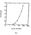
- FIA in heterogeneous phase of the sandwich-type, between the analyte to be determined and two monoclonal antibodies specific towards two epitopes different from the analyte, one can make the determination of ⁇ -fetoprotein in serum.
- the analytical scheme of the assay is as follows: the first antibody MAb, anti-AFP is used as capture antibody and is conjugated with biotin.
- the second antibody Mab 2 anti-AFP is conjugated with the indicator (for example, Dye 7 with maximum absorption at 810 nm.)
- the immunochemical reaction between the analyte and the monoclonal antibodies takes place in the homogeneous phase, in the presence of a solid phase, for example, plates with wells, sensitized with streptavidin, which constitutes half of the unbound/bound separation.
Description

Q is selected from the group consisting of:


| DYE | ABSORPTION (λMAX) | EMISSION (λ MAX) | COMMERCIAL LIGHT EMITTING DIODES/LASER DIODES: EMITTING WAVELENGTH |
| (7) | 741,810 | 847 | 750,810 |

- place in each well 50 µL of sample or reference sample;
100 µL of solution of the STET-Mab1anti-AFP conjugate;
100 µL of the solution of Mab2anti-AFP-dye conjugate; - Incubate for 1 hour at room temperature.
- a series of 5 washes with appropriate buffer is carried out automatically using equipment known in the immunochemical art;
- place in each well 300 µL of a solution (6 M) of guanidine dichloride;
- transfer 300 µL of solution to a cuvette;
- subject sample to excitation at 810 nm and the resulting spectral emission is integrated after correction and normalization.
Claims (14)
- A fluorescent compound and its valence tautomers of the formula:wherein R1 is a functionalised group of the formula -(CH2)jY wherein Y is phthalimido; R2 is a functionalised group of the formula -(CH2)kY', wherein Y' is selected from the group consisting of COOH and phthalimido; M+ is a counterion selected from the group consisting of ammonium, alkali metal cations, and alkaline earth metal cations; n = 1 to 4; m = 1 to 4; j = 2 to 10; k = 2 to 10; and wherein Q is selected from the group consisting of:


 and
and wherein X is selected from the group consisting of hydrogen, F, Cl, Br, I and substituted aryl, wherein said aryl substituent is selected from the group consisting of SO3H, COOH, NH2, CHO, NCS, epoxy and COOZ, wherein Z represents a leaving group and i = 0 or 1.
- The fluorescent compound of claim 1, wherein Q isand R1 and R2 are both phthalimidopropyl.

- A fluorescent compound of claim 1 of the formula:

- A DNA probe labelled with a fluorescent compound of one of claims 1 to 3.
- A method of assay of an analyte in a sample comprising contacting a DNA probe of claim 4 under suitable conditions for binding with the analyte, wherein the binding is representative of the presence or amount of the analyte in the sample and determining the extent of said binding by measuring the fluorescence of the bound DNA probe labelled with said fluorescent compound.
- The method of assay of claim 5, wherein the analyte is separated from the sample prior to contact with the DNA probe.
- The method of assay of claim 6, wherein a second DNA probe, which is not labelled, is attached to a solid phase and the analyte is separated from the sample by contact with said second DNA probe under suitable conditions for binding and the remaining sample is removed prior to contact of analyte with the labelled probe.
- An immunologically binding reagent labelled with a fluorescent compound of one of claims 1 to 3.
- The binding reagent of claim 8, wherein said reagent is an antibody.
- The binding reagent of claim 8, wherein said reagent is an antigen.
- The reagent of claim 9, wherein said antibody is a monoclonal antibody.
- A method of assay of an analyte in a sample comprising contacting an immunologically binding reagent labelled with a fluorescent compound of one of claims 1 to 3 under suitable conditions for binding with the analyte, wherein the binding is representative of the presence or amount of the analyte in the sample and determining the extent of said binding by measuring the fluorescence of the immunologically binding reagent labelled with said fluorescent compound.
- The method of assay of claim 12, wherein the analyte is separated from the sample prior to contact with the immunologically binding reagent.
- The method of assay of claim 13, wherein a second immunologically binding reagent, which is not labelled, is attached to a solid phase and the analyte is separated from the sample by contact with said second immunologically binding reagent under suitable conditions for binding and the remaining sample is removed prior to contact of analyte with the labelled immunologically binding reagent.
Applications Claiming Priority (3)
| Application Number | Priority Date | Filing Date | Title |
|---|---|---|---|
| IT95MI002049A IT1276833B1 (en) | 1995-10-09 | 1995-10-09 | FLUORESCENT DYES OF THE SULFUR BENZ AND INDOCYANINE FAMILY |
| ITMI952049 | 1995-10-09 | ||
| PCT/EP1996/004377 WO1997013810A1 (en) | 1995-10-09 | 1996-10-08 | SULFO BENZ[e]INDOCYANINE FLUORESCENT DYES |
Publications (2)
| Publication Number | Publication Date |
|---|---|
| EP0876428A1 EP0876428A1 (en) | 1998-11-11 |
| EP0876428B1 true EP0876428B1 (en) | 2004-11-17 |
Family
ID=11372321
Family Applications (1)
| Application Number | Title | Priority Date | Filing Date |
|---|---|---|---|
| EP96934591A Expired - Lifetime EP0876428B1 (en) | 1995-10-09 | 1996-10-08 | SULFO BENZ e]INDOCYANINE FLUORESCENT DYES |
Country Status (6)
| Country | Link |
|---|---|
| US (1) | US6136612A (en) |
| EP (1) | EP0876428B1 (en) |
| AU (1) | AU706668B2 (en) |
| DE (1) | DE69626041D1 (en) |
| IT (1) | IT1276833B1 (en) |
| WO (1) | WO1997013810A1 (en) |
Families Citing this family (73)
| Publication number | Priority date | Publication date | Assignee | Title |
|---|---|---|---|---|
| US6593148B1 (en) | 1994-03-01 | 2003-07-15 | Li-Cor, Inc. | Cyanine dye compounds and labeling methods |
| DE19717904A1 (en) | 1997-04-23 | 1998-10-29 | Diagnostikforschung Inst | Acid-labile and enzymatically cleavable dye constructs for diagnostics with near infrared light and for therapy |
| KR100337169B1 (en) * | 1998-07-20 | 2002-05-18 | 최중갑 | Detection method of DNA on polymer gels using counter-dye composition and counter-dye composition for the same |
| US7547721B1 (en) | 1998-09-18 | 2009-06-16 | Bayer Schering Pharma Ag | Near infrared fluorescent contrast agent and fluorescence imaging |
| JP2000095758A (en) * | 1998-09-18 | 2000-04-04 | Schering Ag | Near-infrared, fluorescent contrast medium, and its production |
| US6605434B1 (en) | 1999-03-16 | 2003-08-12 | Human Genome Sciences, Inc. | Direct bacterial lysate sequencing |
| DE19937024A1 (en) | 1999-08-05 | 2001-02-08 | Bayer Ag | Use of acylsulfonamido substituted polymethine dyes as fluorescent dyes and / or markers |
| WO2001050646A1 (en) * | 1999-12-30 | 2001-07-12 | Morphics Technology, Inc. | A configurable multimode despreader for spread spectrum applications |
| US6716994B1 (en) | 2000-01-04 | 2004-04-06 | Applera Corporation | Mobility-Modifying Cyanine Dyes |
| EP1250091B1 (en) * | 2000-01-18 | 2008-04-16 | Mallinckrodt Inc. | Hydrophilic cyanine dyes |
| US7790144B2 (en) | 2000-01-18 | 2010-09-07 | Mallinckrodt Inc. | Receptor-avid exogenous optical contrast and therapeutic agents |
| ATE315066T1 (en) | 2000-09-19 | 2006-02-15 | Li Cor Inc | CYANINE DYES |
| US7597878B2 (en) * | 2000-09-19 | 2009-10-06 | Li-Cor, Inc. | Optical fluorescent imaging |
| US6656451B1 (en) | 2000-10-16 | 2003-12-02 | Mallinckrodt, Inc. | Indole compounds as novel dyes for organ function monitoring |
| US6335450B1 (en) * | 2000-11-09 | 2002-01-01 | Beckman Coulter, Inc. | Efficient cyclic-bridged cyanine dyes |
| US6331632B1 (en) * | 2000-11-09 | 2001-12-18 | Beckman Coulter, Inc. | Cyanine dye phosphoramidites |
| US6710363B1 (en) * | 2000-11-27 | 2004-03-23 | Uview Ultraviolet Systems, Inc. | Detection lamp equipped with light-emitting diode |
| EP1211294B1 (en) * | 2000-11-28 | 2008-08-13 | Visen Medical, Inc. | Improved process and method for the preparation of asymmetric monofunctionalised indocyanine labelling reagents and obtained compounds |
| EP1209205A1 (en) * | 2000-11-28 | 2002-05-29 | Innosense S.r.l. | Improved process and method for the preparation of asymetric monofunctionalised indocyanine labelling reagents and obtained compounds |
| US6800438B2 (en) * | 2000-12-28 | 2004-10-05 | Xerox Corporation | Imager for DNA sequencer |
| JP4319363B2 (en) * | 2001-01-15 | 2009-08-26 | 富士フイルム株式会社 | Negative type image recording material |
| WO2002068537A2 (en) | 2001-02-28 | 2002-09-06 | Stratagene | Fluorescent dye |
| AU2003207438A1 (en) * | 2002-01-02 | 2003-07-24 | Visen Medical, Inc. | Amine functionalized superparamagnetic nanoparticles for the synthesis of bioconjugates and uses therefor |
| EP1485716A1 (en) * | 2002-03-11 | 2004-12-15 | Visen Medical, Inc. | Optical imaging probes |
| AU2003280470A1 (en) * | 2002-07-01 | 2004-01-19 | Guava Technologies, Inc. | Fluorescent dyes, energy transfer couples and methods |
| US20040132092A1 (en) * | 2003-01-03 | 2004-07-08 | Stetson Christopher M. | Determining the density of functional moieties on polymer reagents |
| US20080102036A1 (en) * | 2003-06-04 | 2008-05-01 | Poss Kirtland G | Biocompatible Fluorescent Silicon Nanoparticles |
| ITPZ20030002A1 (en) * | 2003-08-12 | 2005-02-13 | Giuseppe Caputo | COMPOUNDS OF THE CIANINA TYPE WITH AN ALCHINYLIC ARM |
| US8227621B2 (en) * | 2005-06-30 | 2012-07-24 | Li-Cor, Inc. | Cyanine dyes and methods of use |
| DK1937676T3 (en) * | 2005-09-02 | 2017-03-06 | Visen Medical Inc | Biocompatible fluorescent imaging compounds |
| WO2007028037A1 (en) * | 2005-09-02 | 2007-03-08 | Visen Medical, Inc. | Biocompatible n, n-disubstituted sulfonamide-containing fluorescent dye labels |
| EP1934202B1 (en) | 2005-09-02 | 2019-01-09 | Visen Medical, Inc. | Nicotinic acid and picolinic acid derived near-infrared fluorophores |
| US10064584B2 (en) | 2005-12-22 | 2018-09-04 | Visen Medical, Inc. | Combined x-ray and optical tomographic imaging system |
| EP1973575B1 (en) | 2005-12-22 | 2019-07-24 | Visen Medical, Inc. | Biocompatible fluorescent metal oxide nanoparticles |
| ITSV20060002A1 (en) * | 2006-01-19 | 2007-07-20 | Ferrania Technologies Spa | FLUORESCENT DYE OF CIANIN TYPE |
| JP4958461B2 (en) * | 2006-03-30 | 2012-06-20 | 富士フイルム株式会社 | Near-infrared absorbing dye-containing curable composition |
| US20090186366A1 (en) * | 2006-06-28 | 2009-07-23 | Ge Healthcare Bio-Sciences Ab | Method of detecting interactions between protein components |
| DK2118206T3 (en) | 2007-02-09 | 2018-06-18 | Visen Medical Inc | POLYCYCLOF COLORS AND APPLICATION THEREOF |
| WO2008109832A2 (en) * | 2007-03-08 | 2008-09-12 | Visen Medical, Inc. | Viable near-infrared fluorochrome labeled cells and methods of making and using same |
| EP2148702A2 (en) * | 2007-05-16 | 2010-02-03 | GE Healthcare AS | Pentamethine cyanine dyes carrying at least three sulfonic acid groups |
| CA2740602C (en) | 2007-10-19 | 2017-05-02 | Visen Medical, Inc. | Imaging systems featuring waveguiding compensation |
| DK2244741T3 (en) | 2008-01-18 | 2015-05-26 | Visen Medical Inc | Fluorescent imaging agents |
| US8685370B2 (en) | 2008-03-14 | 2014-04-01 | Visen Medical, Inc. | Integrin targeting agents and in-vivo and in-vitro imaging methods using the same |
| US8864821B2 (en) | 2008-11-26 | 2014-10-21 | Visen Medical, Inc. | Methods and compositions for identifying subjects at risk of developing stent thrombosis |
| CN102459469A (en) * | 2009-04-17 | 2012-05-16 | 利康股份有限公司 | Fluorescent imaging with substituted cyanine dyes |
| US9155471B2 (en) | 2009-05-27 | 2015-10-13 | Lumicell, Inc'. | Methods and systems for spatially identifying abnormal cells |
| EP2435096A1 (en) * | 2009-05-27 | 2012-04-04 | Lumicell Diagnostics, Inc. | Methods and systems for spatially identifying abnormal cells |
| AU2010286592B2 (en) | 2009-08-28 | 2015-08-13 | Visen Medical, Inc. | Systems and methods for tomographic imaging in diffuse media using a hybrid inversion technique |
| CA2810822C (en) | 2009-09-22 | 2018-03-06 | Visen Medical, Inc. | Systems and methods for virtual index-matching of diffusive media |
| JP5817073B2 (en) * | 2010-01-28 | 2015-11-18 | 国立大学法人三重大学 | Diagnostic composition and analysis method using novel indocyanine compound |
| PT2530093T (en) * | 2010-01-28 | 2018-12-28 | Univ Nagoya Nat Univ Corp | Indocyanine compound, synthesis method thereof, purification method thereof, diagnostic composition using indocyanine compound, and device for measuring in vivo kinetics and device for visualizing circulation using diagnostic composition |
| DE102010022110A1 (en) | 2010-05-31 | 2011-12-01 | LMU Universität München Department Chemie | Cyanine dyes as a contrast agent to aid eye surgery |
| WO2012054784A1 (en) | 2010-10-20 | 2012-04-26 | Li-Cor, Inc. | Fluorescent imaging with substituted cyanine dyes |
| US9314304B2 (en) | 2010-12-08 | 2016-04-19 | Lumicell, Inc. | Methods and system for image guided cell ablation with microscopic resolution |
| US10053447B2 (en) | 2010-12-21 | 2018-08-21 | Pierce Biotechnology, Inc | Fluorescent compounds |
| CA2835286C (en) | 2011-05-09 | 2021-03-23 | Visen Medical, Inc. | Carbonic anhydrase targeting agents and methods of using same |
| US8889884B1 (en) | 2011-07-14 | 2014-11-18 | Pierce Biotechnology, Inc. | Phosphine derivatives of fluorescent compounds |
| US9249307B2 (en) | 2011-08-16 | 2016-02-02 | Pierce Biotechnology, Inc. | Benzocyanine compounds |
| US9751868B2 (en) | 2012-02-28 | 2017-09-05 | Pierce Biotechnology, Inc. | Benzocyanine compounds |
| EP2804860B1 (en) | 2012-03-02 | 2016-06-15 | Pierce Biotechnology, Inc. | Indole derivatives as labeling dye for biomolecule |
| WO2013148319A1 (en) | 2012-03-30 | 2013-10-03 | Visen Medical, Inc. | Bacterial imaging agents and methods of using same |
| CN104955484B (en) | 2012-08-15 | 2019-01-15 | 文森医学公司 | Prostate-specific antigen medicament and its application method for prostate cancer imaging |
| WO2014035712A1 (en) | 2012-08-28 | 2014-03-06 | Pierce Biotechnology, Inc. | Benzopyrylium compounds |
| EP2906106B1 (en) | 2012-10-15 | 2023-06-14 | VisEn Medical, Inc. | Systems, methods, and apparatus for imaging of diffuse media featuring cross-modality weighting of fluorescent and bioluminescent sources |
| EP2967280A4 (en) | 2013-03-14 | 2016-09-28 | Lumicell Inc | Medical imaging device and methods of use |
| EP3489309A1 (en) | 2013-03-15 | 2019-05-29 | Visen Medical, Inc. | 4,4-disubstituted cyclohexyl bridged heptamethine cyanine dyes and uses thereof |
| AU2014228504C1 (en) | 2013-03-15 | 2019-10-03 | Visen Medical, Inc. | Substituted silaxanthenium red to near-infrared fluorochromes for in vitro and in vivo imaging and detection |
| CN106455979A (en) | 2013-12-31 | 2017-02-22 | 纪念斯隆-凯特琳癌症中心 | Systems, methods, and apparatus for multichannel imaging of fluorescent sources in real time |
| AU2015362663B2 (en) | 2014-12-15 | 2020-07-09 | Cornell University | Cyclic peptides with enhanced nerve-binding selectivity, nanoparticles bound with said cyclic peptides, and use of same for real-time in vivo nerve tissue imaging |
| JP2019509252A (en) | 2015-12-15 | 2019-04-04 | メモリアル スローン ケタリング キャンサー センター | Imaging systems and methods for tissue differentiation, eg, visualization during surgery |
| CA3045007A1 (en) | 2016-11-30 | 2018-06-07 | Memorial Sloan Kettering Cancer Center | Inhibitor-functionalized ultrasmall nanoparticles and methods thereof |
| KR101973456B1 (en) * | 2017-10-17 | 2019-04-29 | 울산과학기술원 | Water-soluble compound targeted to mitochondria, composition comprising them and preparation method thereof |
| EP4015004A1 (en) | 2020-12-18 | 2022-06-22 | Phi Pharma SA | Proteoglycan specific branched peptides |
Family Cites Families (9)
| Publication number | Priority date | Publication date | Assignee | Title |
|---|---|---|---|---|
| US5230781A (en) * | 1984-03-29 | 1993-07-27 | Li-Cor, Inc. | Sequencing near infrared and infrared fluorescence labeled DNA for detecting using laser diodes |
| US5268486A (en) * | 1986-04-18 | 1993-12-07 | Carnegie-Mellon Unversity | Method for labeling and detecting materials employing arylsulfonate cyanine dyes |
| JP2713976B2 (en) * | 1987-04-24 | 1998-02-16 | イーストマン コダック カンパニー | Photographic filter composition |
| CA1337923C (en) * | 1988-07-07 | 1996-01-16 | Richard Lee Parton | Infrared filter dye for photographic element |
| DE3912046B4 (en) * | 1988-09-02 | 2004-03-25 | Carnegie Mellon University | Method of labeling a component of an aqueous liquid and luminescent photostable reaction product |
| ES2067035T3 (en) * | 1989-07-04 | 1995-03-16 | Kubota Kk | DEVICE FOR OPERATION OF A VEHICLE CLUTCH. |
| JP2663033B2 (en) * | 1990-02-22 | 1997-10-15 | 富士写真フイルム株式会社 | Silver halide emulsion |
| EP0626427B1 (en) * | 1993-05-26 | 2001-08-16 | Agfa-Gevaert N.V. | Near infra-red dyes and photographic element containing such dyes |
| US5453505A (en) * | 1994-06-30 | 1995-09-26 | Biometric Imaging, Inc. | N-heteroaromatic ion and iminium ion substituted cyanine dyes for use as fluorescence labels |
-
1995
- 1995-10-09 IT IT95MI002049A patent/IT1276833B1/en active IP Right Grant
-
1996
- 1996-10-08 DE DE69626041T patent/DE69626041D1/en not_active Expired - Lifetime
- 1996-10-08 WO PCT/EP1996/004377 patent/WO1997013810A1/en active IP Right Grant
- 1996-10-08 AU AU72882/96A patent/AU706668B2/en not_active Expired
- 1996-10-08 US US09/043,767 patent/US6136612A/en not_active Expired - Lifetime
- 1996-10-08 EP EP96934591A patent/EP0876428B1/en not_active Expired - Lifetime
Also Published As
| Publication number | Publication date |
|---|---|
| EP0876428A1 (en) | 1998-11-11 |
| US6136612A (en) | 2000-10-24 |
| IT1276833B1 (en) | 1997-11-03 |
| AU7288296A (en) | 1997-04-30 |
| WO1997013810A1 (en) | 1997-04-17 |
| AU706668B2 (en) | 1999-06-17 |
| ITMI952049A1 (en) | 1997-04-09 |
| DE69626041D1 (en) | 2003-03-06 |
| ITMI952049A0 (en) | 1995-10-09 |
Similar Documents
| Publication | Publication Date | Title |
|---|---|---|
| EP0876428B1 (en) | SULFO BENZ e]INDOCYANINE FLUORESCENT DYES | |
| US5569766A (en) | Method for labeling and detecting materials employing arylsulfonate cyanine dyes | |
| US6130094A (en) | Reagents including a carrier and fluorescent labeling complexes with large stokes shift formed by coupling together cyanine and other fluorochromes capable of resonance energy transfer | |
| US5627027A (en) | Cyanine dyes as labeling reagents for detection of biological and other materials by luminescence methods | |
| US7964361B2 (en) | Rigidized trimethine cyanine dyes | |
| JP2898264B2 (en) | Method for labeling or detecting substances using luminescent aryl sulfonate cyanine dye | |
| EP1019721B1 (en) | Chromophores in the preparation of novel tandem conjugates | |
| US5569587A (en) | Method for labeling and detecting materials employing luminescent arysulfonate cyanine dyes | |
| EP0856155B1 (en) | Fluorescent and luminescent labelling compositions and methods for their use | |
| US6686145B1 (en) | Rigidized trimethine cyanine dyes | |
| US7745640B2 (en) | Hydrophilic labels for biomolecules | |
| US20040162423A1 (en) | Benzopyrylo-polymethine-based hydrophilic markers | |
| US20040076979A1 (en) | Dye pair for flourescence resonance energy transfer (fret) measurements | |
| CA2234450C (en) | Sulfo benz [e] indocyanine fluorescent dyes | |
| US4822878A (en) | Cyclic anhydride derivatives of chromophors | |
| US4906749A (en) | Cyclic anhydride derivatives of chromophors | |
| US20190339283A1 (en) | Bioconjugates of heterocyclic compounds | |
| JP2002502034A (en) | Energy transfer dye | |
| GB2373508A (en) | Long wavelength fluorescent dyes with 2-(2-{2-chloro-3-[2-(2,3-dihydro-1H-indol-2-yliden)ethylidene]cyclohex-1-enyl}eth-1-enyl)-1H-benzo[e]indolium skeleton |
Legal Events
| Date | Code | Title | Description |
|---|---|---|---|
| PUAI | Public reference made under article 153(3) epc to a published international application that has entered the european phase |
Free format text: ORIGINAL CODE: 0009012 |
|
| 17P | Request for examination filed |
Effective date: 19980406 |
|
| AK | Designated contracting states |
Kind code of ref document: A1 Designated state(s): DE FR GB IT NL |
|
| 17Q | First examination report despatched |
Effective date: 20000316 |
|
| RAP1 | Party data changed (applicant data changed or rights of an application transferred) |
Owner name: INNOSENSE S.R.L. |
|
| GRAG | Despatch of communication of intention to grant |
Free format text: ORIGINAL CODE: EPIDOS AGRA |
|
| GRAG | Despatch of communication of intention to grant |
Free format text: ORIGINAL CODE: EPIDOS AGRA |
|
| GRAH | Despatch of communication of intention to grant a patent |
Free format text: ORIGINAL CODE: EPIDOS IGRA |
|
| RIN1 | Information on inventor provided before grant (corrected) |
Inventor name: CAPUTO, GIUSEPPE Inventor name: CASSULLO, MARIACRISTINA Inventor name: GRIGNANI, ANDREA Inventor name: DELLA CIANA, LEOPOLDO |
|
| GRAH | Despatch of communication of intention to grant a patent |
Free format text: ORIGINAL CODE: EPIDOS IGRA |
|
| GRAA | (expected) grant |
Free format text: ORIGINAL CODE: 0009210 |
|
| AK | Designated contracting states |
Designated state(s): DE FR GB IT NL |
|
| REG | Reference to a national code |
Ref country code: GB Ref legal event code: FG4D |
|
| REF | Corresponds to: |
Ref document number: 69626041 Country of ref document: DE Date of ref document: 20030306 Kind code of ref document: P |
|
| NLV1 | Nl: lapsed or annulled due to failure to fulfill the requirements of art. 29p and 29m of the patents act | ||
| 29U | Proceedings interrupted after grant according to rule 142 epc |
Effective date: 20021122 |
|
| 29W | Proceedings resumed after grant [after interruption of proceedings according to rule 142 epc] |
Effective date: 20031201 |
|
| 29W | Proceedings resumed after grant [after interruption of proceedings according to rule 142 epc] |
Effective date: 20040601 |
|
| GBPC | Gb: european patent ceased through non-payment of renewal fee |
Effective date: 20031008 |
|
| PUAC | Information related to the publication of a b1 document modified or deleted |
Free format text: ORIGINAL CODE: 0009299EPPU |
|
| RAP2 | Party data changed (patent owner data changed or rights of a patent transferred) |
Owner name: VISEN MEDICAL, INC. |
|
| DB1 | Publication of patent cancelled | ||
| GRAA | (expected) grant |
Free format text: ORIGINAL CODE: 0009210 |
|
| NLXE | Nl: other communications concerning ep-patents (part 3 heading xe) |
Free format text: PAT. BUL. 04/2003 PAT. BUL. 07/2003: PATENTNUMBER 0876428 SHOULD BE DELETED (SEE EUROPEAN PATENT BULLETIN 20040818/34) |
|
| AK | Designated contracting states |
Kind code of ref document: B1 Designated state(s): DE FR GB IT NL |
|
| PG25 | Lapsed in a contracting state [announced via postgrant information from national office to epo] |
Ref country code: IT Free format text: LAPSE BECAUSE OF FAILURE TO SUBMIT A TRANSLATION OF THE DESCRIPTION OR TO PAY THE FEE WITHIN THE PRE;WARNING: LAPSES OF ITALIAN PATENTS WITH EFFECTIVE DATE BEFORE 2007 MAY HAVE OCCURRED AT ANY TIME BEFORE 2007. THE CORRECT EFFECTIVE DATE MAY BE DIFFERENT FROM THE ONE RECORDED.SCRIBED TIME-LIMIT Effective date: 20041117 Ref country code: FR Free format text: LAPSE BECAUSE OF FAILURE TO SUBMIT A TRANSLATION OF THE DESCRIPTION OR TO PAY THE FEE WITHIN THE PRESCRIBED TIME-LIMIT Effective date: 20041117 |
|
| REG | Reference to a national code |
Ref country code: GB Ref legal event code: FG4D |
|
| NLV1 | Nl: lapsed or annulled due to failure to fulfill the requirements of art. 29p and 29m of the patents act | ||
| PLBE | No opposition filed within time limit |
Free format text: ORIGINAL CODE: 0009261 |
|
| STAA | Information on the status of an ep patent application or granted ep patent |
Free format text: STATUS: NO OPPOSITION FILED WITHIN TIME LIMIT |
|
| PG25 | Lapsed in a contracting state [announced via postgrant information from national office to epo] |
Ref country code: GB Free format text: LAPSE BECAUSE OF NON-PAYMENT OF DUE FEES Effective date: 20051008 |
|
| 26N | No opposition filed |
Effective date: 20050818 |
|
| EN | Fr: translation not filed | ||
| GBPC | Gb: european patent ceased through non-payment of renewal fee |
Effective date: 20051008 |
|
| PG25 | Lapsed in a contracting state [announced via postgrant information from national office to epo] |
Ref country code: DE Free format text: LAPSE BECAUSE OF NON-PAYMENT OF DUE FEES Effective date: 20051031 |
|
| PG25 | Lapsed in a contracting state [announced via postgrant information from national office to epo] |
Ref country code: NL Free format text: LAPSE BECAUSE OF NON-PAYMENT OF DUE FEES Effective date: 20041117 |