EP1021132B1 - Endoscopic bone plate positioning device - Google Patents
Endoscopic bone plate positioning device Download PDFInfo
- Publication number
- EP1021132B1 EP1021132B1 EP98918601A EP98918601A EP1021132B1 EP 1021132 B1 EP1021132 B1 EP 1021132B1 EP 98918601 A EP98918601 A EP 98918601A EP 98918601 A EP98918601 A EP 98918601A EP 1021132 B1 EP1021132 B1 EP 1021132B1
- Authority
- EP
- European Patent Office
- Prior art keywords
- bone plate
- attachment element
- plate holder
- endoscope
- endoscope attachment
- Prior art date
- Legal status (The legal status is an assumption and is not a legal conclusion. Google has not performed a legal analysis and makes no representation as to the accuracy of the status listed.)
- Expired - Lifetime
Links
Images


Classifications
-
- A—HUMAN NECESSITIES
- A61—MEDICAL OR VETERINARY SCIENCE; HYGIENE
- A61B—DIAGNOSIS; SURGERY; IDENTIFICATION
- A61B17/00—Surgical instruments, devices or methods, e.g. tourniquets
- A61B17/56—Surgical instruments or methods for treatment of bones or joints; Devices specially adapted therefor
- A61B17/58—Surgical instruments or methods for treatment of bones or joints; Devices specially adapted therefor for osteosynthesis, e.g. bone plates, screws, setting implements or the like
- A61B17/68—Internal fixation devices, including fasteners and spinal fixators, even if a part thereof projects from the skin
- A61B17/80—Cortical plates, i.e. bone plates; Instruments for holding or positioning cortical plates, or for compressing bones attached to cortical plates
- A61B17/808—Instruments for holding or positioning bone plates, or for adjusting screw-to-plate locking mechanisms
-
- A—HUMAN NECESSITIES
- A61—MEDICAL OR VETERINARY SCIENCE; HYGIENE
- A61B—DIAGNOSIS; SURGERY; IDENTIFICATION
- A61B17/00—Surgical instruments, devices or methods, e.g. tourniquets
- A61B17/16—Bone cutting, breaking or removal means other than saws, e.g. Osteoclasts; Drills or chisels for bones; Trepans
- A61B17/17—Guides or aligning means for drills, mills, pins or wires
- A61B17/1728—Guides or aligning means for drills, mills, pins or wires for holes for bone plates or plate screws
-
- A—HUMAN NECESSITIES
- A61—MEDICAL OR VETERINARY SCIENCE; HYGIENE
- A61B—DIAGNOSIS; SURGERY; IDENTIFICATION
- A61B17/00—Surgical instruments, devices or methods, e.g. tourniquets
- A61B17/16—Bone cutting, breaking or removal means other than saws, e.g. Osteoclasts; Drills or chisels for bones; Trepans
- A61B17/17—Guides or aligning means for drills, mills, pins or wires
- A61B17/1739—Guides or aligning means for drills, mills, pins or wires specially adapted for particular parts of the body
- A61B17/176—Guides or aligning means for drills, mills, pins or wires specially adapted for particular parts of the body for the jaw
-
- A—HUMAN NECESSITIES
- A61—MEDICAL OR VETERINARY SCIENCE; HYGIENE
- A61B—DIAGNOSIS; SURGERY; IDENTIFICATION
- A61B17/00—Surgical instruments, devices or methods, e.g. tourniquets
- A61B17/00234—Surgical instruments, devices or methods, e.g. tourniquets for minimally invasive surgery
-
- A—HUMAN NECESSITIES
- A61—MEDICAL OR VETERINARY SCIENCE; HYGIENE
- A61B—DIAGNOSIS; SURGERY; IDENTIFICATION
- A61B17/00—Surgical instruments, devices or methods, e.g. tourniquets
- A61B17/56—Surgical instruments or methods for treatment of bones or joints; Devices specially adapted therefor
- A61B17/58—Surgical instruments or methods for treatment of bones or joints; Devices specially adapted therefor for osteosynthesis, e.g. bone plates, screws, setting implements or the like
- A61B17/68—Internal fixation devices, including fasteners and spinal fixators, even if a part thereof projects from the skin
- A61B17/80—Cortical plates, i.e. bone plates; Instruments for holding or positioning cortical plates, or for compressing bones attached to cortical plates
- A61B17/8061—Cortical plates, i.e. bone plates; Instruments for holding or positioning cortical plates, or for compressing bones attached to cortical plates specially adapted for particular bones
- A61B17/8071—Cortical plates, i.e. bone plates; Instruments for holding or positioning cortical plates, or for compressing bones attached to cortical plates specially adapted for particular bones for the jaw
Definitions
- the present invention relates to a method for the stabilization of bone fractures and in particular to an endoscopic bone plate positioning device for use in such a procedure.
- Endoscopic procedures are minimally invasive procedures which stabilize the fracture using a small incision, and hence decrease the surgical trauma associated with fixing a fracture.
- the present invention relates to a device for implanting a bone plate including an endoscope attachment element having proximal and distal ends and a body member configured and dimensioned to receive at least a portion of an endoscope, and a bone plate holder having one end which is configured and dimensioned to hold at least a portion of the bone plate.
- the bone plate holder is operatively associated with the body member of the endoscope attachment element to move the bone plate holder between a first retracted position and a second implanting position.
- the endoscope attachment element includes a guide track positioned adjacent the body member for receiving the bone plate holder and for providing sliding movement of the bone plate holder between the first and second positions.
- the guide track includes a plurality of bone plate holder positioning catches and the bone plate holder includes a locking button for engaging one of the catches to retain the bone plate holder in the second implanting position.
- the body member of the endoscope attachment element has a distal end which includes a raspatory having an aperture therein, wherein the aperture aligns with the end of the bone plate holder when the bone plate holder is retained in the second implanting position.
- the endoscope attachment element can have an integrated irrigation and suction system as well as a handle for manipulation.
- the device preferably further includes a locking member which has a connecting tube operatively associated with the bone plate to guide and enable fastening operations through the bone plate screw holes.
- a locking member which has a connecting tube operatively associated with the bone plate to guide and enable fastening operations through the bone plate screw holes.
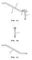
- FIG. 1A shows a locking member 17, an optional component of the device according to the present invention.
- Locking member 17 includes a connecting tube 1 attached to a grip 3 by a shim 2.
- Connecting tube 1 can be a trocar.
- connecting tube 1 has a hollow interior through which a drill bit, screwdriver, screw, or any fastener can be inserted to attach a bone plate 6 ( FIG. 3 ) to bone.
- connecting tube 1 is equipped with a drill bit.
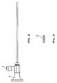
- FIGS. 4A and 4B show an endoscope attachment element 8, component of the device according to the present invention.
- Attachment element 8 has a distal end 15 and a proximal end 16 through which at least a portion of endoscope 4 ( FIG. 2 ) is inserted into a body member 18.
- a handle 10 provides a means for the user to manipulate attachment element 8.
- Attachment element 8 also includes a bone plate holder 7 having one end configured and dimensioned to hold at least a portion of bone plate 6. Bone plate holder 7 is operatively associated with body member 18 of attachment element 8 to move bone plate holder 7 between a first retracted position and a second implanting position.
- Distal end 15 is preferably shaped like a raspatory.
- a guide track 11 is positioned adjacent body member 18 and extends along the length of attachment element 8. Bone plate holder 7 is inserted in guide track 11 and can move along guide track 11 to change the position of bone plate 6 so that one of the screw holes of bone plate 6 aligns with an aperture 14 on attachment element 8. In order to prevent movement of bone plate holder 7 along guide track 11, one of the catches 12 on guide track 11 grasps a locking button 13 located on bone plate holder 7.
- the use of the device according to the present invention will be illustrated using the fixation of a mandibular fracture as an example.
- the surgeon makes a first incision at the mandibular angle.
- the surgeon first reduces the fracture using a surgical tool introduced through the first incision.
- Endoscopic attachment element 8 having a portion of endoscope 4 extending through body member 18 and bone plate holder 7 holding bone plate 6 is introduced through the first incision.
- the fracture and surround area can be viewed using endoscope 4.
- attachment element 8 has an optional integrated irrigation and suction system 9, any impediments to visualization can be removed.
- Connecting tube 1 of locking member 3 is introduced through this second incision and is securely connected to distal end 15 of attachment element 8.
- One way of achieving this secure connection is by providing an end of connecting tube 1 with a first securing element and distal end 15 of attachment element 8 with a second securing element such that the first and second securing elements cooperate to attach connecting tube 1 to attachment element 8.
- a drill bit is introduced through connecting tube 1 to create a hole through which a screw can be used to fix bone plate 6 to the bone. After all screws have been inserted and attachment element 8 and locking member 17 have been detached from each other and removed from the incisions, both incisions are closed.
Abstract
Description
- The present invention relates to a method for the stabilization of bone fractures and in particular to an endoscopic bone plate positioning device for use in such a procedure.
- Most mandibular fractures must be treated operatively. Often it is not possible to fix the fracture from the oral mucous membrane. For this reason, these types of fractures have been treated from a skin incision made in front of the ear.
- One possible serious complication in accessing the mandible from the cheek is damage to the facial nerve which divides into its branches in the parotid salivary gland, i.e., the immediate vicinity of the skin incision. Additionally, access from the exterior carries with it the possibility of aesthetically unappealing scarring.
- The risk of the above complications can be reduced by minimizing the surgical trauma associated with fixing a fracture. Endoscopic procedures are minimally invasive procedures which stabilize the fracture using a small incision, and hence decrease the surgical trauma associated with fixing a fracture. However, because of the small incision, it is difficult to implant the bone plate used to fix the fracture.
- Thus, there exists a need for an improved device for implanting a bone plate to conduct such procedures while minimizing complications.
- From
FR-A-2738375 - The present invention relates to a device for implanting a bone plate including an endoscope attachment element having proximal and distal ends and a body member configured and dimensioned to receive at least a portion of an endoscope, and a bone plate holder having one end which is configured and dimensioned to hold at least a portion of the bone plate. The bone plate holder is operatively associated with the body member of the endoscope attachment element to move the bone plate holder between a first retracted position and a second implanting position.
- In one embodiment, the endoscope attachment element, includes a guide track positioned adjacent the body member for receiving the bone plate holder and for providing sliding movement of the bone plate holder between the first and second positions. Preferably, the guide track includes a plurality of bone plate holder positioning catches and the bone plate holder includes a locking button for engaging one of the catches to retain the bone plate holder in the second implanting position.
- In another embodiment, the body member of the endoscope attachment element has a distal end which includes a raspatory having an aperture therein, wherein the aperture aligns with the end of the bone plate holder when the bone plate holder is retained in the second implanting position.
- The endoscope attachment element can have an integrated irrigation and suction system as well as a handle for manipulation.
- The device preferably further includes a locking member which has a connecting tube operatively associated with the bone plate to guide and enable fastening operations through the bone plate screw holes. In order to secure the connecting tube to the endoscope attachment element once the bone plate has been properly positioned, an end of the connecting tube can have a first securing element and the endoscope attachment element can have a second securing element.
-
-
FIG. 1A is a side view of a locking piece according to the present invention; -
FIG. 1B is side view of a connecting tube according to the present invention; -
FIG. 1C is a side view of a grip according to the present invention; -
FIG. 2 is a side view of an endoscope that can be used with the present invention; -
FIG. 3 is a top view of a bone plate that can be used with the present invention; -
FIG. 4A is a side view an endoscope attachment element according to the present invention; -
FIG. 4B is a bottom view (in the direction of arrow A inFIG. 4A ) of the endoscope attachment element; -
FIG. 4C is a top view of a slider of the endoscope attachment element; -
FIG. 5A is a side view of the device according to the present invention in use; -
FIG. 5B is a top view illustrating how the slider holds the bone plate; and -
FIG. 6 is a schematic showing a mandibular fracture fixed by a bone plate. -
FIG. 1A shows alocking member 17, an optional component of the device according to the present invention.Locking member 17 includes a connecting tube 1 attached to agrip 3 by ashim 2. Connecting tube 1 can be a trocar. As shown inFIG. 1B , connecting tube 1 has a hollow interior through which a drill bit, screwdriver, screw, or any fastener can be inserted to attach a bone plate 6 (FIG. 3 ) to bone. Alternatively, connecting tube 1 is equipped with a drill bit. -
FIGS. 4A and 4B show anendoscope attachment element 8, component of the device according to the present invention.Attachment element 8 has adistal end 15 and aproximal end 16 through which at least a portion of endoscope 4 (FIG. 2 ) is inserted into abody member 18. Ahandle 10 provides a means for the user to manipulateattachment element 8.Attachment element 8 also includes abone plate holder 7 having one end configured and dimensioned to hold at least a portion ofbone plate 6.Bone plate holder 7 is operatively associated withbody member 18 ofattachment element 8 to movebone plate holder 7 between a first retracted position and a second implanting position.Distal end 15 is preferably shaped like a raspatory. - A
guide track 11 is positionedadjacent body member 18 and extends along the length ofattachment element 8.Bone plate holder 7 is inserted inguide track 11 and can move alongguide track 11 to change the position ofbone plate 6 so that one of the screw holes ofbone plate 6 aligns with anaperture 14 onattachment element 8. In order to prevent movement ofbone plate holder 7 alongguide track 11, one of the catches 12 onguide track 11 grasps alocking button 13 located onbone plate holder 7. - The use of the device according to the present invention will be illustrated using the fixation of a mandibular fracture as an example. After surgical preparation which is standard for an endoscopic procedure, the surgeon makes a first incision at the mandibular angle. The surgeon first reduces the fracture using a surgical tool introduced through the first incision.
Endoscopic attachment element 8, having a portion of endoscope 4 extending throughbody member 18 andbone plate holder 7holding bone plate 6 is introduced through the first incision. Using the light from an illumination means 5, the fracture and surround area can be viewed using endoscope 4. Asattachment element 8 has an optional integrated irrigation andsuction system 9, any impediments to visualization can be removed. Oncebone plate 6 is in the desired position,bone plate holder 7 is moved alongguide track 11 until at least one screw hole ofbone plate 6 aligns withaperture 14. In order to prevent loss of this alignment, lockingbutton 13 is fixed to one of the catches 12. - After positioning of
bone plate 6, the surgeon makes a second incision directly above the fracture. Connecting tube 1 of lockingmember 3 is introduced through this second incision and is securely connected todistal end 15 ofattachment element 8. One way of achieving this secure connection is by providing an end of connecting tube 1 with a first securing element anddistal end 15 ofattachment element 8 with a second securing element such that the first and second securing elements cooperate to attach connecting tube 1 toattachment element 8. A drill bit is introduced through connecting tube 1 to create a hole through which a screw can be used to fixbone plate 6 to the bone. After all screws have been inserted andattachment element 8 and lockingmember 17 have been detached from each other and removed from the incisions, both incisions are closed. - While it is apparent that the illustrative embodiments of the invention herein disclosed fulfil the objectives stated above, it will be appreciated that numerous modifications and other embodiments may be devised by those skilled in the art.
Claims (12)
- A device for implanting a bone plate comprising:an endoscope attachment element (8) having proximal and distal ends (16; 15) and a body member (18) configured and dimensioned to receive at least a portion of an endoscope, and a bone plate holder (7) having one end which is configured and dimensioned to hold at least a portion of the bone plate (6);wherein the bone plate holder (7) is operati e associated with the body member (18) of the endoscope attachment element (8) to move the one plate holder (7) relative to the body member (18) between a first retracted position and a second implanting position.
- The device of claim 1, wherein the endoscope attachment element includes a guide track positioned adjacent the body member for receiving the bone plate holder and for providing sliding movement of the bone plate holder between the first and second positions.
- The device of claim 2, wherein the guide track includes a plurality of bone plate holder positioning catches therealong, and the bone plate holder includes a locking button for engaging one of the catches to retain the bone plate holder in the second implanting position.
- The device of claim 3, wherein the body member of the endoscope attachment element has a distal end which includes a raspatory having an aperture therein, wherein the aperture aligns with the end of the bone plate holder when the bone plate holder is retained in the second implanting position.
- The device of claim 1, wherein the endoscope attachment element includes an integrated irrigation and suction system for introducing a. fluid to clear debris from the distal end of the endoscope attachment element.
- The device of claim 1, further comprising a handle for manipulating the endoscope attachment element.
- The device of claim 1, further comprising an endoscope placed at least partially within the body member of the endoscope attachment element for viewing the distal end of the endoscope attachment element.
- The device of claim 1, further comprising a bone plate having at least one screw hole and being retained in position by the bone plate holder.
- The device of claim 8, further comprising a locking member which includes a connecting tube operatively associated with the bone plate to guide and enable fastening operations through the at least one bone plate screw hole.
- The device of claim 9, wherein the locking member includes a grip for manipulation thereof.
- The device of claim 10, wherein the connecting tube is mounted to the grip with a shim.
- The device of claim 10, wherein the connecting tube includes a first securing element and the distal end of the endoscope attachment element has a second securing element, with the first and second securing elements cooperating to securely connect the connecting tube to the endoscope attachment element.
Applications Claiming Priority (3)
| Application Number | Priority Date | Filing Date | Title |
|---|---|---|---|
| DE19717977 | 1997-04-23 | ||
| DE19717977A DE19717977A1 (en) | 1997-04-23 | 1997-04-23 | Lining=up fixture device used to treat jaw or face fractures |
| PCT/US1998/008149 WO1998047437A1 (en) | 1997-04-23 | 1998-04-23 | Endoscopic bone plate positioning device |
Publications (3)
| Publication Number | Publication Date |
|---|---|
| EP1021132A1 EP1021132A1 (en) | 2000-07-26 |
| EP1021132A4 EP1021132A4 (en) | 2009-07-22 |
| EP1021132B1 true EP1021132B1 (en) | 2010-10-06 |
Family
ID=7828034
Family Applications (1)
| Application Number | Title | Priority Date | Filing Date |
|---|---|---|---|
| EP98918601A Expired - Lifetime EP1021132B1 (en) | 1997-04-23 | 1998-04-23 | Endoscopic bone plate positioning device |
Country Status (8)
| Country | Link |
|---|---|
| US (1) | US5947970A (en) |
| EP (1) | EP1021132B1 (en) |
| JP (1) | JP4126089B2 (en) |
| AT (1) | ATE483412T1 (en) |
| AU (1) | AU7149998A (en) |
| CA (1) | CA2287385A1 (en) |
| DE (2) | DE19717977A1 (en) |
| WO (1) | WO1998047437A1 (en) |
Families Citing this family (34)
| Publication number | Priority date | Publication date | Assignee | Title |
|---|---|---|---|---|
| US5938664A (en) * | 1998-03-31 | 1999-08-17 | Zimmer, Inc. | Orthopaedic bone plate |
| US6287313B1 (en) * | 1999-11-23 | 2001-09-11 | Sdgi Holdings, Inc. | Screw delivery system and method |
| WO2002024081A1 (en) | 2000-09-22 | 2002-03-28 | Codman & Shurtleff, Inc. | Self centering bone drill |
| US8475504B2 (en) * | 2007-07-19 | 2013-07-02 | Acumed Llc | Method of bone fixation with slender spanning members disposed outside bone |
| ATE489904T1 (en) * | 2001-06-27 | 2010-12-15 | Depuy Products Inc | MINIMAL INVASIVE ORTHOPEDIC DEVICE |
| US7235077B1 (en) | 2001-11-09 | 2007-06-26 | Board Of Regents Of The University And Community College System Of Nevada On Behalf Of The University Of Nevada, Reno | Bone fixation device and method |
| US20060235408A1 (en) * | 2001-11-09 | 2006-10-19 | Wang Robert C | Apparatus and methods for bone fracture fixation |
| US6660006B2 (en) * | 2002-04-17 | 2003-12-09 | Stryker Spine | Rod persuader |
| US7572276B2 (en) | 2002-05-06 | 2009-08-11 | Warsaw Orthopedic, Inc. | Minimally invasive instruments and methods for inserting implants |
| DE10301691B4 (en) * | 2003-01-17 | 2006-10-12 | Stryker Leibinger Gmbh & Co. Kg | Socket handle and socket system |
| DE10310978B3 (en) * | 2003-03-06 | 2004-08-26 | Karl Storz Gmbh & Co. Kg | Surgical instrument for creating operating field for repair work on fractured human jaw uses endoscope with shaft passing through small incision in cheek to flat plate fixed to jaw |
| WO2005004728A1 (en) * | 2003-07-14 | 2005-01-20 | Synthes Ag Chur | Aiming device |
| US7252673B2 (en) * | 2003-09-10 | 2007-08-07 | Warsaw Orthopedic, Inc. | Devices and methods for inserting spinal implants |
| US7341587B2 (en) * | 2003-11-20 | 2008-03-11 | Warsaw Orthopedic, Inc. | Methods and devices for inserting and engaging vertebral implants in minimally invasive procedures |
| US20060173458A1 (en) * | 2004-10-07 | 2006-08-03 | Micah Forstein | Bone fracture fixation system |
| EP1814474B1 (en) | 2004-11-24 | 2011-09-14 | Samy Abdou | Devices for inter-vertebral orthopedic device placement |
| US7708743B2 (en) * | 2005-04-29 | 2010-05-04 | Warsaw Orthopedic, Inc. | Apparatus and method for positioning an implant during surgery |
| US8753394B2 (en) * | 2005-06-03 | 2014-06-17 | Arthrodisc, L.L.C. | Minimally invasive apparatus to manipulate and revitalize spinal column disc |
| WO2007056516A2 (en) * | 2005-11-09 | 2007-05-18 | Abdou M S | Bone fixation systems and methods of implantation |
| FR2937239B1 (en) * | 2008-10-17 | 2010-11-26 | Fournitures Hospitalieres Ind | SURGICAL DEVICE, IN PARTICULAR FOR THE SURGERY OF THE FOOT |
| US8764806B2 (en) | 2009-12-07 | 2014-07-01 | Samy Abdou | Devices and methods for minimally invasive spinal stabilization and instrumentation |
| US8652180B2 (en) | 2010-09-27 | 2014-02-18 | Acumed Llc | Handle assembly having a radiopaque region to facilitate positioning a bone plate on bone |
| US8900135B2 (en) * | 2011-03-29 | 2014-12-02 | Covidien Lp | Single incision deployable platform |
| US8845728B1 (en) | 2011-09-23 | 2014-09-30 | Samy Abdou | Spinal fixation devices and methods of use |
| US11123117B1 (en) | 2011-11-01 | 2021-09-21 | Nuvasive, Inc. | Surgical fixation system and related methods |
| US20130226240A1 (en) | 2012-02-22 | 2013-08-29 | Samy Abdou | Spinous process fixation devices and methods of use |
| US9198767B2 (en) | 2012-08-28 | 2015-12-01 | Samy Abdou | Devices and methods for spinal stabilization and instrumentation |
| US9320617B2 (en) | 2012-10-22 | 2016-04-26 | Cogent Spine, LLC | Devices and methods for spinal stabilization and instrumentation |
| CN103082987B (en) * | 2013-02-04 | 2015-02-11 | 方玉树 | Lens for bone fracture |
| US10206723B2 (en) | 2013-03-14 | 2019-02-19 | Stryker European Holdings I, Llc | Rod inserter and insertion tube |
| US10857003B1 (en) | 2015-10-14 | 2020-12-08 | Samy Abdou | Devices and methods for vertebral stabilization |
| US10973648B1 (en) | 2016-10-25 | 2021-04-13 | Samy Abdou | Devices and methods for vertebral bone realignment |
| US10744000B1 (en) | 2016-10-25 | 2020-08-18 | Samy Abdou | Devices and methods for vertebral bone realignment |
| US11179248B2 (en) | 2018-10-02 | 2021-11-23 | Samy Abdou | Devices and methods for spinal implantation |
Family Cites Families (29)
| Publication number | Priority date | Publication date | Assignee | Title |
|---|---|---|---|---|
| US3856016A (en) * | 1972-11-03 | 1974-12-24 | H Davis | Method for mechanically applying an occlusion clip to an anatomical tubular structure |
| JPS5641684Y2 (en) * | 1977-11-24 | 1981-09-30 | ||
| US4854302A (en) * | 1987-11-12 | 1989-08-08 | Welch Allyn, Inc. | Video equipped endoscope with needle probe |
| US4994910A (en) * | 1989-07-06 | 1991-02-19 | Acuimage Corporation | Modular endoscopic apparatus with probe |
| US4959065A (en) * | 1989-07-14 | 1990-09-25 | Techmedica, Inc. | Bone plate with positioning member |
| US4923471A (en) * | 1989-10-17 | 1990-05-08 | Timesh, Inc. | Bone fracture reduction and fixation devices with identity tags |
| US5199417A (en) * | 1990-12-21 | 1993-04-06 | Circon Corporation | Endoscope having a deflectable distal section and a semi-rigid proximal section |
| DE4139029C2 (en) * | 1991-11-27 | 1996-05-23 | Erbe Elektromedizin | Device for the coagulation of biological tissues |
| DE9209180U1 (en) * | 1992-07-09 | 1993-11-11 | Medizin Tech Werkstaette Arzt | Handle for probes that can be inserted into endoscopes |
| US5373840A (en) * | 1992-10-02 | 1994-12-20 | Knighton; David R. | Endoscope and method for vein removal |
| US5667478A (en) * | 1992-11-06 | 1997-09-16 | Clarus Medical Systems, Inc. | Surgical instrument with stick-on fiber-optic viewing system and method of using |
| US5334150A (en) * | 1992-11-17 | 1994-08-02 | Kaali Steven G | Visually directed trocar for laparoscopic surgical procedures and method of using same |
| US5423826A (en) * | 1993-02-05 | 1995-06-13 | Danek Medical, Inc. | Anterior cervical plate holder/drill guide and method of use |
| US5377668A (en) * | 1993-04-12 | 1995-01-03 | Optimed Technologies, Inc. | Apparatus and method for endoscopic diagnostics and therapy |
| US5467762A (en) * | 1993-09-13 | 1995-11-21 | United States Surgical Corporation | Optical trocar |
| US5441041A (en) * | 1993-09-13 | 1995-08-15 | United States Surgical Corporation | Optical trocar |
| US5478338A (en) * | 1993-09-24 | 1995-12-26 | Reynard; Michael | Fiber optic sleeve for surgical instruments |
| FR2712798A1 (en) * | 1993-11-22 | 1995-06-02 | Le Foll Dominique | Fastening formation for endoscope against internal body tissue |
| DE4445599A1 (en) * | 1993-11-29 | 1995-09-21 | Etb Endoskopische Technik Gmbh | Endoscopic instrument for endoscopic subfascial discussion of perforans veins |
| US5558622A (en) * | 1994-09-02 | 1996-09-24 | Greenberg Surgical Technologies, Llc | Mandibular border retractor and method for fixating a fractured mandible |
| US5641287A (en) * | 1994-10-25 | 1997-06-24 | Gittleman; Neal B. | Dental tool guidance template and method |
| FR2738475B1 (en) * | 1995-09-11 | 1998-01-16 | Landanger Landos | PLIERS FOR PRESENTING MAXILLO-FACIAL OSTEOSYNTHESIS PLATES |
| US5702463A (en) * | 1996-02-20 | 1997-12-30 | Smith & Nephew Inc. | Tibial prosthesis with polymeric liner and liner insertion/removal instrument |
| US5755721A (en) * | 1996-03-13 | 1998-05-26 | Synthes | Plate holding drill guide and trocar and method of holding a plate |
| US5725523A (en) * | 1996-03-29 | 1998-03-10 | Mueller; Richard L. | Lateral-and posterior-aspect method and apparatus for laser-assisted transmyocardial revascularization and other surgical applications |
| US5700275A (en) * | 1996-04-25 | 1997-12-23 | United States Surgical Corporation | Articulating endoscopic surgical instrument |
| US5700267A (en) * | 1996-08-15 | 1997-12-23 | Kinetikos Medical Incorporated | Method for repairing bone fractures using bone-lock system |
| US5690631A (en) * | 1996-09-11 | 1997-11-25 | Walter Lorenz Surgical, Inc. | Multi-configurable plating system |
| US5732992A (en) * | 1996-12-26 | 1998-03-31 | Exactech, Incorporated | Medical appliance tool providing one hand actuation |
-
1997
- 1997-04-23 DE DE19717977A patent/DE19717977A1/en not_active Withdrawn
-
1998
- 1998-04-23 AT AT98918601T patent/ATE483412T1/en not_active IP Right Cessation
- 1998-04-23 DE DE69841925T patent/DE69841925D1/en not_active Expired - Lifetime
- 1998-04-23 WO PCT/US1998/008149 patent/WO1998047437A1/en active Search and Examination
- 1998-04-23 EP EP98918601A patent/EP1021132B1/en not_active Expired - Lifetime
- 1998-04-23 CA CA002287385A patent/CA2287385A1/en not_active Abandoned
- 1998-04-23 US US09/064,874 patent/US5947970A/en not_active Expired - Fee Related
- 1998-04-23 JP JP54632098A patent/JP4126089B2/en not_active Expired - Fee Related
- 1998-04-23 AU AU71499/98A patent/AU7149998A/en not_active Abandoned
Also Published As
| Publication number | Publication date |
|---|---|
| ATE483412T1 (en) | 2010-10-15 |
| WO1998047437A1 (en) | 1998-10-29 |
| AU7149998A (en) | 1998-11-13 |
| EP1021132A4 (en) | 2009-07-22 |
| JP2002501411A (en) | 2002-01-15 |
| EP1021132A1 (en) | 2000-07-26 |
| DE69841925D1 (en) | 2010-11-18 |
| CA2287385A1 (en) | 1998-10-29 |
| DE19717977A1 (en) | 1998-05-28 |
| US5947970A (en) | 1999-09-07 |
| JP4126089B2 (en) | 2008-07-30 |
Similar Documents
| Publication | Publication Date | Title |
|---|---|---|
| EP1021132B1 (en) | Endoscopic bone plate positioning device | |
| US5558622A (en) | Mandibular border retractor and method for fixating a fractured mandible | |
| US8382802B2 (en) | Systems, methods and devices for placement of bone anchors and connectors | |
| EP1408856B1 (en) | Minimally invasive orthopaedic apparatus | |
| US8579912B2 (en) | Sacroiliac joint fusion alignment guide | |
| US8231661B2 (en) | Systems and methods for minimally invasive facet fusion | |
| US6547795B2 (en) | Surgical guide system for stabilization of the spine | |
| US6007487A (en) | Tissue retractor for use through a cannula | |
| US8764752B2 (en) | Protection sleeve holding mechanism | |
| JP2005506098A (en) | Spinal pedicle screw placement retractor and method | |
| AU2002305696A1 (en) | Minimally invasive orthopaedic apparatus and methods | |
| US20030018340A1 (en) | Method and apparatus for installing cannula | |
| US20230132273A1 (en) | Alignment devices for use in correction of bone deformities | |
| JP2002282263A (en) | Operation instrument kit for excising ostegenetic vertebral arch |
Legal Events
| Date | Code | Title | Description |
|---|---|---|---|
| PUAI | Public reference made under article 153(3) epc to a published international application that has entered the european phase |
Free format text: ORIGINAL CODE: 0009012 |
|
| 17P | Request for examination filed |
Effective date: 19991022 |
|
| AK | Designated contracting states |
Kind code of ref document: A1 Designated state(s): AT BE CH DE ES FI GB LI NL SE |
|
| RAP1 | Party data changed (applicant data changed or rights of an application transferred) |
Owner name: SYNTHES GMBH |
|
| REG | Reference to a national code |
Ref country code: HK Ref legal event code: WD Ref document number: 1027953 Country of ref document: HK |
|
| A4 | Supplementary search report drawn up and despatched |
Effective date: 20090624 |
|
| 17Q | First examination report despatched |
Effective date: 20090902 |
|
| GRAP | Despatch of communication of intention to grant a patent |
Free format text: ORIGINAL CODE: EPIDOSNIGR1 |
|
| GRAS | Grant fee paid |
Free format text: ORIGINAL CODE: EPIDOSNIGR3 |
|
| GRAA | (expected) grant |
Free format text: ORIGINAL CODE: 0009210 |
|
| AK | Designated contracting states |
Kind code of ref document: B1 Designated state(s): AT BE CH DE ES FI GB LI NL SE |
|
| REG | Reference to a national code |
Ref country code: GB Ref legal event code: FG4D |
|
| REG | Reference to a national code |
Ref country code: CH Ref legal event code: NV Representative=s name: DR. LUSUARDI AG Ref country code: CH Ref legal event code: EP |
|
| REF | Corresponds to: |
Ref document number: 69841925 Country of ref document: DE Date of ref document: 20101118 Kind code of ref document: P |
|
| REG | Reference to a national code |
Ref country code: NL Ref legal event code: VDEP Effective date: 20101006 |
|
| PG25 | Lapsed in a contracting state [announced via postgrant information from national office to epo] |
Ref country code: NL Free format text: LAPSE BECAUSE OF FAILURE TO SUBMIT A TRANSLATION OF THE DESCRIPTION OR TO PAY THE FEE WITHIN THE PRESCRIBED TIME-LIMIT Effective date: 20101006 Ref country code: FI Free format text: LAPSE BECAUSE OF FAILURE TO SUBMIT A TRANSLATION OF THE DESCRIPTION OR TO PAY THE FEE WITHIN THE PRESCRIBED TIME-LIMIT Effective date: 20101006 Ref country code: AT Free format text: LAPSE BECAUSE OF FAILURE TO SUBMIT A TRANSLATION OF THE DESCRIPTION OR TO PAY THE FEE WITHIN THE PRESCRIBED TIME-LIMIT Effective date: 20101006 Ref country code: SE Free format text: LAPSE BECAUSE OF FAILURE TO SUBMIT A TRANSLATION OF THE DESCRIPTION OR TO PAY THE FEE WITHIN THE PRESCRIBED TIME-LIMIT Effective date: 20101006 |
|
| PG25 | Lapsed in a contracting state [announced via postgrant information from national office to epo] |
Ref country code: BE Free format text: LAPSE BECAUSE OF FAILURE TO SUBMIT A TRANSLATION OF THE DESCRIPTION OR TO PAY THE FEE WITHIN THE PRESCRIBED TIME-LIMIT Effective date: 20101006 |
|
| PG25 | Lapsed in a contracting state [announced via postgrant information from national office to epo] |
Ref country code: ES Free format text: LAPSE BECAUSE OF FAILURE TO SUBMIT A TRANSLATION OF THE DESCRIPTION OR TO PAY THE FEE WITHIN THE PRESCRIBED TIME-LIMIT Effective date: 20110117 |
|
| PLBE | No opposition filed within time limit |
Free format text: ORIGINAL CODE: 0009261 |
|
| STAA | Information on the status of an ep patent application or granted ep patent |
Free format text: STATUS: NO OPPOSITION FILED WITHIN TIME LIMIT |
|
| 26N | No opposition filed |
Effective date: 20110707 |
|
| REG | Reference to a national code |
Ref country code: DE Ref legal event code: R097 Ref document number: 69841925 Country of ref document: DE Effective date: 20110707 |
|
| PGFP | Annual fee paid to national office [announced via postgrant information from national office to epo] |
Ref country code: GB Payment date: 20140423 Year of fee payment: 17 |
|
| PGFP | Annual fee paid to national office [announced via postgrant information from national office to epo] |
Ref country code: DE Payment date: 20140430 Year of fee payment: 17 Ref country code: CH Payment date: 20140414 Year of fee payment: 17 |
|
| REG | Reference to a national code |
Ref country code: DE Ref legal event code: R119 Ref document number: 69841925 Country of ref document: DE |
|
| REG | Reference to a national code |
Ref country code: CH Ref legal event code: PL |
|
| GBPC | Gb: european patent ceased through non-payment of renewal fee |
Effective date: 20150423 |
|
| PG25 | Lapsed in a contracting state [announced via postgrant information from national office to epo] |
Ref country code: LI Free format text: LAPSE BECAUSE OF NON-PAYMENT OF DUE FEES Effective date: 20150430 Ref country code: CH Free format text: LAPSE BECAUSE OF NON-PAYMENT OF DUE FEES Effective date: 20150430 Ref country code: GB Free format text: LAPSE BECAUSE OF NON-PAYMENT OF DUE FEES Effective date: 20150423 Ref country code: DE Free format text: LAPSE BECAUSE OF NON-PAYMENT OF DUE FEES Effective date: 20151103 |