EP1027604B1 - Extended dynamic range assays - Google Patents
Extended dynamic range assays Download PDFInfo
- Publication number
- EP1027604B1 EP1027604B1 EP98956381A EP98956381A EP1027604B1 EP 1027604 B1 EP1027604 B1 EP 1027604B1 EP 98956381 A EP98956381 A EP 98956381A EP 98956381 A EP98956381 A EP 98956381A EP 1027604 B1 EP1027604 B1 EP 1027604B1
- Authority
- EP
- European Patent Office
- Prior art keywords
- probe
- analyte
- ortho
- label
- labeled
- Prior art date
- Legal status (The legal status is an assumption and is not a legal conclusion. Google has not performed a legal analysis and makes no representation as to the accuracy of the status listed.)
- Expired - Lifetime
Links
Images


Classifications
-
- G—PHYSICS
- G01—MEASURING; TESTING
- G01N—INVESTIGATING OR ANALYSING MATERIALS BY DETERMINING THEIR CHEMICAL OR PHYSICAL PROPERTIES
- G01N33/00—Investigating or analysing materials by specific methods not covered by groups G01N1/00 - G01N31/00
- G01N33/48—Biological material, e.g. blood, urine; Haemocytometers
- G01N33/50—Chemical analysis of biological material, e.g. blood, urine; Testing involving biospecific ligand binding methods; Immunological testing
- G01N33/53—Immunoassay; Biospecific binding assay; Materials therefor
- G01N33/543—Immunoassay; Biospecific binding assay; Materials therefor with an insoluble carrier for immobilising immunochemicals
- G01N33/54306—Solid-phase reaction mechanisms
-
- C—CHEMISTRY; METALLURGY
- C12—BIOCHEMISTRY; BEER; SPIRITS; WINE; VINEGAR; MICROBIOLOGY; ENZYMOLOGY; MUTATION OR GENETIC ENGINEERING
- C12Q—MEASURING OR TESTING PROCESSES INVOLVING ENZYMES, NUCLEIC ACIDS OR MICROORGANISMS; COMPOSITIONS OR TEST PAPERS THEREFOR; PROCESSES OF PREPARING SUCH COMPOSITIONS; CONDITION-RESPONSIVE CONTROL IN MICROBIOLOGICAL OR ENZYMOLOGICAL PROCESSES
- C12Q1/00—Measuring or testing processes involving enzymes, nucleic acids or microorganisms; Compositions therefor; Processes of preparing such compositions
- C12Q1/68—Measuring or testing processes involving enzymes, nucleic acids or microorganisms; Compositions therefor; Processes of preparing such compositions involving nucleic acids
- C12Q1/6813—Hybridisation assays
- C12Q1/6816—Hybridisation assays characterised by the detection means
-
- Y—GENERAL TAGGING OF NEW TECHNOLOGICAL DEVELOPMENTS; GENERAL TAGGING OF CROSS-SECTIONAL TECHNOLOGIES SPANNING OVER SEVERAL SECTIONS OF THE IPC; TECHNICAL SUBJECTS COVERED BY FORMER USPC CROSS-REFERENCE ART COLLECTIONS [XRACs] AND DIGESTS
- Y10—TECHNICAL SUBJECTS COVERED BY FORMER USPC
- Y10S—TECHNICAL SUBJECTS COVERED BY FORMER USPC CROSS-REFERENCE ART COLLECTIONS [XRACs] AND DIGESTS
- Y10S435/00—Chemistry: molecular biology and microbiology
- Y10S435/962—Prevention or removal of interfering materials or reactants or other treatment to enhance results, e.g. determining or preventing nonspecific binding
-
- Y—GENERAL TAGGING OF NEW TECHNOLOGICAL DEVELOPMENTS; GENERAL TAGGING OF CROSS-SECTIONAL TECHNOLOGIES SPANNING OVER SEVERAL SECTIONS OF THE IPC; TECHNICAL SUBJECTS COVERED BY FORMER USPC CROSS-REFERENCE ART COLLECTIONS [XRACs] AND DIGESTS
- Y10—TECHNICAL SUBJECTS COVERED BY FORMER USPC
- Y10S—TECHNICAL SUBJECTS COVERED BY FORMER USPC CROSS-REFERENCE ART COLLECTIONS [XRACs] AND DIGESTS
- Y10S435/00—Chemistry: molecular biology and microbiology
- Y10S435/968—High energy substrates, e.g. fluorescent, chemiluminescent, radioactive
-
- Y—GENERAL TAGGING OF NEW TECHNOLOGICAL DEVELOPMENTS; GENERAL TAGGING OF CROSS-SECTIONAL TECHNOLOGIES SPANNING OVER SEVERAL SECTIONS OF THE IPC; TECHNICAL SUBJECTS COVERED BY FORMER USPC CROSS-REFERENCE ART COLLECTIONS [XRACs] AND DIGESTS
- Y10—TECHNICAL SUBJECTS COVERED BY FORMER USPC
- Y10S—TECHNICAL SUBJECTS COVERED BY FORMER USPC CROSS-REFERENCE ART COLLECTIONS [XRACs] AND DIGESTS
- Y10S436/00—Chemistry: analytical and immunological testing
- Y10S436/80—Fluorescent dyes, e.g. rhodamine
-
- Y—GENERAL TAGGING OF NEW TECHNOLOGICAL DEVELOPMENTS; GENERAL TAGGING OF CROSS-SECTIONAL TECHNOLOGIES SPANNING OVER SEVERAL SECTIONS OF THE IPC; TECHNICAL SUBJECTS COVERED BY FORMER USPC CROSS-REFERENCE ART COLLECTIONS [XRACs] AND DIGESTS
- Y10—TECHNICAL SUBJECTS COVERED BY FORMER USPC
- Y10S—TECHNICAL SUBJECTS COVERED BY FORMER USPC CROSS-REFERENCE ART COLLECTIONS [XRACs] AND DIGESTS
- Y10S436/00—Chemistry: analytical and immunological testing
- Y10S436/811—Test for named disease, body condition or organ function
- Y10S436/814—Pregnancy
-
- Y—GENERAL TAGGING OF NEW TECHNOLOGICAL DEVELOPMENTS; GENERAL TAGGING OF CROSS-SECTIONAL TECHNOLOGIES SPANNING OVER SEVERAL SECTIONS OF THE IPC; TECHNICAL SUBJECTS COVERED BY FORMER USPC CROSS-REFERENCE ART COLLECTIONS [XRACs] AND DIGESTS
- Y10—TECHNICAL SUBJECTS COVERED BY FORMER USPC
- Y10S—TECHNICAL SUBJECTS COVERED BY FORMER USPC CROSS-REFERENCE ART COLLECTIONS [XRACs] AND DIGESTS
- Y10S436/00—Chemistry: analytical and immunological testing
- Y10S436/815—Test for named compound or class of compounds
- Y10S436/817—Steroids or hormones
Definitions
- This invention relates to compositions and methods for detecting analytes which may be present in a wide range of possible concentrations through the use of at least two labeled binding partners or probes, and specifically relates to using labeled binding partners or probes that specifically bind mutually exclusive target regions of the same analyte, such that the use of the at least two binding partners or probes extends the dynamic range of an assay for analyte detection.
- Assays for the detection and/or quantification of analytes exist in many different forms and formats. In many cases, the amount of the analyte in a sample is not great enough for direct detection or quantification. In such cases, a secondary molecule capable of binding to or interacting with the analyte must be used to indicate the presence or amount of analyte in a sample. Unless the secondary molecule is directly detectable, the secondary molecule must be conjugated with a label which will be detected when the secondary molecule binds the analyte.
- binding partners such secondary molecules capable of binding or interacting with analytes are referred to as "binding partners” or “probes.”
- detectable analytes include antibodies, proteins, cell-surface receptors, cytokines, hormones, antigens, nucleic acids, metals, molecular complexes such as polymeric arrangements of proteins or other macromolecules, and the like.
- binding partners or probes for detecting such analytes may be antibodies, proteins, antigens, haptens, nucleic acid probes, chelating agents, enzymes, enzyme substrates, and analogs of these.
- ELISA enzyme-linked immuno-absorption assay
- the analyte is contacted with a primary antibody capable of binding at least one domain or "target region" of the analyte. After the excess antibody is washed free of the resulting analyte: antibody complex, the primary antibody is specifically bound with an enzyme-labeled secondary antibody.
- a chromogenic enzyme substrate is then provided and incubated under conditions favoring enzyme-mediated reaction of the substrate.
- the resulting colored product and its intensity after a reaction time are indications of the presence and amount, respectively, of the analyte originally present in the sample.
- Suitable enzymes used in ELISA are, for example, ⁇ -galactosidase, acid phosphatase, and alkaline phosphatase and suitable enzyme substrates for use with such enzymes include x-gal (5-bromo-4-chloro-3-indolyl- ⁇ -D-galacto-pyranoside) and p-nitrophenyl phosphate, although other enzymes and substrates are well known by those skilled in the art.
- Variations of ELISA exist; for example, the primary antibody may be linked to an enzyme, thus eliminating the secondary antibody step. Nevertheless, these assay methods include common steps of contacting an analyte with a labeled binding partner and subsequently detecting the analyte-bound label as an indication of the presence or amount of the analyte.
- Frengen (US Pat. No. 5,739,042) describes a binary assay system in which two independently determinable forms of solid-supported binding partners (e.g., monoclonal antibodies) are reacted successively rather than simultaneously with analyte and labeled ligand. The analyte concentration is obtained from readings derived from these two forms by comparison to a double standard curve.
- solid-supported binding partners e.g., monoclonal antibodies
- Assays are distinguished based on the type of analytes to be detected, the type of binding partner with which the analyte binds, and the type of label used. Assays can also be classified based on whether the method involves the immobilization of the analyte or the analyte: binding partner complex.
- a probe is allowed to bind its analyte, usually under conditions of probe excess. Either the analyte or the probe may be immobilized to a solid support, thus immobilizing the resulting probe: analyte complex. Often, the complex is formed in the liquid phase and then immobilized.
- probe: analyte complexes After probe: analyte complexes have been immobilized, the excess uncomplexed probes are washed away. It the probes are directly labeled, the label is then detected as an indication of the presence of analyte.
- probe: analyte complexes may be separated from free probe, by means such as gel filtration chromatography, electrophoresis, electrofocusing, or other methods that separate based on the size or charge of the probe: analyte complex.
- homogeneous assays take place wholly in a single phase without a step to separate probe: analyte complex from free probe.
- probe usually either the probe: analyte complex, or the free probe is altered after formation of the complex to permit the separate detection of analyte in the presence of the free probe.
- One way of differentiating free probe from probe that is bound to analyte involves alteration or selective inactivation of the label joined to the probe rather than the free probe itself. Arnold, et al. (US Pat. No.
- This assay method uses multiple antibodies to detect an antigen having multiple binding sites: one antibody is conjugated to a chemiluminescent catalyst that emits light in the presence of suitable chemiluminescent reagents and the other antibody (the "absorber/emitter”) is conjugated to a moiety that absorbs light at one wavelength and emits light at another wavelength.
- the separately-labeled antibodies bind to a single antigen in close proximity, highly efficient energy transfer occurs from the chemiluminescent catalyst conjugated antibody to the absorber/emitter, producing a light signal that is related to the amount of antigen.
- hybrid assays which utilize aspects of both homogeneous and heterogeneous assays, termed “hybrid” assays. These methods may, for example, employ a single-phase selective alteration of the probe or the probe: analyte complex, followed by a separation step to further decrease the level of background in the assay (e.g., see US Pat. No. 5,658,737 to Nelson et al.)
- background generally describes probe or label in the assay which is not bound specifically to analyte and which may mask positive results at low analyte levels.
- background may be provided by a small amount of probe which is not removed during the physical separation of probe: analyte complex from free probe. If the probe is labeled, this small amount of probe will be detected as a residual level of detectable signal.
- background may be provided by the inability to totally alter all free probe or probe-linked label molecules.
- a low level of signal commonly between 0.001% and 10% of the total signal, more commonly between about 0.01% and 1%, is present as a "baseline" below which results cannot be relied on for accuracy.
- the level of background inherent in a particular assay limits the lowest amount of analyte which can be detected and/or measured.
- dynamic range is meant a linear or predictably accurate correspondence between the level of analyte present in the assayed sample and the amount of signal obtained from the label that indicates the analyte's presence. It is readily apparent to those skilled in the art that the dynamic range of an assay cannot extend below the background level contributed by the detection of non-specific label. Thus, the higher the background, the more limited the dynamic range. Moreover, if high amounts of analyte are to be detected, correspondingly high amounts of probe must be used, leading to higher backgrounds.
- an assay's dynamic range may limit an assay's dynamic range. Often, a limitation is the maximum amount of signal capable of being detected or reported by the label-detection device used in the assay. Thus, if an instrument can only accurately read up to, for example, one million counts per second and the sample yields two million counts per second, the extra one million counts are not reported by the instrument, and the upper extent of the assay dynamic range is half of what is necessary to accurately quantify the sample.
- instruments used to detect the labeled probe may have inherent electronic "noise” (sometimes also referred to as “background”) and which can also contribute to limitations on the accuracy of analyte detection.
- noise sometimes also referred to as “background”
- an instrument's electronic signal-to-noise ratio may be improved, noise combines with the inherent assay background described above to further limit the ability to detect or quantify analytes in a wide range of concentrations.
- practitioners of assays generally obtain multiple samples from a single source, or test serial dilutions of a sample to detect the presence of an unknown amount of analyte.
- Some methods increase sensitivity of an assay without extending the dynamic range of the reaction. That is, these methods simply allow one to detect a lesser amount of analyte than previously detected in the same sort of reaction.
- Some methods of increasing sensitivity rely on using mixtures of binding partners. For example, Canfield et al. (US Pat. No. 4,514,505) describe an antigen detection method with enhanced sensitivity that uses mixtures of monoclonal antibodies containing at least two antibodies that can bind simultaneously to at least two different antigenic sites. The amounts of each monoclonal antibody vary, but generally, the preferred amount of each antibody relative to the amount of the other antibodies is substantially the same as the ratio of the binding constants of the antibodies to the antigen to be detected.
- Another assay method uses nucleic acid binding partners to test for bacterial contamination of food by using one or more Salmonella -specific labeled probes to bind to a single Salmonella chromosome (European Pat. App. No. 0114668).
- Salmonella -specific labeled probes to bind to a single Salmonella chromosome (European Pat. App. No. 0114668).
- multiple probes are used to increase the assay sensitivity because each chromosome binds multiple labels.
- One aspect of the present invention is a method of detecting an analyte over a range of concentrations in a single sample.
- This method includes the steps of: providing a probe reagent comprising at least two probes, wherein each probe comprises a target binding moiety and a detectable label, and wherein each probe is present in an amount capable of detecting a range of analyte concentrations that differs from the range of analyte concentrations detectable by another probe in the reagent wherein the probe reagent contains a first probe present in at least a 10-fold molar excess relative to a second probe, such that the probe reagent is capable of detecting a range of analyte concentrations that is greater than the range of analyte concentrations detectable by a single probe in the reagent; and contacting in a single vessel the probe reagent with a sample containing an analyte.
- the method includes the steps of incubating the sample and the probe reagent under conditions favoring specific binding of the target binding moiety of each probe with a target region of the analyte, wherein the target region recognized by each probe differs from the target region recognized by another probe in the probe reagent, thereby producing a specific binding complex comprising the analyte and at least one probe; and detecting the presence of the specific binding complex by detecting a signal from a label indicating the presence of at least one probe in the specific binding complex, thereby indicating the presence of the analyte in the sample within the range of analyte concentrations detectable by the probe in the specific binding complex.
- the detectable label of one probe in the probe reagent results in a signal that is distinguishable from a signal resulting from the detectable label of another probe in the reagent.
- the detecting step measures signal characteristic of kinetics of a reaction.
- the providing step uses a probe reagent in which specific activity of one probe differs from specific activity of another probe in the reagent.
- the providing step uses a probe reagent wherein the detectable label of a first probe results in a signal that is distinguishable from a signal resulting from the detectable label of a second probe in the reagent, and wherein specific activity of the first probe differs from specific activity of the second probe in the reagent.
- the signal resulting from the first probe is distinguishable from the signal resulting from the second probe by measuring kinetics of a reaction in the detecting step.
- the detectable label of at least one probe is a direct label such that the detecting step measures a signal resulting from the label.
- at least one probe has a detectable label that is a fluorescent label, a luminescent label, a radioactive label, a chromophore, an enzyme, or an enzyme substrate.
- the detectable label is a chemiluminescent label.
- at least one probe of the probe reagent has a detectable label that is a ligand capable of specifically binding a signal-producing binding partner.
- the detecting step detects a signal that is light emittance, light absorbance, radioactive decay, a precipitate, or a product of an enzymatic reaction.
- the contacting step uses a probe reagent wherein a first probe is present in at least a 10-fold molar excess relative to a second probe.
- the first probe is present in at least a 100-fold molar excess, or at least a 1,000-fold molar excess, or at least a 10,000-fold molar excess relative to a second-probe.
- the contacting step includes an analyte that is a nucleic acid, a protein, a lipid, a carbohydrate or a compound that is a combination thereof.
- the contacting step includes an analyte that is a nucleic acid, and more preferably, the analyte is RNA.
- at least one probe in the probe reagent is a nucleic acid, a protein, a lipid, a carbohydrate or a compound that is a combination thereof.
- at least one probe is a nucleic acid or analog thereof. More preferably, at least one probe is a DNA, RNA, DNA analog, RNA analog or a molecule that includes a combination thereof.
- compositions for use as a probe reagent for detecting an analyte in a single sample includes at least two probes, wherein each probe comprises a target binding moiety that binds specifically to a target region of the analyte, and a detectable label, wherein each probe binds to a target region that differs from the target region of another probe in the probe reagent, and wherein each probe is present in an amount capable of detecting a range of analyte concentrations that differs from a range of analyte concentrations detectable by another probe in the reagent, such that the probe reagent is capable of detecting the analyte over an extended range of analyte concentrations that is greater than the range of analyte concentrations detectable by a single probe in the reagent and wherein a first probe in the probe reagent is present in an amount that differs by at least about a 10-fold molar excess relative to a second probe in the
- each probe is distinguishable from another probe in the probe reagent because of its specific activity, a property resulting from its detectable label, or a combination thereof.
- Another embodiment of the composition includes, for each probe, a nucleic acid oligonucleotide having a target binding moiety that is a base sequence.
- each probe has a detectable label that is an acridinium ester derivative.
- each probe has a different detectable label, and each probe has a different specific activity compared to another probe in the reagent.
- each probe is a nucleic acid oligonucleotide in which the target binding moiety binds specifically to a target region that is a nucleic acid base sequence, and preferably each probe has a detectable label that is an acridinium ester derivative.
- the present invention is directed to methods and compositions permitting the detection of at least one analyte over a broader total concentration range than is otherwise commonly possible when using a single probe, or using multiple probes in other types of assays (e.g., as described by Taber in EP 114,668).
- the method utilizes two or more labeled probes, each probe targeted to the same analyte in a different target region.
- Each labeled probe is used to detect analyte within a different specified concentration range, and is present in the assay in an amount which corresponds in a defined manner to the maximum molar amount of analyte within that range.
- the probe reagent contains a first probe present in at least a 10-fold molar excess relative to a second probe.
- the labeled probes are present in the assay at different concentrations, which correspond to the different analyte concentration ranges sought to be detected.
- the signals produced by each of the labels are detected by measuring the same type of property change (e.g., light detection).
- the signal produced by each label is independently distinguishable (e.g., by detecting different properties associated with each label, such as light and radioactive decay, or distinguishable measurements of the same property, such as light at different wavelengths).
- each probe may be labeled at the same, or preferably at different specific activities.
- One objective of the invention is a method of detecting analytes and extending the range of concentrations at which said analytes can be detected, by using two or more probes. Each probe is labeled with a detectable label and directed to a different target region of the same analyte.
- the labels to which the probes are joined are separately detectable.
- the labels can be present in the same reaction vessel and each distinguished in the presence of the other.
- Such labels may be of the same general type, such as radionuclides (for example, 32 P and 125 I; the first being detectable by emission of ⁇ particles and the other by emission of ⁇ rays), chemiluminescent labels and fluorescent labels.
- one probe may be labeled with a particular type of label and another probe may be labeled with a different type of label, such as the first probe with radionuclide and the second probe with a fluorescent label.
- the labels on the probes are not separately distinguishable (i.e., the same measurable property is detected), so long as they are present on each probe at different specific activities.
- labeling is meant that a probe is joined to a labeling compound.
- the label can be joined either directly or indirectly.
- An example of indirect labeling is through the use of a bridging molecule, such as a secondary antibody or a bridging oligonucleotide, which is itself either directly or indirectly labeled.
- Direct labeling can occur through covalent bonds or through non-covalent interactions such as hydrogen bonding, hydrophobic and ionic interactions, or through the formation of chelates or coordination complexes.
- concentration or amount of a particular probe corresponding to an amount of analyte which the probe is designed to detect
- concentrations or amounts of the probe and analyte are related in a predictable and reproducable way; for example, by a particular ratio, under a set of assay conditions.
- oligonucleotide or “oligomer” is meant a multimeric compound comprised of nucleosides or nucleoside analogs which have nitrogenous bases, or base analogs, able to specifically bind a nucleic acid analyte to form a stable probe:analyte complex.
- the nucleosides may be linked together by phosphodiester bonds to form a polynucleotide, or may be linked in any other manner that permits the formation of a target-specific complex with a target nucleic acid.
- Other linkages may include, for example, phosphorothioate linkages, methylphosphonate linkages, and peptide bonds.
- Peptide nucleic acids are included in the compounds referred to as oligonucleotides.
- Sugar moieties ribose, deoxyribose
- the bases may be conventional bases (A, G, C, T or U) or analogs thereof (see e.g., The Biochemistry of the Nucleic Acids 5-36, Adams et al., ed., 11 th ed., 1992).
- specific activity units of detectable signal obtained from a label per unit measurement of probe.
- the units of detectable signal may be expressed in counts per minute (cpm), light absorbance units at a specified wavelength, relative light units (“RLU"), units of enzymatic activity or any other unit of measuring the amount of label present.
- the units of probe measurement may be expressed as units of mass (e.g., ⁇ g), or as a measurement of the number of molecules (e.g., ⁇ moles).
- binding partner or "probe” is meant a molecular compound capable of binding to or interacting with an analyte.
- a binding partner or probe is used to indicate the presence or amount of analyte in a sample. Unless the binding partner or probe is inherently or directly detectable, a label is directly or indirectly conjugated to the probe.
- Analytes which may be detected include, for example, nucleic acids, antibodies, proteins, enzymes, lipids, carbohydrates, cell-surface receptors, cytokines, hormones, antigens, metals, molecular complexes such as polymeric arrangements of proteins or other macromolecules, and the like.
- binding partners or probes for detecting such analytes may include, for example, nucleic acid probes, antibodies, antigens, haptens, chelating agents, enzymes, enzyme substrates, proteins and analogs of these.
- target region or “region” is meant any portion of an analyte, continuous or discontinuous, that binds specifically to a probe or binding partner.
- a target region may comprise a nucleotide base sequence that binds specifically a probe.
- the target region may also comprise a secondary or tertiary structure of the analyte to which a probe specifically binds.
- a target region may be an amino acid sequence or a conformational domain of the protein or peptide to which the probe specifically binds.
- a target region does not overlap another target region of the same analyte, so that simultaneously contacting multiple probes to the analyte will not result in competition between probes.
- the methods of the present invention increase the ability to accurately detect the analyte by at least two orders of magnitude.
- the dynamic range of an assay of a single sample may be extended by 3 to 6 orders of magnitude or more.
- These methods allow analyte detection in a single sample without performing sample dilutions or testing replicate samples under different conditions.
- the dynamic response of the assay to an expected analyte level is designed a priori to cover a range of analyte amounts.
- the method preferably allows for sample analysis in a single tube.
- a further object of the invention is to provide compositions for the detection or quantification of an analyte that can be used by a person with a minimum of specialized training in medical, clinical, or scientific methodology.
- the invention provides compositions which can permit the automation of many aspects of assays while minimizing sample handling and risk of error.
- the present invention provides compositions that minimize the level of laboratory personnel training needed to conduct assays, permit assay automation and reduce variability of results.
- a further object of the present invention is to provide a method for detecting or measuring one or more analytes present in a sample that includes the steps of contacting the sample with a probe reagent that includes two or more probes, under conditions favoring the binding of the probes with the analyte.
- Each probe is joined to a label and specifically binds to a separate target region of the analyte, such that each labeled probe is designed to detect the analyte over a different range of analyte concentrations and the amount of each probe present in the probe reagent corresponds to the amount of the analyte sought to be detected by that probe.
- the method includes the step of detecting the presence of at least one label as an indication of the presence or amount of the analyte in the sample, wherein the range of possible analyte amounts in the sample capable of being detected or measured by the probe reagent is greater than the range of analyte amounts in the sample capable of being detected or measured by one labeled probe or a combination of probes as described in the prior art (e.g., see EP 114,668).
- both the contacting and detecting steps are performed in the same vessel.
- the method makes use of separately distinguishable labels.
- the method makes use of two or more labeled probes having different specific activities.
- Probes are present in the probe reagent in different amounts.
- the probe reagent contains a first probe present in at least a 10-fold molar excess relative to a second probe.
- the amount of each probe corresponds to and defines the upper limit of the range of concentrations of the analyte that the labeled probe specifically binds to and detects. This feature is important because the amount of each labeled probe in the assay can define the amount of background present. Also, when the probes differ in their specific activities, the probe having the highest specific activity is generally present in the lowest amount, while the probe having the lowest specific activity is generally present in the highest amount. Thus, background from label associated with unbound probe is minimized.
- compositions and kits for detecting analytes which may be present in a sample within a wide range of possible concentrations.
- Such compositions include a probe reagent that includes two or more labeled probes capable of binding to separate target regions of the analyte.
- the labeled probes in the reagent can detect the analyte over a different range of possible analyte concentrations because the concentration of each probe in the reagent corresponds to the maximal concentration of the analyte to be detected by that particular probe.
- An analyte is capable of binding two or more probes, and each probe is capable of specifically binding to a target region of the analyte.
- a probe and/or analyte may be a nucleic acid or oligonucleotide, protein (e.g., an antigen, antibody, hormone, cytokine or cellular receptor), or a peptide nucleic acid.
- the present invention includes methods, kits, and compositions for the detection and measurement of an analyte which may be present in a sample within a wide concentration range.
- the method obviates the need to dilute samples or make replicate tests of a single sample under different conditions.
- the invention provides a cost-, time-, and labor-effective format for the detection of an analyte capable of binding two or more binding partners.
- the invention involves three observations common to analyte detection assays.
- the first observation is that any assay system employing a label for analyte detection yields a background level of non-specific signal contributed by "free" label.
- free label is meant label that is present in the assay in an unbound form after the assay has been performed, or labeled probes that have failed to bind analyte and yield detectable signal.
- Assay formats typically have an inherent background level which may vary from assay to assay, but is typically in the range of about 0.01% to about 10%, or more preferably in the range of about 0.01% to about 1%, of the total signal measured in the assay. Background may also include electronic noise present in the instrument used to detect the label.
- each instrument or method used to detect label has a maximum level of signal detection, beyond which additional signal is not detected.
- a luminometer may have a maximum detection level of 2 million relative light units ("RLU") and if a sample contains label amounting to 4 million RLU is read in this instrument, it will underestimate the amount of label present in the sample by half (i.e., it will indicate 2 million RLU). This phenomenon is described as loss of dynamic range at the "top end.”
- the third observation is that different labels can be detected separately in the presence of each other.
- the background contributed by a first label will not usually substantially interfere with detection of another independently-detectable label.
- no interference between different labels is advantageous, insubstantial interference does not prevent independent detection of two or more labels.
- Background contributed by labeled probes poses a problem when different probes cannot be distinguished. Background contributed by one probe present in high amounts may obscure low amounts of signal from another probe present in low amounts. This problem can be resolved in two ways, as described in detail below. First, probes having different specific activities can be used, such that one labeled probe present in high amounts will not create significant background if its specific activity is low. Second, if labeled probes having the same specific activities are independently discernable, the background contributed by one labeled probe will not overwhelm the signal obtained by another probe present in low amounts.
- each target region is an indication of an analyte concentration within a range of concentrations.
- a collection of such probes constitutes a sequence of concentration ranges which can be detected.
- the methods of the present invention employ similar but different modes of expanding the concentration range of an assay that detects an analyte using more than one probe or binding partner. To more fully illustrate this, three model systems are described below.
- each probe is labeled with a single type of label. In practice, this label could be different from probe to probe, such as different radionuclides or different fluorescent molecules, but the signal given off by each label is presumed to be indistinguishable from the signal given off by the other labels.
- the mixture of labeled Probes 1, 2 and 3 contact the analyte under conditions that favor binding of all the probes to their respective target regions on the analyte.
- a step of distinguishing the bound probes from unbound probes is included; such a step can be readily determined by one skilled in the art depending on whether a heterogeneous or homogeneous assay system is used (e.g., a physical separation step, or a hydrolysis step that selectively inactivates label on unbound probe).
- the bound labeled probes are each detected under appropriate conditions, which can be readily determined by one skilled in the art depending on the labels used (e.g., by detecting light, radioactive decay or products of an enzyme-substrate reaction). The calculations that follow assume 100% efficiency of detection.
- each probe is targeted to a target region which is present in a single copy for each unit of analyte, e.g., a nucleotide sequence region unique to a target nucleic acid, or a domain unique to a target polypeptide.
- a target region e.g., a nucleotide sequence region unique to a target nucleic acid, or a domain unique to a target polypeptide.
- a plurality of such sites may exist for each target region for which a particular probe is designed to bind.
- the calculations which follow assume a one-to-one correspondence between target regions and analyte, in the case where two or more target regions are present for each unit of analyte, these calculations would differ only in requiring a simple correction to account for this fact. That is, one skilled in the art will realize that the calculations provided below should be multiplied by two for analytes that contain two target regions per analyte, by three for analytes that contain three target regions per ana
- the desired detectable analyte amounts range from 0.01 fmole (1 fmole equals 10 -15 moles) to 10,000 fmoles.
- Three probes at different concentrations are used in the assay: 1 fmole of Probe 1, 100 fmoles of Probe 2, and 10,000 fmoles of Probe 3.
- These probes are labeled at different specific activities that are inversely related to the amount of probe in the assay. For this illustration, assume that Probe 1 is labeled at 10 8 units of label/pmole (10 -12 moles) of probe, Probe 2 with 10 6 units of label/pmole and Probe 3 with 10 4 units of label/pmole. This results in the same amount of label (10 5 units) being contributed to the reaction by each probe.
- the Probe 1:analyte complex produces a signal of 300 units.
- the overall signal detected is 600 units (300 units of background and 300 from Probe 1:analyte complex), yielding a two-fold signal-to-noise ("S/N") ratio at this analyte concentration; a two-fold S/N ratio or greater is generally accepted by those skilled in the art as a positive signal.
- S/N signal-to-noise
- Probe 2 and Probe 3 When 3 attomoles of analyte are present, 3 attomoles each of Probe 2 and Probe 3 also bind the analyte. However, due to their lower specific activities, Probe 2 contributes only 0.3 units of signal, while Probe 3 contributes 0.0003 units. Therefore, the signals from these probes do not contribute significantly to the signal obtained from Probe 1 at this analyte concentration.
- the detectable signal from labeled Probe 2 varies in proportion to target concentration. At such concentrations, all of Probe 1 is bound to its target region, producing a signal equivalent to its 100,000 units of detectable label.
- Probe 1 and Probe 2 in the reaction mixture are bound to analyte producing 100,000 units of detectable signal for each.
- Probe 3 is available to bind with up to 10,000 fmoles of analyte, producing signal from Probe 3:analyte complexes proportionate to the amount of labeled Probe 3 that is bound. For example, when 10,000 fmoles (or 10 pmoles) of analyte are present, Probes 1, 2, and 3 each contribute 100,000 units of detectable signal, totaling 300,000 units of detectable signal.
- Amount of Target to be Detected Probe 1 Units of Signal Probe 2 Units of Signal Probe 3 Units of Signal Total Units of Signal 0.00001 1,000 10 0.1 1,010.1 0.0001 10,000 100 1 1,011 0.001 100,000 1,000 10 101,010 0.01 100,000 10,000 100 110,100 0.1 100,000 100,000 1,000 201,000 1 100,000 100,000 10,000 210,000 10 100,000 100,000 100,000 300,000
- each probe is labeled with a different, separately distinguishable label and the probes are labeled at the same specific activity.
- these labels may be of the same or of different types.
- all the labels may be chemiluminescent labels, or alternatively, one label may be a radioactive label, one a fluorescent label, and one a chemiluminescent label.
- Appropriate detection systems that are specific for each of the labels are well known to those skilled in the art and require only routine testing to optimize detection.
- the same basic assay system and range of analyte concentrations to be detected are used as in Model System 1.
- the probes are present in different amounts to correlate with the different analyte concentration ranges detectable for each probe.
- 1 fmole of Probe 1 is present, 100 fmoles of Probe 2 is present, and 10,000 fmoles of Probe 3 is present.
- each probe is labeled at the same specific activity (here arbitrarily chosen to be 10 8 units of label per pmole of probe), there are 10 5 units of detectable signal from labeled Probe 1, 10 7 units of detectable signal from labeled Probe 2, and 10 9 units of detectable signal from labeled Probe 3 in the reaction mix.
- the background is again arbitrarily set at 0.1% of the maximum detectable signal inherent in the assay.
- the labels in this Model System are distinguishable.
- all of Probe 1 binds to the analyte and resulting in 10 5 units of signal from Probe 1:analyte complexes; and the background for Probe 1 will be 100 units of signal.
- 1 fmole of Probes 2 and 3 also bind to the target, producing 10 5 units of signal from each of their labels. Because the label for each probe is independently detectable, neither Probe 2 or 3 is detected when Probe 1 is specifically detected, and vice versa. Similarly, the background contributed by labels 2 and 3 generally will not affect the detection of label 1, and vice versa, because of the specific and independent detection methods used.
- Probes 1 and 2 are bound completely to analyte, producing 10 5 units of signal from Probe 1;analyte and 10 7 units of signal from Probe 2:analyte complexes detected.
- Probe 3 binds up to 10 5 fmoles (10 pmoles) of analyte; because this probe is labeled at the same specific activity as the other probes, 10 9 units of signal from Probe 3 are detected.
- the background contributed by the label of Probe 3 is 10 9 x 0.001, or 10 6 units of signal.
- this assay format has distinct advantages over the format illustrated in Model System 1. Because the specific activities of the labels in Model System 2 are the same, the same relationship exists between the amount of analyte and the amount of signal generated throughout the dynamic range of the assay for all the probes. Thus, this assay format permits detection of analyte with enhanced sensitivity compared to that of Model System 1.
- This Model System describes detecting analyte across a broad range of potential analyte amounts or concentrations by using probes labeled with distinguishable labels that are joined to different probes, where each probe at a different specific activity.
- this Model System is a hybrid of the conditions of Model Systems 1 and 2 which previously described the features in more detail.
- each probe is present in a different amount (1 fmole of Probe 1, 100 fmoles of Probe 2, and 10,000 fmoles of Probe 3), allowing each probe to detect a specific range of analyte in a sample.
- each probe is labeled at a different specific activity compared to the other probes: Probe 1 has a specific activity of 10 8 units of signal per pmole, Probe 2 has a specific activity of 10 6 units of signal per pmole, and Probe 3 has a specific activity of 10 4 units of signal per pmole.
- the maximum amount of each probe in the assay equals 10 5 units of signal.
- each probe is separately distinguishable.
- each probe is detected without being overwhelmed by signal from the other probes. Because the specific activities of each of the labeled probes are different, the amount of signal obtained while detecting very small amounts of analyte is enhanced, while the amount of signal obtained while detecting very large amounts of analyte is attenuated. This feature is particularly advantageous when the detection method or device used to detect one or more of these labels is limited to a particular dynamic range. For example, in the presently illustrated situation, each of the three probes yields detectable signal in the same range of about 10 3 to 10 5 units of signal. Thus, while the assay detects analyte over six orders of magnitude, a detection device used to detect any one probe:analyte complex only requires accuracy over two orders of magnitude.
- separately distinguishable labels may be detectable using a single instrument or detection method.
- separately detectable chemiluminescent labels are used. Specific chemiluminescent labels may be detected by measuring the emission of light in the same wavelength range over different periods of time, or over the same period of time using mathematical methods to resolve the signals, using a single luminometer. Additionally, the labels are present at different specific activities, which decreases the luminometer dynamic range necessary to detect analytes over a wide range of concentrations.
- any inherent limitations in the dynamic range of the luminometer do not limit the ability of the method to detect analyte over a greater concentration range than would be possible with a single probe, because separately detectable labels are used to cover this enhanced range, with each label being individually detectable within the intrinsic dynamic range of the instrument.
- this example is related to nucleic acid hybridization assays that use oligonucleotide probes labeled with chemiluminescent acridinium ester labels.
- These formats include using chemiluminescent AE derivatives which emit light at different wavelengths, and separately detecting each label; using AE derivatives which react in a light emitting reaction at different rates and separately detecting the labels on the basis of reaction kinetics; and employing labels which react under different reaction conditions, such as pH, temperature, and the like.
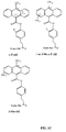
- FIGS. 1A to 1 C show the structures of several acridinium aryl ester compounds to which a linker moiety is attached; these acridinium ester (“AE") compounds are indicated by a shorthand nomenclature as: standard-AE, naphthyl-AE, o -AE, 1 or 3-Me-AE, 2,7-diMe-AE, 4,5-diMe-AE, o -diBr-AE, o -diMe-AE, m -diMe-AE, o -MeO-AE, o -MeO(cinnamyl)-AE, o -Me-AE, o -F-AE, 1 or 3-Me- o -F-AE, and 1 or-3-Me- m -diF-AE (the complete names of the compounds are listed in the Brief Description of FIGS.
- AE acridinium ester
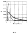
- 2,1-Me- m -diF-AE emits light extremely quickly after initiation of a chemiluminescent reaction
- 1-Me-AE has somewhat slower reaction kinetics, reaching a peak of light emission more slowly and emitting light over a longer time period than that of 1-Me- m -diF-AE
- o -OMe(c)-AE has the slowest reaction kinetics of the three, emitting light over a longer period of time than either of the other compounds.
- FIG. 2 shows a plot of the light emission of each of these compounds, measured in relative light units (RLU), versus time (in 0.2 sec intervals).
- the chemiluminescent reaction was initiated by simultaneously reacting all the labeling compounds with a triggering agent (alkaline hydrogen peroxide).
- FIG. 2 shows that the majority of light emitted from 1-Me- m -diF-AE is produced in the first half second of reaction.
- 1-Me-AE emits light between 0 and 3 sec, after which very little additional light is produced; during the period between 0.5 and 3 sec, very little interference is contributed by the 1-Me- m -diF-AE emission.
- o -OMe(cinnamyl)-AE continues to emit light for at least 5 sec after the 3-sec time point.
- These labels are distinguished experimentally by measuring the light emitted during three defined time "windows", one window for each label.
- One preferred set of windows for these compounds is: 0.0 to 0.6 sec for detection of 1-Me- m -diF-AE, 1.0 to 3.6 sec for detection of 1-Me-AE and 4.0 to 10 sec for detection of o -OMe(cinnamyl)-AE.
- the time course of reaction for each compound is constant, it is possible to predict and subtract light emitted from the other labels in a window used for the measurement of a particular label's signal through a reiterative statistical calculation.
- Such a statistical calculation, as performed for two labels is provided in Nelson et al., US Pat. No. 5,658,737.
- One skilled in the art is capable of using such a calculation for the analysis and correction of signal obtained from three or more labels, as illustrated by the calculated Units of Signal contributed by individual labeled probes in Model System 3 above.
- the 1-Me- m -diF-AE label (Label 1) was used at a relatively high specific activity of 10 8 RLU/pmole, which is useful in the detection of analyte at the low end of the concentration range; the 1-Me-AE label (Label 2) was used at a specific activity of 10 6 RLU/pmole; and the o -OMe(cinnamyl)-AE label (Label 3) was present at a specific activity of 10 4 RLU/pmole.
- Other details of the labels and the assay protocol are shown below in Table 4.
- the actual amount of RLU added for the second and third labels was somewhat greater than 10 5 to assure that 10 5 RLU of label was detected in each reading window. For each reading window, the reaction kinetics were measured in 0.2 second intervals.
- Table 4 The hydrolysis rate mentioned in Table 4 is important only to the homogeneous assay method illustrated in this example, and is not crucial to the general methods or compositions of the present invention.
- Table 5 presents the calculated signals for each label in its respective reading window for different amounts of target, based on the assumption that the labels are completely distinguishable.
- each of these three acridinium ester labels was mixed in the proportions indicated above in a solution containing 30 ⁇ l of 10 mM lithium succinate (pH 5.0) and 0.1% (w/v) lithium lauryl sulfate, 100 ⁇ l of 100 mM lithium succinate (pH 5.0), 8.5% (w/v) lithium lauryl sulfate, 1.5 mM EDTA, 1.5mM EGTA, and 300 ⁇ l of 600 mM sodium borate (pH 8.5), 1% (v/v) TRITON® X-100 (octylphenoxy polyethoxyethanol).
- the present invention may be used in conjunction with four or more separately detectable labels to extend the dynamic range for the detection of two or more analytes in a multiple analyte assay system.
- Labels may be of any type, for example, radioactive, fluorescent, chemiluminescent, chromogeneic, and enzyme- or substrate-linked labels. Examples of multiple analyte assay systems have been described in detail previously (e.g.,US Pat. No. 5,656, 207 to Woodhead et al., and US Pat. No. 5,658,737 to Nelson et al.). Although these references describe particular multiple analyte assay systems, as the previous discussion makes clear, the present invention may be used predictably in a variety of known assay systems.
- At least two labeled probes specific for different target regions of an analyte are used to detect each different analyte.
- the dynamic range of the assay for the detection of each analyte is extended by using at least two labeled probes targeted to different regions of each said analyte.
- each analyte is detected using at least two such probes preferably labeled with distinguishable labels.
- probes targeted to the same analyte are labeled at different specific activities, with the amount of each probe corresponding to the upper limit of the range of analyte amount which that probe is intended to detect.
- Probes 1, 2, 3 and 4 four probes are used (designated Probes 1, 2, 3 and 4) that bind to two different analytes (Analyte 1 and Analyte 2). Probes 1 and 2 specifically bind to Analyte 1 at distinct target regions. Probe 1 is labeled using standard procedures with a fluorescent label, 5-carboxy-fluorescein (excitation at 492 nm and emission at 518 nm) to a specific activity of 10 6 relative fluorescent units (“RFU”) per pmol of probe.
- fluorescent label 5-carboxy-fluorescein (excitation at 492 nm and emission at 518 nm) to a specific activity of 10 6 relative fluorescent units (“RFU”) per pmol of probe.
- Probe 2 is similarly labeled with a different fluorescent label, 6-carboxy rhodamine (excitation at 518 nm and emission at 543 nm) and then mixed with unlabeled Probe 2 to achieve a final specific activity of 10 4 RFU/pmol.
- Probes 3 and 4 specifically bind to Analyte 2 at distinct target regions.
- Probe 3 is labeled with a fluorescent label, 5-(and-6-)-carboxytetramethylrhodamine (excitation at 540 nm and emission at 565 nm) to a specific activity of 10 6 RFU/pmol.
- Probe 4 is labeled with sulforhodamine 101 acid chloride ("Texas Red®"; excitation at 587 nm and emission at 602 nm) and then mixed with unlabeled Probe 4 to achieve a final specific activity of 10 4 RFU/pmol.
- the four labels used in this example are independently distinguishable based on the different wavelengths of their fluorescence emissions. Those skilled in the art will appreciate that other types of labels could readily be substituted for any of these, so long as distinguishable signals are achieved in the combination of labeled probes.
- the probes are mixed together as follows: Probe Amount (pmol) Total RFU at emission wavelength Probe 1 0.1 10 5 at 518 nm Probe 2 10 10 5 at 543 nm Probe 3 0.1 10 5 at 565 nm Probe 4 10 10 5 at 602 nm
- the mixtures are contacted with samples containing different amounts of one or both of the analytes (i.e., Analyte 1, Analyte 2 or Analytes 1 + 2) under conditions that favor binding of the probes to their respective analyte target regions.
- a step of distinguishing bound from unbound probe is performed (e.g., washing), using methods that can readily be determined by one skilled in the art.
- the fluorescence signal of each of the bound labeled probes is detected at the appropriate wavelength.
- the signals detectable for each of the labeled probes are shown in Table 7 for different amounts of analyte (pmol) in the sample.
- background is 0.1% of the total RFU added to the mixture (i.e., 10 2 RFU per label).
- Analyte & Amount (pmol) Probe 1 RFU (at 518 nm) Probe 2 RFU (at 543 nm) Probe 3 RFU (at 565 nm) Probe 4 RFU (at 602 nm) Analyte 1 10 2 10 2 10 2 0.0001 Analyte 1 10 3 10 2 10 2 10 2 0.001 Analyte 1 10 4 10 2 10 2 10 2 0.01 Analyte 1 10 5 10 3 10 2 10 2 0.1 Analyte 1 10 5 10 4 10 2 10 2 1.0 Analyte 1 10 5 10 5 10 2 10 2 10 2 10 Analyte 2 10 2 10 2 10 2 10 2 0.0001 Analyte 2 10 2 10 3 10 2 0.001 Analyte 2 10 2 10 2 10 4 10 2 0.01 Analyte 2 10 2 10 2 10 3 0.1 Analyte 2 10 2 10 2 10 4 1.0 Analyte 2 10 2 10 5 10 5 10 2 10 2 10 2 10 2 10 2 10 2 10 2 0.0001 Analyte
- each probe individually can detect its respective analyte over 3 logs of concentration, and the combination of probes specific for one analyte as described above can detect the analyte over 5 logs of concentration. Furthermore, as shown by the RFU in Table 7, the combination of four probes detects and quantitates both analytes simultaneously and independently over an extended dynamic range of concentrations.
- this type of assay format allows the separate detection and/or quantification of two or more analytes, with each analyte being accurately measured over a wide concentration range.
- labels on Probes 1 and 2 are 5-carboxy-fluorescein and 6-carboxy rhodamine, respectively, and labels on Probes 3 and 4 are 3 H and 32 P, respectively.
- fluorescence signals are measured at 518 nm and 543 nm and then the different radioactive labels are detected and distinguished using any of a variety of well known methods.
- labels on Probes 1 and 2 are 5-carboxy-fluorescein and 6-carboxy rhodamine, respectively, and labels on Probes 3 and 4 are acridinium ester derivatives such as o-F-AE and 2-Me-AE, respectively.
- fluorescence signals are measured at 518 nm and 543 nm, and then the chemiluminescence is measured using well known methods that distinguish the signals from the two different acridinium ester labels.
- labels on Probes 1 and 2 are alkaline phosphatase and horseradish peroxidase, respectively, and the labels on Probes 3 and 4 are 5-(and-6-)-carboxytetramethylrhodamine and Texas Red@, respectively.
- appropriate enzyme substrates are added and allowed to react with the labels of analyte-bound Probes 1 and 2 (e.g., providing p-nitrophenyl phosphate for alkaline phosphatase and measuring the soluble end product by measuring absorbance at 405 nm; and providing o-phenylenediamine for horseradish peroxidase and measuring the soluble end product by measuring absorbance at 450 nm).
- the fluorescence signals of bound Probes 3 and 4 are measured at 565 nm and 602 nm.
- one skilled in the art can readily determine adjustments of specific activity needed for each probe to allow detection of the probes as described above and demonstrated by the information in Table 7.
- This example demonstrates the ability of one embodiment of the present invention to specifically detect analyte across a broad range of analyte amount or concentration.
- Two different probes bearing distinguishable labels were used to detect an analyte in samples, where the analyte concentration varied over a range of five orders of magnitude. Initially, the individual probes were characterized independently, and then the probes were combined in a single analyte-detection assay.
- a synthetic 48-base RNA oligomer was used as the target analyte.
- Two different oligonucleotide probes complementary to non-overlapping, unique areas of the target RNA (Probe 1 of 22 nucleotides, and Probe 2 of 18 nucleotides) were labeled with different chemiluminescent labels.
- Probe 1 was labeled with an N-acridinium phenyl ester having a fluorine substitution at the ortho position of the phenyl ring (i.e., o -F-AE)
- Probe 2 was labeled with an N-acridinium phenyl ester having a methyl group at the 2' position of the acridinium ring (i.e., 2-Me-AE).
- An unsubstituted acridinium ester containing a NHS linker at the 4 position of the phenyl moiety is 4-(2-succinimidyloxycarbonyl ethyl) phenyl-10-methylacridinium 9-carboxylate fluorosulfonate.
- Both Probes 1 and 2 contained modified sugar moieties wherein the 2' position of the ring was substituted with a methoxy group rather than -H (deoxyribose) or -OH (ribose), but use of modified oligonucleotides is not critical to the methods of the present invention, which can be performed with unmodified oligonucleotides as well.
- a chemiluminescent reaction employing the o -F-AE label has sufficiently fast reaction kinetics compared to those of the 2-Me-AE label that the signals from the two compounds following the reaction initiating chemiluminescence can be distinguished from each other by monitoring the resulting light emission over a time penod at regular intervals.
- the respective signals obtained from the two labels can be even more precisely quantified by employing a simple reiterative mathematical technique.
- the oligonucleotides of Probes 1 and 2 were linked to the AE labels through the use of a linker arm, as shown in FIGS. 1A to 1C, attached to the phenyl ring.
- the linker was joined to the oligonucleotide via a non-nucleotide linker, which was incorporated into the oligonucleotide chain using methods previously described in detail in US Pat. No. 5,656,744 to Arnold, et al. In this method the linker arm moiety to which the label will be attached is placed at a predetermined position within the oligonucleotide during synthesis.
- Oligonucleotides were synthesized using well-known solid-phase synthesis methods (e.g., as described in Brown & Brown, "Modern Machine-Aided Methods of Oligodeoxyribonucleotide Synthesis” in Oligonucleotides and Analogues, A Practiced Approach (1991)).
- Acridinium ester derivatives may be joined to the linker arm of the probe using techniques well known in the art, but preferred methods have been previously described in Nelson et al., "Detection of Acridinium Esters by Chemiluminescence” in Non-lsotopic Probe Techniques (Academic Press 1992), and US Pat. No. 5,656,744 to Arnold et al.
- an N-hydroxysuccinimide (“NHS”) ester of acridinium (e.g., 4-(2-succinimidyloxycarbonyl ethyl) phenyl-10-methylacridinium 9-carboxylate fluorosulfonate) is synthesized substantially as described in Weeks et al., 1983, Clin. Chem 29: 1474-1478, and US Pat. No. 5,658,737 to Nelson et al. Reaction of the primary amine of the linker arm:hybridization probe conjugate with the selected NHS-acridinium ester is performed as follows.
- oligonucleotide hybridization probe:linker arm conjugate synthesized as described above is vacuum-dried in a drying centrifuge (e.g., Savant SPEED-VACTM), and then dissolved in 8 ⁇ l of 0.125 M HEPES buffer (pH 8.0) in 50% (v/v) DMSO. To this, 2 ⁇ l of a 25 mM solution of the desired NHS-acridinium ester is added, mixed and incubated at 37 °C for 20 min.
- a drying centrifuge e.g., Savant SPEED-VACTM
- the labeled oligonucleotide is recovered by the addition of 30 ⁇ l of 3 M sodium acetate buffer (pH 5.0), 245 ⁇ l of water, and 5 ⁇ l of 40 mg/ml glycogen, and then adding 640 ⁇ l of chilled 100% ethanol, mixing and incubating on dry ice for 5 to 10 min.
- the precipitated labeled nucleic acids are sedimented in a refrigerated microcentrifuge at 15,000 rpm using a standard rotor head. The supernatant is aspirated off, and the pellet is redissolved in 20 ⁇ l of 0.1 M sodium acetate (pH 5.0) containing 0.1% (w/v) sodium dodecyl sulfate (SDS).
- Probes 1 and 2 conjugated to AE labels substantially as described above, were used at different specific activities.
- Probe 1 was labeled with o -F-AE at a specific activity of about 7 x 10 7 RLU/pmole.
- Probe 2 was labeled with 2-Me-AE at a specific activity of about 1 x 10 8 RLU/pmole and then mixed with unlabeled Probe 2 (as described below) to yield a lower specific activity. This resulted in about a 100-fold difference between the detectable signals of Probe 1 and Probe 2.
- each hybridization reaction of 100 ⁇ l contained 100 mM lithium succinate (pH 5.0), 8.5% (w/v) lithium lauryl sulfate, 1.5 mM EDTA, and 1.5 mM EGTA.
- reaction mixtures were incubated at 50°C for 50 min, and then 300 ⁇ l of 150 mM Na 2 B 4 O 7 (pH 8.6) containing 1% (v/v) TRITON® X-100 were added to each reaction, and the mixtures were incubated at 50°C for 11 min.
- the reaction mixtures were then placed into a luminometer (LEADER® 50) and a chemiluminescent reaction was initiated in each mixture upon the injection of 200 ⁇ l of 0.1% (v/v) H 2 O 2 and 1 mM HNO 3 , followed by injection of 200 ⁇ l of 1.5 N NaOH. Chemiluminescence was measured at a wavelength range from 300 to 650 nm for 2 sec following the second injection.
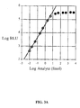
- the o-F-AE label of Probe 1 emitted an increasing amount of light with increasing amount of analyte in the reaction.
- the response was linear from about 0.01 fmoles to about 5 fmoles of the RNA analyte and in excess of 5 fmoles of analyte, the reaction began to plateau.
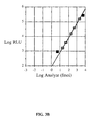
- the 2-Me-AE label of Probe 2 emitted more light when the amount of analyte present in the reaction was increased, but the detectable signal of the 2-Me-AE label of Probe 2 was about 100-fold lower than that of the o-F-AE label of Probe 1.
- target-bound Probe 2 only became detectable when more analyte was present, and a linear response (see FIG. 3B) was seen when analyte was present in a range of from about 20 fmoles to about 2,000 to 5,000 fmoles. From these experiments, standard curves were generated for each of the two labels, present at different specific activities.
- both labeled Probe 1 and labeled Probe 2 were combined in individual hybridization mixtures, each containing different amounts of the RNA analyte.
- the detectable signal of Probe 2 was about 100-fold less than the detectable signal of Probe 1.
- Eleven fmoles o -F-AE labeled Probe 1 and 5 fmoles 2-Me-AE labeled Probe 2 were combined with 15 pmoles of unlabeled Probe 2 in 100 ⁇ l reactions containing a hybridization buffer (100 mM lithium succinate, pH 5.0, 8.5% (w/v) lithium lauryl sulfate, 1.5 mM EDTA, and 1.5 mM EGTA) and different amounts of RNA target, as described in Example 4.
- a hybridization buffer 100 mM lithium succinate, pH 5.0, 8.5% (w/v) lithium lauryl sulfate, 1.5 mM EDTA, and 1.5 mM EGTA
- reaction mixture was incubated at 50°C for 50 min; then, 300 ⁇ l of 150 mM Na 2 B 4 O 7 ( pH 8.6) containing 1% (v/v) TRITON® X-100 were added to each reaction which were incubated at 50°C for 11 min. Then, the reactions were placed into a luminometer (LEADER® 50), and a chemiluminescent reaction was initiated by injecting into each reaction 200 ⁇ l of 0.1% (v/v) H 2 O 2 and 1 mM HNO 3 , followed by 200 ⁇ l of 1.5 N NaOH. Chemiluminescence was then measured for 2 sec, with light collected over 40 millisecond intervals during this period.
- the signal emitted by each label was resolved from the signal generated by the mixture of labels as follows.
- the raw data were subjected to a reiterative mathematical calculation, as described in detail previously (see Nelson, et al., US Pat. No. 5,658,737, at col. 22, lines 37-65) and briefly as follows, using the sum of the light emitted during two interval ranges within the time period (intervals 1 to 5 and intervals 30 to 50, abbreviated as "RLU 1-5 " and "RLU 30-50 ", respectively).
- the intervals are referred to by the number of the 40-millisecond intervals following initiation of the chemiluminescent reaction.
- Samples containing only one labeled probe were used as standards for data analysis.
- the ratio between the sum of the RLU values obtained in the first time interval and the sum of the RLU values obtained in the second time interval was determined ( ⁇ RLU 30-50 / ⁇ RLU 1-5 ).
- the sum of the RLU values in intervals 30 to 50 was divided by the ratio obtained for the 2-Me-AE standard. This results in the calculated amount of RLU contributed in intervals 1 to 5 by the 2-Me-AE-labeled probe. This amount, subtracted from the total RLU in intervals 1 to 5, gives the amount of RLU contributed in these intervals by o -F-AE.
- FIG. 4 also shows that the methods of the present invention permit detection, within the luminometer reporting range, of at least 6 logs of analyte concentrations by using independently detectable labels at different specific activities. Even further expansion of the dynamic range of the assay is possible, for example, by using a greater number of probes linked to independently detectable labels but within the luminometer reporting range of any selected instrument. Thus, an assay can be custom designed to utilize the most accurate reporting range of the instrument or detection method used.
- This example shows that two probes that specifically bind to different target regions of the same analyte, when labeled with the same detectable label but at different specific activities, can be used to detect an extended range of analyte concentrations.
- the probes used in this example are synthetic DNA oligomers having a 2-methoxy backbone, and designated Probe 3 (a 22-mer) and Probe 4 (a 19-mer). Both probes contain nucleotide base sequences that specifically bind to the same analyte but to separate non-overlapping target regions of the analyte.
- the analyte was a synthetic RNA oligomer consisting of 48 bases.
- Both Probes 3 and 4 were labeled with standard AE substantially as described in Example 4. Labeled Probe 4 was mixed with unlabeled Probe 4 to achieve a lower specific activity relative to the specific activity of Probe 3.
- the probe having the higher specific activity (Probe 3) was used to detect the lower range of analyte concentrations, and the probe with the lower specific activity (Probe 4) was used to detect the higher range of analyte concentrations.
- RNA analyte For each probe, a series of hybridization and chemiluminescence detection reactions were performed substantially as described in Example 4, using the RNA analyte combined with either Probe 3 without Probe 4; Probe 4 without Probe 3; or a combination of Probes 3 and 4.
- the amount of analyte added to the reactions was: 0.1, 1.0, 10, 100, 1,000, 10,000 and 100,000 fmol/reaction.
- Each reaction condition was performed in triplicate and the average (mean) RLU results were calculated from the triplicate samples. Triplicate tubes containing no analyte were used to determine background RLU, which were averaged and subtracted from the average of RLU results for each experimental test.
- the reactions were set up substantially as described in Example 4, using the following amounts of probe in 100 ⁇ l reaction mixtures: 50 fmol of labeled Probe 3 were added to those reactions containing Probe 3 alone or combined with Probe 4; 50 fmol of labeled Probe 4 plus 20 pmol of unlabeled Probe 4 were added to those reactions containing Probe 4 alone or combined with Probe 3.
- the hybridization reactions were incubated at 59 °C for 30 min, and then 300 ⁇ l of 150 mM Na2B4O7 (pH 8.7) containing 1% Triton®X-100 was added to each tube which was then incubated at 59°C for 9 min. This was followed by initiating and detecting the chemiluminescent reaction substantially as described in Example 4. Table 10 presents the results of these experiments.
- FIGS. 5A to 5C The results of Table 10 are graphically shown in FIGS. 5A to 5C, using log-log scale graphs of the RLU detected as a function of the analyte present in the sample.
- FIG. 5A the results obtained with Probe 3 alone hybridized to its target are shown, clearly showing a linear response over about a three log range of analyte concentrations (0.1 to 100 fmol).
- FIG. 5B the results obtained with Probe 4 alone hybridized to its target are shown, clearly showing a linear response over about a three log range of analyte concentrations (10 to 10,000 fmol).
- FIG. 5C when both probes were present in the hybridization reactions, the data points fall on two distinct lines.
- FIG. 5C when both probes were present in the hybridization reactions, the data points fall on two distinct lines.
- 5C shows that two probes that produce the same detectable signal and specifically bind to different target regions on the same analyte can be used in a single reaction to produce detectable signal that can be correlated with the concentration of analyte present in a sample over a range of at least 5 orders of magnitude.
- a combination of different probes labeled with the same label but at different specific activities and used in a format as illustrated in this example can detect the presence of analyte over an extended range of analyte concentrations than would be detected with either probe alone.
- the combination of probes can be used to quantify the amount of analyte present in a sample over this extended concentration range.
Abstract
Description
wherein each probe binds to a target region that differs from the target region of another probe in the probe reagent, and wherein each probe is present in an amount capable of detecting a range of analyte concentrations that differs from a range of analyte concentrations detectable by another probe in the reagent, such that the probe reagent is capable of detecting the analyte over an extended range of analyte concentrations that is greater than the range of analyte concentrations detectable by a single probe in the reagent and wherein a first probe in the probe reagent is present in an amount that differs by at least about a 10-fold molar excess relative to a second probe in the probe reagent. In another embodiment, each probe is distinguishable from another probe in the probe reagent because of its specific activity, a property resulting from its detectable label, or a combination thereof. Another embodiment of the composition includes, for each probe, a nucleic acid oligonucleotide having a target binding moiety that is a base sequence. In one embodiment, each probe has a detectable label that is an acridinium ester derivative. Preferably, each probe has a different detectable label, and each probe has a different specific activity compared to another probe in the reagent. In another embodiment, each probe is a nucleic acid oligonucleotide in which the target binding moiety binds specifically to a target region that is a nucleic acid base sequence, and preferably each probe has a detectable label that is an acridinium ester derivative.
| Amount of Target to be Detected (pmoles) | | Probe | 2 Units of | Probe | 3 Units of Signal | Total Units of Signal |
| 0.00001 | 1,000 | 10 | 0.1 | 1,010.1 | ||
| 0.0001 | 10,000 | 100 | 1 | 1,011 | ||
| 0.001 | 100,000 | 1,000 | 10 | 101,010 | ||
| 0.01 | 100,000 | 10,000 | 100 | 110,100 | ||
| 0.1 | 100,000 | 100,000 | 1,000 | 201,000 | ||
| 1 | 100,000 | 100,000 | 10,000 | 210,000 | ||
| 10 | 100,000 | 100,000 | 100,000 | 300,000 |
| Amount of Target to be Detected (pmoles) | | Probe | 2 Units of | Probe | 3 Units of Signal | Total Units of signal |
| 0.00001 | 103 | 103 | 103 | 103 units from | ||
| 103 units from | ||||||
| 103 units from | ||||||
| 0.0001 | 104 | 104 | 104 | 104 units from | ||
| 104 units from | ||||||
| 104 units from | ||||||
| 0.001 | 105 | 105 | 105 | 105 units from | ||
| 105 units from | ||||||
| 105 units from | ||||||
| 0.01 | 105 | 106 | 106 | 105 units from | ||
| 106 units from | ||||||
| 106 units from | ||||||
| 0.1 | 105 | 107 | 107 | 105 units from | ||
| 107 units from | ||||||
| 107 units from | ||||||
| 1 | 105 | 107 | 108 | 105 units from | ||
| 107 units from | ||||||
| 108 units from | ||||||
| 10 | 105 | 107 | 109 | 105 units from | ||
| 107 units from | ||||||
| 109 units from |
| Amount of Target to be Detected (pmoles) | | Probe | 2 Units of | Probe | 3 Units of Signal | Total Units of Signal |
| 0.00001 | 103 | 10 | 0.1 | 103 units from | ||
| 10 units from | ||||||
| 0.1 units from | ||||||
| 0.0001 | 104 | 102 | 1 | 104 units from | ||
| 102 units from | ||||||
| 1 units from | ||||||
| 0.001 | 105 | 104 | 10 | 105 units from | ||
| 104 units from | ||||||
| 10 units from | ||||||
| 0.01 | 105 | 104 | 102 | 105 units from | ||
| 104 units from | ||||||
| 102 units from | ||||||
| 0.1 | 105 | 105 | 103 | 105 units from | ||
| 105 units from | ||||||
| 103 units from | ||||||
| 1 | 105 | 105 | 104 | 105 units from | ||
| 105 units from | ||||||
| 104 units from | ||||||
| 10 | 105 | 105 | 105 | 105 Units from | ||
| 105 units from | ||||||
| 105 units from |

| Target (pmole) | Label 1 (RLU) | Label 2 (RLU) | Label 3 (RLU) |
| 0.00001 | 1000 | 10 | 0.1 |
| 0.00004 | 4000 | 40 | 0.4 |
| 0.00007 | 7000 | 70 | 0.7 |
| 0.0001 | 104 | 100 | 1.0 |
| 0.0004 | 4 x 10⁴ | 400 | 4.0 |
| 0.0007 | 7 x 10⁴ | 700 | 7.0 |
| 0.001 | 105 | 1000 | 10 |
| 0.004 | 105 | 4000 | 40 |
| 0.007 | 105 | 7000 | 70 |
| 0.01 | 105 | 104 | 100 |
| 0.04 | 105 | 4 x 10⁴ | 400 |
| 0.07 | 105 | 7 x 10⁴ | 700 |
| 0.10 | 105 | 105 | 1000 |
| 0.40 | 105 | 105 | 4000 |
| 0.70 | 105 | 105 | 7000 |
| 1.0 | 105 | 105 | 104 |
| 4.0 | 105 | 105 | 4 x 10⁴ |
| 7.0 | 105 | 105 | 7 x 10⁴ |
| 10.0 | 105 | 105 | 105 |
| Target (pmole) | Label 1 (RLU) | Label 2 (RLU) | Label 3 (RLU) |
| 0.00001 | 1,080 | 54 | 63 |
| 0.00004 | 4,601 | 188 | 86 |
| 0.00007 | 7,697 | 311 | 198 |
| 0.0001 | 10,197 | 380 | 234 |
| 0.0004 | 40,959 | 1,308 | 704 |
| 0.0007 | 66,174 | 2,295 | 1,166 |
| 0.001 | 90,979 | 3,117 | 1,810 |
| 0.004 | 93,129 | 8,353 | 2,156 |
| 0.007 | 97,467 | 12,604 | 2.360 |
| 0.01 | 97,010 | 16,285 | 2,516 |
| 0.04 | 144,034 | 61,496 | 5,653 |
| 0.07 | 184,345 | 102,891 | 8,022 |
| 0.10 | 179,248 | 142,326 | 10,524 |
| 0.40 | 180,289 | 146,349 | 14,517 |
| 0.70 | 180,901 | 153,413 | 19,049 |
| 1.0 | 177,833 | 154,494 | 21,023 |
| 4.0 | 197,599 | 232,885 | 59,264 |
| 7.0 | 202,855 | 292,157 | 90,589 |
| 10.0 | 220,880 | 346,219 | 121,487 |
| Probe | Amount (pmol) | Total RFU at |
| Probe | ||
| 1 | 0.1 | 105 at 518 |
| Probe | ||
| 2 | 10 | 105 at 543 |
| Probe | ||
| 3 | 0.1 | 105 at 565 |
| Probe | ||
| 4 | 10 | 105 at 602 nm |
| Analyte & Amount (pmol) | (at 518 nm) | (at 543 nm) | (at 565 nm) | (at 602 nm) |
| | 102 | 102 | 102 | 102 |
| 0.0001 | ||||
| | 103 | 102 | 102 | 102 |
| 0.001 | ||||
| | 104 | 102 | 102 | 102 |
| 0.01 | ||||
| | 105 | 103 | 102 | 102 |
| 0.1 | ||||
| | 105 | 104 | 102 | 102 |
| 1.0 | ||||
| | 105 | 105 | 102 | 102 |
| 10 | ||||
| | 102 | 102 | 102 | 102 |
| 0.0001 | ||||
| | 102 | 102 | 103 | 102 |
| 0.001 | ||||
| | 102 | 102 | 104 | 102 |
| 0.01 | ||||
| | 102 | 102 | 105 | 103 |
| 0.1 | ||||
| | 102 | 102 | 105 | 104 |
| 1.0 | ||||
| | 102 | 102 | 105 | 105 |
| 10 | ||||
| | 102 | 102 | 102 | 102 |
| 0.0001 of each | ||||
| | 103 | 102 | 103 | 102 |
| 0.001 of each | ||||
| | 104 | 102 | 104 | 102 |
| 0.01 of each | ||||
| | 105 | 103 | 105 | 103 |
| 0.1 of each | ||||
| | 105 | 104 | 105 | 104 |
| 1.0 of each | ||||
| | 105 | 105 | 105 | 105 |
| 10 of each |
| Analyte Assayed and Detected in Individual Reaction Mixtures | ||
| Target RNA (fmoles) | RLU Output of Probe 1 (o-F-AE) | RLU Output of Probe 2 (2-Me-AE) |
| 0.00 | 53 | 0 |
| 0.01 | 428 | 0 |
| 0.02 | 912 | 0 |
| 0.05 | 2,209 | 0 |
| 0.2 | 9,181 | 0 |
| 0.5 | 22,968 | 0 |
| 2.0 | 82,545 | 0 |
| 5.0 | 177,543 | 903 |
| 20 | 264,501 | 1,673 |
| 50 | 311,928 | 3,979 |
| 200 | 324,618 | 15,304 |
| 500 | 322,676 | 39,502 |
| 2000 | 351,596 | 158,033 |
| 5000 | 330,214 | 275,699 |
| Analyte Assayed and Detected in Single Reaction Mixtures | ||
| fmoles Target RNA | RLU Output of Probe 1 (o-F-AE) | RLU Output of Probe 2 (2-Me-AE) |
| 0.00 | 59 | 63 |
| 0.01 | 573 | 31 |
| 0.02 | 1,233 | 31 |
| 0.05 | 2,893 | 0 |
| 0.2 | 11,068 | 0 |
| 0.5 | 27,139 | 31 |
| 2.0 | 93,535 | 189 |
| 5.0 | 204,984 | 600 |
| 20.0 | 271,543 | 2,277 |
| 50.0 | 315,697 | 4,428 |
| 200.0 | 314,792 | 15,974 |
| 500.0 | 301,076 | 39,036 |
| 2000.0 | 321,450 | 153,234 |
| 5000.0 | 328,863 | 279,105 |
| fmoles Target RNA | Mean RLU Detected | Mean RLU Detected | Mean RLU Detected |
| 0.1 | 3,630 | Not Tested | 596 |
| 1.0 | 36,758 | Not Tested | 38,250 |
| 10 | 379,672 | 16,270 | 405,427 |
| 100 | 2,054,816 | 122,222 | 2,120,323 |
| 1,000 | 2,047,840 | 1,474,208 | 3,618,762 |
| 10,000 | Not Tested | 10,217,034 | 11,717,176 |
| 100,000 | Not Tested | 10,507,565 | 12,646,943 |
Claims (26)
- A method of detecting an analyte over a range of concentrations in a single sample, comprising the steps of:wherein each labeled probe comprises a target binding moiety and a detectable label,providing a probe reagent comprising at least two different labeled probes,
wherein the target binding moiety of each labeled probe binds specifically to a target region that differs from the target region bound by another labeled probe in the probe reagent, and
wherein each labeled probe is present in an amount capable of detecting a range of analyte concentrations that differs from the range of analyte concentrations detectable by another probe in the reagent, thereby allowing the probe reagent to detect an extended range of analyte concentrations that is greater than the range of analyte concentrations detectable by a single labeled probe in the probe reagent;
contacting in a single vessel the probe reagent with a sample containing the analyte,
wherein the probe reagent contains a first probe present in at least a 10-fold molar excess relative to a second probe;
incubating the sample and the probe reagent under conditions favoring specific binding of the target binding moiety of each labeled probe with its target region of the analyte, thereby producing a specific binding complex comprising the analyte and at least one labeled probe; and
detecting the presence of the specific binding complex by detecting a signal from the label indicating the presence of the at least one probe in the specific binding complex, thereby indicating the presence of the analyte in the sample within the range of analyte concentrations detectable by the probe in the specific binding complex. - The method of claim 1, wherein the contacting and incubating steps are performed in a single vessel.
- The method of claim 1 or 2, wherein the detecting step measures a signal from the detectable label that is characteristic of kinetics of a reaction.
- The method of any one of claims 1 to 3, wherein the providing step uses a probe reagent in which specific activity of one labeled probe differs from specific activity of another labeled probe in the probe reagent.
- The method of any one of claims 1 to 4, wherein the providing step uses a probe reagent wherein at least one labeled probe has a detectable label that is a ligand capable of specifically binding a signal-producing binding partner.
- The method of any one of claims 1 to 4, wherein the providing step uses a probe reagent wherein at least one labeled probe has a detectable label that is a direct label, and the detecting step measures a signal resulting from the direct label.
- The method of any one of claims 1 to 4, wherein the providing step uses a probe reagent wherein at least one labeled probe has a detectable label that is a fluorescent label, luminescent label, radioactive label, chromophore, an enzyme, or an enzyme substrate.
- The method of any one of claims 1 to 6, wherein the providing step uses a probe reagent wherein at least one labeled probe has a detectable label that is a chemiluminescent compound
- The method of any one of claims 1 to 8, wherein the contacting step uses the probe reagent wherein the first labeled probe is present in at least a 100-fold, 1,000-fold or 10,000-fold molar excess relative to the second labeled probe.
- The method of any one of claims 1 to 9, wherein the probe reagent comprises the first labeled probe having a detectable label that is a first chemiluminescent compound and the second labeled probe having a detectable label that is a second chemiluminescent compound, wherein the first chemiluminescent compound and the second chemiluminescent compound are separately distinguishable labels.
- The method of claim 10, wherein the first chemiluminescent compound is selected from the group consisting of standard acridinium ester (AE), naphthyl-AE, ortho-AE, 1- or 3-methyl-AE, 2-methyl-AE, 2,7-dimethyl-AE, 4,5-dimethyl-AE, ortho-dibromo-AE, ortho-dimethyl-AE, meta-dimethyl-AE, ortho-methoxy-AE, ortho-methoxy(cinnamyl)-AE, ortho-methyl-AE, ortho-fluoro-AE, 1- or 3-methyl-ortho-fluoro-AE and 1- or 3-methyl-meta-difluoro-AE; and
wherein the second chemiluminescent compound is selected from the group consisting of standard acridinium ester (AE), naphthyl-AE, ortho-AE, 1- or 3-methyl-AE, 2-methyl-AE, 2,7-dimethyl-AE, 4,5-dimethyl-AE, ortho-dibromo-AE, ortho-dimethyl-AE, meta-dimethyl-AE, ortho-methoxy-AE, ortho-methoxy(cinnamyl)-AE, ortho-methyl-AE, ortho-fluoro-AE, 1- or 3-methyl-ortho-fluoro-AE and 1- or 3-methyl-meta-difluoro-AE,
provided that the first chemiluminescent compound is different and distinguishable from the second chemiluminescent compound. - The method of claim 11, wherein the first chemiluminescent compound is ortho-fluoro-AE, and the second chemiluminescent compound is 2-methyl-AE.
- The method of claim 10, wherein the probe reagent further comprises a third labeled probe having a separately distinguishable label that is a third chemiluminescent compound.
- The method of claim 13, wherein the first chemiluminescent compound is selected from the group consisting of standard acridinium ester (AE), naphthyl-AE, ortho-AE, 1- or 3-methyl-AE, 2-methyl-AE, 2,7-dimethyl-AE, 4,5-dimethyl-AE, ortho-dibromo-AE, ortho-dimethyl-AE, meta-dimethyl-AE, ortho-methoxy-AE, ortho-methoxy(cinnamyl)-AE, ortho-methyl-AE, ortho-fluoro-AE, 1- or 3-methyl-ortho-fluoro-AE and 1- or 3-methyl-meta-difluoro-AE;
wherein the second chemiluminescent compound is selected from the group consisting of standard acridinium ester (AE), naphthyl-AE, ortho-AE, 1- or 3-methyl-AE, 2-methyl-AE, 2,7-dimethyl-AE, 4,5-dimethyl-AE, ortho-dibromo-AE, ortho-dimethyl-AE, meta-dimethyl-AE, ortho-methoxy-AE, ortho-methoxy(cinnamyl)-AE, ortho-methyl-AE, ortho-fluoro-AE, 1- or 3-methyl-ortho-fluoro-AE and 1- or 3-methyl-meta-difluoro-AE; and
wherein the third chemiluminescent compound is selected from the group consisting of standard acridinium ester (AE), naphthyl-AE, ortho-AE, 1- or 3-methyl-AE, 2-methyl-AE, 2,7-dimethyl-AE, 4,5-dimethyl-AE, ortho-dibromo-AE, ortho-dimethyl-AE, meta-dimethyl-AE, ortho-methoxy-AE, ortho-methoxy(cinnamyl)-AE, ortho-methyl-AE, ortho-fluoro-AE, 1- or 3-methyl-ortho-fluoro-AE and 1-or 3-methyl-meta-difluoro-AE,
provided that the first, second and third chemiluminescent compounds differ and are distinguishable from each other. - The method of claim 14, wherein the first chemiluminescent compound is 1- or 3-methyl-meta-difluoro-AE, the second chemiluminescent compound is 1- or 3-methyl-AE, and the third chemiluminescent compound is ortho-methoxy(cinnamyl)-AE.
- The method of any one of claims 1 to 15, wherein the contacting step includes an analyte that is a nucleic acid, protein, lipid, carbohydrate or a compound that is a combination thereof.
- The method of any one of claims 1 to 16, wherein the contacting step includes an analyte that is a DNA or RNA.
- The method of any one of claims 1 to 17, wherein the providing step uses a probe reagent wherein at least one labeled probe is a nucleic acid, protein, lipid, carbohydrate or compound that is a combination thereof.
- The method of any one of claims 1 to 18, wherein the providing step uses a probe reagent wherein at least one labeled probe comprises a DNA, RNA, DNA analog, RNA analog, or a combination thereof.
- A probe reagent for use in the method of any one of claims 1 to 19, comprising at least two different labeled probes, each labeled probe comprising a target binding moiety and a detectable label,
wherein the target binding moiety of each labeled probe binds specifically to a target region that differs from the target region bound by another labeled probe in the probe reagent, and
wherein each labeled probe is present in an amount capable of detecting a range of analyte concentrations that differs from a range of analyte concentrations detectable by another probe in the reagent, and
wherein the probe reagent contains a first probe present in at least a 10-fold molar excess relative to a second probe, thereby allowing the probe reagent to detect the analyte over an extended range of analyte concentrations that is greater than the range of analyte concentrations detectable by a single probe in the probe reagent. - The probe reagent of claim 20, wherein the probe reagent includes a first labeled probe present in an amount that differs by at least a 100-fold molar excess, or at least a 1,000-fold molar excess, or at least a 10,000-fold molar excess relative to a second labeled probe present in the probe reagent.
- The probe reagent of claim 20 or 21, wherein each labeled probe is distinguishable from each other labeled probe in the probe reagent because of:different specific activities of the different labeled probes,a signal resulting from separately distinguishable labels on each of the labeled probes,or a combination thereof.
- The probe reagent of any one of claims 20 to 22, wherein each labeled probe has a different specific activity of label compared to another labeled probe in the probe reagent.
- The probe reagent of claim 23, wherein a labeled probe having a relatively high specific activity is present in a molar amount that is lower than a molar amount of a labeled probe having a relatively low specific activity in the probe reagent.
- The probe reagent of any one of claims 20 to 24, wherein each labeled probe is a nucleic acid oligonucleotide having a target binding moiety that is a base sequence.
- The probe reagent of any one of claims 20 to 25, wherein each labeled probe has a separately detectable label that is an acridinium ester derivative.
Applications Claiming Priority (3)
| Application Number | Priority Date | Filing Date | Title |
|---|---|---|---|
| US962033 | 1997-10-31 | ||
| US08/962,033 US6180340B1 (en) | 1997-10-31 | 1997-10-31 | Extended dynamic range assays |
| PCT/US1998/023088 WO1999023490A1 (en) | 1997-10-31 | 1998-10-30 | Extended dynamic range assays |
Publications (2)
| Publication Number | Publication Date |
|---|---|
| EP1027604A1 EP1027604A1 (en) | 2000-08-16 |
| EP1027604B1 true EP1027604B1 (en) | 2004-08-04 |
Family
ID=25505344
Family Applications (1)
| Application Number | Title | Priority Date | Filing Date |
|---|---|---|---|
| EP98956381A Expired - Lifetime EP1027604B1 (en) | 1997-10-31 | 1998-10-30 | Extended dynamic range assays |
Country Status (9)
| Country | Link |
|---|---|
| US (2) | US6180340B1 (en) |
| EP (1) | EP1027604B1 (en) |
| JP (1) | JP3550358B2 (en) |
| AT (1) | ATE272841T1 (en) |
| AU (1) | AU741568B2 (en) |
| CA (1) | CA2305415C (en) |
| DE (1) | DE69825465T2 (en) |
| ES (1) | ES2226189T3 (en) |
| WO (1) | WO1999023490A1 (en) |
Families Citing this family (66)
| Publication number | Priority date | Publication date | Assignee | Title |
|---|---|---|---|---|
| US7332567B2 (en) | 2001-08-28 | 2008-02-19 | Allergan, Inc. | Fret protease assays for clostridial toxins |
| JP4291143B2 (en) * | 2001-08-31 | 2009-07-08 | ジェン−プロウブ インコーポレイテッド | Affinity-shifted probe for quantification of analyte polynucleotide |
| JP2004012176A (en) * | 2002-06-04 | 2004-01-15 | Hitachi Software Eng Co Ltd | Protein detection method |
| US20040018577A1 (en) * | 2002-07-29 | 2004-01-29 | Emerson Campbell John Lewis | Multiple hybrid immunoassay |
| US7399607B2 (en) | 2004-09-22 | 2008-07-15 | Allergan, Inc. | Fluorescence polarization assays for determining clostridial toxin activity |
| US7396689B2 (en) * | 2005-02-04 | 2008-07-08 | Decision Biomarkers Incorporated | Method of adjusting the working range of a multi-analyte assay |
| US7723127B2 (en) * | 2005-03-03 | 2010-05-25 | Novx Systems Inc. | Immunoassay with extended dynamic range |
| EP1937847A2 (en) | 2005-10-17 | 2008-07-02 | Gen-Probe Incorporated | Compositions and methods to detect legionella pneumophila nucleic acid |
| WO2007134208A2 (en) * | 2006-05-12 | 2007-11-22 | Gen-Probe Incorporated | Compositions and methods to detect enterococci nucleic acid |
| US8728223B2 (en) | 2006-05-16 | 2014-05-20 | Cabot Corporation | Low viscosity, high particulate loading dispersions |
| US8585816B2 (en) * | 2006-05-16 | 2013-11-19 | Cabot Corporation | Low viscosity, high particulate loading dispersions |
| US7595164B2 (en) * | 2007-12-26 | 2009-09-29 | Gen-Probe Incorporated | Compositions and methods to detect Candida albicans nucleic acid |
| GB2464183A (en) * | 2008-09-19 | 2010-04-14 | Singulex Inc | Sandwich assay |
| AU2010276236B2 (en) | 2009-07-21 | 2014-03-20 | Gen-Probe Incorporated | Methods and compositions for quantitative detection of nucleic acid sequences over an extended dynamic range |
| EP2536849B1 (en) | 2010-02-17 | 2016-05-11 | Gen-Probe Incorporated | Compositions and methods to detect atopobium vaginae nucleic acid |
| WO2011133811A2 (en) | 2010-04-21 | 2011-10-27 | Gen-Probe Incorporated | Compositions, methods and kits to detect herpes simplex virus nucleic acid |
| EP2611932A2 (en) | 2010-08-30 | 2013-07-10 | Gen-Probe Incorporated | Compositions, methods and reaction mixtures for the detection of xenotropic murine leukemia virus-related virus |
| EP2625297B1 (en) | 2010-10-04 | 2018-10-10 | Gen-Probe Prodesse, Inc. | Compositions, methods and kits to detect adenovirus nucleic acids |
| US10669569B2 (en) | 2010-10-15 | 2020-06-02 | Navinci Diagnostics Ab | Dynamic range methods |
| CN103703143B (en) | 2011-01-31 | 2016-12-14 | 爱普瑞斯生物公司 | The method of the multiple epi-positions in identification of cell |
| WO2012122571A1 (en) | 2011-03-10 | 2012-09-13 | Gen-Probe Incorporated | Methods and compositions for the selection and optimization of oligonucleotide tag sequences |
| EP2702166B1 (en) | 2011-04-25 | 2018-06-06 | Gen-Probe Incorporated | Compositions and methods for detecting bv-associated bacterial nucleic acid |
| EP2753713B1 (en) | 2011-09-08 | 2017-07-19 | Gen-Probe Incorporated | Compositions and methods for detecting bv-associated bacterial nucleic acid |
| AU2012312169B2 (en) | 2011-09-21 | 2016-01-14 | Gen-Probe Incorporated | Methods for amplifying nucleic acid using tag-mediated displacement |
| DE102011120550B4 (en) | 2011-12-05 | 2013-11-07 | Gen-Probe Prodesse, Inc. | Compositions, methods and kits for the detection of adenovirus nucleic acids |
| CA2865281C (en) | 2012-02-24 | 2021-11-23 | Gen-Probe Prodesse, Inc. | Detection of shiga toxin genes in bacteria |
| AU2013205110B2 (en) | 2012-04-24 | 2016-10-13 | Gen-Probe Incorporated | Compositions, Methods and Kits to Detect Herpes Simplex Virus Nucleic Acids |
| AU2013205064B2 (en) | 2012-06-04 | 2015-07-30 | Gen-Probe Incorporated | Compositions and Methods for Amplifying and Characterizing HCV Nucleic Acid |
| EP2882868B1 (en) | 2012-08-08 | 2019-07-31 | H. Hoffnabb-La Roche Ag | Increasing dynamic range for identifying multiple epitopes in cells |
| KR101445596B1 (en) * | 2012-09-27 | 2014-10-07 | 단국대학교 산학협력단 | Measurement method and measurement kit of antibiotics concentration |
| AU2013205122B2 (en) | 2012-10-11 | 2016-11-10 | Gen-Probe Incorporated | Compositions and Methods for Detecting Human Papillomavirus Nucleic Acid |
| AU2013205090B2 (en) | 2012-12-07 | 2016-07-28 | Gen-Probe Incorporated | Compositions and Methods for Detecting Gastrointestinal Pathogen Nucleic Acid |
| US9157910B2 (en) | 2013-03-15 | 2015-10-13 | Abbott Laboratories | Assay with increased dynamic range |
| JP6389868B2 (en) | 2013-03-15 | 2018-09-12 | ジーピービー デット ホールディングス トゥー エルエルシー | Apparatus and related methods for performing luminescence and fluorescence measurements of samples |
| US9005901B2 (en) | 2013-03-15 | 2015-04-14 | Abbott Laboratories | Assay with internal calibration |
| CN105452488B (en) | 2013-08-14 | 2020-07-14 | 简·探针公司 | Compositions and methods for detecting HEV nucleic acids |
| EP3736346B1 (en) | 2015-01-09 | 2023-12-20 | Gen-Probe Incorporated | Methods and compositions for diagnosing bacterial vaginosis |
| CA2977821A1 (en) | 2015-03-16 | 2016-09-22 | Gen-Probe Incorporated | Methods and compositions for detecting bacterial nucleic acid and diagnosing bacterial vaginosis |
| EP3163287B1 (en) | 2015-10-30 | 2022-03-23 | Sysmex Corporation | Cell information obtaining method and cell information obtaining apparatus |
| EP3400309A2 (en) | 2016-01-04 | 2018-11-14 | Gen-Probe Incorporated | Methods and compositions for detecting candida species |
| US20190284604A1 (en) | 2016-02-05 | 2019-09-19 | Gen-Probe Incorporated | Dried amplification compositions |
| EP3985131A1 (en) | 2016-06-10 | 2022-04-20 | Gen-Probe Incorporated | Compositions and methods for detecting zika virus nucleic acid |
| EP3529381B1 (en) | 2016-10-19 | 2022-07-06 | Gen-Probe Incorporated | Compositions and methods for detecting or quantifying hepatitis c virus |
| AU2017363168B2 (en) | 2016-11-21 | 2023-07-06 | Gen-Probe Incorporated | Compositions and methods for detecting or quantifying hepatitis B virus |
| CA3057154A1 (en) | 2017-03-24 | 2018-09-27 | Gen-Probe Incorporated | Compositions and methods for detecting or quantifying parainfluenza virus |
| CA3056135C (en) | 2017-03-25 | 2024-02-20 | Gen-Probe Incorporated | Compositions, methods and kits to detect adenovirus nucleic acids |
| AU2018281196B2 (en) | 2017-06-07 | 2022-04-28 | Gen-Probe Incorporated | Detecting Babesia species nucleic acid in a sample |
| US11859257B2 (en) | 2017-08-11 | 2024-01-02 | Gen-Probe Incorporated | Compositions and methods for detecting Staphylococcus aureus |
| CN111465707A (en) | 2017-11-17 | 2020-07-28 | 简·探针公司 | Compositions and methods for detecting C1orf43 nucleic acids |
| WO2019118550A1 (en) | 2017-12-13 | 2019-06-20 | Gen-Probe Incorporated | Methods for biological sample processing |
| EP3724354A1 (en) | 2017-12-15 | 2020-10-21 | Gen-Probe Incorporated | Compositions and methods for detecting toxigenic clostridium difficile |
| GB2590210B (en) | 2018-06-13 | 2023-01-25 | Gen Probe Inc | Compositions and methods for detecting group B Streptococcus nucleic acid |
| AU2019314449A1 (en) | 2018-08-01 | 2021-03-11 | Gen-Probe Incorporated | Compositions and methods for detecting nucleic acids of Epstein-Barr virus |
| WO2020041414A1 (en) | 2018-08-21 | 2020-02-27 | Gen-Probe Incorporated | Compositions and methods for amplifying, detecting or quantifying human cytomegalovirus |
| JP7389111B2 (en) | 2018-08-24 | 2023-11-29 | ジェン-プローブ・インコーポレーテッド | Compositions and methods for detecting bacterial nucleic acids and diagnosing bacterial vaginosis |
| WO2020069085A2 (en) | 2018-09-27 | 2020-04-02 | Gen-Probe Incorporated | Compositions and methods for detecting bordetella pertussis and bordetella parapertussis nucleic acid |
| US20220017980A1 (en) | 2018-10-01 | 2022-01-20 | Gen-Probe Incorporated | Compositions and methods for amplifying or detecting varicella-zoster virus |
| WO2020086546A1 (en) | 2018-10-22 | 2020-04-30 | Gen-Probe Incorporated | Compositions and methods for amplifying, detecting or quantifying human polyomavirus bk virus |
| TW202030333A (en) | 2018-12-20 | 2020-08-16 | 美商簡 探針公司 | Compositions and methods for detecting plasmodium species nucleic acid |
| CA3127620A1 (en) | 2019-01-25 | 2020-07-30 | Gen-Probe Incorporated | Detection of drug-resistant mycoplasma genitalium |
| WO2020197987A1 (en) | 2019-03-22 | 2020-10-01 | Gen-Probe Incorporated | Compositions and methods for detecting group a streptococcus |
| EP4146821A1 (en) | 2020-05-07 | 2023-03-15 | Grifols Diagnostic Solutions Inc. | Methods and compositions for detecting sars-cov-2 nucleic acid |
| US20230243001A1 (en) | 2020-07-17 | 2023-08-03 | Gen-Probe Incorporated | Detection of Macrolide-Resistant Mycoplasma Genitalium |
| AU2022237386A1 (en) | 2021-03-15 | 2023-10-05 | Gen-Probe Incorporated | Compositions and methods for biological sample processing |
| WO2023010008A1 (en) | 2021-07-27 | 2023-02-02 | Gen-Probe Incorporated | Compositions and methods for detecting gastrointestinal pathogens |
| WO2024054924A1 (en) | 2022-09-08 | 2024-03-14 | Gen-Probe Incorporated | Method of detecting nucleic acid analytes using dual-specificity primers |
Family Cites Families (21)
| Publication number | Priority date | Publication date | Assignee | Title |
|---|---|---|---|---|
| US4486530A (en) * | 1980-08-04 | 1984-12-04 | Hybritech Incorporated | Immunometric assays using monoclonal antibodies |
| CA1185177A (en) * | 1981-07-17 | 1985-04-09 | Larry E. Morrison | Non-radiative energy transfer immunochemical technique |
| GB2112779B (en) | 1981-12-11 | 1986-10-15 | Welsh Nat School Med | Aryl acridinium esters as luminescent labelling materials |
| US4514505A (en) * | 1982-05-21 | 1985-04-30 | The Trustees Of Columbia University In The City Of New York | Monoclonal antibody mixtures and use thereof for enhanced sensitivity immunoassays |
| US4689295A (en) * | 1983-01-20 | 1987-08-25 | Integrated Genetics, Inc. | Test for Salmonella |
| US4595661A (en) | 1983-11-18 | 1986-06-17 | Beckman Instruments, Inc. | Immunoassays and kits for use therein which include low affinity antibodies for reducing the hook effect |
| US5585481A (en) | 1987-09-21 | 1996-12-17 | Gen-Probe Incorporated | Linking reagents for nucleotide probes |
| US5283174A (en) | 1987-09-21 | 1994-02-01 | Gen-Probe, Incorporated | Homogenous protection assay |
| US5639604A (en) | 1987-09-21 | 1997-06-17 | Gen-Probe Incorporated | Homogeneous protection assay |
| NO164622C (en) | 1988-05-11 | 1990-10-24 | Tore Lindmo | BINAER IMMUNOMETRIC PARTICLE-BASED METHOD FOR MEASURING SPECIFIC SERUM ANTIGENS USING LIQUID FLOW MICROPHOTOMETRY AND A PREPARED TARGET SET UP THEREOF. |
| GB2233450B (en) | 1989-06-24 | 1993-06-30 | Univ Wales Medicine | Detecting or quantifing multiple analytes with luminescent reagents |
| US5656207A (en) | 1989-06-24 | 1997-08-12 | Gen Probe Incorporated | Detecting or quantifying multiple analytes using labelling techniques |
| GB9326238D0 (en) * | 1993-12-23 | 1994-02-23 | Sinvent As | Method of assay |
| GB9326379D0 (en) | 1993-12-23 | 1994-02-23 | Sinvent As | Method of assay |
| EP0709466B1 (en) | 1994-10-28 | 2006-09-27 | Gen-Probe Incorporated | Compositions and methods for the simultaneous detection and quantification of multiple specific nucleic acid sequences |
| US5710029A (en) | 1995-06-07 | 1998-01-20 | Gen-Probe Incorporated | Methods for determining pre-amplification levels of a nucleic acid target sequence from post-amplification levels of product |
| US5731148A (en) | 1995-06-07 | 1998-03-24 | Gen-Probe Incorporated | Adduct protection assay |
| WO1996040990A1 (en) | 1995-06-07 | 1996-12-19 | Gen-Probe Incorporated | Methods and kits for determining pre-amplification levels of a nucleic acid target sequence from post-amplification levels of product |
| AU707818B2 (en) | 1995-06-07 | 1999-07-22 | Gen-Probe Incorporated | Micelle protection assay |
| US5705365A (en) | 1995-06-07 | 1998-01-06 | Gen-Probe Incorporated | Kits for determining pre-amplification levels of a nucleic acid target sequence from post-amplification levels of product |
| US6066458A (en) | 1998-05-18 | 2000-05-23 | Becton, Dickinson And Company | Methods, apparatus and computer program products for determining quantities of nucleic acid sequences in samples using standard curves and amplification ratio estimates |
-
1997
- 1997-10-31 US US08/962,033 patent/US6180340B1/en not_active Expired - Lifetime
-
1998
- 1998-10-30 WO PCT/US1998/023088 patent/WO1999023490A1/en active IP Right Grant
- 1998-10-30 DE DE69825465T patent/DE69825465T2/en not_active Expired - Lifetime
- 1998-10-30 CA CA002305415A patent/CA2305415C/en not_active Expired - Fee Related
- 1998-10-30 AU AU12918/99A patent/AU741568B2/en not_active Ceased
- 1998-10-30 JP JP2000519300A patent/JP3550358B2/en not_active Expired - Fee Related
- 1998-10-30 ES ES98956381T patent/ES2226189T3/en not_active Expired - Lifetime
- 1998-10-30 EP EP98956381A patent/EP1027604B1/en not_active Expired - Lifetime
- 1998-10-30 AT AT98956381T patent/ATE272841T1/en not_active IP Right Cessation
-
2000
- 2000-08-28 US US09/649,636 patent/US6350579B1/en not_active Expired - Lifetime
Also Published As
| Publication number | Publication date |
|---|---|
| CA2305415C (en) | 2007-08-14 |
| AU741568B2 (en) | 2001-12-06 |
| AU1291899A (en) | 1999-05-24 |
| WO1999023490A1 (en) | 1999-05-14 |
| ATE272841T1 (en) | 2004-08-15 |
| DE69825465D1 (en) | 2004-09-09 |
| CA2305415A1 (en) | 1999-04-15 |
| DE69825465T2 (en) | 2005-08-04 |
| US6180340B1 (en) | 2001-01-30 |
| JP3550358B2 (en) | 2004-08-04 |
| EP1027604A1 (en) | 2000-08-16 |
| US6350579B1 (en) | 2002-02-26 |
| JP2002508504A (en) | 2002-03-19 |
| ES2226189T3 (en) | 2005-03-16 |
Similar Documents
| Publication | Publication Date | Title |
|---|---|---|
| EP1027604B1 (en) | Extended dynamic range assays | |
| US5756011A (en) | Detecting or quantifying multiple analytes using labelling techniques | |
| JP6037522B2 (en) | Liquid phase homogeneous assay | |
| FI93781B (en) | Biospecific multiparametric method of determination | |
| EP0619018B1 (en) | Improved membrane for chemiluminescent blotting applications | |
| JP3021038B2 (en) | Detection or quantification of multiple analytes using labeling technology | |
| AU679008B2 (en) | Mixed luminescent conjugate test assays | |
| Scorilas et al. | Polyvinylamine-streptavidin complexes labeled with a europium chelator: a universal detection reagent for solid-phase time resolved fluorometric applications | |
| CN1155723C (en) | Universal test method and reagent kit for quantitative PCR and immune PCR | |
| JP3289907B2 (en) | Chemiluminescence assay for dsDNA antibodies | |
| EP0682254B1 (en) | Enzyme linked chemiluminescent assay | |
| JP7055975B2 (en) | Specific bond chemical luminescence assay | |
| JP4028645B2 (en) | Chemiluminescent enzyme immunoassay method | |
| JP4286357B2 (en) | Chemiluminescent enzyme immunoassay method | |
| JP3745112B2 (en) | Chemiluminescent enzyme immunoassay method | |
| JP3792886B2 (en) | Chemiluminescent enzyme immunoassay method | |
| JP3815905B2 (en) | Enzyme immunoassay | |
| Schweitzer et al. | Electrochemiluminescence: A Technology Evaluation and Assay Reformatting of the Stat6/P578 Protein–Peptide Interaction | |
| JPH0454455A (en) | Biomaterial labeled with multidentate ligand fluorescent chelate | |
| JPH11341997A (en) | Chemiluminescent enzyme immunoassay | |
| JP2000180448A (en) | Method for measuring enzyme immunity |
Legal Events
| Date | Code | Title | Description |
|---|---|---|---|
| PUAI | Public reference made under article 153(3) epc to a published international application that has entered the european phase |
Free format text: ORIGINAL CODE: 0009012 |
|
| 17P | Request for examination filed |
Effective date: 20000530 |
|
| AK | Designated contracting states |
Kind code of ref document: A1 Designated state(s): AT BE CH DE DK ES FR GB IT LI LU NL SE |
|
| 17Q | First examination report despatched |
Effective date: 20020207 |
|
| GRAP | Despatch of communication of intention to grant a patent |
Free format text: ORIGINAL CODE: EPIDOSNIGR1 |
|
| GRAS | Grant fee paid |
Free format text: ORIGINAL CODE: EPIDOSNIGR3 |
|
| GRAA | (expected) grant |
Free format text: ORIGINAL CODE: 0009210 |
|
| AK | Designated contracting states |
Kind code of ref document: B1 Designated state(s): AT BE CH DE DK ES FR GB IT LI LU NL SE |
|
| REG | Reference to a national code |
Ref country code: GB Ref legal event code: FG4D |
|
| REG | Reference to a national code |
Ref country code: CH Ref legal event code: EP |
|
| REF | Corresponds to: |
Ref document number: 69825465 Country of ref document: DE Date of ref document: 20040909 Kind code of ref document: P |
|
| PG25 | Lapsed in a contracting state [announced via postgrant information from national office to epo] |
Ref country code: LU Free format text: LAPSE BECAUSE OF NON-PAYMENT OF DUE FEES Effective date: 20041030 |
|
| PG25 | Lapsed in a contracting state [announced via postgrant information from national office to epo] |
Ref country code: SE Free format text: LAPSE BECAUSE OF FAILURE TO SUBMIT A TRANSLATION OF THE DESCRIPTION OR TO PAY THE FEE WITHIN THE PRESCRIBED TIME-LIMIT Effective date: 20041104 Ref country code: DK Free format text: LAPSE BECAUSE OF FAILURE TO SUBMIT A TRANSLATION OF THE DESCRIPTION OR TO PAY THE FEE WITHIN THE PRESCRIBED TIME-LIMIT Effective date: 20041104 |
|
| REG | Reference to a national code |
Ref country code: CH Ref legal event code: NV Representative=s name: PATENTANWAELTE SCHAAD, BALASS, MENZL & PARTNER AG |
|
| REG | Reference to a national code |
Ref country code: ES Ref legal event code: FG2A Ref document number: 2226189 Country of ref document: ES Kind code of ref document: T3 |
|
| ET | Fr: translation filed | ||
| PLBE | No opposition filed within time limit |
Free format text: ORIGINAL CODE: 0009261 |
|
| STAA | Information on the status of an ep patent application or granted ep patent |
Free format text: STATUS: NO OPPOSITION FILED WITHIN TIME LIMIT |
|
| 26N | No opposition filed |
Effective date: 20050506 |
|
| PGFP | Annual fee paid to national office [announced via postgrant information from national office to epo] |
Ref country code: NL Payment date: 20070920 Year of fee payment: 10 Ref country code: ES Payment date: 20071024 Year of fee payment: 10 |
|
| PGFP | Annual fee paid to national office [announced via postgrant information from national office to epo] |
Ref country code: AT Payment date: 20070918 Year of fee payment: 10 Ref country code: IT Payment date: 20071018 Year of fee payment: 10 |
|
| NLV4 | Nl: lapsed or anulled due to non-payment of the annual fee |
Effective date: 20090501 |
|
| PG25 | Lapsed in a contracting state [announced via postgrant information from national office to epo] |
Ref country code: NL Free format text: LAPSE BECAUSE OF NON-PAYMENT OF DUE FEES Effective date: 20090501 |
|
| PG25 | Lapsed in a contracting state [announced via postgrant information from national office to epo] |
Ref country code: IT Free format text: LAPSE BECAUSE OF NON-PAYMENT OF DUE FEES Effective date: 20081030 Ref country code: AT Free format text: LAPSE BECAUSE OF NON-PAYMENT OF DUE FEES Effective date: 20081030 |
|
| PGFP | Annual fee paid to national office [announced via postgrant information from national office to epo] |
Ref country code: GB Payment date: 20090914 Year of fee payment: 12 |
|
| REG | Reference to a national code |
Ref country code: ES Ref legal event code: FD2A Effective date: 20081031 |
|
| PG25 | Lapsed in a contracting state [announced via postgrant information from national office to epo] |
Ref country code: ES Free format text: LAPSE BECAUSE OF NON-PAYMENT OF DUE FEES Effective date: 20081031 |
|
| PGFP | Annual fee paid to national office [announced via postgrant information from national office to epo] |
Ref country code: DE Payment date: 20091030 Year of fee payment: 12 Ref country code: CH Payment date: 20091026 Year of fee payment: 12 |
|
| PGFP | Annual fee paid to national office [announced via postgrant information from national office to epo] |
Ref country code: FR Payment date: 20091020 Year of fee payment: 12 |
|
| PGFP | Annual fee paid to national office [announced via postgrant information from national office to epo] |
Ref country code: BE Payment date: 20091118 Year of fee payment: 12 |
|
| BERE | Be: lapsed |
Owner name: *GEN-PROBE INC. Effective date: 20101031 |
|
| REG | Reference to a national code |
Ref country code: CH Ref legal event code: PL |
|
| GBPC | Gb: european patent ceased through non-payment of renewal fee |
Effective date: 20101030 |
|
| PG25 | Lapsed in a contracting state [announced via postgrant information from national office to epo] |
Ref country code: LI Free format text: LAPSE BECAUSE OF NON-PAYMENT OF DUE FEES Effective date: 20101031 Ref country code: CH Free format text: LAPSE BECAUSE OF NON-PAYMENT OF DUE FEES Effective date: 20101031 Ref country code: FR Free format text: LAPSE BECAUSE OF NON-PAYMENT OF DUE FEES Effective date: 20101102 |
|
| REG | Reference to a national code |
Ref country code: FR Ref legal event code: ST Effective date: 20110630 |
|
| REG | Reference to a national code |
Ref country code: DE Ref legal event code: R119 Ref document number: 69825465 Country of ref document: DE Effective date: 20110502 |
|
| PG25 | Lapsed in a contracting state [announced via postgrant information from national office to epo] |
Ref country code: GB Free format text: LAPSE BECAUSE OF NON-PAYMENT OF DUE FEES Effective date: 20101030 Ref country code: BE Free format text: LAPSE BECAUSE OF NON-PAYMENT OF DUE FEES Effective date: 20101031 |
|
| PG25 | Lapsed in a contracting state [announced via postgrant information from national office to epo] |
Ref country code: DE Free format text: LAPSE BECAUSE OF NON-PAYMENT OF DUE FEES Effective date: 20110502 |
|
| P01 | Opt-out of the competence of the unified patent court (upc) registered |
Effective date: 20230529 |