EP1415005B1 - Endogenous retroviruses up-regulated in prostate cancer - Google Patents
Endogenous retroviruses up-regulated in prostate cancer Download PDFInfo
- Publication number
- EP1415005B1 EP1415005B1 EP20010996222 EP01996222A EP1415005B1 EP 1415005 B1 EP1415005 B1 EP 1415005B1 EP 20010996222 EP20010996222 EP 20010996222 EP 01996222 A EP01996222 A EP 01996222A EP 1415005 B1 EP1415005 B1 EP 1415005B1
- Authority
- EP
- European Patent Office
- Prior art keywords
- seq
- herv
- sequence
- polypeptide
- sequences
- Prior art date
- Legal status (The legal status is an assumption and is not a legal conclusion. Google has not performed a legal analysis and makes no representation as to the accuracy of the status listed.)
- Expired - Lifetime
Links
- 206010060862 Prostate cancer Diseases 0.000 title claims description 91
- 208000000236 Prostatic Neoplasms Diseases 0.000 title claims description 90
- 230000001105 regulatory effect Effects 0.000 title description 20
- 108020004437 Endogenous Retroviruses Proteins 0.000 title description 6
- 108090000765 processed proteins & peptides Proteins 0.000 claims description 225
- 102000004196 processed proteins & peptides Human genes 0.000 claims description 219
- 229920001184 polypeptide Polymers 0.000 claims description 216
- 238000000034 method Methods 0.000 claims description 130
- 239000000523 sample Substances 0.000 claims description 110
- 230000014509 gene expression Effects 0.000 claims description 105
- 108020004999 messenger RNA Proteins 0.000 claims description 54
- 108091032973 (ribonucleotides)n+m Proteins 0.000 claims description 49
- 239000013610 patient sample Substances 0.000 claims description 27
- 210000002307 prostate Anatomy 0.000 claims description 27
- 108091028043 Nucleic acid sequence Proteins 0.000 claims description 16
- 210000005267 prostate cell Anatomy 0.000 claims description 16
- 239000013642 negative control Substances 0.000 claims description 13
- 241001430294 unidentified retrovirus Species 0.000 claims description 12
- 210000004369 blood Anatomy 0.000 claims description 11
- 239000008280 blood Substances 0.000 claims description 11
- 230000003827 upregulation Effects 0.000 claims description 9
- 238000003757 reverse transcription PCR Methods 0.000 claims description 6
- 108091033319 polynucleotide Proteins 0.000 description 183
- 102000040430 polynucleotide Human genes 0.000 description 183
- 239000002157 polynucleotide Substances 0.000 description 183
- 210000004027 cell Anatomy 0.000 description 150
- 241000192019 Human endogenous retrovirus K Species 0.000 description 148
- 239000002773 nucleotide Substances 0.000 description 92
- 125000003729 nucleotide group Chemical group 0.000 description 92
- 239000012634 fragment Substances 0.000 description 79
- 206010028980 Neoplasm Diseases 0.000 description 72
- 108090000623 proteins and genes Proteins 0.000 description 70
- 150000001413 amino acids Chemical class 0.000 description 68
- 102100027723 Endogenous retrovirus group K member 6 Rec protein Human genes 0.000 description 52
- 238000001514 detection method Methods 0.000 description 52
- 210000001519 tissue Anatomy 0.000 description 52
- 101000580925 Homo sapiens Endogenous retrovirus group K member 6 Rec protein Proteins 0.000 description 51
- 239000002299 complementary DNA Substances 0.000 description 45
- 201000011510 cancer Diseases 0.000 description 42
- 101001064117 Homo sapiens Endogenous retrovirus group K member 6 Env polyprotein Proteins 0.000 description 40
- 101000886139 Homo sapiens Endogenous retrovirus group K member 6 Gag polyprotein Proteins 0.000 description 40
- 101000743323 Homo sapiens Endogenous retrovirus group K member 6 Pro protein Proteins 0.000 description 40
- 101001066687 Homo sapiens Integrase Proteins 0.000 description 40
- 235000001014 amino acid Nutrition 0.000 description 40
- 239000000047 product Substances 0.000 description 39
- 238000012360 testing method Methods 0.000 description 32
- 108020004414 DNA Proteins 0.000 description 30
- 208000023958 prostate neoplasm Diseases 0.000 description 30
- 241001465754 Metazoa Species 0.000 description 24
- 230000027455 binding Effects 0.000 description 24
- 238000009396 hybridization Methods 0.000 description 23
- 238000003752 polymerase chain reaction Methods 0.000 description 20
- 150000007523 nucleic acids Chemical class 0.000 description 18
- 102100039716 Endogenous retrovirus group K member 5 Gag polyprotein Human genes 0.000 description 17
- 101000886140 Homo sapiens Endogenous retrovirus group K member 5 Gag polyprotein Proteins 0.000 description 17
- 101000634517 Homo sapiens Endogenous retrovirus group K member 5 Np9 protein Proteins 0.000 description 17
- 101001064119 Homo sapiens Truncated surface protein Proteins 0.000 description 17
- 238000012163 sequencing technique Methods 0.000 description 17
- 239000000427 antigen Substances 0.000 description 16
- 108091007433 antigens Proteins 0.000 description 16
- 102000036639 antigens Human genes 0.000 description 16
- 230000000295 complement effect Effects 0.000 description 16
- 102000039446 nucleic acids Human genes 0.000 description 16
- 108020004707 nucleic acids Proteins 0.000 description 16
- 102000004169 proteins and genes Human genes 0.000 description 16
- 238000003745 diagnosis Methods 0.000 description 15
- 238000006243 chemical reaction Methods 0.000 description 14
- 235000018102 proteins Nutrition 0.000 description 14
- 241000894007 species Species 0.000 description 14
- 108091035707 Consensus sequence Proteins 0.000 description 13
- 238000003556 assay Methods 0.000 description 12
- 239000013615 primer Substances 0.000 description 12
- 239000000243 solution Substances 0.000 description 12
- 241001213909 Human endogenous retroviruses Species 0.000 description 11
- 102100034349 Integrase Human genes 0.000 description 11
- 238000004458 analytical method Methods 0.000 description 11
- 239000007787 solid Substances 0.000 description 11
- 230000009261 transgenic effect Effects 0.000 description 11
- 210000004881 tumor cell Anatomy 0.000 description 11
- 108700026244 Open Reading Frames Proteins 0.000 description 10
- 150000001875 compounds Chemical class 0.000 description 10
- 238000013519 translation Methods 0.000 description 10
- 230000014616 translation Effects 0.000 description 10
- 108091005804 Peptidases Proteins 0.000 description 9
- 241000700605 Viruses Species 0.000 description 9
- 239000012472 biological sample Substances 0.000 description 9
- 230000003053 immunization Effects 0.000 description 9
- 239000000203 mixture Substances 0.000 description 9
- 238000006467 substitution reaction Methods 0.000 description 9
- 102100038132 Endogenous retrovirus group K member 6 Pro protein Human genes 0.000 description 8
- 239000004365 Protease Substances 0.000 description 8
- 230000003321 amplification Effects 0.000 description 8
- 238000002405 diagnostic procedure Methods 0.000 description 8
- 238000003199 nucleic acid amplification method Methods 0.000 description 8
- 238000003491 array Methods 0.000 description 7
- 239000002131 composite material Substances 0.000 description 7
- 201000010099 disease Diseases 0.000 description 7
- 208000037265 diseases, disorders, signs and symptoms Diseases 0.000 description 7
- 238000002649 immunization Methods 0.000 description 7
- 238000000338 in vitro Methods 0.000 description 7
- 238000001727 in vivo Methods 0.000 description 7
- 238000005259 measurement Methods 0.000 description 7
- 239000013641 positive control Substances 0.000 description 7
- 238000011282 treatment Methods 0.000 description 7
- 108020005544 Antisense RNA Proteins 0.000 description 6
- 108091026890 Coding region Proteins 0.000 description 6
- 102000004190 Enzymes Human genes 0.000 description 6
- 108090000790 Enzymes Proteins 0.000 description 6
- ZHNUHDYFZUAESO-UHFFFAOYSA-N Formamide Chemical compound NC=O ZHNUHDYFZUAESO-UHFFFAOYSA-N 0.000 description 6
- 241000282412 Homo Species 0.000 description 6
- 241000713887 Human endogenous retrovirus Species 0.000 description 6
- 108060003951 Immunoglobulin Proteins 0.000 description 6
- 241000699670 Mus sp. Species 0.000 description 6
- 238000000636 Northern blotting Methods 0.000 description 6
- 241000288906 Primates Species 0.000 description 6
- 108010092799 RNA-directed DNA polymerase Proteins 0.000 description 6
- 230000000890 antigenic effect Effects 0.000 description 6
- 239000000872 buffer Substances 0.000 description 6
- 230000006870 function Effects 0.000 description 6
- 102000018358 immunoglobulin Human genes 0.000 description 6
- 238000002493 microarray Methods 0.000 description 6
- 230000002018 overexpression Effects 0.000 description 6
- 239000002245 particle Substances 0.000 description 6
- 206010004446 Benign prostatic hyperplasia Diseases 0.000 description 5
- 108020004635 Complementary DNA Proteins 0.000 description 5
- 238000002965 ELISA Methods 0.000 description 5
- 101710177291 Gag polyprotein Proteins 0.000 description 5
- 108010061833 Integrases Proteins 0.000 description 5
- 108091092195 Intron Proteins 0.000 description 5
- 101710125418 Major capsid protein Proteins 0.000 description 5
- 241000283973 Oryctolagus cuniculus Species 0.000 description 5
- 208000004403 Prostatic Hyperplasia Diseases 0.000 description 5
- 238000001574 biopsy Methods 0.000 description 5
- 210000002459 blastocyst Anatomy 0.000 description 5
- 239000003184 complementary RNA Substances 0.000 description 5
- 238000002474 experimental method Methods 0.000 description 5
- 230000004927 fusion Effects 0.000 description 5
- 230000013595 glycosylation Effects 0.000 description 5
- 238000006206 glycosylation reaction Methods 0.000 description 5
- 238000003018 immunoassay Methods 0.000 description 5
- 238000002347 injection Methods 0.000 description 5
- 239000007924 injection Substances 0.000 description 5
- 230000000873 masking effect Effects 0.000 description 5
- 239000000463 material Substances 0.000 description 5
- 230000001404 mediated effect Effects 0.000 description 5
- 238000002360 preparation method Methods 0.000 description 5
- 238000003127 radioimmunoassay Methods 0.000 description 5
- 208000001608 teratocarcinoma Diseases 0.000 description 5
- 238000002560 therapeutic procedure Methods 0.000 description 5
- 238000012546 transfer Methods 0.000 description 5
- 239000013598 vector Substances 0.000 description 5
- 230000003612 virological effect Effects 0.000 description 5
- 108010047041 Complementarity Determining Regions Proteins 0.000 description 4
- KCXVZYZYPLLWCC-UHFFFAOYSA-N EDTA Chemical group OC(=O)CN(CC(O)=O)CCN(CC(O)=O)CC(O)=O KCXVZYZYPLLWCC-UHFFFAOYSA-N 0.000 description 4
- 241000124008 Mammalia Species 0.000 description 4
- 241000699666 Mus <mouse, genus> Species 0.000 description 4
- 238000012408 PCR amplification Methods 0.000 description 4
- 229920001213 Polysorbate 20 Polymers 0.000 description 4
- 102100035703 Prostatic acid phosphatase Human genes 0.000 description 4
- 241000283984 Rodentia Species 0.000 description 4
- FAPWRFPIFSIZLT-UHFFFAOYSA-M Sodium chloride Chemical compound [Na+].[Cl-] FAPWRFPIFSIZLT-UHFFFAOYSA-M 0.000 description 4
- 238000002105 Southern blotting Methods 0.000 description 4
- 239000002671 adjuvant Substances 0.000 description 4
- 230000004071 biological effect Effects 0.000 description 4
- 230000000903 blocking effect Effects 0.000 description 4
- 239000003153 chemical reaction reagent Substances 0.000 description 4
- 210000001072 colon Anatomy 0.000 description 4
- 238000012217 deletion Methods 0.000 description 4
- 230000037430 deletion Effects 0.000 description 4
- 239000003814 drug Substances 0.000 description 4
- 239000007850 fluorescent dye Substances 0.000 description 4
- -1 for example Chemical class 0.000 description 4
- 238000002825 functional assay Methods 0.000 description 4
- 230000002068 genetic effect Effects 0.000 description 4
- 210000004408 hybridoma Anatomy 0.000 description 4
- 230000028993 immune response Effects 0.000 description 4
- 238000007901 in situ hybridization Methods 0.000 description 4
- 238000000370 laser capture micro-dissection Methods 0.000 description 4
- 239000011159 matrix material Substances 0.000 description 4
- 230000035772 mutation Effects 0.000 description 4
- 230000001613 neoplastic effect Effects 0.000 description 4
- 238000010606 normalization Methods 0.000 description 4
- 239000000256 polyoxyethylene sorbitan monolaurate Substances 0.000 description 4
- 235000010486 polyoxyethylene sorbitan monolaurate Nutrition 0.000 description 4
- 230000008569 process Effects 0.000 description 4
- 108010043671 prostatic acid phosphatase Proteins 0.000 description 4
- 210000002966 serum Anatomy 0.000 description 4
- 238000010561 standard procedure Methods 0.000 description 4
- 230000001225 therapeutic effect Effects 0.000 description 4
- 238000013518 transcription Methods 0.000 description 4
- 230000035897 transcription Effects 0.000 description 4
- 238000001890 transfection Methods 0.000 description 4
- 238000011269 treatment regimen Methods 0.000 description 4
- QKNYBSVHEMOAJP-UHFFFAOYSA-N 2-amino-2-(hydroxymethyl)propane-1,3-diol;hydron;chloride Chemical compound Cl.OCC(N)(CO)CO QKNYBSVHEMOAJP-UHFFFAOYSA-N 0.000 description 3
- 208000030507 AIDS Diseases 0.000 description 3
- 208000026310 Breast neoplasm Diseases 0.000 description 3
- 108020004705 Codon Proteins 0.000 description 3
- IAZDPXIOMUYVGZ-UHFFFAOYSA-N Dimethylsulphoxide Chemical compound CS(C)=O IAZDPXIOMUYVGZ-UHFFFAOYSA-N 0.000 description 3
- 108700024394 Exon Proteins 0.000 description 3
- OKKJLVBELUTLKV-UHFFFAOYSA-N Methanol Chemical compound OC OKKJLVBELUTLKV-UHFFFAOYSA-N 0.000 description 3
- 241001529936 Murinae Species 0.000 description 3
- 241000699660 Mus musculus Species 0.000 description 3
- 239000000020 Nitrocellulose Substances 0.000 description 3
- 108010038807 Oligopeptides Proteins 0.000 description 3
- 102000015636 Oligopeptides Human genes 0.000 description 3
- 241000700159 Rattus Species 0.000 description 3
- 208000024313 Testicular Neoplasms Diseases 0.000 description 3
- FJWGYAHXMCUOOM-QHOUIDNNSA-N [(2s,3r,4s,5r,6r)-2-[(2r,3r,4s,5r,6s)-4,5-dinitrooxy-2-(nitrooxymethyl)-6-[(2r,3r,4s,5r,6s)-4,5,6-trinitrooxy-2-(nitrooxymethyl)oxan-3-yl]oxyoxan-3-yl]oxy-3,5-dinitrooxy-6-(nitrooxymethyl)oxan-4-yl] nitrate Chemical compound O([C@@H]1O[C@@H]([C@H]([C@H](O[N+]([O-])=O)[C@H]1O[N+]([O-])=O)O[C@H]1[C@@H]([C@@H](O[N+]([O-])=O)[C@H](O[N+]([O-])=O)[C@@H](CO[N+]([O-])=O)O1)O[N+]([O-])=O)CO[N+](=O)[O-])[C@@H]1[C@@H](CO[N+]([O-])=O)O[C@@H](O[N+]([O-])=O)[C@H](O[N+]([O-])=O)[C@H]1O[N+]([O-])=O FJWGYAHXMCUOOM-QHOUIDNNSA-N 0.000 description 3
- 238000013459 approach Methods 0.000 description 3
- 239000013592 cell lysate Substances 0.000 description 3
- 238000012512 characterization method Methods 0.000 description 3
- 210000000349 chromosome Anatomy 0.000 description 3
- 208000029742 colonic neoplasm Diseases 0.000 description 3
- 238000011161 development Methods 0.000 description 3
- 230000000694 effects Effects 0.000 description 3
- MHMNJMPURVTYEJ-UHFFFAOYSA-N fluorescein-5-isothiocyanate Chemical compound O1C(=O)C2=CC(N=C=S)=CC=C2C21C1=CC=C(O)C=C1OC1=CC(O)=CC=C21 MHMNJMPURVTYEJ-UHFFFAOYSA-N 0.000 description 3
- 238000001415 gene therapy Methods 0.000 description 3
- 230000012010 growth Effects 0.000 description 3
- 238000010166 immunofluorescence Methods 0.000 description 3
- 230000002163 immunogen Effects 0.000 description 3
- 238000011534 incubation Methods 0.000 description 3
- 208000015181 infectious disease Diseases 0.000 description 3
- 230000010354 integration Effects 0.000 description 3
- 238000002955 isolation Methods 0.000 description 3
- 238000002372 labelling Methods 0.000 description 3
- 230000003211 malignant effect Effects 0.000 description 3
- 238000004519 manufacturing process Methods 0.000 description 3
- 229910052751 metal Inorganic materials 0.000 description 3
- 239000002184 metal Substances 0.000 description 3
- 230000004048 modification Effects 0.000 description 3
- 238000012986 modification Methods 0.000 description 3
- 229920001220 nitrocellulos Polymers 0.000 description 3
- 229940079938 nitrocellulose Drugs 0.000 description 3
- 210000000056 organ Anatomy 0.000 description 3
- 210000002381 plasma Anatomy 0.000 description 3
- 229920000642 polymer Polymers 0.000 description 3
- 230000002265 prevention Effects 0.000 description 3
- 230000001566 pro-viral effect Effects 0.000 description 3
- 230000002285 radioactive effect Effects 0.000 description 3
- 230000002441 reversible effect Effects 0.000 description 3
- 238000010845 search algorithm Methods 0.000 description 3
- 210000002700 urine Anatomy 0.000 description 3
- 229960005486 vaccine Drugs 0.000 description 3
- 238000005406 washing Methods 0.000 description 3
- YBJHBAHKTGYVGT-ZKWXMUAHSA-N (+)-Biotin Chemical compound N1C(=O)N[C@@H]2[C@H](CCCCC(=O)O)SC[C@@H]21 YBJHBAHKTGYVGT-ZKWXMUAHSA-N 0.000 description 2
- PXFBZOLANLWPMH-UHFFFAOYSA-N 16-Epiaffinine Natural products C1C(C2=CC=CC=C2N2)=C2C(=O)CC2C(=CC)CN(C)C1C2CO PXFBZOLANLWPMH-UHFFFAOYSA-N 0.000 description 2
- NJYVEMPWNAYQQN-UHFFFAOYSA-N 5-carboxyfluorescein Chemical compound C12=CC=C(O)C=C2OC2=CC(O)=CC=C2C21OC(=O)C1=CC(C(=O)O)=CC=C21 NJYVEMPWNAYQQN-UHFFFAOYSA-N 0.000 description 2
- BZTDTCNHAFUJOG-UHFFFAOYSA-N 6-carboxyfluorescein Chemical compound C12=CC=C(O)C=C2OC2=CC(O)=CC=C2C11OC(=O)C2=CC=C(C(=O)O)C=C21 BZTDTCNHAFUJOG-UHFFFAOYSA-N 0.000 description 2
- 241000945470 Arcturus Species 0.000 description 2
- 241000271566 Aves Species 0.000 description 2
- 108090001008 Avidin Proteins 0.000 description 2
- 102100026189 Beta-galactosidase Human genes 0.000 description 2
- 241000283690 Bos taurus Species 0.000 description 2
- 241000283707 Capra Species 0.000 description 2
- 102000014914 Carrier Proteins Human genes 0.000 description 2
- 241000699800 Cricetinae Species 0.000 description 2
- 238000000018 DNA microarray Methods 0.000 description 2
- LFQSCWFLJHTTHZ-UHFFFAOYSA-N Ethanol Chemical compound CCO LFQSCWFLJHTTHZ-UHFFFAOYSA-N 0.000 description 2
- DHMQDGOQFOQNFH-UHFFFAOYSA-N Glycine Chemical compound NCC(O)=O DHMQDGOQFOQNFH-UHFFFAOYSA-N 0.000 description 2
- 101000690301 Homo sapiens Aldo-keto reductase family 1 member C4 Proteins 0.000 description 2
- 101001116548 Homo sapiens Protein CBFA2T1 Proteins 0.000 description 2
- 102100034353 Integrase Human genes 0.000 description 2
- KFZMGEQAYNKOFK-UHFFFAOYSA-N Isopropanol Chemical compound CC(C)O KFZMGEQAYNKOFK-UHFFFAOYSA-N 0.000 description 2
- 108060001084 Luciferase Proteins 0.000 description 2
- 239000005089 Luciferase Substances 0.000 description 2
- 108091027974 Mature messenger RNA Proteins 0.000 description 2
- 206010027476 Metastases Diseases 0.000 description 2
- 208000034176 Neoplasms, Germ Cell and Embryonal Diseases 0.000 description 2
- 108020003217 Nuclear RNA Proteins 0.000 description 2
- 102000043141 Nuclear RNA Human genes 0.000 description 2
- 108010067902 Peptide Library Proteins 0.000 description 2
- 108010004729 Phycoerythrin Proteins 0.000 description 2
- 241000276498 Pollachius virens Species 0.000 description 2
- 208000006994 Precancerous Conditions Diseases 0.000 description 2
- 101710150344 Protein Rev Proteins 0.000 description 2
- 108020005067 RNA Splice Sites Proteins 0.000 description 2
- 239000013614 RNA sample Substances 0.000 description 2
- 240000004808 Saccharomyces cerevisiae Species 0.000 description 2
- 201000010208 Seminoma Diseases 0.000 description 2
- 229920002684 Sepharose Polymers 0.000 description 2
- 108091081024 Start codon Proteins 0.000 description 2
- 101710137500 T7 RNA polymerase Proteins 0.000 description 2
- 239000006180 TBST buffer Substances 0.000 description 2
- 206010067584 Type 1 diabetes mellitus Diseases 0.000 description 2
- 230000002159 abnormal effect Effects 0.000 description 2
- 230000021736 acetylation Effects 0.000 description 2
- 238000006640 acetylation reaction Methods 0.000 description 2
- 238000010171 animal model Methods 0.000 description 2
- 230000001580 bacterial effect Effects 0.000 description 2
- 239000011324 bead Substances 0.000 description 2
- 108010005774 beta-Galactosidase Proteins 0.000 description 2
- 108091008324 binding proteins Proteins 0.000 description 2
- 238000004166 bioassay Methods 0.000 description 2
- 230000033228 biological regulation Effects 0.000 description 2
- 210000001124 body fluid Anatomy 0.000 description 2
- 210000000481 breast Anatomy 0.000 description 2
- 238000010805 cDNA synthesis kit Methods 0.000 description 2
- 239000001506 calcium phosphate Substances 0.000 description 2
- 229910000389 calcium phosphate Inorganic materials 0.000 description 2
- 235000011010 calcium phosphates Nutrition 0.000 description 2
- 238000002512 chemotherapy Methods 0.000 description 2
- 238000010367 cloning Methods 0.000 description 2
- 239000013068 control sample Substances 0.000 description 2
- 230000002950 deficient Effects 0.000 description 2
- 239000000975 dye Substances 0.000 description 2
- 238000001378 electrochemiluminescence detection Methods 0.000 description 2
- 210000002308 embryonic cell Anatomy 0.000 description 2
- 108010078428 env Gene Products Proteins 0.000 description 2
- 108700004025 env Genes Proteins 0.000 description 2
- 230000002255 enzymatic effect Effects 0.000 description 2
- 210000004602 germ cell Anatomy 0.000 description 2
- 102000054751 human RUNX1T1 Human genes 0.000 description 2
- 210000005260 human cell Anatomy 0.000 description 2
- 238000003384 imaging method Methods 0.000 description 2
- 238000003119 immunoblot Methods 0.000 description 2
- 229940072221 immunoglobulins Drugs 0.000 description 2
- 238000001114 immunoprecipitation Methods 0.000 description 2
- 230000002458 infectious effect Effects 0.000 description 2
- 238000011835 investigation Methods 0.000 description 2
- 230000000670 limiting effect Effects 0.000 description 2
- KWGKDLIKAYFUFQ-UHFFFAOYSA-M lithium chloride Chemical compound [Li+].[Cl-] KWGKDLIKAYFUFQ-UHFFFAOYSA-M 0.000 description 2
- 239000003550 marker Substances 0.000 description 2
- 239000012528 membrane Substances 0.000 description 2
- 230000009401 metastasis Effects 0.000 description 2
- MYWUZJCMWCOHBA-VIFPVBQESA-N methamphetamine Chemical compound CN[C@@H](C)CC1=CC=CC=C1 MYWUZJCMWCOHBA-VIFPVBQESA-N 0.000 description 2
- 238000000329 molecular dynamics simulation Methods 0.000 description 2
- 238000012544 monitoring process Methods 0.000 description 2
- 238000002887 multiple sequence alignment Methods 0.000 description 2
- 208000025189 neoplasm of testis Diseases 0.000 description 2
- 210000005170 neoplastic cell Anatomy 0.000 description 2
- 238000011580 nude mouse model Methods 0.000 description 2
- 238000011275 oncology therapy Methods 0.000 description 2
- 238000010647 peptide synthesis reaction Methods 0.000 description 2
- 230000026731 phosphorylation Effects 0.000 description 2
- 238000006366 phosphorylation reaction Methods 0.000 description 2
- SCVFZCLFOSHCOH-UHFFFAOYSA-M potassium acetate Chemical compound [K+].CC([O-])=O SCVFZCLFOSHCOH-UHFFFAOYSA-M 0.000 description 2
- 230000003252 repetitive effect Effects 0.000 description 2
- 230000010076 replication Effects 0.000 description 2
- 230000003362 replicative effect Effects 0.000 description 2
- 238000011160 research Methods 0.000 description 2
- 230000004044 response Effects 0.000 description 2
- 108091008146 restriction endonucleases Proteins 0.000 description 2
- 230000001177 retroviral effect Effects 0.000 description 2
- 238000012552 review Methods 0.000 description 2
- PYWVYCXTNDRMGF-UHFFFAOYSA-N rhodamine B Chemical compound [Cl-].C=12C=CC(=[N+](CC)CC)C=C2OC2=CC(N(CC)CC)=CC=C2C=1C1=CC=CC=C1C(O)=O PYWVYCXTNDRMGF-UHFFFAOYSA-N 0.000 description 2
- 238000012216 screening Methods 0.000 description 2
- 239000011780 sodium chloride Substances 0.000 description 2
- 230000009870 specific binding Effects 0.000 description 2
- 230000004936 stimulating effect Effects 0.000 description 2
- 208000024891 symptom Diseases 0.000 description 2
- 238000003786 synthesis reaction Methods 0.000 description 2
- 230000002194 synthesizing effect Effects 0.000 description 2
- 201000003120 testicular cancer Diseases 0.000 description 2
- 210000001550 testis Anatomy 0.000 description 2
- 230000002103 transcriptional effect Effects 0.000 description 2
- 238000011830 transgenic mouse model Methods 0.000 description 2
- QORWJWZARLRLPR-UHFFFAOYSA-H tricalcium bis(phosphate) Chemical compound [Ca+2].[Ca+2].[Ca+2].[O-]P([O-])([O-])=O.[O-]P([O-])([O-])=O QORWJWZARLRLPR-UHFFFAOYSA-H 0.000 description 2
- 208000035408 type 1 diabetes mellitus 1 Diseases 0.000 description 2
- 210000002845 virion Anatomy 0.000 description 2
- 125000003088 (fluoren-9-ylmethoxy)carbonyl group Chemical group 0.000 description 1
- NKDFYOWSKOHCCO-YPVLXUMRSA-N 20-hydroxyecdysone Chemical compound C1[C@@H](O)[C@@H](O)C[C@]2(C)[C@@H](CC[C@@]3([C@@H]([C@@](C)(O)[C@H](O)CCC(C)(O)C)CC[C@]33O)C)C3=CC(=O)[C@@H]21 NKDFYOWSKOHCCO-YPVLXUMRSA-N 0.000 description 1
- AZIDPTARTNUQSR-UHFFFAOYSA-N 3-(2,5-dioxopyrrol-1-yl)benzoic acid;1-hydroxypyrrolidine-2,5-dione Chemical compound ON1C(=O)CCC1=O.OC(=O)C1=CC=CC(N2C(C=CC2=O)=O)=C1 AZIDPTARTNUQSR-UHFFFAOYSA-N 0.000 description 1
- HUDPLKWXRLNSPC-UHFFFAOYSA-N 4-aminophthalhydrazide Chemical compound O=C1NNC(=O)C=2C1=CC(N)=CC=2 HUDPLKWXRLNSPC-UHFFFAOYSA-N 0.000 description 1
- WQZIDRAQTRIQDX-UHFFFAOYSA-N 6-carboxy-x-rhodamine Chemical compound OC(=O)C1=CC=C(C([O-])=O)C=C1C(C1=CC=2CCCN3CCCC(C=23)=C1O1)=C2C1=C(CCC1)C3=[N+]1CCCC3=C2 WQZIDRAQTRIQDX-UHFFFAOYSA-N 0.000 description 1
- 108010000239 Aequorin Proteins 0.000 description 1
- 108091023043 Alu Element Proteins 0.000 description 1
- 206010002198 Anaphylactic reaction Diseases 0.000 description 1
- 241000972773 Aulopiformes Species 0.000 description 1
- 208000023275 Autoimmune disease Diseases 0.000 description 1
- 238000011725 BALB/c mouse Methods 0.000 description 1
- 108091003079 Bovine Serum Albumin Proteins 0.000 description 1
- 206010006187 Breast cancer Diseases 0.000 description 1
- 101100408682 Caenorhabditis elegans pmt-2 gene Proteins 0.000 description 1
- 241000282465 Canis Species 0.000 description 1
- 208000005623 Carcinogenesis Diseases 0.000 description 1
- 241000700199 Cavia porcellus Species 0.000 description 1
- 241000282693 Cercopithecidae Species 0.000 description 1
- 241001227713 Chiron Species 0.000 description 1
- 206010009944 Colon cancer Diseases 0.000 description 1
- IGXWBGJHJZYPQS-SSDOTTSWSA-N D-Luciferin Chemical compound OC(=O)[C@H]1CSC(C=2SC3=CC=C(O)C=C3N=2)=N1 IGXWBGJHJZYPQS-SSDOTTSWSA-N 0.000 description 1
- 102000053602 DNA Human genes 0.000 description 1
- 239000003155 DNA primer Substances 0.000 description 1
- 238000001712 DNA sequencing Methods 0.000 description 1
- 230000006820 DNA synthesis Effects 0.000 description 1
- 108010014303 DNA-directed DNA polymerase Proteins 0.000 description 1
- 102000016928 DNA-directed DNA polymerase Human genes 0.000 description 1
- CYCGRDQQIOGCKX-UHFFFAOYSA-N Dehydro-luciferin Natural products OC(=O)C1=CSC(C=2SC3=CC(O)=CC=C3N=2)=N1 CYCGRDQQIOGCKX-UHFFFAOYSA-N 0.000 description 1
- 102000016911 Deoxyribonucleases Human genes 0.000 description 1
- 108010053770 Deoxyribonucleases Proteins 0.000 description 1
- 239000006144 Dulbecco’s modified Eagle's medium Substances 0.000 description 1
- 238000012286 ELISA Assay Methods 0.000 description 1
- 102100040519 Endogenous retrovirus group K member 10 Gag polyprotein Human genes 0.000 description 1
- 102100031780 Endonuclease Human genes 0.000 description 1
- 108010042407 Endonucleases Proteins 0.000 description 1
- 101710091045 Envelope protein Proteins 0.000 description 1
- 241000283086 Equidae Species 0.000 description 1
- 241000283073 Equus caballus Species 0.000 description 1
- 241000206602 Eukaryota Species 0.000 description 1
- 241000282324 Felis Species 0.000 description 1
- BJGNCJDXODQBOB-UHFFFAOYSA-N Fivefly Luciferin Natural products OC(=O)C1CSC(C=2SC3=CC(O)=CC=C3N=2)=N1 BJGNCJDXODQBOB-UHFFFAOYSA-N 0.000 description 1
- 241000287828 Gallus gallus Species 0.000 description 1
- 230000010558 Gene Alterations Effects 0.000 description 1
- 206010064571 Gene mutation Diseases 0.000 description 1
- 239000004471 Glycine Substances 0.000 description 1
- 108010043121 Green Fluorescent Proteins Proteins 0.000 description 1
- 102000004144 Green Fluorescent Proteins Human genes 0.000 description 1
- 241001272567 Hominoidea Species 0.000 description 1
- 101000893975 Homo sapiens Endogenous retrovirus group K member 10 Gag polyprotein Proteins 0.000 description 1
- 108010001336 Horseradish Peroxidase Proteins 0.000 description 1
- 241000951357 Human endogenous retrovirus HERV-K(II) Species 0.000 description 1
- 108010021625 Immunoglobulin Fragments Proteins 0.000 description 1
- 102000008394 Immunoglobulin Fragments Human genes 0.000 description 1
- ONIBWKKTOPOVIA-BYPYZUCNSA-N L-Proline Chemical compound OC(=O)[C@@H]1CCCN1 ONIBWKKTOPOVIA-BYPYZUCNSA-N 0.000 description 1
- 108091026898 Leader sequence (mRNA) Proteins 0.000 description 1
- DDWFXDSYGUXRAY-UHFFFAOYSA-N Luciferin Natural products CCc1c(C)c(CC2NC(=O)C(=C2C=C)C)[nH]c1Cc3[nH]c4C(=C5/NC(CC(=O)O)C(C)C5CC(=O)O)CC(=O)c4c3C DDWFXDSYGUXRAY-UHFFFAOYSA-N 0.000 description 1
- 206010025323 Lymphomas Diseases 0.000 description 1
- 241000713333 Mouse mammary tumor virus Species 0.000 description 1
- KWYHDKDOAIKMQN-UHFFFAOYSA-N N,N,N',N'-tetramethylethylenediamine Chemical compound CN(C)CCN(C)C KWYHDKDOAIKMQN-UHFFFAOYSA-N 0.000 description 1
- 241000244206 Nematoda Species 0.000 description 1
- 108700019961 Neoplasm Genes Proteins 0.000 description 1
- 102000048850 Neoplasm Genes Human genes 0.000 description 1
- 108091092724 Noncoding DNA Proteins 0.000 description 1
- 102000008731 Nuclear RNA export factor Human genes 0.000 description 1
- 108050000506 Nuclear RNA export factor Proteins 0.000 description 1
- 108091034117 Oligonucleotide Proteins 0.000 description 1
- 241000577979 Peromyscus spicilegus Species 0.000 description 1
- 206010035226 Plasma cell myeloma Diseases 0.000 description 1
- 208000002151 Pleural effusion Diseases 0.000 description 1
- 239000004793 Polystyrene Substances 0.000 description 1
- ONIBWKKTOPOVIA-UHFFFAOYSA-N Proline Natural products OC(=O)C1CCCN1 ONIBWKKTOPOVIA-UHFFFAOYSA-N 0.000 description 1
- 101710188315 Protein X Proteins 0.000 description 1
- 238000013381 RNA quantification Methods 0.000 description 1
- 241000700157 Rattus norvegicus Species 0.000 description 1
- 108020004511 Recombinant DNA Proteins 0.000 description 1
- 108700008625 Reporter Genes Proteins 0.000 description 1
- 208000007660 Residual Neoplasm Diseases 0.000 description 1
- 241001492360 Retroviral provirus Species 0.000 description 1
- 238000012300 Sequence Analysis Methods 0.000 description 1
- 108010071390 Serum Albumin Proteins 0.000 description 1
- 102000007562 Serum Albumin Human genes 0.000 description 1
- 108091027544 Subgenomic mRNA Proteins 0.000 description 1
- 241000282887 Suidae Species 0.000 description 1
- QAOWNCQODCNURD-UHFFFAOYSA-N Sulfuric acid Chemical compound OS(O)(=O)=O QAOWNCQODCNURD-UHFFFAOYSA-N 0.000 description 1
- 108010006785 Taq Polymerase Proteins 0.000 description 1
- GKLVYJBZJHMRIY-OUBTZVSYSA-N Technetium-99 Chemical compound [99Tc] GKLVYJBZJHMRIY-OUBTZVSYSA-N 0.000 description 1
- 206010057644 Testis cancer Diseases 0.000 description 1
- 108091036066 Three prime untranslated region Proteins 0.000 description 1
- 102000004338 Transferrin Human genes 0.000 description 1
- 108090000901 Transferrin Proteins 0.000 description 1
- 239000007983 Tris buffer Substances 0.000 description 1
- 241000251539 Vertebrata <Metazoa> Species 0.000 description 1
- 108010067390 Viral Proteins Proteins 0.000 description 1
- 208000036142 Viral infection Diseases 0.000 description 1
- JLCPHMBAVCMARE-UHFFFAOYSA-N [3-[[3-[[3-[[3-[[3-[[3-[[3-[[3-[[3-[[3-[[3-[[5-(2-amino-6-oxo-1H-purin-9-yl)-3-[[3-[[3-[[3-[[3-[[3-[[5-(2-amino-6-oxo-1H-purin-9-yl)-3-[[5-(2-amino-6-oxo-1H-purin-9-yl)-3-hydroxyoxolan-2-yl]methoxy-hydroxyphosphoryl]oxyoxolan-2-yl]methoxy-hydroxyphosphoryl]oxy-5-(5-methyl-2,4-dioxopyrimidin-1-yl)oxolan-2-yl]methoxy-hydroxyphosphoryl]oxy-5-(6-aminopurin-9-yl)oxolan-2-yl]methoxy-hydroxyphosphoryl]oxy-5-(6-aminopurin-9-yl)oxolan-2-yl]methoxy-hydroxyphosphoryl]oxy-5-(6-aminopurin-9-yl)oxolan-2-yl]methoxy-hydroxyphosphoryl]oxy-5-(6-aminopurin-9-yl)oxolan-2-yl]methoxy-hydroxyphosphoryl]oxyoxolan-2-yl]methoxy-hydroxyphosphoryl]oxy-5-(5-methyl-2,4-dioxopyrimidin-1-yl)oxolan-2-yl]methoxy-hydroxyphosphoryl]oxy-5-(4-amino-2-oxopyrimidin-1-yl)oxolan-2-yl]methoxy-hydroxyphosphoryl]oxy-5-(5-methyl-2,4-dioxopyrimidin-1-yl)oxolan-2-yl]methoxy-hydroxyphosphoryl]oxy-5-(5-methyl-2,4-dioxopyrimidin-1-yl)oxolan-2-yl]methoxy-hydroxyphosphoryl]oxy-5-(6-aminopurin-9-yl)oxolan-2-yl]methoxy-hydroxyphosphoryl]oxy-5-(6-aminopurin-9-yl)oxolan-2-yl]methoxy-hydroxyphosphoryl]oxy-5-(4-amino-2-oxopyrimidin-1-yl)oxolan-2-yl]methoxy-hydroxyphosphoryl]oxy-5-(4-amino-2-oxopyrimidin-1-yl)oxolan-2-yl]methoxy-hydroxyphosphoryl]oxy-5-(4-amino-2-oxopyrimidin-1-yl)oxolan-2-yl]methoxy-hydroxyphosphoryl]oxy-5-(6-aminopurin-9-yl)oxolan-2-yl]methoxy-hydroxyphosphoryl]oxy-5-(4-amino-2-oxopyrimidin-1-yl)oxolan-2-yl]methyl [5-(6-aminopurin-9-yl)-2-(hydroxymethyl)oxolan-3-yl] hydrogen phosphate Polymers Cc1cn(C2CC(OP(O)(=O)OCC3OC(CC3OP(O)(=O)OCC3OC(CC3O)n3cnc4c3nc(N)[nH]c4=O)n3cnc4c3nc(N)[nH]c4=O)C(COP(O)(=O)OC3CC(OC3COP(O)(=O)OC3CC(OC3COP(O)(=O)OC3CC(OC3COP(O)(=O)OC3CC(OC3COP(O)(=O)OC3CC(OC3COP(O)(=O)OC3CC(OC3COP(O)(=O)OC3CC(OC3COP(O)(=O)OC3CC(OC3COP(O)(=O)OC3CC(OC3COP(O)(=O)OC3CC(OC3COP(O)(=O)OC3CC(OC3COP(O)(=O)OC3CC(OC3COP(O)(=O)OC3CC(OC3COP(O)(=O)OC3CC(OC3COP(O)(=O)OC3CC(OC3COP(O)(=O)OC3CC(OC3COP(O)(=O)OC3CC(OC3CO)n3cnc4c(N)ncnc34)n3ccc(N)nc3=O)n3cnc4c(N)ncnc34)n3ccc(N)nc3=O)n3ccc(N)nc3=O)n3ccc(N)nc3=O)n3cnc4c(N)ncnc34)n3cnc4c(N)ncnc34)n3cc(C)c(=O)[nH]c3=O)n3cc(C)c(=O)[nH]c3=O)n3ccc(N)nc3=O)n3cc(C)c(=O)[nH]c3=O)n3cnc4c3nc(N)[nH]c4=O)n3cnc4c(N)ncnc34)n3cnc4c(N)ncnc34)n3cnc4c(N)ncnc34)n3cnc4c(N)ncnc34)O2)c(=O)[nH]c1=O JLCPHMBAVCMARE-UHFFFAOYSA-N 0.000 description 1
- 230000001594 aberrant effect Effects 0.000 description 1
- 238000002835 absorbance Methods 0.000 description 1
- DZBUGLKDJFMEHC-UHFFFAOYSA-N acridine Chemical class C1=CC=CC2=CC3=CC=CC=C3N=C21 DZBUGLKDJFMEHC-UHFFFAOYSA-N 0.000 description 1
- 230000009471 action Effects 0.000 description 1
- 230000004913 activation Effects 0.000 description 1
- 238000007792 addition Methods 0.000 description 1
- 208000009956 adenocarcinoma Diseases 0.000 description 1
- 238000009098 adjuvant therapy Methods 0.000 description 1
- 238000001042 affinity chromatography Methods 0.000 description 1
- 238000001261 affinity purification Methods 0.000 description 1
- 238000000246 agarose gel electrophoresis Methods 0.000 description 1
- 230000000735 allogeneic effect Effects 0.000 description 1
- 108010004469 allophycocyanin Proteins 0.000 description 1
- VREFGVBLTWBCJP-UHFFFAOYSA-N alprazolam Chemical compound C12=CC(Cl)=CC=C2N2C(C)=NN=C2CN=C1C1=CC=CC=C1 VREFGVBLTWBCJP-UHFFFAOYSA-N 0.000 description 1
- 230000004075 alteration Effects 0.000 description 1
- 229910052782 aluminium Inorganic materials 0.000 description 1
- XAGFODPZIPBFFR-UHFFFAOYSA-N aluminium Chemical compound [Al] XAGFODPZIPBFFR-UHFFFAOYSA-N 0.000 description 1
- 230000036783 anaphylactic response Effects 0.000 description 1
- 208000003455 anaphylaxis Diseases 0.000 description 1
- 230000003302 anti-idiotype Effects 0.000 description 1
- 230000002788 anti-peptide Effects 0.000 description 1
- 230000000692 anti-sense effect Effects 0.000 description 1
- 230000000259 anti-tumor effect Effects 0.000 description 1
- 230000000840 anti-viral effect Effects 0.000 description 1
- 230000009830 antibody antigen interaction Effects 0.000 description 1
- 210000000628 antibody-producing cell Anatomy 0.000 description 1
- 210000003567 ascitic fluid Anatomy 0.000 description 1
- 230000009286 beneficial effect Effects 0.000 description 1
- 230000008901 benefit Effects 0.000 description 1
- RIIWUGSYXOBDMC-UHFFFAOYSA-N benzene-1,2-diamine;hydron;dichloride Chemical compound Cl.Cl.NC1=CC=CC=C1N RIIWUGSYXOBDMC-UHFFFAOYSA-N 0.000 description 1
- 239000012620 biological material Substances 0.000 description 1
- 230000015572 biosynthetic process Effects 0.000 description 1
- 229960002685 biotin Drugs 0.000 description 1
- 235000020958 biotin Nutrition 0.000 description 1
- 239000011616 biotin Substances 0.000 description 1
- 210000004952 blastocoel Anatomy 0.000 description 1
- 238000006664 bond formation reaction Methods 0.000 description 1
- 210000000988 bone and bone Anatomy 0.000 description 1
- 229940098773 bovine serum albumin Drugs 0.000 description 1
- 230000036952 cancer formation Effects 0.000 description 1
- 231100000504 carcinogenesis Toxicity 0.000 description 1
- 238000004113 cell culture Methods 0.000 description 1
- 230000032823 cell division Effects 0.000 description 1
- 230000004663 cell proliferation Effects 0.000 description 1
- 108091092328 cellular RNA Proteins 0.000 description 1
- 230000001413 cellular effect Effects 0.000 description 1
- 210000001175 cerebrospinal fluid Anatomy 0.000 description 1
- 230000000973 chemotherapeutic effect Effects 0.000 description 1
- 238000004587 chromatography analysis Methods 0.000 description 1
- 230000002759 chromosomal effect Effects 0.000 description 1
- 238000003759 clinical diagnosis Methods 0.000 description 1
- 238000002591 computed tomography Methods 0.000 description 1
- 230000021615 conjugation Effects 0.000 description 1
- 239000000356 contaminant Substances 0.000 description 1
- 238000011109 contamination Methods 0.000 description 1
- 238000007796 conventional method Methods 0.000 description 1
- 239000012228 culture supernatant Substances 0.000 description 1
- 238000012258 culturing Methods 0.000 description 1
- ATDGTVJJHBUTRL-UHFFFAOYSA-N cyanogen bromide Chemical compound BrC#N ATDGTVJJHBUTRL-UHFFFAOYSA-N 0.000 description 1
- 230000001351 cycling effect Effects 0.000 description 1
- 235000018417 cysteine Nutrition 0.000 description 1
- XUJNEKJLAYXESH-UHFFFAOYSA-N cysteine Natural products SCC(N)C(O)=O XUJNEKJLAYXESH-UHFFFAOYSA-N 0.000 description 1
- 125000000151 cysteine group Chemical group N[C@@H](CS)C(=O)* 0.000 description 1
- 238000002574 cystoscopy Methods 0.000 description 1
- 238000004925 denaturation Methods 0.000 description 1
- 230000036425 denaturation Effects 0.000 description 1
- 238000013461 design Methods 0.000 description 1
- 238000002059 diagnostic imaging Methods 0.000 description 1
- 230000029087 digestion Effects 0.000 description 1
- 238000003113 dilution method Methods 0.000 description 1
- 230000003292 diminished effect Effects 0.000 description 1
- 229940079593 drug Drugs 0.000 description 1
- 238000007877 drug screening Methods 0.000 description 1
- 230000009977 dual effect Effects 0.000 description 1
- 238000013399 early diagnosis Methods 0.000 description 1
- 239000012636 effector Substances 0.000 description 1
- 210000002969 egg yolk Anatomy 0.000 description 1
- 238000001962 electrophoresis Methods 0.000 description 1
- 238000004520 electroporation Methods 0.000 description 1
- 238000005516 engineering process Methods 0.000 description 1
- 239000003623 enhancer Substances 0.000 description 1
- 101150030339 env gene Proteins 0.000 description 1
- 238000001976 enzyme digestion Methods 0.000 description 1
- 239000003797 essential amino acid Substances 0.000 description 1
- 235000020776 essential amino acid Nutrition 0.000 description 1
- 239000013604 expression vector Substances 0.000 description 1
- 238000000605 extraction Methods 0.000 description 1
- 238000009093 first-line therapy Methods 0.000 description 1
- 238000004401 flow injection analysis Methods 0.000 description 1
- 238000001215 fluorescent labelling Methods 0.000 description 1
- 230000037433 frameshift Effects 0.000 description 1
- 108700004026 gag Genes Proteins 0.000 description 1
- 101150098622 gag gene Proteins 0.000 description 1
- 239000000499 gel Substances 0.000 description 1
- 238000001502 gel electrophoresis Methods 0.000 description 1
- 238000012239 gene modification Methods 0.000 description 1
- 239000005090 green fluorescent protein Substances 0.000 description 1
- 239000003102 growth factor Substances 0.000 description 1
- ZRALSGWEFCBTJO-UHFFFAOYSA-O guanidinium Chemical compound NC(N)=[NH2+] ZRALSGWEFCBTJO-UHFFFAOYSA-O 0.000 description 1
- 230000036541 health Effects 0.000 description 1
- 238000012333 histopathological diagnosis Methods 0.000 description 1
- 108700018336 human endogenous retrovirus K10 protease Proteins 0.000 description 1
- 229910052588 hydroxylapatite Inorganic materials 0.000 description 1
- 230000003100 immobilizing effect Effects 0.000 description 1
- 210000000987 immune system Anatomy 0.000 description 1
- 230000000984 immunochemical effect Effects 0.000 description 1
- 230000002998 immunogenetic effect Effects 0.000 description 1
- 230000005847 immunogenicity Effects 0.000 description 1
- 230000016784 immunoglobulin production Effects 0.000 description 1
- 230000006872 improvement Effects 0.000 description 1
- 230000002779 inactivation Effects 0.000 description 1
- 239000003112 inhibitor Substances 0.000 description 1
- 230000002401 inhibitory effect Effects 0.000 description 1
- 238000003780 insertion Methods 0.000 description 1
- 230000037431 insertion Effects 0.000 description 1
- 230000003993 interaction Effects 0.000 description 1
- 230000003834 intracellular effect Effects 0.000 description 1
- 238000001990 intravenous administration Methods 0.000 description 1
- 229910052747 lanthanoid Inorganic materials 0.000 description 1
- 150000002602 lanthanoids Chemical class 0.000 description 1
- 239000004816 latex Substances 0.000 description 1
- 229920000126 latex Polymers 0.000 description 1
- 231100000225 lethality Toxicity 0.000 description 1
- 208000032839 leukemia Diseases 0.000 description 1
- 230000029226 lipidation Effects 0.000 description 1
- 239000002502 liposome Substances 0.000 description 1
- 239000012160 loading buffer Substances 0.000 description 1
- HWYHZTIRURJOHG-UHFFFAOYSA-N luminol Chemical compound O=C1NNC(=O)C2=C1C(N)=CC=C2 HWYHZTIRURJOHG-UHFFFAOYSA-N 0.000 description 1
- 210000001165 lymph node Anatomy 0.000 description 1
- 239000006166 lysate Substances 0.000 description 1
- 238000002595 magnetic resonance imaging Methods 0.000 description 1
- 210000004962 mammalian cell Anatomy 0.000 description 1
- 210000001161 mammalian embryo Anatomy 0.000 description 1
- 238000004949 mass spectrometry Methods 0.000 description 1
- 230000007246 mechanism Effects 0.000 description 1
- 239000002609 medium Substances 0.000 description 1
- 150000002739 metals Chemical class 0.000 description 1
- 230000001394 metastastic effect Effects 0.000 description 1
- 206010061289 metastatic neoplasm Diseases 0.000 description 1
- 235000013336 milk Nutrition 0.000 description 1
- 239000008267 milk Substances 0.000 description 1
- 210000004080 milk Anatomy 0.000 description 1
- 230000002438 mitochondrial effect Effects 0.000 description 1
- 238000002156 mixing Methods 0.000 description 1
- 238000010369 molecular cloning Methods 0.000 description 1
- 238000000302 molecular modelling Methods 0.000 description 1
- 239000003068 molecular probe Substances 0.000 description 1
- 229940126619 mouse monoclonal antibody Drugs 0.000 description 1
- 201000006417 multiple sclerosis Diseases 0.000 description 1
- 239000003471 mutagenic agent Substances 0.000 description 1
- 231100000707 mutagenic chemical Toxicity 0.000 description 1
- 201000000050 myeloid neoplasm Diseases 0.000 description 1
- 230000009826 neoplastic cell growth Effects 0.000 description 1
- 230000007935 neutral effect Effects 0.000 description 1
- 230000009871 nonspecific binding Effects 0.000 description 1
- 238000011330 nucleic acid test Methods 0.000 description 1
- 230000006548 oncogenic transformation Effects 0.000 description 1
- 239000008188 pellet Substances 0.000 description 1
- XYJRXVWERLGGKC-UHFFFAOYSA-D pentacalcium;hydroxide;triphosphate Chemical compound [OH-].[Ca+2].[Ca+2].[Ca+2].[Ca+2].[Ca+2].[O-]P([O-])([O-])=O.[O-]P([O-])([O-])=O.[O-]P([O-])([O-])=O XYJRXVWERLGGKC-UHFFFAOYSA-D 0.000 description 1
- 230000008823 permeabilization Effects 0.000 description 1
- 102000013415 peroxidase activity proteins Human genes 0.000 description 1
- 108040007629 peroxidase activity proteins Proteins 0.000 description 1
- 239000000546 pharmaceutical excipient Substances 0.000 description 1
- 230000001766 physiological effect Effects 0.000 description 1
- INAAIJLSXJJHOZ-UHFFFAOYSA-N pibenzimol Chemical compound C1CN(C)CCN1C1=CC=C(N=C(N2)C=3C=C4NC(=NC4=CC=3)C=3C=CC(O)=CC=3)C2=C1 INAAIJLSXJJHOZ-UHFFFAOYSA-N 0.000 description 1
- 239000013612 plasmid Substances 0.000 description 1
- 239000013600 plasmid vector Substances 0.000 description 1
- 108700004029 pol Genes Proteins 0.000 description 1
- 102000054765 polymorphisms of proteins Human genes 0.000 description 1
- 229920002223 polystyrene Polymers 0.000 description 1
- 235000011056 potassium acetate Nutrition 0.000 description 1
- 239000002244 precipitate Substances 0.000 description 1
- 238000012545 processing Methods 0.000 description 1
- 238000004393 prognosis Methods 0.000 description 1
- 201000001514 prostate carcinoma Diseases 0.000 description 1
- 238000002731 protein assay Methods 0.000 description 1
- 238000000159 protein binding assay Methods 0.000 description 1
- 210000001938 protoplast Anatomy 0.000 description 1
- 238000000746 purification Methods 0.000 description 1
- 230000005855 radiation Effects 0.000 description 1
- 239000002534 radiation-sensitizing agent Substances 0.000 description 1
- 238000001959 radiotherapy Methods 0.000 description 1
- 238000003259 recombinant expression Methods 0.000 description 1
- 238000005215 recombination Methods 0.000 description 1
- 230000006798 recombination Effects 0.000 description 1
- 210000000664 rectum Anatomy 0.000 description 1
- 230000009467 reduction Effects 0.000 description 1
- 239000011347 resin Substances 0.000 description 1
- 229920005989 resin Polymers 0.000 description 1
- 230000004043 responsiveness Effects 0.000 description 1
- 229940064914 retrovir Drugs 0.000 description 1
- 238000010839 reverse transcription Methods 0.000 description 1
- 210000003705 ribosome Anatomy 0.000 description 1
- 235000019515 salmon Nutrition 0.000 description 1
- 239000006152 selective media Substances 0.000 description 1
- 230000035945 sensitivity Effects 0.000 description 1
- 150000003384 small molecules Chemical class 0.000 description 1
- 239000007790 solid phase Substances 0.000 description 1
- 210000004988 splenocyte Anatomy 0.000 description 1
- 230000002269 spontaneous effect Effects 0.000 description 1
- 239000007858 starting material Substances 0.000 description 1
- 238000007920 subcutaneous administration Methods 0.000 description 1
- 239000006228 supernatant Substances 0.000 description 1
- 239000013589 supplement Substances 0.000 description 1
- 238000001356 surgical procedure Methods 0.000 description 1
- 229940056501 technetium 99m Drugs 0.000 description 1
- 238000012956 testing procedure Methods 0.000 description 1
- ABZLKHKQJHEPAX-UHFFFAOYSA-N tetramethylrhodamine Chemical compound C=12C=CC(N(C)C)=CC2=[O+]C2=CC(N(C)C)=CC=C2C=1C1=CC=CC=C1C([O-])=O ABZLKHKQJHEPAX-UHFFFAOYSA-N 0.000 description 1
- MPLHNVLQVRSVEE-UHFFFAOYSA-N texas red Chemical compound [O-]S(=O)(=O)C1=CC(S(Cl)(=O)=O)=CC=C1C(C1=CC=2CCCN3CCCC(C=23)=C1O1)=C2C1=C(CCC1)C3=[N+]1CCCC3=C2 MPLHNVLQVRSVEE-UHFFFAOYSA-N 0.000 description 1
- 239000012581 transferrin Substances 0.000 description 1
- 230000009466 transformation Effects 0.000 description 1
- LENZDBCJOHFCAS-UHFFFAOYSA-N tris Chemical compound OCC(N)(CO)CO LENZDBCJOHFCAS-UHFFFAOYSA-N 0.000 description 1
- 238000001419 two-dimensional polyacrylamide gel electrophoresis Methods 0.000 description 1
- 238000002604 ultrasonography Methods 0.000 description 1
- 241000701447 unidentified baculovirus Species 0.000 description 1
- 210000003708 urethra Anatomy 0.000 description 1
- 210000003932 urinary bladder Anatomy 0.000 description 1
- 230000009385 viral infection Effects 0.000 description 1
- XLYOFNOQVPJJNP-UHFFFAOYSA-N water Substances O XLYOFNOQVPJJNP-UHFFFAOYSA-N 0.000 description 1
- HBOMLICNUCNMMY-XLPZGREQSA-N zidovudine Chemical compound O=C1NC(=O)C(C)=CN1[C@@H]1O[C@H](CO)[C@@H](N=[N+]=[N-])C1 HBOMLICNUCNMMY-XLPZGREQSA-N 0.000 description 1
Images


Classifications
-
- C—CHEMISTRY; METALLURGY
- C07—ORGANIC CHEMISTRY
- C07K—PEPTIDES
- C07K14/00—Peptides having more than 20 amino acids; Gastrins; Somatostatins; Melanotropins; Derivatives thereof
- C07K14/005—Peptides having more than 20 amino acids; Gastrins; Somatostatins; Melanotropins; Derivatives thereof from viruses
-
- A—HUMAN NECESSITIES
- A61—MEDICAL OR VETERINARY SCIENCE; HYGIENE
- A61P—SPECIFIC THERAPEUTIC ACTIVITY OF CHEMICAL COMPOUNDS OR MEDICINAL PREPARATIONS
- A61P13/00—Drugs for disorders of the urinary system
- A61P13/08—Drugs for disorders of the urinary system of the prostate
-
- A—HUMAN NECESSITIES
- A61—MEDICAL OR VETERINARY SCIENCE; HYGIENE
- A61P—SPECIFIC THERAPEUTIC ACTIVITY OF CHEMICAL COMPOUNDS OR MEDICINAL PREPARATIONS
- A61P15/00—Drugs for genital or sexual disorders; Contraceptives
-
- A—HUMAN NECESSITIES
- A61—MEDICAL OR VETERINARY SCIENCE; HYGIENE
- A61P—SPECIFIC THERAPEUTIC ACTIVITY OF CHEMICAL COMPOUNDS OR MEDICINAL PREPARATIONS
- A61P25/00—Drugs for disorders of the nervous system
-
- A—HUMAN NECESSITIES
- A61—MEDICAL OR VETERINARY SCIENCE; HYGIENE
- A61P—SPECIFIC THERAPEUTIC ACTIVITY OF CHEMICAL COMPOUNDS OR MEDICINAL PREPARATIONS
- A61P3/00—Drugs for disorders of the metabolism
- A61P3/08—Drugs for disorders of the metabolism for glucose homeostasis
- A61P3/10—Drugs for disorders of the metabolism for glucose homeostasis for hyperglycaemia, e.g. antidiabetics
-
- A—HUMAN NECESSITIES
- A61—MEDICAL OR VETERINARY SCIENCE; HYGIENE
- A61P—SPECIFIC THERAPEUTIC ACTIVITY OF CHEMICAL COMPOUNDS OR MEDICINAL PREPARATIONS
- A61P31/00—Antiinfectives, i.e. antibiotics, antiseptics, chemotherapeutics
- A61P31/12—Antivirals
- A61P31/14—Antivirals for RNA viruses
-
- A—HUMAN NECESSITIES
- A61—MEDICAL OR VETERINARY SCIENCE; HYGIENE
- A61P—SPECIFIC THERAPEUTIC ACTIVITY OF CHEMICAL COMPOUNDS OR MEDICINAL PREPARATIONS
- A61P35/00—Antineoplastic agents
-
- A—HUMAN NECESSITIES
- A61—MEDICAL OR VETERINARY SCIENCE; HYGIENE
- A61P—SPECIFIC THERAPEUTIC ACTIVITY OF CHEMICAL COMPOUNDS OR MEDICINAL PREPARATIONS
- A61P37/00—Drugs for immunological or allergic disorders
- A61P37/02—Immunomodulators
- A61P37/04—Immunostimulants
-
- C—CHEMISTRY; METALLURGY
- C12—BIOCHEMISTRY; BEER; SPIRITS; WINE; VINEGAR; MICROBIOLOGY; ENZYMOLOGY; MUTATION OR GENETIC ENGINEERING
- C12N—MICROORGANISMS OR ENZYMES; COMPOSITIONS THEREOF; PROPAGATING, PRESERVING, OR MAINTAINING MICROORGANISMS; MUTATION OR GENETIC ENGINEERING; CULTURE MEDIA
- C12N7/00—Viruses; Bacteriophages; Compositions thereof; Preparation or purification thereof
-
- C—CHEMISTRY; METALLURGY
- C12—BIOCHEMISTRY; BEER; SPIRITS; WINE; VINEGAR; MICROBIOLOGY; ENZYMOLOGY; MUTATION OR GENETIC ENGINEERING
- C12Q—MEASURING OR TESTING PROCESSES INVOLVING ENZYMES, NUCLEIC ACIDS OR MICROORGANISMS; COMPOSITIONS OR TEST PAPERS THEREFOR; PROCESSES OF PREPARING SUCH COMPOSITIONS; CONDITION-RESPONSIVE CONTROL IN MICROBIOLOGICAL OR ENZYMOLOGICAL PROCESSES
- C12Q1/00—Measuring or testing processes involving enzymes, nucleic acids or microorganisms; Compositions therefor; Processes of preparing such compositions
- C12Q1/68—Measuring or testing processes involving enzymes, nucleic acids or microorganisms; Compositions therefor; Processes of preparing such compositions involving nucleic acids
- C12Q1/6876—Nucleic acid products used in the analysis of nucleic acids, e.g. primers or probes
- C12Q1/6883—Nucleic acid products used in the analysis of nucleic acids, e.g. primers or probes for diseases caused by alterations of genetic material
- C12Q1/6886—Nucleic acid products used in the analysis of nucleic acids, e.g. primers or probes for diseases caused by alterations of genetic material for cancer
-
- G—PHYSICS
- G01—MEASURING; TESTING
- G01N—INVESTIGATING OR ANALYSING MATERIALS BY DETERMINING THEIR CHEMICAL OR PHYSICAL PROPERTIES
- G01N33/00—Investigating or analysing materials by specific methods not covered by groups G01N1/00 - G01N31/00
- G01N33/48—Biological material, e.g. blood, urine; Haemocytometers
- G01N33/50—Chemical analysis of biological material, e.g. blood, urine; Testing involving biospecific ligand binding methods; Immunological testing
- G01N33/53—Immunoassay; Biospecific binding assay; Materials therefor
- G01N33/574—Immunoassay; Biospecific binding assay; Materials therefor for cancer
- G01N33/57407—Specifically defined cancers
- G01N33/57434—Specifically defined cancers of prostate
-
- A—HUMAN NECESSITIES
- A61—MEDICAL OR VETERINARY SCIENCE; HYGIENE
- A61K—PREPARATIONS FOR MEDICAL, DENTAL OR TOILETRY PURPOSES
- A61K39/00—Medicinal preparations containing antigens or antibodies
- A61K2039/505—Medicinal preparations containing antigens or antibodies comprising antibodies
-
- A—HUMAN NECESSITIES
- A61—MEDICAL OR VETERINARY SCIENCE; HYGIENE
- A61K—PREPARATIONS FOR MEDICAL, DENTAL OR TOILETRY PURPOSES
- A61K39/00—Medicinal preparations containing antigens or antibodies
- A61K2039/51—Medicinal preparations containing antigens or antibodies comprising whole cells, viruses or DNA/RNA
- A61K2039/53—DNA (RNA) vaccination
-
- A—HUMAN NECESSITIES
- A61—MEDICAL OR VETERINARY SCIENCE; HYGIENE
- A61K—PREPARATIONS FOR MEDICAL, DENTAL OR TOILETRY PURPOSES
- A61K39/00—Medicinal preparations containing antigens or antibodies
-
- C—CHEMISTRY; METALLURGY
- C12—BIOCHEMISTRY; BEER; SPIRITS; WINE; VINEGAR; MICROBIOLOGY; ENZYMOLOGY; MUTATION OR GENETIC ENGINEERING
- C12N—MICROORGANISMS OR ENZYMES; COMPOSITIONS THEREOF; PROPAGATING, PRESERVING, OR MAINTAINING MICROORGANISMS; MUTATION OR GENETIC ENGINEERING; CULTURE MEDIA
- C12N2740/00—Reverse transcribing RNA viruses
- C12N2740/00011—Details
- C12N2740/10011—Retroviridae
- C12N2740/10021—Viruses as such, e.g. new isolates, mutants or their genomic sequences
-
- C—CHEMISTRY; METALLURGY
- C12—BIOCHEMISTRY; BEER; SPIRITS; WINE; VINEGAR; MICROBIOLOGY; ENZYMOLOGY; MUTATION OR GENETIC ENGINEERING
- C12N—MICROORGANISMS OR ENZYMES; COMPOSITIONS THEREOF; PROPAGATING, PRESERVING, OR MAINTAINING MICROORGANISMS; MUTATION OR GENETIC ENGINEERING; CULTURE MEDIA
- C12N2740/00—Reverse transcribing RNA viruses
- C12N2740/00011—Details
- C12N2740/10011—Retroviridae
- C12N2740/10022—New viral proteins or individual genes, new structural or functional aspects of known viral proteins or genes
-
- C—CHEMISTRY; METALLURGY
- C12—BIOCHEMISTRY; BEER; SPIRITS; WINE; VINEGAR; MICROBIOLOGY; ENZYMOLOGY; MUTATION OR GENETIC ENGINEERING
- C12Q—MEASURING OR TESTING PROCESSES INVOLVING ENZYMES, NUCLEIC ACIDS OR MICROORGANISMS; COMPOSITIONS OR TEST PAPERS THEREFOR; PROCESSES OF PREPARING SUCH COMPOSITIONS; CONDITION-RESPONSIVE CONTROL IN MICROBIOLOGICAL OR ENZYMOLOGICAL PROCESSES
- C12Q1/00—Measuring or testing processes involving enzymes, nucleic acids or microorganisms; Compositions therefor; Processes of preparing such compositions
- C12Q1/70—Measuring or testing processes involving enzymes, nucleic acids or microorganisms; Compositions therefor; Processes of preparing such compositions involving virus or bacteriophage
- C12Q1/701—Specific hybridization probes
- C12Q1/702—Specific hybridization probes for retroviruses
-
- C—CHEMISTRY; METALLURGY
- C12—BIOCHEMISTRY; BEER; SPIRITS; WINE; VINEGAR; MICROBIOLOGY; ENZYMOLOGY; MUTATION OR GENETIC ENGINEERING
- C12Q—MEASURING OR TESTING PROCESSES INVOLVING ENZYMES, NUCLEIC ACIDS OR MICROORGANISMS; COMPOSITIONS OR TEST PAPERS THEREFOR; PROCESSES OF PREPARING SUCH COMPOSITIONS; CONDITION-RESPONSIVE CONTROL IN MICROBIOLOGICAL OR ENZYMOLOGICAL PROCESSES
- C12Q2600/00—Oligonucleotides characterized by their use
- C12Q2600/158—Expression markers
-
- G—PHYSICS
- G01—MEASURING; TESTING
- G01N—INVESTIGATING OR ANALYSING MATERIALS BY DETERMINING THEIR CHEMICAL OR PHYSICAL PROPERTIES
- G01N2333/00—Assays involving biological materials from specific organisms or of a specific nature
- G01N2333/005—Assays involving biological materials from specific organisms or of a specific nature from viruses
- G01N2333/08—RNA viruses
- G01N2333/15—Retroviridae, e.g. bovine leukaemia virus, feline leukaemia virus, feline leukaemia virus, human T-cell leukaemia-lymphoma virus
Definitions
- the present invention relates to the diagnosis of cancer, particularly prostate cancer.
- it relates to a subgroup of human endogenous retroviruses (HERVs) which show up-regulated expression in tumors, particularly prostate tumors.
- HERVs human endogenous retroviruses
- Prostate cancer is the most common type of cancer in men in the USA.
- Benign prostatic hyperplasia (BPH) is the abnormal growth of benign prostate cells in which the prostate grows and pushes against the urethra and bladder, blocking the normal flow of urine. More than half of the men in the USA between the ages of 60 and 70 and as many as 90 percent between the ages of 70 and 90 have symptoms of BPH. Although this condition is seldom a threat to life, it may require treatment to relieve symptoms.
- Prostate cancer may remain in the prostate gland, or it may spread to nearby lymph nodes and may also spread to the bones, bladder, rectum, and other organs.
- Prostate cancer is diagnosed by measuring the levels of pmstate-specific antigen (PSA) and prostatic acid phosphatase (PAP) in the blood.
- PSA pmstate-specific antigen
- PAP prostatic acid phosphatase
- the level of PSA in blood may rise in men who have prostate cancer, BPH, or an infection in the prostate.
- the level of PAP rises above normal in many prostate cancer patients, especially if the cancer has spread beyond the prostate.
- PSA or PAP levels may also indicate other, non-cancerous problems.
- HERVs human endogenous retroviruses
- the invention provides a method for diagnosing cancer, especially prostate cancer, the method comprising the step of detecting the presence or absence of an expression product of a HML-2 endogenous retrovirus in a patient sample, wherein the patient sample contains prostate cells and for wherein the patient is suspected of having prostate cancer wherein up-regulation of expression of at least 150% relative to a negative control is indicative of prostate cancer.
- the HML-2 expression product which is detected is either a mRNA transcript or a polypeptide translated from such a transcript
- These expression products may be detected directly or indirectly.
- a direct test uses an assay which detects HML-2 RNA or polypeptide in a patient sample.
- An indirect test uses an assay which detects biomolecules which are not directly expressed in vivo from HML-2 e.g. an assay to detect cDNA which has been reverse-transcribed from a HML-2 mRNA, or an assay to detect an antibody which has been raised in response to a HML-2 polypeptide.
- the patient sample will generally comprise cells, preferably, prostate cells. These may be present in a sample of tissue, preferably, prostate tissue, or may be cells, preferably, prostate cells which have escaped into circulation ( e.g. during metastasis). Instead of or as well as comprising prostate cells, the sample may comprise virions which contain mRNA from HML-2.
- the patient sample may comprise cells, preferably, prostate cells and/or virions (as described above for mRNA), or may comprise antibodies which recognize HML-2 polypeptides. Such antibodies will typically be present in circulation.
- the patient sample is tissue sample (e.g. a biopsy), preferably, a prostate sample (e.g . a biopsy) or a blood sample.
- tissue sample e.g. a biopsy
- prostate sample e.g. a biopsy
- blood sample e.g. a blood sample
- the patient is generally a human, preferably human male, and more preferably an adult human male.
- Expression products may be detected in the patient sample itself, or it may be detected in material derived from the sample (e.g . the supernatant of a cell lysate, or a RNA extract, or cDNA generated from a RNA extract, or polypeptides translated from a RNA extract, or cells derived from culture of cells extracted from a patient etc. ). These are still considered to be "patient samples" within the meaning of the invention.
- Methods of the invention can be conducted in vitro or in vivo.
- patient samples include isolated cells, whole tissues, or bodily fluids (e.g. blood, plasma, serum, urine, pleural effusions, cerebro-spinal fluid, etc .)
- bodily fluids e.g. blood, plasma, serum, urine, pleural effusions, cerebro-spinal fluid, etc .
- the diagnostic method of the invention is based on mRNA detection, it typically involves detecting a RNA comprising six basic regions. From 5' to 3', these are:
- PCA-mRNA molecules prostate cancer associated mRNA
- PCAVs prostate cancer associated viruses
- PCA-mRNAs include all six of these regions, most HERVs are defective in that they have accumulated multiple stop codons, frameshifts, or larger deletions etc. This means that many PCA-mRNAs do not include all six regions. As all PCA-mRNAs are transcribed under the control of the same group of LTRs, however, transcription of all PCA-mRNAs is up-regulated in prostate tumors even though the mRNA may not encode functional polypeptides.
- the mRNA to be detected has the formula N 1 -N 2 -N 3 -N 4 -N 5 -polyA, wherein:
- N 1 , N 2 , N 3 , N 4 or N 5 needs to be present, it is preferred that two, three, four or five of these regions are present. It is preferred that at least one of N 1 and/or N 5 is present.
- N 1 is preferably present in the mRNA to be detected ( i.e . the invention is preferably based on the detection of mRNA driven by a 5' LTR). More preferably, at least N 1 -N 2 is present.
- N 1 is present, it is preferably at the 5' end of the mRNA (i.e . 5'- N 1 -).
- N 5 is present, it is preferably immediately before a 3' polyA tail ( i.e .... -N 5 -polyA-3').
- N 4 preferably comprises a polypeptide-coding sequence (e.g . encoding a HML-2 polypeptide). Examples of HML-2 polypeptide-coding sequences are described below.
- the RNA will generally have a 5' cap.
- the method of the invention preferably comprises an initial step of: (a) extracting RNA (e.g . mRNA) from a patient sample; (b) removing DNA from a patient sample without removing mRNA; and/or (c) removing or disrupting DNA which comprises SEQ ID 4, but not RNA which comprises SEQ ID 4, from a patient sample.
- RNA e.g . mRNA
- the method of the invention preferably comprises an initial step of: (a) extracting RNA (e.g . mRNA) from a patient sample; (b) removing DNA from a patient sample without removing mRNA; and/or (c) removing or disrupting DNA which comprises SEQ ID 4, but not RNA which comprises SEQ ID 4, from a patient sample.
- RNA-specific assay can be used which is not affected by the presence of homologous DNA.
- RNA may be enriched e.g . using oligo-dT techniques.
- Methods for removing DNA from biological samples without removing mRNA are well known [ e.g . appendix C of ref. 2] and include DNase digestion.
- Methods for removing DNA, but not RNA, comprising PCA-mRNA sequences will use a reagent which is specific to a sequence within a PCA-mRNA e.g . a restriction enzyme which recognizes a DNA sequence within SEQ ID 4, but which does not cleave the corresponding RNA sequence.
- a reagent which is specific to a sequence within a PCA-mRNA e.g . a restriction enzyme which recognizes a DNA sequence within SEQ ID 4, but which does not cleave the corresponding RNA sequence.
- Methods for specifically purifying PCA-mRNAs from a sample may also be used.
- One such method uses an affinity support which binds to PCA-mRNAs.
- the affinity support may include a polypeptide sequence which binds to the PCAV-mRNA e.g . the cORF polypeptide, which binds to the LTR of HERV-K mRNAs in a sequence-specific manner, or HIV Rev protein, which has been shown to recognize the HERV-K LTR [3].
- RNA-specific detection technique Detecting the presence or absence of a particular RNA sequence in a sample [ e.g . refs. 2 & 8]. If a sample contains genomic PCAV DNA, the detection technique will generally be RNA-specific; if the sample contains no PCAV DNA, the detection technique may or may not be RNA-specific.
- Hybridization-based detection techniques may be used, in which a polynucleotide probe complementary to a region of PCA-mRNA is contacted with a RNA-containing sample under hybridizing conditions. Detection of hybridization indicates that nucleic acid complementary to the probe is present.
- Hybridization techniques for use with RNA include Northern blots, in situ hybridization and arrays.
- Sequencing may also be used, in which the sequence(s) of RNA molecules in a sample are obtained. These techniques reveal directly whether a sequence of interest is present in a sample. Sequence determination of the 5' end of a RNA corresponding to N 1 will generally be adequate.
- Amplification-based techniques may also be used. These include PCR, SDA, SSSR, LCR, TMA, NASBA, T7 amplification etc.
- the technique preferably gives exponential amplification.
- a preferred technique for use with RNA is RT-PCR [ e.g . see chapter 15 of ref. 2]. RT-PCR of mRNA from prostate cells is reported in references 4, 5, 6 & 7.
- RNA RNA which are derived from RNA
- a typical indirect method of detecting mRNA is to prepare cDNA by reverse transcription and then to directly detect the cDNA.
- Direct detection of cDNA will generally use the same techniques as described above for direct detection of RNA (but it will be appreciated that methods such as RT-PCR are not suitable for DNA detection and that cDNA is doublo-stranded, so detection techniques can be based on a sequence, on its complement, or on the double-stranded molecule).
- the method will involve detecting expression of a polypeptide encoded by a PCAV-mRNA. This will typically involve detecting one or more of the following HML-2 polypeptides: gag, prt, pol, env, cORF. Although some PCA-mRNAs encode all of these polypeptides (e.g. ERVK6 [1]), the polypeptide-coding regions of most HERVs (including PCAVs) contain mutations which mean that one or more coding-regions in the mRNA transcript are either mutated or absent. Thus not all PCAVs have the ability to encode all HML-2 polypeptides.
- transcripts which encode HML-2 polypeptides are generated by alternative splicing of the full-length mRNA copy of the endogenous genome [ e.g. Figure 4 of ref. 143].
- HML-2 gag polypeptide is encoded by the first long ORF in a complete HML-2 genome [140]. Full-length gag polypeptide is proteolytically cleaved.
- gag nucleotide sequences are: SEQ IDs 7, 8, 9 & 11 [HERV-K(CH)]; SEQ ID 85 [HERV-K108]; SEQ ID 91 [HERV-K(C7)]; SEQ ID 97 [BFRV-K(II)]; SEQ ID 102 [HERV-K10].
- gag polypeptide sequences are: SEQ IDs 46, 47, 48, 49, 56 & 57 [HERV-K(CH)]; SEQ ID 92 [HERV-K(C7)]; SEQ ID 98 [HERV-K(II)]; SEQ IDs 103 & 104 [HERV-K10]; SEQ ID 146 [ ⁇ ERVK66'].
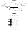
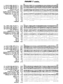
- gag polypeptide sequences An alignment of gag polypeptide sequences is shown in Figure 7 .
- HML-2 prt polypeptide is encoded by the second long ORF in a complete HML-2 genome. It is translated as a gag-prt fusion polypeptide. The fusion polypeptide is proteolytically cleaved to give a protease.
- prt nucleotide sequences are: SEQ ID 86 [HERV-K(108)]; SEQ ID 99 [HERV-K(II)]; SEQ ID 105 [HERV-K10].
- prt polypeptide sequences are: SEQ ID 106 [HERV-K10]; SEQ ID 147 ['ERVK6'].
- HML-2 pol polypeptide is encoded by the third long ORF in a complete HML-2 genome. It is translated as a gag-prt-pol fusion polypeptide. The fusion polypeptide is proteolytically cleaved to give three pol products - reverse transcriptase, endonuclease and integrase [14].
- pol nucleotide sequences are: SEQ ID 87 [HERV-K(108)]; SEQ ID 93 [HERV-K(C7)]; SEQ ID 100 [HERV-K(II)]; SEQ ID 107 [HERV-K10].
- polypeptide sequences are: SEQ ID 94 [HERV-K(C7)]; SEQ ID 108 [HERV-K10]; SEQ ID 148 ['ERVK6'].
- HML-2 env polypeptide is encoded by the fourth long ORF in a complete HML-2 genome.
- the translated polypeptide is proteolytically cleaved.
- env nucleotide sequences are: SEQ ID 88 [HERV-K(108)]; SEQ ID 95 [HERV-K(C7)]; SEQ ID 101 [HERV-K(II)]; SEQ ID 107 [HERV-K10].
- env polypeptide sequences are: SEQ ID 96 [HERV-K(C7)]; SEQ ID 108 [HERV-K10] ; SEQ ID 149 ['ERVK6'].
- HML-2 cORP polypeptide is encoded by an ORF which shares the same 5' region and start codon as env. After amino acid 87, a splicing event removes env-coding sequences and the cORF-coding sequence continues in the reading frame +1 relative to that of env [15,16; see below]. cORF has also been called Rec [17].
- cORF nucleotide sequences are: SEQ ID 89 and SEQ ID 90 [HERV-K(108)]
- Examples of cORF polypeptide sequences are SEQ ID 109.
- Suitable techniques include standard immunohistological methods, immunoprecipitation, immunofluorescence, ELISA, RIA, FIA, etc.
- the invention provides a method for detecting the presence of and/or measuring a level of a polypeptide in a biological sample, wherein the method uses an antibody specific for the polypeptide.
- the method generally comprises the steps of: a) contacting the sample with an antibody specific for the polypeptide; and b) detecting binding between the antibody and polypeptides in the sample.
- Polypeptides can also be detected by functional assays e.g . assays to detect binding activity or enzymatic activity.
- functional assays e.g . assays to detect binding activity or enzymatic activity.
- a functional assay for cORF is disclosed in references 16,129 & 130.
- a functional assay for the protease is disclosed in reference 140.
- polypeptides can be separated using 2D-PAGE and polypeptide spots can be sequenced ( e.g . by mass spectroscopy) in order to identify if a sequence is present in a target polypeptide.
- Detection methods may be adapted for use in vivo (e.g. to locate or identify sites where cancer cells are present).
- an antibody specific for a target polypeptide is administered to an individual ( e.g . by injection) and the antibody is located using standard imaging techniques (e.g. magnetic resonance imaging, computed tomography scanning, etc .). Appropriate labels (e.g. spin labels etc .) will be used. Using these techniques, cancer cells are differentially labeled.
- An immunofluorescence assay can be easily performed on cells without the need for purification of the target polypeptide.
- the cells are first fixed onto a solid support, such as a microscope slide or microtiter well.
- the membranes of the cells are then permeablized in order to permit entry of polypeptide-specific antibody (NB: fixing and permeabilization can be achieved together).
- NB fixing and permeabilization can be achieved together.
- the fixed cells are exposed to an antibody which is specific for the encoded polypeptide and which is fluorescently labeled.
- the presence of this label e.g. visualized under a microscope
- polypeptides may be preferred to detect molecules which are produced by the body in response to a polypeptide (i.e . indirect detection of a polypeptide). This will typically involve the detection of antibodies, so the patient sample will generally be a blood sample. Antibodies can be detected by conventional immunoassay techniques e.g. using PCAV polypeptides which will typically be immobilized.
- Antibodies against HERV-K polypeptides have been detected in humans [143].
- references to a percentage sequence identity between two amino acid sequences means that, when aligned, that percentage of amino acids are the same in comparing the two sequences.
- This alignment and the percent homology or sequence identity can be determined using software programs known in the art, for example those described in section 7.7.18 of reference 11.
- a preferred alignment is determined by the Smith-Waterman homology search algorithm using an affine gap search with a gap open penalty of 12 and a gap extension penalty of 2, BLOSUM matrix of 62.
- the Smith-Waterman homology search algorithm is taught in reference 32.
- polypeptide refers to amino acid polymers of any length.
- the polymer may be linear or branched, it may comprise modified amino acids, and it may be interrupted by non-amino acids.
- the terms also encompass an amino acid polymer that has been modified naturally or by intervention; for example, disulfide bond formation, glycosylation, lipidation, acetylation, phosphorylation, or any other manipulation or modification, such as conjugation with a labeling component
- polypeptides containing one or more analogs of an amino acid including, for example, unnatural amino acids, etc .
- Polypeptides can occur as single chains or associated chains.
- Polypeptides of the invention can be naturally or non-naturally glycosylated (i.e. the polypeptide has a glycosylation pattern that differs from the glycosylation pattern found in the corresponding naturally occurring polypeptide).
- Mutants can include amino acid substitutions, additions or deletions.
- the amino acid substitutions can be conservative amino acid substitutions or substitutions to eliminate non-essential amino acids, such as to alter a glycosylation site, a phosphorylation site or an acetylation site, or to minimize misfolding by substitution or deletion of one or more cysteine residues that are not necessary for function.
- Conservative amino acid substitutions are those that preserve the general charge, hydrophobicity/hydrophilicity, and/or steric bulk of the amino acid substituted.
- Variants can be designed so as to retain or have enhanced biological activity of a particular region of the polypeptide ( e.g.
- amino acid alterations for production of variants can be based upon the accessibility (interior vs. exterior) of the amino acid (e.g . ref. 33), the thermostability of the variant polypeptide (e.g. ref. 34), desired glycosylation sites (e.g . ref. 35), desired disulfide bridges ( e.g . refs. 36 & 37), desired metal binding sites ( e.g . refs.38 & 39), and desired substitutions with in proline loops ( e.g . ref. 40). Cysteine-depleted muteins can be produced as disclosed in reference 41.
- Antibodies be polyclonal or monoclonal and may be produced by any suitable means ( e.g . by recombinant expression).
- Antibodies may include a label.
- the label may be detectable directly, such as a radioactive or fluorescent label.
- the label may be detectable indirectly, such as an enzyme whose products are detectable (e.g . luciferase, ß-galactosidase, peroxidase etc .).
- Antibodies may be attached to a solid support.
- Antibodies be prepared by administering (e.g. injecting) a polypeptide of the invention to an appropriate animal (e.g. a rabbit, hamster, mouse or other rodent).
- an appropriate animal e.g. a rabbit, hamster, mouse or other rodent.
- Antigen-binding fragments of antibodies include Fv, scFv, Fc, Fab, F(ab') 2 etc.
- the antibodies may be chimeric or humanized [ e.g. refs. 42 & 43], or fully human antibodies may be used. Because humanized antibodies are far less immunogenic in humans than the original non-human monoclonal antibodies, they can be used for the treatment of humans with far less risk of anaphylaxis. Thus, these antibodies may be preferred in therapeutic applications that involve in vivo administation to a human such as, use as radiation sensitizers for the treatment of neoplastic disease or use in methods to reduce the side effects of cancer therapy.
- Humanized antibodies may be achieved by a variety of methods including, for example:
- CDRs are amino acid sequences which together define the binding affinity and specificity of a Fv region of a native immunoglobulin binding site [ e.g. refs. 51 & 52].
- constant region refers to the portion of the antibody molecule that confers effector functions.
- mouse constant regions are substituted by human constant regions.
- the constant regions of humanized antibodies are derived from human immunoglobulins.
- the heavy chain constant region can be selected from any of the 5 isotypes: alpha, delta, epsilon, gamma or mu.
- One method of humanizing antibodies comprises aligning the heavy and light chain sequences of a non-human antibody to human heavy and light chain sequences, replacing the non-human framework residues with human framework residues based on such alignment, molecular modeling of the conformation of the humanized sequence in comparison to the conformation of the non-human parent antibody, and repeated back mutation of residues in the framework region which disturb the structure of the non-human CDRs until the predicted conformation of the CDRs in the humanized sequence model closely approximates the conformation of the non-human CDRs of the parent non-human antibody.
- Such humanized antibodies may be further derivatized to facilitate uptake and clearance e.g, via Ashwell receptors. [refs. 53 & 54]
- Humanized or fully-human antibodies can also be produced using transgenic animals that are engineered to contain human immunoglobulin loci.
- ref. 55 discloses transgenic animals having a human Ig locus wherein the animals do not produce functional endogenous immunoglobulins due to the inactivation of endogenous heavy and light chain loci.
- Ref. 56 also discloses transgenic non-primate mammalian hosts capable of mounting an immune response to an immunogen, wherein the antibodies have primate constant and/or variable regions, and wherein the endogenous immunoglobulin-encoding loci are substituted or inactivated.
- Ref. 57 discloses the use of the Cre/Lox system to modify the immunoglobulin locus in a mammal, such as to replace all or a portion of the constant or variable region to form a modified antibody molecule.
- Ref. 58 discloses non-human mammalian hosts having inactivated endogenous Ig loci and functional human Ig loci.
- Ref. 59 discloses methods of making transgenic mice in which the mice lack endogenous heavy claims, and express an exogenous immunoglobulin locus comprising one or more xenogeneic constant regions.
- an immune response can be produced to a PCAV polypeptide, and antibody-producing cells can be removed from the animal and used to produce hybridomas that secrete human monoclonal antibodies.
- Immunization protocols, adjuvants, and the like are known in the art, and are used in immunization of, for example, a transgenic mouse as described in ref. 60.
- the monoclonal antibodies can be tested for the ability to inhibit or neutralize the biological activity or physiological effect of the corresponding polypeptide.
- HML-2 transcripts are up-regulated in tumors, including prostate tumors.
- a reference point is needed i.e. a control.
- Analysis of the control sample gives a standard level of RNA and/or protein expression against which a patient sample can be compared.
- a negative control gives a background or basal level of expression against which a patient sample can be compared.
- Higher levels of expression product relative to a negative control indicate that the patient from whom the sample was taken has, for example, prostate cancer.
- negative controls would include lifetime baseline levels of expression or the expression level observed in pooled normals.
- equivalent levels of expression product indicate that the patient does not have a HML-2-related cancer such as prostate cancer.
- a positive control gives a level of expression against which a patient sample can be compared. Equivalent or higher levels of expression product relative to a positive control indicate that the patient from whom the sample was taken has cancer such as prostate cancer. Conversely, lower levels of expression product indicate that the patient does not have a HML-2 related cancer such as prostate cancer.
- a negative control will generally comprise cells which are not from a tumor cell, e.g. a prostate tumor cell.
- a negative control will generally be a blood sample from a patient who does not have a prostate tumor.
- the negative control could be a sample from the same patient as the patient sample, but from a tissue in which HML-2 expression is not up-regulated e.g. a non-tumor non-prostate cell.
- the negative control could be a prostate cell from the same patient as the patient sample, but taken at an earlier stage in the patient's life.
- the negative control could be a cell from a patient without a prostate tumor. This cell may or may not be a prostate cell.
- the negative control cell could be a prostate cell from a patient with BPH.
- a positive control will generally comprise cells from a tumor cell e.g . a prostate tumor.
- a negative control will generally be a blood sample from a patient who has a prostate tumor.
- the positive control could be a prostate tumor cell from the same patient as the patient sample, but taken at an earlier stage in the patient's life ( e.g . to monitor remission).
- the positive control could be a cell from another patient with a prostate tumor.
- the positive control could be a prostate cell line.
- HML-2 expression in the control can be assessed at the same time as expression in the patient sample. Alternatively, HML-2 expression in the control can be assessed separately (earlier or later).
- control may be an absolute value i.e . a level of expression which has been empirically determined from samples taken from prostate tumor patients ( e.g . under standard conditions).
- the up-regulation relative to the control (100%) will usually be at least 150% ( e.g. 200%, 250%, 300%, 400%, 500%, 600% or more).
- the invention provides a method for diagnosing prostate cancer. It will be appreciated that "diagnosis" according to the invention can range from a definite clinical diagnosis of disease to an indication that the patient should undergo further testing which may lead to a definite diagnosis.
- diagnosis can range from a definite clinical diagnosis of disease to an indication that the patient should undergo further testing which may lead to a definite diagnosis.
- the method of the invention can be used as part of a screening process, with positive samples being subjected to further analysis.
- diagnosis includes monitoring the progress of cancer in a patient already known to have the cancer.
- Cancer can also be staged by the methods of the invention.
- the cancer is prostate cancer.
- the efficacy of a treatment regimen (therametrics) of a cancer associated can also monitored by the method of the invention e.g. to determine its efficacy.
- Susceptibility to a cancer can also be detected e.g. where up-regulation of expression has occurred, but before cancer has developed. Prognostic methods are also encompassed.
- Endogenous retroviruses were identified in human genomic DNA by their homology to retroviruses of other vertebrates [131, 132]. It is believed that the human genome probably contains numerous copies of endogenous proviral DNAs, but little is known about their function. Most HERV families have relatively few members (1-50) but one family (HERV-H) consists of ⁇ 1000 copies per haploid genome distributed on all chromosomes. The large numbers and general transcriptional activity of HERVs in embryonic and tumor cell lines suggest that they could act as disease-causing insertional mutagens or affect adj acent gene expression in a neutral or beneficial way.
- the K family of human endogenous retroviruses is well known [133]. It is related to the mouse mammary tumor virus (MMTV) and is present in the genomes of humans, apes and old world monkeys, but several human HERV-K proviruses are unique to humans [134].
- the HERV-K family is present at 30-50 full-length copies per haploid human genome and possesses long open reading frames that potentially are translated into viral proteins [135, 136].
- Two types of proviral genomes are known, which differ by the presence (type 2) or absence (type 1) of a stretch of 292 nucleotides in the overlapping boundary of the pol and env genes [137].
- HERV-K10 has been shown to encode a full-length gag homologous 73 kDa protein and a functional protease [140].
- Gag proteins released in form of particles from HERV-K have been identified in the cell culture supernatant of the teratocarcinoma derived cell line Tera 1. These retrovirus-like particles (termed “human teratocarcinoma derived virus” or HTDV) have been shown to have a 90% sequence homology to the HERV-K10 genome [138, 143].
- retroviral proviruses While the HERV-K family is present in the genome of every human cell, a high level of expression of mRNAs, proteins and particles is observed only in human teratocarcinoma cell lines [144]. In other tissues and cell lines, only a basal level of expression of mRNA has been demonstrated even using very sensitive methods.
- the expression of retroviral proviruses is generally regulated by elements of the 5' long terminal repeat (LTR). Furthermore, the activation of expression of an endogenous retrovirus may trigger the expression of a downstream gene that triggers a neoplastic effect.
- HML-2 is a subgroup of the HERV-K family [146].
- HERV isolates which are members of the HML-2 subgroup include HERV-K10 [137,142], the 27 HML-2 viruses shown in Figure 4 of reference 147, HERV-K(C7) [148], HERV-K(II) [145], HERV-K(CH)
- the invention is based on the finding that HML-2 mRNA expression is up-regulated in prostate tumors. Because HML-2 is a well-recognized family, the skilled person will be able to determine without difficulty whether any particular endogenous retroviruses is or is not a HML-2.
- Preferred members of the HML-2 family for use in accordance with the present invention are those whose proviral genome has an LTR which has at least 75% sequence identity to SEQ ID 150 (the LTR sequence from HML-2.HOM [1]).
- Example LTRs include SEQ IDs 151-154.
- the present invention is based on the discovery of elevated levels of multiple HML-2 polynucleotides in prostate tumor samples as compared to normal prostate tissue.
- One particular HML-2 whose mRNA was found to be up-regulated is designated herein as ⁇ HERV-K(CH)'.
- HERV-K(CH) Sequences from HERV-K(CH) are shown in SEQ IDs 14-39 and have been deposited with the ATCC (see Table 7). The skilled person will be able to classify any further HERV as HERV-K(CH) or not based on sequence identity to these HERV-K(CH) polynucleotides. Preferably such a comparison is to one or more, or all, of the polynucleotide sequences disclosed herein or of the polynucleotide inserts in the ATCC-deposited isolates.
- sequence identity based on a comparison to any one or more, or all, of the sequences in SEQ IDs 7-10 and SEQ IDs 14-39 taking into consideration the spontaneous mutation rate associated with retroviral replication.
- the differences in the sequences are consistent with a HERV-K(CH) isolate or consistent with another HERV.
- HERV-K(CH) is therefore a specific member of the HML-2 subgroup which can be used in the invention as described above. It can also be used in methods previously described in relation to HERV-K e.g . the diagnosis of testicular cancer [142], autoimmune diseases, multiple sclerosis [149], insulin-dependent diabetes mellitus (IDDM) [150] etc.
- the disclosure provides an isolated polynucleotide comprising: (a) the nucleotide sequence of any of SEQ IDs 7-10; (b) the nucleotide sequence of any of SEQ IDs 27-39; (c) the complement of a nucleotide sequence of any of SEQ IDs 7-10; or (d) the complement of the nucleotide sequence of any of SEQ IDs 27-39.
- the disclosure also provides an isolated polynucleotide comprising a fragment of: (a) a nucleotide sequence shown in SEQ IDs 7-10; (b) the nucleotide sequence shown in any of SEQ IDs 27-39; (c) the complement of a nucleotide sequence shown in SEQ IDs 7-10; or (d) the complement of the nucleotide sequence shown in any of SEQ IDs 27-39.
- the fragment is preferably at least x nucleotides in length, wherein x is at least 7 ( e.g. at least 8, 9, 10,11, 12, 13, 14,15, 16,17, 18, 19, 20, 21, 22, 23, 24, 25, 30, 35, 40, 45, 50, 60, 70, 75, 80, 90,100 etc .).
- the value of x may be between about 150 and about 200 or be between about 250 and about 300.
- the value of x may be about 350, about 400, about 450, about 500, about 550, about 600, about 650, about 700, or about 750.
- the value of x may be less than 2000 ( e.g . less than 1000, 500, 100, or 50).
- the fragment is preferably neither one of the following sequences nor a fragment of one of the following sequences: (i) the nucleotide sequence shown in SEQ ID 42; (ii) the nucleotide sequence shown in SEQ ID 43; (iii) the nucleotide sequence shown in SEQ ID 44; (iv) the nucleotide sequence shown in SEQ ID 45; (v) a known polynucleotide; or (vi) a polynucleotide known as of 7th December 2000 (e.g . a polynucleotide available in a public database such as GenBank of GeneSeq before 7th December 2000).
- the fragment is preferably a contiguous sequence of one of polynucleotides of (a), (b), (c) or (d) that remains unmasked following application of a masking program for masking low complexity (e.g . XBLAST) to the sequence ( i.e . one would select an unmasked region, as indicated by the polynucleotides outside the poly-n stretches of the masked sequence produced by the masking program).
- a masking program for masking low complexity e.g . XBLAST
- probes are particularly useful as probes.
- Probes can be used, for example, to determine the presence or absence of a polynucleotide of the invention (or variants thereof) in a sample.
- probes particularly labeled probes of DNA sequences, one can isolate homologous or related genes.
- the source of homologous genes can be any species e.g. primate species, Particularly human; rodents, such as rats and mice; canines; felines; bovines; ovines; equines; yeast; nematodes; etc .
- Probes from more than one polynucleotide sequence of the disclosure can hybridize with the same nucleic acid if the nucleic acid from which they were derived corresponds to a single sequence ( e.g . more than one can hybridize to a single cDNA derived from the same mRNA).
- Preferred fragments e.g . for the identification of HERV-K(CH) polynucleotides associated with cancer
- SEQ ID 59 from gag region
- SEQ IDs 60-70 from pol region
- SEQ IDs 71-82 from 3' pol region
- Preferred fragments e.g . for the simultaneous identification of HERV-K(CH) polynucleotides, HERV-KII polynucleotides and/or HERV-K10 polynucleotides
- SEQ IDs 83 & 84 from gag region
- Polynucleotide probes unique to HERV-K(CH), HERV-KII and HERV-K10 gag regions are provided in Table 1; polynucleotide probes unique to HERV-K(CH), HERV-KII, and HERV-K10 protease 3' and polymerase 5' regions are provided in Table 2; polynucleotide probes unique to HERV-K(CH), HERV-KII, and HERV-K10 3' pol only regions are provided in Table 3.
- the disclosure also provides an isolated polynucleotide comprising (a) a segment that is a fragment of the sequence shown in SEQ IDs 7-10 or SEQ IDs 27-39, wherein (i) said fragment is at least 10 nucleotides in length and (ii) corresponds identically in its entirety to a portion of SEQ ID 44 and/or 45; and, optionally, (b) one or more segments flanking the segment defined in (a), wherein the presence of said optional segment(s) causes said polynucleotide to not correspond identically to any portion of a sequence shown in SEQ IDs 7-10 or SEQ IDs 27-39.
- the optional flanking segments share less than 40% sequence identity to the nucleic acid sequences shown in SEQ IDs 7-10, SEQ ID 44 and/or SEQ ID 45. In other embodiments, the optional flanking segments have no contiguous sequence of 10, 12, 15 or 20 nucleotides in common with SEQ IDs 7-10, SEQ ID 44 and/or SEQ ID 45. In yet other embodiments, the optional flanking segment is not present. In further embodiments, a fragment of the polynucleotide sequence is up to at least 30,40,50,60,70,80,90,100,200,300,400, 500, 1000, or 1500 nucleotide in length.
- the disclosure also provides an isolated polynucleotide having formula 5'-A-B-C-3', wherein: A is a nucleotide sequence consisting of ⁇ nucleotides; B is a nucleotide sequence consisting of a fragment of b nucleotides from (i) the nucleotide sequence shown in SEQ IDs 7-10, (ii) the nucleotide sequence shown in any of SEQ IDs 27-39, (iii) the complement of the nucleotide sequence shown in SEQ IDs 7-10, or (iv) the complement of the nucleotide sequence shown in any of SEQ IDs 27-39; C is a nucleotide sequence consisting of c nucleotides; and wherein said polynucleotide is not a fragment of (i) the nucleotide sequence shown in SEQ IDs 7-10, (ii) the nucleotide sequence shown in any of SEQ IDs 27-39, (iii) the
- a+c is at least 1 (e.g . at least 2, 3, 4, 5, 6, 7, 8, 9,10, 11, 12, 13, 14, 15, 16, 17, 18, 19, 20, 21, 22, 23, 24, 25, 26, 27, 28, 29, 30, 35, 40, 45, 50, 60, 70, 80, 90, 100 etc.) and b is at least 7 ( e.g. at least 8, 9, 10, 11, 12,13, 14, 15, 16,17, 18,19, 20, 21, 22, 23, 24, 25, 30, 35, 40, 45, 50, 60, 70, 80, 90, 100 etc.). It is preferred that the value of a + b + c is at least 9 ( e.g .
- a + b + c is at most 200 ( e.g . at most 190, 180, 170, 160, 150, 140, 130, 120, 110, 100, 90, 80, 70, 60, 50, 40, 30, 25, 20, 19, 18, 17, 16, 15, 14, 13, 12, 11, 10, 9).
- a and/or C may comprise a promoter sequence (or its complement).
- a polynucleotide having at least s % identity to: (a) SEQ IDs 7-10; (b) a fragment ofx nucleotides of SEQ IDs 7-10; (c) SEQ IDs 11-13; (b) a fragment of x nucleotides of SEQ IDs 11-13.
- the value of s is at least 50 (e.g. at least 55, 60, 65, 70, 75, 80, 85, 90, 91, 92, 93, 94, 9S, 96, 97, 98, 99, 99.5, 99.9 etc.).
- the value ofx is at least 7 (e.g. 8, 9, 10,11, 12,13, 14,15, 16, 17, 18, 19, 20, 21, 22, 23, 24, 25, 30, 35, 40, 45, 50, 60, 70, 80, 90, 100 etc.).
- polynucleotides include naturally-occurring variants (e.g . degenerate variants, allelic variants, etc .), homologs, orthologs, and functional mutants.
- Variants can be identified by hybridization of putative variants with the polynucleotide sequences disclosed in SEQ IDs 14-39 herein, preferably by hybridization under stringent conditions. For example, by using appropriate wash conditions, variants can be identified where the allelic variant exhibits at most about 25-30% base pair (bp) mismatches relative to the selected polynucleotide probe. In general, allelic variants contain 15-25% bp mismatches, and can contain as little as even 5-15%, or 2-5%, or 1-2% bp mismatches, as well as a single bp mismatch.
- bp base pair
- the disclosure also encompasses homologs corresponding to any one of the polynucleotide sequences provided herein, where the source of homologous genes can be any mammalian species (e.g . primate species, particularly human; rodents, such as rats, etc.). Between mammalian species ( e.g . human and primate), homologs generally have substantial sequence similarity (e.g . at least 75% sequence identity, usually at least 90%, more usually at least 95%) between nucleotide sequences. Sequence similarity is calculated based on a reference sequence, which may be a subset of a larger sequence, such as a conserved motif, coding region, flanking region, domain, etc . A reference sequence will usually be at least about 18 contiguous nt long, more usually at least about 30 nt long, and may extend to the complete sequence that is being compared. Algorithms for sequence analysis are known in the art.
- a preferred HERV-K(CH) isolate is an isolate sequence which is shown in SEQ IDs 7-10.
- Another typical classification of the relationship of retroviruses is based on the amino acid sequence similarities in the reverse transcriptase protein.
- an even more preferred class of HERV-K(CH) isolates are those having an amino acid sequence identity of at least 90%, more preferably 95% to the 5' polymerase region encoded by the nucleotide sequence shown in SEQ ID 12, as determined by the Smith-Waterman homology search algorithm using an affine gap search with a gap open penalty of 12 and a gap extension penalty of 2, BLOSUM matrix of 62.
- these prostate cancer-associated polynucleotide sequences define a class of human endogenous retroviruses, designated herein as HERV-K(CH), whose members comprise variations which, without wanted to be bound by theory, may be due to the presence of polymorphisms or allelic variations.
- an isolated polynucleotide comprising a polynucleotide that selectively hybridizes, relative to a known polynucleotide, to: (a) the nucleotide sequence shown in SEQ IDs 7-10; (b) the nucleotide sequence shown in any of SEQ IDs 27-39; (c) the complement of the nucleotide sequence shown in SEQ IDs 7-10; (d) the complement of the nucleotide sequence shown in any of SEQ IDs 27-39; (e) a fragment of the nucleotide sequence shown in SEQ IDs 7-10; (f) a fragment of the nucleotide sequence shown in any of SEQ IDs 27-39; (g) the complement of a fragment of the nucleotide sequence shown in SEQ IDs 7-10; (h) the complement of a fragment of the nucleotide sequence shown in any of SEQ IDs 27-39; (j) a nucleotide sequence shown in SEQ IDs
- the fragment of (e), (f), (g) or (h) is preferably at least x nucleotides in length, wherein x is as defined in H.1.2 above, and is preferably not one of the sequences (i), (ii), (iii), (iv), (v) or (vi) as defined H.1.2 above.
- Hybridization reactions can be performed under conditions of different "stringency", as described in B.4 above.
- the polynucleotide hybridizes under low stringency conditions; in other embodiments it hybridizes under intermediate stringency conditions; in other embodiments, it hybridizes under high stringency conditions.
- an isolated polynucleotide comprising: (a) a HERV-K(CH) cDNA insert as deposited at the ATCC and having an ATCC accession number given in Table 7; (b) a HERV-K(CH) sequence as shown in any one of SEQ IDs 14-26; (c) a HERV-K(CH) sequence as shown in any one of SEQ IDs 27-39; or (d) a fragment of (a), (b) or (c).
- the fragment of (d) is preferably at least x nucleotides in length, wherein x is at least 7 (e.g. at least 8, 9, 10, 11, 12, 13, 14, 15, 16, 17, 18, 19, 20, 21, 22, 23, 24, 25, 30, 35, 40, 45, 50, 60, 70, 80, 90, 100 etc.).
- Preferred polynucleotides are those having a sequence set forth in any one of the polynucleotide sequences SEQ IDs 7-10 and SEQ IDs 14-39 provided herein; polynucleotides obtained from the biological materials described herein, in particular, polynucleotide sequences present in the isolates deposited with the ATCC and having ATCC accession numbers given in Table 7 or other biological sources (particularly human sources) or by hybridization to the above mentioned sequences under stringent conditions (particularly conditions of high stringency); genes corresponding to the provided polynucleotides; variants of the provided polynucleotides and their corresponding genes particularly those variants that retain a biological activity of the encoded gene product ( e.g .
- the isolated polynucleotides preferably comprise a polynucleotide having a HERV-K(CH) sequence.
- a polynucleotide can encode all or a part of a polypeptide, such as the gag region, 5' pol region or 3' pol region of a human endogenous retrovirus. Double or single stranded fragments can be obtained from the DNA sequence by chemically synthesizing oligonucleotides in accordance with conventional methods, by restriction enzyme digestion, by PCR amplification, etc .
- Polynucleotides can be cDNAs or genomic DNAs, as well as fragments thereof, particularly fragments that encode a biologically active gene product and/or are useful in the methods disclosed herein ( e.g . in diagnosis, as a unique identifier of a differentially expressed gene of interest, etc ).
- cDNA as used herein is intended to include all nucleic acids that share the arrangement of sequence elements found in native mature mRNA species, where sequence elements are exons and 3' and 5' non-coding regions. Normally mRNA species have contiguous exons, with the intervening introns, when present, being removed by nuclear RNA splicing, to create a continuous open reading frame encoding a polypeptide.
- mRNA species can also exist with both exons and introns, where the introns may be removed by alternative splicing. Furthermore it should be noted that different species of mRNAs encoded by the same genomic sequence can exist at varying levels in a cell, and detection of these various levels of mRNA species can be indicative of differential expression of the encoded gene product in the cell.
- a genomic sequence of interest comprises the nucleic acid present between the initiation codon and the stop codon, as defined in the listed sequences, including all of the introns that are normally present in a native chromosome. It can further include the 3' and 5' untranslated regions found in the mature mRNA. It can further include specific transcriptional and translational regulatory sequences, such as promoters, enhancers, etc ., including about 1 kb, but possibly more, of flanking genomic DNA at either the 5' and 3' end of the transcribed region.
- the genomic DNA can be isolated as a fragment of 100 kbp or smaller; and substantially free of flanking chromosomal sequence.
- the genomic DNA flanking the coding region, either 3' and 5', or internal regulatory sequences as sometimes found in introns contains sequences required for proper tissue, stage-specific, or disease-state specific expression.
- Polynucleotides can be provided as linear molecules or within circular molecules, and can be provided within autonomously replicating molecules (vectors) or within molecules without replication sequences. Expression of the polynucleotides can be regulated by their own or by other regulatory sequences known in the art.
- the polynucleotides can be introduced into suitable host cells using a variety of techniques available in the art, such as transferrin polycation-mediated DNA transfer, transfection with naked or encapsulated nucleic acids, liposome-mediated DNA transfer, intracellular transportation of DNA-coated latex beads, protoplast fusion, viral infection, electroporation, gene gun, calcium phosphate-mediated transfection, and the like.
- a polynucleotide sequence that is "shown in” or “depicted in” a SEQ ID NO or Figure means that the sequence is present as an identical contiguous sequence in the SEQ ID NO or Figure.
- the term encompasses portions, or regions of the SEQ ID NO or Figure as well as the entire sequence contained within the SEQ ID NO or Figure.
- Deduced polypeptides encoded by the HERV-K(CH) polynucleotides include the gag translations shown in SEQ IDS 46-49 and the 3' pol translations shown in SEQ IDs 50-55.
- a polypeptide sequence encoded by the polynucleotide having the sequence shown in SEQ ID 15 is provided in SEQ ID 56; a polypeptide sequence encoded by the polynucleotide having the sequence shown in SEQ ID 14, is shown in SEQ ID 57.
- a consensus 3' pol polypeptide sequence encoded by the polynucleotides having the sequence shown in SEQ IDs 21-27, inclusive, is provided in SEQ ID 58.
- polypeptides encompassed by the present disclosure include those encoded by polynucleotides of the disclosure e.g. SEQ IDs 7-10 and SEQ IDs 14-39, as well as polynucleotides deposited with the ATCC as disclosed herein, as well as nucleic acids that, by virtue of the degeneracy of the genetic code, are not identical in sequence to the disclosed polynucleotides and encode the polypeptides.
- the disclosure includes within its scope a polypeptide encoded by a polynucleotide having the sequence of any one of the polynucleotide sequences provided herein, or a variant thereof.
- a single large gag polypeptide is synthesized (e.g . a 73 kDa gag protein in HERV-K10) which is subsequently cleaved into multiple functional peptides by a functional protease encoded by the pol or protease region of the genome.
- a functional protease encoded by the pol or protease region of the genome Overexpression of sequences corresponding to both gag and pol domains of the HERV-K(CH) suggest such a mechanism. Sequences corresponding to the env and the nuclear RNA transport protein cORF region of the HERV-K(CH) genome may also be overexpressed.
- the polypeptides encoded by the open reading frames within the over-expressed polynucleotide sequences may play a significant role in the progression of prostate tumors.
- an isolated polypeptide comprising a fragment of: (a) a polypeptide sequence encoded within a HERV-K(CH) open reading frame; (b) a polypeptide sequence encoded by a polynucleotide shown in SEQ ID 11, 12 or 13; or (c) an amino acid sequence as shown in any one of SEQ IDs 46-49, 50-55, 56-57 or 58.
- the fragment is preferably at least x amino acids in length, wherein x is at least 5 ( e.g . at least 6,7, 8, 9, 10, 11, 12, 13, 14, 15, 16, 17, 18, 19, 20, 21, 22, 23, 24, 25, 30, 35, 40, 45, 50, 60, 70, 75, 80, 90, 100, 125, 150, 200, 300, 400, 500 or more etc .).
- x is at least 5 ( e.g . at least 6,7, 8, 9, 10, 11, 12, 13, 14, 15, 16, 17, 18, 19, 20, 21, 22, 23, 24, 25, 30, 35, 40, 45, 50, 60, 70, 75, 80, 90, 100, 125, 150, 200, 300, 400, 500 or more etc .).
- the value of x will typically not exceed 1000.
- the fragment may include an epitope e.g . an epitope of the amino acid sequence shown in SEQ IDs 56, 57 or 58.
- SEQ IDs 46-49 provide a translation of the HERV-K(CH) polynucleotides having a sequence shown in SEQ IDs 14, 15, 16 and 40 (the sequence of SEQ ID 40 is from a polynucleotide found in a normal prostate library) corresponding to polynucleotides encoding the gag region.
- SEQ IDs 50-55 provide a translation of the HERV-K(CH) polynucleotides having a sequence shown in SEQ IDs 21-26, inclusive, corresponding to the 3' region of pol.
- SEQ IDs 56 & 57 provide translations of the HERV-K(CH) polynucleotide of SEQ ID 15 and SEQ ID 14, respectively.
- SEQ ID 58 provides a consensus translation of the polynucleotide from the 3' pol region (SEQ IDs 21-26, inclusive).
- polypeptide fragments such as, epitopes, of at least 5 amino acids, at least 6 amino acids, at least 8 amino acids, at least 10 amino acids, at least 11 amino acids, at least 12 amino acids, at least 13 amino acids, at least 14 amino acids and at least 15 amino acids of the translations shown in SEQ IDs 46-49 and 50-55.
- the HERV-K(CH) epitopes of the amino acid sequence as shown in SEQ IDs 56-58 were determined by the Jameson-Wolf antigenic index
- 3' pol SEQ ID 58
- Jameson-Wolf algorithm amino acids: 1-10; 15-35; 45-55; 60-85; 100-115; 125-140; 170-190; 195-215; 230-268.
- Additional epitope-containing fragments include amino acids 1-8; 2-10; 1-15; 5-15; 7-15; 10-20; 12-20; 15-23; 20-28; 28-35; 15-30; 15-40; 20-30; 45-52; 48-55; 60-68; 60-70; 65-73; 70-78; 75-83; 70-80; 65-75; 68-75; 75-85; 78-85; 65-85; 60-75; 100-108; 103-110; 105-113; 108-115; 125-133; 128-135; 132-140; 170-178; 175-182; 180-187; 182-190; 195-202; 200-208; 205-212; 208-215; 230-237; 235-242; 240-247; 245-252; 250-257; 255-262; 260-268; 230-250; 235-255; 240-260; 245-268; 230-245; 235-245; 235-250; 240-255; 245-260; 250-2
- gag The following regions in gag (SEQ ID 56) were determined to be antigenic by Jameson-Wolf algorithm: amino acids: 1-40; 45-60; 80-105; 130-145; 147-183; 186-220; 245-253; 255-288. Additional epitope-containing fragments include amino acids 1-8; 2-10; 1-15; 5-15; 7-15; 10-20; 12-20; 15-23; 20-28; 28-35; 30-37; 33-40; 1-20; 20-40; 1-15; 15-30; 15-40; 45-52; 50-57; 55-62; 50-60; 1-60; 80-87; 85-92; 80-90; 90-97; 95-102; 98-105; 85-100; 90-105; 80-100; 85-105; 130-137; 135-142; 140-147; 145-152; 150-157; 155-162; 160-167; 165-172; 170-177; 175-183; 180-187; 185-192; 190-197; 195-202; 200-207;
- gag The following regions in gag (SEQ ID 57) were determined to be antigenic by Jameson-Wolf algorithm: amino acids: 1-40; 80-105; 145-180; 185-225; 240-335. Additional epitope-containing fragments include amino acids 1-8; 2-10; 1-15; 5-15; 7-15; 10-20; 12-20; 15-23; 20-28; 28-35; 30-37; 33-40; 1-20; 20-40; 1-15; 15-30; 15-40; 80-87; 85-92; 80-90; 90-97; 95-102; 98-105; 85-100; 90-105; 80-100; 85-105; 145-152; 150-157; 155-162; 160-167; 165-172; 170-177; 175-182; 180-187; 185-192; 190-197; 195-202; 200-207; 205-212; 210-217; 215-212; 218-225; 145-160; 150-165; 155-170; 160-175; 170-185;
- isolated polypeptide having formula 5'-A-B-C-3', wherein: A is an amino acid sequence consisting of a amino acids; B is an amino acid sequence consisting of a fragment of b amino acids from (i) the amino acid sequence encoded by a polynucleotide shown in SEQ ID 11, 12 or 13; (ii) any one of SEQ IDs 46-49, 50-55, 56-57 or 58; C is an amino acid sequence consisting of c amino acids; and wherein said polypeptide is not a fragment of the amino acid sequence defined in (i) or (ii).
- a+c is at least 1 (e.g . at least 2, 3, 4, 5, 6, 7, 8, 9,10,11, 12,13,14, 15, 16,17, 18, 19, 20, 21, 22, 23, 24, 25, 26, 27, 28, 29, 30, 35, 40, 45, 50, 60, 70, 80, 90, 100 etc.) and b is at least 7 ( e.g . at least 8, 9, 10, 11, 12, 13, 14, 15, 16, 17, 18, 19, 20, 21, 22, 23, 24, 25, 30, 35, 40, 45, 50, 60, 70, 80, 90, 100 etc .). It is preferred that the value of a + b + c is at least 9 ( e.g .
- a + b + c is at most 200 ( e.g . at most 190, 180, 170, 160,150, 140, 130, 120, 110, 100, 90, 80, 70, 60, 50, 40, 30, 25, 20, 19, 18, 17, 16, 15, 14, 13, 12, 11, 10, 9).
- polypeptide having at least s% identity to: (a) the polypeptide sequences encoded by SEQ IDs 7-45; (b) a fragment of x amino acids of the polypeptide sequences encoded by SEQ IDs 7-45; (c) the polypeptide sequences SEQ IDs 46-58; (d) a fragment of x amino acids of the polypeptide sequences SEQ IDs 46-58.
- the value of s is at least 35 ( e.g .
- the value ofx is at least 7 ( e.g. 8, 9,10,11,12,13, 14, 15, 16,17,18, 19, 20, 21, 22, 23, 24, 25, 30, 35, 40, 45, 50, 60, 70, 80, 90,100.
- polypeptides include naturally-occurring variants (e.g . allelic variants, etc .), homologs, orthologs, and functional mutants.
- Variants of the naturally-occurring polypeptides wherein such variants are homologous or substantially similar to the naturally occurring polypeptide, can be of an origin of the same or different species as the naturally occurring polypeptide (e.g . human, murine, or some other species that naturally expresses the recited polypeptide, usually a mammalian species).
- These polypeptide variants are encoded by polynucleotides, and the genetic code can be used to select appropriate codons to construct the corresponding variants.
- Polypeptides such as those shown in SEQ IDs 46-58, encoded by HERV-K(CH) polynucleotides are differentially expressed in prostate cancer cells. Such polypeptides are referred to herein as "polypeptides associated with prostate cancer" or "HERV-K(CH) polypeptides".
- the polypeptides can be used to generate antibodies specific for a polypeptide associated with prostate cancer, which antibodies are in turn useful in diagnostic methods, prognostic methods, therametric methods, and the like as discussed in more detail herein. Polypeptides are also useful as targets for therapeutic intervention, as discussed in more detail herein.
- the isolated polypeptides preferably comprise a polypeptide having a HERV-K(CH) sequence.
- Polypeptides such as polypeptides of the gag regions or polypeptides of the pol regions, encoded by the polynucleotides disclosed herein, such as polynucleotides having the sequences as shown in SEQ IDs 7-10 and SEQ IDs 14-39, and in isolates deposited with the ATCC and having ATCC accession numbers given in Table 7 and/or their corresponding full length genes, can be used to screen peptide libraries to identify binding partners, such as receptors, from among the encoded polypeptides.
- Peptide libraries can be synthesized according to methods known in the art ( e.g . see refs. 151 & 152).
- polypeptide refers to both the full length polypeptide encoded by the recited polynucleotide, the polypeptide encoded by the gene represented by the recited polynucleotide, as well as portions or fragments thereof.
- a polypeptide sequence that is "shown in” or “depicted in” a SEQ ID NO or Figure means that the sequence is present as an identical contiguous sequence in the SEQ ID NO or Figure.
- the term encompasses portions, or regions of the SEQ ID NO or Figure as well as the entire sequence contained within the SEQ ID NO or Figure.
- the present disclosure also provides isolated antibodies or antigen binding fragments thereof, that bind to a polypeptide disclosed herein.
- the present disclosure also provides isolated antibodies or antigen binding fragments thereof, that bind to a polypeptide encoded by a polynucleotide disclosure herein.
- the present disclosure also provides isolated antibodies that bind to a polypeptide, or antigen binding fragment thereof, encoded by a polynucleotide made by the method comprising the following steps i) immunizing a host animal with a composition comprising said polypeptide, or antigen binding fragment thereof, and ii) collecting cells from said host expressing antibodies against the antigen or antigen binding fragment thereof.
- antibodies that bind to a polypeptide, or antigen binding fragment thereof, encoded by a polynucleotide disclosed herein made by the method comprising the following steps: providing a cell line producing an antibody, wherein said antibody binds to a polypeptide, or antigen binding fragment thereof, encoded by a polynucleotide disclosed herein and culturing said cell line under conditions wherein said antibodies are produced.
- the antibodies are collected and monoclonal antibodies are produced using the collected host cells or genetic material derived from the collected host cells.
- the antibody is a polyclonal antibody.
- the antibody is attached to a solid surface or further comprises a detectable label.
- Antibodies may be isolated antibodies, that bind a polypeptide encoded by a polynucleotide described herein. Antibodies can be provided in a composition comprising the antibody and a buffer and/or a pharmaceutically acceptable excipient. Antibodies specific for a polypeptide associated with cancer are useful in a variety of diagnostic and therapeutic methods, as discussed in detail herein.
- Expression products of a polynucleotide described herein, as well as the corresponding mRNA (particularly mRNAs having distinct secondary and/or tertiary structures), cDNA, or complete gene, or fragments of said expression products can be prepared and used for raising antibodies for experimental, diagnostic, and therapeutic purposes.
- a polynucleotide described herein, as well as the corresponding mRNA (particularly mRNAs having distinct secondary and/or tertiary structures), cDNA, or complete gene, or fragments of said expression products can be prepared and used for raising antibodies for experimental, diagnostic, and therapeutic purposes.
- polynucleotides to which a corresponding gene has not been assigned this provides an additional method of identifying the corresponding gene.
- the polynucleotide or related cDNA is expressed as described above, and antibodies are prepared.
- These antibodies are specific to an epitope on the polypeptide encoded by the polynucleotide, and can precipitate or bind to the corresponding native polypeptide in a cell or tissue preparation or in a cell-free extract of an in vitro expression system.
- Polyclonal or monoclonal antibodies to the HERV -K(CH) polypeptides or an epitope thereof can be made for use in immunoassays by any of a number of methods know in the art.
- epitope reference is made to an antigenic determinant of a polypeptide.
- the presence of an epitope is demonstrated by the ability of an antibody to bind a polypeptide with specificity. Two antibodies are considered to be directed to the same epitope if they cross block each others binding to the same polypeptide.
- One approach for preparing antibodies to a polypeptide is the selection and preparation of an amino acid sequence of all or part of the polypeptide, chemically synthesizing the sequence and injecting it into an appropriate animal, typically a rabbit, hamster or a mouse.
- Oligopeptides can be selected as candidates for the production of an antibody to the HERV-K(CH) polypeptide based upon the oligopeptides lying in hydrophilic regions, which are thus likely to be exposed in the mature polypeptide. Additional oligopeptides can be determined using, for example, the Antigenicity Index [30].
- humanized monoclonal antibodies are provided, wherein the antibodies are specific for HERV-K(CH) polypeptides and do not appreciably bind other HERV polypeptides.
- the phrase "humanized antibody” refers to an antibody derived from a non-human antibody, typically a mouse monoclonal antibody.
- a humanized antibody may be derived from a chimeric antibody that retains or substantially retains the antigen-binding properties of the parental, non-human, antibody but which exhibits diminished immunogenicity in humans as compared to the parental antibody.
- chimeric antibody refers to an antibody containing sequence derived from two different antibodies (see, e.g. ref. 153) which typically originate from different species. Most typically, chimeric antibodies comprise human and murine antibody fragments, generally human constant and mouse variable regions.
- Methods for preparation of the human or primate HERV-K(CH) or an epitope thereof include, but are not limited to chemical synthesis, recombinant DNA techniques or isolation from biological samples. Chemical synthesis of a peptide can be performed, for example, by the classical Merrifeld method of solid phase peptide synthesis [154] or the FMOC strategy on a Rapid Automated Multiple Peptide Synthesis system (E.I. du Pont de Nemours Company, Wilmington, DE) [155].
- Polyclonal antibodies can be prepared by immunizing rabbits or other animals by injecting antigen followed by subsequent boosts at appropriate intervals. The animals are bled and sera assayed against purified HERV-K(CH) usually by ELISA or by bioassay based upon the ability to block the action of HERV-K(CH). When using avian species, e.g. chicken, turkey and the like, the antibody can be isolated from the yolk of the egg. Monoclonal antibodies can be prepared after the method of Milstein and Kohler by fusing splenocytes from immunized mice with continuously replicating tumor cells such as myeloma or lymphoma cells. [156, 157, 158]. The hybridoma cells so formed are then cloned by limiting dilution methods and supernates assayed for antibody production by ELISA, RIA or bioassay.
- antibodies either polyclonal or monoclonal, to the HERV-K(CH) polypeptides can be produced by any suitable method known in the art as discussed above.
- murine or human monoclonal antibodies can be produced by hybridoma technology or, alternatively, the HERV-K(CH) polypeptides, or an immunologically active fragment thereof, or an anti-idiotypic antibody, or fragment thereof can be administered to an animal to elicit the production of antibodies capable of recognizing and binding to the HERV-K(CH) polypeptides.
- Such antibodies can be from any class of antibodies including, but not limited to IgG, IgA, IgM, IgD, and IgE or in the case of avian species, IgY and from any subclass of antibodies.
- the invention provides methods for diagnosing the presence of prostate cancer in a test sample associated with expression of a polynucleotide in a test cell sample, comprising the steps of: i) detecting a level of expression of at least one polynucleotide of the invention, or a fragment thereof, or at least one polynucleotide found in an isolate selected from the group consisting of ATCC accession numbers given in Table 7, or a fragment thereof; and ii) comparing said level of expression of the polynucleotide in the test sample with a level of expression of polynucleotide in the control cell sample, wherein differential expression of the polynucleotide in the test cell sample relative to the level of polynucleotide expression in the control cell sample is indicative of the presence of cancer in the test cell sample.
- the detecting is measuring the level of an RNA transcript; measuring the level of a polynucleotide; or measuring by a method including PCR, TMA, bDNA, NAT or Nasba.
- the polynucleotide is attached to a solid support.
- compositions comprising a test cell sample and an isolated polynucleotide disclosed herein.
- the present invention further provides methods for detecting prostate cancer associated with expression of a polypeptide in a test cell sample, comprising the steps of: i) detecting a level of expression of at least one polypeptide disclosed herein, or a fragment thereof and ii) comparing said level of expression of the polypeptide in the test sample with a level of expression of polypeptide in the control cell sample, wherein an altered level of expression of the polypeptide in the test cell sample relative to the level of expression of the polypeptide in the control cell sample is indicative of the presence of cancer in the test cell sample.
- the present disclosure also provides methods for detecting prostate cancer associated with the presence of an antibody in a test cell sample, comprising the steps of: i) detecting a level of an antibody and ii) comparing said level of said antibody in the test sample with a level of said antibody in the control cell sample, wherein an altered level of antibody in said test cell sample relative to the level of antibody in the control cell sample is indicative of the presence of cancer in the test cell sample.
- This disclosure also provides methods for detecting prostate cancer associated with elevated levels of HERV-K(CH) polynucleotides, by means of (i) detecting polynucleotides having at least 65%, at least 70%, at least 75%, at least 80%, at least 85%, at least 90% at least 91%, at least 92%, at least 93%, at least 94%, at least 95%, at least 96%, at least 97%, at least 98%, at least 99% or at least 100% identity to the polynucleotide shown in SEQ IDs 7-10 or to polynucleotides in isolates deposited with the ATCC and having ATCC deposit accession numbers PTA-2561, PTA-2572, PTA-2566, PTA-2571, PTA-2562, PTA-2573, PTA-2560, PTA-2565, PTA-2568, PTA-2564, PTA-2569, PTA-2567, PTA-2559, PTA-2563, PTA-2570, as measured by the alignment program GCG Gap (Suite
- the treatment regimen of a prostate cancer associated with elevated levels of HERV-K(CH) polynucleotides may also monitored by detecting levels of the polynucleotides and polypeptides in order to assess the staging of the cancer and/or efficacy of particular cancer therapies.
- the present invention provides methods of using the polynucleotides described herein for detecting prostate cancer cells, facilitating diagnosis of prostate cancer and the severity of a cancer (e.g . tumor grade, tumor burden, and the like) in a subject, facilitating a determination of the prognosis of a subject, and assessing the responsiveness of the subject to therapy (e.g . by providing a measure of therapeutic effect through, for example, assessing tumor burden during or following a chemotherapeutic regimen).
- Detection can be based on detection of a polynucleotide that is differentially expressed in a prostate cancer cell and/or detection of a polypeptide encoded by a polynucleotide that is differentially expressed in a prostate cancer cell.
- the detection methods of the invention can be conducted in vitro or in vivo, on isolated cells, or in whole tissues or a bodily fluid e.g . blood, plasma, serum, urine, and the like).
- kits for detecting the presence and/or a level of a polynucleotide that is differentially expressed in a cancer cell e.g . by detection of an mRNA encoded by the differentially expressed gene of interest
- a polypeptide encoded thereby in a biological sample.
- Procedures using these kits can be performed by clinical laboratories, experimental laboratories, medical practitioners, or private individuals.
- the kits of the disclosure for detecting a polypeptide encoded by a polynucleotide that is differentially expressed in a prostate cancer cell may comprise a moiety that specifically binds the polypeptide, which may be an antibody that binds the polypeptide or fragment thereof.
- kits of the disclosure used for detecting a polynucleotide that is differentially expressed in a prostate cancer cell may comprise a moiety that specifically hybridizes to such a polynucleotide.
- the kit may optionally provide additional components that are useful in the procedure, including, but not limited to, buffers, developing reagents, labels, reacting surfaces, means for detection, control samples, standards, instructions, and interpretive information.
- kits for detecting prostate cancer comprising at least one of polynucleotides having the sequence as shown in SEQ IDs 7-10, SEQ IDs 14-39, or fragments thereof, or having the sequence found in an isolate deposited with the ATCC and having ATCC accession numbers PTA-2561, PTA-2572, PTA-2566, PTA-2571, PTA-2562, PTA-2573, PTA-2560, PTA-2565, PTA-2568, PTA-2564, PTA-2569, PTA-2567, PTA-2559, PTA-2563, PTA-2570 or fragments thereof.
- methods are provided for detecting a polypeptide encoded by a gene differentially expressed in a prostate cancer cell.
- Any of a variety of known methods can be used for detection, including, but not limited to, immunoassay, using antibody that binds the polypeptide, e.g. by enzyme-linked immunosorbent assay (ELISA), radioimmunoassay (RIA), and the like; and functional assays for the encoded polypeptide, e.g. binding activity or enzymatic activity.
- ELISA enzyme-linked immunosorbent assay
- RIA radioimmunoassay
- the detection methods and other methods described herein can be readily varied. Such variations are within the intended scope of the invention.
- the probe for use in detection can be immobilized on a solid support, and the test sample contacted with the immobilized probe. Binding of the test sample to the probe can then be detected in a variety of ways, e.g . by detecting a detectable label bound to the test sample to facilitate detected of test sample-immobilized probe complexes.
- the present invention further provides methods for detecting the presence of and/or measuring a level of a polypeptide in a biological sample, which polypeptide is encoded by a polynucleotide that is differentially expressed in a prostate cancer cell, using an antibody specific for the encoded polypeptide.
- the methods generally comprise: a) contacting the sample with an antibody specific for a polypeptide encoded by a polynucleotide that is differentially expressed in a prostate cancer cell; and b) detecting binding between the antibody and molecules of the sample.
- Detection of specific binding of the antibody specific for the encoded prostate cancer-associated polypeptide, when compared to a suitable control is an indication that encoded polypeptide is present in the sample.
- Suitable controls include a sample known not to contain the encoded polypeptide or known not to contain elevated levels of the polypeptide; such as normal prostate tissue, and a sample contacted with an antibody not specific for the encoded polypeptide, e.g. an anti-idiotype antibody.
- a variety of methods to detect specific antibody-antigen interactions are known in the art and can be used in the method, including, but not limited to, standard immunohistological methods, immunoprecipitation, an enzyme immunoassay, and a radioimmunoassay.
- the specific antibody will be detectably labeled, either directly or indirectly.
- Direct labels include radioisotopes; enzymes whose products are detectable (e.g. luciferase, ß-galactosidase, and the like); fluorescent labels (e.g. fluorescein isothiocyanate, rhodamine, phycoerythrin, and the like); fluorescence emitting metals, e.g. 152 Eu, or others of the lanthanide series, attached to the antibody through metal chelating groups such as EDTA; chemiluminescent compounds, e.g. luminol, isoluminol, acridinium salts, and the like; bioluminescent compounds, e.g.
- luciferin aequorin (green fluorescent protein), and the like.
- the antibody may be attached (coupled) to an insoluble support, such as a polystyrene plate or a bead.
- Indirect labels include second antibodies specific for antibodies specific for the encoded polypeptide ("first specific antibody"), wherein the second antibody is labeled as described above; and members of specific binding pairs, e.g. biotin-avidin, and the like.
- the biological sample may be brought into contact with and immobilized on a solid support or carrier, such as nitrocellulose, that is capable of immobilizing cells, cell particles, or soluble proteins.
- the support may then be washed with suitable buffers, followed by contacting with a detectably-labeled first specific antibody. Detection methods are known in the art and will be chosen as appropriate to the signal emitted by the detectable label. Detection is generally accomplished in comparison to suitable controls, and to appropriate standards.
- the methods are adapted for use in vivo, e.g. to locate or identify sites where cancer cells, such as prostate cancer cells, are present.
- methods are provided for detecting a cancer cell by detecting expression in the cell of a transcript that is differentially expressed in a cancer cell.
- Any of a variety of known methods can be used for detection, including, but not limited to, detection of a transcript by hybridization with a polynucleotide that hybridizes to a polynucleotide that is differentially expressed in a prostate cancer cell; detection of a transcript by a polymerase chain reaction using specific oligonucleotide primers; in situ hybridization of a cell using as a probe a polynucleotide that hybridizes to a gene that is differentially expressed in a prostate cancer cell.
- the methods can be used to detect and/or measure mRNA levels of a gene that is differentially expressed in a prostate cancer cell.
- the methods comprise: a) contacting a sample with a polynucleotide that corresponds to a differentially expressed gene described herein under conditions that allow hybridization; and b) detecting hybridization, if any.
- Detection of differential hybridization when compared to a suitable control, is an indication of the presence in the sample of a polynucleotide that is differentially expressed in a cancer cell.
- Appropriate controls include, for example, a sample which is known not to contain a polynucleotide that is differentially expressed in a cancer cell, and use of a labeled polynucleotide of the same "sense" as the polynucleotide that is differentially expressed in the cancer cell.
- the cancer cell is a prostate cancer cell. Conditions that allow hybridization are known in the art, and have been described in more detail above.
- Detection can also be accomplished by any known method, including, but not limited to, in situ hybridization, PCR (polymerase chain reaction), RT-PCR (reverse transcription-PCR), TMA, bDNA, and Nasba and "Northern” or RNA blotting, or combinations of such techniques, using a suitably labeled polynucleotide.
- PCR polymerase chain reaction
- RT-PCR reverse transcription-PCR
- TMA reverse transcription-PCR
- bDNA reverse transcription-PCR
- Nasba and "Northern” or RNA blotting or combinations of such techniques, using a suitably labeled polynucleotide.
- a variety of labels and labeling methods for polynucleotides are known in the art and can be used in the assay methods of the invention. Specific hybridization can be determined by comparison to appropriate controls.
- Polynucleotide generally comprising at least 10 nt, at least 12nt or at least 15 contiguous nucleotides of a polynucleotide provided herein, such as, for example, those having the sequence as depicted in SEQ IDs 7-10, and 3-28, are used for a variety of purposes, such as probes for detection of and/or measurement of, transcription levels of a polynucleotide that is differentially expressed in a prostate cancer cell.
- a probe that hybridizes specifically to a polynucleotide disclosed herein should provide a detection signal at least 5-, 10-, or 20-fold higher than the background hybridization provided with other unrelated sequences.
- probe as used herein is meant to refer to a polynucleotide sequence used to detect a differentially expressed gene product in a test sample.
- the probe can be detectably labeled and contacted with, for example, an array comprising immobilized polynucleotides obtained from a test sample (e.g . mRNA).
- the probe can be immobilized on an array and the test sample detectably labeled.
- Nucleotide probes are used to detect expression of a gene corresponding to the provided polynucleotide.
- Northern blots mRNA is separated electrophoretically and contacted with a probe. A probe is detected as hybridizing to an mRNA species of a particular size. The amount of hybridization can be quantitated to determine relative amounts of expression, for example under a particular condition.
- Probes are used for in situ hybridization to cells to detect expression. Probes can also be used in vivo for diagnostic detection of hybridizing sequences. Probes are typically labeled with a radioactive isotope. Other types of detectable labels can be used such as chromophores, fluorophores, and enzymes. Other examples of nucleotide hybridization assays are described in refs. 185 and 186.
- PCR is another means for detecting small amounts of target nucleic acids (see, e.g . refs. 187, 188 & 189).
- Two primer polynucleotides nucleotides that hybridize with the target nucleic acids are used to prime the reaction.
- the primers can be composed of sequence within or 3' and 5' to the HERV-K(CH) polynucleotides disclosed herein. Alternatively, if the primers are 3' and 5' to these polynucleotides, they need not hybridize to them or the complements.
- the amplified target nucleic acids can be detected by methods known in the art (e.g . Southern blot).
- mRNA or cDNA can also be detected by traditional blotting techniques (e.g . Southern blot, Northern blot, etc.) described in ref. 8 ( e.g . without PCR amplification).
- traditional blotting techniques e.g . Southern blot, Northern blot, etc.
- mRNA or cDNA generated from mRNA using a polymerase enzyme can be purified and separated using gel electrophoresis, and transferred to a solid support, such as nitrocellulose. The solid support is exposed to a labeled probe, washed to remove any unhybridized probe, and duplexes containing the labeled probe are detected.
- Methods using PCR amplification can be performed on the DNA from a single cell, although it is convenient to use at least about 10 5 cells.
- the use of the polymerase chain reaction is described in ref. 190, and a review of techniques may be found in pages 14.2 to 14.33 of reference 8.
- a detectable label may be included in the amplification reaction. Suitable detectable labels include fluorochromes,( e.g .
- fluorescein isothiocyanate FITC
- rhodamine Texas Red
- phycoerythrin allophycocyanin
- 6-carboxyfluorescein (6-FAM)
- 6-carboxy-X-rhodamine ROX
- 2',7'-dimethoxy-4',5'-dichloro-6-carboxyfluorescein 5-carboxyfluorescein
- TAMRA N,N,N',N'-tetramethyl-6-carboxyrhodamine
- BEX 6-carboxy-2',4',7',4,7-hexachlorofluorescein
- the label may be a two stage system, where the polynucleotides is conjugated to biotin, haptens, etc . having a high affinity binding partner, e.g . avidin, specific antibodies, etc ., where the binding partner is conjugated to a detectable label.
- the label may be conjugated to one or both of the primers.
- the pool of nucleotides used in the amplification is labeled, so as to incorporate the label into the amplification product.
- the present invention further relates to methods of detecting/diagnosing a neoplastic or preneoplastic condition in a mammal (for example, a human).
- Examples of conditions that can be detected/diagnosed in accordance with these methods include, but are not limited to prostate cancers. Polynucleotides corresponding to genes that exhibit the appropriate expression pattern can be used to detect prostate cancer in a subject Reference 191 reviews markers of cancer.
- One detection/diagnostic method comprises: (a) obtaining from a mammal (eg a human) a biological sample, (b) detecting the presence in the sample of a HERV-K(CH) polypeptide and (c) comparing the amount of product present with that in a control sample.
- a mammal eg a human
- detecting the presence in the sample of a HERV-K(CH) polypeptide and comparing the amount of product present with that in a control sample.
- the presence in the sample of elevated levels of a HERV-K(CH) gene product indicates that the subject has a neoplastic or preneoplastic condition.
- the compound is preferably a binding protein, e.g. an antibody, polyclonal or monoclonal, or antigen binding fragment thereof, which can be labeled with a detectable marker (eg fluorophore, chromophore or isotope, etc).
- a detectable marker eg fluorophore, chromophore or isotope, etc.
- the compound can be attached to a solid support. Determination of formation of the complex can be effected by contacting the complex with a further compound (eg an antibody) that specifically binds to the first compound (or complex). Like the first compound, the further compound can be attached to a solid support and/or can be labeled with a detectable marker.
- HERV-K(CH) polypeptide in accordance with the present invention makes possible the identification of subjects (patients) that are likely to benefit from adjuvant therapy.
- a biological sample from a post-primary therapy subject e.g. subject having undergone surgery
- tissue from the cut site of a surgically removed tumor can be examined (e.g . by immunofluorescence), the presence of elevated levels of product (relative to the surrounding tissue) being indicative of incomplete removal of the tumor.
- Subjects undergoing non-surgical therapy can also be monitored, the presence in samples from such subjects of elevated levels of HERV-K(CH) polypeptide being indicative of the need for continued treatment
- Staging of the disease can also be effected, for example, by prostate biopsy e.g . with antibody specific for a HERV-K(CH) polypeptide.
- a kit can be used in the detection of a HERV-K(CH) polypeptide.
- the kit can comprise a compound that specifically binds a HERV-K(CH) polypeptide, such as, for example, binding proteins including antibodies or binding fragments thereof (e.g. F(ab') 2 fragments) disposed within a container means.
- the kit can further comprise ancillary reagents, for processing the binding assay.
- composition comprising X may consist exclusively of X or may include something additional e.g . X + Y.
- Neoplastic cells refer to cells which exhibit relatively autonomous growth, so that they exhibit an aberrant growth phenotype characterized by a significant loss of control of cell proliferation (i.e. de-regulated cell division).
- Neoplastic cells can be malignant or benign and include prostate cancer derived tissue.
- Candidate polynucleotides that may represent genes differentially expressed in cancer were obtained from both publicly-available sources and from cDNA libraries generated from selected cell lines and patient tissues.
- a normalized cDNA library was prepared from one patient tumor tissue and cloned polynucleotides for spotting on microarrays were isolated from the library. Normal and tumor tissues from 13 patients were processed to generate T7 RNA polymerase transcribed polynucleotides, which were, in turn, assessed for expression in the microarrays.
- the tissues that served as sources for these libraries and polynucleotides are summarized in Table 4.
- Normalization The objective of normalization is to generate a cDNA library in which all transcripts expressed in a particular cell type or tissue are equally represented [refs. 192 & 193], and therefore isolation of as few as 30,000 recombinant clones in an optimally normalized library may represent the entire gene expression repertoire of a cell, estimated to number 10,000 per cell.
- the source materials for generating the normalized prostate libraries were cryopreserved prostate tumor tissue from a patient with Gleason grade 3+3 adenocarcinoma and normal prostate biopsies from a pool of at-risk subjects under medical surveillance.
- Prostate epithelia were harvested directly from frozen sections of tissue by laser capture microdissection (LCM, Arcturus Engineering Inc., Mountain View, CA), carried out according to methods well known in the art (e.g . ref. 194), to provide substantially homogenous cell samples.
- LCM laser capture microdissection
- Single-stranded cDNA (“normalized” cDNA) was purified by hydroxyapatite chromatography (#130-0520, BioRad, Hercules, CA) following the manufacturer's recommended procedures, amplified and converted to double-stranded cDNA by three cycles of PCR amplification, and cloned into plasmid vectors using standard procedures (ref 8). All primers/adaptors used in the normalization and cloning process are provided by the manufacturer in the SMARTTM PCR cDNA synthesis kit (ClonTech, Palo Alto, CA). Supercompetent cells (XL-2 Blue Ultracompetent Cells, Stratagene, California) were transfected with the normalized cDNA libraries, plated on plated on solid media and grown overnight at 36°C.
- masking does not influence the final search results, except to eliminate sequences of relative little interest due to their low complexity, and to eliminate multiple "hits" based on similarity to repetitive regions common to multiple sequences e.g . Alu repeats.
- the remaining sequences were then used in a BLASTN vs . GenBank search.
- the sequences were also used as query sequence in a BLASTX vs . NRP (non-redundant proteins) database search.
- sequence identity analysis The number of times a sequence is represented in a library is determined by performing sequence identity analysis on cloned cDNA sequences and assigning transcript identity to each isolated clone.
- each sequence was checked to see if it was a mitochondrial, bacterial or ribosomal contaminant. Such sequences were excluded from the subsequent analysis.
- sequence artifacts e.g. vector and repetitive elements
- sequences were compared via BLAST [198] to GenBank and EST databases for gene identification and were compared with each other via FastA [199] to calculate the frequency of cDNA appearance in the normalized cDNA library.
- the sequences were also searched against the GenBank and GeneSeq nucleotide databases using the BLASTN program (BLASTN 1.3MP [198]).
- the sequences were analyzed against a non-redundant protein (NRP) database with the BLASTX program (BLASTX 1.3MP [198]).
- NRP non-redundant protein
- This protein database is a combination of the Swiss-Prot, PIR, and NCBI GenPept protein databases.
- the BLASTX program was run using the default BLOSUM-62 substitution matrix with the filter parameter: "xnu+seg". The score cutoff utilized was 75.
- cDNA sequences representing a variety of candidate genes to be screened for differential expression in prostate cancer were assayed by hybridization on polynucleotide arrays.
- the cDNA sequences included cDNA clones isolated from cell lines or tissues as described above.
- the cDNA sequences analyzed also included polynucleotides comprising sequence overlap with sequences in the Unigene database, and which encode a variety gene products of various origins, functionality, and levels of characterization.
- cDNAs were spotted onto reflective slides (Amersham) according to methods well known in the art at a density of 9,216 spots per slide representing 4608 sequences (including controls) spotted in duplicate, with approximately 0.8 ⁇ l of an approximately 200ng/ ⁇ l solution of cDNA.
- PCR products of selected cDNA clones corresponding to the gene products of interest were prepared in a 50% DMSO solution. These PCR products were spotted onto Amersham aluminum microarray slides at a density of 9216 clones per array using a Molecular Dynamics Generation III spotting robot. Clones were spotted in duplicate, for a total of 4608 different sequences per chip.
- cDNA probes were prepared from total RNA obtained by laser capture microdissection (LCM, Arcturus Enginering Inc., Mountain View, CA) of tumor tissue samples and normal tissue samples isolated from the patients described above.
- RNA was first reverse transcribed into cDNA using a primer containing a T7 RNA polymerase promoter, followed by second strand DNA synthesis.
- cDNA was then transcribed in vitro to produce antisense RNA using the T7 promoter-mediated expression (e.g . ref. 200), and the antisense RNA was then converted into cDNA.
- the second set of cDNAs were again transcribed in vitro, using the T7 promoter, to provide antisense RNA.
- This antisense RNA was then fluorescently labeled, or the RNA was again converted into cDNA, allowing for third round of T7-mediated amplification to produce more antisense RNA.
- Probes were labeled by making fluorescently labeled cDNA from the RNA starting material. Fluorescently-labeled cDNAs prepared from the tumor RNA sample were compared to fluorescently labeled cDNAs prepared from normal cell RNA sample. For example, the cDNA probes from the normal cells were labeled with Cy3 fluorescent dye (green) and cDNA probes prepared from the tumor cells were labeled with Cy5 fluorescent dye (red).
- the differential expression assay was performed by mixing equal amounts of probes from tumor cells and normal cells of the same patient.
- the arrays were pre-hybridized by incubation for about 2 hrs at 60°C in 5X SSC/0.2% SDS/1 mM EDTA, and then washed three times in water and twice in isopropanol.
- the probe mixture was then hybridized to the array under conditions of high stringency (overnight at 42°C in 50% formamide, 5X SSC, and 0.2% SDS. After hybridization, the array was washed at 55°C three times as follows: 1) first wash in 1X SSC/0.2% SDS; 2) second wash in 0.1X SSC/0.2% SDS; and 3) third wash in 0.1X SSC.
- the arrays were then scanned for green and red fluorescence using a Molecular Dynamics Generation III dual color laser-scanner/detector.
- the images were processed using BioDiscovery Autogene software, and the data from each scan set normalized. The experiment was repeated, this time labeling the two probes with the opposite color in order to perform the assay in both "color directions.” Each experiment was sometimes repeated with two more slides (one in each color direction).
- the data from each scan was normalized, and the level fluorescence for each sequence on the array expressed as a ratio of the geometric mean of 8 replicate spots/genes from the four arrays or 4 replicate spots/gene from 2 arrays or some other permutation.
- Table 6 summarizes the results for gene products differentially expressed in the prostate tumor samples relative to normal cells.
- the ratio of differential expression is expressed as the normalized hybridization signal associated with the tumor probe divided by the normalized hybridization signal with the normal probe; thus, a ratio greater than 1 indicates that the gene product is increased in expression in cancerous cells relative to normal cells, while a ratio of less than 1 indicates the opposite.
- the results from each patient are identified by "Pat” with the corresponding patient identification number.
- “Concordance” indicates the % of patients in which differential expression of the selected gene product in tumor cells was at least a two-fold different from normal cells.
- Table 6 provides those gene products that were differentially expressed and were classified as gag, 5'-pol (reverse transcriptase) and 3'-pol (integrase) related sequences. It may be possible to examine the function of these gene products in development of cancer and metastasis through use of small molecule inhibitors known to affect the activity of such enzymes.
- PCR products of 16 different clones from one prostate tumor patient were sequenced. PCR products from these and other clones from the same library were spotted on DNA microarrays. RNA from 13 prostate tumor patients were assayed on the microarrays and then the full inserts of some of the 16 clones were sequenced (Table 6).
- the 16 isolates were initially determined in a first pass sequencing reaction to have the sequences as shown in SEQ IDs 27-39, inclusive.
- the isolate from the normal prostate tissue was initially determined in a first pass sequencing reaction to have the sequence as shown in SEQ ID 41.
- a first pass sequencing reaction refers to a high-throughput process, where PCR reactions generate the sequencing template then sequencing is performed with one of the PCR primers, in a single direction.
- a search of public databases revealed that these 16 isolates have some degree of identity to regions of the human endogenous retrovirus HERV-K(II) sequence disclosed in Genbank accession number AB047240 and shown in SEQ ID 44, and also to HERV-K(10), but are nonetheless unique.
- the isolates were subjected to a second round of nucleic acid sequencing and were found to have the sequences as shown in SEQ IDs 14-26, inclusive.
- the isolate from the normal prostate tissue was subjected to a second round of nucleic acid sequencing and found to have the sequence as shown in SEQ ID 40.
- This second round of sequencing is a customized process, where sequencing is performed on purified dsDNA template in a DNA vector. Sequencing is done from both ends of the template, forward and reverse, with primers designed from the flanking regions of the vector, and new primers are synthesized for every additional reaction needed to span the entire insert.
- Genbank disclosure of HERV-K(II) provides only an incomplete characterization of its genetic features and no association with any disease.
- the Genbank disclosure characterizes HERV-KII as having a gag gene located at nucleotide 2113-4116 and an env gene located at nucleotide 7437-8174.
- Detailed analysis of the reported HERV-K(II) sequence indicates that the HERV-K(II) genome includes regions related to gag, protease, 5'-end of pol (reverse transcriptase) and 3'-end of pol (integrase) domains of a retrovirus.
- the location of the protease gene is from about nucleotide 3917 to about 4920 and the location of the polymerase domain is from about nucleotide 4797 to about 7468.
- SEQ IDs 7, 8, 9 and 10 Composite HERV-K(CH) polynucleotide sequences are shown in SEQ IDs 7, 8, 9 and 10 and Figure 1 provides a schematic illustration of a human endogenous retrovirus and the HERV-K(CH) species within the schematic illustration.
- SEQ ID 7 is a composite sequence of the polynucleotides SEQ IDs 14-16, inclusive, and has a consensus sequence as shown in SEQ ID 11. This region corresponds to the gag region of a human endogenous retrovirus.
- SEQ IDs 8 and 9 are composites sequence of the polynucleotides having a sequence as shown in SEQ IDs 17-20, inclusive, and has a consensus sequence as shown in SEQ ID 12. This region corresponds to the 5' pol region of a human endogenous retrovirus.
- SEQ ID 10 is a composite sequence of the polynucleotides having a sequence as shown in SEQ IDs 21-26, inclusive, and has a consensus sequence as shown in SEQ ID 13. This region corresponds to the 3' pol region of a human endogenous retrovirus
- Homology to HERV-K(II) gag region varied from 87% to 99%.
- Homology to HERV-K(II) 5'-pol (reverse transcriptase) region varied from 87% to 97%.
- Homology to HERV-K(II) 3'-pol (integrase) region was approximately 89%.
- the homology of the gag region clones was approximately 79%, the 5'-pol region between 81 % and 89% and the 3'-pol region was approximately 89%.
- Table 5 illustrates the homology of the sequences of the individual clones with the corresponding HERV-K(II) and HERV-K(10) regions.
- Consensus polynucleotide sequences SEQ IDs 11-13 were generated with Multiple Sequence Alignment (MSA), a web implementation of the GCG Pileup and Pretty programs.
- MSA Multiple Sequence Alignment
- the program uses a clustering algorithm similar to the Clustal program described in reference 201.
- the default values for the alignments and consensus extraction were 8 for gap open and 2 for gap extension.
- the poling plurality or minimum number of like sequences specified to assign a residue to the consensus sequence was 2.
- the "N" represents where there is no qualifying minimum representative base. i.e. at least two sequences with the same base at that site.
- HERV-K(CH) is a member of the HML-2 subgroup of the HERV-K family.
- HERV-K(H) and HERV-K(10) are also members of this sub-group.
- HML-2 endogenous retroviruses are up-regulated in prostate tumors.
- Tumor samples taken from patients with breast and colon cancer were investigated for up-regulation of HML-2 and HML-6 HERV-K viruses using the microarray techniques described above.
- HML-2 viruses are up-regulated in tissue from prostate tumors, but not from colon or breast tumors.
- HML-6 expression is not up-regulated in any of the tumors.
- HERV-K(CH) sequences in human prostate cancer cells and tissues.
- DNA from prostate cancer tissue and other human cancer tissues, human colon, normal human tissues including non-cancerous prostate, and from other human cell lines are extracted following the procedure of ref. 202.
- the DNA is re-suspended in a solution containing 0.05 M Tris HCl buffer, pH 7.8, and 0.1 mM EDTA, and the amount of DNA recovered is determined by microfluorometry using Hoechst 33258 dye [ref. 203].
- PCR Polymerase chain reaction
- Thermocycling is performed in a DNA cycler by denaturation at 94° C. for 3 min. followed by either 35 or 50 cycles of 94° C. for 1.5 min., 50° C. for 2 min. and 72° C. for 3 min.
- the ability of the PCR to amplify the selected regions of the HERV-K(CH) gene is tested by using a cloned HERV-K(CH) polynucleotide(s) as a positive template(s).
- Optimal Mg 2+ , primer concentrations and requirements for the different cycling temperatures are determined with these templates.
- the master mix recommended by the manufacturer is used. To detect possible contamination of the master mix components, reactions without template are routinely tested.
- Southern blotting and hybridization are performed as described in reference 204, using the cloned sequences labeled by the random primer procedure [205]. Prehybridization and hybridization are performed in a solution containing 6xSSPE, 5% Denhardt's, 0.5% SDS, 50% formamide, 100 ⁇ g/ml denaturated salmon testis DNA, incubated for 18 hrs at 42° C., followed by washings with 2xSSC and 0.5% SDS at room temperature and at 37° C. and finally in 0.1xSSC with 0.5% SDS at 68° C. for 30 min (ref. 8). For paraffin-embedded tissue sections the conditions described in ref. 206 are followed using primers designed to detect a 250 bp sequence.
- restriction fragments from the HERV-K(CH) cDNA are cloned into the expression vector pMT2 (pages 16.17-16.22 of ref. 8) and transfected into COS cells grown in DMEM supplemented with 10% FCS. Transfections are performed employing calcium phosphate techniques (pages 16.32-16.40 of ref. 8) and cell lysates are prepared forty-eight hours after transfection from both transfected and untransfected COS cells. Lysates are subjected to analysis by immunoblotting using anti-peptide antibody.
- Polypeptides unique to HERV-K(CH) are synthesized or isolated from bacterial or other (e.g. yeast, baculovirus) expression systems and conjugated to rabbit serum albumin (RSA) with m-maleimido benzoic acid N-hydroxysuccinimide ester (MBS) (Pierce, Rockford, Ill.). Immunization protocols with these peptides are performed according to standard methods. Initially, a pre-bleed of the rabbits is performed prior to immunization. The first immunization includes Freund's complete adjuvant and 500 ⁇ g conjugated peptide or 100 ⁇ g purified peptide. All subsequent immunizations, performed four weeks after the previous injection, include Freund's incomplete adjuvant with the same amount of protein. Bleeds are conducted seven to ten days after the immunizations.
- RSA rabbit serum albumin
- MFS m-maleimido benzoic acid N-hydroxysuccinimide ester
- the corresponding HERV-K(CH) polypeptide is conjugated to RSA with MBS, and coupled to CNBr-activated Sepharose (Pharmacia, Sweden).
- Antiserum is diluted 10-fold in 10 mM Tris-HCl, pH 7.5, and incubated overnight with the affinity matrix. After washing, bound antibodies are eluted from the resin with 100 mM glycine, pH 2.5.
- the following procedure is employed. After the recombinant HERV-K(CH) pol or gag or env related polypeptides are purified, the recombinant polypeptide is diluted in PBS to a concentration of 5 ⁇ g/ml (500 ng/100 ⁇ l). 100 microliters of the diluted antigen solution is added to each well of a 96-well Immulon 1 plate (Dynatech Laboratories, Chantilly, Va.), and the plate is then incubated for 1 hour at room temperature, or overnight at 4° C., and washed 3 times with 0.05% Tween 20 in PBS.
- Blocking to reduce nonspecific binding of antibodies is accomplished by adding to each well 200 ⁇ l of a 1% solution of bovine serum albumin in PBS/Tween 20 and incubation for 1 hour. After aspiration of the blocking solution, 100 ⁇ l of the primary antibody solution (anticoagulated whole blood, plasma, or serum), diluted in the range of 1/16 to 1/2048 in blocking solution, is added and incubated for 1 hour at room temperature or overnight at 4° C.
- the primary antibody solution anticoagulated whole blood, plasma, or serum
- the wells are then washed 3 times, and 100 ⁇ l goat anti-human IgG antibody conjugated to horseradish peroxidase (organon Teknika, Durham, N.C.), diluted 1/500 or 1/1000 in PBS/Tween 20, 100 ⁇ l of o-phenylenediamine dihydrochloride (OPD, Sigma) solution is added to each well and incubated for 5-15 minutes.
- OPD solution is prepared by dissolving a 5 mg OPD tablet in 50 ml 1% methanol in H 2 O and adding 50 ⁇ l 30% H 2 O 2 immediately before use. The reaction is stopped by adding 25 1 of 4M H 2 SO 4 Absorbance are read at 490 nm in a microplate reader (Bio-Rad).
- the present invention also relates to a method of stimulating an immune response against cells that express HERV-K(CH) polypeptides in a patient using HERV-K(CH) gag, and/or pol polypeptides of the invention that acts as an antigen produced by or associated with a malignant cell.
- This aspect of the invention provides a method of stimulating an immune response in a human against prostate cells or cells that express a HERV-K(CH) pol or gag polynucleotides and polypeptides.
- the method comprises the step of administering to a human an immunogenic amount of a polypeptide comprising: (a) the amino acid sequence of a human endogenous retrovirus HERV-K(CH) polypeptide or (b) a mutein or variant of a polypeptide comprising the amino acid sequence of a human endogenous retrovirus HERV-K(CH) polypeptide.
- HERV-K(CH) nucleic acids are used to generate genetically modified non-human animals, or site specific gene modifications thereof, in cell lines, for the study of function or regulation of prostate tumor-related genes, or to create animal models of diseases, including prostate cancer.
- the term "transgenic” is intended to encompass genetically modified animals having an exogenous HERV-K(CH) gene(s) that is stably transmitted in the host cells where the gene(s) may be altered in sequence to produce a modified polypeptide, or having an exogenous HERV-K(CH) LTR promoter operably linked to a reporter gene.
- Transgenic animals may be made through a nucleic acid construct randomly integrated into the genome.
- Vectors for stable integration include plasmids, retroviruses and other animal viruses, YACs, and the like.
- transgenic mammals e.g . cows, pigs, goats, horses, etc.
- rodents e.g . rats, mice, etc.
- the modified cells or animals are useful in the study of HERV-K(CH) gene function and regulation.
- a series of small deletions and/or substitutions may be made in the HERV-K(CH) gene to determine the role of different domains in prostate tumorigenesis.
- Specific constructs of interest include, but are not limited to, anti-sense constructs to block HERV-K(CH) gene expression, expression of dominant negative HERV-K(CH) gene mutations, and over-expression of a HERV-K(CH) gene.
- Expression of a HERV-K(CH) gene or variants thereof in cells or tissues where it is not normally expressed or at abnormal times of development is provided.
- polypeptides derived from HERV-K(CH) in cells in which it is otherwise not normally produced, changes in cellular behavior can be induced.
- DNA constructs for random integration need not include regions of homology to mediate recombination. Conveniently, markers for positive and negative selection are included. For various techniques for transfecting mammalian cells, see ref. 207.
- ES cells For embryonic stem (ES) cells, an ES cell line is employed, or embryonic cells is obtained freshly from a host, e.g. mouse, rat, guinea pig, etc. Such cells are grown on an appropriate fibroblast-feeder layer or grown in the presence of appropriate growth factors, such as leukemia inhibiting factor (LIF).
- LIF leukemia inhibiting factor
- ES cells When ES cells are transformed, they may be used to produce transgenic animals. After transformation, the cells are plated onto a feeder layer in an appropriate medium. Cells containing the construct may be detected by employing a selective medium. After sufficient time for colonies to grow, they are picked and analyzed for the occurrence of integration of the construct. Those colonies that are positive may then be used for embryo manipulation and blastocyst injection.
- LIF leukemia inhibiting factor
- Blastocysts are obtained from 4 to 6 week old superovulated females.
- the ES cells are trypsinized, and the modified cells are injected into the blastocoel of the blastocyst. After injection, the blastocysts are returned to each uterine horn of pseudopregnant females. Females are then allowed to go to term and the resulting chimeric animals screened for cells bearing the construct.
- chimeric progeny can be readily detected.
- the chimeric animals are screened for the presence of the modified gene and males and females having the modification are mated to produce homozygous progeny. If the gene alterations cause lethality at some point in development, tissues or organs are maintained as allogeneic or congenic grafts or transplants, or in in vitro culture.
- the transgenic animals may be any non-human mammal, such as laboratory animals, domestic animals, etc.
- the transgenic animals are used in functional studies, drug screening, etc., e.g. to determine the effect of a candidate drug on prostate cancer, to test potential therapeutics or treatment regimens, etc.
- the present invention encompasses the use of antibodies to HERV-K(CH) polypeptides to accurately stage prostate cancer patients at initial presentation and for early detection of metastatic spread of prostate cancer. Radioimmunoscintigraphy using monoclonal antibodies specific for HERV-K(CH) gag or HERV-K(CH) pol or portions thereof or other HERV-K(CH) polypeptides can provide an additional tumor-specific diagnostic test.
- the monoclonal antibodies of the instant invention are used for histopathological diagnosis of prostate carcinomas.
- Subcutaneous human xenografts of prostate cancer cells in nude mice is used to test whether a technetium-99m ( 99m Tc)-labeled monoclonal antibody of the invention can successfully image the xenografted prostate cancer by external gamma scintography as described for seminoma cells in ref. 208.
- a technetium-99m ( 99m Tc)-labeled monoclonal antibody of the invention can successfully image the xenografted prostate cancer by external gamma scintography as described for seminoma cells in ref. 208.
- Each monoclonal antibody specific for a HERV-K(CH) polypeptide is purified from ascitic fluid of BALB/c mice bearing hybridoma tumors by affinity chromatography on polypeptide A-Sepharose. Purified antibodies, including control monoclonal antibodies such as an avidin-specific monoclonal antibody [209] are labeled with 99m Tc following reduction, using the methods of refs.
- Nude mice bearing human prostate cancer cells are injected intraperitoneally with 200-500 ⁇ Ci of 99m Tc-labeled antibody. Twenty-four hours after injection, images of the mice are obtained using a Siemens ZLC3700 gamma camera equipped with a 6 mm pinhole collimator set approximately 8 cm from the animal. To determine monoclonal antibody biodistribution following imaging, the normal organs and tumors are removed, weighed, and the radioactivity of the tissues and a sample of the injectate are measured. Additionally, HERV-K(CH) -specific antibodies conjugated to antitumor compounds are used as prostate cancer-specific chemotherapy.
- GenBank ID HERV HML Subgroup Result AB047240 K HML-2 65 AF164611 K HML-2 63 AF164612 K HML-2 63 AF079797 K HML-6 3 BC005351 H - 0 XM_054932 H - 0 TABLE 10 - Expression of HERV-K viruses in colon and breast tumors
- the "Result" columns give the % of patient samples which showed up-regulation of the GenBank sequence given in the first column in tumor tissue relative to non-tumor tissue.
Description
- The present invention relates to the diagnosis of cancer, particularly prostate cancer. In particular, it relates to a subgroup of human endogenous retroviruses (HERVs) which show up-regulated expression in tumors, particularly prostate tumors.
- Prostate cancer is the most common type of cancer in men in the USA. Benign prostatic hyperplasia (BPH) is the abnormal growth of benign prostate cells in which the prostate grows and pushes against the urethra and bladder, blocking the normal flow of urine. More than half of the men in the USA between the ages of 60 and 70 and as many as 90 percent between the ages of 70 and 90 have symptoms of BPH. Although this condition is seldom a threat to life, it may require treatment to relieve symptoms.
- Cancer that begins in the prostate is called primary prostate cancer (or prostatic cancer). Prostate cancer may remain in the prostate gland, or it may spread to nearby lymph nodes and may also spread to the bones, bladder, rectum, and other organs. Prostate cancer is diagnosed by measuring the levels of pmstate-specific antigen (PSA) and prostatic acid phosphatase (PAP) in the blood. The level of PSA in blood may rise in men who have prostate cancer, BPH, or an infection in the prostate. The level of PAP rises above normal in many prostate cancer patients, especially if the cancer has spread beyond the prostate. However, one cannot diagnose prostate cancer with these tests alone because elevated PSA or PAP levels may also indicate other, non-cancerous problems.
- In order to help determine whether conditions of the prostate are benign or malignant further tests such as transrectal ultrasonography, intravenous pyelogram, and cystoscopy are usually performed. If these test results suggest that cancer may be present, the patient must undergo a biopsy as the only sure way to diagnose prostate cancer. Consequently, it is desirable to provide a simple and direct test for the early detection and diagnosis of prostate cancer without having to undergo multiple rounds of cumbersome testing procedures. It is also desirable and necessary to provide compositions and methods for the prevention and/or treatment of prostate cancer.
- It is an object of the invention to provide materials that can be used in the prevention, treatment and diagnosis of prostate cancer. It is a further object to provide improvements in the prevention, treatment and diagnosis of prostate cancer.
- It has been found that human endogenous retroviruses (HERVs) of the HML-2 subgroup of the HERV-K family show up-regulated expression in prostate tumors. This finding can be used in prostate cancer screening, diagnosis and therapy.
- The invention provides a method for diagnosing cancer, especially prostate cancer, the method comprising the step of detecting the presence or absence of an expression product of a HML-2 endogenous retrovirus in a patient sample, wherein the patient sample contains prostate cells and for wherein the patient is suspected of having prostate cancer wherein up-regulation of expression of at least 150% relative to a negative control is indicative of prostate cancer.
- The HML-2 expression product which is detected is either a mRNA transcript or a polypeptide translated from such a transcript These expression products may be detected directly or indirectly. A direct test uses an assay which detects HML-2 RNA or polypeptide in a patient sample. An indirect test uses an assay which detects biomolecules which are not directly expressed in vivo from HML-2 e.g. an assay to detect cDNA which has been reverse-transcribed from a HML-2 mRNA, or an assay to detect an antibody which has been raised in response to a HML-2 polypeptide.
- Where the diagnostic method of the invention is based on HML-2 mRNA, the patient sample will generally comprise cells, preferably, prostate cells. These may be present in a sample of tissue, preferably, prostate tissue, or may be cells, preferably, prostate cells which have escaped into circulation (e.g. during metastasis). Instead of or as well as comprising prostate cells, the sample may comprise virions which contain mRNA from HML-2.
- Where the diagnostic method of the invention is based on HML-2 polypeptides, the patient sample may comprise cells, preferably, prostate cells and/or virions (as described above for mRNA), or may comprise antibodies which recognize HML-2 polypeptides. Such antibodies will typically be present in circulation.
- In general, therefore, the patient sample is tissue sample (e.g. a biopsy), preferably, a prostate sample (e.g. a biopsy) or a blood sample.
- The patient is generally a human, preferably human male, and more preferably an adult human male.
- Expression products may be detected in the patient sample itself, or it may be detected in material derived from the sample (e.g. the supernatant of a cell lysate, or a RNA extract, or cDNA generated from a RNA extract, or polypeptides translated from a RNA extract, or cells derived from culture of cells extracted from a patient etc.). These are still considered to be "patient samples" within the meaning of the invention.
- Methods of the invention can be conducted in vitro or in vivo.
- Other possible sources of patient samples include isolated cells, whole tissues, or bodily fluids (e.g. blood, plasma, serum, urine, pleural effusions, cerebro-spinal fluid, etc.)
- Where the diagnostic method of the invention is based on mRNA detection, it typically involves detecting a RNA comprising six basic regions. From 5' to 3', these are:
- 1. A sequence which has at least 75% identity to SEQ ID 155 (e.g. 76%, 77%, 78%, 79%, 80%, 81%, 82%, 83%, 84%, 85%, 86%, 87%, 88%, 89%, 90%, 91%, 92%, 93%, 94%, 95%, 96%, 97%, 98%, 99%, 99.5%, 99.9%, 100% identity); or a sequence which has at least 50% identity to SEQ ID 155 (e.g. 51%, 52%, 53%, 54%, 55%, 56%, 57%, 58%, 59%, 60%, 61%, 62%, 63%, 64%, 65%, 66%, 67%, 68%, 69%, 70%, 71%, 72%, 73%, 74%, 75%, 76%, 77%, 78%, 79%, 80%, 81%, 82%, 83%, 84%, 85%, 86%, 87%, 88%, 89%, 90%, 91%, 92%, 93%, 94%, 95%, 96%, 97%, 98%, 99%, 99.5%, 99.9%, 100% identity) and is expressed at least 1.5 fold (e.g. 2, 2.5, 5, 10, 20, 50, etc., fold) higher level relative to expression in a normal (i.e., non cancerous) cell with at least a 95% confidence level; or a sequence which has at least 80% identity (e.g. 81%, 82%, 83%, 84%, 85%, 86%, 87%, 88%, 89%, 90%, 91%, 92%, 93%, 94%, 95%, 96%, 97%, 98%, 99%, 99.5%, 99.9%, 100% identity) to at least a 20 contiguous nucleotide fragment (e.g. 25, 30, 35, 40, 45, 50, 55, 60, 65, 70, 75, 80, 85, 90, 95, 100, 110, 115, 120, 125, 130, 135, 140, 145, etc., contiguous nucleotides) of
SEQ ID 155; or a sequence which has at least 80% identity (e.g. 81%, 82%, 83%, 84%, 85%, 86%, 87%, 88%, 89%, 90%, 91%, 92%, 93%, 94%, 95%, 96%, 97%, 98%, 99%, 99.5%, 99.9%, 100% identity) to at least a 20 contiguous nucleotide fragment (e.g. 25,30,35,40,45, 50, 55, 60, 65, 70, 75, 80, 85, 90, 95, 100, 110, 115, 120, 125, 130, 135, 140, 145, etc., contiguous nucleotides) ofSEQ ID 155 and is expressed at least 1.5 fold (e.g. 2, 2.5, 5, 10, 20, 50, etc., fold) higher level relative to expression in a normal (i.e., non cancerous) cell with at least a 95% confidence level. This sequence will typically be at the 5' end of the RNA.SEQ ID 155 is the nucleotide sequence of the start of R region in the LTR of the 'ERVK6' HML-2 virus [ref. 1]. This portion of the R region is found in all full-length HML-2 transcripts. - 2. A downstream region comprising a sequence which has at least 75% sequence identity to SEQ ID 156 (e.g. 76%, 77%, 78%, 79%, 80%, 81%, 82%, 83%, 84%, 85%, 86%, 87%, 88%, 89%, 90%, 91%, 92%, 93%, 94%, 95%, 96%, 97%, 98%, 99%, 99.5%, 99.9%, 100% identity); or a sequence which has at least 50% identity to SEQ ID 156 (e.g. 51 %, 52%, 53%, 54%, 55%, 56%, 57%, 58%, 59%, 60%, 61%, 62%, 63%, 64%, 65%, 66%, 67%, 68%, 69%, 70%, 71%, 72%, 73%, 74%, 75%, 76%, 77%, 78%, 79%, 80%, 81%, 82%, 83%, 84%, 85%, 86%, 87%, 88%, 89%, 90%, 91%, 92%, 93%, 94%, 95%, 96%, 97%, 98%, 99%, 99.5%, 99.9%, 100% identity) and is expressed at least 1.5 fold (e.g. 2, 2.5, 5, 10, 20, S0, etc., fold) higher level relative to expression in a normal (i.e., non cancerous) cell with at least a 95% confidence level; or a sequence which has at least 80% identity (e.g. 81%, 82%, 83%, 84%, 85%, 86%, 87%, 88%, 89%, 90%, 91%, 92%, 93%, 94%, 95%, 96%, 97%, 98%, 99%, 99.5%, 99.9%, 100% identity) to at least a 20 contiguous nucleotide fragment (e.g. 25, 30, 35, 40, 45, 50, 55, 60, 65, 70, 75, 80, 85, 90, 95, 100, 110, 115, 120, 125, 130, 135, 140, 145, 150, 155,160, 165,170, 175, 180,185,190, 195, 200, 205, 210, 215, 220, 225, 230, 235, 240, 245, 250, 255, etc., contiguous nucleotides) of
SEQ ID 156; or a sequence which has at least 80% identity (e.g. 81%, 82%, 83%, 84%, 85%, 86%, 87%, 88%, 89%, 90%, 91%, 92%, 93%, 94%, 95%, 96%, 97%, 98%, 99%, 99.5%, 99.9%, 100% identity) to at least a 20 contiguous nucleotide fragment (e.g. 25, 30, 35, 40, 45, 50, 55, 60, 65, 70, 75, 80, 85, 90, 95, 100, 110, 115,120,125,130,135,140,145,150,155,160,165,170,175,180,185,190, 195, 200, 245, 210, 215, 220, 225, 230, 235, 240, 245, 250, 255, etc., contiguous nucleotides) ofSEQ ID 156 and is expressed at least 1.5 fold (e.g. 2, 2.5, 5, 10, 20, 50, etc., fold) higher level relative to expression in a normal (i.e., non cancerous) cell with at least a 95% confidence level.SEQ ID 156 is the nucleotide sequence of the RU5 region downstream ofSEQ ID 155 in the ERVK6 LTR. This region is found in full-length HML-2 transcripts, but may not be present in all mRNAs transcribed from a HML-2 LTR promoter. - 3. A downstream region comprising a sequence which has at least 75% sequence identity to SEQ ID 6 (e.g. 76%, 77%, 78%, 79%, 80%, 81%, 82%, 83%, 84%, 85%, 86%, 87%, 88%, 89%, 90%, 91%, 92%, 93%, 94%, 95%, 96%, 97%, 98%, 99%, 99.5%, 99.9%, 100% identity); or a sequence which has at least 50% identity to SEQ ID 6 (e.g. 51%, 52%, 53%, 54%, 55%, 56%, 57%, 58%, 59%, 60%, 61%, 62%, 63%, 64%, 65%, 66%, 67%, 68%, 69%, 70%, 71%, 72%, 73%, 74%, 75%, 76%, 77%, 78%, 79%, 80%, 81%, 82%, 83%, 84%, 85%, 86%, 87%, 88%, 89%, 90%, 91%, 92%, 93%, 94%, 95%, 96%, 97%, 98%, 99%, 99.5%, 99.9%, 100% identity) and is expressed at least 1.5 fold (e.g. 2, 2.5, 5, 10, 20, 50, etc., fold) higher level relative to expression in a normal (i.e., non cancerous) cell with at least a 95% confidence level; or a sequence which has at least 80% identity (e.g. 81%, 82%, 83%, 84%, 85%, 86%, 87%, 88%, 89%, 90%, 91%, 92%, 93%, 94%, 95%, 96%, 97%, 98%, 99%, 99.5%, 99.9%, 100% identity) to at least a 20 contiguous nucleotide fragment (e.g. 25, 30, 35, 40, 45, 50, 55, 60, 65, 70, 75, 80, 85, 90, 95, 100, etc., contiguous nucleotides) of
SEQ ID 6; or a sequence which has at least 80% identity (e.g. 81%, 82%, 83%, 84%, 85%, 86%, 87%, 88%, 89%, 90%, 91%, 92%, 93%, 94%, 95%, 96%, 97%, 98%, 99%, 99.5%, 99.9%, 100% identity) to at least a 20 contiguous nucleotide fragment (e.g. 25, 30, 35, 40, 45, 50, 55, 60, 65, 70, 75, 80, 85, 90, 95, 100, etc., contiguous nucleotides) ofSEQ m 6 and is expressed at least 1.5 fold (e.g. 2, 2.5, 5, 10, 20, 50, etc., fold) higher level relative to expression in a normal (i.e., non cancerous) cell with at least a 95% confidence level.SEQ ID 6 is the nucleotide sequence of the region of the ERVK6 virus between the U5 region and the first 5' splice site. This region is found in full-length HML-2 transcripts, but has been lost by some variants and, likeregion 2 above, may not be present in all mRNAs transcribed from a HML-2 LTR promoter. - 4. A downstream region comprising any RNA sequence. This region will typically comprise the coding sequence of one or more HML-2 polypeptides, but may alternatively comprise: a mutant viral coding sequence; a viral or non-viral non-coding sequence; or a non-viral coding sequence. Transcription of any of these sequences can come under the control of a HML-2 LTR.
- 5. A downstream region comprising a sequence which has at least 75% sequence identity to SEQ ID 5 (e.g. 76%, 77%, 78%, 79%, 80%, 81%, 82%, 83%, 84%, 85%, 86%, 87%, 88%, 89%, 90%, 91 %, 92%, 93%, 94%, 95%, 96%, 97%, 98%, 99%, 99.5%, 99.9%, 100% identity); or a sequence which has at least 50% identity to SEQ ID 5 (e.g. 51%, 52%, 53%, 54%, 55%, 56%, 57%, 58%, 59%, 60%, 61%, 62%, 63%, 64%, 65%, 66%, 67%, 68%, 69%, 70%, 71%, 72%, 73%, 74%, 75%, 76%, 77%, 78%, 79%, 80%, 81%, 82%, 83%, 84%, 85%, 86%, 87%, 88%, 89%, 90%, 91%, 92%, 93%, 94%, 95%, 96%, 97%, 98%, 99%, 99.5%, 99.9%, 100% identity) and is expressed at least 1.5 fold (e.g. 2, 2.5, 5, 10, 20, 50, etc., fold) higher level relative to expression in a normal (i.e., non cancerous) cell with at least a 95% confidence level; or a sequence which has at least 80% identity (e.g. 81%, 82%, 83%, 84%, 85%, 86%, 87%, 88%, 89%, 90%, 91%, 92%, 93%, 94%, 95%, 96%, 97%, 98%, 99%, 99.5%, 99.9%, 100% identity) to at least a 20 contiguous nucleotide fragment (e.g. 25, 30, 35, 40, 45, 50, 55, 60, 65, 70, 75, 80, 85, 90, 95, 100, 110, 115, 120, 125, 130, 135, 140, 145, 150, 155, 160, 165, 170, 175, 180, 185, 190, 195, 200, 205, 210, 215, 220, 225, 230, 235, 240, 245, 250, 255, 260, 265, 270, 275, 280, 285, 290, 295, 300, 350, 400, 450, 500, 550, 600, 650, 700, 750, 800, etc., contiguous nucleotides) of
SEQ ID 5; or a sequence which has at least 80% identity (e.g. 81%, 82%, 83%, 84%, 85%, 86%, 87%, 88%, 89%, 90%, 91%, 92%, 93%, 94%, 95%, 96%, 97%, 98%, 99%, 99.5%, 99.9%, 100% identity) to at least a 20 contiguous nucleotide fragment (e.g., 25, 30, 35, 40, 45, 50, 55, 60, 65, 70, 75, 80, 85, 90, 95, 100, 110, 115, 120, 125, 130, 135, 140, 145, 150, 155, 160, 165, 170, 175, 180, 185, 190, 195, 200, 205, 210, 215, 220, 225, 230, 235, 240, 245, 250, 255, 260, 265, 270, 275, 280, 285, 290, 295, 300, 350, 400, 450, 500, 550, 600, 650, 700, 750, 800, etc., contiguous nucleotides) ofSEQ ID 5 and is expressed at least 1.5 fold (e.g. 2, 2.5, 5, 10, 20, 50, etc., fold) higher level relative to expression in a normal (i.e., non cancerous) cell with at least a 95% confidence level.SEQ ID 5 is the nucleotide sequence of the U3R region in the 3' end of ERVK6. This sequence will typically be near the 3' end of the RNA, immediately preceding any polyA tail. - 6. A 3' polyA tail.
- The percent identity of the sequences described above are determined by the Smith-Waterman algorithm using the default parameters: open gap penalty = -20 and extension penalty =-5.
- These mRNA molecules are referred to below as "PCA-mRNA" molecules ("prostate cancer associated mRNA"), and endogenous viruses which express these PCA-mRNAs are referred to as PCAVs ("prostate cancer associated viruses"). Nevertheless, said PCAVs may also be associated with other types of cancer.
- Although some PCA-mRNAs include all six of these regions, most HERVs are defective in that they have accumulated multiple stop codons, frameshifts, or larger deletions etc. This means that many PCA-mRNAs do not include all six regions. As all PCA-mRNAs are transcribed under the control of the same group of LTRs, however, transcription of all PCA-mRNAs is up-regulated in prostate tumors even though the mRNA may not encode functional polypeptides.
- Where a mRNA to be detected is driven by 5' LTR of HML-2 in genomic DNA, the first of these regions will always be present, but the remaining five are optional. Conversely, where a mRNA to be detected is controlled by 3' LTR of HML-2, the fifth of these regions will always be present, but the remaining five are optional.
- In general, therefore, the mRNA to be detected has the formula N1-N2-N3-N4-N5-polyA, wherein:
- N1 has at least 75% sequence identity to
SEQ ID 155; or has at least 50% identity toSEQ ID 155 and is expressed at least 1.5 fold higher relative to expression in a normal (i.e., non cancerous) cell with at least a 95% confidence level; or has at least 80% identity to at least a 20 contiguous nucleotide fragment ofSEQ ID 155; or has at least 80% identity to at least a 20 contiguous nucleotide fragment ofSEQ ID 155 and is expressed at least 1.5 fold higher relative to expression in a normal (i.e., non cancerous) cell with at least a 95% confidence level; - N2 has at least 75% sequence identity to
SEQ ID 156; or has at least 50% identity toSEQ ID 156 and is expressed at least 1.5 fold higher relative to expression in a normal (i.e., non cancerous) cell with at least a 95% confidence level; or has at least 80% identity to at least a 20 contiguous nucleotide fragment ofSEQ ID 156; or has at least 80% identity to at least a 20 contiguous nucleotide fragment ofSEQ ID 156 and is expressed at least 1.5 fold higher relative to expression in a normal (i.e., non cancerous) cell with at least a 95% confidence level; - N3 has at least 75% sequence identity to
SEQ ID 6; or has at least 50% identity toSEQ ID 6 and is expressed at least 1.5 fold higher relative to expression in a normal (i.e., non cancerous) cell with at least a 95% confidence level; or has at least 80% identity to at least a 20 contiguous nucleotide fragment ofSEQ ID 6; or has at least 80% identity to at least a 20 contiguous nucleotide fragment ofSEQ ID 6 and is expressed at least 1.5 fold higher relative to expression in a normal (i.e., non cancerous) cell with at least a 95% confidence level; - N4 comprises any RNA sequence;
- N5 has at least 75% sequence identity to
SEQ ID 5; or has at least 50% identity toSEQ ID 5 and is expressed at least 1.5 fold higher relative to expression in a normal (i.e., non cancerous) cell with at least a 95% confidence level; or has at least 80% identity to at least a 20 contiguous nucleotide fragment ofSEQ ID 5; or has at least 80% identity to at least a 20 contiguous nucleotide fragment ofSEQ ID 5 and is expressed at least 1.5 fold higher relative to expression in a normal (i.e., non cancerous) cell with at least a 95% confidence level; and - at least one of N1, N2, N3, N4 or N5 is present, but polyA is optional.
- Although only at least one of N1, N2, N3, N4 or N5 needs to be present, it is preferred that two, three, four or five of these regions are present. It is preferred that at least one of N1 and/or N5 is present.
- N1 is preferably present in the mRNA to be detected (i.e. the invention is preferably based on the detection of mRNA driven by a 5' LTR). More preferably, at least N1-N2 is present.
- Where N1 is present, it is preferably at the 5' end of the mRNA (i.e. 5'- N1-...).
- Where N5 is present, it is preferably immediately before a 3' polyA tail (i.e.... -N5-polyA-3').
- Where N4 is present, it preferably comprises a polypeptide-coding sequence (e.g. encoding a HML-2 polypeptide). Examples of HML-2 polypeptide-coding sequences are described below.
- The RNA will generally have a 5' cap.
- Where diagnosis is based on mRNA detection, the method of the invention preferably comprises an initial step of: (a) extracting RNA (e.g. mRNA) from a patient sample; (b) removing DNA from a patient sample without removing mRNA; and/or (c) removing or disrupting DNA which comprises
SEQ ID 4, but not RNA which comprisesSEQ ID 4, from a patient sample. This is necessary because the genomes of both normal and cancerous prostate cells contain multiple PCAV DNA templates, whereas increased PCA-mRNA levels are only found in cancerous cells. As an alternative, a RNA-specific assay can be used which is not affected by the presence of homologous DNA. - Methods for extracting RNA from biological samples are well known [e.g. refs. 2 & 8] and include methods based on guanidinium buffers, lithium chloride, SDS/potassium acetate etc. After total cellular RNA has been extracted, mRNA may be enriched e.g. using oligo-dT techniques.
- Methods for removing DNA from biological samples without removing mRNA are well known [e.g. appendix C of ref. 2] and include DNase digestion.
- Methods for removing DNA, but not RNA, comprising PCA-mRNA sequences will use a reagent which is specific to a sequence within a PCA-mRNA e.g. a restriction enzyme which recognizes a DNA sequence within
SEQ ID 4, but which does not cleave the corresponding RNA sequence. - Methods for specifically purifying PCA-mRNAs from a sample may also be used. One such method uses an affinity support which binds to PCA-mRNAs. The affinity support may include a polypeptide sequence which binds to the PCAV-mRNA e.g. the cORF polypeptide, which binds to the LTR of HERV-K mRNAs in a sequence-specific manner, or HIV Rev protein, which has been shown to recognize the HERV-K LTR [3].
- Various techniques are available for detecting the presence or absence of a particular RNA sequence in a sample [e.g. refs. 2 & 8]. If a sample contains genomic PCAV DNA, the detection technique will generally be RNA-specific; if the sample contains no PCAV DNA, the detection technique may or may not be RNA-specific.
- Hybridization-based detection techniques may be used, in which a polynucleotide probe complementary to a region of PCA-mRNA is contacted with a RNA-containing sample under hybridizing conditions. Detection of hybridization indicates that nucleic acid complementary to the probe is present. Hybridization techniques for use with RNA include Northern blots, in situ hybridization and arrays.
- Sequencing may also be used, in which the sequence(s) of RNA molecules in a sample are obtained. These techniques reveal directly whether a sequence of interest is present in a sample. Sequence determination of the 5' end of a RNA corresponding to N1 will generally be adequate.
- Amplification-based techniques may also be used. These include PCR, SDA, SSSR, LCR, TMA, NASBA, T7 amplification etc. The technique preferably gives exponential amplification. A preferred technique for use with RNA is RT-PCR [e.g. see
chapter 15 of ref. 2]. RT-PCR of mRNA from prostate cells is reported inreferences - Rather than detect RNA directly, it may be preferred to detect molecules which are derived from RNA (i.e. indirect detection of RNA). A typical indirect method of detecting mRNA is to prepare cDNA by reverse transcription and then to directly detect the cDNA. Direct detection of cDNA will generally use the same techniques as described above for direct detection of RNA (but it will be appreciated that methods such as RT-PCR are not suitable for DNA detection and that cDNA is doublo-stranded, so detection techniques can be based on a sequence, on its complement, or on the double-stranded molecule).
- Where the method is based on polypeptide detection, it will involve detecting expression of a polypeptide encoded by a PCAV-mRNA. This will typically involve detecting one or more of the following HML-2 polypeptides: gag, prt, pol, env, cORF. Although some PCA-mRNAs encode all of these polypeptides (e.g. ERVK6 [1]), the polypeptide-coding regions of most HERVs (including PCAVs) contain mutations which mean that one or more coding-regions in the mRNA transcript are either mutated or absent. Thus not all PCAVs have the ability to encode all HML-2 polypeptides.
- The transcripts which encode HML-2 polypeptides are generated by alternative splicing of the full-length mRNA copy of the endogenous genome [e.g.
Figure 4 of ref. 143]. - HML-2 gag polypeptide is encoded by the first long ORF in a complete HML-2 genome [140]. Full-length gag polypeptide is proteolytically cleaved.
- Examples of gag nucleotide sequences are:
SEQ IDs 7, 8, 9 & 11 [HERV-K(CH)]; SEQ ID 85 [HERV-K108]; SEQ ID 91 [HERV-K(C7)]; SEQ ID 97 [BFRV-K(II)]; SEQ ID 102 [HERV-K10]. - Examples of gag polypeptide sequences are:
SEQ IDs 46, 47, 48, 49, 56 & 57 [HERV-K(CH)]; SEQ ID 92 [HERV-K(C7)]; SEQ ID 98 [HERV-K(II)]; SEQ IDs 103 & 104 [HERV-K10]; SEQ ID 146 [`ERVK66']. - An alignment of gag polypeptide sequences is shown in
Figure 7 . - HML-2 prt polypeptide is encoded by the second long ORF in a complete HML-2 genome. It is translated as a gag-prt fusion polypeptide. The fusion polypeptide is proteolytically cleaved to give a protease.
- Examples of prt nucleotide sequences are: SEQ ID 86 [HERV-K(108)]; SEQ ID 99 [HERV-K(II)]; SEQ ID 105 [HERV-K10].
- Examples of prt polypeptide sequences are: SEQ ID 106 [HERV-K10]; SEQ ID 147 ['ERVK6'].
- HML-2 pol polypeptide is encoded by the third long ORF in a complete HML-2 genome. It is translated as a gag-prt-pol fusion polypeptide. The fusion polypeptide is proteolytically cleaved to give three pol products - reverse transcriptase, endonuclease and integrase [14].
- Examples of pol nucleotide sequences are: SEQ ID 87 [HERV-K(108)]; SEQ ID 93 [HERV-K(C7)]; SEQ ID 100 [HERV-K(II)]; SEQ ID 107 [HERV-K10].
- Examples of pol polypeptide sequences are: SEQ ID 94 [HERV-K(C7)]; SEQ ID 108 [HERV-K10]; SEQ ID 148 ['ERVK6'].
- An alignment of pol polypeptide sequences is shown in
Figure 8 . - HML-2 env polypeptide is encoded by the fourth long ORF in a complete HML-2 genome. The translated polypeptide is proteolytically cleaved.
- Examples of env nucleotide sequences are: SEQ ID 88 [HERV-K(108)]; SEQ ID 95 [HERV-K(C7)]; SEQ ID 101 [HERV-K(II)]; SEQ ID 107 [HERV-K10].
- Examples of env polypeptide sequences are: SEQ ID 96 [HERV-K(C7)]; SEQ ID 108 [HERV-K10] ; SEQ ID 149 ['ERVK6'].
- Alignments of env polynucleotide and polypeptide sequences are shown in
Figures 6 and9 . - HML-2 cORP polypeptide is encoded by an ORF which shares the same 5' region and start codon as env. After
amino acid 87, a splicing event removes env-coding sequences and the cORF-coding sequence continues in the reading frame +1 relative to that of env [15,16; see below]. cORF has also been called Rec [17]. - Examples of cORF nucleotide sequences are: SEQ ID 89 and SEQ ID 90 [HERV-K(108)]
- Examples of cORF polypeptide sequences are SEQ ID 109.
- Various techniques are available for detecting the presence or absence of a particular polypeptides in a sample. These are generally immunoassay techniques which are based on the specific interaction between an antibody and an antigenic amino acid sequence in the polypeptide. Suitable techniques include standard immunohistological methods, immunoprecipitation, immunofluorescence, ELISA, RIA, FIA, etc.
- In general, therefore, the invention provides a method for detecting the presence of and/or measuring a level of a polypeptide in a biological sample, wherein the method uses an antibody specific for the polypeptide. The method generally comprises the steps of: a) contacting the sample with an antibody specific for the polypeptide; and b) detecting binding between the antibody and polypeptides in the sample.
- Polypeptides can also be detected by functional assays e.g. assays to detect binding activity or enzymatic activity. For instance, a functional assay for cORF is disclosed in references 16,129 & 130. A functional assay for the protease is disclosed in reference 140.
- Another way for detecting polypeptides is to use standard proteomics techniques e.g. purify or separate polypeptides and then use peptide sequencing. For example, polypeptides can be separated using 2D-PAGE and polypeptide spots can be sequenced (e.g. by mass spectroscopy) in order to identify if a sequence is present in a target polypeptide.
- Detection methods may be adapted for use in vivo (e.g. to locate or identify sites where cancer cells are present). In these embodiments, an antibody specific for a target polypeptide is administered to an individual (e.g. by injection) and the antibody is located using standard imaging techniques (e.g. magnetic resonance imaging, computed tomography scanning, etc.). Appropriate labels (e.g. spin labels etc.) will be used. Using these techniques, cancer cells are differentially labeled.
- An immunofluorescence assay can be easily performed on cells without the need for purification of the target polypeptide. The cells are first fixed onto a solid support, such as a microscope slide or microtiter well. The membranes of the cells are then permeablized in order to permit entry of polypeptide-specific antibody (NB: fixing and permeabilization can be achieved together). Next, the fixed cells are exposed to an antibody which is specific for the encoded polypeptide and which is fluorescently labeled. The presence of this label (e.g. visualized under a microscope) identifies cells which express the target PCAV polypeptide. To increase the sensitivity of the assay, it is possible to use a second antibody to bind to the anti-PCAV antibody, with the label being carried by the second antibody. [18]
- Rather than detect polypeptides directly, it may be preferred to detect molecules which are produced by the body in response to a polypeptide (i.e. indirect detection of a polypeptide). This will typically involve the detection of antibodies, so the patient sample will generally be a blood sample. Antibodies can be detected by conventional immunoassay techniques e.g. using PCAV polypeptides which will typically be immobilized.
- Antibodies against HERV-K polypeptides have been detected in humans [143].
- References to a percentage sequence identity between two amino acid sequences means that, when aligned, that percentage of amino acids are the same in comparing the two sequences. This alignment and the percent homology or sequence identity can be determined using software programs known in the art, for example those described in section 7.7.18 of reference 11. A preferred alignment is determined by the Smith-Waterman homology search algorithm using an affine gap search with a gap open penalty of 12 and a gap extension penalty of 2, BLOSUM matrix of 62. The Smith-Waterman homology search algorithm is taught in
reference 32. - The term "polypeptide" refers to amino acid polymers of any length. The polymer may be linear or branched, it may comprise modified amino acids, and it may be interrupted by non-amino acids. The terms also encompass an amino acid polymer that has been modified naturally or by intervention; for example, disulfide bond formation, glycosylation, lipidation, acetylation, phosphorylation, or any other manipulation or modification, such as conjugation with a labeling component Also included within the definition are, for example, polypeptides containing one or more analogs of an amino acid (including, for example, unnatural amino acids, etc.), as well as other modifications known in the art. Polypeptides can occur as single chains or associated chains. Polypeptides of the invention can be naturally or non-naturally glycosylated (i.e. the polypeptide has a glycosylation pattern that differs from the glycosylation pattern found in the corresponding naturally occurring polypeptide).
- Mutants can include amino acid substitutions, additions or deletions. The amino acid substitutions can be conservative amino acid substitutions or substitutions to eliminate non-essential amino acids, such as to alter a glycosylation site, a phosphorylation site or an acetylation site, or to minimize misfolding by substitution or deletion of one or more cysteine residues that are not necessary for function. Conservative amino acid substitutions are those that preserve the general charge, hydrophobicity/hydrophilicity, and/or steric bulk of the amino acid substituted. Variants can be designed so as to retain or have enhanced biological activity of a particular region of the polypeptide (e.g. a functional domain and/or, where the polypeptide is a member of a polypeptide family, a region associated with a consensus sequence). Selection of amino acid alterations for production of variants can be based upon the accessibility (interior vs. exterior) of the amino acid (e.g. ref. 33), the thermostability of the variant polypeptide (e.g. ref. 34), desired glycosylation sites (e.g. ref. 35), desired disulfide bridges (e.g. refs. 36 & 37), desired metal binding sites (e.g. refs.38 & 39), and desired substitutions with in proline loops (e.g. ref. 40). Cysteine-depleted muteins can be produced as disclosed in reference 41.
- Antibodies be polyclonal or monoclonal and may be produced by any suitable means (e.g. by recombinant expression).
- Antibodies may include a label. The label may be detectable directly, such as a radioactive or fluorescent label. Alternatively, the label may be detectable indirectly, such as an enzyme whose products are detectable (e.g. luciferase, ß-galactosidase, peroxidase etc.).
- Antibodies may be attached to a solid support.
- Antibodies be prepared by administering (e.g. injecting) a polypeptide of the invention to an appropriate animal (e.g. a rabbit, hamster, mouse or other rodent).
- Antigen-binding fragments of antibodies include Fv, scFv, Fc, Fab, F(ab')2 etc.
- To increase compatibility with the human immune system, the antibodies may be chimeric or humanized [e.g. refs. 42 & 43], or fully human antibodies may be used. Because humanized antibodies are far less immunogenic in humans than the original non-human monoclonal antibodies, they can be used for the treatment of humans with far less risk of anaphylaxis. Thus, these antibodies may be preferred in therapeutic applications that involve in vivo administation to a human such as, use as radiation sensitizers for the treatment of neoplastic disease or use in methods to reduce the side effects of cancer therapy.
- Humanized antibodies may be achieved by a variety of methods including, for example:
- (1) grafting non-human complementarity determining regions (CDRs) onto a human framework and constant region ("humanizing"), with the optional transfer of one or more framework residues from the non-human antibody; (2) transplanting entire non-human variable domains, but "cloaking" them with a human-like surface by replacement of surface residues ("veneering"). In the present invention, humanized antibodies will include both "humanized" and "veneered" antibodies. [44, 45, 46, 47, 48, 49, 50].
- CDRs are amino acid sequences which together define the binding affinity and specificity of a Fv region of a native immunoglobulin binding site [e.g. refs. 51 & 52].
- The phrase "constant region" refers to the portion of the antibody molecule that confers effector functions. In chimeric antibodies, mouse constant regions are substituted by human constant regions. The constant regions of humanized antibodies are derived from human immunoglobulins. The heavy chain constant region can be selected from any of the 5 isotypes: alpha, delta, epsilon, gamma or mu.
- One method of humanizing antibodies comprises aligning the heavy and light chain sequences of a non-human antibody to human heavy and light chain sequences, replacing the non-human framework residues with human framework residues based on such alignment, molecular modeling of the conformation of the humanized sequence in comparison to the conformation of the non-human parent antibody, and repeated back mutation of residues in the framework region which disturb the structure of the non-human CDRs until the predicted conformation of the CDRs in the humanized sequence model closely approximates the conformation of the non-human CDRs of the parent non-human antibody. Such humanized antibodies may be further derivatized to facilitate uptake and clearance e.g, via Ashwell receptors. [refs. 53 & 54]
- Humanized or fully-human antibodies can also be produced using transgenic animals that are engineered to contain human immunoglobulin loci. For example, ref. 55 discloses transgenic animals having a human Ig locus wherein the animals do not produce functional endogenous immunoglobulins due to the inactivation of endogenous heavy and light chain loci. Ref. 56 also discloses transgenic non-primate mammalian hosts capable of mounting an immune response to an immunogen, wherein the antibodies have primate constant and/or variable regions, and wherein the endogenous immunoglobulin-encoding loci are substituted or inactivated. Ref. 57 discloses the use of the Cre/Lox system to modify the immunoglobulin locus in a mammal, such as to replace all or a portion of the constant or variable region to form a modified antibody molecule. Ref. 58 discloses non-human mammalian hosts having inactivated endogenous Ig loci and functional human Ig loci. Ref. 59 discloses methods of making transgenic mice in which the mice lack endogenous heavy claims, and express an exogenous immunoglobulin locus comprising one or more xenogeneic constant regions.
- Using a transgenic animal described above, an immune response can be produced to a PCAV polypeptide, and antibody-producing cells can be removed from the animal and used to produce hybridomas that secrete human monoclonal antibodies. Immunization protocols, adjuvants, and the like are known in the art, and are used in immunization of, for example, a transgenic mouse as described in ref. 60. The monoclonal antibodies can be tested for the ability to inhibit or neutralize the biological activity or physiological effect of the corresponding polypeptide.
- HML-2 transcripts are up-regulated in tumors, including prostate tumors. To detect such up-regulation, a reference point is needed i.e. a control. Analysis of the control sample gives a standard level of RNA and/or protein expression against which a patient sample can be compared.
- A negative control gives a background or basal level of expression against which a patient sample can be compared. Higher levels of expression product relative to a negative control indicate that the patient from whom the sample was taken has, for example, prostate cancer. Typically, for prostate cancer, for example, negative controls would include lifetime baseline levels of expression or the expression level observed in pooled normals. Conversely, equivalent levels of expression product indicate that the patient does not have a HML-2-related cancer such as prostate cancer.
- A positive control gives a level of expression against which a patient sample can be compared. Equivalent or higher levels of expression product relative to a positive control indicate that the patient from whom the sample was taken has cancer such as prostate cancer. Conversely, lower levels of expression product indicate that the patient does not have a HML-2 related cancer such as prostate cancer.
- For direct or indirect RNA measurement, or for direct polypeptide measurement, a negative control will generally comprise cells which are not from a tumor cell, e.g. a prostate tumor cell. For indirect polypeptide measurement, a negative control will generally be a blood sample from a patient who does not have a prostate tumor. The negative control could be a sample from the same patient as the patient sample, but from a tissue in which HML-2 expression is not up-regulated e.g. a non-tumor non-prostate cell. The negative control could be a prostate cell from the same patient as the patient sample, but taken at an earlier stage in the patient's life. The negative control could be a cell from a patient without a prostate tumor. This cell may or may not be a prostate cell. The negative control cell could be a prostate cell from a patient with BPH.
- For direct or indirect RNA measurement, or for direct polypeptide measurement, a positive control will generally comprise cells from a tumor cell e.g. a prostate tumor. For indirect polypeptide measurement, a negative control will generally be a blood sample from a patient who has a prostate tumor. The positive control could be a prostate tumor cell from the same patient as the patient sample, but taken at an earlier stage in the patient's life (e.g. to monitor remission). The positive control could be a cell from another patient with a prostate tumor. The positive control could be a prostate cell line.
- Other suitable positive and negative controls will be apparent to the skilled person.
- HML-2 expression in the control can be assessed at the same time as expression in the patient sample. Alternatively, HML-2 expression in the control can be assessed separately (earlier or later).
- Rather than actually compare two samples, however, the control may be an absolute value i.e. a level of expression which has been empirically determined from samples taken from prostate tumor patients (e.g. under standard conditions).
- The up-regulation relative to the control (100%) will usually be at least 150% (e.g. 200%, 250%, 300%, 400%, 500%, 600% or more).
- The invention provides a method for diagnosing prostate cancer. It will be appreciated that "diagnosis" according to the invention can range from a definite clinical diagnosis of disease to an indication that the patient should undergo further testing which may lead to a definite diagnosis. For example, the method of the invention can be used as part of a screening process, with positive samples being subjected to further analysis.
- Furthermore, diagnosis includes monitoring the progress of cancer in a patient already known to have the cancer. Cancer can also be staged by the methods of the invention. The cancer is prostate cancer.
- The efficacy of a treatment regimen (therametrics) of a cancer associated can also monitored by the method of the invention e.g. to determine its efficacy.
- Susceptibility to a cancer can also be detected e.g. where up-regulation of expression has occurred, but before cancer has developed. Prognostic methods are also encompassed.
- All of these techniques fall within the general meaning of "diagnosis" in the present invention.
- Genomes of all eukaryotes contain multiple copies of sequences related to infectious retroviruses. These endogenous retroviruses have been well studied in mice where both true infectious forms and thousands of defective retrovirus-like elements (e.g. the IAP and Etn sequence families) exist. Some members of the IAP and Etn families are "active" retrotransposons since insertions of these elements have been documented which cause germ line mutations or oncogenic transformation.
- Endogenous retroviruses were identified in human genomic DNA by their homology to retroviruses of other vertebrates [131, 132]. It is believed that the human genome probably contains numerous copies of endogenous proviral DNAs, but little is known about their function. Most HERV families have relatively few members (1-50) but one family (HERV-H) consists of ~1000 copies per haploid genome distributed on all chromosomes. The large numbers and general transcriptional activity of HERVs in embryonic and tumor cell lines suggest that they could act as disease-causing insertional mutagens or affect adj acent gene expression in a neutral or beneficial way.
- The K family of human endogenous retroviruses (HERV-K) is well known [133]. It is related to the mouse mammary tumor virus (MMTV) and is present in the genomes of humans, apes and old world monkeys, but several human HERV-K proviruses are unique to humans [134]. The HERV-K family is present at 30-50 full-length copies per haploid human genome and possesses long open reading frames that potentially are translated into viral proteins [135, 136]. Two types of proviral genomes are known, which differ by the presence (type 2) or absence (type 1) of a stretch of 292 nucleotides in the overlapping boundary of the pol and env genes [137]. Some members of the HERV-K family are known to code for the gag protein and retroviral particles, which are both detectable in germ cell tumors and derived cell lines [138]. Analysis of the RNA expression pattern of full-length HERV-K has also identified a doubly-spliced RNA that encodes a 105 amino acid protein termed central ORF ('cORF') which is a sequence-specific nuclear RNA export factor that is functionally equivalent to the Rev protein of HIV [139]. HERV-K10 has been shown to encode a full-length gag homologous 73 kDa protein and a functional protease [140].
- Patients suffering from germ cell tumors show high antibody titers against HERV-K gag and env proteins at the time of tumor detection [141]. In normal testis and testicular tumors the HERV-K transmembrane envelope protein has been detected both in germ cells and tumor cells, but not in the surrounding tissue. In the case of testicular tumor, correlations between the expression of the env-specific mRNA, the presence of the transmembrane env, cORF and gag proteins and antibodies against HERV-K specific peptides in the serum of the patients, have been reported.
Reference 142 reports that HERV-K10 gag and/or env proteins are synthesized in seminoma cells and that patients with those tumors exhibit relatively high antibody titers against gag and/or env. - Gag proteins released in form of particles from HERV-K have been identified in the cell culture supernatant of the teratocarcinoma derived
cell line Tera 1. These retrovirus-like particles (termed "human teratocarcinoma derived virus" or HTDV) have been shown to have a 90% sequence homology to the HERV-K10 genome [138, 143]. - While the HERV-K family is present in the genome of every human cell, a high level of expression of mRNAs, proteins and particles is observed only in human teratocarcinoma cell lines [144]. In other tissues and cell lines, only a basal level of expression of mRNA has been demonstrated even using very sensitive methods. The expression of retroviral proviruses is generally regulated by elements of the 5' long terminal repeat (LTR). Furthermore, the activation of expression of an endogenous retrovirus may trigger the expression of a downstream gene that triggers a neoplastic effect.
- The sequence of HERV-K(II), which locates to
chromosome 3, has been disclosed [145]. - HML-2 is a subgroup of the HERV-K family [146]. HERV isolates which are members of the HML-2 subgroup include HERV-K10 [137,142], the 27 HML-2 viruses shown in
Figure 4 of reference 147, HERV-K(C7) [148], HERV-K(II) [145], HERV-K(CH) Table 11 provides a list of all known members of the HML-2 subgroup of the HERV-K family as determined by searching the DoubleTwist database containing all genomic contigs with the sequence AF074086 using the Smith-Waterman algorithm with the default parameters: open gap penalty = -20 and extension penalty = -5. - The invention is based on the finding that HML-2 mRNA expression is up-regulated in prostate tumors. Because HML-2 is a well-recognized family, the skilled person will be able to determine without difficulty whether any particular endogenous retroviruses is or is not a HML-2. Preferred members of the HML-2 family for use in accordance with the present invention are those whose proviral genome has an LTR which has at least 75% sequence identity to SEQ ID 150 (the LTR sequence from HML-2.HOM [1]). Example LTRs include SEQ IDs 151-154.
- The present invention is based on the discovery of elevated levels of multiple HML-2 polynucleotides in prostate tumor samples as compared to normal prostate tissue. One particular HML-2 whose mRNA was found to be up-regulated is designated herein as `HERV-K(CH)'.
- Sequences from HERV-K(CH) are shown in SEQ IDs 14-39 and have been deposited with the ATCC (see Table 7). The skilled person will be able to classify any further HERV as HERV-K(CH) or not based on sequence identity to these HERV-K(CH) polynucleotides. Preferably such a comparison is to one or more, or all, of the polynucleotide sequences disclosed herein or of the polynucleotide inserts in the ATCC-deposited isolates. Alternatively, the skilled artisan can determine the sequence identity based on a comparison to any one or more, or all, of the sequences in SEQ IDs 7-10 and SEQ IDs 14-39 taking into consideration the spontaneous mutation rate associated with retroviral replication. Thus, it will be apparent when the differences in the sequences are consistent with a HERV-K(CH) isolate or consistent with another HERV.
- HERV-K(CH) is therefore a specific member of the HML-2 subgroup which can be used in the invention as described above. It can also be used in methods previously described in relation to HERV-K e.g. the diagnosis of testicular cancer [142], autoimmune diseases, multiple sclerosis [149], insulin-dependent diabetes mellitus (IDDM) [150] etc.
- The disclosure provides an isolated polynucleotide comprising: (a) the nucleotide sequence of any of SEQ IDs 7-10; (b) the nucleotide sequence of any of SEQ IDs 27-39; (c) the complement of a nucleotide sequence of any of SEQ IDs 7-10; or (d) the complement of the nucleotide sequence of any of SEQ IDs 27-39.
- The disclosure also provides an isolated polynucleotide comprising a fragment of: (a) a nucleotide sequence shown in SEQ IDs 7-10; (b) the nucleotide sequence shown in any of SEQ IDs 27-39; (c) the complement of a nucleotide sequence shown in SEQ IDs 7-10; or (d) the complement of the nucleotide sequence shown in any of SEQ IDs 27-39.
- The fragment is preferably at least x nucleotides in length, wherein x is at least 7 (e.g. at least 8, 9, 10,11, 12, 13, 14,15, 16,17, 18, 19, 20, 21, 22, 23, 24, 25, 30, 35, 40, 45, 50, 60, 70, 75, 80, 90,100 etc.). The value of x may be between about 150 and about 200 or be between about 250 and about 300. The value of x may be about 350, about 400, about 450, about 500, about 550, about 600, about 650, about 700, or about 750. The value of x may be less than 2000 (e.g. less than 1000, 500, 100, or 50).
- The fragment is preferably neither one of the following sequences nor a fragment of one of the following sequences: (i) the nucleotide sequence shown in SEQ ID 42; (ii) the nucleotide sequence shown in SEQ ID 43; (iii) the nucleotide sequence shown in SEQ ID 44; (iv) the nucleotide sequence shown in SEQ ID 45; (v) a known polynucleotide; or (vi) a polynucleotide known as of 7th December 2000 (e.g. a polynucleotide available in a public database such as GenBank of GeneSeq before 7th December 2000).
- The fragment is preferably a contiguous sequence of one of polynucleotides of (a), (b), (c) or (d) that remains unmasked following application of a masking program for masking low complexity (e.g. XBLAST) to the sequence (i.e. one would select an unmasked region, as indicated by the polynucleotides outside the poly-n stretches of the masked sequence produced by the masking program).
- These polynucleotides are particularly useful as probes. In general, a probe in which x=15 represents sufficient sequence for unique identification. Probes can be used, for example, to determine the presence or absence of a polynucleotide of the invention (or variants thereof) in a sample. By using probes, particularly labeled probes of DNA sequences, one can isolate homologous or related genes. The source of homologous genes can be any species e.g. primate species, Particularly human; rodents, such as rats and mice; canines; felines; bovines; ovines; equines; yeast; nematodes; etc.
- Probes from more than one polynucleotide sequence of the disclosure can hybridize with the same nucleic acid if the nucleic acid from which they were derived corresponds to a single sequence (e.g. more than one can hybridize to a single cDNA derived from the same mRNA).
- Preferred fragments (e.g. for the identification of HERV-K(CH) polynucleotides associated with cancer) which do not correspond identically in their entirety to any portion of the sequence(s) shown in SEQ IDs 42-45 are: SEQ ID 59 (from gag region), SEQ IDs 60-70 (from pol region) and SEQ IDs 71-82 (from 3' pol region).
- Preferred fragments (e.g. for the simultaneous identification of HERV-K(CH) polynucleotides, HERV-KII polynucleotides and/or HERV-K10 polynucleotides) which do correspond identically in their entirety to any portion of the sequence(s) shown in SEQ IDs 44 & 45 are SEQ
IDs 83 & 84 (from gag region). - Polynucleotide probes unique to HERV-K(CH), HERV-KII and HERV-K10 gag regions are provided in Table 1; polynucleotide probes unique to HERV-K(CH), HERV-KII, and HERV-K10 protease 3' and polymerase 5' regions are provided in Table 2; polynucleotide probes unique to HERV-K(CH), HERV-KII, and HERV-K10 3' pol only regions are provided in Table 3.
- The disclosure also provides an isolated polynucleotide comprising (a) a segment that is a fragment of the sequence shown in SEQ IDs 7-10 or SEQ IDs 27-39, wherein (i) said fragment is at least 10 nucleotides in length and (ii) corresponds identically in its entirety to a portion of SEQ ID 44 and/or 45; and, optionally, (b) one or more segments flanking the segment defined in (a), wherein the presence of said optional segment(s) causes said polynucleotide to not correspond identically to any portion of a sequence shown in SEQ IDs 7-10 or SEQ IDs 27-39. In some embodiments, the optional flanking segments share less than 40% sequence identity to the nucleic acid sequences shown in SEQ IDs 7-10, SEQ ID 44 and/or SEQ ID 45. In other embodiments, the optional flanking segments have no contiguous sequence of 10, 12, 15 or 20 nucleotides in common with SEQ IDs 7-10, SEQ ID 44 and/or SEQ ID 45. In yet other embodiments, the optional flanking segment is not present. In further embodiments, a fragment of the polynucleotide sequence is up to at least 30,40,50,60,70,80,90,100,200,300,400, 500, 1000, or 1500 nucleotide in length.
- The disclosure also provides an isolated polynucleotide having formula 5'-A-B-C-3', wherein: A is a nucleotide sequence consisting of α nucleotides; B is a nucleotide sequence consisting of a fragment of b nucleotides from (i) the nucleotide sequence shown in SEQ IDs 7-10, (ii) the nucleotide sequence shown in any of SEQ IDs 27-39, (iii) the complement of the nucleotide sequence shown in SEQ IDs 7-10, or (iv) the complement of the nucleotide sequence shown in any of SEQ IDs 27-39; C is a nucleotide sequence consisting of c nucleotides; and wherein said polynucleotide is not a fragment of (i) the nucleotide sequence shown in SEQ IDs 7-10, (ii) the nucleotide sequence shown in any of SEQ IDs 27-39, (iii) the complement of the nucleotide sequence shown in SEQ IDs 7-10, or (iv) the complement of the nucleotide sequence shown in any of SEQ IDs 27-39.
- In this polynucleotide, a+c is at least 1 (e.g. at least 2, 3, 4, 5, 6, 7, 8, 9,10, 11, 12, 13, 14, 15, 16, 17, 18, 19, 20, 21, 22, 23, 24, 25, 26, 27, 28, 29, 30, 35, 40, 45, 50, 60, 70, 80, 90, 100 etc.) and b is at least 7 (e.g. at least 8, 9, 10, 11, 12,13, 14, 15, 16,17, 18,19, 20, 21, 22, 23, 24, 25, 30, 35, 40, 45, 50, 60, 70, 80, 90, 100 etc.). It is preferred that the value of a+b+c is at least 9 (e.g. at least 10, 11,12, 13, 14, 15,16, 17, 18, 19, 20, 21, 22, 23, 24, 25, 30, 35, 40, 45, 50, 60, 70, 80, 90,100 etc.). It is preferred that the value of a+b+c is at most 200 (e.g. at most 190, 180, 170, 160, 150, 140, 130, 120, 110, 100, 90, 80, 70, 60, 50, 40, 30, 25, 20, 19, 18, 17, 16, 15, 14, 13, 12, 11, 10, 9).
- A and/or C may comprise a promoter sequence (or its complement).
- Also provided is a polynucleotide having at least s% identity to: (a) SEQ IDs 7-10; (b) a fragment ofx nucleotides of SEQ IDs 7-10; (c) SEQ IDs 11-13; (b) a fragment of x nucleotides of SEQ IDs 11-13. The value of s is at least 50 (e.g. at least 55, 60, 65, 70, 75, 80, 85, 90, 91, 92, 93, 94, 9S, 96, 97, 98, 99, 99.5, 99.9 etc.). The value ofx is at least 7 (e.g. 8, 9, 10,11, 12,13, 14,15, 16, 17, 18, 19, 20, 21, 22, 23, 24, 25, 30, 35, 40, 45, 50, 60, 70, 80, 90, 100 etc.).
- These polynucleotides include naturally-occurring variants (e.g. degenerate variants, allelic variants, etc.), homologs, orthologs, and functional mutants.
- Variants can be identified by hybridization of putative variants with the polynucleotide sequences disclosed in SEQ IDs 14-39 herein, preferably by hybridization under stringent conditions. For example, by using appropriate wash conditions, variants can be identified where the allelic variant exhibits at most about 25-30% base pair (bp) mismatches relative to the selected polynucleotide probe. In general, allelic variants contain 15-25% bp mismatches, and can contain as little as even 5-15%, or 2-5%, or 1-2% bp mismatches, as well as a single bp mismatch.
- The disclosure also encompasses homologs corresponding to any one of the polynucleotide sequences provided herein, where the source of homologous genes can be any mammalian species (e.g. primate species, particularly human; rodents, such as rats, etc.). Between mammalian species (e.g. human and primate), homologs generally have substantial sequence similarity (e.g. at least 75% sequence identity, usually at least 90%, more usually at least 95%) between nucleotide sequences. Sequence similarity is calculated based on a reference sequence, which may be a subset of a larger sequence, such as a conserved motif, coding region, flanking region, domain, etc. A reference sequence will usually be at least about 18 contiguous nt long, more usually at least about 30 nt long, and may extend to the complete sequence that is being compared. Algorithms for sequence analysis are known in the art.
- A preferred HERV-K(CH) isolate is an isolate sequence which is shown in SEQ IDs 7-10. Another preferred class of HERV-K(CH) isolates are those having a nucleotide sequence identity of at least 90%, preferably at least 95% to the 3' polymerase region shown in SEQ ID 13 which relates to integrase, as measured by the alignment program GCG Gap (Suite Version 10.1) using the default parameters: open gap = 3 and extend gap = 1. Another preferred class of HERV-K(CH) isolates are those having a nucleotide sequence identity of at least 98%, more preferably at least 99% to the 5' polymerase region shown in SEQ ID 12 which relates to reverse transcriptase, as measured by the alignment program GCG Gap (Suite Version 10.1) using the default parameters: open gap = 3 and extend gap = 1. Another typical classification of the relationship of retroviruses is based on the amino acid sequence similarities in the reverse transcriptase protein. Thus, an even more preferred class of HERV-K(CH) isolates are those having an amino acid sequence identity of at least 90%, more preferably 95% to the 5' polymerase region encoded by the nucleotide sequence shown in SEQ ID 12, as determined by the Smith-Waterman homology search algorithm using an affine gap search with a gap open penalty of 12 and a gap extension penalty of 2, BLOSUM matrix of 62. Thus, these prostate cancer-associated polynucleotide sequences define a class of human endogenous retroviruses, designated herein as HERV-K(CH), whose members comprise variations which, without wanted to be bound by theory, may be due to the presence of polymorphisms or allelic variations.
- Disclosed is an isolated polynucleotide comprising a polynucleotide that selectively hybridizes, relative to a known polynucleotide, to: (a) the nucleotide sequence shown in SEQ IDs 7-10; (b) the nucleotide sequence shown in any of SEQ IDs 27-39; (c) the complement of the nucleotide sequence shown in SEQ IDs 7-10; (d) the complement of the nucleotide sequence shown in any of SEQ IDs 27-39; (e) a fragment of the nucleotide sequence shown in SEQ IDs 7-10; (f) a fragment of the nucleotide sequence shown in any of SEQ IDs 27-39; (g) the complement of a fragment of the nucleotide sequence shown in SEQ IDs 7-10; (h) the complement of a fragment of the nucleotide sequence shown in any of SEQ IDs 27-39; (j) a nucleotide sequence shown in SEQ IDs 14-39; or (k) polynucleotides found in ATCC deposits having ATCC accession numbers given in Table 7. The fragment of (e), (f), (g) or (h) is preferably at least x nucleotides in length, wherein x is as defined in H.1.2 above, and is preferably not one of the sequences (i), (ii), (iii), (iv), (v) or (vi) as defined H.1.2 above.
- Hybridization reactions can be performed under conditions of different "stringency", as described in B.4 above. In some embodiments, the polynucleotide hybridizes under low stringency conditions; in other embodiments it hybridizes under intermediate stringency conditions; in other embodiments, it hybridizes under high stringency conditions.
- Also disclosed is an isolated polynucleotide comprising: (a) a HERV-K(CH) cDNA insert as deposited at the ATCC and having an ATCC accession number given in Table 7; (b) a HERV-K(CH) sequence as shown in any one of SEQ IDs 14-26; (c) a HERV-K(CH) sequence as shown in any one of SEQ IDs 27-39; or (d) a fragment of (a), (b) or (c). The fragment of (d) is preferably at least x nucleotides in length, wherein x is at least 7 (e.g. at least 8, 9, 10, 11, 12, 13, 14, 15, 16, 17, 18, 19, 20, 21, 22, 23, 24, 25, 30, 35, 40, 45, 50, 60, 70, 80, 90, 100 etc.).
- Preferred polynucleotides are those having a sequence set forth in any one of the polynucleotide sequences SEQ IDs 7-10 and SEQ IDs 14-39 provided herein; polynucleotides obtained from the biological materials described herein, in particular, polynucleotide sequences present in the isolates deposited with the ATCC and having ATCC accession numbers given in Table 7 or other biological sources (particularly human sources) or by hybridization to the above mentioned sequences under stringent conditions (particularly conditions of high stringency); genes corresponding to the provided polynucleotides; variants of the provided polynucleotides and their corresponding genes particularly those variants that retain a biological activity of the encoded gene product (e.g. a biological activity ascribed to a gene product corresponding to the provided polynucleotides as a result of the assignment of the gene product to a protein family(ies) and/or identification of a functional domain present in the gene product). Other polynucleotides and polynucleotide compositions contemplated by and within the scope of the disclosure will be readily apparent to one of ordinary skill in the art when provided with the disclosure here.
- General features of the polynucleotides described in this section H.1 are the same as those described in section B.4 above.
- The isolated polynucleotides preferably comprise a polynucleotide having a HERV-K(CH) sequence.
- A polynucleotide can encode all or a part of a polypeptide, such as the gag region, 5' pol region or 3' pol region of a human endogenous retrovirus. Double or single stranded fragments can be obtained from the DNA sequence by chemically synthesizing oligonucleotides in accordance with conventional methods, by restriction enzyme digestion, by PCR amplification, etc.
- Polynucleotides can be cDNAs or genomic DNAs, as well as fragments thereof, particularly fragments that encode a biologically active gene product and/or are useful in the methods disclosed herein (e.g. in diagnosis, as a unique identifier of a differentially expressed gene of interest, etc). The term "cDNA" as used herein is intended to include all nucleic acids that share the arrangement of sequence elements found in native mature mRNA species, where sequence elements are exons and 3' and 5' non-coding regions. Normally mRNA species have contiguous exons, with the intervening introns, when present, being removed by nuclear RNA splicing, to create a continuous open reading frame encoding a polypeptide. mRNA species can also exist with both exons and introns, where the introns may be removed by alternative splicing. Furthermore it should be noted that different species of mRNAs encoded by the same genomic sequence can exist at varying levels in a cell, and detection of these various levels of mRNA species can be indicative of differential expression of the encoded gene product in the cell.
- A genomic sequence of interest comprises the nucleic acid present between the initiation codon and the stop codon, as defined in the listed sequences, including all of the introns that are normally present in a native chromosome. It can further include the 3' and 5' untranslated regions found in the mature mRNA. It can further include specific transcriptional and translational regulatory sequences, such as promoters, enhancers, etc., including about 1 kb, but possibly more, of flanking genomic DNA at either the 5' and 3' end of the transcribed region. The genomic DNA can be isolated as a fragment of 100 kbp or smaller; and substantially free of flanking chromosomal sequence. The genomic DNA flanking the coding region, either 3' and 5', or internal regulatory sequences as sometimes found in introns, contains sequences required for proper tissue, stage-specific, or disease-state specific expression.
- Polynucleotides can be provided as linear molecules or within circular molecules, and can be provided within autonomously replicating molecules (vectors) or within molecules without replication sequences. Expression of the polynucleotides can be regulated by their own or by other regulatory sequences known in the art. The polynucleotides can be introduced into suitable host cells using a variety of techniques available in the art, such as transferrin polycation-mediated DNA transfer, transfection with naked or encapsulated nucleic acids, liposome-mediated DNA transfer, intracellular transportation of DNA-coated latex beads, protoplast fusion, viral infection, electroporation, gene gun, calcium phosphate-mediated transfection, and the like.
- A polynucleotide sequence that is "shown in" or "depicted in" a SEQ ID NO or Figure means that the sequence is present as an identical contiguous sequence in the SEQ ID NO or Figure. The term encompasses portions, or regions of the SEQ ID NO or Figure as well as the entire sequence contained within the SEQ ID NO or Figure.
- Disclosed is an isolated polypeptide: (a) encoded within a HEPV-K(CH) open reading frame; (b) encoded by a polynucleotide shown in SEQ ID 11,12 or 13; or (c) comprising an amino acid sequence as shown in any one of SEQ IDs 46-49, 50-55, 56-57 or 58.
- Deduced polypeptides encoded by the HERV-K(CH) polynucleotides include the gag translations shown in SEQ IDS 46-49 and the 3' pol translations shown in SEQ IDs 50-55. A polypeptide sequence encoded by the polynucleotide having the sequence shown in
SEQ ID 15 is provided inSEQ ID 56; a polypeptide sequence encoded by the polynucleotide having the sequence shown in SEQ ID 14, is shown in SEQ ID 57. A consensus 3' pol polypeptide sequence encoded by the polynucleotides having the sequence shown in SEQ IDs 21-27, inclusive, is provided inSEQ ID 58. - The polypeptides encompassed by the present disclosure include those encoded by polynucleotides of the disclosure e.g. SEQ IDs 7-10 and SEQ IDs 14-39, as well as polynucleotides deposited with the ATCC as disclosed herein, as well as nucleic acids that, by virtue of the degeneracy of the genetic code, are not identical in sequence to the disclosed polynucleotides and encode the polypeptides. Thus, the disclosure includes within its scope a polypeptide encoded by a polynucleotide having the sequence of any one of the polynucleotide sequences provided herein, or a variant thereof.
- While the over-expression of the polynucleotides associated with prostate tumor is observed, elevated levels of expression of the polypeptides encoded by these polynucleotides may likely play a role in prostate tumors.
- Typically, in retroviruses, a single large gag polypeptide is synthesized (e.g. a 73 kDa gag protein in HERV-K10) which is subsequently cleaved into multiple functional peptides by a functional protease encoded by the pol or protease region of the genome. Overexpression of sequences corresponding to both gag and pol domains of the HERV-K(CH) suggest such a mechanism. Sequences corresponding to the env and the nuclear RNA transport protein cORF region of the HERV-K(CH) genome may also be overexpressed. The polypeptides encoded by the open reading frames within the over-expressed polynucleotide sequences may play a significant role in the progression of prostate tumors.
- The detection of these polypeptides by antibodies or other reagents that specifically recognize them may aid in the early diagnosis of prostate tumor or any other cancers associated with the overexpression of these HERV-K(CH) sequences.
- Disclosed is an isolated polypeptide comprising a fragment of: (a) a polypeptide sequence encoded within a HERV-K(CH) open reading frame; (b) a polypeptide sequence encoded by a polynucleotide shown in SEQ ID 11, 12 or 13; or (c) an amino acid sequence as shown in any one of SEQ IDs 46-49, 50-55, 56-57 or 58.
- The fragment is preferably at least x amino acids in length, wherein x is at least 5 (e.g. at least 6,7, 8, 9, 10, 11, 12, 13, 14, 15, 16, 17, 18, 19, 20, 21, 22, 23, 24, 25, 30, 35, 40, 45, 50, 60, 70, 75, 80, 90, 100, 125, 150, 200, 300, 400, 500 or more etc.). The value of x will typically not exceed 1000.
- The fragment may include an epitope e.g. an epitope of the amino acid sequence shown in
SEQ IDs - SEQ IDs 46-49 provide a translation of the HERV-K(CH) polynucleotides having a sequence shown in
SEQ IDs SEQ ID 40 is from a polynucleotide found in a normal prostate library) corresponding to polynucleotides encoding the gag region. SEQ IDs 50-55 provide a translation of the HERV-K(CH) polynucleotides having a sequence shown in SEQ IDs 21-26, inclusive, corresponding to the 3' region of pol.SEQ IDs 56 & 57 provide translations of the HERV-K(CH) polynucleotide ofSEQ ID 15 and SEQ ID 14, respectively.SEQ ID 58 provides a consensus translation of the polynucleotide from the 3' pol region (SEQ IDs 21-26, inclusive). Encompassed with the present disclosure are polypeptide fragments, such as, epitopes, of at least 5 amino acids, at least 6 amino acids, at least 8 amino acids, at least 10 amino acids, at least 11 amino acids, at least 12 amino acids, at least 13 amino acids, at least 14 amino acids and at least 15 amino acids of the translations shown in SEQ IDs 46-49 and 50-55. In a preferred embodiment, the HERV-K(CH) epitopes of the amino acid sequence as shown in SEQ IDs 56-58 were determined by the Jameson-Wolf antigenic index - The following regions in 3' pol (SEQ ID 58) were determined to be antigenic by Jameson-Wolf algorithm: amino acids: 1-10; 15-35; 45-55; 60-85; 100-115; 125-140; 170-190; 195-215; 230-268. Additional epitope-containing fragments include amino acids 1-8; 2-10; 1-15; 5-15; 7-15; 10-20; 12-20; 15-23; 20-28; 28-35; 15-30; 15-40; 20-30; 45-52; 48-55; 60-68; 60-70; 65-73; 70-78; 75-83; 70-80; 65-75; 68-75; 75-85; 78-85; 65-85; 60-75; 100-108; 103-110; 105-113; 108-115; 125-133; 128-135; 132-140; 170-178; 175-182; 180-187; 182-190; 195-202; 200-208; 205-212; 208-215; 230-237; 235-242; 240-247; 245-252; 250-257; 255-262; 260-268; 230-250; 235-255; 240-260; 245-268; 230-245; 235-245; 235-250; 240-255; 245-260; 250-268; 15-55; 170-215; 45-85.
- The following regions in gag (SEQ ID 56) were determined to be antigenic by Jameson-Wolf algorithm: amino acids: 1-40; 45-60; 80-105; 130-145; 147-183; 186-220; 245-253; 255-288. Additional epitope-containing fragments include amino acids 1-8; 2-10; 1-15; 5-15; 7-15; 10-20; 12-20; 15-23; 20-28; 28-35; 30-37; 33-40; 1-20; 20-40; 1-15; 15-30; 15-40; 45-52; 50-57; 55-62; 50-60; 1-60; 80-87; 85-92; 80-90; 90-97; 95-102; 98-105; 85-100; 90-105; 80-100; 85-105; 130-137; 135-142; 140-147; 145-152; 150-157; 155-162; 160-167; 165-172; 170-177; 175-183; 180-187; 185-192; 190-197; 195-202; 200-207; 205-212; 210-217; 213-220; 185-220; 190-220; 195-220; 200-220; 205-220; 255-262; 260-267; 265-272; 270-277; 275-282; 280-288; 245-288; 250-288; 260-288; 265-288; 270-288.
- The following regions in gag (SEQ ID 57) were determined to be antigenic by Jameson-Wolf algorithm: amino acids: 1-40; 80-105; 145-180; 185-225; 240-335. Additional epitope-containing fragments include amino acids 1-8; 2-10; 1-15; 5-15; 7-15; 10-20; 12-20; 15-23; 20-28; 28-35; 30-37; 33-40; 1-20; 20-40; 1-15; 15-30; 15-40; 80-87; 85-92; 80-90; 90-97; 95-102; 98-105; 85-100; 90-105; 80-100; 85-105; 145-152; 150-157; 155-162; 160-167; 165-172; 170-177; 175-182; 180-187; 185-192; 190-197; 195-202; 200-207; 205-212; 210-217; 215-212; 218-225; 145-160; 150-165; 155-170; 160-175; 170-185; 180-225; 185-225; 190-225; 195-225; 200-225; 205-225; 210-225; 215-225; 240-247; 245-252; 250-257; 255-262; 260-267; 265-272; 270-277; 275-282; 280-287; 285-292; 290-297; 295-302; 300-307; 305-312; 310-317; 315-322; 320-327; 325-332; 328-335; 245-285; 250-285; 260-285; 265-285; 270-295; 275-300; 280-305; 285-310; 295-315; 300-320; 305-325; 325-335; 245-335; 250-335; 255-335; 260-335; 270-335; 275-335; 280-335; 285-335; 290-335; 295-335; 305-335; 310-335; 315-335; 320-335.
- Also disclosed is isolated polypeptide having formula 5'-A-B-C-3', wherein: A is an amino acid sequence consisting of a amino acids; B is an amino acid sequence consisting of a fragment of b amino acids from (i) the amino acid sequence encoded by a polynucleotide shown in SEQ ID 11, 12 or 13; (ii) any one of SEQ IDs 46-49, 50-55, 56-57 or 58; C is an amino acid sequence consisting of c amino acids; and wherein said polypeptide is not a fragment of the amino acid sequence defined in (i) or (ii).
- In this polypeptide, a+c is at least 1 (e.g. at least 2, 3, 4, 5, 6, 7, 8, 9,10,11, 12,13,14, 15, 16,17, 18, 19, 20, 21, 22, 23, 24, 25, 26, 27, 28, 29, 30, 35, 40, 45, 50, 60, 70, 80, 90, 100 etc.) and b is at least 7 (e.g. at least 8, 9, 10, 11, 12, 13, 14, 15, 16, 17, 18, 19, 20, 21, 22, 23, 24, 25, 30, 35, 40, 45, 50, 60, 70, 80, 90, 100 etc.). It is preferred that the value of a+b+c is at least 9 (e.g. at least 10, 11, 12, 13, 14, 15, 16, 17, 18, 19, 20, 21, 22, 23, 24, 25, 30, 35, 40, 45, 50, 60, 70, 80, 90, 100 etc.). It is preferred that the value of a+b+c is at most 200 (e.g. at most 190, 180, 170, 160,150, 140, 130, 120, 110, 100, 90, 80, 70, 60, 50, 40, 30, 25, 20, 19, 18, 17, 16, 15, 14, 13, 12, 11, 10, 9).
- Also disclosed is a polypeptide having at least s% identity to: (a) the polypeptide sequences encoded by SEQ IDs 7-45; (b) a fragment of x amino acids of the polypeptide sequences encoded by SEQ IDs 7-45; (c) the polypeptide sequences SEQ IDs 46-58; (d) a fragment of x amino acids of the polypeptide sequences SEQ IDs 46-58. The value of s is at least 35 (e.g. at least 40, 45, 50, 55, 60, 65, 70, 75, 80, 81, 82, 83, 84, 85, 86, 87, 88, 89, 90, 91, 92, 93, 94, 95, 96, 97, 98, 99, 99.5, 99.9 etc.). The value ofx is at least 7 (e.g. 8, 9,10,11,12,13, 14, 15, 16,17,18, 19, 20, 21, 22, 23, 24, 25, 30, 35, 40, 45, 50, 60, 70, 80, 90,100.
- These polypeptides include naturally-occurring variants (e.g. allelic variants, etc.), homologs, orthologs, and functional mutants.
- Variants of the naturally-occurring polypeptides, wherein such variants are homologous or substantially similar to the naturally occurring polypeptide, can be of an origin of the same or different species as the naturally occurring polypeptide (e.g. human, murine, or some other species that naturally expresses the recited polypeptide, usually a mammalian species). These polypeptide variants are encoded by polynucleotides, and the genetic code can be used to select appropriate codons to construct the corresponding variants.
- Polypeptides, such as those shown in SEQ IDs 46-58, encoded by HERV-K(CH) polynucleotides are differentially expressed in prostate cancer cells. Such polypeptides are referred to herein as "polypeptides associated with prostate cancer" or "HERV-K(CH) polypeptides". The polypeptides can be used to generate antibodies specific for a polypeptide associated with prostate cancer, which antibodies are in turn useful in diagnostic methods, prognostic methods, therametric methods, and the like as discussed in more detail herein. Polypeptides are also useful as targets for therapeutic intervention, as discussed in more detail herein.
- General features of the polypeptides described in this section H.2 are the same as those described in section C.3 above.
- The isolated polypeptides preferably comprise a polypeptide having a HERV-K(CH) sequence.
- Polypeptides, such as polypeptides of the gag regions or polypeptides of the pol regions, encoded by the polynucleotides disclosed herein, such as polynucleotides having the sequences as shown in SEQ IDs 7-10 and SEQ IDs 14-39, and in isolates deposited with the ATCC and having ATCC accession numbers given in Table 7 and/or their corresponding full length genes, can be used to screen peptide libraries to identify binding partners, such as receptors, from among the encoded polypeptides. Peptide libraries can be synthesized according to methods known in the art (e.g. see refs. 151 & 152).
- In general, the term "polypeptide" as used herein refers to both the full length polypeptide encoded by the recited polynucleotide, the polypeptide encoded by the gene represented by the recited polynucleotide, as well as portions or fragments thereof.
- A polypeptide sequence that is "shown in" or "depicted in" a SEQ ID NO or Figure means that the sequence is present as an identical contiguous sequence in the SEQ ID NO or Figure. The term encompasses portions, or regions of the SEQ ID NO or Figure as well as the entire sequence contained within the SEQ ID NO or Figure.
- The present disclosure also provides isolated antibodies or antigen binding fragments thereof, that bind to a polypeptide disclosed herein. The present disclosure also provides isolated antibodies or antigen binding fragments thereof, that bind to a polypeptide encoded by a polynucleotide disclosure herein. The present disclosure also provides isolated antibodies that bind to a polypeptide, or antigen binding fragment thereof, encoded by a polynucleotide made by the method comprising the following steps i) immunizing a host animal with a composition comprising said polypeptide, or antigen binding fragment thereof, and ii) collecting cells from said host expressing antibodies against the antigen or antigen binding fragment thereof. Also provided are isolated antibodies that bind to a polypeptide, or antigen binding fragment thereof, encoded by a polynucleotide disclosed herein made by the method comprising the following steps: providing a cell line producing an antibody, wherein said antibody binds to a polypeptide, or antigen binding fragment thereof, encoded by a polynucleotide disclosed herein and culturing said cell line under conditions wherein said antibodies are produced. In additional embodiments, the antibodies are collected and monoclonal antibodies are produced using the collected host cells or genetic material derived from the collected host cells. In additional embodiments, the antibody is a polyclonal antibody. In a further embodiment, the antibody is attached to a solid surface or further comprises a detectable label.
- Antibodies, may be isolated antibodies, that bind a polypeptide encoded by a polynucleotide described herein. Antibodies can be provided in a composition comprising the antibody and a buffer and/or a pharmaceutically acceptable excipient. Antibodies specific for a polypeptide associated with cancer are useful in a variety of diagnostic and therapeutic methods, as discussed in detail herein.
- Expression products of a polynucleotide described herein, as well as the corresponding mRNA (particularly mRNAs having distinct secondary and/or tertiary structures), cDNA, or complete gene, or fragments of said expression products can be prepared and used for raising antibodies for experimental, diagnostic, and therapeutic purposes. For polynucleotides to which a corresponding gene has not been assigned, this provides an additional method of identifying the corresponding gene. The polynucleotide or related cDNA is expressed as described above, and antibodies are prepared. These antibodies are specific to an epitope on the polypeptide encoded by the polynucleotide, and can precipitate or bind to the corresponding native polypeptide in a cell or tissue preparation or in a cell-free extract of an in vitro expression system.
- Polyclonal or monoclonal antibodies to the HERV -K(CH) polypeptides or an epitope thereof can be made for use in immunoassays by any of a number of methods know in the art. By epitope reference is made to an antigenic determinant of a polypeptide. The presence of an epitope is demonstrated by the ability of an antibody to bind a polypeptide with specificity. Two antibodies are considered to be directed to the same epitope if they cross block each others binding to the same polypeptide.
- One approach for preparing antibodies to a polypeptide is the selection and preparation of an amino acid sequence of all or part of the polypeptide, chemically synthesizing the sequence and injecting it into an appropriate animal, typically a rabbit, hamster or a mouse.
- Oligopeptides can be selected as candidates for the production of an antibody to the HERV-K(CH) polypeptide based upon the oligopeptides lying in hydrophilic regions, which are thus likely to be exposed in the mature polypeptide. Additional oligopeptides can be determined using, for example, the Antigenicity Index [30].
- In other embodiments, humanized monoclonal antibodies are provided, wherein the antibodies are specific for HERV-K(CH) polypeptides and do not appreciably bind other HERV polypeptides. The phrase "humanized antibody" refers to an antibody derived from a non-human antibody, typically a mouse monoclonal antibody. Alternatively, a humanized antibody may be derived from a chimeric antibody that retains or substantially retains the antigen-binding properties of the parental, non-human, antibody but which exhibits diminished immunogenicity in humans as compared to the parental antibody. The phrase "chimeric antibody," as used herein, refers to an antibody containing sequence derived from two different antibodies (see, e.g. ref. 153) which typically originate from different species. Most typically, chimeric antibodies comprise human and murine antibody fragments, generally human constant and mouse variable regions.
- Methods for preparation of the human or primate HERV-K(CH) or an epitope thereof include, but are not limited to chemical synthesis, recombinant DNA techniques or isolation from biological samples. Chemical synthesis of a peptide can be performed, for example, by the classical Merrifeld method of solid phase peptide synthesis [154] or the FMOC strategy on a Rapid Automated Multiple Peptide Synthesis system (E.I. du Pont de Nemours Company, Wilmington, DE) [155].
- Polyclonal antibodies can be prepared by immunizing rabbits or other animals by injecting antigen followed by subsequent boosts at appropriate intervals. The animals are bled and sera assayed against purified HERV-K(CH) usually by ELISA or by bioassay based upon the ability to block the action of HERV-K(CH). When using avian species, e.g. chicken, turkey and the like, the antibody can be isolated from the yolk of the egg. Monoclonal antibodies can be prepared after the method of Milstein and Kohler by fusing splenocytes from immunized mice with continuously replicating tumor cells such as myeloma or lymphoma cells. [156, 157, 158]. The hybridoma cells so formed are then cloned by limiting dilution methods and supernates assayed for antibody production by ELISA, RIA or bioassay.
- The unique ability of antibodies to recognize and specifically bind to target polypeptides provides an approach for treating an overexpression of the polypeptide.
- Specific antibodies, either polyclonal or monoclonal, to the HERV-K(CH) polypeptides can be produced by any suitable method known in the art as discussed above. For example, murine or human monoclonal antibodies can be produced by hybridoma technology or, alternatively, the HERV-K(CH) polypeptides, or an immunologically active fragment thereof, or an anti-idiotypic antibody, or fragment thereof can be administered to an animal to elicit the production of antibodies capable of recognizing and binding to the HERV-K(CH) polypeptides. Such antibodies can be from any class of antibodies including, but not limited to IgG, IgA, IgM, IgD, and IgE or in the case of avian species, IgY and from any subclass of antibodies.
- The invention provides methods for diagnosing the presence of prostate cancer in a test sample associated with expression of a polynucleotide in a test cell sample, comprising the steps of: i) detecting a level of expression of at least one polynucleotide of the invention, or a fragment thereof, or at least one polynucleotide found in an isolate selected from the group consisting of ATCC accession numbers given in Table 7, or a fragment thereof; and ii) comparing said level of expression of the polynucleotide in the test sample with a level of expression of polynucleotide in the control cell sample, wherein differential expression of the polynucleotide in the test cell sample relative to the level of polynucleotide expression in the control cell sample is indicative of the presence of cancer in the test cell sample.
- In yet other embodiments of the present invention, the detecting is measuring the level of an RNA transcript; measuring the level of a polynucleotide; or measuring by a method including PCR, TMA, bDNA, NAT or Nasba. In further embodiments, the polynucleotide is attached to a solid support.
- Also disclosed are compositions comprising a test cell sample and an isolated polynucleotide disclosed herein. The present invention further provides methods for detecting prostate cancer associated with expression of a polypeptide in a test cell sample, comprising the steps of: i) detecting a level of expression of at least one polypeptide disclosed herein, or a fragment thereof and ii) comparing said level of expression of the polypeptide in the test sample with a level of expression of polypeptide in the control cell sample, wherein an altered level of expression of the polypeptide in the test cell sample relative to the level of expression of the polypeptide in the control cell sample is indicative of the presence of cancer in the test cell sample. The present disclosure also provides methods for detecting prostate cancer associated with the presence of an antibody in a test cell sample, comprising the steps of: i) detecting a level of an antibody and ii) comparing said level of said antibody in the test sample with a level of said antibody in the control cell sample, wherein an altered level of antibody in said test cell sample relative to the level of antibody in the control cell sample is indicative of the presence of cancer in the test cell sample.
- This disclosure also provides methods for detecting prostate cancer associated with elevated levels of HERV-K(CH) polynucleotides, by means of (i) detecting polynucleotides having at least 65%, at least 70%, at least 75%, at least 80%, at least 85%, at least 90% at least 91%, at least 92%, at least 93%, at least 94%, at least 95%, at least 96%, at least 97%, at least 98%, at least 99% or at least 100% identity to the polynucleotide shown in SEQ IDs 7-10 or to polynucleotides in isolates deposited with the ATCC and having ATCC deposit accession numbers PTA-2561, PTA-2572, PTA-2566, PTA-2571, PTA-2562, PTA-2573, PTA-2560, PTA-2565, PTA-2568, PTA-2564, PTA-2569, PTA-2567, PTA-2559, PTA-2563, PTA-2570, as measured by the alignment program GCG Gap (Suite Version 10.1) using the default parameters: open gap = 3 and extend gap =1 or polynucleotides hybridizing under high stringency conditions to the polynucleotide shown in SEQ IDs 7-10; (ii) detecting polypeptides, or fragments thereof encoded by the sequences of (i); and (iii) detecting antibodies specific for one or more of the polypeptides. Furthermore, (iv) detecting particles associated with overexpression of HERV-K(CH) polynucleotides may also be used in the diagnosis of prostate cancer, and monitoring its progression.
- The treatment regimen of a prostate cancer associated with elevated levels of HERV-K(CH) polynucleotides may also monitored by detecting levels of the polynucleotides and polypeptides in order to assess the staging of the cancer and/or efficacy of particular cancer therapies.
- The present invention provides methods of using the polynucleotides described herein for detecting prostate cancer cells, facilitating diagnosis of prostate cancer and the severity of a cancer (e.g. tumor grade, tumor burden, and the like) in a subject, facilitating a determination of the prognosis of a subject, and assessing the responsiveness of the subject to therapy (e.g. by providing a measure of therapeutic effect through, for example, assessing tumor burden during or following a chemotherapeutic regimen). Detection can be based on detection of a polynucleotide that is differentially expressed in a prostate cancer cell and/or detection of a polypeptide encoded by a polynucleotide that is differentially expressed in a prostate cancer cell. The detection methods of the invention can be conducted in vitro or in vivo, on isolated cells, or in whole tissues or a bodily fluid e.g. blood, plasma, serum, urine, and the like).
- The detection methods can be provided as part of a kit. Thus, the disclosure further provides kits for detecting the presence and/or a level of a polynucleotide that is differentially expressed in a cancer cell (e.g. by detection of an mRNA encoded by the differentially expressed gene of interest), and/or a polypeptide encoded thereby, in a biological sample. Procedures using these kits can be performed by clinical laboratories, experimental laboratories, medical practitioners, or private individuals. The kits of the disclosure for detecting a polypeptide encoded by a polynucleotide that is differentially expressed in a prostate cancer cell may comprise a moiety that specifically binds the polypeptide, which may be an antibody that binds the polypeptide or fragment thereof. The kits of the disclosure used for detecting a polynucleotide that is differentially expressed in a prostate cancer cell may comprise a moiety that specifically hybridizes to such a polynucleotide. The kit may optionally provide additional components that are useful in the procedure, including, but not limited to, buffers, developing reagents, labels, reacting surfaces, means for detection, control samples, standards, instructions, and interpretive information.
- Accordingly, the present disclosure provides kits for detecting prostate cancer comprising at least one of polynucleotides having the sequence as shown in SEQ IDs 7-10, SEQ IDs 14-39, or fragments thereof, or having the sequence found in an isolate deposited with the ATCC and having ATCC accession numbers PTA-2561, PTA-2572, PTA-2566, PTA-2571, PTA-2562, PTA-2573, PTA-2560, PTA-2565, PTA-2568, PTA-2564, PTA-2569, PTA-2567, PTA-2559, PTA-2563, PTA-2570 or fragments thereof.
- In some embodiments, methods are provided for detecting a polypeptide encoded by a gene differentially expressed in a prostate cancer cell. Any of a variety of known methods can be used for detection, including, but not limited to, immunoassay, using antibody that binds the polypeptide, e.g. by enzyme-linked immunosorbent assay (ELISA), radioimmunoassay (RIA), and the like; and functional assays for the encoded polypeptide, e.g. binding activity or enzymatic activity.
- As will be readily apparent to the ordinarily skilled artisan upon reading the present specification, the detection methods and other methods described herein can be readily varied. Such variations are within the intended scope of the invention. For example, in the above detection scheme, the probe for use in detection can be immobilized on a solid support, and the test sample contacted with the immobilized probe. Binding of the test sample to the probe can then be detected in a variety of ways, e.g. by detecting a detectable label bound to the test sample to facilitate detected of test sample-immobilized probe complexes.
- The present invention further provides methods for detecting the presence of and/or measuring a level of a polypeptide in a biological sample, which polypeptide is encoded by a polynucleotide that is differentially expressed in a prostate cancer cell, using an antibody specific for the encoded polypeptide. The methods generally comprise: a) contacting the sample with an antibody specific for a polypeptide encoded by a polynucleotide that is differentially expressed in a prostate cancer cell; and b) detecting binding between the antibody and molecules of the sample.
- Detection of specific binding of the antibody specific for the encoded prostate cancer-associated polypeptide, when compared to a suitable control is an indication that encoded polypeptide is present in the sample. Suitable controls include a sample known not to contain the encoded polypeptide or known not to contain elevated levels of the polypeptide; such as normal prostate tissue, and a sample contacted with an antibody not specific for the encoded polypeptide, e.g. an anti-idiotype antibody. A variety of methods to detect specific antibody-antigen interactions are known in the art and can be used in the method, including, but not limited to, standard immunohistological methods, immunoprecipitation, an enzyme immunoassay, and a radioimmunoassay. In general, the specific antibody will be detectably labeled, either directly or indirectly. Direct labels include radioisotopes; enzymes whose products are detectable (e.g. luciferase, ß-galactosidase, and the like); fluorescent labels (e.g. fluorescein isothiocyanate, rhodamine, phycoerythrin, and the like); fluorescence emitting metals, e.g. 152Eu, or others of the lanthanide series, attached to the antibody through metal chelating groups such as EDTA; chemiluminescent compounds, e.g. luminol, isoluminol, acridinium salts, and the like; bioluminescent compounds, e.g. luciferin, aequorin (green fluorescent protein), and the like. The antibody may be attached (coupled) to an insoluble support, such as a polystyrene plate or a bead. Indirect labels include second antibodies specific for antibodies specific for the encoded polypeptide ("first specific antibody"), wherein the second antibody is labeled as described above; and members of specific binding pairs, e.g. biotin-avidin, and the like. The biological sample may be brought into contact with and immobilized on a solid support or carrier, such as nitrocellulose, that is capable of immobilizing cells, cell particles, or soluble proteins. The support may then be washed with suitable buffers, followed by contacting with a detectably-labeled first specific antibody. Detection methods are known in the art and will be chosen as appropriate to the signal emitted by the detectable label. Detection is generally accomplished in comparison to suitable controls, and to appropriate standards.
- In some embodiments, the methods are adapted for use in vivo, e.g. to locate or identify sites where cancer cells, such as prostate cancer cells, are present.
- In some embodiments, methods are provided for detecting a cancer cell by detecting expression in the cell of a transcript that is differentially expressed in a cancer cell. Any of a variety of known methods can be used for detection, including, but not limited to, detection of a transcript by hybridization with a polynucleotide that hybridizes to a polynucleotide that is differentially expressed in a prostate cancer cell; detection of a transcript by a polymerase chain reaction using specific oligonucleotide primers; in situ hybridization of a cell using as a probe a polynucleotide that hybridizes to a gene that is differentially expressed in a prostate cancer cell. The methods can be used to detect and/or measure mRNA levels of a gene that is differentially expressed in a prostate cancer cell. In some embodiments, the methods comprise: a) contacting a sample with a polynucleotide that corresponds to a differentially expressed gene described herein under conditions that allow hybridization; and b) detecting hybridization, if any.
- Detection of differential hybridization, when compared to a suitable control, is an indication of the presence in the sample of a polynucleotide that is differentially expressed in a cancer cell. Appropriate controls include, for example, a sample which is known not to contain a polynucleotide that is differentially expressed in a cancer cell, and use of a labeled polynucleotide of the same "sense" as the polynucleotide that is differentially expressed in the cancer cell. The cancer cell is a prostate cancer cell. Conditions that allow hybridization are known in the art, and have been described in more detail above. Detection can also be accomplished by any known method, including, but not limited to, in situ hybridization, PCR (polymerase chain reaction), RT-PCR (reverse transcription-PCR), TMA, bDNA, and Nasba and "Northern" or RNA blotting, or combinations of such techniques, using a suitably labeled polynucleotide. A variety of labels and labeling methods for polynucleotides are known in the art and can be used in the assay methods of the invention. Specific hybridization can be determined by comparison to appropriate controls.
- Polynucleotide generally comprising at least 10 nt, at least 12nt or at least 15 contiguous nucleotides of a polynucleotide provided herein, such as, for example, those having the sequence as depicted in SEQ IDs 7-10, and 3-28, are used for a variety of purposes, such as probes for detection of and/or measurement of, transcription levels of a polynucleotide that is differentially expressed in a prostate cancer cell. A probe that hybridizes specifically to a polynucleotide disclosed herein should provide a detection signal at least 5-, 10-, or 20-fold higher than the background hybridization provided with other unrelated sequences. It should be noted that "probe" as used herein is meant to refer to a polynucleotide sequence used to detect a differentially expressed gene product in a test sample. As will be readily appreciated by the ordinarily skilled artisan, the probe can be detectably labeled and contacted with, for example, an array comprising immobilized polynucleotides obtained from a test sample (e.g. mRNA). Alternatively, the probe can be immobilized on an array and the test sample detectably labeled. These and other variations of the methods of the invention are well within the skill in the art and are within the scope of the invention.
- Nucleotide probes are used to detect expression of a gene corresponding to the provided polynucleotide. In Northern blots, mRNA is separated electrophoretically and contacted with a probe. A probe is detected as hybridizing to an mRNA species of a particular size. The amount of hybridization can be quantitated to determine relative amounts of expression, for example under a particular condition. Probes are used for in situ hybridization to cells to detect expression. Probes can also be used in vivo for diagnostic detection of hybridizing sequences. Probes are typically labeled with a radioactive isotope. Other types of detectable labels can be used such as chromophores, fluorophores, and enzymes. Other examples of nucleotide hybridization assays are described in refs. 185 and 186.
- PCR is another means for detecting small amounts of target nucleic acids (see, e.g. refs. 187, 188 & 189). Two primer polynucleotides nucleotides that hybridize with the target nucleic acids are used to prime the reaction. The primers can be composed of sequence within or 3' and 5' to the HERV-K(CH) polynucleotides disclosed herein. Alternatively, if the primers are 3' and 5' to these polynucleotides, they need not hybridize to them or the complements. After amplification of the target with a thermostable polymerase, the amplified target nucleic acids can be detected by methods known in the art (e.g. Southern blot). mRNA or cDNA can also be detected by traditional blotting techniques (e.g. Southern blot, Northern blot, etc.) described in ref. 8 (e.g. without PCR amplification). In general, mRNA or cDNA generated from mRNA using a polymerase enzyme can be purified and separated using gel electrophoresis, and transferred to a solid support, such as nitrocellulose. The solid support is exposed to a labeled probe, washed to remove any unhybridized probe, and duplexes containing the labeled probe are detected.
- Methods using PCR amplification can be performed on the DNA from a single cell, although it is convenient to use at least about 105 cells. The use of the polymerase chain reaction is described in ref. 190, and a review of techniques may be found in pages 14.2 to 14.33 of reference 8. A detectable label may be included in the amplification reaction. Suitable detectable labels include fluorochromes,(e.g. fluorescein isothiocyanate (FITC), rhodamine, Texas Red, phycoerythrin, allophycocyanin, 6-carboxyfluorescein (6-FAM), 6-carboxy-X-rhodamine (ROX), 2',7'-dimethoxy-4',5'-dichloro-6-carboxyfluorescein, 5-carboxyfluorescein (5-FAM), N,N,N',N'-tetramethyl-6-carboxyrhodamine (TAMRA), or 6-carboxy-2',4',7',4,7-hexachlorofluorescein (BEX)), radioactive labels, (e.g. 32P, 35S, 3H, etc.), and the like. The label may be a two stage system, where the polynucleotides is conjugated to biotin, haptens, etc. having a high affinity binding partner, e.g. avidin, specific antibodies, etc., where the binding partner is conjugated to a detectable label. The label may be conjugated to one or both of the primers. Alternatively, the pool of nucleotides used in the amplification is labeled, so as to incorporate the label into the amplification product.
- The present invention further relates to methods of detecting/diagnosing a neoplastic or preneoplastic condition in a mammal (for example, a human).
- Examples of conditions that can be detected/diagnosed in accordance with these methods include, but are not limited to prostate cancers. Polynucleotides corresponding to genes that exhibit the appropriate expression pattern can be used to detect prostate cancer in a
subject Reference 191 reviews markers of cancer. - One detection/diagnostic method comprises: (a) obtaining from a mammal (eg a human) a biological sample, (b) detecting the presence in the sample of a HERV-K(CH) polypeptide and (c) comparing the amount of product present with that in a control sample. In accordance with this method, the presence in the sample of elevated levels of a HERV-K(CH) gene product indicates that the subject has a neoplastic or preneoplastic condition.
- The compound is preferably a binding protein, e.g. an antibody, polyclonal or monoclonal, or antigen binding fragment thereof, which can be labeled with a detectable marker (eg fluorophore, chromophore or isotope, etc). Where appropriate, the compound can be attached to a solid support. Determination of formation of the complex can be effected by contacting the complex with a further compound (eg an antibody) that specifically binds to the first compound (or complex). Like the first compound, the further compound can be attached to a solid support and/or can be labeled with a detectable marker.
- The identification of elevated levels of HERV-K(CH) polypeptide in accordance with the present invention makes possible the identification of subjects (patients) that are likely to benefit from adjuvant therapy. For example, a biological sample from a post-primary therapy subject (e.g. subject having undergone surgery) can be screened for the presence of circulating HERV-K(CH) polypeptide, the presence of elevated levels of the polypeptide, determined by studies of normal populations, being indicative of residual tumor tissue. Similarly, tissue from the cut site of a surgically removed tumor can be examined (e.g. by immunofluorescence), the presence of elevated levels of product (relative to the surrounding tissue) being indicative of incomplete removal of the tumor. The ability to identify such subjects makes it possible to tailor therapy to the needs of the particular subject. Subjects undergoing non-surgical therapy (e.g. chemotherapy or radiation therapy) can also be monitored, the presence in samples from such subjects of elevated levels of HERV-K(CH) polypeptide being indicative of the need for continued treatment Staging of the disease (for example, for purposes of optimizing treatment regimens) can also be effected, for example, by prostate biopsy e.g. with antibody specific for a HERV-K(CH) polypeptide.
- A kit can be used in the detection of a HERV-K(CH) polypeptide. The kit can comprise a compound that specifically binds a HERV-K(CH) polypeptide, such as, for example, binding proteins including antibodies or binding fragments thereof (e.g. F(ab')2 fragments) disposed within a container means. The kit can further comprise ancillary reagents, for processing the binding assay.
- The term "comprising" means "including" as well as "consisting" e.g. a composition "comprising" X may consist exclusively of X or may include something additional e.g. X + Y.
- The term "about" in relation to a numerical value x means, for example, x±10%.
- The terms "neoplastic cells", "neoplasia", "tumor", "tumor cells", "cancer" and "cancer cells", (used interchangeably) refer to cells which exhibit relatively autonomous growth, so that they exhibit an aberrant growth phenotype characterized by a significant loss of control of cell proliferation (i.e. de-regulated cell division). Neoplastic cells can be malignant or benign and include prostate cancer derived tissue.
-
-
Figure 1 is a schematic representation of a human endogenous retrovirus with a depiction of the HERV-K(CH) polynucleotides and their position relative to the retrovirus. -
Figure 2 is a schematic representation of open reading frames within the HERV-K(HML-2.HOM) (also known as 'ERVK6') genome [1]. -
Figure 3 shows splicing events described in the prior art [16] for HERV-K mRNAs. -
Figure 4 shows splice sites identified near the 5' and 3' ends of the env ORF. The three reading frames are shaded differently. -
Figure 5 shows northern blot analysis of PCAV transcripts in cancer cell lines. The top arrow on the left shows the position of the genomic mRNA transcript. The next arrow shows the position of the env transcript. The bottom two arrows show the positions of other ORFs. The lanes contain RNA from the following cell lines: (1)Tera 1; (2) DU145; (3) PC3; (4) MDA Pca-2b; (5) LNCaP.Tera 1 is a teratocarcinoma cell line; the others are prostatic carcinoma cell lines. -
Figure 6 shows an alignment of env genomic DNA sequences from 27 HERV-K viruses. A consensus sequence (SEQ ID 157) is shown on the bottom line. -
Figures 7-9 show alignments of inferred polypeptide sequences for gag (7), pol (8) and env (9) from various HERV-K viruses, together with consensus sequences (SEQ IDs 158-160). - Certain aspects of the present invention are described in greater detail in the non-limiting examples that follow. The examples are put forth so as to provide those of ordinary skill in the art with a complete disclosure and description of how to make and use the present invention, and are not intended to limit the scope of what the inventors regard as their invention nor are they intended to represent that the experiments below are all and only experiments performed. Efforts have been made to ensure accuracy with respect to numbers used (e.g. amounts, temperature, etc.) but some experimental errors and deviations should be accounted for. Unless indicated otherwise, parts are parts by weight, molecular weight is weight average molecular weight, temperature is in degrees Celsius, and pressure is at or near atmospheric.
- Candidate polynucleotides that may represent genes differentially expressed in cancer were obtained from both publicly-available sources and from cDNA libraries generated from selected cell lines and patient tissues. A normalized cDNA library was prepared from one patient tumor tissue and cloned polynucleotides for spotting on microarrays were isolated from the library. Normal and tumor tissues from 13 patients were processed to generate T7 RNA polymerase transcribed polynucleotides, which were, in turn, assessed for expression in the microarrays. The tissues that served as sources for these libraries and polynucleotides are summarized in Table 4.
- Normalization: The objective of normalization is to generate a cDNA library in which all transcripts expressed in a particular cell type or tissue are equally represented [refs. 192 & 193], and therefore isolation of as few as 30,000 recombinant clones in an optimally normalized library may represent the entire gene expression repertoire of a cell, estimated to number 10,000 per cell. The source materials for generating the normalized prostate libraries were cryopreserved prostate tumor tissue from a patient with
Gleason grade 3+3 adenocarcinoma and normal prostate biopsies from a pool of at-risk subjects under medical surveillance. Prostate epithelia were harvested directly from frozen sections of tissue by laser capture microdissection (LCM, Arcturus Engineering Inc., Mountain View, CA), carried out according to methods well known in the art (e.g. ref. 194), to provide substantially homogenous cell samples. - Total RNA was extracted from LCM-harvested cells using RNeasy™ Protect Kit (Qiagen, Valencia, CA), following manufacturer's recommended procedures. RNA was quantified using RiboGreen™ RNA quantification kit (Molecular Probes, Inc. Eugene, OR). One µg of total RNA was reverse transcribed and PCR amplified using SMART™ PCR cDNA synthesis kit (ClonTech, Palo Alto, CA). The cDNA products were size-selected by agarose gel electrophoresis using standard procedures (ref. 8). The cDNA was extracted using Bio 101 Geneclean® II kit (Qbiogene, Carlsbad, CA). Normalization of the cDNA was carried out using kinetics of hybridization principles: 1.0 µg of cDNA was denatured by heat at 100° C for 10 minutes, then incubated at 42°C for 42 hours in the presence of 120 mM NaCl, 10 mM Tris.HCl (pH=8.0), 5 mM EDTA.NA+ and 50% formamide. Single-stranded cDNA ("normalized" cDNA) was purified by hydroxyapatite chromatography (#130-0520, BioRad, Hercules, CA) following the manufacturer's recommended procedures, amplified and converted to double-stranded cDNA by three cycles of PCR amplification, and cloned into plasmid vectors using standard procedures (ref 8). All primers/adaptors used in the normalization and cloning process are provided by the manufacturer in the SMART™ PCR cDNA synthesis kit (ClonTech, Palo Alto, CA). Supercompetent cells (XL-2 Blue Ultracompetent Cells, Stratagene, California) were transfected with the normalized cDNA libraries, plated on plated on solid media and grown overnight at 36°C.
- Characterization of normalized libraries: The sequences of 10,000 recombinants per library were analyzed by capillary sequencing using the ABI PRISM 3700 DNA Analyzer (Applied Biosystems, California). To determine the representation of transcripts in a library, BLAST analysis was performed on the clone sequences to assign transcript identity to each isolated clone, i.e. the sequences of the isolated polynucleotides were first masked to eliminate low complexity sequences using the XBLAST masking program (refs. 195, 196 and 197). Generally, masking does not influence the final search results, except to eliminate sequences of relative little interest due to their low complexity, and to eliminate multiple "hits" based on similarity to repetitive regions common to multiple sequences e.g. Alu repeats. The remaining sequences were then used in a BLASTN vs. GenBank search. The sequences were also used as query sequence in a BLASTX vs. NRP (non-redundant proteins) database search.
- Automated sequencing reactions were performed using a Perkin-Elmer PRISM Dye Terminator Cycle Sequencing Ready Reaction Kit containing AmpliTaq DNA Polymerase, FS, according to the manufacturer's directions. The reactions were cycled on a GeneAmp PCR System 9600 as per manufacturer's instructions, except that they were annealed at 20° C. or 30° C. for one minute. Sequencing reactions were ethanol precipitated, pellets were resuspended in 8 microliters of loading buffer, 1.5 microliters was loaded on a sequencing gel, and the data was collected by an ABI PRISM 3700 DNA Sequencer. (Applied Biosystems, Foster City, CA).
- The number of times a sequence is represented in a library is determined by performing sequence identity analysis on cloned cDNA sequences and assigning transcript identity to each isolated clone. First, each sequence was checked to see if it was a mitochondrial, bacterial or ribosomal contaminant. Such sequences were excluded from the subsequent analysis. Second, sequence artifacts (e.g. vector and repetitive elements) were masked and/or removed from each sequence.
- The remaining sequences were compared via BLAST [198] to GenBank and EST databases for gene identification and were compared with each other via FastA [199] to calculate the frequency of cDNA appearance in the normalized cDNA library. The sequences were also searched against the GenBank and GeneSeq nucleotide databases using the BLASTN program (BLASTN 1.3MP [198]). Fourth, the sequences were analyzed against a non-redundant protein (NRP) database with the BLASTX program (BLASTX 1.3MP [198]). This protein database is a combination of the Swiss-Prot, PIR, and NCBI GenPept protein databases. The BLASTX program was run using the default BLOSUM-62 substitution matrix with the filter parameter: "xnu+seg". The score cutoff utilized was 75.
- Assembly of overlapping clones into contigs was done using the program Sequencher (Gene Codes Corp.; Ann Arbor, Mich.). The assembled contigs were analyzed using the programs in the GCG package (Genetic Computer Group, University Research Park, 575 Science Drive, Madison, Wis. 53711) Suite Version 10.1.
- Summary of polynucleotides described herein: Table 6 provides a summary of polynucleotides isolated as described above and identified as corresponding to a differentially expressed gene (see below). Specifically, Table 6 provides: 1) the HERVK ORF for each clone ID; 2) the clone ID assigned to each sequence; 3) the % patients having the expression ratio of >/= 2X; >/= 2-5X; >/= 5X; and less than 1/2 X; and the Tumor/Normal mRNA Expression Ratio per patient "Pat", eg, patient 93,
patient 95, patient 96, etc. - cDNA sequences representing a variety of candidate genes to be screened for differential expression in prostate cancer were assayed by hybridization on polynucleotide arrays. The cDNA sequences included cDNA clones isolated from cell lines or tissues as described above. The cDNA sequences analyzed also included polynucleotides comprising sequence overlap with sequences in the Unigene database, and which encode a variety gene products of various origins, functionality, and levels of characterization. cDNAs were spotted onto reflective slides (Amersham) according to methods well known in the art at a density of 9,216 spots per slide representing 4608 sequences (including controls) spotted in duplicate, with approximately 0.8 µl of an approximately 200ng/µl solution of cDNA.
- PCR products of selected cDNA clones corresponding to the gene products of interest were prepared in a 50% DMSO solution. These PCR products were spotted onto Amersham aluminum microarray slides at a density of 9216 clones per array using a Molecular Dynamics Generation III spotting robot. Clones were spotted in duplicate, for a total of 4608 different sequences per chip.
- cDNA probes were prepared from total RNA obtained by laser capture microdissection (LCM, Arcturus Enginering Inc., Mountain View, CA) of tumor tissue samples and normal tissue samples isolated from the patients described above.
- Total RNA was first reverse transcribed into cDNA using a primer containing a T7 RNA polymerase promoter, followed by second strand DNA synthesis. cDNA was then transcribed in vitro to produce antisense RNA using the T7 promoter-mediated expression (e.g. ref. 200), and the antisense RNA was then converted into cDNA. The second set of cDNAs were again transcribed in vitro, using the T7 promoter, to provide antisense RNA. This antisense RNA was then fluorescently labeled, or the RNA was again converted into cDNA, allowing for third round of T7-mediated amplification to produce more antisense RNA. Thus the procedure provided for two or three rounds of in vitro transcription to produce the final RNA used for fluorescent labeling. Probes were labeled by making fluorescently labeled cDNA from the RNA starting material. Fluorescently-labeled cDNAs prepared from the tumor RNA sample were compared to fluorescently labeled cDNAs prepared from normal cell RNA sample. For example, the cDNA probes from the normal cells were labeled with Cy3 fluorescent dye (green) and cDNA probes prepared from the tumor cells were labeled with Cy5 fluorescent dye (red).
- The differential expression assay was performed by mixing equal amounts of probes from tumor cells and normal cells of the same patient. The arrays were pre-hybridized by incubation for about 2 hrs at 60°C in 5X SSC/0.2% SDS/1 mM EDTA, and then washed three times in water and twice in isopropanol. Following pre-hybridization of the array, the probe mixture was then hybridized to the array under conditions of high stringency (overnight at 42°C in 50% formamide, 5X SSC, and 0.2% SDS. After hybridization, the array was washed at 55°C three times as follows: 1) first wash in 1X SSC/0.2% SDS; 2) second wash in 0.1X SSC/0.2% SDS; and 3) third wash in 0.1X SSC.
- The arrays were then scanned for green and red fluorescence using a Molecular Dynamics Generation III dual color laser-scanner/detector. The images were processed using BioDiscovery Autogene software, and the data from each scan set normalized. The experiment was repeated, this time labeling the two probes with the opposite color in order to perform the assay in both "color directions." Each experiment was sometimes repeated with two more slides (one in each color direction). The data from each scan was normalized, and the level fluorescence for each sequence on the array expressed as a ratio of the geometric mean of 8 replicate spots/genes from the four arrays or 4 replicate spots/gene from 2 arrays or some other permutation.
- Table 6 summarizes the results for gene products differentially expressed in the prostate tumor samples relative to normal cells. The ratio of differential expression is expressed as the normalized hybridization signal associated with the tumor probe divided by the normalized hybridization signal with the normal probe; thus, a ratio greater than 1 indicates that the gene product is increased in expression in cancerous cells relative to normal cells, while a ratio of less than 1 indicates the opposite. The results from each patient are identified by "Pat" with the corresponding patient identification number. "Concordance" indicates the % of patients in which differential expression of the selected gene product in tumor cells was at least a two-fold different from normal cells.
- In at least 79% of prostate patients assayed, 8 out of 10 genes, whose expression was elevated by at least 500%, were represented in HERV-K(CH) sequences.
- Table 6 provides those gene products that were differentially expressed and were classified as gag, 5'-pol (reverse transcriptase) and 3'-pol (integrase) related sequences. It may be possible to examine the function of these gene products in development of cancer and metastasis through use of small molecule inhibitors known to affect the activity of such enzymes.
- In order to determine whether there was homology to any known sequences, the PCR products of 16 different clones from one prostate tumor patient were sequenced. PCR products from these and other clones from the same library were spotted on DNA microarrays. RNA from 13 prostate tumor patients were assayed on the microarrays and then the full inserts of some of the 16 clones were sequenced (Table 6).
- The 16 isolates were initially determined in a first pass sequencing reaction to have the sequences as shown in SEQ IDs 27-39, inclusive. The isolate from the normal prostate tissue was initially determined in a first pass sequencing reaction to have the sequence as shown in SEQ ID 41. A first pass sequencing reaction refers to a high-throughput process, where PCR reactions generate the sequencing template then sequencing is performed with one of the PCR primers, in a single direction. A search of public databases revealed that these 16 isolates have some degree of identity to regions of the human endogenous retrovirus HERV-K(II) sequence disclosed in Genbank accession number AB047240 and shown in SEQ ID 44, and also to HERV-K(10), but are nonetheless unique.
- The isolates were subjected to a second round of nucleic acid sequencing and were found to have the sequences as shown in SEQ IDs 14-26, inclusive. The isolate from the normal prostate tissue was subjected to a second round of nucleic acid sequencing and found to have the sequence as shown in
SEQ ID 40. This second round of sequencing is a customized process, where sequencing is performed on purified dsDNA template in a DNA vector. Sequencing is done from both ends of the template, forward and reverse, with primers designed from the flanking regions of the vector, and new primers are synthesized for every additional reaction needed to span the entire insert. - The Genbank disclosure of HERV-K(II) provides only an incomplete characterization of its genetic features and no association with any disease. The Genbank disclosure characterizes HERV-KII as having a gag gene located at nucleotide 2113-4116 and an env gene located at nucleotide 7437-8174. Detailed analysis of the reported HERV-K(II) sequence indicates that the HERV-K(II) genome includes regions related to gag, protease, 5'-end of pol (reverse transcriptase) and 3'-end of pol (integrase) domains of a retrovirus. Specifically, the location of the protease gene is from about nucleotide 3917 to about 4920 and the location of the polymerase domain is from about nucleotide 4797 to about 7468.
- Composite HERV-K(CH) polynucleotide sequences are shown in
SEQ IDs Figure 1 provides a schematic illustration of a human endogenous retrovirus and the HERV-K(CH) species within the schematic illustration. SEQ ID 7 is a composite sequence of the polynucleotides SEQ IDs 14-16, inclusive, and has a consensus sequence as shown in SEQ ID 11. This region corresponds to the gag region of a human endogenous retrovirus.SEQ IDs 8 and 9 are composites sequence of the polynucleotides having a sequence as shown in SEQ IDs 17-20, inclusive, and has a consensus sequence as shown in SEQ ID 12. This region corresponds to the 5' pol region of a human endogenous retrovirus.SEQ ID 10 is a composite sequence of the polynucleotides having a sequence as shown in SEQ IDs 21-26, inclusive, and has a consensus sequence as shown in SEQ ID 13. This region corresponds to the 3' pol region of a human endogenous retrovirus - Homology to HERV-K(II) gag region varied from 87% to 99%. Homology to HERV-K(II) 5'-pol (reverse transcriptase) region varied from 87% to 97%. Homology to HERV-K(II) 3'-pol (integrase) region was approximately 89%. When compared to the human endogenous provirus HERV-K10, the homology of the gag region clones was approximately 79%, the 5'-pol region between 81 % and 89% and the 3'-pol region was approximately 89%. Table 5 illustrates the homology of the sequences of the individual clones with the corresponding HERV-K(II) and HERV-K(10) regions. Because the presence of polyA stretches in the HERV-K(CH) sequences (and deposited isolates) may be an artifact of cloning, the % identity shown in Table 5 was determined with alignments performed with polynucleotides excluding the terminal polyA stretch.
- Consensus polynucleotide sequences SEQ IDs 11-13 were generated with Multiple Sequence Alignment (MSA), a web implementation of the GCG Pileup and Pretty programs. The program uses a clustering algorithm similar to the Clustal program described in reference 201. The default values for the alignments and consensus extraction were 8 for gap open and 2 for gap extension. The poling plurality or minimum number of like sequences specified to assign a residue to the consensus sequence was 2.
- The polynucleotide sequences shown in SEQ IDs 14-16, inclusive, were used for the consensus polynucleotide sequence shown in SEQ ID 11. The polynucleotide sequences shown in SEQ IDs 17-20, inclusive, were used for the consensus polynucleotide sequence shown in SEQ ID 12. The polynucleotide sequences shown in SEQ IDs 21-26, inclusive, were used for the consensus polynucleotide shown in SEQ ID 13. The "N" represents where there is no qualifying minimum representative base. i.e. at least two sequences with the same base at that site.
- Northern blotting of prostate cancer cell lines using nucleotides 243-end of SEQ ID 150 labeled as a probe indicates that they express PCAV transcripts of several sizes, corresponding to both full-length viral genomic sequences and to sub-genomic spliced transcripts (
Figure 5 ). Expression of such transcripts have also been observed in teratocarcinoma cell lines [15], as shown inlane 1 of figure 14. - HERV-K(CH) is a member of the HML-2 subgroup of the HERV-K family. HERV-K(H) and HERV-K(10) are also members of this sub-group.
- The same microarray techniques as described above were used to study the expression of members of the HERV-K family in the HML-2 and HML-6 subgroups in prostate tumor tissue. The expression of HERV-H viruses was also studied.
- The results in table 9 show that HERV-H is not up-regulated in prostate tumors. The HML-6 subgroup of HERV-K is also not up-regulated. The only endogenous retroviruses that are up-regulated in prostate tumors are in the HML-2 subgroup.
- HML-2 endogenous retroviruses are up-regulated in prostate tumors. Tumor samples taken from patients with breast and colon cancer were investigated for up-regulation of HML-2 and HML-6 HERV-K viruses using the microarray techniques described above.
- The results in table 10 show that the HML-2 viruses are up-regulated in tissue from prostate tumors, but not from colon or breast tumors. HML-6 expression is not up-regulated in any of the tumors.
- DNA from prostate cancer tissue and other human cancer tissues, human colon, normal human tissues including non-cancerous prostate, and from other human cell lines are extracted following the procedure of ref. 202. The DNA is re-suspended in a solution containing 0.05 M Tris HCl buffer, pH 7.8, and 0.1 mM EDTA, and the amount of DNA recovered is determined by microfluorometry using Hoechst 33258 dye [ref. 203].
- Polymerase chain reaction (PCR) is performed using Taq polymerase following the conditions recommended by the manufacturer (Perkin Elmer Cetus) with regard to buffer, Mg2+, and nucleotide concentrations. Thermocycling is performed in a DNA cycler by denaturation at 94° C. for 3 min. followed by either 35 or 50 cycles of 94° C. for 1.5 min., 50° C. for 2 min. and 72° C. for 3 min. The ability of the PCR to amplify the selected regions of the HERV-K(CH) gene is tested by using a cloned HERV-K(CH) polynucleotide(s) as a positive template(s). Optimal Mg2+, primer concentrations and requirements for the different cycling temperatures are determined with these templates. The master mix recommended by the manufacturer is used. To detect possible contamination of the master mix components, reactions without template are routinely tested.
- Southern blotting and hybridization are performed as described in reference 204, using the cloned sequences labeled by the random primer procedure [205]. Prehybridization and hybridization are performed in a solution containing 6xSSPE, 5% Denhardt's, 0.5% SDS, 50% formamide, 100 µg/ml denaturated salmon testis DNA, incubated for 18 hrs at 42° C., followed by washings with 2xSSC and 0.5% SDS at room temperature and at 37° C. and finally in 0.1xSSC with 0.5% SDS at 68° C. for 30 min (ref. 8). For paraffin-embedded tissue sections the conditions described in ref. 206 are followed using primers designed to detect a 250 bp sequence.
- To study the polypeptide products of HERV-K(CH) cDNA, restriction fragments from the HERV-K(CH) cDNA are cloned into the expression vector pMT2 (pages 16.17-16.22 of ref. 8) and transfected into COS cells grown in DMEM supplemented with 10% FCS. Transfections are performed employing calcium phosphate techniques (pages 16.32-16.40 of ref. 8) and cell lysates are prepared forty-eight hours after transfection from both transfected and untransfected COS cells. Lysates are subjected to analysis by immunoblotting using anti-peptide antibody.
- In immunoblotting experiments, preparation of cell lysates and electrophoresis are performed according to standard procedures. Protein concentration is determined using BioRad protein assay solutions. After semi-dry electrophoretic transfer to nitro-cellulose, the membranes are blocked in 500 mM NaCl, 20 mM Tris, pH 7.5, 0.05% Tween-20 (TTBS) with 5% dry milk. After washing in TTBS and incubation with secondary antibodies (Amersham), enhanced chemiluminescence (ECL) protocols (Amersham) are performed as described by the manufacturer to facilitate detection.
- Polypeptides, unique to HERV-K(CH) are synthesized or isolated from bacterial or other (e.g. yeast, baculovirus) expression systems and conjugated to rabbit serum albumin (RSA) with m-maleimido benzoic acid N-hydroxysuccinimide ester (MBS) (Pierce, Rockford, Ill.). Immunization protocols with these peptides are performed according to standard methods. Initially, a pre-bleed of the rabbits is performed prior to immunization. The first immunization includes Freund's complete adjuvant and 500 µg conjugated peptide or 100 µg purified peptide. All subsequent immunizations, performed four weeks after the previous injection, include Freund's incomplete adjuvant with the same amount of protein. Bleeds are conducted seven to ten days after the immunizations.
- For affinity purification of the antibodies, the corresponding HERV-K(CH) polypeptide is conjugated to RSA with MBS, and coupled to CNBr-activated Sepharose (Pharmacia, Sweden). Antiserum is diluted 10-fold in 10 mM Tris-HCl, pH 7.5, and incubated overnight with the affinity matrix. After washing, bound antibodies are eluted from the resin with 100 mM glycine, pH 2.5.
- To test blood samples for antibodies that bind specifically to recombinantly produced HERV-K(CH) antigens, the following procedure is employed. After the recombinant HERV-K(CH) pol or gag or env related polypeptides are purified, the recombinant polypeptide is diluted in PBS to a concentration of 5 µg/ml (500 ng/100 µl). 100 microliters of the diluted antigen solution is added to each well of a 96-
well Immulon 1 plate (Dynatech Laboratories, Chantilly, Va.), and the plate is then incubated for 1 hour at room temperature, or overnight at 4° C., and washed 3 times with 0.05% Tween 20 in PBS. Blocking to reduce nonspecific binding of antibodies is accomplished by adding to each well 200 µl of a 1% solution of bovine serum albumin in PBS/Tween 20 and incubation for 1 hour. After aspiration of the blocking solution, 100 µl of the primary antibody solution (anticoagulated whole blood, plasma, or serum), diluted in the range of 1/16 to 1/2048 in blocking solution, is added and incubated for 1 hour at room temperature or overnight at 4° C. The wells are then washed 3 times, and 100µl goat anti-human IgG antibody conjugated to horseradish peroxidase (organon Teknika, Durham, N.C.), diluted 1/500 or 1/1000 in PBS/Tween ml 1% methanol in H2O and adding 50µl 30% H2O2 immediately before use. The reaction is stopped by adding 25 1 of 4M H2SO4 Absorbance are read at 490 nm in a microplate reader (Bio-Rad). - The present invention also relates to a method of stimulating an immune response against cells that express HERV-K(CH) polypeptides in a patient using HERV-K(CH) gag, and/or pol polypeptides of the invention that acts as an antigen produced by or associated with a malignant cell. This aspect of the invention provides a method of stimulating an immune response in a human against prostate cells or cells that express a HERV-K(CH) pol or gag polynucleotides and polypeptides. The method comprises the step of administering to a human an immunogenic amount of a polypeptide comprising: (a) the amino acid sequence of a human endogenous retrovirus HERV-K(CH) polypeptide or (b) a mutein or variant of a polypeptide comprising the amino acid sequence of a human endogenous retrovirus HERV-K(CH) polypeptide.
- HERV-K(CH) nucleic acids are used to generate genetically modified non-human animals, or site specific gene modifications thereof, in cell lines, for the study of function or regulation of prostate tumor-related genes, or to create animal models of diseases, including prostate cancer. The term "transgenic" is intended to encompass genetically modified animals having an exogenous HERV-K(CH) gene(s) that is stably transmitted in the host cells where the gene(s) may be altered in sequence to produce a modified polypeptide, or having an exogenous HERV-K(CH) LTR promoter operably linked to a reporter gene. Transgenic animals may be made through a nucleic acid construct randomly integrated into the genome. Vectors for stable integration include plasmids, retroviruses and other animal viruses, YACs, and the like. Of interest are transgenic mammals, e.g. cows, pigs, goats, horses, etc., and particularly rodents, e.g. rats, mice, etc.
- The modified cells or animals are useful in the study of HERV-K(CH) gene function and regulation. For example, a series of small deletions and/or substitutions may be made in the HERV-K(CH) gene to determine the role of different domains in prostate tumorigenesis. Specific constructs of interest include, but are not limited to, anti-sense constructs to block HERV-K(CH) gene expression, expression of dominant negative HERV-K(CH) gene mutations, and over-expression of a HERV-K(CH) gene. Expression of a HERV-K(CH) gene or variants thereof in cells or tissues where it is not normally expressed or at abnormal times of development is provided. In addition, by providing expression of polypeptides derived from HERV-K(CH) in cells in which it is otherwise not normally produced, changes in cellular behavior can be induced.
- DNA constructs for random integration need not include regions of homology to mediate recombination. Conveniently, markers for positive and negative selection are included. For various techniques for transfecting mammalian cells, see ref. 207.
- For embryonic stem (ES) cells, an ES cell line is employed, or embryonic cells is obtained freshly from a host, e.g. mouse, rat, guinea pig, etc. Such cells are grown on an appropriate fibroblast-feeder layer or grown in the presence of appropriate growth factors, such as leukemia inhibiting factor (LIF). When ES cells are transformed, they may be used to produce transgenic animals. After transformation, the cells are plated onto a feeder layer in an appropriate medium. Cells containing the construct may be detected by employing a selective medium. After sufficient time for colonies to grow, they are picked and analyzed for the occurrence of integration of the construct. Those colonies that are positive may then be used for embryo manipulation and blastocyst injection. Blastocysts are obtained from 4 to 6 week old superovulated females. The ES cells are trypsinized, and the modified cells are injected into the blastocoel of the blastocyst. After injection, the blastocysts are returned to each uterine horn of pseudopregnant females. Females are then allowed to go to term and the resulting chimeric animals screened for cells bearing the construct. By providing for a different phenotype of the blastocyst and the ES cells, chimeric progeny can be readily detected.
- The chimeric animals are screened for the presence of the modified gene and males and females having the modification are mated to produce homozygous progeny. If the gene alterations cause lethality at some point in development, tissues or organs are maintained as allogeneic or congenic grafts or transplants, or in in vitro culture. The transgenic animals may be any non-human mammal, such as laboratory animals, domestic animals, etc. The transgenic animals are used in functional studies, drug screening, etc., e.g. to determine the effect of a candidate drug on prostate cancer, to test potential therapeutics or treatment regimens, etc.
- The present invention encompasses the use of antibodies to HERV-K(CH) polypeptides to accurately stage prostate cancer patients at initial presentation and for early detection of metastatic spread of prostate cancer. Radioimmunoscintigraphy using monoclonal antibodies specific for HERV-K(CH) gag or HERV-K(CH) pol or portions thereof or other HERV-K(CH) polypeptides can provide an additional tumor-specific diagnostic test. The monoclonal antibodies of the instant invention are used for histopathological diagnosis of prostate carcinomas.
- Subcutaneous human xenografts of prostate cancer cells in nude mice is used to test whether a technetium-99m (99mTc)-labeled monoclonal antibody of the invention can successfully image the xenografted prostate cancer by external gamma scintography as described for seminoma cells in ref. 208. Each monoclonal antibody specific for a HERV-K(CH) polypeptide is purified from ascitic fluid of BALB/c mice bearing hybridoma tumors by affinity chromatography on polypeptide A-Sepharose. Purified antibodies, including control monoclonal antibodies such as an avidin-specific monoclonal antibody [209] are labeled with 99mTc following reduction, using the methods of refs. 210 and 211. Nude mice bearing human prostate cancer cells are injected intraperitoneally with 200-500 µCi of 99mTc-labeled antibody. Twenty-four hours after injection, images of the mice are obtained using a Siemens ZLC3700 gamma camera equipped with a 6 mm pinhole collimator set approximately 8 cm from the animal. To determine monoclonal antibody biodistribution following imaging, the normal organs and tumors are removed, weighed, and the radioactivity of the tissues and a sample of the injectate are measured. Additionally, HERV-K(CH) -specific antibodies conjugated to antitumor compounds are used as prostate cancer-specific chemotherapy.
- The materials listed in Table 7 were deposited with the American Type Culture Collection.
- The foregoing description of preferred embodiments of the invention has been presented by way of illustration and example for purposes of clarity and understanding. It is not intended to be exhaustive or to limit the invention to the precise forms disclosed. It is intended that the scope of the invention be defined by the appended claims and their equivalents.
TABLE 1 - GAG protease (5') probes, isolate specific Isolate Nucleotides SEQ ID Isolate Nucleotides SEQ ID K(CH) 1224-1238 161 K10 1490-1510 188 KII 2098-2114 162 1502-1520 189 K10 874-890 163 1522-1538 190 894-908 164 1561-1576 191 910-927 165 1586-1605 192 927-944 166 1620-1635 193 989-1004 167 1653-1669 194 1019-1036 168 1698-1723 195 1046-1063 169 1722-1743 196 1063-1078 170 1748-1762 197 1084-1103 171 1773-1788 198 1131-1145 172 1820-1834 199 1148-1163 173 1872-1887 200 1164-1185 174 1917-1935 201 1206-1223 175 1940-1955 202 1216-1235 176 1955-1969 203 1243-1260 177 1973-1995 204 1258-2375 178 2008-2042 205 1277-1295 179 2049-2064 206 1300-1329 180 2076-2093 207 1347-1361 181 2097-2113 208 1367-1382 182 2122-2139 209 1392-1410 183 2148-2118 210 1412-1428 184 2176-2196 211 1426-1442 185 2198-2212 212 1445-1461 186 2219-2235 213 1463-1477 187 2246-2261 214 TABLE 2 - Protease (3'seg) Polymerase (5'seq) Probes Isolate Nucleotides SEQ ID Isolate Nucleotides SEQ ID K(CH) consensus 170-188 215 K10 11-38 113 205-221 216 37-54 114 253-268 217 70-90 115 316-336 218 226-243 116 401-417 219 249-264 117 490-504 220 308-324 118 538-552 221 327-342 119 872-886 222 381-397 120 K(CH) 109-125 223 440-454 121 1374-1388 224 541-557 122 1402-1416 225 678-698 123 KII 140-159 110 722-741 124 410-426 111 753-767 125 1127-1141 112 771-785 126 854-869 127 872-890 128 1195-1209 129 1308-1323 130 1335-1349 131 1349-1365 132 TABLE 3 - 3' POL probes only Isolate Nucleotides SEQ ID K(CH) consensus 3-17 133 25-39 134 82-104 135 136-151 136 154-169 137 189-203 138 322-337 139 461-475 140 630-645 141 712-727 142 757-771 143 818-833 144 KII 1636-1651 145 TABLE 4 - ORFS and sources of initial isolates/clones from prostate cDNA libraries HERVK ORF Chiron Clone ID Source of Clone gag 035JN002.E02 Prostate Cancer Tissue, Patient 101, Gleason Grade 3+3 gag 035JN013.H09 Prostate Cancer Tissue, Patient 101, Gleason Grade 3+3 gag 035JN023.F12 Prostate Cancer Tissue, Patient 101, Gleason Grade 3+3 gag 037XN001.D10 Normal Prostate Tissue, Pooled from 10 individuals po15' 035JN001.F06 Prostate Cancer Tissue, Patient 101, Gleason Grade 3+3 po15' 035JN003.E06 Prostate Cancer Tissue, Patient 101, Gleason Grade 3+3 po15' 035JN013.C11 Prostate Cancer Tissue, Patient 101, Gleason Grade 3+3 po15' 035JN013.F03 Prostate Cancer Tissue, Patient 101, Gleason Grade 3+3 pro13' 035JN003.G09 Prostate Cancer Tissue, Patient 101, Gleason Grade 3+3 po13' 035JN010.A09 Prostate Cancer Tissue, Patient 101, Gleason Grade 3+3 po13' 035JN015.F06 Prostate Cancer Tissue, Patient 101, Gleason Grade 3+3 po13' 035JN020.B12 Prostate Cancer Tissue, Patient 101, Gleason Grade 3+3 po13' 035JN020.D07 Prostate Cancer Tissue, Patient 101, Gleason Grade 3+3 po13' 035JN022.G09 Prostate Cancer Tissue, Patient 101, Gleason Grade 3+3 po13' 035JN015.H02 Prostate Cancer Tissue, Patient 101, Gleason Grade 3+3 po13' 035JN016.H02 Prostate Cancer Tissue, Patient 101, Gleason Grade 3+3 TABLE 5 - Identity of HERV-K(CH) polynucleotides with HERV-K(II) and HERV-K(10) Clone ID Region % Identity HERV-K(II) % Identity HERV-K(10) 035JN003.G09 3'-pol 89.423 89.423 035JN010.A09 3'-pol 89.663 89.663 035JN015.F06 3'-pol 89.423 89.423 035JN020.B12 3'-pol 89.303 89.303 035JN020.D07 3'-pol 89.614 89.614 035JN022.G09 3'-pol 89.354 89.354 035JN002.E02 gag 99.524 79.881 035JN013.H09 gag 99.017 79.975 035JN023.F12 gag 98.849 79.335 035XN001.D10 gag 87.383 79.947 035JN001.F06 5'-pol 97.211 88.844 035JN003.E06 5'-pol 97.450 86.723 035JN013.C11 5'-pol 97.156 85.444 035JN013.F03 5'-pol 87.962 81.521 TABLE 6 DNA microarray results: 13 patients tumor vs. normal prostate, expression of HERV-K RNA Percent Patient with Expression Ratio Tu Expression Ratio mor/Normal mRNA HERVK ORF Chiron Clone ID >=2x >=2-5x >=5x ' <=halfx Pat 93 Pat 95 Pat 96 Pat 97 Pat 151 Pat 155 Pat 231 Pat 232 Pat 251 Pat 282 Pat 286 Pat 294 Pat 351 gag 035JN002.E02 57.1 42.9 7.1 0.0 4.8 3.0 2.1 1.0 2.3 2.5 1.9 1.7 6.9 1.5 0.6 2.6 2.9 gag 035JN013.H09 78.6 78.6 50.0 0.0 9.3 4.5 5.2 1.4 5.5 13.8 4.2 3.5 31.2 4.5 1.0 12.1 8.6 gag 035JN023.F12 78.6 78.6 57.1 0.0 9.1 4.1 5.1 1.6 5.5 17.0 4.5 3.2 28.2 5.2 1.0 12.7 7.3 qaq 037XN001.D10 64.3 64.3 14.3 0.0 5.4 3.4 2.5 1.5 3.6 4.6 2.9 1.8 10.0 1.7 1.0 3.5 4.3 pol5prime 035JN001.F06 42.9 21.4 7.1 0.0 2.0 2.6 1.8 9.5 2.7 1.8 2.0 1.8 7.8 1.2 1.0 1.9 2.3 pol5prime 035JN003.E06 42.9 21.4 7.1 0.0 2.1 2.6 1.8 1.4 2.6 1.9 2.0 1.7 7.7 1.2 1.0 1.8 2.1 pol5prime 035JN013.C11 85.7 78.6 57.1 0.0 6.9 5.6 6.9 2.0 7.4 24.0 4.8 4.3 37.4 4.4 1.0 13.1 8.8 pol5prime 035JN013.F03 85.7 71.4 21.4 0.0 4.6 3.4 3.7 2.2 4.6 8.4 4.1 3.4 21.8 2.3 1.0 5.0 5.8 pol3prime 035JN003.G09 71.4 57.1 7.1 0.0 4.1 3.3 3.3 1.6 4.9 3.3 2.2 3.5 14.9 1.5 1.0 2.5 3.9 pol3prime 035JN010.A09 85.7 78.6 71.4 0.0 8.0 4.4 12.6 2.1 12.4 55.9 5.1 9.5 70.0 5.8 1.0 26.3 9.7 pol3prime 035JN015.F06 85.7 78.6 71.4 0.0 7.6 4.0 12.8 2.2 11.9 53.4 5.1 8.0 69.7 5.9 1.0 25.3 9.1 pol3prime 035JN020.B12 85.7 78.6 64.3 0.0 7.0 4.0 10.5 2.2 11.9 34.9 5.0 6.8 44.5 5.2 1.0 15.2 8.1 pol3orime 035JN020.D07 85.7 78.6 57.1 0.0 6.0 3.2 8.7 2.0 13.7 22.9 4.6 8.6 58.2 3.8 1.0 15.0 7.6 pol3prime 035JN022.G09 78.6 78.6 57.1 0.0 6.6 4.2 6.6 2.0 8.8 12.7 4.5 5.3 28.0 2.6 1.0 5.9 7.8 pol3prime 035JN015.H02 85.7 78.6 57.1 0.0 7.9 4.2 9.0 2.1 10.7 35.3 4.7 7.5 49.5 4.8 1.0 18.2 8.7 pol3prime 035JN018.H02 71.4 71.4 14.3 0.0 3.8 3.0 3.4 1.9 4.3 5.0 3.0 3.1 14.1 1.7 1.0 2.6 5.0 TABLE 7 - DEPOSITS ATCC = American Type Culture Collection CMCC = Chiron Master Culture Collection All deposits made 10th April 2000 Cell Line CMCC Accession No. ATCC Accession No. 035JN003G09 5400 PTA 2561035JN010A09 5401 PTA 2572 035JN015F06 5402 PTA 2566 035JN015H02 5403 PTA 2571 035JN020B12 5405 PTA 2562 035JN020D07 5406 PTA 2573 035JN022G09 5413 PTA 2560035JN002E02 5404 PTA 2565 035JN013H09 5408 PTA 2568 035JN023F12 5409 PTA 2564 035XN001D10 5410 PTA 2569 035JN001F06 5411 PTA 2567 035JN003E06 5412 PTA 2559 035JN013C11 5407 PTA 2563 035JN013F03 5415 PTA 2570 TABLE 8 - Sequence listing SEQ ID DESCRIPTION 1 U5 region of herv-k(hml-2.hom) [GenBank AF074086] 2 U3 region of herv-k(hml-2.hom) 3 R region of herv-k(hml-2.hom) 4 RU5 region of herv-k(hml-2.hom) 5 U3R region of herv-k(hml-2.hom) 6 Non-coding region between U5 and first 5' splice site of herv-k(hml-2.hom) 7 Composite of three HERV-K(CH) polynucleotides [SEQ IDs 14-16] positioned in the gag region. 8 & 9 Composite of four HERV-K(CH) polynucleotides [SEQ IDs 17-20] positioned in the 5' pol region 10 Composite of six HERV-K(CH) polynucleotides [SEQ IDs 21-26] positioned in the 3' pol region 11 Consensus sequence of HERV-K(CH) gag region 12 Consensus sequence of HERV-K(CH) 5' pol region 13 Consensus sequence of HERV-K(CH) 3' pol region 14 Sequence for clone 035JN002.E02. 15 Sequence for clone 035JN023.F12. 16 Sequence for clone 035JN013.H09. 17 Sequence for clone 035JN013.C11 18 Sequence for clone 035JN003.E06. 19 Sequence for clone 35JN001.F06. 20 Sequence for clone 035JN013.F03. 21 Sequence for clone 035JN020.D07. 22 Sequence for clone 035JN015.F06. 23 Sequence for clone 035JN003.G09. 24 Sequence for clone 035JN020.B12. 25 Sequence for clone 035JN022.G09. 26 Sequence for clone 035JN010.A09. 27 Sequence for clone 0353N002.E02. 28 Sequence for clone 035JN023.F12. 29 Sequence for clone 035JN013.H09. 30 Sequence for clone 035JN013.C11. 31 Sequence for clone 035JN003.E06. 32 Sequence for clone 035JN001.F06. 33 Sequence for clone 035JN013.F03. 34 Sequence for clone 035JN020.D07. 35 Sequence for clone 035JN015.F06. 36 Sequence for clone 035JN003.G09. 37 Sequence for clone 035JN020.B12. 38 Sequence for clone 035JN022.G09. 39 Sequence for clone 035JN010.A09. 40 Sequence for clone 037XN001.D10 and isolated from normal prostate tissue. 41 Sequence for clone 037XN001.D10 and isolated from normal prostate tissue. 42 EST polynucleotide sequence shown in GenBank accession number Q60732. 43 EST polynucleotide sequence SEQ ID 407 of WO 00/04149 44 Polynucleotide sequence for HERV-KII 45 Polynucleotide sequence for HERV-K10 46-49 Amino acid translations of SEQ IDs 11, 14, 15, 16 50-55 Amino acid translations of SEQ IDs 21-26 (note PSFGK motifs) 56-57 Amino acid translations of SEQ IDs 27 & 28 58 Consensus polypeptide sequence inferred from SEQ IDs 21-26 59-82 Polynucleotide probes not in SEQ IDs 42-45 83 & 84 Polynucleotide probes shared with SEQ IDs 42-45 85 HERV-K108 gag CDS 86 HERV-K108 prt CDS 87 HERV-K108 pol CDS 88 HERV-K108 env CDS 89 HERV-K108 cORF 5' CDS 90 HERV-K108 cORF 3' CDS 91 HERV-K(C7) gag CDS 92 HERV-K(C7) gag amino acid sequence 93 HERV-K(C7) pol CDS 94 HERV-K(C7) pol amino acid sequence 95 HERV-K(C7) env CDS 96 HERV-K(C7) env amino acid sequence 97 BERV-K(II) gag CDS 98 HERV-K(H) gag amino acid sequence 99 HERV-K(II) prt CDS 100 HERV-K(II) pol CDS 101 BERV-K(II) env CDS 102 HERV-K10 gag CDS 103 HERV-K10 gag(i) 104 HERV-K10 gag(ii) 105 HERV-K10 prt CDS 106 HERV-K10 prt amino acid sequence 107 HERV-K10 pol/env CDS 108 HERV-K10 pol/env amino acid sequence 109 CORF amino acid sequence 110-132 Table 2 probes (contd at SEQ IDs 215-225) 133-145 Table 3 probes 146 HML-2.HOM ('ERVK6') gag amino acid sequence 147 HML-2.HOM ('ERVK6') prt amino acid sequence 148 HML-2.HOM ('ERVK6') pol amino acid sequence 149 HML-2.HOM ('ERVK6') env amino acid sequence 150 LTR of herv-k(hml-2.hom) 151-154 HML-2 LTR sequences 155 & 156 herv-k(hml-2.hom) RU5 region (5' and 3' regions, respectively) 157 Env consensus nucleic acid sequence ( Figure 6 )158 Gag consensus sequence ( Figure 7 )159 Pol consensus sequence ( Figure 8 )160 Env consensus sequence ( Figure 9 )161-214 Table 1 probes 215-225 Table 2 probes (contd from SEQ IDs 110-132) TABLE 9 - Expression of HERV-H and HERV-K in prostate tumors The "Result" column gives the % of patient samples which showed up-regulation of the GenBank sequence given in the first column in tumor tissue relative to non-tumor tissue. GenBank ID HERV HML Subgroup Result AB047240 K HML-2 65 AF164611 K HML-2 63 AF164612 K HML-2 63 AF079797 K HML-6 3 BC005351 H - 0 XM_054932 H - 0 TABLE 10 - Expression of HERV-K viruses in colon and breast tumors The "Result" columns give the % of patient samples which showed up-regulation of the GenBank sequence given in the first column in tumor tissue relative to non-tumor tissue. GenBank ID HERV HML Subgroup Result Prostate Breast Colon AB047240 K HML-2 65 0 2 AF079797 K HML-6 3 6 0 AF164611 K HML-2 63 0 2 AF164612 K HML-2 63 6 2 
-
- 1 Mayer et al. (1999) Nat. Genet. 21 (3), 257-258 (1999)
- 2 Farrell (1998) RNA Methodologies (Academic Press; ISBN 0-12-249695-7).
- 3 Yang et al. (1999) Proc Natl Acad Sci USA 96(23):13404-8
- 4 Robbins et al. (1997) Clin Lab Sci 10(5):265-71.
- 5 Ylikoski et al. (1999) Clin Chem 45(9):1397-407
- 6 Ylikoski et al. (2001) Biotechniques 30:832-840
- 7 Shirahata & Pegg (1986) J. Biol. Chem. 261(29):13833-7.
- 8 Sambrook et al. (1989) Molecular Cloning: A Laboratory Manual. NY, Cold Spring Harbor Laboratory
- 9 Short protocols in molecular biology (4th edition, 1999) Ausubel et al. eds. ISBN 0-471-32938-X.
- 10
US patent 5,707,829 - 11 Current Protocols in Molecular Biology (F.M. Ausubel et al., eds., 1987) .
- 12
EP-B-0509612 - 13
EP-B-0505012 - 14 Berkhout et al. (1999) J. Virol. 73:2365-2375.
- 15 Löwer et al. (1995) J. Virol. 69:141-149.
- 16 Magin et al. (1999) J. Virol. 73:9496-9507.
- 17 Magin-Lachmann (2001) J. Virol. 75(21):10359-71.
- 18 Hashido et al. (1992) Biochem. Biophys. Res. Comm. 187:1241-1248.
- 19 Geysen et al. (1984) PNAS USA 81:3998-4002.
- 20 Carter (1994) Methods Mol Biol 36:207-23.
- 21 Jameson, BA et al., 1988, CABIOS 4(1):181-186.
- 22 Raddrizzani & Hammer (2000) Brief Bioinform 1(2):179-89.
- 23 De Lalla et al. (1999) J. Immunol. 163:1725-29.
- 24 Brusic et al. (1998) Bioinformatics 14(2):121-30
- 25 Meister et al. (1995) Vaccine 13(6):581-91.
- 26 Roberts et al. (1996) AIDS Res Hum Retroviruses 12(7):593-610.
- 27 Maksyutov & Zagrebelnaya (1993) Comput Appl Biosci 9(3):291-7.
- 28 Feller & de la Cruz (1991) Nature 349(6311):720-1.
- 29 Hopp (1993) Peptide Research 6:183-190.
- 30 Welling et al. (1985) FEBS Lett. 188:215-218.
- 31 Davenport et al. (1995) Immunogenetics 42:392-297.
- 32 Smith and Waterman, Adv. Appl. Math. (1981) 2: 482-489.
- 33 Go et al, Int. J. Peptide Protein Res. (1980) 15:211
- 34 Querol et al., Prot. Eng. (1996) 9:265
- 35 Olsen and Thomsen, J. Gen. Microbiol. (1991) 137:579
- 36 Clarke et al., Biochemistry (1993) 32:4322
- 37 Wakarchuk et al., Protein Eng. (1994) 7:1379
- 38 Toma et al., Biochemistry (1991) 30:97
- 39 Haezerbrouck et al., Protein Eng. (1993) 6:643
- 40 Masul et al., Appl. Env. Microbiol. (1994) 60:3579
- 41
US patent 4,959,314 - 42 Breedveld (2000) Lancet 355(9205):735-740.
- 43 Gorman & Clark (1990) Semin. Immuno/. 2:457-466
- 44 Jones et al., Nature 321:522-525 (1986)
- 45 Morrison et al., Proc. Natl. Acad. Sci, US.A., 81:6851-6855 (1984)
- 46 Morrison and Oi, Adv. Immunol., 44:65-92 (1988)
- 47 Verhoeyer et al., Science 239:1534-1536 (1988)
- 48 Padlan, Molec. Immun. 28:489-498 (1991)
- 49 Padlan, Molec. Immunol. 31(3):169-217 (1994).
- 50 Kettleborough, C.A. et al., Protein Eng. 4(7):773-83 (1991).
- 51 Chothia et al., J. Mol. Biol. 196:901-917 (1987)
- 52 Kabat et al., U.S. Dept. of Health and Human Services NIH Publication No. 91-3242 (1991)
- 53
US patent 5,530,101 . - 54
US patent 5,585,089 . - 55
WO 98/24893 - 56
WO 91/10741 - 57
WO 96/30498 - 58
WO 94/02602 - 59
US Patent 5,939,598 . - 60
WO 96/33735 - 61
WO 93/14778 - 62 Findeis et al., Trends Biotechnol. (1993) 11:202
- 63 Chiou et al. (1994) Gene Therapeutics: Methods And Applications Of Direct Gene Transfer. ed. Wolff
- 64 Wu et al., J. Biol. Chem. (1988) 263:621
- 65 Wu et al., J. Biol. Chem. (1994) 269:542
- 66 Zenke et al., Proc. Natl. Acad. Sci. (USA) (1990) 87:3655
- 67 Wu et al., J. Biol. Chem. (1991) 266:338
- 68 Jolly, Cancer Gene Therapy (1994) 1:51
- 69 Kimura, Human Gene Therapy (1994) 5:845
- 70 Connelly, Human Gene Therapy (1995) 1:185
- 71 Kaplitt, Nature Genetics (1994) 6:148
- 72
WO 90/07936 - 73
WO 94/03622 - 74
WO 93/25698 - 75
WO 93/25234 - 76
US patent 5,219,740 - 77
WO 93/11230 - 78
WO 93/10218 - 79
US patent 4,777,127 - 80
GB Patent No. 2,200,651 - 81
EP-A- 0 345 242 - 82
WO 91/02805 - 83
WO 94/12649 - 84
WO 93/03769 - 85
WO 93/19191 - 86
WO 94/28938 - 87
WO 95/11984 - 88
WO 95/00655 - 89 Curiel, Hum. Gene Ther. (1992) 3:147
- 90 Wu, J. Biol. Chem. (1989) 264:16985
- 91
US patent 5,814,482 - 92
WO 95/07994 - 93
WO 96/17072 - 94
WO 95/30763 - 95
WO 97/42338 - 96
WO 90/11092 - 97
US patent 5,580,859 - 98
US patent 5,422,120 - 99
WO 95/13796 - 100
WO 94/23697 - 101
WO 91/14445 - 102
EP 0524968 - 103 Philip, Mol. Cell Biol. (1994) 14:2411
- 104 Woffendin, Proc. Natl. Acad. Sci. (1994) 91:11581
- 105
US patent 5,206,152 - 106
WO 92/11033 - 107
US patent 5,149,655 - 108
US patent 5,206,152 - 109
WO 92/11033 - 110
WO90/14837 - 111 Vaccine Design - the subunit and adjuvant approach (1995) ed. Powell & Newman
- 112
WO00/07621 - 113
GB-2220221 - 114
EP-A-0689454 - 115
EP-A-0835318 - 116
EP-A-0735898 - 117
EP-A-0761231 - 118
WO99/52549 - 119
WO01/21207 - 120
WO01/21152 - 121
WO00/62800 - 122
WO00/23105 - 123
WO99/11241 - 124
WO98/57659 - 125
WO93/13202 - 126 McSharry (1999) Antiviral Res 43(1):1-21.
- 127 Kuhelj et al. (2001) J Biol Chem 276(20):16674-82.
- 128 Schommer et al. (1996) J Gen Virol 77:375-379.
- 129 Magin et al. (2000) Virology 274:11-16.
- 130 Boese et al. (2001) FEBS Lett 493(2-3):117-21.
- 131 Larsson, E., et al., Current Topics in Microbiology and Immunology 148:115 (1989)
- 132 Mariani-Costantini, et al., J. Virol. 63:4982 (1989) and Shih, et al., Virology 182:495 (1991)
- 133 Tönjes et al. (1996) J. AIDS Hum. Retrovir. 13(Suppl 1):S261-S267.
- 134 Barbulescu et al., Curr. Biol. 9:861 (1999)
- 135 Ono, et al., J. Virol. 58:937 (1986)
- 136 Löwer et al., Proc. Natl. Acad. Sci USA 90:4480 (1993)
- 137 Ono et al., (1986) J. Virol. 60:589
- 138 Boller, et al., Virol. 196:349 (1993)
- 139 Yang et al., Proc. Natl. Acad. Sci USA 96:13404 (1999)
- 140 Mueller- Lantzsch et al., AIDS Research and Human Retroviruses 9:343-350 (1993)
- 141 Herbst et al., Amer. J. Pathol. 149:1727 (1996)
- 142
US patent 5,858,723 - 143 Löwer et al., Proc. Natl. Acad. Sci USA 93:5177 (1996)
- 144 Löwer et al., Virology 192:501 (1993)
- 145 Genbank accession number AB047240
- 146 Andersson et al. (1999) J. Gen. Virol. 80:255-260 .
- 147 Zsíros et al. (1998) J. Gen. Virol. 79:61-70.
- 148 Tönjes et al. (1999) J. Virol. 73:9187-9195.
- 149 Johnston et al. (2001) Ann Neurol 50(4):434-42.
- 150 Medstrand et al. (1998) J Virol 72(12):9782-7.
- 151
US patent 5,010,175 - 152 International patent application
WO 91/17823 - 153
US Patent 4,816,567 . -
154 Merrifeld, J. Am. Chem. Soc. 85:2149, 1963 - 155 Caprino and Han, J. Org. Chem. 37:3404, 1972
- 156 Milstein and Kohler, Nature 256:495-497, 1975
- 157 Gulfre and Milstein, Methods in Enzymology: Immunochemical Techniques 73:1-46
- 158 Langone and Banatis eds., Academic Press, 1981
- 159 Altschul et al. Nucleic Acids Res. (1997) 25:3389-3402
- 160 Brutlag et al. Comp. Chem. (1993) 17:203
- 161 Schena et al. (1996) Proc Natl Acad Sci U S A. 93(20):10614-9
- 162 Schena et al. (1995) Science 270(5235):467-70
- 163 Shalon et al. (1996) Genome Res. 6(7):639-45
- 164
US patent 5,807,522 - 165 European patent application
0799897 - 166
WO 97/29212 - 167
WO 97/27317 - 168 European patent application
0785280 - 169
WO 97/02357 - 170
US patent 5,593,839 - 171
US patent 5,578,832 - 172 European patent application
0728520 - 173
US patent 5,599,695 - 174 European patent application
0721016 . - 175
US patent 5,556,752 - 176
WO 95/22058 - 177
US patent 5,631,734 - 178 Pappalarado et al., Sem. Radiation Oncol. (1998) 8:217
- 179 Ramsay Nature Biotechnol. (1998) 16:40
- 180
US patent 5,134,854 - 181
US patent 5,445,934 - 182
WO 95/35505 - 183
US patent 5,631,734 - 184
US patent 5,800,992 - 185
WO92/02526 - 186
US patent 5,124,246 . -
187 Mullis et al., Meth. Enzymol. (1987) 155:335 - 188
US patent 4,683,195 - 189
US patent 4,683,202 - 190 Saiki et al. (1985) Science 239:487
- 191 Hanahan et al. Cell 100:57-70 (2000)
- 192 Weissman SM Mol Biol. Med. 4(3),133-143 (1987
- 193 Patanjali, et al. Proc. Natl. Acad. Sci. USA 88 (1991)
- 194 Simone et al. Am J Pathol. 156(2):445-52 (2000)
- 195 Claverie (1996) Meth. Enzymol. 266:212-227.
- 196 Automated DNA Sequencing and Analysis Techniques Adams et al., eds., Chap. 36, p. 267 Academic
- Press, San Diego, 1994
- 197 Claverie et al. Comput. Chem. (1993) 17:191
- 198 Altschul et. al, J. Mol. Biol., 215:403-410, 1990
- 199 Pearson & Lipman, PNAS, 85:2444, 1988
- 200 Luo et al. (1999) Nature Med 5:117-122
- 201 Higgins & )
- 202 Delli Bovi et al. (1986, Cancer Res. 46:6333-6338)
- 203 Cesarone, C. et al., Anal Biochem 100:188-197 (1979)
- 204 Southern, E. M., J. Mol. Biol. 98:503-517 (1975)
- 205 Feinberg, A. P., et al., 1983, Anal. Biochem. 132:6-13
- 206 Wright and Manos (1990, in "PCR Protocols", Innis et al., eds., Academic Press, pp. 153-158)
- 207 Keown et al., Methods in Enzymology 185:527-537 (1990)
- 208 Marks, et al., Brit. J. Urol. 75:225 (1995)
- 209 Skea, et al., J. Immunol. 151:3557 (1993)
- 210 Mather, et al., J. Nucl. Med. 31:692 (1990)
- 211 Zhang et al., Nucl. Med. Biol. 19:607 (1992)
-


- SEQ ID 59:
TAGGCCTTTGAGGGA - SEQ ID 60:
CATTAGAAAAAGGACATTG - SEQ ID 61:
TTGGAATTCTGTTTGTA - SEQ ID 62:
TAACTGAGCCATTAAT - SEQ ID 63:
AGCCATGGTCCCCTTTAATTA - SEQ ID 64:
TTTTACCACACCAGCCT - SEQ ID 65:
TTGTCAGCTCAAGCT - SEQ ID 66:
TACATCGTTCACTAT - SEQ ID 67:
TTAAAAGCATTAAAT - SEQ ID 68:
AGAAGTCCCAATTGAGG - SEQ ID 69:
GGTCTTGCCGATTTT - SEQ ID 70:
ACAATCGTTACCACA - SEQ ID 71:
AAAAGAATGAGTCAT - SEQ ID 72:
CAGTATCACTTGACT - SEQ ID 73:
TTTTAATCAGTCTATTAACATTG - SEQ ID 74:
AAAGGATATTGAGAGA - SEQ ID 75:
CCTAATCAAATACATT - SEQ ID 76:
CGCTGTTTAATTTGT - SEQ ID 77:
TGCATTCATGGAAGCA - SEQ ID 78:
ACTCAGGAGGCAAGA - SEQ ID 79:
TTAAGAGACATTTATT - SEQ ID 80:
TAAAGCAGTTCAAAAA - SEQ ID 81:
AATAGGAATTCTCTA - SEQ ID 82:
AAAGCTCAATTGGTTA - SEQ ID 83:
TAGGAGGACAAGTTAGAACATTTGG - SEQ ID 84:
AAAATGTTATAATTGTGGTCAAAT


- SEQ ID 110:
GAAAAAAATCAAAAAAAGAA - SEQ ID 111:
AGCCATTAATGCCATAA - SEQ ID 112:
TAAATAGGATCACTT - SEQ ID 113:
GGTGCGGAAATCACCATGCCCGCTCCAT - SEQ ID 114:
ATTATATAGCCCCACGAG - SEQ ID 115:
CAAGATGGGATATATACCAGG - SEQ ID 116:
AAAACAGAAAAACCGGTG - SEQ ID 117:
AAATCAGTGGCCGCTA - SEQ ID 118:
AGTTAGAAAAGGGTCAC - SEQ ID 119:
TGAGCCTTCGTTCTCA - SEQ ID 120:
AGGCAAATGGCATACGT - SEQ ID 121:
GGCCTCTCCAACCCG - SEQ ID 122:
GAGCAGGATTGTGAAAA - SEQ ID 123:
TCTTCAACCAGTGAGAGAAAA - SEQ ID 124:
ATTATATTGATGATATTTTA - SEQ ID 125:
AACGAAAGATAAATT - SEQ ID 126:
TGACTGTTATACATT - SEQ ID 127:
TTCATTATTTAGGGAT - SEQ ID 128:
AGATAGAAAATAGAAAAAT - SEQ ID 129:
ATTATTCAAAATACT - SEQ ID 130:
AATAACAAAATTATGT - SEQ ID 131:
AGACAAAATAGTTGT - SEQ ID 132:
TCCCTTTAACCAAGGAA - SEQ ID 133:
AAAAGAATGAGTCAT - SEQ ID 134:
CAGTATCACTTGACT - SEQ ID 135:
TTTTAATCAGTCTATTAACATTG - SEQ ID 136:
AAAGGATATTGAGAGA - SEQ ID 137:
CCTAATCAAATACATT - SEQ ID 138:
CGCTGTTTAATTTGT - SEQ ID 139:
TGCATTCATGGAAGCA - SEQ ID 140:
ACTCAGGAGGCAAGA - SEQ ID 141:
TTAAGAGACATTTATT - SEQ ID 142:
TAAAGCAGTTCAAAAA - SEQ ID 143:
AATAGGAATTCTCTA - SEQ ID 144:
AAAGCTCAATTGGTTA - SEQ ID 145:
ACGGACGATCATTTAA


- SEQ ID 161:
TAGGCCTTTGAGGGA - SEQ ID 162:
TAGGCCTTATTTTAGGG - SEQ ID 163:
GAGAAGGAGCCCAAGAG - SEQ ID 164:
GAGCCTCCCACAGTT - SEQ ID 165:
AGGCCAGATACAAGTCT - SEQ ID 166:
TTTTCGATAAAAATGCTA - SEQ ID 167:
TTATATGAGGACATTA - SEQ ID 168:
TTATGGACATAGACTCAT - SEQ ID 169:
TTGGGAGATTCTGGCAAA - SEQ ID 170:
AATCGTCTCTCTCACC - SEQ ID 171:
AATTTTTACAATTTAAGACT - SEQ ID 172:
GTCCGAAGAAATAGG - SEQ ID 173:
TGCCAATCCTCCAGTT - SEQ ID 174:
AACATAGATGCAGATCAACTAT - SEQ ID 175:
AGTACTATTAGTCAACAA - SEQ ID 176:
GTCAACAAGCATTAATGCAA - SEQ ID 177:
CCATTGAGCAAGTTAGAG - SEQ ID 178:
GAGCTATCTGCCTTAGAG - SEQ ID 179:
CTTGGGAAAAAATCCAAGAC - SEQ ID 180:
GAAGTACCTGCCCCTCATTTAA-TACAGTAA - SEQ ID 181:
CCCTACCCTGATTTT - SEQ ID 182:
AAGGCTCCAAGATGTT - SEQ ID 183:
TCAATTGCCGATGAAAAAG - SEQ ID 184:
CGGTAAGGTCATAGTGG - SEQ ID 185:
TGGAGTTGATGGCATAT - SEQ ID 186:
AAACGCCAATCCTGAGT - SEQ ID 187:
TCAATCAGCCATTAA - SEQ ID 188:
AAAGGTTCCTGCAGGATCAGA - SEQ ID 189:
AGGATCAGATGTAATCTCA - SEQ ID 190:
AATATGTAAAAGCCTGT - SEQ ID 191:
ATAAAGCTATGCTTAT - SEQ ID 192:
AATAACAGGAGTTGTTTTAG - SEQ ID 193:
ACATTTGGAGGAAAAT - SEQ ID 194:
ATTGGTCACTTAAAAAA - SEQ ID 195:
ATTGGTCACTTAAAAAA - SEQ ID 196:
GGTAGAGAGCCACCTGACTTAT - SEQ ID 197:
AAGATGTAAAAAAGG - SEQ ID 198:
GCTAGTCAATGTCGTT - SEQ ID 199:
GGGAAACGAGCAAAG - SEQ ID 200:
CCAATTCAGCCATTTG - SEQ ID 201:
CCACTGTCCCAAGTGTTTC - SEQ ID 202:
AATAAGCCAGTTACCA - SEQ ID 203:
ACAATACAACAATTG - SEQ ID 204:
CTCACCACAAGCGGCAGTGCAGC - SEQ ID 205:
TACTATACAAGCAGTCTCTCTGCTTCCAGGGGAGC - SEQ ID 206:
AAAAAATCCCTACAGG - SEQ ID 207:
CACTGCCTGAGGGGACTG - SEQ ID 208:
GACTAATCTTGGGAAGA - SEQ ID 209:
AAATCTAAAAGGAGTTCA - SEQ ID 210:
CTAGTGTGGTTGATTCAGACT - SEQ ID 211:
CGAAATTCAATTGGTTATTA - SEQ ID 212:
TCTTCAATTCCTTGG - SEQ ID 213:
AGTCCAAGAGACAGGAT - SEQ ID 214:
TTATTACTCCTGCCATATA - SEQ ID 215:
CATTAGAAAAAGGACATTG - SEQ ID 216:
TTGGAATTCTGTTTGTA - SEQ ID 217:
TAACTGAGCCATTAAT - SEQ ID 218:
AGCCATGGTCCCCTTTAATTA - SEQ ID 219:
TTTTACCACACCAGCCT - SEQ ID 220:
TTGTCAGCTCAAGCT - SEQ ID 221:
TACATCGTTCACTAT - SEQ ID 222:
TTAAAAGCATTAAAT - SEQ ID 223:
AGAAGTCCCAATTGAGG - SEQ ID 224:
GGTCTTGCCGATTTT - SEQ ID 225:
ACAATCGTTACCACA
Claims (13)
- A method for diagnosing prostate cancer, the method comprising the step of detecting the presence or absence of an expression product of a HML-2 endogenous retrovirus in a patient sample, wherein the patient sample contains prostate cells and/or wherein the patient is suspected of having prostate cancer wherein up-regulation of expression of at least 150% relative to a negative control is indicative of prostate cancer.
- The method of claim 1, wherein the expression product is a RNA or a polypeptide.
- The method of any preceding claim, wherein the patient sample is a prostate sample or a blood sample.
- The method of any preceding claim, wherein the expression product is a RNA having the following formula: N1-N2-N3-N4-N5-polyA, wherein: N1 has at least 75% sequence identity to SEQ ID 155; N2 has at least 75% sequence identity to SEQ ID 156; N3 has at least 75% sequence identity to SEQ ID 6; N4 comprises any RNA sequence; N5 has at least 75% sequence identity to SEQ ID 5; and at least one of N1 or N5 is present, but N2, N3, N4 and polyA are optional.
- The method of claim 4, wherein the RNA comprises N1.
- The method of claim 5, wherein N1 is at the 5' end of the RNA.
- The method of claim 4, wherein N4 comprises a polypeptide-coding sequence.
- The method of claim 2, wherein the polypeptide is encoded by a mRNA having the following formula: N1-N2-N3-N4-N5-polyA, as defined in claim 4.
- The method of claim 8, wherein the mRNA encodes one or more of the following HML-2 polypeptides: gag, prt, pol, env, cORF, tat.
- The method of claim 8 or claim 9, wherein the polypeptide is detected using an antibody.
- The method of any one of claims 1 to 6, wherein said step is preceded by a step of enriching RNA in the patient sample.
- The method of any one of claims 1 to 6 or 11, wherein the expression product is detected using PCR, SDA, SSSR, LCR, TMA or NASBA.
- The method of claim 12, wherein the PCR is RT-PCR.
Priority Applications (2)
| Application Number | Priority Date | Filing Date | Title |
|---|---|---|---|
| EP20100176402 EP2336368A1 (en) | 2000-12-07 | 2001-12-07 | Endogenous retroviruses up-regulated in prostate cancer |
| EP20100176900 EP2339035A1 (en) | 2000-12-07 | 2001-12-07 | Endogenous retroviruses up-regulated in prostate cancer |
Applications Claiming Priority (5)
| Application Number | Priority Date | Filing Date | Title |
|---|---|---|---|
| US25183000P | 2000-12-07 | 2000-12-07 | |
| US251830P | 2000-12-07 | ||
| US16604 | 2001-12-07 | ||
| US10/016,604 US7776523B2 (en) | 2000-12-07 | 2001-12-07 | Endogenous retroviruses up-regulated in prostate cancer |
| PCT/US2001/047824 WO2002046477A2 (en) | 2000-12-07 | 2001-12-07 | Endogenous retroviruses up-regulated in prostate cancer |
Related Child Applications (2)
| Application Number | Title | Priority Date | Filing Date |
|---|---|---|---|
| EP10176402.5 Division-Into | 2010-09-13 | ||
| EP10176900.8 Division-Into | 2010-09-15 |
Publications (2)
| Publication Number | Publication Date |
|---|---|
| EP1415005A2 EP1415005A2 (en) | 2004-05-06 |
| EP1415005B1 true EP1415005B1 (en) | 2012-11-21 |
Family
ID=26688828
Family Applications (3)
| Application Number | Title | Priority Date | Filing Date |
|---|---|---|---|
| EP20100176900 Withdrawn EP2339035A1 (en) | 2000-12-07 | 2001-12-07 | Endogenous retroviruses up-regulated in prostate cancer |
| EP20100176402 Withdrawn EP2336368A1 (en) | 2000-12-07 | 2001-12-07 | Endogenous retroviruses up-regulated in prostate cancer |
| EP20010996222 Expired - Lifetime EP1415005B1 (en) | 2000-12-07 | 2001-12-07 | Endogenous retroviruses up-regulated in prostate cancer |
Family Applications Before (2)
| Application Number | Title | Priority Date | Filing Date |
|---|---|---|---|
| EP20100176900 Withdrawn EP2339035A1 (en) | 2000-12-07 | 2001-12-07 | Endogenous retroviruses up-regulated in prostate cancer |
| EP20100176402 Withdrawn EP2336368A1 (en) | 2000-12-07 | 2001-12-07 | Endogenous retroviruses up-regulated in prostate cancer |
Country Status (7)
| Country | Link |
|---|---|
| US (2) | US7776523B2 (en) |
| EP (3) | EP2339035A1 (en) |
| JP (4) | JP2004535765A (en) |
| AU (1) | AU2002227365A1 (en) |
| CA (1) | CA2430379A1 (en) |
| ES (1) | ES2397627T3 (en) |
| WO (1) | WO2002046477A2 (en) |
Families Citing this family (46)
| Publication number | Priority date | Publication date | Assignee | Title |
|---|---|---|---|---|
| WO2002046477A2 (en) * | 2000-12-07 | 2002-06-13 | Chiron Corporation | Endogenous retroviruses up-regulated in prostate cancer |
| AT411262B (en) * | 2001-09-27 | 2003-11-25 | Wolff Klaus Dr | HUMAN ENDOGENIC RETROVIRUS |
| DE60235413D1 (en) * | 2001-12-07 | 2010-04-01 | Novartis Vaccines & Diagnostic | IN PROSTATE CANCER HIGHLY REGULATED ENDOGENIC RETROVIRUS |
| US20060275747A1 (en) * | 2001-12-07 | 2006-12-07 | Hardy Stephen F | Endogenous retrovirus up-regulated in prostate cancer |
| EP1521594B1 (en) | 2001-12-07 | 2013-10-02 | Novartis Vaccines and Diagnostics, Inc. | Endogenous retrovirus polypeptides linked to oncogenic transformation |
| AU2003276679A1 (en) * | 2002-06-13 | 2003-12-31 | Chiron Corporation | Vectors for expression of hml-2 polypeptides |
| EP1613263A4 (en) * | 2003-01-31 | 2007-11-28 | Pathologica Llc | Monitoring and treatment of amyotrophic lateral sclerosis |
| EP1606387A4 (en) * | 2003-03-04 | 2008-04-23 | Alexion Pharma Inc | Vectors used to create hybrid constant regions |
| CA2501301A1 (en) * | 2005-03-18 | 2006-09-18 | Her Majesty The Queen In Right Of Canada As Represented By The Minister Of Health | Human endogenous foamy retrovirus and uses thereof |
| AT502292B1 (en) | 2005-05-11 | 2010-04-15 | Avir Green Hills Biotechnology | MELANOMA DIAGNOSIS |
| US20100105085A1 (en) * | 2006-08-29 | 2010-04-29 | Nir Dotan | Method of diagnosing and stratifying anti-phospholipid syndrome |
| US10260106B2 (en) | 2007-01-10 | 2019-04-16 | Henry Ford Health System | Methods and compositions for identification of prostate cancer markers |
| WO2008086478A2 (en) * | 2007-01-10 | 2008-07-17 | Henry Ford Health System | Methods and compositions for identification of prostate cancer markers |
| US20110053157A1 (en) * | 2008-02-01 | 2011-03-03 | The General Hospital Corporation | Use of microvesicles in diagnosis, prognosis and treatment of medical diseases and conditions |
| US20130029339A1 (en) | 2009-09-09 | 2013-01-31 | The General Hospital Corporation | Use of microvesicles in analyzing kras mutations |
| WO2011031877A1 (en) | 2009-09-09 | 2011-03-17 | The General Hospital Corporation | Use of microvesicles in analyzing nucleic acid profiles |
| CN102576012B (en) * | 2009-09-18 | 2015-07-15 | 阿斯图特医药公司 | Methods and composition for diagnosis and prognosis of renal injury and renal failure |
| EP3578205A1 (en) | 2010-08-06 | 2019-12-11 | ModernaTX, Inc. | A pharmaceutical formulation comprising engineered nucleic acids and medical use thereof |
| US20140045915A1 (en) | 2010-08-31 | 2014-02-13 | The General Hospital Corporation | Cancer-related biological materials in microvesicles |
| CA2813466A1 (en) | 2010-10-01 | 2012-04-05 | Moderna Therapeutics, Inc. | Modified nucleosides, nucleotides, and nucleic acids that disrupt major groove binding partner interactions |
| JP6219721B2 (en) | 2010-11-10 | 2017-10-25 | エキソソーム ダイアグノスティックス インコーポレイテッド | Methods for isolation of nucleic acid-containing particles and extraction of nucleic acids from the particles |
| CA2831613A1 (en) | 2011-03-31 | 2012-10-04 | Moderna Therapeutics, Inc. | Delivery and formulation of engineered nucleic acids |
| US9464124B2 (en) | 2011-09-12 | 2016-10-11 | Moderna Therapeutics, Inc. | Engineered nucleic acids and methods of use thereof |
| CN110511939A (en) | 2011-10-03 | 2019-11-29 | 现代泰克斯公司 | Nucleosides, nucleotide and nucleic acid of modification and application thereof |
| EP2791160B1 (en) | 2011-12-16 | 2022-03-02 | ModernaTX, Inc. | Modified mrna compositions |
| FR2984360B1 (en) * | 2011-12-20 | 2017-11-24 | Biomerieux Sa | METHOD FOR IN VITRO DIAGNOSIS OR PROGNOSIS OF COLON CANCER |
| FR2984359B1 (en) * | 2011-12-20 | 2017-11-24 | Biomerieux Sa | METHOD FOR THE DIAGNOSIS OR PROGNOSIS, IN VITRO, OF PROSTATE CANCER |
| FR2984363B1 (en) * | 2011-12-20 | 2017-11-24 | Biomerieux Sa | METHOD FOR IN VITRO DIAGNOSIS OR PROGNOSIS OF BREAST CANCER |
| FR2984361B1 (en) * | 2011-12-20 | 2017-11-24 | Biomerieux Sa | METHOD FOR IN VITRO DIAGNOSIS OR PROGNOSIS OF TESTICLE CANCER |
| FR2984362B1 (en) * | 2011-12-20 | 2017-11-24 | Biomerieux Sa | METHOD FOR IN VITRO DIAGNOSIS OR PROGNOSIS OF LUNG CANCER |
| FR2984364A1 (en) * | 2011-12-20 | 2013-06-21 | Biomerieux Sa | METHOD FOR IN VITRO DIAGNOSIS OR PROGNOSIS OF OVARIAN CANCER |
| US9572897B2 (en) | 2012-04-02 | 2017-02-21 | Modernatx, Inc. | Modified polynucleotides for the production of cytoplasmic and cytoskeletal proteins |
| EP2833923A4 (en) | 2012-04-02 | 2016-02-24 | Moderna Therapeutics Inc | Modified polynucleotides for the production of proteins |
| US9283287B2 (en) | 2012-04-02 | 2016-03-15 | Moderna Therapeutics, Inc. | Modified polynucleotides for the production of nuclear proteins |
| US9878056B2 (en) | 2012-04-02 | 2018-01-30 | Modernatx, Inc. | Modified polynucleotides for the production of cosmetic proteins and peptides |
| WO2014004385A2 (en) * | 2012-06-25 | 2014-01-03 | Board Of Regents, The University Of Texas System | Anti-cancer vaccines |
| PT2922554T (en) | 2012-11-26 | 2022-06-28 | Modernatx Inc | Terminally modified rna |
| US8980864B2 (en) | 2013-03-15 | 2015-03-17 | Moderna Therapeutics, Inc. | Compositions and methods of altering cholesterol levels |
| WO2015048744A2 (en) | 2013-09-30 | 2015-04-02 | Moderna Therapeutics, Inc. | Polynucleotides encoding immune modulating polypeptides |
| SG11201602503TA (en) | 2013-10-03 | 2016-04-28 | Moderna Therapeutics Inc | Polynucleotides encoding low density lipoprotein receptor |
| EP3981437A1 (en) | 2014-04-23 | 2022-04-13 | ModernaTX, Inc. | Nucleic acid vaccines |
| JP6830660B2 (en) * | 2015-04-03 | 2021-02-17 | 国立大学法人京都大学 | Screening methods for cancer treatments |
| EP3403095B1 (en) * | 2016-03-15 | 2019-08-07 | Laboratory Corporation of America Holdings | Methods of assessing protein interactions between cells |
| US20180057890A1 (en) * | 2016-05-19 | 2018-03-01 | Llew Keltner | Methods of diagnosis and therapeutic targeting of clinically intractable malignant tumors |
| US20230064896A1 (en) * | 2017-10-17 | 2023-03-02 | University Of Manitoba | Endogenous retrovirus-k (ervk) encodes an alternate envelope protein |
| WO2019090355A1 (en) * | 2017-11-06 | 2019-05-09 | Children's National Medical Center | Cells expressing antibodies and methods of treatment using the same |
Family Cites Families (96)
| Publication number | Priority date | Publication date | Assignee | Title |
|---|---|---|---|---|
| US4816567A (en) | 1983-04-08 | 1989-03-28 | Genentech, Inc. | Recombinant immunoglobin preparations |
| WO1995013796A1 (en) | 1993-11-16 | 1995-05-26 | Depotech Corporation | Vesicles with controlled release of actives |
| US4959314A (en) | 1984-11-09 | 1990-09-25 | Cetus Corporation | Cysteine-depleted muteins of biologically active proteins |
| US4683195A (en) | 1986-01-30 | 1987-07-28 | Cetus Corporation | Process for amplifying, detecting, and/or-cloning nucleic acid sequences |
| US4683202A (en) | 1985-03-28 | 1987-07-28 | Cetus Corporation | Process for amplifying nucleic acid sequences |
| US4777127A (en) | 1985-09-30 | 1988-10-11 | Labsystems Oy | Human retrovirus-related products and methods of diagnosing and treating conditions associated with said retrovirus |
| GB8702816D0 (en) | 1987-02-07 | 1987-03-11 | Al Sumidaie A M K | Obtaining retrovirus-containing fraction |
| US5219740A (en) | 1987-02-13 | 1993-06-15 | Fred Hutchinson Cancer Research Center | Retroviral gene transfer into diploid fibroblasts for gene therapy |
| US5124246A (en) | 1987-10-15 | 1992-06-23 | Chiron Corporation | Nucleic acid multimers and amplified nucleic acid hybridization assays using same |
| US5206152A (en) | 1988-04-08 | 1993-04-27 | Arch Development Corporation | Cloning and expression of early growth regulatory protein genes |
| US5010175A (en) | 1988-05-02 | 1991-04-23 | The Regents Of The University Of California | General method for producing and selecting peptides with specific properties |
| US5422120A (en) | 1988-05-30 | 1995-06-06 | Depotech Corporation | Heterovesicular liposomes |
| AP129A (en) | 1988-06-03 | 1991-04-17 | Smithkline Biologicals S A | Expression of retrovirus gag protein eukaryotic cells |
| GB8815795D0 (en) | 1988-07-02 | 1988-08-10 | Bkl Extrusions Ltd | Glazing bead |
| US5530101A (en) | 1988-12-28 | 1996-06-25 | Protein Design Labs, Inc. | Humanized immunoglobulins |
| EP0832980B1 (en) | 1989-01-23 | 2002-06-19 | Chiron Corporation | Recombinant therapies for infection and hyperproliferative disorders |
| ATE277193T1 (en) | 1989-03-21 | 2004-10-15 | Vical Inc | EXPRESSION OF EXOGENEOUS POLYNUCLEOTIDE SEQUENCES IN VERTEBRATES |
| US5703055A (en) | 1989-03-21 | 1997-12-30 | Wisconsin Alumni Research Foundation | Generation of antibodies through lipid mediated DNA delivery |
| JPH0832638B2 (en) | 1989-05-25 | 1996-03-29 | カイロン コーポレイション | Adjuvant formulation comprising submicron oil droplet emulsion |
| US5800992A (en) | 1989-06-07 | 1998-09-01 | Fodor; Stephen P.A. | Method of detecting nucleic acids |
| US5143854A (en) | 1989-06-07 | 1992-09-01 | Affymax Technologies N.V. | Large scale photolithographic solid phase synthesis of polypeptides and receptor binding screening thereof |
| AU648261B2 (en) | 1989-08-18 | 1994-04-21 | Novartis Vaccines And Diagnostics, Inc. | Recombinant retroviruses delivering vector constructs to target cells |
| US5585362A (en) | 1989-08-22 | 1996-12-17 | The Regents Of The University Of Michigan | Adenovirus vectors for gene therapy |
| DE69133566T2 (en) | 1990-01-12 | 2007-12-06 | Amgen Fremont Inc. | Formation of xenogenic antibodies |
| ZA911974B (en) | 1990-03-21 | 1994-08-22 | Res Dev Foundation | Heterovesicular liposomes |
| US5252296A (en) | 1990-05-15 | 1993-10-12 | Chiron Corporation | Method and apparatus for biopolymer synthesis |
| US5149655A (en) | 1990-06-21 | 1992-09-22 | Agracetus, Inc. | Apparatus for genetic transformation |
| WO1992002526A1 (en) | 1990-07-27 | 1992-02-20 | Chiron Corporation | Large comb-type branched polynucleotides |
| AU657111B2 (en) | 1990-12-20 | 1995-03-02 | Dana-Farber Cancer Institute | Control of gene expression by ionizing radiation |
| EP0509112B1 (en) | 1991-04-16 | 1997-01-22 | Siemens Aktiengesellschaft | Output buffer amplifier |
| ES2197145T3 (en) | 1991-08-20 | 2004-01-01 | The Government Of The Usa As Represented By The Secretary Of The Deptm. Of Health And Human Services | TRANSFER OF GENES MEDIATED BY ADENOVIRUS TO GASTROINTESTINAL. |
| WO1993010218A1 (en) | 1991-11-14 | 1993-05-27 | The United States Government As Represented By The Secretary Of The Department Of Health And Human Services | Vectors including foreign genes and negative selective markers |
| GB9125623D0 (en) | 1991-12-02 | 1992-01-29 | Dynal As | Cell modification |
| IT1253009B (en) | 1991-12-31 | 1995-07-10 | Sclavo Ricerca S R L | DETOXIFIED IMMUNOGENIC MUTANTS OF COLERIC TOXIN AND TOXIN LT, THEIR PREPARATION AND USE FOR THE PREPARATION OF VACCINES |
| CA2128616A1 (en) | 1992-01-23 | 1993-08-05 | Gary H. Rhodes | Ex vivo gene transfer |
| FR2688514A1 (en) | 1992-03-16 | 1993-09-17 | Centre Nat Rech Scient | Defective recombinant adenoviruses expressing cytokines and antitumour drugs containing them |
| WO1993025234A1 (en) | 1992-06-08 | 1993-12-23 | The Regents Of The University Of California | Methods and compositions for targeting specific tissue |
| EP0644946A4 (en) | 1992-06-10 | 1997-03-12 | Us Health | Vector particles resistant to inactivation by human serum. |
| SG49909A1 (en) | 1992-06-25 | 1998-06-15 | Smithkline Beecham Biolog | Vaccine composition containing adjuvants |
| ES2301158T3 (en) | 1992-07-24 | 2008-06-16 | Amgen Fremont Inc. | XENOGENIC ANTIBODY PRODUCTION. |
| GB2269175A (en) | 1992-07-31 | 1994-02-02 | Imperial College | Retroviral vectors |
| CA2592997A1 (en) | 1992-12-03 | 1994-06-09 | Genzyme Corporation | Pseudo-adenovirus vectors |
| DE69405551T3 (en) | 1993-03-23 | 2005-10-20 | Smithkline Beecham Biologicals S.A. | 3-0-DEAZYLATED MONOPHOSPHORYL LIPID A-CONTAINING VACCINE COMPOSITIONS |
| US5759573A (en) | 1993-04-22 | 1998-06-02 | Depotech Corporation | Cyclodextrin liposomes encapsulating pharmacologic compounds and methods for their use |
| CA2166118C (en) | 1993-06-24 | 2007-04-17 | Frank L. Graham | Adenovirus vectors for gene therapy |
| US5858659A (en) | 1995-11-29 | 1999-01-12 | Affymetrix, Inc. | Polymorphism detection |
| EP0814154B1 (en) | 1993-09-15 | 2009-07-29 | Novartis Vaccines and Diagnostics, Inc. | Recombinant alphavirus vectors |
| US6015686A (en) | 1993-09-15 | 2000-01-18 | Chiron Viagene, Inc. | Eukaryotic layered vector initiation systems |
| DE69435223D1 (en) | 1993-10-25 | 2009-09-03 | Canji Inc | Recombinant adenovirus vector and method of use |
| GB9326253D0 (en) | 1993-12-23 | 1994-02-23 | Smithkline Beecham Biolog | Vaccines |
| US5631734A (en) | 1994-02-10 | 1997-05-20 | Affymetrix, Inc. | Method and apparatus for detection of fluorescently labeled materials |
| US5578832A (en) | 1994-09-02 | 1996-11-26 | Affymetrix, Inc. | Method and apparatus for imaging a sample on a device |
| EP0772689B1 (en) | 1994-05-09 | 2007-12-19 | Oxford Biomedica (UK) Limited | Retroviral vectors having a reduced recombination rate |
| US5571639A (en) | 1994-05-24 | 1996-11-05 | Affymax Technologies N.V. | Computer-aided engineering system for design of sequence arrays and lithographic masks |
| US5807522A (en) | 1994-06-17 | 1998-09-15 | The Board Of Trustees Of The Leland Stanford Junior University | Methods for fabricating microarrays of biological samples |
| US5556752A (en) | 1994-10-24 | 1996-09-17 | Affymetrix, Inc. | Surface-bound, unimolecular, double-stranded DNA |
| US6974666B1 (en) | 1994-10-21 | 2005-12-13 | Appymetric, Inc. | Methods of enzymatic discrimination enhancement and surface-bound double-stranded DNA |
| WO1997027317A1 (en) | 1996-01-23 | 1997-07-31 | Affymetrix, Inc. | Nucleic acid analysis techniques |
| WO1996017072A2 (en) | 1994-11-30 | 1996-06-06 | Chiron Viagene, Inc. | Recombinant alphavirus vectors |
| US5599695A (en) | 1995-02-27 | 1997-02-04 | Affymetrix, Inc. | Printing molecular library arrays using deprotection agents solely in the vapor phase |
| US6130364A (en) | 1995-03-29 | 2000-10-10 | Abgenix, Inc. | Production of antibodies using Cre-mediated site-specific recombination |
| EP0822830B1 (en) | 1995-04-27 | 2008-04-02 | Amgen Fremont Inc. | Human anti-IL-8 antibodies, derived from immunized xenomice |
| US5856174A (en) | 1995-06-29 | 1999-01-05 | Affymetrix, Inc. | Integrated nucleic acid diagnostic device |
| GB9513261D0 (en) | 1995-06-29 | 1995-09-06 | Smithkline Beecham Biolog | Vaccines |
| US5707829A (en) | 1995-08-11 | 1998-01-13 | Genetics Institute, Inc. | DNA sequences and secreted proteins encoded thereby |
| US5858723A (en) | 1995-12-05 | 1999-01-12 | Behringwerke Aktiengesellschaft | Polypeptides and antibodies for diagnosing and treating seminoma |
| AU1575697A (en) | 1996-01-10 | 1997-08-01 | Corixa Corporation | Compositions and methods for the treatment and diagnosis of cancer |
| AU2189397A (en) | 1996-02-08 | 1997-08-28 | Affymetrix, Inc. | Chip-based speciation and phenotypic characterization of microorganisms |
| US6458530B1 (en) | 1996-04-04 | 2002-10-01 | Affymetrix Inc. | Selecting tag nucleic acids |
| ATE424463T1 (en) | 1996-05-06 | 2009-03-15 | Oxford Biomedica Ltd | RECOMBINATION-INCAPABLE RETROVIRAL VECTORS |
| CA2722378C (en) | 1996-12-03 | 2015-02-03 | Amgen Fremont Inc. | Human antibodies that bind tnf.alpha. |
| GB9712347D0 (en) | 1997-06-14 | 1997-08-13 | Smithkline Beecham Biolog | Vaccine |
| WO1999011241A1 (en) | 1997-09-05 | 1999-03-11 | Smithkline Beecham Biologicals S.A. | Oil in water emulsions containing saponins |
| US6747137B1 (en) * | 1998-02-13 | 2004-06-08 | Genome Therapeutics Corporation | Nucleic acid sequences relating to Candida albicans for diagnostics and therapeutics |
| CN1296416A (en) | 1998-04-09 | 2001-05-23 | 史密丝克莱恩比彻姆生物有限公司 | Adjuvant compositions |
| GB9817052D0 (en) | 1998-08-05 | 1998-09-30 | Smithkline Beecham Biolog | Vaccine |
| US7357936B1 (en) | 1998-10-16 | 2008-04-15 | Smithkline Beecham Biologicals, Sa | Adjuvant systems and vaccines |
| IL145982A0 (en) | 1999-04-19 | 2002-07-25 | Smithkline Beecham Biolog | Vaccines |
| AU5047800A (en) | 1999-05-28 | 2000-12-18 | Ludwig Institute For Cancer Research | Breast, gastric and prostate cancer associated antigens and uses therefor |
| EP1074617A3 (en) | 1999-07-29 | 2004-04-21 | Research Association for Biotechnology | Primers for synthesising full-length cDNA and their use |
| WO2001021152A1 (en) | 1999-09-24 | 2001-03-29 | Smithkline Beecham Biologicals S.A. | Adjuvant comprising a polyxyethylene alkyl ether or ester and at least one nonionic surfactant |
| IL148672A0 (en) | 1999-09-24 | 2002-09-12 | Smithkline Beecham Biolog | Use of combination of polyxyethylene sorbitan ester and octoxynol as adjuvant and its use in vaccines |
| AU2074201A (en) | 1999-12-08 | 2001-06-18 | Millennium Pharmaceuticals, Inc. | Novel genes, compositions, kits, and methods for identification, assessment, prevention, and therapy of cervical cancer |
| NO314091B1 (en) | 2000-01-12 | 2003-01-27 | Biotec Pharmacon Asa | Heat-labile uracil DNA glycosylase, DNA sequence encoding the enzyme, microorganism containing the DNA sequence, and use of the enzyme |
| WO2001055309A2 (en) | 2000-01-31 | 2001-08-02 | Human Genome Sciences, Inc. | Nucleic acids, proteins, and antibodies |
| WO2001057252A2 (en) | 2000-02-04 | 2001-08-09 | Aeomica, Inc. | Methods and apparatus for high-throughput detection and characterization of alternatively spliced genes |
| WO2001060860A2 (en) | 2000-02-17 | 2001-08-23 | Millennium Predictive Medicine, Inc. | Genes differentially expressed in human prostate cancer and their use |
| EP1263956A2 (en) * | 2000-03-09 | 2002-12-11 | Chiron Corporation | Human genes and gene expression products |
| CA2403909A1 (en) | 2000-03-27 | 2001-10-04 | Corixa Corporation | Compositions and methods for the therapy and diagnosis of prostate cancer |
| US6436703B1 (en) * | 2000-03-31 | 2002-08-20 | Hyseq, Inc. | Nucleic acids and polypeptides |
| WO2002046477A2 (en) * | 2000-12-07 | 2002-06-13 | Chiron Corporation | Endogenous retroviruses up-regulated in prostate cancer |
| US20060275747A1 (en) * | 2001-12-07 | 2006-12-07 | Hardy Stephen F | Endogenous retrovirus up-regulated in prostate cancer |
| EP1521594B1 (en) * | 2001-12-07 | 2013-10-02 | Novartis Vaccines and Diagnostics, Inc. | Endogenous retrovirus polypeptides linked to oncogenic transformation |
| AU2003276679A1 (en) * | 2002-06-13 | 2003-12-31 | Chiron Corporation | Vectors for expression of hml-2 polypeptides |
| WO2005098041A2 (en) * | 2004-03-26 | 2005-10-20 | University Of Florida Research Foundation, Inc. | Detection and treatment of fibrotic disorders |
| EP1904918A1 (en) | 2005-06-29 | 2008-04-02 | Nokia Corporation | Smarter printing |
-
2001
- 2001-12-07 WO PCT/US2001/047824 patent/WO2002046477A2/en active Application Filing
- 2001-12-07 ES ES01996222T patent/ES2397627T3/en not_active Expired - Lifetime
- 2001-12-07 EP EP20100176900 patent/EP2339035A1/en not_active Withdrawn
- 2001-12-07 EP EP20100176402 patent/EP2336368A1/en not_active Withdrawn
- 2001-12-07 EP EP20010996222 patent/EP1415005B1/en not_active Expired - Lifetime
- 2001-12-07 US US10/016,604 patent/US7776523B2/en not_active Expired - Fee Related
- 2001-12-07 CA CA 2430379 patent/CA2430379A1/en not_active Abandoned
- 2001-12-07 JP JP2002548193A patent/JP2004535765A/en active Pending
- 2001-12-07 AU AU2002227365A patent/AU2002227365A1/en not_active Abandoned
-
2008
- 2008-02-28 JP JP2008048814A patent/JP2008212152A/en not_active Withdrawn
-
2010
- 2010-06-21 US US12/819,648 patent/US20110020352A1/en not_active Abandoned
-
2012
- 2012-03-30 JP JP2012080484A patent/JP2012125260A/en not_active Withdrawn
-
2013
- 2013-03-28 JP JP2013068150A patent/JP2013165722A/en active Pending
Also Published As
| Publication number | Publication date |
|---|---|
| JP2008212152A (en) | 2008-09-18 |
| EP2339035A1 (en) | 2011-06-29 |
| EP1415005A2 (en) | 2004-05-06 |
| ES2397627T3 (en) | 2013-03-08 |
| JP2013165722A (en) | 2013-08-29 |
| US7776523B2 (en) | 2010-08-17 |
| WO2002046477A2 (en) | 2002-06-13 |
| WO2002046477A3 (en) | 2004-02-19 |
| AU2002227365A1 (en) | 2002-06-18 |
| JP2004535765A (en) | 2004-12-02 |
| EP2336368A1 (en) | 2011-06-22 |
| CA2430379A1 (en) | 2002-06-13 |
| US20100136522A1 (en) | 2010-06-03 |
| US20110020352A1 (en) | 2011-01-27 |
| JP2012125260A (en) | 2012-07-05 |
Similar Documents
| Publication | Publication Date | Title |
|---|---|---|
| EP1415005B1 (en) | Endogenous retroviruses up-regulated in prostate cancer | |
| EP1578919B1 (en) | Endogenous retrovirus up-regulated in prostate cancer | |
| EP1521594B1 (en) | Endogenous retrovirus polypeptides linked to oncogenic transformation | |
| EP1876241A2 (en) | Compositions and methods for the treatment and diagnosis of breast cancer | |
| US20060275747A1 (en) | Endogenous retrovirus up-regulated in prostate cancer | |
| JP4270523B2 (en) | Compositions and methods for the treatment and diagnosis of breast cancer | |
| WO1997025431A1 (en) | Compositions and methods for the treatment and diagnosis of cancer | |
| AU2009200751A1 (en) | Novel compositions and methods in cancer | |
| JP2005520543A (en) | Novel compositions and methods in cancer | |
| JP2005508154A (en) | Retroviruses isolated from mantle histocytes of mantle cell lymphoma | |
| JP2008289472A (en) | Composition and method for treating and diagnosing breast cancer | |
| JP2006068004A (en) | env GENE OF NEW HUMAN ENDOGENOUS RETROVIRUS HC2 | |
| CZ9903550A3 (en) | Preparations and method of treating and diagnosis of mastocarcinoma |
Legal Events
| Date | Code | Title | Description |
|---|---|---|---|
| PUAI | Public reference made under article 153(3) epc to a published international application that has entered the european phase |
Free format text: ORIGINAL CODE: 0009012 |
|
| 17P | Request for examination filed |
Effective date: 20030618 |
|
| AK | Designated contracting states |
Kind code of ref document: A2 Designated state(s): AT BE CH CY DE DK ES FI FR GB GR IE IT LI LU MC NL PT SE TR |
|
| AX | Request for extension of the european patent |
Extension state: AL LT LV MK RO SI |
|
| RAP1 | Party data changed (applicant data changed or rights of an application transferred) |
Owner name: NOVARTIS VACCINES AND DIAGNOSTICS, INC. |
|
| REG | Reference to a national code |
Ref country code: DE Ref legal event code: R079 Ref document number: 60147413 Country of ref document: DE Free format text: PREVIOUS MAIN CLASS: C12Q0001700000 Ipc: C07K0014150000 |
|
| RIC1 | Information provided on ipc code assigned before grant |
Ipc: C07K 14/15 20060101AFI20120330BHEP Ipc: C12Q 1/68 20060101ALI20120330BHEP Ipc: C12N 7/00 20060101ALI20120330BHEP Ipc: G01N 33/574 20060101ALI20120330BHEP |
|
| GRAP | Despatch of communication of intention to grant a patent |
Free format text: ORIGINAL CODE: EPIDOSNIGR1 |
|
| DAX | Request for extension of the european patent (deleted) | ||
| GRAS | Grant fee paid |
Free format text: ORIGINAL CODE: EPIDOSNIGR3 |
|
| GRAA | (expected) grant |
Free format text: ORIGINAL CODE: 0009210 |
|
| AK | Designated contracting states |
Kind code of ref document: B1 Designated state(s): AT BE CH CY DE DK ES FI FR GB GR IE IT LI LU MC NL PT SE TR |
|
| REG | Reference to a national code |
Ref country code: GB Ref legal event code: FG4D |
|
| REG | Reference to a national code |
Ref country code: CH Ref legal event code: NV Representative=s name: E. BLUM AND CO. AG PATENT- UND MARKENANWAELTE , CH Ref country code: CH Ref legal event code: EP |
|
| REG | Reference to a national code |
Ref country code: AT Ref legal event code: REF Ref document number: 585038 Country of ref document: AT Kind code of ref document: T Effective date: 20121215 |
|
| REG | Reference to a national code |
Ref country code: IE Ref legal event code: FG4D |
|
| REG | Reference to a national code |
Ref country code: DE Ref legal event code: R096 Ref document number: 60147413 Country of ref document: DE Effective date: 20130117 |
|
| REG | Reference to a national code |
Ref country code: NL Ref legal event code: T3 |
|
| REG | Reference to a national code |
Ref country code: SE Ref legal event code: TRGR |
|
| REG | Reference to a national code |
Ref country code: ES Ref legal event code: FG2A Ref document number: 2397627 Country of ref document: ES Kind code of ref document: T3 Effective date: 20130308 |
|
| REG | Reference to a national code |
Ref country code: AT Ref legal event code: MK05 Ref document number: 585038 Country of ref document: AT Kind code of ref document: T Effective date: 20121121 |
|
| PG25 | Lapsed in a contracting state [announced via postgrant information from national office to epo] |
Ref country code: FI Free format text: LAPSE BECAUSE OF FAILURE TO SUBMIT A TRANSLATION OF THE DESCRIPTION OR TO PAY THE FEE WITHIN THE PRESCRIBED TIME-LIMIT Effective date: 20121121 |
|
| PG25 | Lapsed in a contracting state [announced via postgrant information from national office to epo] |
Ref country code: GR Free format text: LAPSE BECAUSE OF FAILURE TO SUBMIT A TRANSLATION OF THE DESCRIPTION OR TO PAY THE FEE WITHIN THE PRESCRIBED TIME-LIMIT Effective date: 20130222 Ref country code: BE Free format text: LAPSE BECAUSE OF FAILURE TO SUBMIT A TRANSLATION OF THE DESCRIPTION OR TO PAY THE FEE WITHIN THE PRESCRIBED TIME-LIMIT Effective date: 20121121 Ref country code: PT Free format text: LAPSE BECAUSE OF FAILURE TO SUBMIT A TRANSLATION OF THE DESCRIPTION OR TO PAY THE FEE WITHIN THE PRESCRIBED TIME-LIMIT Effective date: 20130321 |
|
| PGFP | Annual fee paid to national office [announced via postgrant information from national office to epo] |
Ref country code: NL Payment date: 20121225 Year of fee payment: 12 |
|
| PG25 | Lapsed in a contracting state [announced via postgrant information from national office to epo] |
Ref country code: AT Free format text: LAPSE BECAUSE OF FAILURE TO SUBMIT A TRANSLATION OF THE DESCRIPTION OR TO PAY THE FEE WITHIN THE PRESCRIBED TIME-LIMIT Effective date: 20121121 |
|
| PG25 | Lapsed in a contracting state [announced via postgrant information from national office to epo] |
Ref country code: DK Free format text: LAPSE BECAUSE OF FAILURE TO SUBMIT A TRANSLATION OF THE DESCRIPTION OR TO PAY THE FEE WITHIN THE PRESCRIBED TIME-LIMIT Effective date: 20121121 Ref country code: MC Free format text: LAPSE BECAUSE OF NON-PAYMENT OF DUE FEES Effective date: 20121231 |
|
| REG | Reference to a national code |
Ref country code: IE Ref legal event code: MM4A |
|
| PLBE | No opposition filed within time limit |
Free format text: ORIGINAL CODE: 0009261 |
|
| STAA | Information on the status of an ep patent application or granted ep patent |
Free format text: STATUS: NO OPPOSITION FILED WITHIN TIME LIMIT |
|
| 26N | No opposition filed |
Effective date: 20130822 |
|
| PG25 | Lapsed in a contracting state [announced via postgrant information from national office to epo] |
Ref country code: IE Free format text: LAPSE BECAUSE OF NON-PAYMENT OF DUE FEES Effective date: 20121207 |
|
| REG | Reference to a national code |
Ref country code: DE Ref legal event code: R097 Ref document number: 60147413 Country of ref document: DE Effective date: 20130822 |
|
| PG25 | Lapsed in a contracting state [announced via postgrant information from national office to epo] |
Ref country code: TR Free format text: LAPSE BECAUSE OF FAILURE TO SUBMIT A TRANSLATION OF THE DESCRIPTION OR TO PAY THE FEE WITHIN THE PRESCRIBED TIME-LIMIT Effective date: 20121121 |
|
| PGFP | Annual fee paid to national office [announced via postgrant information from national office to epo] |
Ref country code: SE Payment date: 20121227 Year of fee payment: 12 |
|
| PG25 | Lapsed in a contracting state [announced via postgrant information from national office to epo] |
Ref country code: LU Free format text: LAPSE BECAUSE OF NON-PAYMENT OF DUE FEES Effective date: 20121207 Ref country code: CY Free format text: LAPSE BECAUSE OF FAILURE TO SUBMIT A TRANSLATION OF THE DESCRIPTION OR TO PAY THE FEE WITHIN THE PRESCRIBED TIME-LIMIT Effective date: 20121121 |
|
| REG | Reference to a national code |
Ref country code: NL Ref legal event code: V1 Effective date: 20140701 |
|
| REG | Reference to a national code |
Ref country code: SE Ref legal event code: EUG |
|
| PG25 | Lapsed in a contracting state [announced via postgrant information from national office to epo] |
Ref country code: SE Free format text: LAPSE BECAUSE OF NON-PAYMENT OF DUE FEES Effective date: 20131208 |
|
| PG25 | Lapsed in a contracting state [announced via postgrant information from national office to epo] |
Ref country code: NL Free format text: LAPSE BECAUSE OF NON-PAYMENT OF DUE FEES Effective date: 20140701 |
|
| PGFP | Annual fee paid to national office [announced via postgrant information from national office to epo] |
Ref country code: CH Payment date: 20141229 Year of fee payment: 14 Ref country code: GB Payment date: 20141229 Year of fee payment: 14 |
|
| PGFP | Annual fee paid to national office [announced via postgrant information from national office to epo] |
Ref country code: IT Payment date: 20141230 Year of fee payment: 14 |
|
| PGFP | Annual fee paid to national office [announced via postgrant information from national office to epo] |
Ref country code: DE Payment date: 20141230 Year of fee payment: 14 Ref country code: ES Payment date: 20141231 Year of fee payment: 14 |
|
| PGFP | Annual fee paid to national office [announced via postgrant information from national office to epo] |
Ref country code: FR Payment date: 20141230 Year of fee payment: 14 |
|
| REG | Reference to a national code |
Ref country code: DE Ref legal event code: R119 Ref document number: 60147413 Country of ref document: DE |
|
| REG | Reference to a national code |
Ref country code: CH Ref legal event code: PL |
|
| GBPC | Gb: european patent ceased through non-payment of renewal fee |
Effective date: 20151207 |
|
| REG | Reference to a national code |
Ref country code: FR Ref legal event code: ST Effective date: 20160831 |
|
| PG25 | Lapsed in a contracting state [announced via postgrant information from national office to epo] |
Ref country code: LI Free format text: LAPSE BECAUSE OF NON-PAYMENT OF DUE FEES Effective date: 20151231 Ref country code: DE Free format text: LAPSE BECAUSE OF NON-PAYMENT OF DUE FEES Effective date: 20160701 Ref country code: GB Free format text: LAPSE BECAUSE OF NON-PAYMENT OF DUE FEES Effective date: 20151207 Ref country code: CH Free format text: LAPSE BECAUSE OF NON-PAYMENT OF DUE FEES Effective date: 20151231 |
|
| PG25 | Lapsed in a contracting state [announced via postgrant information from national office to epo] |
Ref country code: FR Free format text: LAPSE BECAUSE OF NON-PAYMENT OF DUE FEES Effective date: 20151231 |
|
| PG25 | Lapsed in a contracting state [announced via postgrant information from national office to epo] |
Ref country code: IT Free format text: LAPSE BECAUSE OF NON-PAYMENT OF DUE FEES Effective date: 20151207 |
|
| PG25 | Lapsed in a contracting state [announced via postgrant information from national office to epo] |
Ref country code: ES Free format text: LAPSE BECAUSE OF NON-PAYMENT OF DUE FEES Effective date: 20151208 |
|
| REG | Reference to a national code |
Ref country code: ES Ref legal event code: FD2A Effective date: 20180710 |