EP1534335B1 - Fcgammariib-specific antibodies and methods of use thereof - Google Patents
Fcgammariib-specific antibodies and methods of use thereof Download PDFInfo
- Publication number
- EP1534335B1 EP1534335B1 EP03788456A EP03788456A EP1534335B1 EP 1534335 B1 EP1534335 B1 EP 1534335B1 EP 03788456 A EP03788456 A EP 03788456A EP 03788456 A EP03788456 A EP 03788456A EP 1534335 B1 EP1534335 B1 EP 1534335B1
- Authority
- EP
- European Patent Office
- Prior art keywords
- antibody
- antibodies
- cells
- fragment
- fcγriib
- Prior art date
- Legal status (The legal status is an assumption and is not a legal conclusion. Google has not performed a legal analysis and makes no representation as to the accuracy of the status listed.)
- Expired - Lifetime
Links
- 238000000034 method Methods 0.000 title abstract description 206
- 241000282414 Homo sapiens Species 0.000 claims abstract description 153
- 239000012634 fragment Substances 0.000 claims abstract description 151
- 230000001225 therapeutic effect Effects 0.000 claims abstract description 87
- 239000000203 mixture Substances 0.000 claims abstract description 36
- 230000002708 enhancing effect Effects 0.000 claims abstract description 32
- 229960005486 vaccine Drugs 0.000 claims abstract description 24
- 206010028980 Neoplasm Diseases 0.000 claims description 285
- 108010021472 Fc gamma receptor IIB Proteins 0.000 claims description 242
- 102100029205 Low affinity immunoglobulin gamma Fc region receptor II-b Human genes 0.000 claims description 232
- 201000011510 cancer Diseases 0.000 claims description 141
- 239000000427 antigen Substances 0.000 claims description 105
- 108091007433 antigens Proteins 0.000 claims description 104
- 102000036639 antigens Human genes 0.000 claims description 104
- 230000000694 effects Effects 0.000 claims description 101
- 230000001404 mediated effect Effects 0.000 claims description 96
- 102100029204 Low affinity immunoglobulin gamma Fc region receptor II-a Human genes 0.000 claims description 94
- 108010021468 Fc gamma receptor IIA Proteins 0.000 claims description 85
- 230000027455 binding Effects 0.000 claims description 84
- 238000011282 treatment Methods 0.000 claims description 72
- 239000003814 drug Substances 0.000 claims description 51
- 108090000765 processed proteins & peptides Proteins 0.000 claims description 49
- 210000004408 hybridoma Anatomy 0.000 claims description 42
- 102000004196 processed proteins & peptides Human genes 0.000 claims description 41
- 230000004913 activation Effects 0.000 claims description 40
- 229920001184 polypeptide Polymers 0.000 claims description 35
- 210000003719 b-lymphocyte Anatomy 0.000 claims description 34
- 230000011664 signaling Effects 0.000 claims description 34
- 210000003630 histaminocyte Anatomy 0.000 claims description 33
- 230000001419 dependent effect Effects 0.000 claims description 32
- 230000001472 cytotoxic effect Effects 0.000 claims description 30
- 229940124597 therapeutic agent Drugs 0.000 claims description 25
- 108091008875 B cell receptors Proteins 0.000 claims description 24
- 230000004048 modification Effects 0.000 claims description 24
- 238000012986 modification Methods 0.000 claims description 24
- 231100000433 cytotoxic Toxicity 0.000 claims description 23
- 230000028993 immune response Effects 0.000 claims description 21
- 102100029193 Low affinity immunoglobulin gamma Fc region receptor III-A Human genes 0.000 claims description 18
- -1 GAGE-2 Proteins 0.000 claims description 16
- 102000008394 Immunoglobulin Fragments Human genes 0.000 claims description 16
- 108010021625 Immunoglobulin Fragments Proteins 0.000 claims description 16
- 230000020411 cell activation Effects 0.000 claims description 14
- 230000019491 signal transduction Effects 0.000 claims description 11
- 101710099301 Low affinity immunoglobulin gamma Fc region receptor III-A Proteins 0.000 claims description 10
- 230000007783 downstream signaling Effects 0.000 claims description 9
- 210000005260 human cell Anatomy 0.000 claims description 9
- 230000003834 intracellular effect Effects 0.000 claims description 9
- 230000004663 cell proliferation Effects 0.000 claims description 7
- 239000008194 pharmaceutical composition Substances 0.000 claims description 7
- 230000022534 cell killing Effects 0.000 claims description 6
- 230000036755 cellular response Effects 0.000 claims description 6
- 231100000599 cytotoxic agent Toxicity 0.000 claims description 5
- 108010001857 Cell Surface Receptors Proteins 0.000 claims description 4
- 101001012157 Homo sapiens Receptor tyrosine-protein kinase erbB-2 Proteins 0.000 claims description 4
- 102100034256 Mucin-1 Human genes 0.000 claims description 4
- 108010008707 Mucin-1 Proteins 0.000 claims description 4
- 102100030086 Receptor tyrosine-protein kinase erbB-2 Human genes 0.000 claims description 4
- 230000009460 calcium influx Effects 0.000 claims description 4
- 239000003937 drug carrier Substances 0.000 claims description 4
- 238000002360 preparation method Methods 0.000 claims description 4
- 239000002619 cytotoxin Substances 0.000 claims description 3
- 101710112752 Cytotoxin Proteins 0.000 claims description 2
- 102000006240 membrane receptors Human genes 0.000 claims 2
- 101800000504 3C-like protease Proteins 0.000 claims 1
- 102100036464 Activated RNA polymerase II transcriptional coactivator p15 Human genes 0.000 claims 1
- 102100035526 B melanoma antigen 1 Human genes 0.000 claims 1
- 102000015735 Beta-catenin Human genes 0.000 claims 1
- 108060000903 Beta-catenin Proteins 0.000 claims 1
- 108010025464 Cyclin-Dependent Kinase 4 Proteins 0.000 claims 1
- 102000013701 Cyclin-Dependent Kinase 4 Human genes 0.000 claims 1
- 102100039717 G antigen 1 Human genes 0.000 claims 1
- 101000874316 Homo sapiens B melanoma antigen 1 Proteins 0.000 claims 1
- 101000886137 Homo sapiens G antigen 1 Proteins 0.000 claims 1
- 102100023315 N-acetyllactosaminide beta-1,6-N-acetylglucosaminyl-transferase Human genes 0.000 claims 1
- 108010056664 N-acetyllactosaminide beta-1,6-N-acetylglucosaminyltransferase Proteins 0.000 claims 1
- 102100034640 PWWP domain-containing DNA repair factor 3A Human genes 0.000 claims 1
- 108050007154 PWWP domain-containing DNA repair factor 3A Proteins 0.000 claims 1
- 101800000607 p15 Proteins 0.000 claims 1
- 239000012636 effector Substances 0.000 abstract description 75
- 210000004027 cell Anatomy 0.000 description 319
- 238000003556 assay Methods 0.000 description 65
- 108090000623 proteins and genes Proteins 0.000 description 65
- 208000037265 diseases, disorders, signs and symptoms Diseases 0.000 description 60
- 210000004881 tumor cell Anatomy 0.000 description 52
- 230000010056 antibody-dependent cellular cytotoxicity Effects 0.000 description 50
- 241000699670 Mus sp. Species 0.000 description 49
- 102000009438 IgE Receptors Human genes 0.000 description 48
- 108010073816 IgE Receptors Proteins 0.000 description 48
- 210000001616 monocyte Anatomy 0.000 description 46
- 230000014509 gene expression Effects 0.000 description 43
- 235000018102 proteins Nutrition 0.000 description 41
- 102000004169 proteins and genes Human genes 0.000 description 41
- 102000005962 receptors Human genes 0.000 description 41
- 108020003175 receptors Proteins 0.000 description 41
- 230000006870 function Effects 0.000 description 39
- 230000026731 phosphorylation Effects 0.000 description 38
- 238000006366 phosphorylation reaction Methods 0.000 description 38
- 208000023275 Autoimmune disease Diseases 0.000 description 36
- 108060003951 Immunoglobulin Proteins 0.000 description 36
- 102000018358 immunoglobulin Human genes 0.000 description 36
- 125000003275 alpha amino acid group Chemical group 0.000 description 34
- 235000001014 amino acid Nutrition 0.000 description 33
- 239000003795 chemical substances by application Substances 0.000 description 33
- 150000001720 carbohydrates Chemical class 0.000 description 31
- 201000010099 disease Diseases 0.000 description 31
- 208000035475 disorder Diseases 0.000 description 29
- 230000005764 inhibitory process Effects 0.000 description 29
- 210000002540 macrophage Anatomy 0.000 description 28
- 108020004414 DNA Proteins 0.000 description 27
- 241000699666 Mus <mouse, genus> Species 0.000 description 25
- 230000000069 prophylactic effect Effects 0.000 description 25
- 206010039073 rheumatoid arthritis Diseases 0.000 description 25
- 230000002401 inhibitory effect Effects 0.000 description 23
- 241000283707 Capra Species 0.000 description 22
- 238000006467 substitution reaction Methods 0.000 description 22
- 239000013598 vector Substances 0.000 description 22
- 206010003445 Ascites Diseases 0.000 description 21
- 230000009089 cytolysis Effects 0.000 description 21
- 235000014633 carbohydrates Nutrition 0.000 description 20
- 150000007523 nucleic acids Chemical group 0.000 description 20
- 230000002265 prevention Effects 0.000 description 20
- 206010020751 Hypersensitivity Diseases 0.000 description 19
- 229940024606 amino acid Drugs 0.000 description 19
- 125000000539 amino acid group Chemical group 0.000 description 19
- 239000000126 substance Substances 0.000 description 19
- 230000001270 agonistic effect Effects 0.000 description 18
- 150000001413 amino acids Chemical class 0.000 description 18
- 238000001727 in vivo Methods 0.000 description 18
- 208000027866 inflammatory disease Diseases 0.000 description 18
- 102000039446 nucleic acids Human genes 0.000 description 18
- 108020004707 nucleic acids Proteins 0.000 description 18
- 210000001519 tissue Anatomy 0.000 description 18
- 206010057249 Phagocytosis Diseases 0.000 description 17
- 210000004369 blood Anatomy 0.000 description 17
- 239000008280 blood Substances 0.000 description 17
- 230000001965 increasing effect Effects 0.000 description 17
- 238000004519 manufacturing process Methods 0.000 description 17
- 125000003729 nucleotide group Chemical group 0.000 description 17
- 230000008782 phagocytosis Effects 0.000 description 17
- 230000004044 response Effects 0.000 description 17
- 102000009109 Fc receptors Human genes 0.000 description 16
- 108010087819 Fc receptors Proteins 0.000 description 16
- 208000009956 adenocarcinoma Diseases 0.000 description 16
- 230000003042 antagnostic effect Effects 0.000 description 16
- 230000000903 blocking effect Effects 0.000 description 16
- 230000004054 inflammatory process Effects 0.000 description 16
- 201000001441 melanoma Diseases 0.000 description 16
- 239000002773 nucleotide Substances 0.000 description 16
- 108091033319 polynucleotide Proteins 0.000 description 16
- 102000040430 polynucleotide Human genes 0.000 description 16
- 239000002157 polynucleotide Substances 0.000 description 16
- 208000026310 Breast neoplasm Diseases 0.000 description 15
- 206010061218 Inflammation Diseases 0.000 description 15
- 206010033128 Ovarian cancer Diseases 0.000 description 15
- 230000003213 activating effect Effects 0.000 description 15
- 229940079593 drug Drugs 0.000 description 15
- 238000005516 engineering process Methods 0.000 description 15
- 206010006187 Breast cancer Diseases 0.000 description 14
- 238000002965 ELISA Methods 0.000 description 14
- 101000917826 Homo sapiens Low affinity immunoglobulin gamma Fc region receptor II-a Proteins 0.000 description 14
- 102100024952 Protein CBFA2T1 Human genes 0.000 description 14
- 239000002671 adjuvant Substances 0.000 description 14
- 239000013604 expression vector Substances 0.000 description 14
- 230000013595 glycosylation Effects 0.000 description 14
- 238000006206 glycosylation reaction Methods 0.000 description 14
- 230000010534 mechanism of action Effects 0.000 description 14
- 239000006228 supernatant Substances 0.000 description 14
- 241001465754 Metazoa Species 0.000 description 13
- 230000000259 anti-tumor effect Effects 0.000 description 13
- 206010003246 arthritis Diseases 0.000 description 13
- 210000003651 basophil Anatomy 0.000 description 13
- 102000037865 fusion proteins Human genes 0.000 description 13
- 108020001507 fusion proteins Proteins 0.000 description 13
- 101150029707 ERBB2 gene Proteins 0.000 description 12
- OUYCCCASQSFEME-QMMMGPOBSA-N L-tyrosine Chemical compound OC(=O)[C@@H](N)CC1=CC=C(O)C=C1 OUYCCCASQSFEME-QMMMGPOBSA-N 0.000 description 12
- 206010027476 Metastases Diseases 0.000 description 12
- 241001529936 Murinae Species 0.000 description 12
- 230000001413 cellular effect Effects 0.000 description 12
- 230000009401 metastasis Effects 0.000 description 12
- 238000011275 oncology therapy Methods 0.000 description 12
- 230000007115 recruitment Effects 0.000 description 12
- OUYCCCASQSFEME-UHFFFAOYSA-N tyrosine Natural products OC(=O)C(N)CC1=CC=C(O)C=C1 OUYCCCASQSFEME-UHFFFAOYSA-N 0.000 description 12
- 230000000172 allergic effect Effects 0.000 description 11
- 230000006907 apoptotic process Effects 0.000 description 11
- 208000010668 atopic eczema Diseases 0.000 description 11
- 208000015181 infectious disease Diseases 0.000 description 11
- 210000001503 joint Anatomy 0.000 description 11
- 239000003446 ligand Substances 0.000 description 11
- 229940126619 mouse monoclonal antibody Drugs 0.000 description 11
- 238000003127 radioimmunoassay Methods 0.000 description 11
- 230000027425 release of sequestered calcium ion into cytosol Effects 0.000 description 11
- 238000010561 standard procedure Methods 0.000 description 11
- 238000002560 therapeutic procedure Methods 0.000 description 11
- 238000012286 ELISA Assay Methods 0.000 description 10
- 206010061535 Ovarian neoplasm Diseases 0.000 description 10
- 241000700605 Viruses Species 0.000 description 10
- 208000026935 allergic disease Diseases 0.000 description 10
- 230000000295 complement effect Effects 0.000 description 10
- 238000004132 cross linking Methods 0.000 description 10
- 238000009396 hybridization Methods 0.000 description 10
- 238000000338 in vitro Methods 0.000 description 10
- 230000003993 interaction Effects 0.000 description 10
- 210000004980 monocyte derived macrophage Anatomy 0.000 description 10
- 230000035772 mutation Effects 0.000 description 10
- 239000000047 product Substances 0.000 description 10
- 210000002966 serum Anatomy 0.000 description 10
- 208000024891 symptom Diseases 0.000 description 10
- 102000004190 Enzymes Human genes 0.000 description 9
- 108090000790 Enzymes Proteins 0.000 description 9
- 101000917824 Homo sapiens Low affinity immunoglobulin gamma Fc region receptor II-b Proteins 0.000 description 9
- SHZGCJCMOBCMKK-DHVFOXMCSA-N L-fucopyranose Chemical compound C[C@@H]1OC(O)[C@@H](O)[C@H](O)[C@@H]1O SHZGCJCMOBCMKK-DHVFOXMCSA-N 0.000 description 9
- 241000124008 Mammalia Species 0.000 description 9
- 241000699660 Mus musculus Species 0.000 description 9
- 230000007815 allergy Effects 0.000 description 9
- 210000000481 breast Anatomy 0.000 description 9
- 238000002512 chemotherapy Methods 0.000 description 9
- 210000004978 chinese hamster ovary cell Anatomy 0.000 description 9
- 229940127089 cytotoxic agent Drugs 0.000 description 9
- 230000006378 damage Effects 0.000 description 9
- 238000001514 detection method Methods 0.000 description 9
- 229940088598 enzyme Drugs 0.000 description 9
- 230000012010 growth Effects 0.000 description 9
- 230000002519 immonomodulatory effect Effects 0.000 description 9
- 230000003053 immunization Effects 0.000 description 9
- 238000002347 injection Methods 0.000 description 9
- 239000007924 injection Substances 0.000 description 9
- 208000032839 leukemia Diseases 0.000 description 9
- 210000005170 neoplastic cell Anatomy 0.000 description 9
- 230000003389 potentiating effect Effects 0.000 description 9
- 238000012216 screening Methods 0.000 description 9
- 206010041823 squamous cell carcinoma Diseases 0.000 description 9
- 201000009030 Carcinoma Diseases 0.000 description 8
- SHZGCJCMOBCMKK-UHFFFAOYSA-N D-mannomethylose Natural products CC1OC(O)C(O)C(O)C1O SHZGCJCMOBCMKK-UHFFFAOYSA-N 0.000 description 8
- 102000043136 MAP kinase family Human genes 0.000 description 8
- 108091054455 MAP kinase family Proteins 0.000 description 8
- 108010046938 Macrophage Colony-Stimulating Factor Proteins 0.000 description 8
- 102000007651 Macrophage Colony-Stimulating Factor Human genes 0.000 description 8
- 206010042674 Swelling Diseases 0.000 description 8
- 239000002246 antineoplastic agent Substances 0.000 description 8
- 230000001580 bacterial effect Effects 0.000 description 8
- 102000007478 beta-N-Acetylhexosaminidases Human genes 0.000 description 8
- 108010085377 beta-N-Acetylhexosaminidases Proteins 0.000 description 8
- 230000002950 deficient Effects 0.000 description 8
- 238000009169 immunotherapy Methods 0.000 description 8
- 239000003550 marker Substances 0.000 description 8
- 210000000056 organ Anatomy 0.000 description 8
- 210000003819 peripheral blood mononuclear cell Anatomy 0.000 description 8
- 238000000746 purification Methods 0.000 description 8
- 230000001105 regulatory effect Effects 0.000 description 8
- 238000012552 review Methods 0.000 description 8
- 230000028327 secretion Effects 0.000 description 8
- QZAYGJVTTNCVMB-UHFFFAOYSA-N serotonin Chemical compound C1=C(O)C=C2C(CCN)=CNC2=C1 QZAYGJVTTNCVMB-UHFFFAOYSA-N 0.000 description 8
- 230000008961 swelling Effects 0.000 description 8
- 241000894006 Bacteria Species 0.000 description 7
- VYZAMTAEIAYCRO-BJUDXGSMSA-N Chromium-51 Chemical compound [51Cr] VYZAMTAEIAYCRO-BJUDXGSMSA-N 0.000 description 7
- 102000004127 Cytokines Human genes 0.000 description 7
- 108090000695 Cytokines Proteins 0.000 description 7
- PNNNRSAQSRJVSB-SLPGGIOYSA-N Fucose Natural products C[C@H](O)[C@@H](O)[C@H](O)[C@H](O)C=O PNNNRSAQSRJVSB-SLPGGIOYSA-N 0.000 description 7
- 108010073807 IgG Receptors Proteins 0.000 description 7
- FBOZXECLQNJBKD-ZDUSSCGKSA-N L-methotrexate Chemical compound C=1N=C2N=C(N)N=C(N)C2=NC=1CN(C)C1=CC=C(C(=O)N[C@@H](CCC(O)=O)C(O)=O)C=C1 FBOZXECLQNJBKD-ZDUSSCGKSA-N 0.000 description 7
- 206010025323 Lymphomas Diseases 0.000 description 7
- 241000283973 Oryctolagus cuniculus Species 0.000 description 7
- 208000002151 Pleural effusion Diseases 0.000 description 7
- 210000001744 T-lymphocyte Anatomy 0.000 description 7
- 230000008901 benefit Effects 0.000 description 7
- 238000006243 chemical reaction Methods 0.000 description 7
- 210000000987 immune system Anatomy 0.000 description 7
- 230000002163 immunogen Effects 0.000 description 7
- 238000007912 intraperitoneal administration Methods 0.000 description 7
- 230000036210 malignancy Effects 0.000 description 7
- 230000003211 malignant effect Effects 0.000 description 7
- 239000000463 material Substances 0.000 description 7
- 229960000485 methotrexate Drugs 0.000 description 7
- 150000002482 oligosaccharides Chemical class 0.000 description 7
- 230000002611 ovarian Effects 0.000 description 7
- 238000002823 phage display Methods 0.000 description 7
- 229920001223 polyethylene glycol Polymers 0.000 description 7
- 238000010186 staining Methods 0.000 description 7
- 238000011830 transgenic mouse model Methods 0.000 description 7
- 108010022366 Carcinoembryonic Antigen Proteins 0.000 description 6
- 102100025475 Carcinoembryonic antigen-related cell adhesion molecule 5 Human genes 0.000 description 6
- 108091026890 Coding region Proteins 0.000 description 6
- AOJJSUZBOXZQNB-TZSSRYMLSA-N Doxorubicin Chemical compound O([C@H]1C[C@@](O)(CC=2C(O)=C3C(=O)C=4C=CC=C(C=4C(=O)C3=C(O)C=21)OC)C(=O)CO)[C@H]1C[C@H](N)[C@H](O)[C@H](C)O1 AOJJSUZBOXZQNB-TZSSRYMLSA-N 0.000 description 6
- 101150064015 FAS gene Proteins 0.000 description 6
- 108010054477 Immunoglobulin Fab Fragments Proteins 0.000 description 6
- 102000001706 Immunoglobulin Fab Fragments Human genes 0.000 description 6
- 108091028043 Nucleic acid sequence Proteins 0.000 description 6
- 239000004698 Polyethylene Substances 0.000 description 6
- 239000002202 Polyethylene glycol Substances 0.000 description 6
- 108020004511 Recombinant DNA Proteins 0.000 description 6
- 208000003837 Second Primary Neoplasms Diseases 0.000 description 6
- 238000007792 addition Methods 0.000 description 6
- 239000002260 anti-inflammatory agent Substances 0.000 description 6
- 229940121363 anti-inflammatory agent Drugs 0.000 description 6
- 230000001640 apoptogenic effect Effects 0.000 description 6
- 230000001363 autoimmune Effects 0.000 description 6
- 210000000845 cartilage Anatomy 0.000 description 6
- 238000012512 characterization method Methods 0.000 description 6
- 230000001086 cytosolic effect Effects 0.000 description 6
- 238000002474 experimental method Methods 0.000 description 6
- 210000002950 fibroblast Anatomy 0.000 description 6
- 238000003018 immunoassay Methods 0.000 description 6
- 230000001939 inductive effect Effects 0.000 description 6
- 210000004072 lung Anatomy 0.000 description 6
- 210000004962 mammalian cell Anatomy 0.000 description 6
- 230000007246 mechanism Effects 0.000 description 6
- 230000037361 pathway Effects 0.000 description 6
- 239000013612 plasmid Substances 0.000 description 6
- 239000000243 solution Substances 0.000 description 6
- 230000004936 stimulating effect Effects 0.000 description 6
- 238000007920 subcutaneous administration Methods 0.000 description 6
- 235000000346 sugar Nutrition 0.000 description 6
- 230000003612 virological effect Effects 0.000 description 6
- 208000010839 B-cell chronic lymphocytic leukemia Diseases 0.000 description 5
- 102000001301 EGF receptor Human genes 0.000 description 5
- 108060006698 EGF receptor Proteins 0.000 description 5
- 201000008808 Fibrosarcoma Diseases 0.000 description 5
- 102100039620 Granulocyte-macrophage colony-stimulating factor Human genes 0.000 description 5
- 208000031422 Lymphocytic Chronic B-Cell Leukemia Diseases 0.000 description 5
- 230000004988 N-glycosylation Effects 0.000 description 5
- 208000002193 Pain Diseases 0.000 description 5
- 241000276498 Pollachius virens Species 0.000 description 5
- 206010039491 Sarcoma Diseases 0.000 description 5
- 108060008682 Tumor Necrosis Factor Proteins 0.000 description 5
- 102000000852 Tumor Necrosis Factor-alpha Human genes 0.000 description 5
- 238000002835 absorbance Methods 0.000 description 5
- 208000037979 autoimmune inflammatory disease Diseases 0.000 description 5
- 239000011324 bead Substances 0.000 description 5
- 230000037396 body weight Effects 0.000 description 5
- 210000000988 bone and bone Anatomy 0.000 description 5
- 208000032852 chronic lymphocytic leukemia Diseases 0.000 description 5
- 239000002299 complementary DNA Substances 0.000 description 5
- 230000016396 cytokine production Effects 0.000 description 5
- 238000012217 deletion Methods 0.000 description 5
- 230000037430 deletion Effects 0.000 description 5
- 238000003745 diagnosis Methods 0.000 description 5
- 238000010790 dilution Methods 0.000 description 5
- 239000012895 dilution Substances 0.000 description 5
- 210000003743 erythrocyte Anatomy 0.000 description 5
- 229940072221 immunoglobulins Drugs 0.000 description 5
- 230000002757 inflammatory effect Effects 0.000 description 5
- 230000000670 limiting effect Effects 0.000 description 5
- 125000005647 linker group Chemical group 0.000 description 5
- 238000007726 management method Methods 0.000 description 5
- 239000002609 medium Substances 0.000 description 5
- 210000000822 natural killer cell Anatomy 0.000 description 5
- 210000000440 neutrophil Anatomy 0.000 description 5
- 230000014207 opsonization Effects 0.000 description 5
- 230000008569 process Effects 0.000 description 5
- 238000001959 radiotherapy Methods 0.000 description 5
- 230000009467 reduction Effects 0.000 description 5
- 210000003491 skin Anatomy 0.000 description 5
- 239000007787 solid Substances 0.000 description 5
- 238000001356 surgical procedure Methods 0.000 description 5
- 238000012360 testing method Methods 0.000 description 5
- 230000004614 tumor growth Effects 0.000 description 5
- 238000005406 washing Methods 0.000 description 5
- YBJHBAHKTGYVGT-ZKWXMUAHSA-N (+)-Biotin Chemical compound N1C(=O)N[C@@H]2[C@H](CCCCC(=O)O)SC[C@@H]21 YBJHBAHKTGYVGT-ZKWXMUAHSA-N 0.000 description 4
- MTCFGRXMJLQNBG-REOHCLBHSA-N (2S)-2-Amino-3-hydroxypropansäure Chemical compound OC[C@H](N)C(O)=O MTCFGRXMJLQNBG-REOHCLBHSA-N 0.000 description 4
- STQGQHZAVUOBTE-UHFFFAOYSA-N 7-Cyan-hept-2t-en-4,6-diinsaeure Natural products C1=2C(O)=C3C(=O)C=4C(OC)=CC=CC=4C(=O)C3=C(O)C=2CC(O)(C(C)=O)CC1OC1CC(N)C(O)C(C)O1 STQGQHZAVUOBTE-UHFFFAOYSA-N 0.000 description 4
- 241000972773 Aulopiformes Species 0.000 description 4
- 102100022005 B-lymphocyte antigen CD20 Human genes 0.000 description 4
- OYPRJOBELJOOCE-UHFFFAOYSA-N Calcium Chemical compound [Ca] OYPRJOBELJOOCE-UHFFFAOYSA-N 0.000 description 4
- 241000759568 Corixa Species 0.000 description 4
- 201000009051 Embryonal Carcinoma Diseases 0.000 description 4
- 229920001917 Ficoll Polymers 0.000 description 4
- 108010017213 Granulocyte-Macrophage Colony-Stimulating Factor Proteins 0.000 description 4
- NTYJJOPFIAHURM-UHFFFAOYSA-N Histamine Chemical compound NCCC1=CN=CN1 NTYJJOPFIAHURM-UHFFFAOYSA-N 0.000 description 4
- 101000897405 Homo sapiens B-lymphocyte antigen CD20 Proteins 0.000 description 4
- 108700005091 Immunoglobulin Genes Proteins 0.000 description 4
- 102100030931 Ladinin-1 Human genes 0.000 description 4
- 206010058467 Lung neoplasm malignant Diseases 0.000 description 4
- 101150018665 MAPK3 gene Proteins 0.000 description 4
- OVRNDRQMDRJTHS-RTRLPJTCSA-N N-acetyl-D-glucosamine Chemical compound CC(=O)N[C@H]1C(O)O[C@H](CO)[C@@H](O)[C@@H]1O OVRNDRQMDRJTHS-RTRLPJTCSA-N 0.000 description 4
- 108010029485 Protein Isoforms Proteins 0.000 description 4
- 102000001708 Protein Isoforms Human genes 0.000 description 4
- 241000700159 Rattus Species 0.000 description 4
- 108010003723 Single-Domain Antibodies Proteins 0.000 description 4
- 230000002159 abnormal effect Effects 0.000 description 4
- 230000021736 acetylation Effects 0.000 description 4
- 238000006640 acetylation reaction Methods 0.000 description 4
- RJURFGZVJUQBHK-UHFFFAOYSA-N actinomycin D Natural products CC1OC(=O)C(C(C)C)N(C)C(=O)CN(C)C(=O)C2CCCN2C(=O)C(C(C)C)NC(=O)C1NC(=O)C1=C(N)C(=O)C(C)=C2OC(C(C)=CC=C3C(=O)NC4C(=O)NC(C(N5CCCC5C(=O)N(C)CC(=O)N(C)C(C(C)C)C(=O)OC4C)=O)C(C)C)=C3N=C21 RJURFGZVJUQBHK-UHFFFAOYSA-N 0.000 description 4
- 239000013566 allergen Substances 0.000 description 4
- 230000004075 alteration Effects 0.000 description 4
- 230000000890 antigenic effect Effects 0.000 description 4
- 238000013459 approach Methods 0.000 description 4
- 230000015572 biosynthetic process Effects 0.000 description 4
- 239000011575 calcium Substances 0.000 description 4
- 229910052791 calcium Inorganic materials 0.000 description 4
- 230000003915 cell function Effects 0.000 description 4
- 238000003776 cleavage reaction Methods 0.000 description 4
- 230000001268 conjugating effect Effects 0.000 description 4
- 230000021615 conjugation Effects 0.000 description 4
- 238000012258 culturing Methods 0.000 description 4
- STQGQHZAVUOBTE-VGBVRHCVSA-N daunorubicin Chemical compound O([C@H]1C[C@@](O)(CC=2C(O)=C3C(=O)C=4C=CC=C(C=4C(=O)C3=C(O)C=21)OC)C(C)=O)[C@H]1C[C@H](N)[C@H](O)[C@H](C)O1 STQGQHZAVUOBTE-VGBVRHCVSA-N 0.000 description 4
- 210000004443 dendritic cell Anatomy 0.000 description 4
- 238000011161 development Methods 0.000 description 4
- 230000018109 developmental process Effects 0.000 description 4
- 230000004069 differentiation Effects 0.000 description 4
- LOKCTEFSRHRXRJ-UHFFFAOYSA-I dipotassium trisodium dihydrogen phosphate hydrogen phosphate dichloride Chemical compound P(=O)(O)(O)[O-].[K+].P(=O)(O)([O-])[O-].[Na+].[Na+].[Cl-].[K+].[Cl-].[Na+] LOKCTEFSRHRXRJ-UHFFFAOYSA-I 0.000 description 4
- OGPBJKLSAFTDLK-UHFFFAOYSA-N europium atom Chemical compound [Eu] OGPBJKLSAFTDLK-UHFFFAOYSA-N 0.000 description 4
- 238000001943 fluorescence-activated cell sorting Methods 0.000 description 4
- 239000008187 granular material Substances 0.000 description 4
- 239000003102 growth factor Substances 0.000 description 4
- 230000003394 haemopoietic effect Effects 0.000 description 4
- 238000001794 hormone therapy Methods 0.000 description 4
- 239000012642 immune effector Substances 0.000 description 4
- 230000016784 immunoglobulin production Effects 0.000 description 4
- 229940121354 immunomodulator Drugs 0.000 description 4
- 230000006698 induction Effects 0.000 description 4
- 238000003780 insertion Methods 0.000 description 4
- 230000037431 insertion Effects 0.000 description 4
- 210000000265 leukocyte Anatomy 0.000 description 4
- 206010025135 lupus erythematosus Diseases 0.000 description 4
- 230000001613 neoplastic effect Effects 0.000 description 4
- 229920001542 oligosaccharide Polymers 0.000 description 4
- 210000001672 ovary Anatomy 0.000 description 4
- 102000002574 p38 Mitogen-Activated Protein Kinases Human genes 0.000 description 4
- 108010068338 p38 Mitogen-Activated Protein Kinases Proteins 0.000 description 4
- 239000002953 phosphate buffered saline Substances 0.000 description 4
- 238000012545 processing Methods 0.000 description 4
- 230000001737 promoting effect Effects 0.000 description 4
- 230000002285 radioactive effect Effects 0.000 description 4
- 239000012857 radioactive material Substances 0.000 description 4
- 238000011160 research Methods 0.000 description 4
- 235000019515 salmon Nutrition 0.000 description 4
- 239000000523 sample Substances 0.000 description 4
- 230000007017 scission Effects 0.000 description 4
- 229940076279 serotonin Drugs 0.000 description 4
- 210000004989 spleen cell Anatomy 0.000 description 4
- 230000000638 stimulation Effects 0.000 description 4
- 101150047061 tag-72 gene Proteins 0.000 description 4
- 238000013518 transcription Methods 0.000 description 4
- 230000035897 transcription Effects 0.000 description 4
- 241000701161 unidentified adenovirus Species 0.000 description 4
- IAKHMKGGTNLKSZ-INIZCTEOSA-N (S)-colchicine Chemical compound C1([C@@H](NC(C)=O)CC2)=CC(=O)C(OC)=CC=C1C1=C2C=C(OC)C(OC)=C1OC IAKHMKGGTNLKSZ-INIZCTEOSA-N 0.000 description 3
- QKNYBSVHEMOAJP-UHFFFAOYSA-N 2-amino-2-(hydroxymethyl)propane-1,3-diol;hydron;chloride Chemical compound Cl.OCC(N)(CO)CO QKNYBSVHEMOAJP-UHFFFAOYSA-N 0.000 description 3
- 208000024893 Acute lymphoblastic leukemia Diseases 0.000 description 3
- 208000014697 Acute lymphocytic leukaemia Diseases 0.000 description 3
- 206010002198 Anaphylactic reaction Diseases 0.000 description 3
- 241000201370 Autographa californica nucleopolyhedrovirus Species 0.000 description 3
- 108060005980 Collagenase Proteins 0.000 description 3
- 102000029816 Collagenase Human genes 0.000 description 3
- 208000001333 Colorectal Neoplasms Diseases 0.000 description 3
- 102000053602 DNA Human genes 0.000 description 3
- 108010092160 Dactinomycin Proteins 0.000 description 3
- KCXVZYZYPLLWCC-UHFFFAOYSA-N EDTA Chemical compound OC(=O)CN(CC(O)=O)CCN(CC(O)=O)CC(O)=O KCXVZYZYPLLWCC-UHFFFAOYSA-N 0.000 description 3
- 241000196324 Embryophyta Species 0.000 description 3
- 241000588724 Escherichia coli Species 0.000 description 3
- 229910052693 Europium Inorganic materials 0.000 description 3
- 238000012413 Fluorescence activated cell sorting analysis Methods 0.000 description 3
- 208000032612 Glial tumor Diseases 0.000 description 3
- 206010018338 Glioma Diseases 0.000 description 3
- 102000005720 Glutathione transferase Human genes 0.000 description 3
- 108010070675 Glutathione transferase Proteins 0.000 description 3
- 241000238631 Hexapoda Species 0.000 description 3
- 108010002350 Interleukin-2 Proteins 0.000 description 3
- 102000000588 Interleukin-2 Human genes 0.000 description 3
- 108010055717 JNK Mitogen-Activated Protein Kinases Proteins 0.000 description 3
- 208000003456 Juvenile Arthritis Diseases 0.000 description 3
- 206010059176 Juvenile idiopathic arthritis Diseases 0.000 description 3
- ONIBWKKTOPOVIA-BYPYZUCNSA-N L-Proline Chemical compound OC(=O)[C@@H]1CCCN1 ONIBWKKTOPOVIA-BYPYZUCNSA-N 0.000 description 3
- 101710177601 Ladinin-1 Proteins 0.000 description 3
- 101710157723 Low affinity immunoglobulin gamma Fc region receptor II-a Proteins 0.000 description 3
- 101150024075 Mapk1 gene Proteins 0.000 description 3
- 102000012750 Membrane Glycoproteins Human genes 0.000 description 3
- 108010090054 Membrane Glycoproteins Proteins 0.000 description 3
- OKKJLVBELUTLKV-UHFFFAOYSA-N Methanol Chemical compound OC OKKJLVBELUTLKV-UHFFFAOYSA-N 0.000 description 3
- NWIBSHFKIJFRCO-WUDYKRTCSA-N Mytomycin Chemical compound C1N2C(C(C(C)=C(N)C3=O)=O)=C3[C@@H](COC(N)=O)[C@@]2(OC)[C@@H]2[C@H]1N2 NWIBSHFKIJFRCO-WUDYKRTCSA-N 0.000 description 3
- OVRNDRQMDRJTHS-UHFFFAOYSA-N N-acelyl-D-glucosamine Natural products CC(=O)NC1C(O)OC(CO)C(O)C1O OVRNDRQMDRJTHS-UHFFFAOYSA-N 0.000 description 3
- MBLBDJOUHNCFQT-LXGUWJNJSA-N N-acetylglucosamine Natural products CC(=O)N[C@@H](C=O)[C@@H](O)[C@H](O)[C@H](O)CO MBLBDJOUHNCFQT-LXGUWJNJSA-N 0.000 description 3
- 206010061309 Neoplasm progression Diseases 0.000 description 3
- 208000015914 Non-Hodgkin lymphomas Diseases 0.000 description 3
- 230000004989 O-glycosylation Effects 0.000 description 3
- KDLHZDBZIXYQEI-UHFFFAOYSA-N Palladium Chemical compound [Pd] KDLHZDBZIXYQEI-UHFFFAOYSA-N 0.000 description 3
- 108010004729 Phycoerythrin Proteins 0.000 description 3
- 208000007452 Plasmacytoma Diseases 0.000 description 3
- 208000006664 Precursor Cell Lymphoblastic Leukemia-Lymphoma Diseases 0.000 description 3
- ONIBWKKTOPOVIA-UHFFFAOYSA-N Proline Natural products OC(=O)C1CCCN1 ONIBWKKTOPOVIA-UHFFFAOYSA-N 0.000 description 3
- 102100031426 Ras GTPase-activating protein 1 Human genes 0.000 description 3
- 108050004017 Ras GTPase-activating protein 1 Proteins 0.000 description 3
- 208000033464 Reiter syndrome Diseases 0.000 description 3
- 240000004808 Saccharomyces cerevisiae Species 0.000 description 3
- 201000010208 Seminoma Diseases 0.000 description 3
- 208000005718 Stomach Neoplasms Diseases 0.000 description 3
- 206010067584 Type 1 diabetes mellitus Diseases 0.000 description 3
- JXLYSJRDGCGARV-WWYNWVTFSA-N Vinblastine Natural products O=C(O[C@H]1[C@](O)(C(=O)OC)[C@@H]2N(C)c3c(cc(c(OC)c3)[C@]3(C(=O)OC)c4[nH]c5c(c4CCN4C[C@](O)(CC)C[C@H](C3)C4)cccc5)[C@@]32[C@H]2[C@@]1(CC)C=CCN2CC3)C JXLYSJRDGCGARV-WWYNWVTFSA-N 0.000 description 3
- 208000019790 abdominal distention Diseases 0.000 description 3
- 239000002253 acid Substances 0.000 description 3
- 230000033289 adaptive immune response Effects 0.000 description 3
- 235000004279 alanine Nutrition 0.000 description 3
- 238000004458 analytical method Methods 0.000 description 3
- 230000002052 anaphylactic effect Effects 0.000 description 3
- 230000036783 anaphylactic response Effects 0.000 description 3
- 208000003455 anaphylaxis Diseases 0.000 description 3
- 230000000340 anti-metabolite Effects 0.000 description 3
- 229940100197 antimetabolite Drugs 0.000 description 3
- 239000002256 antimetabolite Substances 0.000 description 3
- 230000008512 biological response Effects 0.000 description 3
- 238000001815 biotherapy Methods 0.000 description 3
- 210000004204 blood vessel Anatomy 0.000 description 3
- 230000006369 cell cycle progression Effects 0.000 description 3
- 230000001684 chronic effect Effects 0.000 description 3
- 208000037976 chronic inflammation Diseases 0.000 description 3
- 230000006020 chronic inflammation Effects 0.000 description 3
- 238000010367 cloning Methods 0.000 description 3
- 229960002424 collagenase Drugs 0.000 description 3
- 210000001072 colon Anatomy 0.000 description 3
- 150000001875 compounds Chemical class 0.000 description 3
- 230000003013 cytotoxicity Effects 0.000 description 3
- 231100000135 cytotoxicity Toxicity 0.000 description 3
- 238000002784 cytotoxicity assay Methods 0.000 description 3
- 231100000263 cytotoxicity test Toxicity 0.000 description 3
- 229960000975 daunorubicin Drugs 0.000 description 3
- 230000034994 death Effects 0.000 description 3
- 238000001212 derivatisation Methods 0.000 description 3
- 201000001981 dermatomyositis Diseases 0.000 description 3
- 238000012137 double-staining Methods 0.000 description 3
- 229960004679 doxorubicin Drugs 0.000 description 3
- 210000001671 embryonic stem cell Anatomy 0.000 description 3
- 210000003979 eosinophil Anatomy 0.000 description 3
- 150000002148 esters Chemical class 0.000 description 3
- 238000011156 evaluation Methods 0.000 description 3
- 239000012530 fluid Substances 0.000 description 3
- UIWYJDYFSGRHKR-UHFFFAOYSA-N gadolinium atom Chemical compound [Gd] UIWYJDYFSGRHKR-UHFFFAOYSA-N 0.000 description 3
- 210000003958 hematopoietic stem cell Anatomy 0.000 description 3
- 230000003463 hyperproliferative effect Effects 0.000 description 3
- 230000001771 impaired effect Effects 0.000 description 3
- 238000011534 incubation Methods 0.000 description 3
- 210000004969 inflammatory cell Anatomy 0.000 description 3
- 230000015788 innate immune response Effects 0.000 description 3
- 238000011081 inoculation Methods 0.000 description 3
- 230000004068 intracellular signaling Effects 0.000 description 3
- 230000002147 killing effect Effects 0.000 description 3
- 238000002372 labelling Methods 0.000 description 3
- 230000004807 localization Effects 0.000 description 3
- 230000007774 longterm Effects 0.000 description 3
- 208000020816 lung neoplasm Diseases 0.000 description 3
- 210000004698 lymphocyte Anatomy 0.000 description 3
- 230000002934 lysing effect Effects 0.000 description 3
- 238000013507 mapping Methods 0.000 description 3
- 108010082117 matrigel Proteins 0.000 description 3
- 210000004379 membrane Anatomy 0.000 description 3
- 239000012528 membrane Substances 0.000 description 3
- 210000005033 mesothelial cell Anatomy 0.000 description 3
- 229910021645 metal ion Inorganic materials 0.000 description 3
- 238000010369 molecular cloning Methods 0.000 description 3
- 238000012544 monitoring process Methods 0.000 description 3
- 238000010172 mouse model Methods 0.000 description 3
- 201000006417 multiple sclerosis Diseases 0.000 description 3
- 238000002703 mutagenesis Methods 0.000 description 3
- 231100000350 mutagenesis Toxicity 0.000 description 3
- 201000008968 osteosarcoma Diseases 0.000 description 3
- 210000000496 pancreas Anatomy 0.000 description 3
- 230000001323 posttranslational effect Effects 0.000 description 3
- 230000035755 proliferation Effects 0.000 description 3
- 230000000644 propagated effect Effects 0.000 description 3
- 230000006337 proteolytic cleavage Effects 0.000 description 3
- 230000005855 radiation Effects 0.000 description 3
- 208000002574 reactive arthritis Diseases 0.000 description 3
- 238000003259 recombinant expression Methods 0.000 description 3
- 230000006798 recombination Effects 0.000 description 3
- 230000002829 reductive effect Effects 0.000 description 3
- 230000019254 respiratory burst Effects 0.000 description 3
- 230000003248 secreting effect Effects 0.000 description 3
- 230000035945 sensitivity Effects 0.000 description 3
- 150000003384 small molecules Chemical class 0.000 description 3
- 230000009870 specific binding Effects 0.000 description 3
- 238000009987 spinning Methods 0.000 description 3
- 238000003786 synthesis reaction Methods 0.000 description 3
- 210000001685 thyroid gland Anatomy 0.000 description 3
- 238000013519 translation Methods 0.000 description 3
- 230000001960 triggered effect Effects 0.000 description 3
- 230000005751 tumor progression Effects 0.000 description 3
- 229960003048 vinblastine Drugs 0.000 description 3
- JXLYSJRDGCGARV-XQKSVPLYSA-N vincaleukoblastine Chemical compound C([C@@H](C[C@]1(C(=O)OC)C=2C(=CC3=C([C@]45[C@H]([C@@]([C@H](OC(C)=O)[C@]6(CC)C=CCN([C@H]56)CC4)(O)C(=O)OC)N3C)C=2)OC)C[C@@](C2)(O)CC)N2CCC2=C1NC1=CC=CC=C21 JXLYSJRDGCGARV-XQKSVPLYSA-N 0.000 description 3
- 229960004528 vincristine Drugs 0.000 description 3
- OGWKCGZFUXNPDA-UHFFFAOYSA-N vincristine Natural products C1C(CC)(O)CC(CC2(C(=O)OC)C=3C(=CC4=C(C56C(C(C(OC(C)=O)C7(CC)C=CCN(C67)CC5)(O)C(=O)OC)N4C=O)C=3)OC)CN1CCC1=C2NC2=CC=CC=C12 OGWKCGZFUXNPDA-UHFFFAOYSA-N 0.000 description 3
- OGWKCGZFUXNPDA-XQKSVPLYSA-N vincristine Chemical compound C([N@]1C[C@@H](C[C@]2(C(=O)OC)C=3C(=CC4=C([C@]56[C@H]([C@@]([C@H](OC(C)=O)[C@]7(CC)C=CCN([C@H]67)CC5)(O)C(=O)OC)N4C=O)C=3)OC)C[C@@](C1)(O)CC)CC1=C2NC2=CC=CC=C12 OGWKCGZFUXNPDA-XQKSVPLYSA-N 0.000 description 3
- MZOFCQQQCNRIBI-VMXHOPILSA-N (3s)-4-[[(2s)-1-[[(2s)-1-[[(1s)-1-carboxy-2-hydroxyethyl]amino]-4-methyl-1-oxopentan-2-yl]amino]-5-(diaminomethylideneamino)-1-oxopentan-2-yl]amino]-3-[[2-[[(2s)-2,6-diaminohexanoyl]amino]acetyl]amino]-4-oxobutanoic acid Chemical compound OC[C@@H](C(O)=O)NC(=O)[C@H](CC(C)C)NC(=O)[C@H](CCCN=C(N)N)NC(=O)[C@H](CC(O)=O)NC(=O)CNC(=O)[C@@H](N)CCCCN MZOFCQQQCNRIBI-VMXHOPILSA-N 0.000 description 2
- NALREUIWICQLPS-UHFFFAOYSA-N 7-imino-n,n-dimethylphenothiazin-3-amine;hydrochloride Chemical compound [Cl-].C1=C(N)C=C2SC3=CC(=[N+](C)C)C=CC3=NC2=C1 NALREUIWICQLPS-UHFFFAOYSA-N 0.000 description 2
- 208000016557 Acute basophilic leukemia Diseases 0.000 description 2
- 201000003076 Angiosarcoma Diseases 0.000 description 2
- 206010002556 Ankylosing Spondylitis Diseases 0.000 description 2
- 208000036487 Arthropathies Diseases 0.000 description 2
- 206010003571 Astrocytoma Diseases 0.000 description 2
- 206010003827 Autoimmune hepatitis Diseases 0.000 description 2
- 230000003844 B-cell-activation Effects 0.000 description 2
- 206010004146 Basal cell carcinoma Diseases 0.000 description 2
- 102100023995 Beta-nerve growth factor Human genes 0.000 description 2
- 108010006654 Bleomycin Proteins 0.000 description 2
- 101001011741 Bos taurus Insulin Proteins 0.000 description 2
- 208000011691 Burkitt lymphomas Diseases 0.000 description 2
- 201000000274 Carcinosarcoma Diseases 0.000 description 2
- 102000014914 Carrier Proteins Human genes 0.000 description 2
- 108010078791 Carrier Proteins Proteins 0.000 description 2
- 241000701489 Cauliflower mosaic virus Species 0.000 description 2
- 102000000844 Cell Surface Receptors Human genes 0.000 description 2
- 108010039939 Cell Wall Skeleton Proteins 0.000 description 2
- 208000006545 Chronic Obstructive Pulmonary Disease Diseases 0.000 description 2
- 241000272194 Ciconiiformes Species 0.000 description 2
- 206010009900 Colitis ulcerative Diseases 0.000 description 2
- 206010009944 Colon cancer Diseases 0.000 description 2
- 208000032170 Congenital Abnormalities Diseases 0.000 description 2
- 208000011231 Crohn disease Diseases 0.000 description 2
- WQZGKKKJIJFFOK-QTVWNMPRSA-N D-mannopyranose Chemical compound OC[C@H]1OC(O)[C@@H](O)[C@@H](O)[C@@H]1O WQZGKKKJIJFFOK-QTVWNMPRSA-N 0.000 description 2
- 102000009058 Death Domain Receptors Human genes 0.000 description 2
- 108010049207 Death Domain Receptors Proteins 0.000 description 2
- BWGNESOTFCXPMA-UHFFFAOYSA-N Dihydrogen disulfide Chemical compound SS BWGNESOTFCXPMA-UHFFFAOYSA-N 0.000 description 2
- 102100037832 Docking protein 1 Human genes 0.000 description 2
- 208000001640 Fibromyalgia Diseases 0.000 description 2
- ZHNUHDYFZUAESO-UHFFFAOYSA-N Formamide Chemical compound NC=O ZHNUHDYFZUAESO-UHFFFAOYSA-N 0.000 description 2
- WQZGKKKJIJFFOK-GASJEMHNSA-N Glucose Chemical compound OC[C@H]1OC(O)[C@H](O)[C@@H](O)[C@@H]1O WQZGKKKJIJFFOK-GASJEMHNSA-N 0.000 description 2
- 102100041003 Glutamate carboxypeptidase 2 Human genes 0.000 description 2
- 102000003886 Glycoproteins Human genes 0.000 description 2
- 108090000288 Glycoproteins Proteins 0.000 description 2
- 108010017080 Granulocyte Colony-Stimulating Factor Proteins 0.000 description 2
- 102100039619 Granulocyte colony-stimulating factor Human genes 0.000 description 2
- 208000015023 Graves' disease Diseases 0.000 description 2
- 208000030836 Hashimoto thyroiditis Diseases 0.000 description 2
- 101710154606 Hemagglutinin Proteins 0.000 description 2
- 208000001258 Hemangiosarcoma Diseases 0.000 description 2
- 208000035186 Hemolytic Autoimmune Anemia Diseases 0.000 description 2
- 102100026122 High affinity immunoglobulin gamma Fc receptor I Human genes 0.000 description 2
- 108010093488 His-His-His-His-His-His Proteins 0.000 description 2
- 208000017604 Hodgkin disease Diseases 0.000 description 2
- 208000010747 Hodgkins lymphoma Diseases 0.000 description 2
- 101000805172 Homo sapiens Docking protein 1 Proteins 0.000 description 2
- 101000892862 Homo sapiens Glutamate carboxypeptidase 2 Proteins 0.000 description 2
- 101000913074 Homo sapiens High affinity immunoglobulin gamma Fc receptor I Proteins 0.000 description 2
- 101000917858 Homo sapiens Low affinity immunoglobulin gamma Fc region receptor III-A Proteins 0.000 description 2
- 101000917839 Homo sapiens Low affinity immunoglobulin gamma Fc region receptor III-B Proteins 0.000 description 2
- 101000934338 Homo sapiens Myeloid cell surface antigen CD33 Proteins 0.000 description 2
- 108010009817 Immunoglobulin Constant Regions Proteins 0.000 description 2
- 102000009786 Immunoglobulin Constant Regions Human genes 0.000 description 2
- 208000022559 Inflammatory bowel disease Diseases 0.000 description 2
- 108010047761 Interferon-alpha Proteins 0.000 description 2
- 102000006992 Interferon-alpha Human genes 0.000 description 2
- 108090000467 Interferon-beta Proteins 0.000 description 2
- 102000000589 Interleukin-1 Human genes 0.000 description 2
- 108010002352 Interleukin-1 Proteins 0.000 description 2
- 108090000978 Interleukin-4 Proteins 0.000 description 2
- 108090001005 Interleukin-6 Proteins 0.000 description 2
- 102000004889 Interleukin-6 Human genes 0.000 description 2
- 208000012659 Joint disease Diseases 0.000 description 2
- 206010023230 Joint stiffness Diseases 0.000 description 2
- AYFVYJQAPQTCCC-GBXIJSLDSA-N L-threonine Chemical compound C[C@@H](O)[C@H](N)C(O)=O AYFVYJQAPQTCCC-GBXIJSLDSA-N 0.000 description 2
- 208000018142 Leiomyosarcoma Diseases 0.000 description 2
- GQYIWUVLTXOXAJ-UHFFFAOYSA-N Lomustine Chemical compound ClCCN(N=O)C(=O)NC1CCCCC1 GQYIWUVLTXOXAJ-UHFFFAOYSA-N 0.000 description 2
- KDXKERNSBIXSRK-UHFFFAOYSA-N Lysine Natural products NCCCCC(N)C(O)=O KDXKERNSBIXSRK-UHFFFAOYSA-N 0.000 description 2
- 239000004472 Lysine Substances 0.000 description 2
- 208000010190 Monoclonal Gammopathy of Undetermined Significance Diseases 0.000 description 2
- 108010063954 Mucins Proteins 0.000 description 2
- 102000015728 Mucins Human genes 0.000 description 2
- 206010057269 Mucoepidermoid carcinoma Diseases 0.000 description 2
- 201000003793 Myelodysplastic syndrome Diseases 0.000 description 2
- 208000033776 Myeloid Acute Leukemia Diseases 0.000 description 2
- 102100025243 Myeloid cell surface antigen CD33 Human genes 0.000 description 2
- 108010025020 Nerve Growth Factor Proteins 0.000 description 2
- 206010029260 Neuroblastoma Diseases 0.000 description 2
- 208000010191 Osteitis Deformans Diseases 0.000 description 2
- 208000003076 Osteolysis Diseases 0.000 description 2
- 101710093908 Outer capsid protein VP4 Proteins 0.000 description 2
- 101710135467 Outer capsid protein sigma-1 Proteins 0.000 description 2
- 229910019142 PO4 Inorganic materials 0.000 description 2
- 208000027868 Paget disease Diseases 0.000 description 2
- 206010061902 Pancreatic neoplasm Diseases 0.000 description 2
- 241001494479 Pecora Species 0.000 description 2
- 206010034277 Pemphigoid Diseases 0.000 description 2
- 208000031845 Pernicious anaemia Diseases 0.000 description 2
- 208000007641 Pinealoma Diseases 0.000 description 2
- 206010035226 Plasma cell myeloma Diseases 0.000 description 2
- 108010038512 Platelet-Derived Growth Factor Proteins 0.000 description 2
- 102000010780 Platelet-Derived Growth Factor Human genes 0.000 description 2
- 101710182846 Polyhedrin Proteins 0.000 description 2
- 229920000388 Polyphosphate Polymers 0.000 description 2
- 208000000236 Prostatic Neoplasms Diseases 0.000 description 2
- 239000004365 Protease Substances 0.000 description 2
- 101710176177 Protein A56 Proteins 0.000 description 2
- 201000004681 Psoriasis Diseases 0.000 description 2
- 201000001263 Psoriatic Arthritis Diseases 0.000 description 2
- 208000036824 Psoriatic arthropathy Diseases 0.000 description 2
- 206010037660 Pyrexia Diseases 0.000 description 2
- 208000006265 Renal cell carcinoma Diseases 0.000 description 2
- 108010039491 Ricin Proteins 0.000 description 2
- 102000014400 SH2 domains Human genes 0.000 description 2
- 108050003452 SH2 domains Proteins 0.000 description 2
- 206010040070 Septic Shock Diseases 0.000 description 2
- MTCFGRXMJLQNBG-UHFFFAOYSA-N Serine Natural products OCC(N)C(O)=O MTCFGRXMJLQNBG-UHFFFAOYSA-N 0.000 description 2
- 208000021386 Sjogren Syndrome Diseases 0.000 description 2
- FAPWRFPIFSIZLT-UHFFFAOYSA-M Sodium chloride Chemical compound [Na+].[Cl-] FAPWRFPIFSIZLT-UHFFFAOYSA-M 0.000 description 2
- 208000006045 Spondylarthropathies Diseases 0.000 description 2
- 108091081024 Start codon Proteins 0.000 description 2
- AYFVYJQAPQTCCC-UHFFFAOYSA-N Threonine Natural products CC(O)C(N)C(O)=O AYFVYJQAPQTCCC-UHFFFAOYSA-N 0.000 description 2
- 239000004473 Threonine Substances 0.000 description 2
- IQFYYKKMVGJFEH-XLPZGREQSA-N Thymidine Chemical compound O=C1NC(=O)C(C)=CN1[C@@H]1O[C@H](CO)[C@@H](O)C1 IQFYYKKMVGJFEH-XLPZGREQSA-N 0.000 description 2
- 102000003978 Tissue Plasminogen Activator Human genes 0.000 description 2
- 108090000373 Tissue Plasminogen Activator Proteins 0.000 description 2
- 241000723873 Tobacco mosaic virus Species 0.000 description 2
- 108060008683 Tumor Necrosis Factor Receptor Proteins 0.000 description 2
- YJQCOFNZVFGCAF-UHFFFAOYSA-N Tunicamycin II Natural products O1C(CC(O)C2C(C(O)C(O2)N2C(NC(=O)C=C2)=O)O)C(O)C(O)C(NC(=O)C=CCCCCCCCCC(C)C)C1OC1OC(CO)C(O)C(O)C1NC(C)=O YJQCOFNZVFGCAF-UHFFFAOYSA-N 0.000 description 2
- VGQOVCHZGQWAOI-UHFFFAOYSA-N UNPD55612 Natural products N1C(O)C2CC(C=CC(N)=O)=CN2C(=O)C2=CC=C(C)C(O)=C12 VGQOVCHZGQWAOI-UHFFFAOYSA-N 0.000 description 2
- 201000006704 Ulcerative Colitis Diseases 0.000 description 2
- 208000007097 Urinary Bladder Neoplasms Diseases 0.000 description 2
- 208000002495 Uterine Neoplasms Diseases 0.000 description 2
- 206010047115 Vasculitis Diseases 0.000 description 2
- 208000027418 Wounds and injury Diseases 0.000 description 2
- 230000001594 aberrant effect Effects 0.000 description 2
- RJURFGZVJUQBHK-IIXSONLDSA-N actinomycin D Chemical compound C[C@H]1OC(=O)[C@H](C(C)C)N(C)C(=O)CN(C)C(=O)[C@@H]2CCCN2C(=O)[C@@H](C(C)C)NC(=O)[C@H]1NC(=O)C1=C(N)C(=O)C(C)=C2OC(C(C)=CC=C3C(=O)N[C@@H]4C(=O)N[C@@H](C(N5CCC[C@H]5C(=O)N(C)CC(=O)N(C)[C@@H](C(C)C)C(=O)O[C@@H]4C)=O)C(C)C)=C3N=C21 RJURFGZVJUQBHK-IIXSONLDSA-N 0.000 description 2
- 239000012190 activator Substances 0.000 description 2
- 230000001154 acute effect Effects 0.000 description 2
- 230000001464 adherent effect Effects 0.000 description 2
- 125000003295 alanine group Chemical group N[C@@H](C)C(=O)* 0.000 description 2
- 229940100198 alkylating agent Drugs 0.000 description 2
- 239000002168 alkylating agent Substances 0.000 description 2
- 230000009435 amidation Effects 0.000 description 2
- 238000007112 amidation reaction Methods 0.000 description 2
- 238000010171 animal model Methods 0.000 description 2
- VGQOVCHZGQWAOI-HYUHUPJXSA-N anthramycin Chemical compound N1[C@@H](O)[C@@H]2CC(\C=C\C(N)=O)=CN2C(=O)C2=CC=C(C)C(O)=C12 VGQOVCHZGQWAOI-HYUHUPJXSA-N 0.000 description 2
- 230000003110 anti-inflammatory effect Effects 0.000 description 2
- 230000000845 anti-microbial effect Effects 0.000 description 2
- 230000000840 anti-viral effect Effects 0.000 description 2
- 229940125715 antihistaminic agent Drugs 0.000 description 2
- 239000000739 antihistaminic agent Substances 0.000 description 2
- 239000004599 antimicrobial Substances 0.000 description 2
- 230000005775 apoptotic pathway Effects 0.000 description 2
- 208000006673 asthma Diseases 0.000 description 2
- 201000003710 autoimmune thrombocytopenic purpura Diseases 0.000 description 2
- 238000000376 autoradiography Methods 0.000 description 2
- 239000007640 basal medium Substances 0.000 description 2
- 108010005774 beta-Galactosidase Proteins 0.000 description 2
- 230000004071 biological effect Effects 0.000 description 2
- 239000012472 biological sample Substances 0.000 description 2
- 229960002685 biotin Drugs 0.000 description 2
- 235000020958 biotin Nutrition 0.000 description 2
- 239000011616 biotin Substances 0.000 description 2
- 229960001561 bleomycin Drugs 0.000 description 2
- OYVAGSVQBOHSSS-UAPAGMARSA-O bleomycin A2 Chemical compound N([C@H](C(=O)N[C@H](C)[C@@H](O)[C@H](C)C(=O)N[C@@H]([C@H](O)C)C(=O)NCCC=1SC=C(N=1)C=1SC=C(N=1)C(=O)NCCC[S+](C)C)[C@@H](O[C@H]1[C@H]([C@@H](O)[C@H](O)[C@H](CO)O1)O[C@@H]1[C@H]([C@@H](OC(N)=O)[C@H](O)[C@@H](CO)O1)O)C=1N=CNC=1)C(=O)C1=NC([C@H](CC(N)=O)NC[C@H](N)C(N)=O)=NC(N)=C1C OYVAGSVQBOHSSS-UAPAGMARSA-O 0.000 description 2
- 210000001772 blood platelet Anatomy 0.000 description 2
- 206010006007 bone sarcoma Diseases 0.000 description 2
- IXIBAKNTJSCKJM-BUBXBXGNSA-N bovine insulin Chemical compound C([C@@H](C(=O)N[C@@H](CC(C)C)C(=O)N[C@H]1CSSC[C@H]2C(=O)N[C@@H](C)C(=O)N[C@@H](CO)C(=O)N[C@H](C(=O)N[C@H](C(N[C@@H](CO)C(=O)N[C@@H](CC(C)C)C(=O)N[C@@H](CC=3C=CC(O)=CC=3)C(=O)N[C@@H](CCC(N)=O)C(=O)N[C@@H](CC(C)C)C(=O)N[C@@H](CCC(O)=O)C(=O)N[C@@H](CC(N)=O)C(=O)N[C@@H](CC=3C=CC(O)=CC=3)C(=O)N[C@@H](CSSC[C@H](NC(=O)[C@H](C(C)C)NC(=O)[C@H](CC(C)C)NC(=O)[C@H](CC=3C=CC(O)=CC=3)NC(=O)[C@H](CC(C)C)NC(=O)[C@H](C)NC(=O)[C@H](CCC(O)=O)NC(=O)[C@H](C(C)C)NC(=O)[C@H](CC(C)C)NC(=O)[C@H](CC=3NC=NC=3)NC(=O)[C@H](CO)NC(=O)CNC1=O)C(=O)NCC(=O)N[C@@H](CCC(O)=O)C(=O)N[C@@H](CCCNC(N)=N)C(=O)NCC(=O)N[C@@H](CC=1C=CC=CC=1)C(=O)N[C@@H](CC=1C=CC=CC=1)C(=O)N[C@@H](CC=1C=CC(O)=CC=1)C(=O)N[C@@H]([C@@H](C)O)C(=O)N1[C@@H](CCC1)C(=O)N[C@@H](CCCCN)C(=O)N[C@@H](C)C(O)=O)C(=O)N[C@@H](CC(N)=O)C(O)=O)=O)CSSC[C@@H](C(N2)=O)NC(=O)[C@H](CCC(N)=O)NC(=O)[C@H](CCC(O)=O)NC(=O)[C@H](C(C)C)NC(=O)[C@@H](NC(=O)CN)[C@@H](C)CC)C(C)C)NC(=O)[C@H](CCC(N)=O)NC(=O)[C@H](CC(N)=O)NC(=O)[C@@H](NC(=O)[C@@H](N)CC=1C=CC=CC=1)C(C)C)C1=CN=CN1 IXIBAKNTJSCKJM-BUBXBXGNSA-N 0.000 description 2
- 201000008275 breast carcinoma Diseases 0.000 description 2
- 208000035269 cancer or benign tumor Diseases 0.000 description 2
- 229910052799 carbon Inorganic materials 0.000 description 2
- 125000003178 carboxy group Chemical group [H]OC(*)=O 0.000 description 2
- 230000024245 cell differentiation Effects 0.000 description 2
- 210000004520 cell wall skeleton Anatomy 0.000 description 2
- 210000003169 central nervous system Anatomy 0.000 description 2
- 239000002738 chelating agent Substances 0.000 description 2
- 238000007385 chemical modification Methods 0.000 description 2
- 208000019069 chronic childhood arthritis Diseases 0.000 description 2
- DQLATGHUWYMOKM-UHFFFAOYSA-L cisplatin Chemical compound N[Pt](N)(Cl)Cl DQLATGHUWYMOKM-UHFFFAOYSA-L 0.000 description 2
- 208000029664 classic familial adenomatous polyposis Diseases 0.000 description 2
- 201000010897 colon adenocarcinoma Diseases 0.000 description 2
- 208000029742 colonic neoplasm Diseases 0.000 description 2
- 230000001276 controlling effect Effects 0.000 description 2
- 238000007796 conventional method Methods 0.000 description 2
- 239000003246 corticosteroid Substances 0.000 description 2
- 229960001334 corticosteroids Drugs 0.000 description 2
- 230000008878 coupling Effects 0.000 description 2
- 238000010168 coupling process Methods 0.000 description 2
- 238000005859 coupling reaction Methods 0.000 description 2
- CZZLLDDFQKSALH-UHFFFAOYSA-N cpg 8954 Chemical compound O=C1NC(=O)C(C)=CN1C1OC(COP(O)(=O)OC2C(OC(C2)N2C3=C(C(NC(N)=N3)=O)N=C2)COP(O)(=O)OC2C(OC(C2)N2C(N=C(N)C=C2)=O)COP(O)(=O)OC2C(OC(C2)N2C(NC(=O)C(C)=C2)=O)COP(O)(=O)OC2C(OC(C2)N2C3=C(C(NC(N)=N3)=O)N=C2)COP(O)(=O)OC2C(OC(C2)N2C(N=C(N)C=C2)=O)COP(O)(=O)OC2C(OC(C2)N2C3=NC=NC(N)=C3N=C2)COP(O)(=O)OC2C(OC(C2)N2C3=C(C(NC(N)=N3)=O)N=C2)COP(O)(=O)OC2C(OC(C2)N2C(N=C(N)C=C2)=O)COP(O)(=O)OC2C(OC(C2)N2C3=NC=NC(N)=C3N=C2)COP(O)(=O)OC2C(OC(C2)N2C3=C(C(NC(N)=N3)=O)N=C2)COP(O)(=O)OC2C(OC(C2)N2C3=C(C(NC(N)=N3)=O)N=C2)COP(O)(=O)OC2C(OC(C2)N2C3=C(C(NC(N)=N3)=O)N=C2)COP(O)(=O)OC2C(OC(C2)N2C3=C(C(NC(N)=N3)=O)N=C2)CO)C(OP(O)(=O)OCC2C(CC(O2)N2C3=C(C(NC(N)=N3)=O)N=C2)OP(O)(=O)OCC2C(CC(O2)N2C3=C(C(NC(N)=N3)=O)N=C2)OP(O)(=O)OCC2C(CC(O2)N2C3=C(C(NC(N)=N3)=O)N=C2)OP(O)(=O)OCC2C(CC(O2)N2C3=C(C(NC(N)=N3)=O)N=C2)OP(O)(=O)OCC2C(CC(O2)N2C3=C(C(NC(N)=N3)=O)N=C2)OP(O)(=O)OCC2C(CC(O2)N2C3=C(C(NC(N)=N3)=O)N=C2)OP(O)(=O)OCC2C(CC(O2)N2C3=C(C(NC(N)=N3)=O)N=C2)O)C1 CZZLLDDFQKSALH-UHFFFAOYSA-N 0.000 description 2
- 239000002254 cytotoxic agent Substances 0.000 description 2
- 229960000640 dactinomycin Drugs 0.000 description 2
- 230000007423 decrease Effects 0.000 description 2
- CFCUWKMKBJTWLW-UHFFFAOYSA-N deoliosyl-3C-alpha-L-digitoxosyl-MTM Natural products CC=1C(O)=C2C(O)=C3C(=O)C(OC4OC(C)C(O)C(OC5OC(C)C(O)C(OC6OC(C)C(O)C(C)(O)C6)C5)C4)C(C(OC)C(=O)C(O)C(C)O)CC3=CC2=CC=1OC(OC(C)C1O)CC1OC1CC(O)C(O)C(C)O1 CFCUWKMKBJTWLW-UHFFFAOYSA-N 0.000 description 2
- 239000003599 detergent Substances 0.000 description 2
- 230000003292 diminished effect Effects 0.000 description 2
- 230000006806 disease prevention Effects 0.000 description 2
- 238000012377 drug delivery Methods 0.000 description 2
- 230000008030 elimination Effects 0.000 description 2
- 238000003379 elimination reaction Methods 0.000 description 2
- 210000002889 endothelial cell Anatomy 0.000 description 2
- 239000002158 endotoxin Substances 0.000 description 2
- 206010016256 fatigue Diseases 0.000 description 2
- 238000000684 flow cytometry Methods 0.000 description 2
- GNBHRKFJIUUOQI-UHFFFAOYSA-N fluorescein Chemical compound O1C(=O)C2=CC=CC=C2C21C1=CC=C(O)C=C1OC1=CC(O)=CC=C21 GNBHRKFJIUUOQI-UHFFFAOYSA-N 0.000 description 2
- MHMNJMPURVTYEJ-UHFFFAOYSA-N fluorescein-5-isothiocyanate Chemical compound O1C(=O)C2=CC(N=C=S)=CC=C2C21C1=CC=C(O)C=C1OC1=CC(O)=CC=C21 MHMNJMPURVTYEJ-UHFFFAOYSA-N 0.000 description 2
- 230000022244 formylation Effects 0.000 description 2
- 238000006170 formylation reaction Methods 0.000 description 2
- 230000004927 fusion Effects 0.000 description 2
- 229930182830 galactose Natural products 0.000 description 2
- 206010017758 gastric cancer Diseases 0.000 description 2
- 210000001035 gastrointestinal tract Anatomy 0.000 description 2
- RWSXRVCMGQZWBV-WDSKDSINSA-N glutathione Chemical compound OC(=O)[C@@H](N)CCC(=O)N[C@@H](CS)C(=O)NCC(O)=O RWSXRVCMGQZWBV-WDSKDSINSA-N 0.000 description 2
- 230000036541 health Effects 0.000 description 2
- 201000005787 hematologic cancer Diseases 0.000 description 2
- 208000024200 hematopoietic and lymphoid system neoplasm Diseases 0.000 description 2
- 229940022353 herceptin Drugs 0.000 description 2
- 229960001340 histamine Drugs 0.000 description 2
- 230000001744 histochemical effect Effects 0.000 description 2
- 238000002744 homologous recombination Methods 0.000 description 2
- 230000006801 homologous recombination Effects 0.000 description 2
- 230000001900 immune effect Effects 0.000 description 2
- 230000036737 immune function Effects 0.000 description 2
- 230000036039 immunity Effects 0.000 description 2
- 238000002649 immunization Methods 0.000 description 2
- 238000003364 immunohistochemistry Methods 0.000 description 2
- 230000008595 infiltration Effects 0.000 description 2
- 238000001764 infiltration Methods 0.000 description 2
- 230000004968 inflammatory condition Effects 0.000 description 2
- 108091008042 inhibitory receptors Proteins 0.000 description 2
- 208000014674 injury Diseases 0.000 description 2
- CDAISMWEOUEBRE-GPIVLXJGSA-N inositol Chemical compound O[C@H]1[C@H](O)[C@@H](O)[C@H](O)[C@H](O)[C@@H]1O CDAISMWEOUEBRE-GPIVLXJGSA-N 0.000 description 2
- 229960000367 inositol Drugs 0.000 description 2
- 238000011835 investigation Methods 0.000 description 2
- 230000001788 irregular Effects 0.000 description 2
- 201000002215 juvenile rheumatoid arthritis Diseases 0.000 description 2
- 229920006008 lipopolysaccharide Polymers 0.000 description 2
- 206010024627 liposarcoma Diseases 0.000 description 2
- 201000005202 lung cancer Diseases 0.000 description 2
- 201000005296 lung carcinoma Diseases 0.000 description 2
- 208000029791 lytic metastatic bone lesion Diseases 0.000 description 2
- 230000005291 magnetic effect Effects 0.000 description 2
- 208000015486 malignant pancreatic neoplasm Diseases 0.000 description 2
- 208000027202 mammary Paget disease Diseases 0.000 description 2
- 239000011572 manganese Substances 0.000 description 2
- GLVAUDGFNGKCSF-UHFFFAOYSA-N mercaptopurine Chemical compound S=C1NC=NC2=C1NC=N2 GLVAUDGFNGKCSF-UHFFFAOYSA-N 0.000 description 2
- 230000002503 metabolic effect Effects 0.000 description 2
- 229910052751 metal Inorganic materials 0.000 description 2
- 239000002184 metal Substances 0.000 description 2
- 150000002739 metals Chemical class 0.000 description 2
- 125000002496 methyl group Chemical group [H]C([H])([H])* 0.000 description 2
- 244000005700 microbiome Species 0.000 description 2
- 230000003278 mimic effect Effects 0.000 description 2
- CFCUWKMKBJTWLW-BKHRDMLASA-N mithramycin Chemical compound O([C@@H]1C[C@@H](O[C@H](C)[C@H]1O)OC=1C=C2C=C3C[C@H]([C@@H](C(=O)C3=C(O)C2=C(O)C=1C)O[C@@H]1O[C@H](C)[C@@H](O)[C@H](O[C@@H]2O[C@H](C)[C@H](O)[C@H](O[C@@H]3O[C@H](C)[C@@H](O)[C@@](C)(O)C3)C2)C1)[C@H](OC)C(=O)[C@@H](O)[C@@H](C)O)[C@H]1C[C@@H](O)[C@H](O)[C@@H](C)O1 CFCUWKMKBJTWLW-BKHRDMLASA-N 0.000 description 2
- 229960004857 mitomycin Drugs 0.000 description 2
- 201000005328 monoclonal gammopathy of uncertain significance Diseases 0.000 description 2
- 229940035032 monophosphoryl lipid a Drugs 0.000 description 2
- 208000029766 myalgic encephalomeyelitis/chronic fatigue syndrome Diseases 0.000 description 2
- 206010028417 myasthenia gravis Diseases 0.000 description 2
- 210000000066 myeloid cell Anatomy 0.000 description 2
- 210000005036 nerve Anatomy 0.000 description 2
- 229940053128 nerve growth factor Drugs 0.000 description 2
- 208000007538 neurilemmoma Diseases 0.000 description 2
- 238000011580 nude mouse model Methods 0.000 description 2
- 208000016517 ovarian serous tumor Diseases 0.000 description 2
- 230000000242 pagocytic effect Effects 0.000 description 2
- 201000002528 pancreatic cancer Diseases 0.000 description 2
- 208000008443 pancreatic carcinoma Diseases 0.000 description 2
- 208000004019 papillary adenocarcinoma Diseases 0.000 description 2
- 230000005298 paramagnetic effect Effects 0.000 description 2
- 239000002245 particle Substances 0.000 description 2
- 230000006320 pegylation Effects 0.000 description 2
- 210000003200 peritoneal cavity Anatomy 0.000 description 2
- 230000002085 persistent effect Effects 0.000 description 2
- 230000000144 pharmacologic effect Effects 0.000 description 2
- 239000010452 phosphate Substances 0.000 description 2
- 229960003171 plicamycin Drugs 0.000 description 2
- 229920000642 polymer Polymers 0.000 description 2
- 239000001205 polyphosphate Substances 0.000 description 2
- 235000011176 polyphosphates Nutrition 0.000 description 2
- 125000002924 primary amino group Chemical group [H]N([H])* 0.000 description 2
- AQHHHDLHHXJYJD-UHFFFAOYSA-N propranolol Chemical compound C1=CC=C2C(OCC(O)CNC(C)C)=CC=CC2=C1 AQHHHDLHHXJYJD-UHFFFAOYSA-N 0.000 description 2
- 230000001681 protective effect Effects 0.000 description 2
- 208000005069 pulmonary fibrosis Diseases 0.000 description 2
- RXWNCPJZOCPEPQ-NVWDDTSBSA-N puromycin Chemical compound C1=CC(OC)=CC=C1C[C@H](N)C(=O)N[C@H]1[C@@H](O)[C@H](N2C3=NC=NC(=C3N=C2)N(C)C)O[C@@H]1CO RXWNCPJZOCPEPQ-NVWDDTSBSA-N 0.000 description 2
- 238000005215 recombination Methods 0.000 description 2
- 238000011084 recovery Methods 0.000 description 2
- 230000010076 replication Effects 0.000 description 2
- 230000004043 responsiveness Effects 0.000 description 2
- 201000009410 rhabdomyosarcoma Diseases 0.000 description 2
- 230000001235 sensitizing effect Effects 0.000 description 2
- 230000036303 septic shock Effects 0.000 description 2
- 125000005629 sialic acid group Chemical group 0.000 description 2
- 235000021309 simple sugar Nutrition 0.000 description 2
- 238000002741 site-directed mutagenesis Methods 0.000 description 2
- 210000004988 splenocyte Anatomy 0.000 description 2
- 201000005671 spondyloarthropathy Diseases 0.000 description 2
- 230000007480 spreading Effects 0.000 description 2
- 238000003892 spreading Methods 0.000 description 2
- 208000017572 squamous cell neoplasm Diseases 0.000 description 2
- 210000000130 stem cell Anatomy 0.000 description 2
- 201000011549 stomach cancer Diseases 0.000 description 2
- 239000000758 substrate Substances 0.000 description 2
- 230000008093 supporting effect Effects 0.000 description 2
- 210000001258 synovial membrane Anatomy 0.000 description 2
- 206010042863 synovial sarcoma Diseases 0.000 description 2
- 201000000596 systemic lupus erythematosus Diseases 0.000 description 2
- 229940126585 therapeutic drug Drugs 0.000 description 2
- WYWHKKSPHMUBEB-UHFFFAOYSA-N tioguanine Chemical compound N1C(N)=NC(=S)C2=C1N=CN2 WYWHKKSPHMUBEB-UHFFFAOYSA-N 0.000 description 2
- 229960000187 tissue plasminogen activator Drugs 0.000 description 2
- 231100000331 toxic Toxicity 0.000 description 2
- 230000002588 toxic effect Effects 0.000 description 2
- 206010044412 transitional cell carcinoma Diseases 0.000 description 2
- 238000002054 transplantation Methods 0.000 description 2
- 102000003298 tumor necrosis factor receptor Human genes 0.000 description 2
- ZHSGGJXRNHWHRS-VIDYELAYSA-N tunicamycin Chemical compound O([C@H]1[C@@H]([C@H]([C@@H](O)[C@@H](CC(O)[C@@H]2[C@H]([C@@H](O)[C@@H](O2)N2C(NC(=O)C=C2)=O)O)O1)O)NC(=O)/C=C/CC(C)C)[C@H]1O[C@H](CO)[C@@H](O)[C@H](O)[C@H]1NC(C)=O ZHSGGJXRNHWHRS-VIDYELAYSA-N 0.000 description 2
- MEYZYGMYMLNUHJ-UHFFFAOYSA-N tunicamycin Natural products CC(C)CCCCCCCCCC=CC(=O)NC1C(O)C(O)C(CC(O)C2OC(C(O)C2O)N3C=CC(=O)NC3=O)OC1OC4OC(CO)C(O)C(O)C4NC(=O)C MEYZYGMYMLNUHJ-UHFFFAOYSA-N 0.000 description 2
- 125000001493 tyrosinyl group Chemical group [H]OC1=C([H])C([H])=C(C([H])=C1[H])C([H])([H])C([H])(N([H])[H])C(*)=O 0.000 description 2
- 210000003932 urinary bladder Anatomy 0.000 description 2
- 230000002227 vasoactive effect Effects 0.000 description 2
- 230000003442 weekly effect Effects 0.000 description 2
- 238000001262 western blot Methods 0.000 description 2
- 239000011701 zinc Substances 0.000 description 2
- DIGQNXIGRZPYDK-WKSCXVIASA-N (2R)-6-amino-2-[[2-[[(2S)-2-[[2-[[(2R)-2-[[(2S)-2-[[(2R,3S)-2-[[2-[[(2S)-2-[[2-[[(2S)-2-[[(2S)-2-[[(2R)-2-[[(2S,3S)-2-[[(2R)-2-[[(2S)-2-[[(2S)-2-[[(2S)-2-[[2-[[(2S)-2-[[(2R)-2-[[2-[[2-[[2-[(2-amino-1-hydroxyethylidene)amino]-3-carboxy-1-hydroxypropylidene]amino]-1-hydroxy-3-sulfanylpropylidene]amino]-1-hydroxyethylidene]amino]-1-hydroxy-3-sulfanylpropylidene]amino]-1,3-dihydroxypropylidene]amino]-1-hydroxyethylidene]amino]-1-hydroxypropylidene]amino]-1,3-dihydroxypropylidene]amino]-1,3-dihydroxypropylidene]amino]-1-hydroxy-3-sulfanylpropylidene]amino]-1,3-dihydroxybutylidene]amino]-1-hydroxy-3-sulfanylpropylidene]amino]-1-hydroxypropylidene]amino]-1,3-dihydroxypropylidene]amino]-1-hydroxyethylidene]amino]-1,5-dihydroxy-5-iminopentylidene]amino]-1-hydroxy-3-sulfanylpropylidene]amino]-1,3-dihydroxybutylidene]amino]-1-hydroxy-3-sulfanylpropylidene]amino]-1,3-dihydroxypropylidene]amino]-1-hydroxyethylidene]amino]-1-hydroxy-3-sulfanylpropylidene]amino]-1-hydroxyethylidene]amino]hexanoic acid Chemical compound C[C@@H]([C@@H](C(=N[C@@H](CS)C(=N[C@@H](C)C(=N[C@@H](CO)C(=NCC(=N[C@@H](CCC(=N)O)C(=NC(CS)C(=N[C@H]([C@H](C)O)C(=N[C@H](CS)C(=N[C@H](CO)C(=NCC(=N[C@H](CS)C(=NCC(=N[C@H](CCCCN)C(=O)O)O)O)O)O)O)O)O)O)O)O)O)O)O)N=C([C@H](CS)N=C([C@H](CO)N=C([C@H](CO)N=C([C@H](C)N=C(CN=C([C@H](CO)N=C([C@H](CS)N=C(CN=C(C(CS)N=C(C(CC(=O)O)N=C(CN)O)O)O)O)O)O)O)O)O)O)O)O DIGQNXIGRZPYDK-WKSCXVIASA-N 0.000 description 1
- LAQPKDLYOBZWBT-NYLDSJSYSA-N (2s,4s,5r,6r)-5-acetamido-2-{[(2s,3r,4s,5s,6r)-2-{[(2r,3r,4r,5r)-5-acetamido-1,2-dihydroxy-6-oxo-4-{[(2s,3s,4r,5s,6s)-3,4,5-trihydroxy-6-methyloxan-2-yl]oxy}hexan-3-yl]oxy}-3,5-dihydroxy-6-(hydroxymethyl)oxan-4-yl]oxy}-4-hydroxy-6-[(1r,2r)-1,2,3-trihydrox Chemical compound O[C@H]1[C@H](O)[C@H](O)[C@H](C)O[C@H]1O[C@H]([C@@H](NC(C)=O)C=O)[C@@H]([C@H](O)CO)O[C@H]1[C@H](O)[C@@H](O[C@]2(O[C@H]([C@H](NC(C)=O)[C@@H](O)C2)[C@H](O)[C@H](O)CO)C(O)=O)[C@@H](O)[C@@H](CO)O1 LAQPKDLYOBZWBT-NYLDSJSYSA-N 0.000 description 1
- FDKXTQMXEQVLRF-ZHACJKMWSA-N (E)-dacarbazine Chemical compound CN(C)\N=N\c1[nH]cnc1C(N)=O FDKXTQMXEQVLRF-ZHACJKMWSA-N 0.000 description 1
- VSNHCAURESNICA-NJFSPNSNSA-N 1-oxidanylurea Chemical compound N[14C](=O)NO VSNHCAURESNICA-NJFSPNSNSA-N 0.000 description 1
- RTQWWZBSTRGEAV-PKHIMPSTSA-N 2-[[(2s)-2-[bis(carboxymethyl)amino]-3-[4-(methylcarbamoylamino)phenyl]propyl]-[2-[bis(carboxymethyl)amino]propyl]amino]acetic acid Chemical compound CNC(=O)NC1=CC=C(C[C@@H](CN(CC(C)N(CC(O)=O)CC(O)=O)CC(O)=O)N(CC(O)=O)CC(O)=O)C=C1 RTQWWZBSTRGEAV-PKHIMPSTSA-N 0.000 description 1
- GOJUJUVQIVIZAV-UHFFFAOYSA-N 2-amino-4,6-dichloropyrimidine-5-carbaldehyde Chemical group NC1=NC(Cl)=C(C=O)C(Cl)=N1 GOJUJUVQIVIZAV-UHFFFAOYSA-N 0.000 description 1
- 108050001326 26S Proteasome regulatory subunit 6A Proteins 0.000 description 1
- 102100029510 26S proteasome regulatory subunit 6A Human genes 0.000 description 1
- OMRLTNCLYHKQCK-DHGKCCLASA-N 4-nitrophenyl N-acetyl-beta-D-glucosaminide Chemical compound CC(=O)N[C@@H]1[C@@H](O)[C@H](O)[C@@H](CO)O[C@H]1OC1=CC=C([N+]([O-])=O)C=C1 OMRLTNCLYHKQCK-DHGKCCLASA-N 0.000 description 1
- 102100030310 5,6-dihydroxyindole-2-carboxylic acid oxidase Human genes 0.000 description 1
- CJIJXIFQYOPWTF-UHFFFAOYSA-N 7-hydroxycoumarin Natural products O1C(=O)C=CC2=CC(O)=CC=C21 CJIJXIFQYOPWTF-UHFFFAOYSA-N 0.000 description 1
- ZCYVEMRRCGMTRW-UHFFFAOYSA-N 7553-56-2 Chemical compound [I] ZCYVEMRRCGMTRW-UHFFFAOYSA-N 0.000 description 1
- 108010066676 Abrin Proteins 0.000 description 1
- 108010022752 Acetylcholinesterase Proteins 0.000 description 1
- 102000012440 Acetylcholinesterase Human genes 0.000 description 1
- 206010069754 Acquired gene mutation Diseases 0.000 description 1
- 206010000599 Acromegaly Diseases 0.000 description 1
- 206010000830 Acute leukaemia Diseases 0.000 description 1
- 208000031261 Acute myeloid leukaemia Diseases 0.000 description 1
- 208000036762 Acute promyelocytic leukaemia Diseases 0.000 description 1
- 208000026872 Addison Disease Diseases 0.000 description 1
- 102100029457 Adenine phosphoribosyltransferase Human genes 0.000 description 1
- 108010024223 Adenine phosphoribosyltransferase Proteins 0.000 description 1
- 208000010507 Adenocarcinoma of Lung Diseases 0.000 description 1
- 241000321096 Adenoides Species 0.000 description 1
- 108010000239 Aequorin Proteins 0.000 description 1
- 208000008190 Agammaglobulinemia Diseases 0.000 description 1
- 229920000936 Agarose Polymers 0.000 description 1
- 102000002260 Alkaline Phosphatase Human genes 0.000 description 1
- 108020004774 Alkaline Phosphatase Proteins 0.000 description 1
- 208000032671 Allergic granulomatous angiitis Diseases 0.000 description 1
- 208000001446 Anaplastic Thyroid Carcinoma Diseases 0.000 description 1
- 206010002240 Anaplastic thyroid cancer Diseases 0.000 description 1
- 102400000068 Angiostatin Human genes 0.000 description 1
- 108010079709 Angiostatins Proteins 0.000 description 1
- 208000003343 Antiphospholipid Syndrome Diseases 0.000 description 1
- 101001084702 Arabidopsis thaliana Histone H2B.10 Proteins 0.000 description 1
- 208000006820 Arthralgia Diseases 0.000 description 1
- BSYNRYMUTXBXSQ-UHFFFAOYSA-N Aspirin Chemical compound CC(=O)OC1=CC=CC=C1C(O)=O BSYNRYMUTXBXSQ-UHFFFAOYSA-N 0.000 description 1
- 241000587155 Athene Species 0.000 description 1
- 206010050245 Autoimmune thrombocytopenia Diseases 0.000 description 1
- 241000271566 Aves Species 0.000 description 1
- 108090001008 Avidin Proteins 0.000 description 1
- 208000003950 B-cell lymphoma Diseases 0.000 description 1
- 208000028564 B-cell non-Hodgkin lymphoma Diseases 0.000 description 1
- 102100024222 B-lymphocyte antigen CD19 Human genes 0.000 description 1
- 238000011729 BALB/c nude mouse Methods 0.000 description 1
- 208000032791 BCR-ABL1 positive chronic myelogenous leukemia Diseases 0.000 description 1
- 241000193830 Bacillus <bacterium> Species 0.000 description 1
- 235000014469 Bacillus subtilis Nutrition 0.000 description 1
- 208000023328 Basedow disease Diseases 0.000 description 1
- 102100032412 Basigin Human genes 0.000 description 1
- 208000009137 Behcet syndrome Diseases 0.000 description 1
- 208000035821 Benign schwannoma Diseases 0.000 description 1
- DWRXFEITVBNRMK-UHFFFAOYSA-N Beta-D-1-Arabinofuranosylthymine Natural products O=C1NC(=O)C(C)=CN1C1C(O)C(O)C(CO)O1 DWRXFEITVBNRMK-UHFFFAOYSA-N 0.000 description 1
- 102100026189 Beta-galactosidase Human genes 0.000 description 1
- 208000008439 Biliary Liver Cirrhosis Diseases 0.000 description 1
- 208000033222 Biliary cirrhosis primary Diseases 0.000 description 1
- 206010073106 Bone giant cell tumour malignant Diseases 0.000 description 1
- 206010065553 Bone marrow failure Diseases 0.000 description 1
- 208000003174 Brain Neoplasms Diseases 0.000 description 1
- 206010006143 Brain stem glioma Diseases 0.000 description 1
- 206010055113 Breast cancer metastatic Diseases 0.000 description 1
- COVZYZSDYWQREU-UHFFFAOYSA-N Busulfan Chemical compound CS(=O)(=O)OCCCCOS(C)(=O)=O COVZYZSDYWQREU-UHFFFAOYSA-N 0.000 description 1
- 102100024217 CAMPATH-1 antigen Human genes 0.000 description 1
- 102100032912 CD44 antigen Human genes 0.000 description 1
- 108010065524 CD52 Antigen Proteins 0.000 description 1
- 201000002829 CREST Syndrome Diseases 0.000 description 1
- 102000016843 Calbindin 2 Human genes 0.000 description 1
- 108010028326 Calbindin 2 Proteins 0.000 description 1
- 241000282836 Camelus dromedarius Species 0.000 description 1
- OKTJSMMVPCPJKN-UHFFFAOYSA-N Carbon Chemical compound [C] OKTJSMMVPCPJKN-UHFFFAOYSA-N 0.000 description 1
- 208000005623 Carcinogenesis Diseases 0.000 description 1
- 206010007275 Carcinoid tumour Diseases 0.000 description 1
- 208000031229 Cardiomyopathies Diseases 0.000 description 1
- DLGOEMSEDOSKAD-UHFFFAOYSA-N Carmustine Chemical compound ClCCNC(=O)N(N=O)CCCl DLGOEMSEDOSKAD-UHFFFAOYSA-N 0.000 description 1
- 241000700199 Cavia porcellus Species 0.000 description 1
- 231100000023 Cell-mediated cytotoxicity Toxicity 0.000 description 1
- 206010057250 Cell-mediated cytotoxicity Diseases 0.000 description 1
- 102100025064 Cellular tumor antigen p53 Human genes 0.000 description 1
- 206010008342 Cervix carcinoma Diseases 0.000 description 1
- 241001227713 Chiron Species 0.000 description 1
- 102000009016 Cholera Toxin Human genes 0.000 description 1
- 108010049048 Cholera Toxin Proteins 0.000 description 1
- 208000005243 Chondrosarcoma Diseases 0.000 description 1
- 201000009047 Chordoma Diseases 0.000 description 1
- 208000006332 Choriocarcinoma Diseases 0.000 description 1
- 208000030939 Chronic inflammatory demyelinating polyneuropathy Diseases 0.000 description 1
- 208000006344 Churg-Strauss Syndrome Diseases 0.000 description 1
- 108020004705 Codon Proteins 0.000 description 1
- 208000015943 Coeliac disease Diseases 0.000 description 1
- 102100035176 Coiled-coil alpha-helical rod protein 1 Human genes 0.000 description 1
- 208000011038 Cold agglutinin disease Diseases 0.000 description 1
- 206010009868 Cold type haemolytic anaemia Diseases 0.000 description 1
- 102100031162 Collagen alpha-1(XVIII) chain Human genes 0.000 description 1
- 108091035707 Consensus sequence Proteins 0.000 description 1
- 206010010774 Constipation Diseases 0.000 description 1
- 241000557626 Corvus corax Species 0.000 description 1
- 241000186216 Corynebacterium Species 0.000 description 1
- 208000009798 Craniopharyngioma Diseases 0.000 description 1
- 208000019707 Cryoglobulinemic vasculitis Diseases 0.000 description 1
- UHDGCWIWMRVCDJ-CCXZUQQUSA-N Cytarabine Chemical compound O=C1N=C(N)C=CN1[C@H]1[C@@H](O)[C@H](O)[C@@H](CO)O1 UHDGCWIWMRVCDJ-CCXZUQQUSA-N 0.000 description 1
- IGXWBGJHJZYPQS-SSDOTTSWSA-N D-Luciferin Chemical compound OC(=O)[C@H]1CSC(C=2SC3=CC=C(O)C=C3N=2)=N1 IGXWBGJHJZYPQS-SSDOTTSWSA-N 0.000 description 1
- MMWCIQZXVOZEGG-XJTPDSDZSA-N D-myo-Inositol 1,4,5-trisphosphate Chemical compound O[C@@H]1[C@H](O)[C@@H](OP(O)(O)=O)[C@H](OP(O)(O)=O)[C@@H](O)[C@@H]1OP(O)(O)=O MMWCIQZXVOZEGG-XJTPDSDZSA-N 0.000 description 1
- 101150074155 DHFR gene Proteins 0.000 description 1
- 230000004544 DNA amplification Effects 0.000 description 1
- 230000004543 DNA replication Effects 0.000 description 1
- 230000006820 DNA synthesis Effects 0.000 description 1
- XPDXVDYUQZHFPV-UHFFFAOYSA-N Dansyl Chloride Chemical compound C1=CC=C2C(N(C)C)=CC=CC2=C1S(Cl)(=O)=O XPDXVDYUQZHFPV-UHFFFAOYSA-N 0.000 description 1
- WEAHRLBPCANXCN-UHFFFAOYSA-N Daunomycin Natural products CCC1(O)CC(OC2CC(N)C(O)C(C)O2)c3cc4C(=O)c5c(OC)cccc5C(=O)c4c(O)c3C1 WEAHRLBPCANXCN-UHFFFAOYSA-N 0.000 description 1
- CYCGRDQQIOGCKX-UHFFFAOYSA-N Dehydro-luciferin Natural products OC(=O)C1=CSC(C=2SC3=CC(O)=CC=C3N=2)=N1 CYCGRDQQIOGCKX-UHFFFAOYSA-N 0.000 description 1
- 201000004624 Dermatitis Diseases 0.000 description 1
- 206010012468 Dermatitis herpetiformis Diseases 0.000 description 1
- 108010053187 Diphtheria Toxin Proteins 0.000 description 1
- 102000016607 Diphtheria Toxin Human genes 0.000 description 1
- 206010061818 Disease progression Diseases 0.000 description 1
- 206010059866 Drug resistance Diseases 0.000 description 1
- 206010058314 Dysplasia Diseases 0.000 description 1
- 102100035813 E3 ubiquitin-protein ligase CBL Human genes 0.000 description 1
- MBYXEBXZARTUSS-QLWBXOBMSA-N Emetamine Natural products O(C)c1c(OC)cc2c(c(C[C@@H]3[C@H](CC)CN4[C@H](c5c(cc(OC)c(OC)c5)CC4)C3)ncc2)c1 MBYXEBXZARTUSS-QLWBXOBMSA-N 0.000 description 1
- 206010014733 Endometrial cancer Diseases 0.000 description 1
- 206010014759 Endometrial neoplasm Diseases 0.000 description 1
- 108010079505 Endostatins Proteins 0.000 description 1
- 208000018428 Eosinophilic granulomatosis with polyangiitis Diseases 0.000 description 1
- 206010014967 Ependymoma Diseases 0.000 description 1
- 101800003838 Epidermal growth factor Proteins 0.000 description 1
- YQYJSBFKSSDGFO-UHFFFAOYSA-N Epihygromycin Natural products OC1C(O)C(C(=O)C)OC1OC(C(=C1)O)=CC=C1C=C(C)C(=O)NC1C(O)C(O)C2OCOC2C1O YQYJSBFKSSDGFO-UHFFFAOYSA-N 0.000 description 1
- 241000283074 Equus asinus Species 0.000 description 1
- 241000283073 Equus caballus Species 0.000 description 1
- 241000160765 Erebia ligea Species 0.000 description 1
- 208000031637 Erythroblastic Acute Leukemia Diseases 0.000 description 1
- 208000036566 Erythroleukaemia Diseases 0.000 description 1
- 208000000461 Esophageal Neoplasms Diseases 0.000 description 1
- 208000006168 Ewing Sarcoma Diseases 0.000 description 1
- 208000010201 Exanthema Diseases 0.000 description 1
- 108010074860 Factor Xa Proteins 0.000 description 1
- 201000006107 Familial adenomatous polyposis Diseases 0.000 description 1
- 241000724791 Filamentous phage Species 0.000 description 1
- BJGNCJDXODQBOB-UHFFFAOYSA-N Fivefly Luciferin Natural products OC(=O)C1CSC(C=2SC3=CC(O)=CC=C3N=2)=N1 BJGNCJDXODQBOB-UHFFFAOYSA-N 0.000 description 1
- PXGOKWXKJXAPGV-UHFFFAOYSA-N Fluorine Chemical compound FF PXGOKWXKJXAPGV-UHFFFAOYSA-N 0.000 description 1
- GHASVSINZRGABV-UHFFFAOYSA-N Fluorouracil Chemical compound FC1=CNC(=O)NC1=O GHASVSINZRGABV-UHFFFAOYSA-N 0.000 description 1
- 206010016935 Follicular thyroid cancer Diseases 0.000 description 1
- 241000233866 Fungi Species 0.000 description 1
- 229910052688 Gadolinium Inorganic materials 0.000 description 1
- 208000022072 Gallbladder Neoplasms Diseases 0.000 description 1
- GYHNNYVSQQEPJS-UHFFFAOYSA-N Gallium Chemical compound [Ga] GYHNNYVSQQEPJS-UHFFFAOYSA-N 0.000 description 1
- 241000287828 Gallus gallus Species 0.000 description 1
- 108700004714 Gelonium multiflorum GEL Proteins 0.000 description 1
- 108700028146 Genetic Enhancer Elements Proteins 0.000 description 1
- 208000034951 Genetic Translocation Diseases 0.000 description 1
- 208000021309 Germ cell tumor Diseases 0.000 description 1
- 208000007465 Giant cell arteritis Diseases 0.000 description 1
- 206010018364 Glomerulonephritis Diseases 0.000 description 1
- 206010018404 Glucagonoma Diseases 0.000 description 1
- 108010024636 Glutathione Proteins 0.000 description 1
- 201000005569 Gout Diseases 0.000 description 1
- 206010018634 Gouty Arthritis Diseases 0.000 description 1
- 108010026389 Gramicidin Proteins 0.000 description 1
- 206010072579 Granulomatosis with polyangiitis Diseases 0.000 description 1
- 208000003807 Graves Disease Diseases 0.000 description 1
- 108010051696 Growth Hormone Proteins 0.000 description 1
- 206010019851 Hepatotoxicity Diseases 0.000 description 1
- 229910052689 Holmium Inorganic materials 0.000 description 1
- 241000282412 Homo Species 0.000 description 1
- 101000773083 Homo sapiens 5,6-dihydroxyindole-2-carboxylic acid oxidase Proteins 0.000 description 1
- 101000980825 Homo sapiens B-lymphocyte antigen CD19 Proteins 0.000 description 1
- 101000868273 Homo sapiens CD44 antigen Proteins 0.000 description 1
- 101000721661 Homo sapiens Cellular tumor antigen p53 Proteins 0.000 description 1
- 101000746373 Homo sapiens Granulocyte-macrophage colony-stimulating factor Proteins 0.000 description 1
- 101000878602 Homo sapiens Immunoglobulin alpha Fc receptor Proteins 0.000 description 1
- 101000878605 Homo sapiens Low affinity immunoglobulin epsilon Fc receptor Proteins 0.000 description 1
- 101000917821 Homo sapiens Low affinity immunoglobulin gamma Fc region receptor II-c Proteins 0.000 description 1
- 101000798109 Homo sapiens Melanotransferrin Proteins 0.000 description 1
- 101000946889 Homo sapiens Monocyte differentiation antigen CD14 Proteins 0.000 description 1
- 101000581981 Homo sapiens Neural cell adhesion molecule 1 Proteins 0.000 description 1
- 101000600766 Homo sapiens Podoplanin Proteins 0.000 description 1
- 101000738771 Homo sapiens Receptor-type tyrosine-protein phosphatase C Proteins 0.000 description 1
- 101000884271 Homo sapiens Signal transducer CD24 Proteins 0.000 description 1
- 101000914496 Homo sapiens T-cell antigen CD7 Proteins 0.000 description 1
- 101000830596 Homo sapiens Tumor necrosis factor ligand superfamily member 15 Proteins 0.000 description 1
- 101001074035 Homo sapiens Zinc finger protein GLI2 Proteins 0.000 description 1
- 108010001336 Horseradish Peroxidase Proteins 0.000 description 1
- 241000701024 Human betaherpesvirus 5 Species 0.000 description 1
- 241000725303 Human immunodeficiency virus Species 0.000 description 1
- 101100321817 Human parvovirus B19 (strain HV) 7.5K gene Proteins 0.000 description 1
- 241000257303 Hymenoptera Species 0.000 description 1
- 206010020983 Hypogammaglobulinaemia Diseases 0.000 description 1
- 108010091358 Hypoxanthine Phosphoribosyltransferase Proteins 0.000 description 1
- 102100029098 Hypoxanthine-guanine phosphoribosyltransferase Human genes 0.000 description 1
- HEFNNWSXXWATRW-UHFFFAOYSA-N Ibuprofen Chemical compound CC(C)CC1=CC=C(C(C)C(O)=O)C=C1 HEFNNWSXXWATRW-UHFFFAOYSA-N 0.000 description 1
- 201000009794 Idiopathic Pulmonary Fibrosis Diseases 0.000 description 1
- 102000009490 IgG Receptors Human genes 0.000 description 1
- 102100026120 IgG receptor FcRn large subunit p51 Human genes 0.000 description 1
- 101710177940 IgG receptor FcRn large subunit p51 Proteins 0.000 description 1
- 208000001718 Immediate Hypersensitivity Diseases 0.000 description 1
- 108010067060 Immunoglobulin Variable Region Proteins 0.000 description 1
- 102000017727 Immunoglobulin Variable Region Human genes 0.000 description 1
- 102100038005 Immunoglobulin alpha Fc receptor Human genes 0.000 description 1
- 206010062016 Immunosuppression Diseases 0.000 description 1
- 208000005726 Inflammatory Breast Neoplasms Diseases 0.000 description 1
- 206010021980 Inflammatory carcinoma of the breast Diseases 0.000 description 1
- 206010022004 Influenza like illness Diseases 0.000 description 1
- 108020005350 Initiator Codon Proteins 0.000 description 1
- 229930010555 Inosine Natural products 0.000 description 1
- UGQMRVRMYYASKQ-KQYNXXCUSA-N Inosine Chemical compound O[C@@H]1[C@H](O)[C@@H](CO)O[C@H]1N1C2=NC=NC(O)=C2N=C1 UGQMRVRMYYASKQ-KQYNXXCUSA-N 0.000 description 1
- 108010008212 Integrin alpha4beta1 Proteins 0.000 description 1
- 102100026720 Interferon beta Human genes 0.000 description 1
- 102000003996 Interferon-beta Human genes 0.000 description 1
- 102000014150 Interferons Human genes 0.000 description 1
- 108010050904 Interferons Proteins 0.000 description 1
- 108010002386 Interleukin-3 Proteins 0.000 description 1
- 108010002586 Interleukin-7 Proteins 0.000 description 1
- 102000004890 Interleukin-8 Human genes 0.000 description 1
- 108090001007 Interleukin-8 Proteins 0.000 description 1
- 208000037396 Intraductal Noninfiltrating Carcinoma Diseases 0.000 description 1
- 206010073086 Iris melanoma Diseases 0.000 description 1
- 208000009164 Islet Cell Adenoma Diseases 0.000 description 1
- 206010023203 Joint destruction Diseases 0.000 description 1
- 208000007766 Kaposi sarcoma Diseases 0.000 description 1
- 108010076876 Keratins Proteins 0.000 description 1
- 102000011782 Keratins Human genes 0.000 description 1
- 208000008839 Kidney Neoplasms Diseases 0.000 description 1
- QNAYBMKLOCPYGJ-REOHCLBHSA-N L-alanine Chemical compound C[C@H](N)C(O)=O QNAYBMKLOCPYGJ-REOHCLBHSA-N 0.000 description 1
- 125000000769 L-threonyl group Chemical group [H]N([H])[C@]([H])(C(=O)[*])[C@](O[H])(C([H])([H])[H])[H] 0.000 description 1
- 108090001090 Lectins Proteins 0.000 description 1
- 102000004856 Lectins Human genes 0.000 description 1
- 201000001779 Leukocyte adhesion deficiency Diseases 0.000 description 1
- 208000030514 Leukocyte adhesion deficiency type II Diseases 0.000 description 1
- NNJVILVZKWQKPM-UHFFFAOYSA-N Lidocaine Chemical compound CCN(CC)CC(=O)NC1=C(C)C=CC=C1C NNJVILVZKWQKPM-UHFFFAOYSA-N 0.000 description 1
- 102100038007 Low affinity immunoglobulin epsilon Fc receptor Human genes 0.000 description 1
- 102100029206 Low affinity immunoglobulin gamma Fc region receptor II-c Human genes 0.000 description 1
- 108060001084 Luciferase Proteins 0.000 description 1
- 239000005089 Luciferase Substances 0.000 description 1
- DDWFXDSYGUXRAY-UHFFFAOYSA-N Luciferin Natural products CCc1c(C)c(CC2NC(=O)C(=C2C=C)C)[nH]c1Cc3[nH]c4C(=C5/NC(CC(=O)O)C(C)C5CC(=O)O)CC(=O)c4c3C DDWFXDSYGUXRAY-UHFFFAOYSA-N 0.000 description 1
- 229910052765 Lutetium Inorganic materials 0.000 description 1
- 102000008072 Lymphokines Human genes 0.000 description 1
- 108010074338 Lymphokines Proteins 0.000 description 1
- 102000004083 Lymphotoxin-alpha Human genes 0.000 description 1
- 108090000542 Lymphotoxin-alpha Proteins 0.000 description 1
- 206010025538 Malignant ascites Diseases 0.000 description 1
- PWHULOQIROXLJO-UHFFFAOYSA-N Manganese Chemical compound [Mn] PWHULOQIROXLJO-UHFFFAOYSA-N 0.000 description 1
- 208000009018 Medullary thyroid cancer Diseases 0.000 description 1
- 208000000172 Medulloblastoma Diseases 0.000 description 1
- 102000007557 Melanoma-Specific Antigens Human genes 0.000 description 1
- 108010071463 Melanoma-Specific Antigens Proteins 0.000 description 1
- 102100032239 Melanotransferrin Human genes 0.000 description 1
- 206010027406 Mesothelioma Diseases 0.000 description 1
- 102000003792 Metallothionein Human genes 0.000 description 1
- 108090000157 Metallothionein Proteins 0.000 description 1
- 206010054949 Metaplasia Diseases 0.000 description 1
- 206010051676 Metastases to peritoneum Diseases 0.000 description 1
- 102000029749 Microtubule Human genes 0.000 description 1
- 108091022875 Microtubule Proteins 0.000 description 1
- VFKZTMPDYBFSTM-KVTDHHQDSA-N Mitobronitol Chemical compound BrC[C@@H](O)[C@@H](O)[C@H](O)[C@H](O)CBr VFKZTMPDYBFSTM-KVTDHHQDSA-N 0.000 description 1
- 229930192392 Mitomycin Natural products 0.000 description 1
- 208000003250 Mixed connective tissue disease Diseases 0.000 description 1
- ZOKXTWBITQBERF-AKLPVKDBSA-N Molybdenum Mo-99 Chemical compound [99Mo] ZOKXTWBITQBERF-AKLPVKDBSA-N 0.000 description 1
- 102100035877 Monocyte differentiation antigen CD14 Human genes 0.000 description 1
- 208000003445 Mouth Neoplasms Diseases 0.000 description 1
- 206010073101 Mucinous breast carcinoma Diseases 0.000 description 1
- 208000034578 Multiple myelomas Diseases 0.000 description 1
- 101000985214 Mus musculus 4-hydroxyphenylpyruvate dioxygenase Proteins 0.000 description 1
- 101100066431 Mus musculus Fcgr2 gene Proteins 0.000 description 1
- 206010052904 Musculoskeletal stiffness Diseases 0.000 description 1
- 208000033761 Myelogenous Chronic BCR-ABL Positive Leukemia Diseases 0.000 description 1
- SQVRNKJHWKZAKO-PFQGKNLYSA-N N-acetyl-beta-neuraminic acid Chemical compound CC(=O)N[C@@H]1[C@@H](O)C[C@@](O)(C(O)=O)O[C@H]1[C@H](O)[C@H](O)CO SQVRNKJHWKZAKO-PFQGKNLYSA-N 0.000 description 1
- FDJKUWYYUZCUJX-PGIATKPXSA-M N-glycoloylneuraminate Chemical compound OC[C@@H](O)[C@@H](O)[C@@H]1OC(O)(C([O-])=O)C[C@H](O)[C@H]1NC(=O)CO FDJKUWYYUZCUJX-PGIATKPXSA-M 0.000 description 1
- 238000011789 NOD SCID mouse Methods 0.000 description 1
- 108091061960 Naked DNA Proteins 0.000 description 1
- 206010028813 Nausea Diseases 0.000 description 1
- 206010028851 Necrosis Diseases 0.000 description 1
- 208000034176 Neoplasms, Germ Cell and Embryonal Diseases 0.000 description 1
- 102100027347 Neural cell adhesion molecule 1 Human genes 0.000 description 1
- 206010029488 Nodular melanoma Diseases 0.000 description 1
- 101710163270 Nuclease Proteins 0.000 description 1
- 239000004677 Nylon Substances 0.000 description 1
- XDMCWZFLLGVIID-SXPRBRBTSA-N O-(3-O-D-galactosyl-N-acetyl-beta-D-galactosaminyl)-L-serine Chemical compound CC(=O)N[C@H]1[C@H](OC[C@H]([NH3+])C([O-])=O)O[C@H](CO)[C@H](O)[C@@H]1OC1[C@H](O)[C@@H](O)[C@@H](O)[C@@H](CO)O1 XDMCWZFLLGVIID-SXPRBRBTSA-N 0.000 description 1
- 201000010133 Oligodendroglioma Diseases 0.000 description 1
- 108020005187 Oligonucleotide Probes Proteins 0.000 description 1
- 101710160107 Outer membrane protein A Proteins 0.000 description 1
- 208000007571 Ovarian Epithelial Carcinoma Diseases 0.000 description 1
- 238000012408 PCR amplification Methods 0.000 description 1
- 206010053869 POEMS syndrome Diseases 0.000 description 1
- 108090000526 Papain Proteins 0.000 description 1
- 206010033701 Papillary thyroid cancer Diseases 0.000 description 1
- 201000011152 Pemphigus Diseases 0.000 description 1
- 102000057297 Pepsin A Human genes 0.000 description 1
- 108090000284 Pepsin A Proteins 0.000 description 1
- 102000035195 Peptidases Human genes 0.000 description 1
- 108091005804 Peptidases Proteins 0.000 description 1
- 108010067902 Peptide Library Proteins 0.000 description 1
- 208000009565 Pharyngeal Neoplasms Diseases 0.000 description 1
- 108091000080 Phosphotransferase Proteins 0.000 description 1
- 241000235648 Pichia Species 0.000 description 1
- 206010050487 Pinealoblastoma Diseases 0.000 description 1
- 208000010067 Pituitary ACTH Hypersecretion Diseases 0.000 description 1
- 208000007913 Pituitary Neoplasms Diseases 0.000 description 1
- 208000020627 Pituitary-dependent Cushing syndrome Diseases 0.000 description 1
- 102000010995 Pleckstrin homology domains Human genes 0.000 description 1
- 108050001185 Pleckstrin homology domains Proteins 0.000 description 1
- 208000007048 Polymyalgia Rheumatica Diseases 0.000 description 1
- 239000004743 Polypropylene Substances 0.000 description 1
- 239000004793 Polystyrene Substances 0.000 description 1
- 229910052777 Praseodymium Inorganic materials 0.000 description 1
- 208000012654 Primary biliary cholangitis Diseases 0.000 description 1
- 102100033237 Pro-epidermal growth factor Human genes 0.000 description 1
- 102000003946 Prolactin Human genes 0.000 description 1
- 108010057464 Prolactin Proteins 0.000 description 1
- 229910052773 Promethium Inorganic materials 0.000 description 1
- 206010060862 Prostate cancer Diseases 0.000 description 1
- 108010072866 Prostate-Specific Antigen Proteins 0.000 description 1
- 102100038358 Prostate-specific antigen Human genes 0.000 description 1
- 102000009516 Protein Serine-Threonine Kinases Human genes 0.000 description 1
- 108010009341 Protein Serine-Threonine Kinases Proteins 0.000 description 1
- 102000004022 Protein-Tyrosine Kinases Human genes 0.000 description 1
- 108090000412 Protein-Tyrosine Kinases Proteins 0.000 description 1
- 101000762949 Pseudomonas aeruginosa (strain ATCC 15692 / DSM 22644 / CIP 104116 / JCM 14847 / LMG 12228 / 1C / PRS 101 / PAO1) Exotoxin A Proteins 0.000 description 1
- 101710132082 Pyrimidine/purine nucleoside phosphorylase Proteins 0.000 description 1
- 102100037422 Receptor-type tyrosine-protein phosphatase C Human genes 0.000 description 1
- 108010008281 Recombinant Fusion Proteins Proteins 0.000 description 1
- 102000007056 Recombinant Fusion Proteins Human genes 0.000 description 1
- 208000015634 Rectal Neoplasms Diseases 0.000 description 1
- 201000000582 Retinoblastoma Diseases 0.000 description 1
- 208000009921 Rheumatoid Nodule Diseases 0.000 description 1
- 102000006382 Ribonucleases Human genes 0.000 description 1
- 108010083644 Ribonucleases Proteins 0.000 description 1
- 230000018199 S phase Effects 0.000 description 1
- 238000011579 SCID mouse model Methods 0.000 description 1
- AUVVAXYIELKVAI-UHFFFAOYSA-N SJ000285215 Natural products N1CCC2=CC(OC)=C(OC)C=C2C1CC1CC2C3=CC(OC)=C(OC)C=C3CCN2CC1CC AUVVAXYIELKVAI-UHFFFAOYSA-N 0.000 description 1
- 241000235070 Saccharomyces Species 0.000 description 1
- 208000004337 Salivary Gland Neoplasms Diseases 0.000 description 1
- 241001222774 Salmonella enterica subsp. enterica serovar Minnesota Species 0.000 description 1
- 229910052772 Samarium Inorganic materials 0.000 description 1
- 206010039710 Scleroderma Diseases 0.000 description 1
- BUGBHKTXTAQXES-UHFFFAOYSA-N Selenium Chemical compound [Se] BUGBHKTXTAQXES-UHFFFAOYSA-N 0.000 description 1
- 102100038081 Signal transducer CD24 Human genes 0.000 description 1
- 241000700584 Simplexvirus Species 0.000 description 1
- 108020004682 Single-Stranded DNA Proteins 0.000 description 1
- 208000000453 Skin Neoplasms Diseases 0.000 description 1
- 206010041067 Small cell lung cancer Diseases 0.000 description 1
- 102220497176 Small vasohibin-binding protein_T47D_mutation Human genes 0.000 description 1
- 208000004346 Smoldering Multiple Myeloma Diseases 0.000 description 1
- 208000021712 Soft tissue sarcoma Diseases 0.000 description 1
- 108010056088 Somatostatin Proteins 0.000 description 1
- 102000005157 Somatostatin Human genes 0.000 description 1
- 102100038803 Somatotropin Human genes 0.000 description 1
- 241000256251 Spodoptera frugiperda Species 0.000 description 1
- 206010072148 Stiff-Person syndrome Diseases 0.000 description 1
- 108010090804 Streptavidin Proteins 0.000 description 1
- ZSJLQEPLLKMAKR-UHFFFAOYSA-N Streptozotocin Natural products O=NN(C)C(=O)NC1C(O)OC(CO)C(O)C1O ZSJLQEPLLKMAKR-UHFFFAOYSA-N 0.000 description 1
- NINIDFKCEFEMDL-UHFFFAOYSA-N Sulfur Chemical compound [S] NINIDFKCEFEMDL-UHFFFAOYSA-N 0.000 description 1
- 206010042553 Superficial spreading melanoma stage unspecified Diseases 0.000 description 1
- 102000000551 Syk Kinase Human genes 0.000 description 1
- 108010016672 Syk Kinase Proteins 0.000 description 1
- 230000024932 T cell mediated immunity Effects 0.000 description 1
- 108091008874 T cell receptors Proteins 0.000 description 1
- 102000016266 T-Cell Antigen Receptors Human genes 0.000 description 1
- 208000031673 T-Cell Cutaneous Lymphoma Diseases 0.000 description 1
- 102100027208 T-cell antigen CD7 Human genes 0.000 description 1
- 206010042971 T-cell lymphoma Diseases 0.000 description 1
- 208000027585 T-cell non-Hodgkin lymphoma Diseases 0.000 description 1
- 208000001106 Takayasu Arteritis Diseases 0.000 description 1
- GKLVYJBZJHMRIY-OUBTZVSYSA-N Technetium-99 Chemical compound [99Tc] GKLVYJBZJHMRIY-OUBTZVSYSA-N 0.000 description 1
- 206010043276 Teratoma Diseases 0.000 description 1
- 208000024313 Testicular Neoplasms Diseases 0.000 description 1
- WDLRUFUQRNWCPK-UHFFFAOYSA-N Tetraxetan Chemical compound OC(=O)CN1CCN(CC(O)=O)CCN(CC(O)=O)CCN(CC(O)=O)CC1 WDLRUFUQRNWCPK-UHFFFAOYSA-N 0.000 description 1
- 108010022394 Threonine synthase Proteins 0.000 description 1
- 108090000190 Thrombin Proteins 0.000 description 1
- 208000031981 Thrombocytopenic Idiopathic Purpura Diseases 0.000 description 1
- 102000006601 Thymidine Kinase Human genes 0.000 description 1
- 108020004440 Thymidine kinase Proteins 0.000 description 1
- 102100031372 Thymidine phosphorylase Human genes 0.000 description 1
- 208000024770 Thyroid neoplasm Diseases 0.000 description 1
- ATJFFYVFTNAWJD-UHFFFAOYSA-N Tin Chemical compound [Sn] ATJFFYVFTNAWJD-UHFFFAOYSA-N 0.000 description 1
- 108700019146 Transgenes Proteins 0.000 description 1
- YZCKVEUIGOORGS-NJFSPNSNSA-N Tritium Chemical compound [3H] YZCKVEUIGOORGS-NJFSPNSNSA-N 0.000 description 1
- 206010073104 Tubular breast carcinoma Diseases 0.000 description 1
- 102100024587 Tumor necrosis factor ligand superfamily member 15 Human genes 0.000 description 1
- 102100031988 Tumor necrosis factor ligand superfamily member 6 Human genes 0.000 description 1
- 108050002568 Tumor necrosis factor ligand superfamily member 6 Proteins 0.000 description 1
- 206010045240 Type I hypersensitivity Diseases 0.000 description 1
- 206010053613 Type IV hypersensitivity reaction Diseases 0.000 description 1
- GBOGMAARMMDZGR-UHFFFAOYSA-N UNPD149280 Natural products N1C(=O)C23OC(=O)C=CC(O)CCCC(C)CC=CC3C(O)C(=C)C(C)C2C1CC1=CC=CC=C1 GBOGMAARMMDZGR-UHFFFAOYSA-N 0.000 description 1
- 208000006105 Uterine Cervical Neoplasms Diseases 0.000 description 1
- 201000005969 Uveal melanoma Diseases 0.000 description 1
- 206010046851 Uveitis Diseases 0.000 description 1
- 208000009311 VIPoma Diseases 0.000 description 1
- 241000700618 Vaccinia virus Species 0.000 description 1
- 206010047112 Vasculitides Diseases 0.000 description 1
- 208000014070 Vestibular schwannoma Diseases 0.000 description 1
- 229940122803 Vinca alkaloid Drugs 0.000 description 1
- 206010047642 Vitiligo Diseases 0.000 description 1
- 206010047741 Vulval cancer Diseases 0.000 description 1
- 208000004354 Vulvar Neoplasms Diseases 0.000 description 1
- 208000033559 Waldenström macroglobulinemia Diseases 0.000 description 1
- 208000008383 Wilms tumor Diseases 0.000 description 1
- FHNFHKCVQCLJFQ-NJFSPNSNSA-N Xenon-133 Chemical compound [133Xe] FHNFHKCVQCLJFQ-NJFSPNSNSA-N 0.000 description 1
- 208000012018 Yolk sac tumor Diseases 0.000 description 1
- 229910052769 Ytterbium Inorganic materials 0.000 description 1
- VWQVUPCCIRVNHF-OUBTZVSYSA-N Yttrium-90 Chemical compound [90Y] VWQVUPCCIRVNHF-OUBTZVSYSA-N 0.000 description 1
- HCHKCACWOHOZIP-UHFFFAOYSA-N Zinc Chemical compound [Zn] HCHKCACWOHOZIP-UHFFFAOYSA-N 0.000 description 1
- 102100035558 Zinc finger protein GLI2 Human genes 0.000 description 1
- ZSZXYWFCIKKZBT-ZVDPZPSOSA-N [(2r)-3-[[(2s,3s,5r,6s)-2,6-dihydroxy-3,4,5-triphosphonooxycyclohexyl]oxy-hydroxyphosphoryl]oxy-2-hexadecanoyloxypropyl] hexadecanoate Chemical compound CCCCCCCCCCCCCCCC(=O)OC[C@@H](OC(=O)CCCCCCCCCCCCCCC)COP(O)(=O)OC1[C@H](O)[C@H](OP(O)(O)=O)C(OP(O)(O)=O)[C@H](OP(O)(O)=O)[C@H]1O ZSZXYWFCIKKZBT-ZVDPZPSOSA-N 0.000 description 1
- CJGYSWNGNKCJSB-YVLZZHOMSA-M [(4ar,6r,7r,7ar)-6-[6-(butanoylamino)purin-9-yl]-2-oxido-2-oxo-4a,6,7,7a-tetrahydro-4h-furo[3,2-d][1,3,2]dioxaphosphinin-7-yl] butanoate Chemical compound C([C@H]1O2)OP([O-])(=O)O[C@H]1[C@@H](OC(=O)CCC)[C@@H]2N1C(N=CN=C2NC(=O)CCC)=C2N=C1 CJGYSWNGNKCJSB-YVLZZHOMSA-M 0.000 description 1
- 238000009825 accumulation Methods 0.000 description 1
- 229940022698 acetylcholinesterase Drugs 0.000 description 1
- 229960001138 acetylsalicylic acid Drugs 0.000 description 1
- 150000007513 acids Chemical class 0.000 description 1
- 208000004064 acoustic neuroma Diseases 0.000 description 1
- 208000017733 acquired polycythemia vera Diseases 0.000 description 1
- 206010000583 acral lentiginous melanoma Diseases 0.000 description 1
- 229930183665 actinomycin Natural products 0.000 description 1
- 230000009471 action Effects 0.000 description 1
- 208000021841 acute erythroid leukemia Diseases 0.000 description 1
- 239000012082 adaptor molecule Substances 0.000 description 1
- 210000002534 adenoid Anatomy 0.000 description 1
- 208000002517 adenoid cystic carcinoma Diseases 0.000 description 1
- 201000008395 adenosquamous carcinoma Diseases 0.000 description 1
- 208000020990 adrenal cortex carcinoma Diseases 0.000 description 1
- 210000004100 adrenal gland Anatomy 0.000 description 1
- 201000005188 adrenal gland cancer Diseases 0.000 description 1
- 208000024447 adrenal gland neoplasm Diseases 0.000 description 1
- 208000007128 adrenocortical carcinoma Diseases 0.000 description 1
- 230000009824 affinity maturation Effects 0.000 description 1
- 230000002776 aggregation Effects 0.000 description 1
- 238000004220 aggregation Methods 0.000 description 1
- 230000009285 allergic inflammation Effects 0.000 description 1
- 208000004631 alopecia areata Diseases 0.000 description 1
- 102000012086 alpha-L-Fucosidase Human genes 0.000 description 1
- 108010061314 alpha-L-Fucosidase Proteins 0.000 description 1
- ANBQYFIVLNNZCU-CQCLMDPOSA-N alpha-L-Fucp-(1->2)-[alpha-D-GalpNAc-(1->3)]-beta-D-Galp-(1->3)-[alpha-L-Fucp-(1->4)]-beta-D-GlcpNAc-(1->3)-beta-D-Galp Chemical compound O[C@H]1[C@H](O)[C@H](O)[C@H](C)O[C@H]1O[C@H]1[C@H](O[C@H]2[C@@H]([C@@H](O[C@@H]3[C@@H]([C@@H](O)[C@@H](O)[C@@H](CO)O3)NC(C)=O)[C@@H](O)[C@@H](CO)O2)O[C@H]2[C@H]([C@H](O)[C@H](O)[C@H](C)O2)O)[C@@H](NC(C)=O)[C@H](O[C@H]2[C@H]([C@@H](CO)O[C@@H](O)[C@@H]2O)O)O[C@@H]1CO ANBQYFIVLNNZCU-CQCLMDPOSA-N 0.000 description 1
- WNROFYMDJYEPJX-UHFFFAOYSA-K aluminium hydroxide Chemical compound [OH-].[OH-].[OH-].[Al+3] WNROFYMDJYEPJX-UHFFFAOYSA-K 0.000 description 1
- 125000003277 amino group Chemical group 0.000 description 1
- 229940126575 aminoglycoside Drugs 0.000 description 1
- 230000003321 amplification Effects 0.000 description 1
- 230000003698 anagen phase Effects 0.000 description 1
- 208000007502 anemia Diseases 0.000 description 1
- 239000002870 angiogenesis inducing agent Substances 0.000 description 1
- 239000004037 angiogenesis inhibitor Substances 0.000 description 1
- 238000002399 angioplasty Methods 0.000 description 1
- MWPLVEDNUUSJAV-UHFFFAOYSA-N anthracene Chemical compound C1=CC=CC2=CC3=CC=CC=C3C=C21 MWPLVEDNUUSJAV-UHFFFAOYSA-N 0.000 description 1
- 229940045799 anthracyclines and related substance Drugs 0.000 description 1
- 239000003242 anti bacterial agent Substances 0.000 description 1
- 230000002424 anti-apoptotic effect Effects 0.000 description 1
- 230000003302 anti-idiotype Effects 0.000 description 1
- 230000000947 anti-immunosuppressive effect Effects 0.000 description 1
- 230000005809 anti-tumor immunity Effects 0.000 description 1
- 229940088710 antibiotic agent Drugs 0.000 description 1
- 230000005875 antibody response Effects 0.000 description 1
- 238000009175 antibody therapy Methods 0.000 description 1
- 238000011230 antibody-based therapy Methods 0.000 description 1
- 230000009227 antibody-mediated cytotoxicity Effects 0.000 description 1
- 230000030741 antigen processing and presentation Effects 0.000 description 1
- 239000003080 antimitotic agent Substances 0.000 description 1
- 239000003443 antiviral agent Substances 0.000 description 1
- 125000000613 asparagine group Chemical group N[C@@H](CC(N)=O)C(=O)* 0.000 description 1
- FZCSTZYAHCUGEM-UHFFFAOYSA-N aspergillomarasmine B Natural products OC(=O)CNC(C(O)=O)CNC(C(O)=O)CC(O)=O FZCSTZYAHCUGEM-UHFFFAOYSA-N 0.000 description 1
- 208000010216 atopic IgE responsiveness Diseases 0.000 description 1
- 201000000448 autoimmune hemolytic anemia Diseases 0.000 description 1
- 208000027625 autoimmune inner ear disease Diseases 0.000 description 1
- 208000006424 autoimmune oophoritis Diseases 0.000 description 1
- 208000036923 autoimmune primary adrenal insufficiency Diseases 0.000 description 1
- 230000005784 autoimmunity Effects 0.000 description 1
- 208000003373 basosquamous carcinoma Diseases 0.000 description 1
- 230000009286 beneficial effect Effects 0.000 description 1
- CXQCLLQQYTUUKJ-ALWAHNIESA-N beta-D-GalpNAc-(1->4)-[alpha-Neup5Ac-(2->8)-alpha-Neup5Ac-(2->3)]-beta-D-Galp-(1->4)-beta-D-Glcp-(1<->1')-Cer(d18:1/18:0) Chemical compound O[C@@H]1[C@@H](O)[C@H](OC[C@H](NC(=O)CCCCCCCCCCCCCCCCC)[C@H](O)\C=C\CCCCCCCCCCCCC)O[C@H](CO)[C@H]1O[C@H]1[C@H](O)[C@@H](O[C@]2(O[C@H]([C@H](NC(C)=O)[C@@H](O)C2)[C@H](O)[C@@H](CO)O[C@]2(O[C@H]([C@H](NC(C)=O)[C@@H](O)C2)[C@H](O)[C@H](O)CO)C(O)=O)C(O)=O)[C@@H](O[C@H]2[C@@H]([C@@H](O)[C@@H](O)[C@@H](CO)O2)NC(C)=O)[C@@H](CO)O1 CXQCLLQQYTUUKJ-ALWAHNIESA-N 0.000 description 1
- 102000005936 beta-Galactosidase Human genes 0.000 description 1
- IQFYYKKMVGJFEH-UHFFFAOYSA-N beta-L-thymidine Natural products O=C1NC(=O)C(C)=CN1C1OC(CO)C(O)C1 IQFYYKKMVGJFEH-UHFFFAOYSA-N 0.000 description 1
- 238000010256 biochemical assay Methods 0.000 description 1
- 230000033228 biological regulation Effects 0.000 description 1
- 229910052797 bismuth Inorganic materials 0.000 description 1
- JCXGWMGPZLAOME-UHFFFAOYSA-N bismuth atom Chemical compound [Bi] JCXGWMGPZLAOME-UHFFFAOYSA-N 0.000 description 1
- 210000002459 blastocyst Anatomy 0.000 description 1
- 230000017531 blood circulation Effects 0.000 description 1
- 238000009534 blood test Methods 0.000 description 1
- RSIHSRDYCUFFLA-DYKIIFRCSA-N boldenone Chemical compound O=C1C=C[C@]2(C)[C@H]3CC[C@](C)([C@H](CC4)O)[C@@H]4[C@@H]3CCC2=C1 RSIHSRDYCUFFLA-DYKIIFRCSA-N 0.000 description 1
- 208000018420 bone fibrosarcoma Diseases 0.000 description 1
- 210000001185 bone marrow Anatomy 0.000 description 1
- 210000004556 brain Anatomy 0.000 description 1
- 201000007476 breast mucinous carcinoma Diseases 0.000 description 1
- 201000000135 breast papillary carcinoma Diseases 0.000 description 1
- 208000003362 bronchogenic carcinoma Diseases 0.000 description 1
- 239000000872 buffer Substances 0.000 description 1
- 208000000594 bullous pemphigoid Diseases 0.000 description 1
- 229960002092 busulfan Drugs 0.000 description 1
- 239000001506 calcium phosphate Substances 0.000 description 1
- 229910000389 calcium phosphate Inorganic materials 0.000 description 1
- 235000011010 calcium phosphates Nutrition 0.000 description 1
- 229940112129 campath Drugs 0.000 description 1
- 230000036952 cancer formation Effects 0.000 description 1
- 238000002619 cancer immunotherapy Methods 0.000 description 1
- 238000009566 cancer vaccine Methods 0.000 description 1
- 229940022399 cancer vaccine Drugs 0.000 description 1
- 231100000357 carcinogen Toxicity 0.000 description 1
- 231100000504 carcinogenesis Toxicity 0.000 description 1
- 239000003183 carcinogenic agent Substances 0.000 description 1
- 208000002458 carcinoid tumor Diseases 0.000 description 1
- 229960005243 carmustine Drugs 0.000 description 1
- 239000000969 carrier Substances 0.000 description 1
- 229960000590 celecoxib Drugs 0.000 description 1
- RZEKVGVHFLEQIL-UHFFFAOYSA-N celecoxib Chemical compound C1=CC(C)=CC=C1C1=CC(C(F)(F)F)=NN1C1=CC=C(S(N)(=O)=O)C=C1 RZEKVGVHFLEQIL-UHFFFAOYSA-N 0.000 description 1
- 238000000423 cell based assay Methods 0.000 description 1
- 238000004113 cell culture Methods 0.000 description 1
- 230000022131 cell cycle Effects 0.000 description 1
- 230000011712 cell development Effects 0.000 description 1
- 230000032823 cell division Effects 0.000 description 1
- 230000010261 cell growth Effects 0.000 description 1
- 230000004709 cell invasion Effects 0.000 description 1
- 210000000170 cell membrane Anatomy 0.000 description 1
- 230000012292 cell migration Effects 0.000 description 1
- 230000009134 cell regulation Effects 0.000 description 1
- 239000002458 cell surface marker Substances 0.000 description 1
- 230000005890 cell-mediated cytotoxicity Effects 0.000 description 1
- 230000006364 cellular survival Effects 0.000 description 1
- 239000001913 cellulose Substances 0.000 description 1
- 229920002678 cellulose Polymers 0.000 description 1
- 238000005119 centrifugation Methods 0.000 description 1
- 201000010881 cervical cancer Diseases 0.000 description 1
- 210000003679 cervix uteri Anatomy 0.000 description 1
- 230000008859 change Effects 0.000 description 1
- 239000013522 chelant Substances 0.000 description 1
- NDAYQJDHGXTBJL-MWWSRJDJSA-N chembl557217 Chemical compound C1=CC=C2C(C[C@H](NC(=O)[C@@H](CC(C)C)NC(=O)[C@H](CC=3C4=CC=CC=C4NC=3)NC(=O)[C@@H](CC(C)C)NC(=O)[C@H](CC=3C4=CC=CC=C4NC=3)NC(=O)[C@@H](CC(C)C)NC(=O)[C@H](CC=3C4=CC=CC=C4NC=3)NC(=O)[C@@H](C(C)C)NC(=O)[C@H](C(C)C)NC(=O)[C@@H](C(C)C)NC(=O)[C@H](C)NC(=O)[C@H](NC(=O)CNC(=O)[C@@H](NC=O)C(C)C)CC(C)C)C(=O)NCCO)=CNC2=C1 NDAYQJDHGXTBJL-MWWSRJDJSA-N 0.000 description 1
- 239000003153 chemical reaction reagent Substances 0.000 description 1
- 230000000973 chemotherapeutic effect Effects 0.000 description 1
- 229960004630 chlorambucil Drugs 0.000 description 1
- JCKYGMPEJWAADB-UHFFFAOYSA-N chlorambucil Chemical compound OC(=O)CCCC1=CC=C(N(CCCl)CCCl)C=C1 JCKYGMPEJWAADB-UHFFFAOYSA-N 0.000 description 1
- 208000006990 cholangiocarcinoma Diseases 0.000 description 1
- HVYWMOMLDIMFJA-DPAQBDIFSA-N cholesterol group Chemical group [C@@H]1(CC[C@H]2[C@@H]3CC=C4C[C@@H](O)CC[C@]4(C)[C@H]3CC[C@]12C)[C@H](C)CCCC(C)C HVYWMOMLDIMFJA-DPAQBDIFSA-N 0.000 description 1
- 238000004587 chromatography analysis Methods 0.000 description 1
- 210000000349 chromosome Anatomy 0.000 description 1
- 201000005795 chronic inflammatory demyelinating polyneuritis Diseases 0.000 description 1
- 208000024207 chronic leukemia Diseases 0.000 description 1
- 208000025302 chronic primary adrenal insufficiency Diseases 0.000 description 1
- 229960004316 cisplatin Drugs 0.000 description 1
- 229910017052 cobalt Inorganic materials 0.000 description 1
- 239000010941 cobalt Substances 0.000 description 1
- GUTLYIVDDKVIGB-UHFFFAOYSA-N cobalt atom Chemical compound [Co] GUTLYIVDDKVIGB-UHFFFAOYSA-N 0.000 description 1
- 229960001338 colchicine Drugs 0.000 description 1
- 238000004440 column chromatography Methods 0.000 description 1
- 238000009096 combination chemotherapy Methods 0.000 description 1
- 238000002967 competitive immunoassay Methods 0.000 description 1
- 230000004540 complement-dependent cytotoxicity Effects 0.000 description 1
- 238000012790 confirmation Methods 0.000 description 1
- 210000002808 connective tissue Anatomy 0.000 description 1
- 239000000599 controlled substance Substances 0.000 description 1
- 239000007822 coupling agent Substances 0.000 description 1
- 229940111134 coxibs Drugs 0.000 description 1
- 230000002089 crippling effect Effects 0.000 description 1
- 239000003431 cross linking reagent Substances 0.000 description 1
- 201000003278 cryoglobulinemia Diseases 0.000 description 1
- 201000007241 cutaneous T cell lymphoma Diseases 0.000 description 1
- 208000035250 cutaneous malignant susceptibility to 1 melanoma Diseases 0.000 description 1
- 239000003255 cyclooxygenase 2 inhibitor Substances 0.000 description 1
- 208000002445 cystadenocarcinoma Diseases 0.000 description 1
- 229960000684 cytarabine Drugs 0.000 description 1
- GBOGMAARMMDZGR-TYHYBEHESA-N cytochalasin B Chemical compound C([C@H]1[C@@H]2[C@@H](C([C@@H](O)[C@@H]3/C=C/C[C@H](C)CCC[C@@H](O)/C=C/C(=O)O[C@@]23C(=O)N1)=C)C)C1=CC=CC=C1 GBOGMAARMMDZGR-TYHYBEHESA-N 0.000 description 1
- GBOGMAARMMDZGR-JREHFAHYSA-N cytochalasin B Natural products C[C@H]1CCC[C@@H](O)C=CC(=O)O[C@@]23[C@H](C=CC1)[C@H](O)C(=C)[C@@H](C)[C@@H]2[C@H](Cc4ccccc4)NC3=O GBOGMAARMMDZGR-JREHFAHYSA-N 0.000 description 1
- 231100000409 cytocidal Toxicity 0.000 description 1
- 230000000445 cytocidal effect Effects 0.000 description 1
- 210000000805 cytoplasm Anatomy 0.000 description 1
- 239000000824 cytostatic agent Substances 0.000 description 1
- 230000001085 cytostatic effect Effects 0.000 description 1
- 210000001151 cytotoxic T lymphocyte Anatomy 0.000 description 1
- 239000003145 cytotoxic factor Substances 0.000 description 1
- 230000003247 decreasing effect Effects 0.000 description 1
- RSIHSRDYCUFFLA-UHFFFAOYSA-N dehydrotestosterone Natural products O=C1C=CC2(C)C3CCC(C)(C(CC4)O)C4C3CCC2=C1 RSIHSRDYCUFFLA-UHFFFAOYSA-N 0.000 description 1
- 230000002939 deleterious effect Effects 0.000 description 1
- 238000000432 density-gradient centrifugation Methods 0.000 description 1
- 239000005547 deoxyribonucleotide Substances 0.000 description 1
- 125000002637 deoxyribonucleotide group Chemical group 0.000 description 1
- 230000003831 deregulation Effects 0.000 description 1
- 230000001627 detrimental effect Effects 0.000 description 1
- 229960000633 dextran sulfate Drugs 0.000 description 1
- 206010012601 diabetes mellitus Diseases 0.000 description 1
- 239000000032 diagnostic agent Substances 0.000 description 1
- 229940039227 diagnostic agent Drugs 0.000 description 1
- 238000010586 diagram Methods 0.000 description 1
- 102000004419 dihydrofolate reductase Human genes 0.000 description 1
- 230000005750 disease progression Effects 0.000 description 1
- 238000010494 dissociation reaction Methods 0.000 description 1
- 230000005593 dissociations Effects 0.000 description 1
- 231100000673 dose–response relationship Toxicity 0.000 description 1
- 230000003828 downregulation Effects 0.000 description 1
- 208000028715 ductal breast carcinoma in situ Diseases 0.000 description 1
- 230000001909 effect on DNA Effects 0.000 description 1
- 238000004520 electroporation Methods 0.000 description 1
- 238000010828 elution Methods 0.000 description 1
- 210000002257 embryonic structure Anatomy 0.000 description 1
- 229960002694 emetine Drugs 0.000 description 1
- AUVVAXYIELKVAI-CKBKHPSWSA-N emetine Chemical compound N1CCC2=CC(OC)=C(OC)C=C2[C@H]1C[C@H]1C[C@H]2C3=CC(OC)=C(OC)C=C3CCN2C[C@@H]1CC AUVVAXYIELKVAI-CKBKHPSWSA-N 0.000 description 1
- AUVVAXYIELKVAI-UWBTVBNJSA-N emetine Natural products N1CCC2=CC(OC)=C(OC)C=C2[C@H]1C[C@H]1C[C@H]2C3=CC(OC)=C(OC)C=C3CCN2C[C@H]1CC AUVVAXYIELKVAI-UWBTVBNJSA-N 0.000 description 1
- 206010014599 encephalitis Diseases 0.000 description 1
- 210000003372 endocrine gland Anatomy 0.000 description 1
- 210000001900 endoderm Anatomy 0.000 description 1
- 208000001991 endodermal sinus tumor Diseases 0.000 description 1
- 201000003914 endometrial carcinoma Diseases 0.000 description 1
- 230000003511 endothelial effect Effects 0.000 description 1
- 239000003623 enhancer Substances 0.000 description 1
- 229940116977 epidermal growth factor Drugs 0.000 description 1
- 230000008995 epigenetic change Effects 0.000 description 1
- 208000037828 epithelial carcinoma Diseases 0.000 description 1
- 210000002919 epithelial cell Anatomy 0.000 description 1
- 210000000981 epithelium Anatomy 0.000 description 1
- 230000003628 erosive effect Effects 0.000 description 1
- 239000003797 essential amino acid Substances 0.000 description 1
- 235000020776 essential amino acid Nutrition 0.000 description 1
- 229960005542 ethidium bromide Drugs 0.000 description 1
- ZMMJGEGLRURXTF-UHFFFAOYSA-N ethidium bromide Chemical compound [Br-].C12=CC(N)=CC=C2C2=CC=C(N)C=C2[N+](CC)=C1C1=CC=CC=C1 ZMMJGEGLRURXTF-UHFFFAOYSA-N 0.000 description 1
- 125000001495 ethyl group Chemical group [H]C([H])([H])C([H])([H])* 0.000 description 1
- 229960005420 etoposide Drugs 0.000 description 1
- VJJPUSNTGOMMGY-MRVIYFEKSA-N etoposide Chemical compound COC1=C(O)C(OC)=CC([C@@H]2C3=CC=4OCOC=4C=C3[C@@H](O[C@H]3[C@@H]([C@@H](O)[C@@H]4O[C@H](C)OC[C@H]4O3)O)[C@@H]3[C@@H]2C(OC3)=O)=C1 VJJPUSNTGOMMGY-MRVIYFEKSA-N 0.000 description 1
- 210000003527 eukaryotic cell Anatomy 0.000 description 1
- 201000005884 exanthem Diseases 0.000 description 1
- 230000010222 extracellular calcium influx Effects 0.000 description 1
- 201000006569 extramedullary plasmacytoma Diseases 0.000 description 1
- 210000001508 eye Anatomy 0.000 description 1
- 208000024519 eye neoplasm Diseases 0.000 description 1
- 108010052621 fas Receptor Proteins 0.000 description 1
- 102000018823 fas Receptor Human genes 0.000 description 1
- 230000001605 fetal effect Effects 0.000 description 1
- 201000008825 fibrosarcoma of bone Diseases 0.000 description 1
- 239000012997 ficoll-paque Substances 0.000 description 1
- 210000001145 finger joint Anatomy 0.000 description 1
- 239000011737 fluorine Substances 0.000 description 1
- 229910052731 fluorine Inorganic materials 0.000 description 1
- 229960002949 fluorouracil Drugs 0.000 description 1
- 230000003325 follicular Effects 0.000 description 1
- 201000003444 follicular lymphoma Diseases 0.000 description 1
- 239000012737 fresh medium Substances 0.000 description 1
- YFHXZQPUBCBNIP-UHFFFAOYSA-N fura-2 Chemical compound CC1=CC=C(N(CC(O)=O)CC(O)=O)C(OCCOC=2C(=CC=3OC(=CC=3C=2)C=2OC(=CN=2)C(O)=O)N(CC(O)=O)CC(O)=O)=C1 YFHXZQPUBCBNIP-UHFFFAOYSA-N 0.000 description 1
- ZZUFCTLCJUWOSV-UHFFFAOYSA-N furosemide Chemical compound C1=C(Cl)C(S(=O)(=O)N)=CC(C(O)=O)=C1NCC1=CC=CO1 ZZUFCTLCJUWOSV-UHFFFAOYSA-N 0.000 description 1
- 229910052733 gallium Inorganic materials 0.000 description 1
- GIVLTTJNORAZON-HDBOBKCLSA-N ganglioside GM2 (18:0) Chemical compound O[C@@H]1[C@@H](O)[C@H](OC[C@H](NC(=O)CCCCCCCCCCCCCCCCC)[C@H](O)\C=C\CCCCCCCCCCCCC)O[C@H](CO)[C@H]1O[C@H]1[C@H](O)[C@@H](O[C@]2(O[C@H]([C@H](NC(C)=O)[C@@H](O)C2)[C@H](O)[C@H](O)CO)C(O)=O)[C@@H](O[C@H]2[C@@H]([C@@H](O)[C@@H](O)[C@@H](CO)O2)NC(C)=O)[C@@H](CO)O1 GIVLTTJNORAZON-HDBOBKCLSA-N 0.000 description 1
- PFJKOHUKELZMLE-VEUXDRLPSA-N ganglioside GM3 Chemical compound O[C@@H]1[C@@H](O)[C@H](OC[C@@H]([C@H](O)/C=C/CCCCCCCCCCCCC)NC(=O)CCCCCCCCCCCCC\C=C/CCCCCCCC)O[C@H](CO)[C@H]1O[C@H]1[C@H](O)[C@@H](O[C@]2(O[C@H]([C@H](NC(C)=O)[C@@H](O)C2)[C@H](O)[C@H](O)CO)C(O)=O)[C@@H](O)[C@@H](CO)O1 PFJKOHUKELZMLE-VEUXDRLPSA-N 0.000 description 1
- 150000002270 gangliosides Chemical class 0.000 description 1
- 201000006585 gastric adenocarcinoma Diseases 0.000 description 1
- 230000002496 gastric effect Effects 0.000 description 1
- 208000015419 gastrin-producing neuroendocrine tumor Diseases 0.000 description 1
- 201000000052 gastrinoma Diseases 0.000 description 1
- 230000004545 gene duplication Effects 0.000 description 1
- 230000002068 genetic effect Effects 0.000 description 1
- 229910052732 germanium Inorganic materials 0.000 description 1
- GNPVGFCGXDBREM-UHFFFAOYSA-N germanium atom Chemical compound [Ge] GNPVGFCGXDBREM-UHFFFAOYSA-N 0.000 description 1
- 239000011521 glass Substances 0.000 description 1
- 239000003862 glucocorticoid Substances 0.000 description 1
- 239000008103 glucose Substances 0.000 description 1
- ZDXPYRJPNDTMRX-UHFFFAOYSA-N glutamine Natural products OC(=O)C(N)CCC(N)=O ZDXPYRJPNDTMRX-UHFFFAOYSA-N 0.000 description 1
- 125000000404 glutamine group Chemical group N[C@@H](CCC(N)=O)C(=O)* 0.000 description 1
- 235000003969 glutathione Nutrition 0.000 description 1
- 229960003180 glutathione Drugs 0.000 description 1
- PCHJSUWPFVWCPO-UHFFFAOYSA-N gold Chemical compound [Au] PCHJSUWPFVWCPO-UHFFFAOYSA-N 0.000 description 1
- 229910052737 gold Inorganic materials 0.000 description 1
- 239000010931 gold Substances 0.000 description 1
- 239000000122 growth hormone Substances 0.000 description 1
- 201000009277 hairy cell leukemia Diseases 0.000 description 1
- 210000004247 hand Anatomy 0.000 description 1
- 210000002216 heart Anatomy 0.000 description 1
- 208000025750 heavy chain disease Diseases 0.000 description 1
- 239000000185 hemagglutinin Substances 0.000 description 1
- 201000002222 hemangioblastoma Diseases 0.000 description 1
- 208000006359 hepatoblastoma Diseases 0.000 description 1
- 206010073071 hepatocellular carcinoma Diseases 0.000 description 1
- 231100000844 hepatocellular carcinoma Toxicity 0.000 description 1
- 231100000304 hepatotoxicity Toxicity 0.000 description 1
- 230000007686 hepatotoxicity Effects 0.000 description 1
- KJZYNXUDTRRSPN-UHFFFAOYSA-N holmium atom Chemical compound [Ho] KJZYNXUDTRRSPN-UHFFFAOYSA-N 0.000 description 1
- 229940088597 hormone Drugs 0.000 description 1
- 239000005556 hormone Substances 0.000 description 1
- 235000020256 human milk Nutrition 0.000 description 1
- 210000004251 human milk Anatomy 0.000 description 1
- BHEPBYXIRTUNPN-UHFFFAOYSA-N hydridophosphorus(.) (triplet) Chemical compound [PH] BHEPBYXIRTUNPN-UHFFFAOYSA-N 0.000 description 1
- 230000007062 hydrolysis Effects 0.000 description 1
- 238000006460 hydrolysis reaction Methods 0.000 description 1
- 125000002887 hydroxy group Chemical group [H]O* 0.000 description 1
- 125000002349 hydroxyamino group Chemical group [H]ON([H])[*] 0.000 description 1
- 230000009610 hypersensitivity Effects 0.000 description 1
- 229960001001 ibritumomab tiuxetan Drugs 0.000 description 1
- 229960001680 ibuprofen Drugs 0.000 description 1
- 230000000984 immunochemical effect Effects 0.000 description 1
- 229940127121 immunoconjugate Drugs 0.000 description 1
- 230000009851 immunogenic response Effects 0.000 description 1
- 230000003308 immunostimulating effect Effects 0.000 description 1
- 230000001506 immunosuppresive effect Effects 0.000 description 1
- 229960003444 immunosuppressant agent Drugs 0.000 description 1
- 239000003018 immunosuppressive agent Substances 0.000 description 1
- 230000008676 import Effects 0.000 description 1
- 230000006872 improvement Effects 0.000 description 1
- 238000000099 in vitro assay Methods 0.000 description 1
- 238000011065 in-situ storage Methods 0.000 description 1
- 229910052738 indium Inorganic materials 0.000 description 1
- APFVFJFRJDLVQX-UHFFFAOYSA-N indium atom Chemical compound [In] APFVFJFRJDLVQX-UHFFFAOYSA-N 0.000 description 1
- 239000012678 infectious agent Substances 0.000 description 1
- 201000004653 inflammatory breast carcinoma Diseases 0.000 description 1
- 230000028709 inflammatory response Effects 0.000 description 1
- 206010022000 influenza Diseases 0.000 description 1
- 230000004941 influx Effects 0.000 description 1
- 239000003112 inhibitor Substances 0.000 description 1
- 230000000977 initiatory effect Effects 0.000 description 1
- 150000002484 inorganic compounds Chemical class 0.000 description 1
- 229960003786 inosine Drugs 0.000 description 1
- 102000030582 inositol polyphosphate 5-phosphatase Human genes 0.000 description 1
- 108060004006 inositol polyphosphate 5-phosphatase Proteins 0.000 description 1
- 206010022498 insulinoma Diseases 0.000 description 1
- 229940047124 interferons Drugs 0.000 description 1
- 229940100601 interleukin-6 Drugs 0.000 description 1
- 229940096397 interleukin-8 Drugs 0.000 description 1
- XKTZWUACRZHVAN-VADRZIEHSA-N interleukin-8 Chemical compound C([C@H](NC(=O)[C@H](CC(O)=O)NC(=O)[C@H](CC=1C2=CC=CC=C2NC=1)NC(=O)[C@@H](NC(C)=O)CCSC)C(=O)N[C@@H](CC(O)=O)C(=O)N[C@@H](CC(O)=O)C(=O)N[C@@H](CC(C)C)C(=O)N[C@@H](CC(N)=O)C(=O)N[C@@H](CC=1C=CC=CC=1)C(=O)N[C@@H]([C@@H](C)O)C(=O)NCC(=O)N[C@@H](CCSC)C(=O)N1[C@H](CCC1)C(=O)N1[C@H](CCC1)C(=O)N[C@@H](C)C(=O)N[C@H](CC(O)=O)C(=O)N[C@H](CCC(O)=O)C(=O)N[C@H](CC(O)=O)C(=O)N[C@H](CC=1C=CC(O)=CC=1)C(=O)N[C@H](CO)C(=O)N1[C@H](CCC1)C(N)=O)C1=CC=CC=C1 XKTZWUACRZHVAN-VADRZIEHSA-N 0.000 description 1
- 208000036971 interstitial lung disease 2 Diseases 0.000 description 1
- 201000002696 invasive tubular breast carcinoma Diseases 0.000 description 1
- 239000011630 iodine Substances 0.000 description 1
- 229910052740 iodine Inorganic materials 0.000 description 1
- JDNTWHVOXJZDSN-UHFFFAOYSA-N iodoacetic acid Chemical compound OC(=O)CI JDNTWHVOXJZDSN-UHFFFAOYSA-N 0.000 description 1
- 238000005342 ion exchange Methods 0.000 description 1
- 238000004255 ion exchange chromatography Methods 0.000 description 1
- 150000002500 ions Chemical class 0.000 description 1
- 201000002529 islet cell tumor Diseases 0.000 description 1
- 208000018934 joint symptom Diseases 0.000 description 1
- 210000005067 joint tissue Anatomy 0.000 description 1
- 210000003734 kidney Anatomy 0.000 description 1
- 210000000244 kidney pelvis Anatomy 0.000 description 1
- 210000003127 knee Anatomy 0.000 description 1
- 238000011005 laboratory method Methods 0.000 description 1
- 208000003849 large cell carcinoma Diseases 0.000 description 1
- 150000002611 lead compounds Chemical class 0.000 description 1
- 239000002523 lectin Substances 0.000 description 1
- 206010024217 lentigo Diseases 0.000 description 1
- 230000003902 lesion Effects 0.000 description 1
- 201000011486 lichen planus Diseases 0.000 description 1
- 229960004194 lidocaine Drugs 0.000 description 1
- 108020001756 ligand binding domains Proteins 0.000 description 1
- 210000004185 liver Anatomy 0.000 description 1
- 208000014018 liver neoplasm Diseases 0.000 description 1
- 238000011068 loading method Methods 0.000 description 1
- 229960002247 lomustine Drugs 0.000 description 1
- 230000001050 lubricating effect Effects 0.000 description 1
- HWYHZTIRURJOHG-UHFFFAOYSA-N luminol Chemical compound O=C1NNC(=O)C2=C1C(N)=CC=C2 HWYHZTIRURJOHG-UHFFFAOYSA-N 0.000 description 1
- 201000005249 lung adenocarcinoma Diseases 0.000 description 1
- OHSVLFRHMCKCQY-UHFFFAOYSA-N lutetium atom Chemical compound [Lu] OHSVLFRHMCKCQY-UHFFFAOYSA-N 0.000 description 1
- 208000037829 lymphangioendotheliosarcoma Diseases 0.000 description 1
- 208000012804 lymphangiosarcoma Diseases 0.000 description 1
- 239000006166 lysate Substances 0.000 description 1
- 125000003588 lysine group Chemical group [H]N([H])C([H])([H])C([H])([H])C([H])([H])C([H])([H])C([H])(N([H])[H])C(*)=O 0.000 description 1
- 201000004593 malignant giant cell tumor Diseases 0.000 description 1
- 210000005075 mammary gland Anatomy 0.000 description 1
- 229910052748 manganese Inorganic materials 0.000 description 1
- 238000004949 mass spectrometry Methods 0.000 description 1
- 208000000516 mast-cell leukemia Diseases 0.000 description 1
- 201000008749 mast-cell sarcoma Diseases 0.000 description 1
- 239000011159 matrix material Substances 0.000 description 1
- 238000002483 medication Methods 0.000 description 1
- 208000030163 medullary breast carcinoma Diseases 0.000 description 1
- 208000023356 medullary thyroid gland carcinoma Diseases 0.000 description 1
- 229960001924 melphalan Drugs 0.000 description 1
- SGDBTWWWUNNDEQ-LBPRGKRZSA-N melphalan Chemical compound OC(=O)[C@@H](N)CC1=CC=C(N(CCCl)CCCl)C=C1 SGDBTWWWUNNDEQ-LBPRGKRZSA-N 0.000 description 1
- 210000001806 memory b lymphocyte Anatomy 0.000 description 1
- 206010027191 meningioma Diseases 0.000 description 1
- 229960001428 mercaptopurine Drugs 0.000 description 1
- 108020004999 messenger RNA Proteins 0.000 description 1
- 230000004060 metabolic process Effects 0.000 description 1
- 230000015689 metaplastic ossification Effects 0.000 description 1
- 230000001394 metastastic effect Effects 0.000 description 1
- 206010061289 metastatic neoplasm Diseases 0.000 description 1
- MYWUZJCMWCOHBA-VIFPVBQESA-N methamphetamine Chemical compound CN[C@@H](C)CC1=CC=CC=C1 MYWUZJCMWCOHBA-VIFPVBQESA-N 0.000 description 1
- HPNSFSBZBAHARI-UHFFFAOYSA-N micophenolic acid Natural products OC1=C(CC=C(C)CCC(O)=O)C(OC)=C(C)C2=C1C(=O)OC2 HPNSFSBZBAHARI-UHFFFAOYSA-N 0.000 description 1
- 210000004688 microtubule Anatomy 0.000 description 1
- 108010071421 milk fat globule Proteins 0.000 description 1
- 229960005485 mitobronitol Drugs 0.000 description 1
- 230000036456 mitotic arrest Effects 0.000 description 1
- 229960001156 mitoxantrone Drugs 0.000 description 1
- KKZJGLLVHKMTCM-UHFFFAOYSA-N mitoxantrone Chemical compound O=C1C2=C(O)C=CC(O)=C2C(=O)C2=C1C(NCCNCCO)=CC=C2NCCNCCO KKZJGLLVHKMTCM-UHFFFAOYSA-N 0.000 description 1
- 239000003607 modifier Substances 0.000 description 1
- 238000003032 molecular docking Methods 0.000 description 1
- 229940051875 mucins Drugs 0.000 description 1
- 230000036457 multidrug resistance Effects 0.000 description 1
- 210000003205 muscle Anatomy 0.000 description 1
- 229960000951 mycophenolic acid Drugs 0.000 description 1
- HPNSFSBZBAHARI-RUDMXATFSA-N mycophenolic acid Chemical compound OC1=C(C\C=C(/C)CCC(O)=O)C(OC)=C(C)C2=C1C(=O)OC2 HPNSFSBZBAHARI-RUDMXATFSA-N 0.000 description 1
- 201000005962 mycosis fungoides Diseases 0.000 description 1
- 201000000050 myeloid neoplasm Diseases 0.000 description 1
- 208000010125 myocardial infarction Diseases 0.000 description 1
- 208000001611 myxosarcoma Diseases 0.000 description 1
- ZTLGJPIZUOVDMT-UHFFFAOYSA-N n,n-dichlorotriazin-4-amine Chemical compound ClN(Cl)C1=CC=NN=N1 ZTLGJPIZUOVDMT-UHFFFAOYSA-N 0.000 description 1
- OHDXDNUPVVYWOV-UHFFFAOYSA-N n-methyl-1-(2-naphthalen-1-ylsulfanylphenyl)methanamine Chemical compound CNCC1=CC=CC=C1SC1=CC=CC2=CC=CC=C12 OHDXDNUPVVYWOV-UHFFFAOYSA-N 0.000 description 1
- 230000008693 nausea Effects 0.000 description 1
- 230000017074 necrotic cell death Effects 0.000 description 1
- 239000013642 negative control Substances 0.000 description 1
- 230000015286 negative regulation of phagocytosis Effects 0.000 description 1
- 230000027405 negative regulation of phosphorylation Effects 0.000 description 1
- 230000009826 neoplastic cell growth Effects 0.000 description 1
- 201000008383 nephritis Diseases 0.000 description 1
- 201000001119 neuropathy Diseases 0.000 description 1
- 230000007823 neuropathy Effects 0.000 description 1
- 229910052757 nitrogen Inorganic materials 0.000 description 1
- OSTGTTZJOCZWJG-UHFFFAOYSA-N nitrosourea Chemical compound NC(=O)N=NO OSTGTTZJOCZWJG-UHFFFAOYSA-N 0.000 description 1
- 201000000032 nodular malignant melanoma Diseases 0.000 description 1
- 208000002154 non-small cell lung carcinoma Diseases 0.000 description 1
- 210000004882 non-tumor cell Anatomy 0.000 description 1
- 238000003199 nucleic acid amplification method Methods 0.000 description 1
- 210000004940 nucleus Anatomy 0.000 description 1
- 229920001778 nylon Polymers 0.000 description 1
- 201000002575 ocular melanoma Diseases 0.000 description 1
- 239000002751 oligonucleotide probe Substances 0.000 description 1
- 230000003571 opsonizing effect Effects 0.000 description 1
- 201000005737 orchitis Diseases 0.000 description 1
- 150000002894 organic compounds Chemical class 0.000 description 1
- 229940029358 orthoclone okt3 Drugs 0.000 description 1
- 201000009234 osteosclerotic myeloma Diseases 0.000 description 1
- 230000002018 overexpression Effects 0.000 description 1
- 229940094443 oxytocics prostaglandins Drugs 0.000 description 1
- 229910052763 palladium Inorganic materials 0.000 description 1
- 208000021255 pancreatic insulinoma Diseases 0.000 description 1
- 208000022102 pancreatic neuroendocrine neoplasm Diseases 0.000 description 1
- 229940055729 papain Drugs 0.000 description 1
- 235000019834 papain Nutrition 0.000 description 1
- 201000010198 papillary carcinoma Diseases 0.000 description 1
- 230000003076 paracrine Effects 0.000 description 1
- 244000045947 parasite Species 0.000 description 1
- 244000052769 pathogen Species 0.000 description 1
- 230000008506 pathogenesis Effects 0.000 description 1
- 230000001717 pathogenic effect Effects 0.000 description 1
- 230000007170 pathology Effects 0.000 description 1
- 201000001976 pemphigus vulgaris Diseases 0.000 description 1
- 229940111202 pepsin Drugs 0.000 description 1
- 210000001428 peripheral nervous system Anatomy 0.000 description 1
- 208000033808 peripheral neuropathy Diseases 0.000 description 1
- 208000010918 peritoneal neoplasm Diseases 0.000 description 1
- 239000012071 phase Substances 0.000 description 1
- NBIIXXVUZAFLBC-UHFFFAOYSA-K phosphate Chemical compound [O-]P([O-])([O-])=O NBIIXXVUZAFLBC-UHFFFAOYSA-K 0.000 description 1
- 102000020233 phosphotransferase Human genes 0.000 description 1
- 201000003113 pineoblastoma Diseases 0.000 description 1
- 206010035059 pineocytoma Diseases 0.000 description 1
- 208000031223 plasma cell leukemia Diseases 0.000 description 1
- 230000004983 pleiotropic effect Effects 0.000 description 1
- 108700028325 pokeweed antiviral Proteins 0.000 description 1
- 229920001481 poly(stearyl methacrylate) Polymers 0.000 description 1
- 229920002401 polyacrylamide Polymers 0.000 description 1
- 230000008488 polyadenylation Effects 0.000 description 1
- 201000006292 polyarteritis nodosa Diseases 0.000 description 1
- 208000037244 polycythemia vera Diseases 0.000 description 1
- 208000005987 polymyositis Diseases 0.000 description 1
- 229920001155 polypropylene Polymers 0.000 description 1
- 229920002223 polystyrene Polymers 0.000 description 1
- 239000004800 polyvinyl chloride Substances 0.000 description 1
- 229920000915 polyvinyl chloride Polymers 0.000 description 1
- 238000002600 positron emission tomography Methods 0.000 description 1
- 102000035123 post-translationally modified proteins Human genes 0.000 description 1
- 108091005626 post-translationally modified proteins Proteins 0.000 description 1
- PUDIUYLPXJFUGB-UHFFFAOYSA-N praseodymium atom Chemical compound [Pr] PUDIUYLPXJFUGB-UHFFFAOYSA-N 0.000 description 1
- 238000001556 precipitation Methods 0.000 description 1
- 239000002243 precursor Substances 0.000 description 1
- 208000016800 primary central nervous system lymphoma Diseases 0.000 description 1
- 208000025638 primary cutaneous T-cell non-Hodgkin lymphoma Diseases 0.000 description 1
- 230000037452 priming Effects 0.000 description 1
- 229960004919 procaine Drugs 0.000 description 1
- MFDFERRIHVXMIY-UHFFFAOYSA-N procaine Chemical compound CCN(CC)CCOC(=O)C1=CC=C(N)C=C1 MFDFERRIHVXMIY-UHFFFAOYSA-N 0.000 description 1
- 230000002250 progressing effect Effects 0.000 description 1
- 230000000750 progressive effect Effects 0.000 description 1
- 230000000770 proinflammatory effect Effects 0.000 description 1
- 229940097325 prolactin Drugs 0.000 description 1
- 230000002062 proliferating effect Effects 0.000 description 1
- VQMWBBYLQSCNPO-UHFFFAOYSA-N promethium atom Chemical compound [Pm] VQMWBBYLQSCNPO-UHFFFAOYSA-N 0.000 description 1
- 238000011321 prophylaxis Methods 0.000 description 1
- 229960003712 propranolol Drugs 0.000 description 1
- 150000003180 prostaglandins Chemical class 0.000 description 1
- 210000002307 prostate Anatomy 0.000 description 1
- 230000009979 protective mechanism Effects 0.000 description 1
- 230000009145 protein modification Effects 0.000 description 1
- 229950010131 puromycin Drugs 0.000 description 1
- 238000002708 random mutagenesis Methods 0.000 description 1
- 206010037844 rash Diseases 0.000 description 1
- 230000036647 reaction Effects 0.000 description 1
- 238000010188 recombinant method Methods 0.000 description 1
- 229940116176 remicade Drugs 0.000 description 1
- 208000015347 renal cell adenocarcinoma Diseases 0.000 description 1
- 229940107685 reopro Drugs 0.000 description 1
- 230000002207 retinal effect Effects 0.000 description 1
- 229910052702 rhenium Inorganic materials 0.000 description 1
- WUAPFZMCVAUBPE-UHFFFAOYSA-N rhenium atom Chemical compound [Re] WUAPFZMCVAUBPE-UHFFFAOYSA-N 0.000 description 1
- PYWVYCXTNDRMGF-UHFFFAOYSA-N rhodamine B Chemical compound [Cl-].C=12C=CC(=[N+](CC)CC)C=C2OC2=CC(N(CC)CC)=CC=C2C=1C1=CC=CC=C1C(O)=O PYWVYCXTNDRMGF-UHFFFAOYSA-N 0.000 description 1
- 229910052703 rhodium Inorganic materials 0.000 description 1
- 239000010948 rhodium Substances 0.000 description 1
- MHOVAHRLVXNVSD-UHFFFAOYSA-N rhodium atom Chemical compound [Rh] MHOVAHRLVXNVSD-UHFFFAOYSA-N 0.000 description 1
- 229960000371 rofecoxib Drugs 0.000 description 1
- RZJQGNCSTQAWON-UHFFFAOYSA-N rofecoxib Chemical compound C1=CC(S(=O)(=O)C)=CC=C1C1=C(C=2C=CC=CC=2)C(=O)OC1 RZJQGNCSTQAWON-UHFFFAOYSA-N 0.000 description 1
- 201000007416 salivary gland adenoid cystic carcinoma Diseases 0.000 description 1
- KZUNJOHGWZRPMI-UHFFFAOYSA-N samarium atom Chemical compound [Sm] KZUNJOHGWZRPMI-UHFFFAOYSA-N 0.000 description 1
- 201000000306 sarcoidosis Diseases 0.000 description 1
- 229910052706 scandium Inorganic materials 0.000 description 1
- SIXSYDAISGFNSX-UHFFFAOYSA-N scandium atom Chemical compound [Sc] SIXSYDAISGFNSX-UHFFFAOYSA-N 0.000 description 1
- 231100000241 scar Toxicity 0.000 description 1
- 206010039667 schwannoma Diseases 0.000 description 1
- 238000007423 screening assay Methods 0.000 description 1
- 201000008407 sebaceous adenocarcinoma Diseases 0.000 description 1
- 239000006152 selective media Substances 0.000 description 1
- 239000011669 selenium Substances 0.000 description 1
- 229910052711 selenium Inorganic materials 0.000 description 1
- 230000007727 signaling mechanism Effects 0.000 description 1
- 238000009097 single-agent therapy Methods 0.000 description 1
- 238000001542 size-exclusion chromatography Methods 0.000 description 1
- 238000004513 sizing Methods 0.000 description 1
- 208000000587 small cell lung carcinoma Diseases 0.000 description 1
- 208000010721 smoldering plasma cell myeloma Diseases 0.000 description 1
- 239000011734 sodium Substances 0.000 description 1
- 239000011780 sodium chloride Substances 0.000 description 1
- 239000001509 sodium citrate Substances 0.000 description 1
- NLJMYIDDQXHKNR-UHFFFAOYSA-K sodium citrate Chemical compound O.O.[Na+].[Na+].[Na+].[O-]C(=O)CC(O)(CC([O-])=O)C([O-])=O NLJMYIDDQXHKNR-UHFFFAOYSA-K 0.000 description 1
- 238000002415 sodium dodecyl sulfate polyacrylamide gel electrophoresis Methods 0.000 description 1
- 210000004872 soft tissue Anatomy 0.000 description 1
- 238000005063 solubilization Methods 0.000 description 1
- 230000007928 solubilization Effects 0.000 description 1
- 210000001082 somatic cell Anatomy 0.000 description 1
- 230000037439 somatic mutation Effects 0.000 description 1
- NHXLMOGPVYXJNR-ATOGVRKGSA-N somatostatin Chemical compound C([C@H]1C(=O)N[C@H](C(N[C@@H](CO)C(=O)N[C@@H](CSSC[C@@H](C(=O)N[C@@H](CCCCN)C(=O)N[C@@H](CC(N)=O)C(=O)N[C@@H](CC=2C=CC=CC=2)C(=O)N[C@@H](CC=2C=CC=CC=2)C(=O)N[C@@H](CC=2C3=CC=CC=C3NC=2)C(=O)N[C@@H](CCCCN)C(=O)N[C@H](C(=O)N1)[C@@H](C)O)NC(=O)CNC(=O)[C@H](C)N)C(O)=O)=O)[C@H](O)C)C1=CC=CC=C1 NHXLMOGPVYXJNR-ATOGVRKGSA-N 0.000 description 1
- 229960000553 somatostatin Drugs 0.000 description 1
- 238000001179 sorption measurement Methods 0.000 description 1
- 150000003408 sphingolipids Chemical class 0.000 description 1
- 210000000952 spleen Anatomy 0.000 description 1
- 230000003393 splenic effect Effects 0.000 description 1
- 210000001845 splenic macrophage Anatomy 0.000 description 1
- 230000010473 stable expression Effects 0.000 description 1
- 150000003431 steroids Chemical class 0.000 description 1
- 210000002784 stomach Anatomy 0.000 description 1
- 229960001052 streptozocin Drugs 0.000 description 1
- ZSJLQEPLLKMAKR-GKHCUFPYSA-N streptozocin Chemical compound O=NN(C)C(=O)N[C@H]1[C@@H](O)O[C@H](CO)[C@@H](O)[C@@H]1O ZSJLQEPLLKMAKR-GKHCUFPYSA-N 0.000 description 1
- 229910052712 strontium Inorganic materials 0.000 description 1
- CIOAGBVUUVVLOB-UHFFFAOYSA-N strontium atom Chemical compound [Sr] CIOAGBVUUVVLOB-UHFFFAOYSA-N 0.000 description 1
- 229910052717 sulfur Inorganic materials 0.000 description 1
- 239000011593 sulfur Substances 0.000 description 1
- 208000030457 superficial spreading melanoma Diseases 0.000 description 1
- 239000013595 supernatant sample Substances 0.000 description 1
- 230000004083 survival effect Effects 0.000 description 1
- 230000002459 sustained effect Effects 0.000 description 1
- 201000010965 sweat gland carcinoma Diseases 0.000 description 1
- 229940036185 synagis Drugs 0.000 description 1
- 208000011580 syndromic disease Diseases 0.000 description 1
- 210000002437 synoviocyte Anatomy 0.000 description 1
- 229940037128 systemic glucocorticoids Drugs 0.000 description 1
- RCINICONZNJXQF-MZXODVADSA-N taxol Chemical compound O([C@@H]1[C@@]2(C[C@@H](C(C)=C(C2(C)C)[C@H](C([C@]2(C)[C@@H](O)C[C@H]3OC[C@]3([C@H]21)OC(C)=O)=O)OC(=O)C)OC(=O)[C@H](O)[C@@H](NC(=O)C=1C=CC=CC=1)C=1C=CC=CC=1)O)C(=O)C1=CC=CC=C1 RCINICONZNJXQF-MZXODVADSA-N 0.000 description 1
- 229910052713 technetium Inorganic materials 0.000 description 1
- GKLVYJBZJHMRIY-UHFFFAOYSA-N technetium atom Chemical compound [Tc] GKLVYJBZJHMRIY-UHFFFAOYSA-N 0.000 description 1
- 206010043207 temporal arteritis Diseases 0.000 description 1
- 230000002123 temporal effect Effects 0.000 description 1
- NRUKOCRGYNPUPR-QBPJDGROSA-N teniposide Chemical compound COC1=C(O)C(OC)=CC([C@@H]2C3=CC=4OCOC=4C=C3[C@@H](O[C@H]3[C@@H]([C@@H](O)[C@@H]4O[C@@H](OC[C@H]4O3)C=3SC=CC=3)O)[C@@H]3[C@@H]2C(OC3)=O)=C1 NRUKOCRGYNPUPR-QBPJDGROSA-N 0.000 description 1
- 229960001278 teniposide Drugs 0.000 description 1
- 208000001608 teratocarcinoma Diseases 0.000 description 1
- 238000012956 testing procedure Methods 0.000 description 1
- 229960002372 tetracaine Drugs 0.000 description 1
- GKCBAIGFKIBETG-UHFFFAOYSA-N tetracaine Chemical compound CCCCNC1=CC=C(C(=O)OCCN(C)C)C=C1 GKCBAIGFKIBETG-UHFFFAOYSA-N 0.000 description 1
- 229910052716 thallium Inorganic materials 0.000 description 1
- BKVIYDNLLOSFOA-UHFFFAOYSA-N thallium Chemical compound [Tl] BKVIYDNLLOSFOA-UHFFFAOYSA-N 0.000 description 1
- 229960004072 thrombin Drugs 0.000 description 1
- 230000001732 thrombotic effect Effects 0.000 description 1
- 229940104230 thymidine Drugs 0.000 description 1
- 201000002510 thyroid cancer Diseases 0.000 description 1
- 208000030901 thyroid gland follicular carcinoma Diseases 0.000 description 1
- 208000030045 thyroid gland papillary carcinoma Diseases 0.000 description 1
- 208000019179 thyroid gland undifferentiated (anaplastic) carcinoma Diseases 0.000 description 1
- 229960003087 tioguanine Drugs 0.000 description 1
- 230000030968 tissue homeostasis Effects 0.000 description 1
- 238000004448 titration Methods 0.000 description 1
- 239000003053 toxin Substances 0.000 description 1
- 231100000765 toxin Toxicity 0.000 description 1
- 230000002103 transcriptional effect Effects 0.000 description 1
- 238000001890 transfection Methods 0.000 description 1
- 238000012546 transfer Methods 0.000 description 1
- 230000007704 transition Effects 0.000 description 1
- 230000032258 transport Effects 0.000 description 1
- 238000011269 treatment regimen Methods 0.000 description 1
- XETCRXVKJHBPMK-MJSODCSWSA-N trehalose 6,6'-dimycolate Chemical compound C([C@@H]1[C@H]([C@H](O)[C@@H](O)[C@@H](O[C@@H]2[C@@H]([C@@H](O)[C@H](O)[C@@H](COC(=O)C(CCCCCCCCCCC3C(C3)CCCCCCCCCCCCCCCCCC)C(O)CCCCCCCCCCCCCCCCCCCCCCCCC)O2)O)O1)O)OC(=O)C(C(O)CCCCCCCCCCCCCCCCCCCCCCCCC)CCCCCCCCCCC1CC1CCCCCCCCCCCCCCCCCC XETCRXVKJHBPMK-MJSODCSWSA-N 0.000 description 1
- QORWJWZARLRLPR-UHFFFAOYSA-H tricalcium bis(phosphate) Chemical compound [Ca+2].[Ca+2].[Ca+2].[O-]P([O-])([O-])=O.[O-]P([O-])([O-])=O QORWJWZARLRLPR-UHFFFAOYSA-H 0.000 description 1
- 239000001226 triphosphate Substances 0.000 description 1
- 235000011178 triphosphate Nutrition 0.000 description 1
- UNXRWKVEANCORM-UHFFFAOYSA-N triphosphoric acid Chemical compound OP(O)(=O)OP(O)(=O)OP(O)(O)=O UNXRWKVEANCORM-UHFFFAOYSA-N 0.000 description 1
- 229910052722 tritium Inorganic materials 0.000 description 1
- 230000001228 trophic effect Effects 0.000 description 1
- 229940046728 tumor necrosis factor alpha inhibitor Drugs 0.000 description 1
- 239000002452 tumor necrosis factor alpha inhibitor Substances 0.000 description 1
- 208000029729 tumor suppressor gene on chromosome 11 Diseases 0.000 description 1
- 230000002476 tumorcidal effect Effects 0.000 description 1
- ORHBXUUXSCNDEV-UHFFFAOYSA-N umbelliferone Chemical compound C1=CC(=O)OC2=CC(O)=CC=C21 ORHBXUUXSCNDEV-UHFFFAOYSA-N 0.000 description 1
- HFTAFOQKODTIJY-UHFFFAOYSA-N umbelliferone Natural products Cc1cc2C=CC(=O)Oc2cc1OCC=CC(C)(C)O HFTAFOQKODTIJY-UHFFFAOYSA-N 0.000 description 1
- 230000004222 uncontrolled growth Effects 0.000 description 1
- 241000701447 unidentified baculovirus Species 0.000 description 1
- 241001515965 unidentified phage Species 0.000 description 1
- VBEQCZHXXJYVRD-GACYYNSASA-N uroanthelone Chemical compound C([C@@H](C(=O)N[C@H](C(=O)N[C@@H](CS)C(=O)N[C@@H](CC(N)=O)C(=O)N[C@@H](CS)C(=O)N[C@H](C(=O)N[C@@H]([C@@H](C)CC)C(=O)NCC(=O)N[C@@H](CC=1C=CC(O)=CC=1)C(=O)N[C@@H](CO)C(=O)NCC(=O)N[C@@H](CC(O)=O)C(=O)N[C@@H](CCCNC(N)=N)C(=O)N[C@@H](CS)C(=O)N[C@@H](CCC(N)=O)C(=O)N[C@@H]([C@@H](C)O)C(=O)N[C@@H](CCCNC(N)=N)C(=O)N[C@@H](CC(O)=O)C(=O)N[C@@H](CC(C)C)C(=O)N[C@@H](CCCNC(N)=N)C(=O)N[C@@H](CC=1C2=CC=CC=C2NC=1)C(=O)N[C@@H](CC=1C2=CC=CC=C2NC=1)C(=O)N[C@@H](CCC(O)=O)C(=O)N[C@@H](CC(C)C)C(=O)N[C@@H](CCCNC(N)=N)C(O)=O)C(C)C)[C@@H](C)O)NC(=O)[C@H](CO)NC(=O)[C@H](CC(O)=O)NC(=O)[C@H](CC(C)C)NC(=O)[C@H](CO)NC(=O)[C@H](CCC(O)=O)NC(=O)[C@@H](NC(=O)[C@H](CC=1NC=NC=1)NC(=O)[C@H](CCSC)NC(=O)[C@H](CS)NC(=O)[C@@H](NC(=O)CNC(=O)CNC(=O)[C@H](CC(N)=O)NC(=O)[C@H](CC(C)C)NC(=O)[C@H](CS)NC(=O)[C@H](CC=1C=CC(O)=CC=1)NC(=O)CNC(=O)[C@H](CC(O)=O)NC(=O)[C@H](CC=1C=CC(O)=CC=1)NC(=O)[C@H](CO)NC(=O)[C@H](CO)NC(=O)[C@H]1N(CCC1)C(=O)[C@H](CS)NC(=O)CNC(=O)[C@H]1N(CCC1)C(=O)[C@H](CC=1C=CC(O)=CC=1)NC(=O)[C@H](CO)NC(=O)[C@@H](N)CC(N)=O)C(C)C)[C@@H](C)CC)C1=CC=C(O)C=C1 VBEQCZHXXJYVRD-GACYYNSASA-N 0.000 description 1
- 208000037965 uterine sarcoma Diseases 0.000 description 1
- 210000004291 uterus Anatomy 0.000 description 1
- 208000013139 vaginal neoplasm Diseases 0.000 description 1
- 230000002792 vascular Effects 0.000 description 1
- 210000005166 vasculature Anatomy 0.000 description 1
- 239000003981 vehicle Substances 0.000 description 1
- 208000008662 verrucous carcinoma Diseases 0.000 description 1
- 230000035899 viability Effects 0.000 description 1
- 201000005102 vulva cancer Diseases 0.000 description 1
- 230000004580 weight loss Effects 0.000 description 1
- 208000016261 weight loss Diseases 0.000 description 1
- 210000000707 wrist Anatomy 0.000 description 1
- 210000003857 wrist joint Anatomy 0.000 description 1
- NAWDYIZEMPQZHO-UHFFFAOYSA-N ytterbium Chemical compound [Yb] NAWDYIZEMPQZHO-UHFFFAOYSA-N 0.000 description 1
- 229910052725 zinc Inorganic materials 0.000 description 1
Images

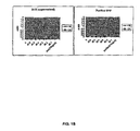
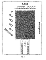
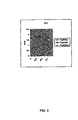


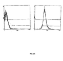


Classifications
-
- C—CHEMISTRY; METALLURGY
- C07—ORGANIC CHEMISTRY
- C07K—PEPTIDES
- C07K16/00—Immunoglobulins [IGs], e.g. monoclonal or polyclonal antibodies
- C07K16/18—Immunoglobulins [IGs], e.g. monoclonal or polyclonal antibodies against material from animals or humans
- C07K16/32—Immunoglobulins [IGs], e.g. monoclonal or polyclonal antibodies against material from animals or humans against translation products of oncogenes
-
- A—HUMAN NECESSITIES
- A61—MEDICAL OR VETERINARY SCIENCE; HYGIENE
- A61P—SPECIFIC THERAPEUTIC ACTIVITY OF CHEMICAL COMPOUNDS OR MEDICINAL PREPARATIONS
- A61P1/00—Drugs for disorders of the alimentary tract or the digestive system
-
- A—HUMAN NECESSITIES
- A61—MEDICAL OR VETERINARY SCIENCE; HYGIENE
- A61P—SPECIFIC THERAPEUTIC ACTIVITY OF CHEMICAL COMPOUNDS OR MEDICINAL PREPARATIONS
- A61P11/00—Drugs for disorders of the respiratory system
- A61P11/06—Antiasthmatics
-
- A—HUMAN NECESSITIES
- A61—MEDICAL OR VETERINARY SCIENCE; HYGIENE
- A61P—SPECIFIC THERAPEUTIC ACTIVITY OF CHEMICAL COMPOUNDS OR MEDICINAL PREPARATIONS
- A61P13/00—Drugs for disorders of the urinary system
- A61P13/12—Drugs for disorders of the urinary system of the kidneys
-
- A—HUMAN NECESSITIES
- A61—MEDICAL OR VETERINARY SCIENCE; HYGIENE
- A61P—SPECIFIC THERAPEUTIC ACTIVITY OF CHEMICAL COMPOUNDS OR MEDICINAL PREPARATIONS
- A61P17/00—Drugs for dermatological disorders
- A61P17/06—Antipsoriatics
-
- A—HUMAN NECESSITIES
- A61—MEDICAL OR VETERINARY SCIENCE; HYGIENE
- A61P—SPECIFIC THERAPEUTIC ACTIVITY OF CHEMICAL COMPOUNDS OR MEDICINAL PREPARATIONS
- A61P19/00—Drugs for skeletal disorders
- A61P19/02—Drugs for skeletal disorders for joint disorders, e.g. arthritis, arthrosis
-
- A—HUMAN NECESSITIES
- A61—MEDICAL OR VETERINARY SCIENCE; HYGIENE
- A61P—SPECIFIC THERAPEUTIC ACTIVITY OF CHEMICAL COMPOUNDS OR MEDICINAL PREPARATIONS
- A61P27/00—Drugs for disorders of the senses
- A61P27/02—Ophthalmic agents
-
- A—HUMAN NECESSITIES
- A61—MEDICAL OR VETERINARY SCIENCE; HYGIENE
- A61P—SPECIFIC THERAPEUTIC ACTIVITY OF CHEMICAL COMPOUNDS OR MEDICINAL PREPARATIONS
- A61P27/00—Drugs for disorders of the senses
- A61P27/16—Otologicals
-
- A—HUMAN NECESSITIES
- A61—MEDICAL OR VETERINARY SCIENCE; HYGIENE
- A61P—SPECIFIC THERAPEUTIC ACTIVITY OF CHEMICAL COMPOUNDS OR MEDICINAL PREPARATIONS
- A61P29/00—Non-central analgesic, antipyretic or antiinflammatory agents, e.g. antirheumatic agents; Non-steroidal antiinflammatory drugs [NSAID]
-
- A—HUMAN NECESSITIES
- A61—MEDICAL OR VETERINARY SCIENCE; HYGIENE
- A61P—SPECIFIC THERAPEUTIC ACTIVITY OF CHEMICAL COMPOUNDS OR MEDICINAL PREPARATIONS
- A61P35/00—Antineoplastic agents
-
- A—HUMAN NECESSITIES
- A61—MEDICAL OR VETERINARY SCIENCE; HYGIENE
- A61P—SPECIFIC THERAPEUTIC ACTIVITY OF CHEMICAL COMPOUNDS OR MEDICINAL PREPARATIONS
- A61P37/00—Drugs for immunological or allergic disorders
- A61P37/02—Immunomodulators
-
- A—HUMAN NECESSITIES
- A61—MEDICAL OR VETERINARY SCIENCE; HYGIENE
- A61P—SPECIFIC THERAPEUTIC ACTIVITY OF CHEMICAL COMPOUNDS OR MEDICINAL PREPARATIONS
- A61P37/00—Drugs for immunological or allergic disorders
- A61P37/08—Antiallergic agents
-
- C—CHEMISTRY; METALLURGY
- C07—ORGANIC CHEMISTRY
- C07K—PEPTIDES
- C07K16/00—Immunoglobulins [IGs], e.g. monoclonal or polyclonal antibodies
- C07K16/18—Immunoglobulins [IGs], e.g. monoclonal or polyclonal antibodies against material from animals or humans
- C07K16/28—Immunoglobulins [IGs], e.g. monoclonal or polyclonal antibodies against material from animals or humans against receptors, cell surface antigens or cell surface determinants
- C07K16/2803—Immunoglobulins [IGs], e.g. monoclonal or polyclonal antibodies against material from animals or humans against receptors, cell surface antigens or cell surface determinants against the immunoglobulin superfamily
- C07K16/283—Immunoglobulins [IGs], e.g. monoclonal or polyclonal antibodies against material from animals or humans against receptors, cell surface antigens or cell surface determinants against the immunoglobulin superfamily against Fc-receptors, e.g. CD16, CD32, CD64
-
- C—CHEMISTRY; METALLURGY
- C07—ORGANIC CHEMISTRY
- C07K—PEPTIDES
- C07K16/00—Immunoglobulins [IGs], e.g. monoclonal or polyclonal antibodies
- C07K16/18—Immunoglobulins [IGs], e.g. monoclonal or polyclonal antibodies against material from animals or humans
- C07K16/28—Immunoglobulins [IGs], e.g. monoclonal or polyclonal antibodies against material from animals or humans against receptors, cell surface antigens or cell surface determinants
- C07K16/2896—Immunoglobulins [IGs], e.g. monoclonal or polyclonal antibodies against material from animals or humans against receptors, cell surface antigens or cell surface determinants against molecules with a "CD"-designation, not provided for elsewhere
-
- A—HUMAN NECESSITIES
- A61—MEDICAL OR VETERINARY SCIENCE; HYGIENE
- A61K—PREPARATIONS FOR MEDICAL, DENTAL OR TOILETRY PURPOSES
- A61K39/00—Medicinal preparations containing antigens or antibodies
- A61K2039/505—Medicinal preparations containing antigens or antibodies comprising antibodies
-
- C—CHEMISTRY; METALLURGY
- C07—ORGANIC CHEMISTRY
- C07K—PEPTIDES
- C07K2317/00—Immunoglobulins specific features
- C07K2317/20—Immunoglobulins specific features characterized by taxonomic origin
- C07K2317/24—Immunoglobulins specific features characterized by taxonomic origin containing regions, domains or residues from different species, e.g. chimeric, humanized or veneered
-
- C—CHEMISTRY; METALLURGY
- C07—ORGANIC CHEMISTRY
- C07K—PEPTIDES
- C07K2317/00—Immunoglobulins specific features
- C07K2317/30—Immunoglobulins specific features characterized by aspects of specificity or valency
- C07K2317/34—Identification of a linear epitope shorter than 20 amino acid residues or of a conformational epitope defined by amino acid residues
-
- C—CHEMISTRY; METALLURGY
- C07—ORGANIC CHEMISTRY
- C07K—PEPTIDES
- C07K2317/00—Immunoglobulins specific features
- C07K2317/70—Immunoglobulins specific features characterized by effect upon binding to a cell or to an antigen
- C07K2317/73—Inducing cell death, e.g. apoptosis, necrosis or inhibition of cell proliferation
- C07K2317/732—Antibody-dependent cellular cytotoxicity [ADCC]
Definitions
- the present disclosure relates to antibodies or fragments thereof that specifically bind Fc ⁇ RIIB, particularly human Fc ⁇ RIIB, with greater affinity than said antibodies or fragments thereof bind Fc ⁇ RIIA, particularly human Fc ⁇ RIIA.
- the disclosure provides methods of enhancing the therapeutic effect of therapeutic antibodies by administering the antibodies of the invention to enhance the effector function of the therapeutic antibodies.
- the disclosure also provides methods of enhancing efficacy of a vaccine composition by administering the antibodies of the invention.
- the interaction of antibody-antigen complexes with cells of the immune system results in a wide array of responses, ranging from effector functions such as antibody-dependent cytotoxicity, mast cell degranulation, and phagocytosis to immunomodulatory signals such as regulating lymphocyte proliferation and antibody secretion. All these interactions are initiated through the binding of the Fc domain of antibodies or immune complexes to specialized cell surface receptors on hematopoietic cells.
- effector functions such as antibody-dependent cytotoxicity, mast cell degranulation, and phagocytosis
- immunomodulatory signals such as regulating lymphocyte proliferation and antibody secretion. All these interactions are initiated through the binding of the Fc domain of antibodies or immune complexes to specialized cell surface receptors on hematopoietic cells.
- the diversity of cellular responses triggered by antibodies and immune complexes results from the structural heterogeneity of Fc receptors.
- Fc receptors share structurally related ligand binding domains which presumably mediate intracellular signaling.
- the Fc receptors members of the immunoglobulin gene superfamily of proteins, are surface glycoproteins that can bind the Fc portion of immunoglobulin molecules. Each member of the family recognizes immunoglobulins of one or more isotypes through a recognition domain on the a chain of the Fc receptor. Fc receptors are defined by their specificity for immunoglobulin subtypes. Fc receptors for IgG are referred to as Fc ⁇ R, for IgE as Fc ⁇ R, and for IgA as Fc ⁇ R.
- Different accessory cells bear Fc receptors for antibodies of different isotype, and the isotype of the antibody determines which accessory cells will be engaged in a given response (reviewed by Ravetch J.V. et al. 1991, Annu. Rev.
- Each member of this family is an integral membrane glycoprotein, possessing extracellular domains related to a C2-set of immunoglobulin-related domains, a single membrane spanning domain and an intracytoplasmic domain of variable length.
- Fc ⁇ Rs There are three known Fc ⁇ Rs, designated Fc ⁇ RI(CD64), Fc ⁇ RII(CD32), and Fc ⁇ RIII(CD16).
- the three receptors are encoded by distinct genes; however, the extensive homology between the three family members suggest they arose from a common progenitor perhaps by gene duplication. This invention specifically focuses on Fc ⁇ RII(CD32).
- Fc ⁇ RII proteins are 40KDa integral membrane glycoproteins which bind only the complexed IgG due to a low affinity for monomeric Ig (10 6 M -1 ). This receptor is the most widely expressed Fc ⁇ R, present on all hematopoietic cells, including monocytes, macrophages, B cells, NK cells, neutrophils, mast cells, and platelets. Fc ⁇ RII has only two immunoglobulin-like regions in its immunoglobulin binding chain and hence a much lower affinity for IgG than Fc ⁇ RI. There are three human Fc ⁇ RII genes (Fc ⁇ RII-A, Fc ⁇ RII-B, Fc ⁇ RII-C), all of which bind IgG in aggregates or immune complexes.
- the fundamental difference is that the A isoform initiates intracellular signaling leading to cell activation such as phagocytosis and respiratory burst, whereas the B isoform initiates inhibitory signals, e.g. , inhibiting B-cell activation.
- Both activating and inhibitory signals are transduced through the Fc ⁇ Rs following ligation. These diametrically opposing functions result from structural differences among the different receptor isoforms.
- Two distinct domains within the cytoplasmic signaling domains of the receptor called i mmunoreceptor t yrosine based a ctivation m otifs (ITAMs) or i mmunoreceptor t yrosine based i nhibitory m otifs (ITIMS) account for the different responses.
- ITAM-containing Fc ⁇ R complexes include Fc ⁇ RI, Fc ⁇ RIIA, Fc ⁇ RIIIA, whereas ITIM-containing complexes only include Fc ⁇ RIIB.
- Fc ⁇ RIIA gene Human neutrophils express the Fc ⁇ RIIA gene.
- Fc ⁇ RIIA clustering via immune complexes or specific antibody cross-linking serves to aggregate ITAMs along with receptor-associated kinases which facilitate ITAM phosphorylation.
- ITAM phosphorylation serves as a docking site for Syk kinase, activation of which results in activation of downstream substrates ( e.g. , PI 3 K). Cellular activation leads to release of proinflammatory mediators.
- the Fc ⁇ RIIB gene is expressed on B lymphocytes; its extracellular domain is 96% identical to Fc ⁇ RIIA and binds IgG complexes in an indistinguishable manner.
- the presence of an ITIM in the cytoplasmic domain of Fc ⁇ RIIB defines this inhibitory subclass of Fc ⁇ R. Recently the molecular basis of this inhibition was established.
- the ITIM in Fc ⁇ RIIB When colligated along with an activating Fc ⁇ R, the ITIM in Fc ⁇ RIIB becomes phosphorylated and attracts the SH2 domain of the inosital polyphosphate 5'-phosphatase (SHIP), which hydrolyzes phosphoinositol messengers released as a consequence of ITAM-containing Fc ⁇ R- mediated tyrosine kinase activation, consequently preventing the influx of intracellular Ca ++ .
- SHIP inosital polyphosphate 5'-phosphatase
- crosslinking of Fc ⁇ RIIB dampens the activating response to Fc ⁇ R ligation and inhibits cellular responsiveness. B cell activation, B cell proliferation and antibody secretion is thus aborted.
- a neoplasm, or tumor is a neoplastic mass resulting from abnormal uncontrolled cell growth which can be benign or malignant. Benign tumors generally remain localized. Malignant tumors are collectively termed cancers.
- malignant generally means that the tumor can invade and destroy neighboring body structures and spread to distant sites to cause death (for review, see Robbins and Angell, 1976, Basic Pathology, 2d Ed., W.B. Saunders Co., Philadelphia, pp. 68-122 ). Cancer can arise in many sites of the body and behave differently depending upon its origin. Cancerous cells destroy the part of the body in which they originate and then spread to other part(s) of the body where they start new growth and cause more destruction.
- Lung and prostate cancer are the top cancer killers for men in the United States.
- Lung and breast cancer are the top cancer killers for women in the United States.
- One in two men in the United States will be diagnosed with cancer at some time during his lifetime.
- One in three women in the United States will be diagnosed with cancer at some time during her lifetime.
- cancer therapy may involve surgery, chemotherapy, hormonal therapy and/or radiation treatment to eradicate neoplastic cells in a patient (See, for example, Stockdale, 1998, "Principles of Cancer Patient Management", in Scientific American: Medicine, vol. 3, Rubenstein and Federman, eds., Chapter 12, Section IV ).
- cancer therapy could also involve biological therapy or immunotherapy. All of these approaches pose significant drawbacks for the patient.
- Surgery for example, may be contraindicated due to the health of the patient or may be unacceptable to the patient. Additionally, surgery may not completely remove the neoplastic tissue. Radiation therapy is only effective when the neoplastic tissue exhibits a higher sensitivity to radiation than normal tissue, and radiation therapy can also often elicit serious side effects.
- Hormonal therapy is rarely given as a single agent and although can be effective, is often used to prevent or delay recurrence of cancer after other treatments have removed the majority of the cancer cells.
- Biological therapies/immunotherapies are limited in number and may produce side effects such as rashes or swellings, flu-like symptoms, including fever, chills and fatigue, digestive tract problems or allergic reactions.
- chemotherapeutic agents available for treatment of cancer.
- a significant majority of cancer chemotherapeutics act by inhibiting DNA synthesis, either directly, or indirectly by inhibiting the biosynthesis of the deoxyribonucleotide triphosphate precursors, to prevent DNA replication and concomitant cell division (See, for example, Gilman et al., Goodman and Gilman's: The Pharmacological Basis of Therapeutics, Eighth Ed. (Pergamom Press, New York, 1990 )).
- agents which include alkylating agents, such as nitrosourea, anti-metabolites, such as methotrexate and hydroxyurea, and other agents, such as etoposides, campathecins, bleomycin, doxorubicin, daunorubicin, etc ., although not necessarily cell cycle specific, kill cells during S phase because of their effect on DNA replication.
- agents specifically colchicine and the vinca alkaloids, such as vinblastine and vincristine, interfere with microtubule assembly resulting in mitotic arrest.
- Chemotherapy protocols generally involve administration of a combination of chemotherapeutic agents to increase the efficacy of treatment.
- chemotherapeutic agents Despite the availability of a variety of chemotherapeutic agents, chemotherapy has many drawbacks ( See, for example, Stockdale, 1998, "Principles Of Cancer Patient Management” in Scientific American Medicine, vol. 3, Rubenstein and Federman, eds., ch. 12, sect. 10 ). Almost all chemotherapeutic agents are toxic, and chemotherapy causes significant, and often dangerous, side effects, including severe nausea, bone marrow depression, immunosuppression, etc . Additionally, even with administration of combinations of chemotherapeutic agents, many tumor cells are resistant or develop resistance to the chemotherapeutic agents.
- those cells resistant to the particular chemotherapeutic agents used in the treatment protocol often prove to be resistant to other drugs, even those agents that act by mechanisms different from the mechanisms of action of the drugs used in the specific treatment; this phenomenon is termed pleiotropic drug or multidrug resistance.
- drug resistance many cancers prove refractory to standard chemotherapeutic treatment protocols.
- Inflammation is a process by which the body's white blood cells and chemicals protect our bodies from infection by foreign substances, such as bacteria and viruses. It is usually characterized by pain, swelling, warmth and redness of the affected area. Chemicals known as cytokines and prostaglandins control this process, and are released in an ordered and self-limiting cascade into the blood or affected tissues. This release of chemicals increases the blood flow to the area of injury or infection, and may result in the redness and warmth. Some of the chemicals cause a leak of fluid into the tissues, resulting in swelling. This protective process may stimulate nerves and cause pain. These changes, when occurring for a limited period in the relevant area, work to the benefit of the body.
- autoimmune and/or inflammatory disorders the immune system triggers an inflammatory response when there are no foreign substances to fight and the body's normally protective immune system causes damage to its own tissues by mistakenly attacking self.
- autoimmune disorders which affect the body in different ways.
- the brain is affected in individuals with multiple sclerosis
- the gut is affected in individuals with Crohn's disease
- the synovium, bone and cartilage of various joints are affected in individuals with rheumatoid arthritis.
- the autoimmune disorder may affect only one organ or tissue type or may affect multiple organs and tissues.
- Organs and tissues commonly affected by autoimmune disorders include red blood cells, blood vessels, connective tissues, endocrine glands (e.g. , the thyroid or pancreas), muscles, joints, and skin.
- autoimmune disorders include, but are not limited to, Hashimoto's thyroiditis, pernicious anemia, Addison's disease, type 1 diabetes, rheumatoid arthritis, systemic lupus erythematosus, dermatomyositis, Sjogren's syndrome, dermatomyositis, lupus erythematosus, multiple sclerosis, autoimmune inner ear disease myasthenia gravis, Reiter's syndrome, Graves disease, autoimmune hepatitis, familial adenomatous polyposis and ulcerative colitis.
- Rheumatoid arthritis and juvenile rheumatoid arthritis are types of inflammatory arthritis.
- Arthritis is a general term that describes inflammation in joints. Some, but not all, types of arthritis are the result of misdirected inflammation.
- other types of arthritis associated with inflammation include the following: psoriatic arthritis, Reiter's syndrome, ankylosing spondylitis arthritis, and gouty arthritis.
- Rheumatoid arthritis is a type of chronic arthritis that occurs in joints on both sides of the body (such as both hands, wrists or knees). This symmetry helps distinguish rheumatoid arthritis from other types of arthritis.
- rheumatoid arthritis may occasionally affect the skin, eyes, lungs, heart, blood or nerves.
- Rheumatoid arthritis affects about 1% of the world's population and is potentially disabling. There are approximately 2.9 million incidences of rheumatoid arthritis in the United States. Two to three times more women are affected than men. The typical age that rheumatoid arthritis occurs is between 25 and 50. Juvenile rheumatoid arthritis affects 71,000 young Americans (aged eighteen and under), affecting six times as many girls as boys.
- Rheumatoid arthritis is an autoimmune disorder where the body's immune system improperly identifies the synovial membranes that secrete the lubricating fluid in the joints as foreign. Inflammation results, and the cartilage and tissues in and around the joints are damaged or destroyed. In severe cases, this inflammation extends to other joint tissues and surrounding cartilage, where it may erode or destroy bone and cartilage and lead to joint deformities. The body replaces damaged tissue with scar tissue, causing the normal spaces within the joints to become narrow and the bones to fuse together. Rheumatoid arthritis creates stiffness, swelling, fatigue, anemia, weight loss, fever, and often, crippling pain.
- Some common symptoms of rheumatoid arthritis include joint stiffness upon awakening that lasts an hour or longer; swelling in a specific finger or wrist joints; swelling in the soft tissue around the joints; and swelling on both sides of the joint. Swelling can occur with or without pain, and can worsen progressively or remain the same for years before progressing.
- the diagnosis of rheumatoid arthritis is based on a combination of factors, including: the specific location and symmetry of painful joints, the presence of joint stiffness in the morning, the presence of bumps and nodules under the skin (rheumatoid nodules), results of X-ray tests that suggest rheumatoid arthritis, and/or positive results of a blood test called the rheumatoid factor.
- rheumatoid factor Many, but not all, people with rheumatoid arthritis have the rheumatoid-factor antibody in their blood.
- the rheumatoid factor may be present in people who do not have rheumatoid arthritis.
- the typical course of the disease is one of persistent but fluctuating joint symptoms, and after about 10 years, 90% of sufferers will show structural damage to bone and cartilage. A small percentage will have a short illness that clears up completely, and another small percentage will have very severe disease with many joint deformities, and occasionally other manifestations of the disease.
- the inflammatory process causes erosion or destruction of bone and cartilage in the joints.
- rheumatoid arthritis there is an autoimmune cycle of persistent antigen presentation, T-cell stimulation, cytokine secretion, synovial cell activation, and joint destruction.
- the disease has a major impact on both the individual and society, causing significant pain, impaired function and disability, as well as costing millions of dollars in healthcare expenses and lost wages. (See, for example, the NIH website and the NIAID website).
- the first line of treatment of any arthritis is usually anti-inflammatories, such as aspirin, ibuprofen and Cox-2 inhibitors such as celecoxib and rofecoxib.
- anti-inflammatories such as aspirin, ibuprofen and Cox-2 inhibitors such as celecoxib and rofecoxib.
- Stecond line drugs include gold, methotrexate and steroids. Although these are well-established treatments for arthritis, very few patients remit on these lines of treatment alone. Recent advances in the understanding of the pathogenesis of rheumatoid arthritis have led to the use of methotrexate in combination with antibodies to cytokines or recombinant soluble receptors.
- recombinant soluble receptors for tumor necrosis factor (TNF)- ⁇ have been used in combination with methotrexate in the treatment of arthritis.
- TNF tumor necrosis factor
- only about 50% of the patients treated with a combination of methotrexate and anti-TNF- ⁇ agents such as recombinant soluble receptors for TNF- ⁇ show clinically significant improvement.
- Many patients remain refractory despite treatment.
- Difficult treatment issues still remain for patients with rheumatoid arthritis.
- Many current treatments have a high incidence of side effects or cannot completely prevent disease progression. So far, no treatment is ideal, and there is no cure. Novel therapeutics are needed that more effectively treat rheumatoid arthritis and other autoimmune disorders.
- Immune-mediated allergic (hypersensitivity) reactions are classified into four types (I-IV) according to the underlying mechanisms leading to the expression of the allergic symptoms.
- Type I allergic reactions are characterized by IgE-mediated release of vasoactive substances such as histamine from mast cells and basophils. The release of these substances and the subsequent manifestation of allergic symptoms are initiated by the cross-linking of allergen-bound IgE to its receptor on the surface of mast cells and basophils.
- IgE-mediated release of vasoactive substances such as histamine from mast cells and basophils.
- the release of these substances and the subsequent manifestation of allergic symptoms are initiated by the cross-linking of allergen-bound IgE to its receptor on the surface of mast cells and basophils.
- IgE antibodies specific for the allergen in individuals suffering from type I allergic reactions, exposure to an allergen for a second time leads to the production of high levels of IgE antibodies specific for the allergen as a result of the involvement of memory B and T cells in the 3-cell interaction required for IgE
- IgE antibodies produced cause an increase in the cross-linking of IgE receptors on mast cells and basophils by allergen-bound IgE, which in turn leads to the activation of these cells and the release of the pharmacological mediators that are responsible for the clinical manifestations of type I allergic diseases.
- the high affinity receptor (Fc ⁇ RI) is expressed on the surface of mast cells and basophils.
- the low affinity receptor (Fc ⁇ RII/CD23) is expressed on many cell types including B cells, T cells, macrophages, eosinophils and Langerhan cells.
- the high affinity IgE receptor consists of three subunits (alpha, beta and gamma chains).
- IgE structures required for IgE to bind to the Fc ⁇ RI on mast cells and basophils is of utmost importance in devising strategies for treatment or prevention of IgE-mediated allergies.
- the elucidation of the IgE receptor-binding site could lead to the identification of peptides or small molecules that block the binding of IgE to receptor-bearing cells in vivo.
- IgE-mediated allergic reactions are treated with drugs such as antihistamines and corticosteroids which attempt to alleviate the symptoms associated with allergic reactions by counteracting the effects of the vasoactive substances released from mast cells and basophils.
- drugs such as antihistamines and corticosteroids which attempt to alleviate the symptoms associated with allergic reactions by counteracting the effects of the vasoactive substances released from mast cells and basophils.
- High doses of antihistamines and corticosteroids have deleterious side effects (e.g. , central nervous system disturbance, constipation, etc ).
- other methods for treating type I allergic reactions are needed.
- Fc ⁇ RIIA and Fc ⁇ RIIB The extracellular domains of Fc ⁇ RIIA and Fc ⁇ RIIB are 95% identical and thus they share numerous epitopes. However, Fc ⁇ RIIA and Fc ⁇ RIIB exhibit very different activities. The fundamental difference is that the Fc ⁇ RIIA initiates intracellular signaling leading to cell activation such as phagocytosis and respiratory burst, whereas the Fc ⁇ RIIB initiates inhibitory signaling.
- antibodies that distinguish between native human Fc ⁇ RIIA and native human Fc ⁇ RIIB expressed on human cells have not been identified , (see, Weinrich et al., Hybridoma 15(1996) 109-116 ; Micklem et al., J. Immunol.
- the invention concerns the embodiments characterized in the claims.
- it relates to an isolated IgG antibody or fragment thereof that specifically binds the extracellular domain of human Fc ⁇ RIIB endogenously expressed on a human cell with at least 10 times greater affinity than said variable domain binds Fc ⁇ RIIA endogenously expressed on a human cell, wherein said specific binding is via the variable domain.
- the antibodies of the invention compete for binding to Fc ⁇ RIIB with the antibody produced by hybridoma cell line 2B6, having ATCC accession number PTA-4591, or with the antibody produced by hybridoma cell line 3H7, having ATCC accession number PTA-4592.
- the invention also relates to an isolated IgG antibody or fragment thereof that specifically binds the extracellular domain of human Fc ⁇ RIIB endogenously expressed on a human cell with at least 10 times greater affinity than said variable domain binds Fc ⁇ RIIA endogenously expressed on a human cell, wherein said specific binding is via the variable domain, which variable domain blocks binding of an Ig-Fc to Fc ⁇ RIIB and enhances activation of B cells, for use in the treatment of cancer.
- the invention also relates to an isolated IgG antibody or fragment thereof that specifically binds the extracellular domain of human Fc ⁇ RIIB endogenously expressed on a human cell with at least 10 times greater affinity than said variable domain binds Fc ⁇ RIIA endogenously expressed on a human cell, wherein said specific binding is via the variable domain for use in a vaccine composition, said antibody or fragment thereof being in an amount effective to enhance the immune response of a subject to said vaccine composition.
- the invention further relates to pharmaceutical compositions comprising a therapeutically relevant amount of an antibody or fragment of the invention and a pharmaceutically acceptable carrier.
- Disclosed herein is an isolated antibody or a fragment thereof that specifically binds Fc ⁇ RIIB, particularly human Fc ⁇ RIIB, more particularly native human Fc ⁇ RIIB with a greater affinity than said antibody or a fragment thereof binds Fc ⁇ RIIA, particularly human Fc ⁇ RIIA, more particularly native human Fc ⁇ RIIA.
- “native” means Fc ⁇ RIIB or Fc ⁇ RIIA which is endogenously expressed on the cell or recombinantly expressed in a mammalian cell but not expressed in a bacterial cell or isolated and denatured.
- the antibody or a fragment thereof binds Fc ⁇ RIIB with at least 2 times greater affinity than said antibody or a fragment thereof binds Fc ⁇ RIIsA. In other embodiments, the antibody or a fragment thereof binds Fc ⁇ RIIB with at least 4 times, at least 6 times, at least 8 times, at least 10 times, at least 100 times, at least 1000 times, at least 10 4 , at least 10 5 , at least 10 6 , at least 10 7 , or at least 10 8 times greater affinity than said antibody or a fragment thereof binds Fc ⁇ RIIA In a preferred embodiment, said antibody or a fragment thereof binds Fc ⁇ RIIB with 100 times, 1000 times, 10 4 times, 10 5 times, 10 6 times, 10 7 times, or 10 8 times greater affinity than said antibody or a fragment thereof binds Fc ⁇ RIIA.
- these binding affinities are determined with the monomeric IgG, and not the aggregated IgG, and binding is via the variable domain (e.g. , Fab fragments have similar binding characteristic).
- the antibody disclosed herein is not the monoclonal antibody designated KB61, as disclosed in Pulford et al, 1986 Immunology 57: 71-76 or the monoclonal antibody II8D2 disclosed in Weinrich et al, 1996, Hybridoma 15: 109-116 .
- the antibody disclosed herein does not bind to the same epitope as and/or compete for binding with KD61 or II8D2.
- the antibody disclosed herein does not bind the amino sequence SDPNFSI corresponding to positions 135 to 141 of the Fc ⁇ RIIB2 isoform.
- an isolated antibody or a fragment thereof that specifically binds Fc ⁇ RIIB with a greater affinity than said antibody or a fragment thereof binds Fc ⁇ RIIA, as determined by any standard method known in the art for assessing specificities.
- the disclosure relates to an isolated antibody or a fragment thereof that specifically binds Fc ⁇ RIIB with a greater affinity than said antibody or a fragment thereof binds Fc ⁇ RIIA, as determined, for example, by western blot or radioimmunoassay.
- the disclosure relates to an isolated antibody or a fragment thereof that specifically binds Fc ⁇ RIIB with a greater affinity than said antibody or a fragment thereof binds Fc ⁇ RIIA, as determined in an ELISA assay, in the linear range for Fc ⁇ RIIB binding.
- the invention relates to an isolated antibody or a fragment thereof that specifically binds Fc ⁇ RIIB, produced in either a bacterial or mammalian system, with a greater affinity than said antibody or a fragment thereof binds Fc ⁇ RIIA, as determined in an ELISA assay.
- the disclosure relates to an isolated antibody or a fragment thereof that specifically binds Fc ⁇ RIIB with a greater affinity than said antibody or a fragment thereof binds Fc ⁇ RIIA, and the constant domain of said antibody further has an enhanced affinity for at least one or more Fc activation receptors.
- said Fc activation receptor is Fc ⁇ RIII.
- said antibody or a fragment thereof blocks the IgG binding site of Fc ⁇ RIIB and blocks the binding of aggregated labeled IgGs to Fc ⁇ RIIB in, for example, a blocking ELISA assay.
- said antibody or a fragment thereof blocks the binding of aggregated labeled IgGs in an ELISA blocking assay by at least 50%, 60%, 70%, 80%, 90%, 95%, 99%, or 99.9%.
- the antibody or a fragment thereof completely blocks the binding of said aggregated labeled IgG in said ELISA assay.
- said antibody or a fragment thereof blocks the IgG binding site of Fc ⁇ RIIB and blocks the binding of aggregated labeled IgG to Fc ⁇ RIIB, as determined by a double-staining FACS assay.
- the disclosure encompasses the use of antibodies that modulate (i.e. , agonize or antagonize) the activity of Fc ⁇ RIIB.
- the antibodies of the invention agonize at least one activity of Fc ⁇ RIIB, i.e. , elicit signaling.
- agonistic antibodies of the invention may mimic clustering of Fc ⁇ RIIB leading to dampening of the activating response to Fc ⁇ R ligation and inhibition of cellular responsiveness.
- the antibodies of the disclosure antagonize at least one activity of Fc ⁇ RIIB, i.e. , block signaling.
- the antibodies block the binding of aggregated IgGs to Fc ⁇ RIIB.
- the disclosure provides antibodies that inhibit Fc ⁇ RI-induced mast cell activation.
- the disclosure further provides anti-Fc ⁇ RIIB antibodies that inhibit Fc ⁇ RIIA-mediated macrophage activation in monocytic cells.
- the disclosure also provides anti-Fc ⁇ RIIB antibodies that inhibit B-cell receptor mediated signaling.
- the anti-Fc ⁇ RIIB antibodies block the ligand binding site of Fc ⁇ RIIB.
- the blocking activity can block the negative regulation of immune-complex-triggered activation and consequently enhance the immune response.
- the enhanced immune response is an increase in antibody-dependent cellular response.
- the anti-Fc ⁇ RIIB antibodies block crosslinking of Fc ⁇ RIIB receptors to B cell and/or Fc receptors, leading to B cell, mast cell, dendritic cell, or macrophage activation.
- the disclosure encompasses the production of novel monoclonal antibodies with specificities for Fc ⁇ RIIB relative to Fc ⁇ RIIA.
- the disclosure provides a method for producing Fc ⁇ RIIB monoclonal antibodies that specifically bind Fc ⁇ RIIB, particularly human Fc ⁇ RIIB, with a greater affinity than said monoclonal antibodies bind Fc ⁇ RIIA, particularly human Fc ⁇ RIIA, said method comprising: (a) immunizing one or more Fc ⁇ RIIA transgenic mice with purified Fc ⁇ RIIB or an immunogenic fragment thereof; (b) producing hybridoma cells lines from spleen cells of said one or more mice; (c) screening said hybridoma cell lines for one or more hybridoma cell lines that produce antibodies that specifically bind Fc ⁇ RIIB with a greater affinity than the antibodies bind Fc ⁇ RIIA.
- the disclosure encompasses any antibody produced by said method.
- the disclosure provides a method for producing Fc ⁇ RIIB monoclonal antibodies that specifically bind Fc ⁇ RIIB, particularly human Fc ⁇ RIIB, with a greater affinity than said monoclonal antibodies bind Fc ⁇ RIIA, particularly human Fc ⁇ RIIA, said method comprising: (a) immunizing one or more Fc ⁇ RIIA transgenic mice with purified Fc ⁇ RIIB or an immunogenic fragment thereof; (b) booster immunizing said mice for a time sufficient to elicit an immune response; (c) producing hybridoma cells lines from spleen cells of said one or more mice; (d) screening said hybridoma cell lines for one or more hybridoma cell lines that produce antibodies that specifically bind Fc ⁇ RIIB with a greater affinity than the antibodies bind Fc ⁇ RIIA.
- said mice are booster immunized at least four times over a period of four months.
- said mice are immunized with purified Fc ⁇ RIIB, which has been mixed with adjuvants known in the art to enhance immune response in said mice.
- said immunogenic fragment is the soluble extracellular domain of Fc ⁇ RIIB.
- the hybridoma cell lines can be screened using standard techniques known in the art (e.g. , ELISA).
- the invention provides a monoclonal antibody produced by clone 2B6 or 3H7, having ATCC accession numbers PTA-4591 and PTA-4592, respectively.
- the disclosure provides an isolated antibody or a fragment thereof that competes for binding with the monoclonal antibody produced by clone 2B6 or 3H7 and binds Fc ⁇ RIIB with a greater affinity than said antibody or a fragment thereof binds Fc ⁇ RIIA, and/or binds to the same epitope of Fc ⁇ RIIB as the monoclonal antibody produced from clone 2B6 or 3H7 and binds Fc ⁇ RIIB with a greater affinity than said antibody or a fragment thereof binds Fc ⁇ RIIA.
- the disclosure provides hybridoma cell line 2B6 or 3H7, having ATCC accession numbers PTA-4591 and PTA-4592, respectively.
- the methods disclosed herein also encompass polynucleotides that encode the antibodies of the disclosure.
- the disclosure provides an isolated nucleic acid sequence encoding a heavy chain or a light chain of an antibody or a fragment thereof that specifically binds Fc ⁇ RIIB with greater affinity than said antibody or a fragment thereof binds Fc ⁇ RIIA.
- the disclosure also relates to a vector comprising said nucleic acid.
- the disclosure further provides a vector comprising a first nucleic acid molecule encoding a heavy chain and a second nucleic acid molecule encoding a light chain, said heavy chain and light chain being of an antibody or a fragment thereof that specifically binds Fc ⁇ RIIB with greater affinity than said antibody or a fragment thereof binds Fc ⁇ RIIA.
- said vector is an expression vector.
- the disclosure further provides host cells containing the vectors of or polynucleotides encoding the antibodies of the invention.
- the disclosure encompasses polynucleotides encoding heavy and light chains of the antibodies produced by the deposited hybridoma clones, having ATCC accession numbers PTA-4591 and PTA-4592, respectively, or portions thereof, e.g. , CDRs, variable domains, etc .
- the disclosure further provides methods for the production of antibodies of the invention or fragments thereof.
- the antibodies disclosed herein or fragments thereof can be produced by any method known in the art for the production of antibodies, in particular, by secretion from cultured hybridoma cells, chemical synthesis or by recombinant expression techniques known in the art.
- the disclosure relates to a method for recombinantly producing a Fc ⁇ RIIB-specific antibody, said method comprising:
- the antibodies disclosed herein are monoclonal antibodies, and more preferably, humanized or human antibodies.
- the antibodies bind to the extracellular domain of human Fc ⁇ RIIB.
- the antibodies specifically or selectively recognize one or more epitopes of Fc ⁇ RIIB.
- Another embodiment of the disclosure encompasses the use of phage display technology to increase the affinity of the antibodies disclosed herein for Fc ⁇ RIIB. Any screening method known in the art can be used to identify mutant antibodies with increased avidity for Fc ⁇ RIIB ( e.g. , ELISA).
- antibodies are screened using antibody screening assays well known in the art (e.g. , BIACORE assays) to identify antibodies with K off rate less than 3x10 -3 s -1 .
- the disclosure encompasses the use of the antibodies disclosed herein to detect the presence of Fc ⁇ RIIB specifically (i.e. , Fc ⁇ RIIB and not Fc ⁇ RIIA) in a biological sample.
- Activation and inhibitory Fc receptors are critical for the balanced function of these receptors and proper cellular immune responses.
- the disclosure encompasses the use of the antibodies disclosed herein for the treatment of any disease related to loss of such balance and regulated control in the Fc receptor signaling pathway.
- the Fc ⁇ RIIB antibodies of the disclosure have uses in regulating the immune response, e.g. , in inhibiting immune response in connection with autoimmune or inflammatory disease, or allergic response.
- the Fc ⁇ RIIB antibodies of the disclosure can also be used to alter certain effector functions to enhance, for example, therapeutic antibody-mediated cytotoxicity.
- the antibodies disclosed herein are useful for prevention or treatment of cancer, for example, in one embodiment, as a single agent therapy.
- the antibodies are useful for prevention or treatment of B-cell malignancies, particularly non-Hodgkin's lymphoma or chronic lymphocytic leukemia.
- the antibodies are useful for prevention or treatment of cancer, particularly in potentiating the cytotoxic activity of cancer antigen-specific therapeutic antibodies with cytotoxic activity to enhance tumor cell killing and/or enhancing antibody dependent cytotoxic cellular ("ADCC") activity, complement dependent cytotoxic (“CDC”) activity, or phagocytosis of the therapeutic antibodies.
- ADCC antibody dependent cytotoxic cellular
- CDC complement dependent cytotoxic
- the disclosure provides a method of treating cancer in a patient having a cancer characterized by a cancer antigen, said method comprising administering to said patient a therapeutically effective amount of a first antibody or a fragment thereof that specifically binds Fc ⁇ RIIB with greater affinity than said antibody or a fragment thereof binds Fc ⁇ RIIA, and a second antibody that specifically binds said cancer antigen and is cytotoxic.
- the disclosure also provides a method of treating cancer in a patient having a cancer characterized by a cancer antigen, said method comprising administering to said patient a therapeutically effective amount of an antibody or a fragment thereof that specifically binds Fc ⁇ RIIB with greater affinity than said antibody or a fragment thereof binds Fc ⁇ RIIA, and the constant domain of which further has an increased affinity for one or more Fc activation receptors, when the antibody is monomeric, such as Fc ⁇ RIIIA, and an antibody that specifically binds said cancer antigen and is cytotoxic.
- said Fc activation receptor is Fc ⁇ RIIIA.
- the disclosure provides a method of enhancing an antibody mediated cytotoxic effect in a subject being treated with a cytotoxic antibody, said method comprising administering to said patient an antibody disclosed herein or a fragment thereof, in an amount sufficient to enhance the cytotoxic effect of said cytotoxic antibody.
- the disclosure provides a method of enhancing an antibody-mediated cytotoxic effect in a subject being treated with a cytotoxic antibody, said method comprising administering to said patient an antibody disclosed herein or a fragment thereof, further having an enhanced affinity for an Fc activation receptor, when monomeric, in an amount sufficient to enhance the cytotoxic effect of said cytotoxic antibody.
- the disclosure provides a method further comprising the administration of one or more additional cancer therapies.
- the disclosure further provides a pharmaceutical composition comprising (i) a therapeutically effective amount of the antibody or a fragment thereof that specifically binds Fc ⁇ RIIB with greater affinity than said antibody or a fragment thereof binds Fc ⁇ RIIA; and (ii) a pharmaceutically acceptable carrier.
- the disclosure additionally provides a pharmaceutical composition comprising (i) a therapeutically effective amount of the antibody or a fragment thereof that specifically binds Fc ⁇ RIIB with greater affinity than said antibody or a fragment thereof binds Fc ⁇ RIIA; (ii) a cytotoxic antibody that specifically binds a cancer antigen; and (iii) a pharmaceutically acceptable carrier.
- the disclosure encompasses the use of the antibodies disclosed herein in combination with any therapeutic antibody that mediates its therapeutic effect through cell killing to potentiate the antibody's therapeutic activity.
- the antibodies potentiate the antibody's therapeutic activity by enhancing antibody-mediated effector function.
- the antibodies potentiate the cytotoxic antibody's therapeutic activity by enhancing phagocytosis and opsonization of the targeted tumor cells.
- the antibodies potentiate the antibody's therapeutic activity by enhancing antibody-dependent cell-mediated cytotoxicity ("ADCC”) in destruction of the targeted tumor cells.
- ADCC antibody-dependent cell-mediated cytotoxicity
- the disclosure encompasses use of the antibodies disclosed herein in combination with a therapeutic antibody that does not mediate its therapeutic effect through cell killing to potentiate the antibody's therapeutic activity.
- the disclosure encompasses use of the antibodies in combination with a therapeutic apoptosis inducing antibody with agonisitc activity, e.g. , anti-Fas antibody.
- Therapeutic apoptosis inducing antibodies may be specific for any death receptor known in the art for the modulation of apoptotic pathway, e.g. , TNFR receptor family member.
- the disclosure encompasses using the antibodies disclosed herein to block macrophage mediated tumor cell progression and metastasis.
- the antibodies are particularly useful in the treatment of solid tumors, where macrophage infiltration occurs.
- the antagonistic antibodies are particularly useful for controlling, e.g. , reducing or eliminating, tumor cell metastasis, by reducing or eliminating the population of macrophages that are localized at the tumor site.
- the disclosure further encompasses antibodies that effectively deplete or eliminate immune effector cells other than macrophages that express Fc ⁇ RIIB, e.g. , dendritic cells. Effective depletion or elimination of immune effector cells using the antibodies of the invention may range from a reduction in population of the effector cells by 50%, 60%, 70%, 80%, preferably 90%, and most preferably 99%.
- the agonistic antibodies disclosed herein are particularly useful for the treatment of tumors of non-hematopoietic origin, including tumors of melanoma cells.
- the disclosure encompasses use of the antibodies disclosed herein in combination with therapeutic antibodies that immunospecifically bind to tumor antigens that are not expressed on the tumor cells themselves, but rather on the surrounding reactive and tumor supporting, non-malignant cells comprising the tumor stroma.
- an antibody disclosed herein is used in combination with an antibody that immunospecifically binds a tumor antigen on a fibroblast cell, e.g. , fibroblast activation protein (FAP).
- FAP fibroblast activation protein
- the disclosure provides a method of treating an autoimmune disorder in a patient in need thereof, said method comprising administering to said patient a therapeutically effective amount of one or more antibodies disclosed herein.
- the disclosure also provides a method of treating an autoimmune disorder in a patient in need thereof, said method further comprising administering to said patient a therapeutically effective amount of one or more anti-inflammatory agents, and/or one or more immunomodulatory agents.
- the disclosure also provides a method of treating an inflammatory disorder in a patient in need thereof, said method comprising administering to said patient a therapeutically effective amount of one or more antibodies disclosed herein.
- the disclosure also provides a method of treating an inflammatory disorder in a patient in need thereof, said method further comprising administering to said patient a therapeutically effective amount of one or more anti-inflammatory agents, and/or one or more immunomodulatory agents.
- the disclosure provides a method of enhancing an immune response to a vaccine composition in a subject, said method comprising administering to said subject an antibody or a fragment thereof that specifically binds Fc ⁇ RIIB with greater affinity than said antibody or a fragment thereof binds Fc ⁇ RIIA, and a vaccine composition, such that said antibody or a fragment thereof is administered in an amount effective to enhance the immune response to said vaccine composition in said subject.
- the antibodies disclosed herein may be used to enhance a humoral and/or cell mediated response against the antigen(s) of the vaccine composition.
- the antibodies disclosed herein may be used in combination with any vaccines known in the art.
- the disclosure encompasses the use of the antibodies disclosed herein to either prevent or treat a particular disorder, where an enhanced immune response against a particular antigen or antigens is effective to treat or prevent the disease or disorder.
- the disclosure also provides a method for enhancing immune therapy for an infectious agent wherein the antibodies disclosed herein are administered to a patient that is already infected by a pathogen, such as HIV or HSV, to enhance opsonization and phagocytosis of infected cells.
- a pathogen such as HIV or HSV
- the disclosure provides a method of treating diseases with impaired apoptotic mediated signaling, e.g. , cancer, autoimmune disease
- the invention encompasses a method of treating a disease with deficient Fas-mediated apoptosis, said method comprising administering an antibody disclosed herein in combination with an anti-Fas antibody.
- the disclosure provides a method of diagnosis of an autoimmune disease in a subject comprising: (i) contacting a biological sample from said subject with an effective amount of an antibody of the invention; and (ii) detecting binding of said antibody or a fragment thereof, wherein detection of said detectable marker above a background or standard level indicates that said subject has an autoimmune disease.
- the disclosure further provides a method for treating or preventing an IgE-mediated allergic disorder in a patient in need thereof, comprising administering to said patient a therapeutically effective amount of the agonistic antibodies disclosed herein.
- the disclosure also provides a method for treating or preventing an IgE-mediated allergic disorder in a patient in need thereof, comprising administering to said patient the antibodies disclosed herein in combination with other therapeutic antibodies or vaccine compositions used for the treatment or prevention of IgE-mediated allergic disorders.
- the term “specifically binds to Fc ⁇ RIIB” and analogous terms refer to antibodies or fragments thereof that specifically bind to Fc ⁇ RIIB or a fragment thereof and do not specifically bind to other Fc receptors, in particular to Fc ⁇ RIIA. Further it is understood to one skilled in the art, that an antibody that specifically binds to Fc ⁇ RIIB, may bind through the variable domain or the constant domain of the antibody. If the antibody that specifically binds to Fc ⁇ RIIB binds through its variable domain, it is understood to one skilled in the art that it is not aggregated, i.e., is monomeric.
- An antibody that specifically binds to Fc ⁇ RIIB may bind to other peptides or polypeptides with lower affinity as determined by, e.g. , immunoassays, BIAcore, or other assays known in the art.
- antibodies or fragments that specifically bind to Fc ⁇ RIIB or a fragment thereof do not cross-react with other antigens.
- Antibodies or fragments that specifically bind to Fc ⁇ RIIB can be identified, for example, by immunoassays, BIAcore, or other techniques known to those of skill in the art.
- An antibody or a fragment thereof binds specifically to a Fc ⁇ RIIB when it binds to Fc ⁇ RIIB with higher affinity than to any cross-reactive antigen as determined using experimental techniques, such as western blots, radioimmunoassays (RIA) and enzyme-linked immunosorbent assays (ELISAs). See, e.g., Paul, ed., 1989, Fundamental Immunology Second Edition, Raven Press, New York at pages 332-336 for a discussion regarding antibody specificity.
- antibody and “antibodies” refer to monoclonal antibodies, multispecific antibodies, human antibodies, humanized antibodies, synthetic antibodies, chimeric antibodies, camelized antibodies, single-chain Fvs (scFv), single chain antibodies, Fab fragments, F(ab') fragments, disulfide-linked Fvs (sdFv), intrabodies, and anti-idiotypic (anti-Id) antibodies (including, e.g ., anti-Id and anti-anti-Id antibodies to antibodies of the invention), and epitope-binding fragments of any of the above.
- scFv single-chain Fvs
- Fab fragments F(ab') fragments
- disulfide-linked Fvs sdFv
- intrabodies and anti-idiotypic antibodies (including, e.g ., anti-Id and anti-anti-Id antibodies to antibodies of the invention), and epitope-binding fragments of any of the above.
- antibodies include immunoglobulin molecules and immunologically active fragments of immunoglobulin molecules, i.e., molecules that contain an antigen binding site.
- Immunoglobulin molecules can be of any type (e.g ., IgG, IgE, IgM, IgD, IgA and IgY), class (e.g ., IgG 1 , IgG 2 , IgG 3 , IgG 4 , IgA 1 and IgA 2 ) or subclass.
- derivative in the context of polypeptides or proteins refers to a polypeptide or protein that comprises an amino acid sequence which has been altered by the introduction of amino acid residue substitutions, deletions or additions.
- derivative also refers to a polypeptide or protein which has been modified, i.e, by the covalent attachment of any type of molecule to the polypeptide or protein.
- an antibody may be modified, e.g., by glycosylation, acetylation, pegylation, phosphorylation, amidation, derivatization by known protecting/blocking groups, proteolytic cleavage, linkage to a cellular ligand or other protein, etc.
- a derivative polypeptide or protein may be produced by chemical modifications using techniques known to those of skill in the art, including, but not limited to specific chemical cleavage, acetylation, formylation, metabolic synthesis of tunicamycin, etc. Further, a derivative polypeptide or protein derivative possesses a similar or identical function as the polypeptide or protein from which it was derived.
- derivative in the context of a non-proteinaceous derivative refers to a second organic or inorganic molecule that is formed based upon the structure of a first organic or inorganic molecule.
- a derivative of an organic molecule includes, but is not limited to, a molecule modified, e.g ., by the addition or deletion of a hydroxyl, methyl, ethyl, carboxyl or amine group.
- An organic molecule may also be esterified, alkylated and/or phosphorylated.
- the terms “disorder” and “disease” are used interchangeably to refer to a condition in a subject.
- autoimmune disease is used interchangeably with the term “autoimmune disorder” to refer to a condition in a subject characterized by cellular, tissue and/or organ injury caused by an immunologic reaction of the subject to its own cells, tissues and/or organs.
- inflammatory disease is used interchangeably with the term “inflammatory disorder” to refer to a condition in a subject characterized by inflammation, preferably chronic inflammation.
- Autoimmune disorders may or may not be associated with inflammation.
- inflammation may or may not be caused by an autoimmune disorder.
- certain disorders may be characterized as both autoimmune and inflammatory disorders.
- cancer refers to a neoplasm or tumor resulting from abnormal uncontrolled growth of cells.
- cancer explicitly includes, leukemias and lymphomas.
- cancer refers to a benign tumor, which has remained localized.
- cancer refers to a malignant tumor, which has invaded and destroyed neighboring body structures and spread to distant sites.
- the cancer is associated with a specific cancer antigen.
- immunomodulatory agent refers to an agent that modulates a host's immune system.
- an immunomodulatory agent is an immunosuppressant agent.
- an immunomodulatory agent is an immunostimulatory agent.
- Immunomodatory agents include, but are not limited to, small molecules, peptides, polypeptides, fusion proteins, antibodies, inorganic molecules, mimetic agents, and organic molecules.
- epitope refers to a fragment of a polypeptide or protein having antigenic or immunogenic activity in an animal, preferably in a mammal, and most preferably in a human.
- An epitope having immunogenic activity is a fragment of a polypeptide or protein that elicits an antibody response in an animal.
- An epitope having antigenic activity is a fragment of a polypeptide or protein to which an antibody immunospecifically binds as determined by any method well-known to one of skill in the art, for example by immunoassays. Antigenic epitopes need not necessarily be immunogenic.
- fragment refers to a peptide or polypeptide comprising an amino acid sequence of at least 5 contiguous amino acid residues, at least 10 contiguous amino acid residues, at least 15 contiguous amino acid residues, at least 20 contiguous amino acid residues, at least 25 contiguous amino acid residues, at least 40 contiguous amino acid residues, at least 50 contiguous amino acid residues, at least 60 contiguous amino residues, at least 70 contiguous amino acid residues, at least contiguous 80 amino acid residues, at least contiguous 90 amino acid residues, at least contiguous 100 amino acid residues, at least contiguous 125 amino acid residues, at least 150 contiguous amino acid residues, at least contiguous 175 amino acid residues, at least contiguous 200 amino acid residues, or at least contiguous 250 amino acid residues of the amino acid sequence of another polypeptide.
- a fragment of a polypeptide retains at least one function of the polypeptide
- nucleic acids and "nucleotide sequences” include DNA molecules (e.g ., cDNA or genomic DNA), RNA molecules (e.g ., mRNA), combinations of DNA and RNA molecules or hybrid DNA/RNA molecules, and analogs of DNA or RNA molecules.
- Such analogs can be generated using, for example, nucleotide analogs, which include, but are not limited to, inosine or tritylated bases.
- Such analogs can also comprise DNA or RNA molecules comprising modified backbones that lend beneficial attributes to the molecules such as, for example, nuclease resistance or an increased ability to cross cellular membranes.
- nucleic acids or nucleotide sequences can be single-stranded, double-stranded, may contain both single-stranded and double-stranded portions, and may contain triple-stranded portions, but preferably is double-stranded DNA.
- a "therapeutically effective amount” refers to that amount of the therapeutic agent sufficient to treat or manage a disease or disorder associated with Fc ⁇ RIIB and any disease related to the loss of regulation in the Fc receptor signaling pathway or to enhance the therapeutic efficacy of another therapy, e.g ., therapeutic antibody, vaccine therapy, etc .
- a therapeutically effective amount may refer to the amount of therapeutic agent sufficient to delay or minimize the onset of disease, e.g ., delay or minimize the spread of cancer.
- a therapeutically effective amount may also refer to the amount of the therapeutic agent that provides a therapeutic benefit in the treatment or management of a disease.
- a therapeutically effective amount with respect to a therapeutic agent of the invention means that amount of therapeutic agent alone, or in combination with other therapies, that provides a therapeutic benefit in the treatment or management of a disease, e.g., sufficient to enhance the therapeutic efficacy of a therapeutic antibody sufficient to treat or manage a disease.
- an amount of Fc ⁇ RIIB antibody disclosed herein can encompass an amount that improves overall therapy, reduces or avoids unwanted effects, or enhances the therapeutic efficacy of or synergies with another therapeutic agent.
- prophylactic agent and “prophylactic agents” refer to any agent(s) which can be used in the prevention of a disorder, or prevention of recurrence or spread of a disorder.
- a prophylactically effective amount may refer to the amount of prophylactic agent sufficient to prevent the recurrence or spread of hyperproliferative disease, particularly cancer, or the occurrence of such in a patient, including but not limited to those predisposed to hyperproliferative disease, for example those genetically predisposed to cancer or previously exposed to carcinogens.
- a prophylactically effective amount may also refer to the amount of the prophylactic agent that provides a prophylactic benefit in the prevention of disease.
- a prophylactically effective amount with respect to a prophylactic agent of the invention means that amount of prophylactic agent alone, or in combination with other agents, that provides a prophylactic benefit in the prevention of disease.
- the term can encompass an amount that improves overall prophylaxis or enhances the prophylactic efficacy of or synergies with another prophylactic agent, such as but not limited to a therapeutic antibody.
- the terms “prevent”, “preventing” and “prevention” refer to the prevention of the recurrence or onset of one or more symptoms of a disorder in a subject resulting from the administration of a prophylactic or therapeutic agent.
- the term “in combination” refers to the use of more than one prophylactic and/or therapeutic agents.
- the use of the term “in combination” does not restrict the order in which prophylactic and/or therapeutic agents are administered to a subject with a disorder.
- a first prophylactic or therapeutic agent can be administered prior to (e.g ., 5 minutes, 15 minutes, 30 minutes, 45 minutes, 1 hours, 2 hours, 4 hours, 6 hours, 12 hours, 24 hours, 48 hours, 72 hours, 96 hours, 1 week, 2 weeks, 3 weeks, 4 weeks, 5 weeks, 6 weeks, 8 weeks, or 12 weeks before), concomitantly with, or subsequent to ( e.g ., 5 minutes, 15 minutes, 30 minutes, 45 minutes, 1 hour, 2 hours, 4 hours, 6 hours, 12 hours, 24 hours, 48 hours, 72 hours, 96 hours, 1 week, 2 weeks, 3 weeks, 4 weeks, 5 weeks, 6 weeks, 8 weeks, or 12 weeks after) the administration of a second prophylactic or therapeutic agent to a subject with a disorder.
- the disclosure encompasses antibodies (preferably monoclonal antibodies) or fragments thereof that specifically bind Fc ⁇ IIB, preferably human Fc ⁇ IIB, more preferably human native Fc ⁇ IIB with a greater affinity than said antibodies or fragments thereof bind Fc ⁇ IIa, preferably, human Fc ⁇ IIB, more preferably native human Fc ⁇ IIB.
- the antibodies disclosed herein bind the extracellular domain of native human Fc ⁇ RIIB.
- the antibodies or fragments thereof bind to Fc ⁇ IIB with an affinity greater than two-fold, four fold, 6 fold, 10 fold, 20 fold, 50 fold, 100 fold 1000 fold, 10 4 fold, 10 5 fold, 10 6 fold, 10 7 fold, or 10 8 fold than said antibodies or fragments thereof bind Fc ⁇ RIIA.
- the antibody is a mouse monoclonal antibody produced by clone 2B6 or 3H7, having ATCC accession numbers PTA-4591 and PTA-4592, respectively.
- Hybridomas producing antibodies of the invention have been deposited with the American Type Culture Collection (10801 University Boulevard, Manassas, Virginia 20110) on August 13, 2002 under the provisions of the Budapest Treaty on the International Recognition of the Deposit of Microorganisms for the Purposes of Patent Procedures, and assigned accession numbers PTA-4591 and PTA-4592, respectively.
- the disclosure encompasses an antibody with the heavy chain having the amino acid sequence of SEQ ID No 2 and the light chain having the amino acid sequence of SEQ ID No. 4.
- the antibodies disclosed herein are human or have been humanized, preferably a humanized version of the antibody produced by clone 3H7 or 2B6.
- the antibodies disclosure herein further do not bind Fc activation receptors, e.g. , Fc ⁇ IIIA, Fc ⁇ IIIB, etc.
- the antibodies disclosures herein, or fragments thereof agonize at least one activity Fc ⁇ IIB.
- said activity is inhibition of B cell receptor-mediated signaling.
- the agonistic antibodies disclosed herein inhibit activation of B cells, B cell proliferation, antibody production, intracellular calcium influx ofB cells, cell cycle progression, or activity of one or more downstream signaling molecules in the Fc ⁇ IIB signal transduction pathway.
- the agonistic antibodies enhance phosphorylation of Fc ⁇ IIB or SHIP recruitment.
- the agonistic antibodies inhibit MAP kinase activity or Akt recruitment in the B cell receptor-mediated signaling pathway.
- the agonistic antibodies agonize Fc ⁇ RIIB-mediated inhibition of Fc ⁇ RI signaling.
- said antibodies inhibit Fc ⁇ I-induced mast cell activation, calcium mobilization, degranulation, cytokine production, or serotonin release.
- the agonistic antibodies stimulate phosphorylation of Fc ⁇ IIB, stimulate recruitment of SHIP, stimulate SHIP phosphorylation and its association with Shc, or inhibit activation of MAP kinase family members ( e.g. , Erk1, Erk2,1NK, p38, etc .).
- the agonistic antibodies enhance tyrosine phosphorylation of p62dok and its association with SHIP and rasGAP.
- the agonistic antibodies inhibit Fc ⁇ R-mediatcd phagocytosis in monocytes or macrophages.
- the antibodies disclosed herein or fragments thereof antagonize at least one activity of Fc ⁇ IIB.
- said activity is activation of B cell receptor-mediated signaling.
- the antagonistic antibodies enhance B cell activity, B cell proliferation, antibody production, intracellular calcium influx, or activity of one or more downstream signaling molecules in the Fc ⁇ RIIB signal transduction pathway.
- the antagonistic antibodies decrease phosphorylation of Fc ⁇ RIIB or SHIP recruitment
- the antagonistic antibodies enhance MAP kinase activity or Akt recruitment in the B cell receptor mediated signaling pathway.
- the antagonistic antibodies antagonize Fc ⁇ RIIB-mediated inhibition of Fc ⁇ RI signaling.
- the antagonistic antibodies enhance Fc ⁇ RI-induced mast cell activation, calcium mobilization, degranulation, cytokine production, or serotonin release.
- the antagonistic antibodies inhibit phosphorylation of Fc ⁇ IIB, inhibit recruitment of SHIP, inhibit SHIP phosphorylation and its association with Shc, enhance activation of MAP kinase family members ( e.g ., Erk1, Erk2, JNK, p38, etc .).
- the antagonistic antibodies inhibit tyrosine phosphorylation of p62dok and its association with SHIP and rasGAP.
- the antagonistic antibodies enhance Fc ⁇ R-mediated phagocytosis in monocytes or macrophages.
- the antagonistic antibodies prevent phagocytosis, clearance of opsonized particles by splenic macrophages.
- Antibodies disclosed herein are not limited to, monoclonal antibodies, synthetic antibodies, recombinantly produced antibodies, multispecific antibodies, human antibodies, humanized antibodies, chimeric antibodies, camelized antibodies, single-chain Fvs (scFv), single chain antibodies. Fab fragments, F(ab') fragments, disulfide-linked Fvs (sdFv), intrabodies, and epitope-binding fragments of any of the above.
- antibodies used in the methods disclosed herein include immunoglobulin molecules and immunologically active portions of immunoglobulin molecules, i.e., molecules that contain an antigen binding site that immunospecifically binds to Fc ⁇ RIIB with greater affinity than said immunoglobulin molecule binds Fc ⁇ RIIB.
- the antibodies used in the methods disclosed herein may be from any animal origin including birds and mammals (e.g ., human, non-human primate, murine, donkey, sheep, rabbit, goat, guinea pig, camel, horse, or chicken).
- the antibodies are human or humanized monoclonal antibodies.
- "human” antibodies include antibodies having the amino acid sequence of a human immunoglobulin and include antibodies isolated from human immunoglobulin libraries or libraries of synthetic human immunoglobulin coding sequences or from mice that express antibodies from human genes.
- the antibodies used in the methods disclosed herein may be monospecific, bispecific, trispecific or of greater multispecificity.
- Multispecific antibodies may immunospecifically bind to different epitopes of Fc ⁇ RIIB or immunospecifically bind to both an epitope of Fc ⁇ RIIB as well a heterologous epitope, such as a heterologous polypeptide or solid support material.
- a heterologous epitope such as a heterologous polypeptide or solid support material.
- the antibodies disclosed herein are multi-specific with specifcities for Fc ⁇ RIIB and for a cancer antigen or any other cell surface marker specific for a cell designed to be killed, e.g., in treating or preventing a particular disease or disorder, or for other Fc receptors, e.g ., Fc ⁇ IIA, Fc ⁇ IIB, etc.
- an antibody used in the methods disclosed herein is an antibody or an antigen-binding fragment thereof (e.g ., comprising one or more complementarily determining regions (CDRs), preferably all 6 CDRs) of the antibody produced by clone 2B6 or 3H7 with ATCC accession numbers PTA-4591 and PTA-4592, respectively ( e.g ., the heavy chain CDR3).
- CDRs complementarily determining regions
- an antibody used in the methods disclosed herein binds to the same epitope as the mouse monoclonal antibody produced from clone 2B6 or 3H7 with ATCC accession numbers PTA-4591 and PTA-4592, respectively and/or competes with the mouse monoclonal antibody produced from clone 2B6 or 3H7 with ATCC accession numbers PTA-4591 and PTA-4592, respectively as determined e.g., in an ELISA assay or other appropriate competitive immunoassay, and also binds Fc ⁇ RIIB with a greater affinity than said antibody or a fragment thereof binds Fc ⁇ RIIB.
- the antibodies used in the methods disclosed herein include derivatives that are modified, i.e , by the covalent attachment of any type of molecule to the antibody such that covalent attachment.
- the antibody derivatives include antibodies that have been modified, e.g ., by glycosylation, acetylation, pegylation, phosphorylation, amidation, derivatization by known protecting/blocking groups, proteolytic cleavage, linkage to a cellular ligand or other protein, etc . Any of numerous chemical modifications may be carried out by known techniques, including, but not limited to, specific chemical cleavage, acetylation, formylation, metabolic synthesis of tunicamycin, etc . Additionally, the derivative may contain one or more non-classical amino acids.
- Human antibodies can be made by a variety of methods known in the art including phage display methods described above using antibody libraries derived from human immunoglobulin sequences. See also U.S. Patent Nos. 4,444,887 and 4,716,111 ; and International Publication Nos. WO 98/46645 , WO 98/50433 , WO 98/24893 , WO 98/16654 , WO 96/34096 , WO 96/33735 , and WO 91/10741 .
- Human antibodies can also be produced using transgenic mice which are incapable of expressing functional endogenous immunoglobulins, but which can express human immunoglobulin genes.
- the human heavy and light chain immunoglobulin gene complexes may be introduced randomly or by homologous recombination into mouse embryonic stem cells.
- the human variable region, constant region, and diversity region may be introduced into mouse embryonic stem cells in addition to the human heavy and light chain genes.
- the mouse heavy and light chain immunoglobulin genes may be rendered non-functional separately or simultaneously with the introduction of human immunoglobulin loci by homologous recombination. In particular, homozygous deletion of the J H region prevents endogenous antibody production.
- the modified embryonic stem cells are expanded and microinjected into blastocysts to produce chimeric mice.
- the chimeric mice are then bred to produce homozygous offspring which express human antibodies.
- the transgenic mice are immunized using conventional methodologies with a selected antigen, e.g ., all or a portion of a polypeptide of the invention.
- Monoclonal antibodies directed against the antigen can be obtained from the immunized, transgcnic mice using conventional hybridoma technology,
- the human immunoglobulin transgenes harbored by the transgenic mice rearrange during B cell differentiation, and subsequently undergo class switching and somatic mutation.
- a chimeric antibody is a molecule in which different portions of the antibody are derived from different immunoglobulin molecules such as antibodies having a variable region derived from a non-human antibody and a human immunoglobulin constant region.
- Methods for producing chimeric antibodies are known in the art. See e.g., Morrison, 1985, Science 229:1202 ; Oi et al., 1986, BioTechniques 4:214 ; Gillies et al., 1989, J. Immunol. Methods 125:191-202 ; and U.S. Patent Nos. 6,311,415 , 5,807.715 , 4.816,567 , and 4,816,397 .
- Chimeric antibodies comprising one or more CDRs from a non-human species and framework regions from a human immunoglobulin molecule can be produced using a variety of techniques known in the art including, for example, CDR-grafting ( EP 239,400 ; International Publication No. WO 91/09967 ; and U.S. Patent Nos.
- framework residues in the framework regions will be substituted with the corresponding residue from the CDR donor antibody to alter, preferably improve, antigen binding.
- These framework substitutions are identified by methods well known in the art, e.g ., by modeling of the interactions of the CDR and framework residues to identify framework residues important for antigen binding and sequence comparison to identify unusual framework residues at particular positions. (See, e.g., U.S. Patent No. 5,585,089 ; and Riechmann et al., 1988, Nature 332:323 .)
- a humanized antibody is an antibody, a variant or a fragment thereof which is capable of binding to a predetermined antigen and which comprises a framework region having substantially the amino acid sequence of a human immunoglobulin and a CDR having substantially the amino acid sequence of a non-human immunoglobulin.
- a humanized antibody comprises substantially all of at least one, and typically two, variable domains in which all or substantially all of the CDR regions correspond to those of a non-human immunoglobulin (i.e ., donor antibody) and all or substantially all of the framework regions are those of a human immunoglobulin consensus sequence.
- a humanized antibody also comprises at least a portion of an immunoglobulin constant region (Fc), typically that of a human immunoglobulin.
- the antibody will contain both the light chain as well as at least the variable domain of a heavy chain.
- the antibody also may include the CH1, hinge, CH2, CH3, and CH4 regions of the heavy chain.
- the humanized antibody can be selected from any class of immunoglobulins, including IgM, IgG, IgD, IgA and IgE, and any isotype, including IgG), IgG 2 , IgG 3 and IgG 4 .
- the constant domain is a complement fixing constant domain where it is desired that the humanized antibody exhibit cytotoxic activity, and the class is typically IgG 1 . Where such cytotoxic activity is not desirable, the constant domain may be of the IgG 2 class.
- the humanized antibody may comprise sequences from more than one class or isotype, and selecting particular constant domains to optimize desired effector functions is within the ordinary skill in the art.
- the framework and CDR regions of a humanized antibody need not correspond precisely to the parental sequences, e.g ., the donor CDR or the consensus framework may be mutagenized by substitution, insertion or deletion of at least one residue so that the CDR or framework residue at that site does not correspond to either the consensus or the import antibody. Such mutations, however, will not be extensive. Usually, at least 75% of the humanized antibody residues will correspond to those of the parental framework region (FR) and CDR sequences, more often 90%, and most preferably greater than 95%.
- FR parental framework region
- Humanized antibodies can be produced using variety of techniques known in the art, including but not limited to, CDR-grafting (European Patent No. EP 239,400 ; International Publication No. WO 91/09967 ; and U.S. Patent Nos. 5,225,539 , 5,530,101 , and 5,585,089 ), veneering or resurfacing (European Patent Nos. EP 592,106 and EP 519,596 ; Padlan, 1991, Molecular Immunology 28(4/5):489-498 ; Studnicka et al., 1994, Protein Engineering 7(6):805-814 ; and Roguska et al., 1994, PNAS 91:969-973 ), chain shuffling ( U.S. Patent No.
- framework substitutions are identified by methods well known in the art, e.g ., by modeling of the interactions of the CDR and framework residues to identify framework residues important for antigen binding and sequence comparison to identify unusual framework residues at particular positions. ( See , e . g ., U.S. Patent No. 5,585,089 ; and Riechmann et al, 1988, Nature 332:323 .)
- the antibodies disclosed herein can, in turn, be utilized to generate anti-idiotype antibodies using techniques well known to those skilled in the art. (See, e.g., Greenspan & Bona, 1989, FASEB J. 7:437-444 ; and Nissinoff, 1991, J. Immunol. 147:2429-2438 ).
- the disclosure provides methods employing the use of polynucleotides comprising a nucleotide sequence encoding an antibody disclosed herein or a fragment thereof.
- the disclosure encompasses single domain antibodies, including camelized single domain antibodies ( See e.g., Muyldermans et al., 2001, Trends Biochem. Sci. 26:230 ; Nuttall et al., 2000, Cur. Pharm. Biotech. 1:253 ; Reichmann and Muyldermans, 1999, J. Immunol. Meth. 231:25 ; International Publication Nos. WO 94/04678 and WO 94/25591 ; U.S. Patent No. 6,005,079 ).
- the disclosure provides single domain antibodies comprising two VH domains with modifications such that single domain antibodies are formed.
- the methods disclosed herein also encompass the use of antibodies or fragments thereof that have half lives (e.g ., serum half-lives) in a mammal, preferably a human, of greater than 15 days, preferably greater than 20 days, greater than 25 days, greater than 30 days, greater than 35 days, greater than 40 days, greater than 45 days, greater than 2 months, greater than 3 months, greater than 4 months, or greater than 5 months.
- the increased half-lives of the antibodies or fragments thereof in a mammal, preferably a human results in a higher serum titer of said antibodies or antibody fragments in the mammal, and thus, reduces the frequency of the administration of said antibodies or antibody fragments and/or reduces the concentration of said antibodies or antibody fragments to be administered.
- Antibodies or fragments thereof having increased in vivo half-lives can be generated by techniques known to those of skill in the art. For example, antibodies or fragments thereof with increased in vivo half-lives can be generated by modifying ( e.g ., substituting deleting or adding) amino acid residues identified as involved in the interaction between the Fc domain and the FcRn receptor.
- the antibodies may be engineered by methods described in Ward et al. to increase biological half-lives ( See U.S. 6,277,375 B1 ).
- antibodies disclosed herein may be engineered in the Fc-hinge domain to have increased in vivo or serum half lives.
- Antibodies or fragments thereof with increased in vivo half-lives can be generated by attaching to said antibodies or antibody fragments polymer molecules such as high molecular weight polyethyleneglycol (PEG).
- PEG polymer molecules
- PEG can be attached to said antibodies or antibody fragments with or without a multifunctional linker either through site-specific conjugation of the PEG to the N- or C- terminus of said antibodies or antibody fragments or via epsilon-amino groups present on lysine residues. Linear or branched polymer derivatization that results in minimal loss of biological activity will be used.
- the degree of conjugation will be closely monitored by SDS-PAGE and mass spectrometry to ensure proper conjugation of PEG molecules to the antibodies.
- Unreacted PEG can be separated from ants'body-PEG conjugates by, e.g., size exclusion or ion-exchange chromatography.
- the antibodies disclosed herein may also be modified by the methods and coupling agents described by Davis et al. (See U.S. 4,179,337 ) in order to provide compositions that can be injected into the mammalian circulatory system with substantially no immunogenic response.
- the disclosure also encompasses the use of antibodies or antibody fragments comprising the amino acid sequence of any of the antibodies disclosed herein with mutations (e.g ., one or more amino acid substitutions) in the framework or variable regions.
- mutations in these antibodies maintain or enhance the avidity and/or affinity of the antibodies for the particular antigen(s) to which they immunospecifically bind.
- Standard techniques known to those skilled in the art e.g ., immunoassays can be used to assay the affinity of an antibody for a particular antigen.
- the disclosure encompasses antibodies comprising modifications preferably, in the Fc region that modify the binding affinity of the antibody to one or more Fc ⁇ R.
- Methods for modifying antibodies with modified binding to one or more Fc ⁇ R are known in the art, see, e.g., PCT Publication Nos. WO 99/58572 , WO 99/51642 , WO 98/23289 , WO 89/07142 , WO 88/07089 , and U.S. Patent Nos. 5,843,597 and 5,642,821 .
- the disclosure encompasses any of the mutations disclosed in U.S. Application Nos. 60/439,498, filed January 9, 2003 , and 60/456,041, filed March 19, 2003 .
- the disclosure encompasses antibodies that have altered affinity for an activating Fc ⁇ R, e.g ., Fc ⁇ RIIIA. Preferably such modifications also have an altered Fc-mediated effector function. Modifications that affect Fc-mediated effector function are well known in the art ( See U.S. 6,194,551 ).
- amino acids that can be modified in accordance with the method disclosed herein include but are not limited to Proline 329, Proline 331, and Lysine 322.
- Proline 329,331 and Lysine 322 are preferably replaced with alanine, however, substitution with any other amino acid is contemplated. See International Publication No.: WO 00/42072 and U.S. 6,194,551 .
- the modification of the Fc region comprises one or more mutations in the Fc region.
- the one or more mutations in the Fc region may result in an antibody with an altered antibody-mediated effector function, an altered binding to other Fc receptors (e.g., Fc activation receptors), an altered ADCC activity, or an altered C1q binding activity, or an altered complement dependent cytotoxicity activity, or any combination thereof.
- Oligosaccharides as used herein refer to carbohydrates containing two or more simple sugars and the two terms may be used interchangeably herein.
- Carbohydrate moieties disclosed herein will be described with reference to commonly used nomenclature in the art. For a review of carbohydrate chemistry, see, e.g., Hubbard et al., 1981 Ann. Rev. Biochem., 50: 555-583 . This nomenclature includes for example, Man which represents mannose; GlcNAc which represents 2-N-acetylglucosamine; Gal which represents galactose; Fuc for fucose and Glc for glucose.
- Sialic acids are described by the shorthand notation NeuNAc for 5-N-acetylneuraminic acid, and NeuNGc for 5-glycolneuraminic.
- antibodies contain carbohydrate moeities at conserved positions in the constant region of the heavy chain, and up to 30% of human IgGs have a glycosylated Fab region.
- IgG has a single N-linked biantennary carbohydrate structure at Asn 297 which resides in the CH2 domain ( Jefferis et al., 1998, Immunol. Rev. 163: 59-76 ; Wright et al., 1997, Trends Biotech 15: 26-32 ).
- Human IgG typically has a carbohydrate of the following structure; GicNAc(Fucose)-GIcNAc-Man-(ManGicNAc) 2 .
- the carbohydrate moiety has a galactose and/or galactose-sialic acid at one or both of the terminal GlcNAc and/or a third GlcNac arm (bisecting GlcNAc).
- the antibodies disclosed herein are substantially free of one or more selected sugar groups, e.g ., one or more sialic acid residues, one or more galactose residues, one or more fucose residues.
- An antibody that is substantially free of one or more selected sugar groups may be prepared using common methods known to one skilled in the art, including for example recombinantly producing an antibody in a host cell that is defective in the addition of the selected sugar groups(s) to the carbohydrate moiety of the antibody, such that about 90-100% of the antibody in the composition lacks the selected sugar group(s) attached to the carbohydrate moiety.
- Alternative methods for preparing such antibodies include for example, culturing cells under conditions (which prevent or reduce the addition of one or more selected sugar groups, or post-translational removal of one or more selected sugar groups.
- the disclosure encompasses a method of producing a substantially homogenous antibody preparation, wherein about 80-100% of the antibody in the composition lacks a fucose on its carbohydrate moiety, e.g., the carbohydrate attachment on Asn 297.
- the antibody may be prepared for example by (a) use of an engineered host cell that is deficient in fucose metabolism such that it has a reduced ability to fucosylate proteins expressed therein; (b) culturing cells under conditions which prevent or reduce fusocylation; (c) post-translational removal of fucose, e.g., with a fucosidase enzyme; or (d) purification of the antibody so as to select for the product which is not fucosylated.
- nucleic acid encoding the desired antibody is expressed in a host cell that has a reduced ability to fucosylate the antibody expressed therein.
- the host cell is a dihydrofolate reductase deficient chinese hamster ovary cell (CHO), e.g., a Lec 13 CHO cell (lectin resistant CHO mutant cell line; Ribka & Stanley, 1986, Somatic Cell & Molec. Gen. 12(1); 51-62 ; Ripka et al., 1986 Arch. Biochem. Biophys. 249(2): 533-45 ), CHO-K1, DUX-B 11, CHO-DP12 or CHO-DG44, which has been modified so that the antibody is not substantially fucosylated.
- CHO dihydrofolate reductase deficient chinese hamster ovary cell
- the cell may display altered expression and/or activity for the fucoysltransferase enzyme, or another enzyme or substrate involved in adding fucose to the N-linked oligosaccharide so that the enzyme has a diminished activity and/or reduced expression level in the cell.
- the fucoysltransferase enzyme or another enzyme or substrate involved in adding fucose to the N-linked oligosaccharide so that the enzyme has a diminished activity and/or reduced expression level in the cell.
- the altered carbohydrate modifications modulate one or more of the following: solubilization of the antibody, facilitation of subcellular transport and secretion of the antibody, promotion of antibody assembly, conformational integrity, and antibody-mediated effector function.
- the altered carbohydrate modifications enhance antibody mediated effector function relative to the antibody lacking the carbohydrate modification.
- Carbohydrate modifications that lead to altered antibody mediated effector function are well known in the art (for e.g., see Shields R.L. et al., 2001, J. Biol. Chem. 277(30): 26733-40 ; Davies J. et al., 2001, Biotechnology & Bioengineering, 74(4): 288-294 ).
- the altered carbohydrate modifications enhance the binding of antibodies of the invention to Fc ⁇ RIIB receptor.
- Altering carbohydrate modifications in accordance with the methods disclosed herein includes, for example, increasing the carbohydrate content of the antibody or decreasing the carbohydrate content of the antibody. Methods of altering carbohydrate contents are known to those skilled in the art, see, e.g., Wallick et al., 1988, Journal of Exp. Med. 168(3): 1099-1109 ; Tao et al., 1989 Journal of Immunology, 143(8): 2595-2601 ; Routledge et al., 1995 Transplantation, 60(8): 847-53 ; Elliott et al. 2003; Nature Biotechnology, 21: 414-21 ; Shields et al. 2002 Journal of Biological Chemistry, 277(30): 26733-40
- the disclosure encompasses antibodies comprising one or more glycosylation sites, so that one or more carbohydrate moieties are covalently attached to the antibody.
- the disclosure encompasses antibodies comprising one or more glycosylation sites and one or more modifications in the Fc region, such as those disclosed supra and those known to one skilled in the art.
- the one or more modifications in the Fc region enhance the affinity of the antibody for an activating Fc ⁇ R, e.g ., Fr ⁇ RIIIA, relative to the antibody comprising the wild type Fc regions.
- Antibodies disclosed herein with one or more glycosylation sites and/or one or more modifications in the Fc region have an enhanced antibody mediated effector function, e.g ., enhanced ADCC activity.
- the disclosure further comprises antibodies comprising one or more modifications of amino acids that are directly or indirectly known to interact with a carbohydrate moiety of the antibody, including but not limited to amino acids at positions 241, 243, 244, 245, 245, 249, 256, 258, 260, 262, 264, 265, 296, 299, and 301.
- Amino acids that directly or indirectly interact with a carbohydrate moiety of an antibody are known in the art, see , e.g. , Jefferis et al., 1995 Immunology Letters, 44: 111-7 .
- glycosylation sites may be introduced into the variable and/or constant region of the antibodies disclosed herein.
- glycosylation sites include any specific amino acid sequence in an antibody to which an oligosaccharide (i.e. , carbohydrates containing two or more simple sugars linked together) will specifically and covalently attach. Oligosaccharide side chains are typically linked to the backbone of an antibody via either N-or O-linkages.
- N-linked glycosylation refers to the attachment of an oligosaccharide moiety to the side chain of an asparagine residue.
- O-linked glycosylation refers to the attachment of an oligosaccharide moiety to a hydroxyamino acid, e.g. , serine, threonine.
- the antibodies may comprise one or more glycosylation sites, including N-linked and O-linked glycosylation sites. Any glycosylation site for N-linked or O-linked glycosylation known in the art may be used in accordance with the instant invention.
- N-linked glycosylation site that is useful in accordance with the methods disclosed herein is the amino acid sequence: Asn-X-Thr/Ser, wherein X may be any amino acid and Thr/Ser indicates a threonine or a serine.
- a site or sites may be introduced into an antibody of the invention using methods well known in the art. See , for example, " In Vitro Mutagenesis," Recombinant DNA: A Short Course, J. D. Watson, et al. W.H. Freeman and Company, New York, 1983, chapter 8, pp. 106-116 ,.
- An exemplary method for introducing a glycosylation site into an antibody may comprise: modifying or mutating an amino acid sequence of the antibody so that the desired Asn-X-Thr/Ser sequence is obtained.
- the disclosure encompasses methods of modifying the carbohydrate content of an antibody disclosed herein by adding or deleting a glycosylation site.
- Methods for modifying the carbohydrate content of antibodies are well known in the art see , e.g. , U.S. Patent No. 6,218,149 ; EP 0 359 096 B1 ; U.S. Publication No. US 2002/0028486 ; WO 03/035835 ; U.S. Publication No. 2003/0115614 ; U.S. Patent No. 6,218,149 ; U.S. Patent No. 6,472,511 .
- the disclosure encompasses methods of modifying the carbohydrate content of an antibody disclosed herein by deleting one or more endogenous carbohydrate moieties of the antibody.
- the disclosure further encompasses methods of modifying an effector function of an antibody disclosed herein, wherein the method comprises modifying the carbohydrate content of the antibody using the methods disclosed herein or known in the art.
- the derivatives include less than 15 amino acid substitutions, less than 10 amino acid substitutions, less than 5 amino acid substitutions, less than 4 amino acid substitutions, less than 3 amino acid substitutions, or less than 2 amino acid substitutions relative to the original antibody or fragment thereof.
- the derivatives have conservative amino acid substitutions made at one or more predicted non-essential amino acid residues.
- the disclosure also encompasses antibodies or fragments thereof comprising an amino acid sequence of a variable heavy chain and/or variable light chain that is at least 45%, at least 50%, at least 55%, at least 60%, at least 65%, at least 70%, at least 75%, at least 80%, at least 85%, at least 90%, at least 95%, or at least 99% identical to the amino acid sequence of the variable heavy chain and/or light chain of the mouse monoclonal antibody produced by clone 2B6 or 3H7 having ATCC accession numbers PTA-4591 and PTA-4592, respectively.
- the disclosure further encompasses antibodies or fragments thereof that specifically bind Fc ⁇ RIIB with greater affinity than said antibody or fragment thereof binds Fc ⁇ RIIA, said antibodies or antibody fragments comprising an amino acid sequence of one or more CDRs that is at least 45%, at least 50%, at least 55%, at least 60%, at least 65%, at least 70%, at least 75%, at least 80%, at least 85%, at least 90%, at least 95%, or at least 99% identical to the amino acid sequence of one or more CDRs of the mouse monoclonal antibody produced by clone 2B6 or 3H7 having ATCC accession numbers PTA-4591 and PTA-4592, respectively.
- the determination of percent identity of two amino acid sequences can be determined by any method known to one skilled in the art, including BLAST protein searches.
- the disclosure also encompasses the use of antibodies or antibody fragments that specifically bind Fc ⁇ RIIB with greater affinity than said antibodies or fragments thereof binds Fc ⁇ RIIA, wherein said antibodies or antibody fragments are encoded by a nucleotide sequence that hybridizes to the nucleotide sequence of the mouse monoclonal antibody produced by clone 2B6 or 3H7 having ATCC accession numbers PTA-4591 and PTA-4592, respectively, under stringent conditions.
- the disclosure provides antibodies or fragments thereof that specifically bind Fc ⁇ RIIB with greater affinity than said antibodies or fragments thereof bind Fc ⁇ RIIA, said antibodies or antibody fragments comprising a variable light and/or variable heavy chain encoded by a nucleotide sequence that hybridizes under stringent conditions to the nucleotide sequence of the variable light and/or variable heavy chain of the mouse monoclonal antibody produced by clone 2B6 or 3H7 having ATCC accession numbers PTA-4591 and PTA-4592, respectively, under stringent conditions.
- the disclosure provides antibodies or fragments thereof that specifically bind Fc ⁇ RIIB with greater affinity than said antibodies or fragments thereof bind Fc ⁇ RIIA, said antibodies or antibody fragments comprising one or more CDRs encoded by a nucleotide sequence that hybridizes under stringent conditions to the nucleotide sequence of one or more CDRs of the mouse monoclonal antibody produced by clone 2B6 or 3H7 with ATCC accession numbers PTA-4591 and PTA-4592, respectively.
- Stringent hybridization conditions include, but are not limited to, hybridization to filter-bound DNA in 6X sodium chloride/sodium citrate (SSC) at about 45°C followed by one or more washes in 0.2X SSC/0.1% SDS at about 50-65°C, highly stringent conditions such as hybridization to filter-bound DNA in 6X SSC at about 45°C followed by one or more washes in 0.1X SSC/0.2% SDS at about 60°C, or any other stringent hybridization conditions known to those skilled in the art (see , for example, Ausubel, F.M. et al., eds. 1989 Current Protocols in Molecular Biology, vol. 1, Green Publishing Associates, Inc. and John Wiley and Sons, Inc., NY at pages 6.3.1 to 6.3.6 and 2.10.3 ).
- the disclosure encompasses antibodies recombinantly fused or chemically conjugated (including both covalently and non-covalently conjugations) to heterologous polypeptides (i.e. , an unrelated polypeptide; or portion thereof, preferably at least 10, at least 20, at least 30, at least 40, at least 50, at least 60, at least 70, at least 80, at least 90 or at least 100 amino acids of the polypeptide) to generate fusion proteins.
- the fusion does not necessarily need to be direct, but may occur through linker sequences.
- Antibodies may be used for example to target heterologous polypeptides to particular cell types, either in vitro or in vivo , by fusing or conjugating the antibodies to antibodies specific for particular cell surface receptors.
- Antibodies fused or conjugated to heterologous polypeptides may also be used in in vitro immunoassays and purification methods using methods known in the art. See e.g. , PCT publication Number WO 93/2 1232 ; EP 439,095 ; Naramura et al., Immunol. Lett., 39:91-99, 1994 ; U.S. Patent 5,474,981 ; Gillies et al., PNAS, 89:1428-1432, 1992 ; and Fell et al., J. Immunol., 146:2446-2452, 1991 .
- an antibody may be conjugated to a therapeutic agent or drug moiety that modifies a given biological response.
- Therapeutic agents or drug moieties are not to be construed as limited to classical chemical therapeutic agents.
- the drug moiety may be a protein or polypeptide possessing a desired biological activity.
- proteins may include, for example, a toxin such as abrin, ricin A, pseudomonas exotoxin ( i.e.
- a protein such as tumor necrosis factor, interferons including, but not limited to, ⁇ -interferon (IFN- ⁇ ), ⁇ -interferon (IFN- ⁇ ), nerve growth factor (NGF), platelet derived growth factor (PDGF), tissue plasminogen activator (TPA), an apoptotic agent ( e.g. , TNF- ⁇ , TNF- ⁇ , AIM I as disclosed in PCT Publication No. WO 97/33899 ), AIM II (see, PCT Publication No. WO 97/34911 ), Fas Ligand ( Takahashi et al., J.
- VEGI vascular endothelial growth factor
- a thrombotic agent or an anti-angiogenic agent e.g. , angiostatin or endostatin
- a biological response modifier such as, for example, a lymphokine (e.g. , interleukin-1 ("IL-1"), interleukin-2 ("IL-2"), interleukin-6 (“IL-6”), granulocyte macrophage colony stimulating factor (“GM-CSF”), and granulocyte colony stimulating factor (“G-CSF”)), macrophage colony stimulating factor, (“M-CSF”), or a growth factor (e.g. , growth hormone ("GH”); proteases, or ribonucleases.
- IL-1 interleukin-1
- IL-2 interleukin-2
- IL-6 interleukin-6
- G-CSF granulocyte colony stimulating factor
- M-CSF macrophage colony stimulating factor
- M-CSF macrophage colony stimulating factor
- Antibodies can be fused to marker sequences, such as a peptide to facilitate purification.
- the marker amino acid sequence is a hexa-histidine peptide, such as the tag provided in a pQE vector (QIAGEN, Inc., 9259 Eton Avenue, Chatsworth, CA, 91311), among others, many of which are commercially available.
- a pQE vector QIAGEN, Inc., 9259 Eton Avenue, Chatsworth, CA, 91311
- hexa-histidine provides for convenient purification of the fusion protein.
- peptide tags useful for purification include, but are not limited to, the hemagglutinin "HA” tag, which corresponds to an epitope derived from the influenza hemagglutinin protein ( Wilson et al., Cell, 37:767 1984 ) and the "flag” tag ( Knappik et al., Biotechniques, 17(4):754-761, 1994 ).
- HA hemagglutinin
- compositions comprising heterologous polypeptides fused or conjugated to antibody fragments.
- the heterologous polypeptides may be fused or conjugated to a Fab fragment, Fd fragment, Fv fragment, F(ab) 2 fragment, or portion thereof.
- Methods for fusing or conjugating polypeptides to antibody portions are known in the art. See , e.g. , U.S. Patent Nos. 5,336,603 , 5,622,929 , 5,359,046 , 5,349,053 , 5,447,851 , and 5,112,946 ; EP 307,434 ; EP 367,166 ; International Publication Nos.
- DNA shuffling may be employed to alter the activities of antibodies disclosed herein or fragments thereof (e.g. , antibodies or fragments thereof with higher affinities and lower dissociation rates). See , generally, U.S. Patent Nos. 5,605,793 ; 5,811,238 ; 5,830,721 ; 5,834,252 ; and 5,837,458 , and Patten et al., 1997, Curr. Opinion Biotechnol.
- Antibodies or fragments thereof, or the encoded antibodies or fragments thereof, may be altered by being subjected to random mutagenesis by error-prone PCR, random nucleotide insertion or other methods prior to recombination.
- One or more portions of a polynucleotide encoding an antibody or antibody fragment, which portions specifically bind to Fc ⁇ RIIB may be recombined with one or more components, motifs, sections, parts, domains, fragments, etc. of one or more heterologous molecules.
- the disclosure also encompasses antibodies conjugated to a diagnostic or therapeutic agent or any other molecule for which serum half-life is desired to be increased.
- the antibodies can be used diagnostically to, for example, monitor the development or progression of a disease, disorder or infection as part of a clinical testing procedure to, e.g. , determine the efficacy of a given treatment regimen. Detection can be facilitated by coupling the antibody to a detectable substance. Examples of detectable substances include various enzymes, prosthetic groups, fluorescent materials, luminescent materials, bioluminescent materials, radioactive materials, positron emitting metals, and nonradioactive paramagnetic metal ions.
- the detectable substance may be coupled or conjugated either directly to the antibody or indirectly, through an intermediate (such as, for example, a linker known in the art) using techniques known in the art. See , for example, U.S. Patent No. 4,741,900 for metal ions which can be conjugated to antibodies for use as diagnostics.
- Such diagnosis and detection can be accomplished by coupling the antibody to detectable substances including, but not limited to, various enzymes, enzymes including but not limited to, horseradish peroxidase, alkaline phosphatase, beta-galactosidase, or acetylcholinesterase; prosthetic group complexes such as, but not limited to, streptavidin/biotin and avidin/biotin; fluorescent materials such as, but not limited to, umbelliferone, fluorescein, fluorescein isothiocyanate, rhodamine, dichlorotriazinylamine fluorescein, dansyl chloride or phycoerythrin; luminescent material such as, but not limited to, luminol; bioluminescent materials such as, but not limited to, luciferase, luciferin, and aequorin; radioactive material such as, but not limited to, bismuth ( 213 Bi), carbon ( 14 C), chromium ( 51 Cr), co
- An antibody may be conjugated to a therapeutic moiety such as a cytotoxin (e.g. , a cytostatic or cytocidal agent), a therapeutic agent or a radioactive element (e.g. , alpha-emitters, gamma-emitters, etc. ).
- cytotoxin e.g. , a cytostatic or cytocidal agent
- therapeutic agent e.g. , a cytostatic or cytocidal agent
- a radioactive element e.g. , alpha-emitters, gamma-emitters, etc.
- Cytotoxins or cytotoxic agents include any agent that is detrimental to cells.
- Examples include paclitaxol, cytochalasin B, gramicidin D, ethidium bromide, emetine, mitomycin, etoposide, tenoposide, vincristine, vinblastine, colchicin, doxorubicin, daunorubicin, dihydroxy anthracin dione, mitoxantrone, mithramycin, actinomycin D, 1-dehydrotestosterone, glucocorticoids, procaine, tetracaine, lidocaine, propranolol, and puromycin and analogs or homologs thereof.
- Therapeutic agents include, but are not limited to, antimetabolites (e.g.
- alkylating agents e.g. , mechlorethamina, thioepa chlorambucil, melphalan, carmustine (BSNU) and lomustine (CCNU)
- alkylating agents e.g. , mechlorethamina, thioepa chlorambucil, melphalan, carmustine (BSNU) and lomustine (CCNU)
- cyclothosphamide busulfan, dibromomannitol, streptozotocin, mitomycin C, and cisdichlorodiamine platinum (II) (DDP) cisplatin
- anthracyclines e.g.
- daunorubicin (formerly daunomycin) and doxorubicin
- antibiotics e.g. , dactinomycin (formerly actinomycin), bleomycin, mithramycin, and anthramycin (AMC)
- anti-mitotic agents e.g. , vincristine and vinblastine.
- an antibody can be conjugated to therapeutic moieties such as a radioactive materials or macrocyclic chelators useful for conjugating radiometal ions (see above for examples of radioactive materials).
- the macrocyclic chelator is 1,4,7,10-tetraazacyclododecane-N,N',N",N"-tetraacetic acid (DOTA) which can be attached to the antibody via a linker molecule.
- linker molecules are commonly known in the art and described in Denardo et al., 1998, Clin Cancer Res. 4:2483-90 ; Peterson et al., 1999, Bioconjug. Chem. 10:553 ; and Zimmerman et al., 1999, Nucl. Med. Biol. 26:943-50 .
- An antibody or fragment thereof, with or without a therapeutic moiety conjugated to it, administered alone or in combination with cytotoxic factor(s) and/or cytokine(s) can be used as a therapeutic.
- an antibody can be conjugated to a second antibody to form an antibody heteroconjugate as described by Segal in U.S. Patent No. 4,676,980 .
- Antibodies may also be attached to solid supports, which are particularly useful for immunoassays or purification of the target antigen.
- solid supports include, but are not limited to, glass, cellulose, polyacrylamide, nylon, polystyrene, polyvinyl chloride or polypropylene.
- Monoclonal antibodies can be prepared using a wide variety of techniques known in the art including the use of hybridoma, recombinant, and phage display technologies, or a combination thereof.
- monoclonal antibodies can be produced using hybridoma techniques including those known in the art and taught, for example, in Harlow et al., Antibodies: A Laboratory Manual. (Cold Spring Harbor Laboratory Press, 2nd ed 1988 ); Hammerling, et al., in: Monoclonal Antibodies and T-Cell Hybridomas, pp. 563-681 (Elsevier, N.Y., 1981 ).
- the term “monoclonal antibody” as used herein is not limited to antibodies produced through hybridoma technology.
- the term “monoclonal antibody” refers to an antibody that is derived from a single clone, including any eukaryotic, prokaryotic, or phage clone, and not the method by which it is produced.
- mice can be immunized with an antigen of interest or a cell expressing such an antigen.
- an immune response e.g. , antibodies specific for the antigen are detected in the mouse serum
- the mouse spleen is harvested and splenocytes isolated.
- the splenocytes are then fused by well known techniques to any suitable myeloma cells.
- Hybridomas are selected and cloned by limiting dilution.
- the hybridoma clones are then assayed by methods known in the art for cells that secrete antibodies capable of binding the antigen.
- Ascites fluid which generally contains high levels of antibodies, can be generated by inoculating mice intraperitoneally with positive hybridoma clones.
- the disclosure provides a method for producing monoclonal antibodies that specifically bind Fc ⁇ RIIB with greater affinity than said monoclonal antibodies bind Fc ⁇ RIIA comprising: immunizing one or more Fc ⁇ RIIA transgenic mice ( See U.S. 5,877,396 and U.S. 5,824,487 ) with the purified extracellular domain of human Fc ⁇ RIIB, amino acids 1-180; producing hybridoma cell lines from spleen cells of said mice, screening said hybridoma cells lines for one or more hybridoma cell lines that produce antibodies that specifically bind Fc ⁇ RIIB with greater affinity than said antibodies bind Fc ⁇ RIIA.
- the disclosure provides a method for producing Fc ⁇ RIIB monoclonal antibodies that specifically bind Fc ⁇ RIIB, particularly human Fc ⁇ RIIB, with a greater affinity than said monoclonal antibodies bind Fc ⁇ RIIA, said method further comprising: immunizing one or more Fc ⁇ RIIA transgenic mice with purified Fc ⁇ RIIB or an immunogenic fragment thereof, booster immunizing said mice sufficient number of times to elicit an immune response, producing hybridoma cells lines from spleen cells of said one or more mice, screening said hybridoma cell lines for one or more hybridoma cell lines that produce antibodies that specifically bind Fc ⁇ RIIB with a greater affinity than said antibodies bind Fc ⁇ RIIA.
- said mice are immunized with purified Fc ⁇ RIIB which has been mixed with any adjuvant known in the art to enhance immune response.
- adjuvants that can be used in the methods disclosed herein include, but are not limited to, protein adjuvants; bacterial adjuvants, e.g. , whole bacteria (BCG, Corynebacterium parvum, Salmonella minnesota) and bacterial components including cell wall skeleton, trehalose dimycolate, monophosphoryl lipid A, methanol extractable residue (MER) of tubercle bacillus, complete or incomplete Freund's adjuvant; viral adjuvants; chemical adjuvants, e.g.
- RNA adjuvants that can be used in the methods disclosed herein include, Cholera toxin, paropox proteins, MF-59 (Chiron Corporation; See also Bieg et al., 1999, Autoimmunity, 31(1):15-24 ), MPL® (Corixa Corporation; See also Lodmell D.L et al., 2000 Vaccine, 18: 1059-1066 ; Ulrich et al., 2000, Methods in Molecular Medicine, 273-282 ; Johnson et al., 1999, Journal of Medicinal Chemistry, 42: 4640-4649 ; Baldridge et al., 1999 Methods, 19: 103-107 ), RC-529 adjuvant (Corixa Corporation; the lead compound from Corixa's aminoalkyl glucosaminide 4-phosphate (AGP) chemical library, see also www.corixacom
- Antibody fragments which recognize specific epitopes may be generated by known techniques.
- Fab and F(ab') 2 fragments may be produced by proteolytic cleavage of immunoglobulin molecules, using enzymes such as papain (to produce Fab fragments) or pepsin (to produce F(ab') 2 fragments).
- F(ab') 2 fragments contain the complete light chain, and the variable region, the CH1 region and the hinge region of the heavy chain.
- antibodies can also be generated using various phage display methods known in the art.
- phage display methods functional antibody domains are displayed on the surface of phage particles which carry the polynucleotide sequences encoding them.
- phage can be utilized to display antigen binding domains, such as Fab and Fv or disulfide-bond stabilized Fv, expressed from a repertoire or combinatorial antibody library ( e.g., human or murine).
- Phage expressing an antigen binding domain that binds the antigen of interest can be selected or identified with antigen, e.g. , using labeled antigen or antigen bound or captured to a solid surface or bead.
- Phage used in these methods are typically filamentous phage, including fd and M13.
- the antigen binding domains are expressed as a recombinantly fused protein to either the phage gene III or gene VIII protein.
- Examples of phage display methods that can be used to make the immunoglobulins, or fragments thereof, disclosed herein include those disclosed in Brinkman et al., J. Immunol. Methods, 182:41-50, 1995 ; Ames et al., J. Immunol. Methods, 184:177-186, 1995 ; Kettleborough et al., Eur. J.
- the antibody coding regions from the phage can be isolated and used to generate whole antibodies, including human antibodies, or any other desired fragments, and expressed in any desired host, including mammalian cells, insect cells, plant cells, yeast, and bacteria, e.g., as described in detail below.
- Fab, Fab' and F(ab') 2 fragments can also be employed using methods known in the art such as those disclosed in PCT publication WO 92/22324 ; Mullinax et al., BioTechniques, 12(6):864-869, 1992 ; and Sawai et al., AJRI, 34:26-34,1995 ; and Better et al., Science, 240:1041-1043, 1988 .
- Phage display technology can be used to increase the affinity of an antibody of the invention for Fc ⁇ RIIB. This technique would be useful in obtaining high affinity antibodies that could be used in the combinatorial methods disclosed herein.
- the technology referred to as affinity maturation, employs mutagenesis or CDR walking and re-selection using Fc ⁇ RIIB or an antigenic fragment thereof to identify antibodies that bind with higher affinity to the antigen when compared with the initial or parental antibody ( See , e.g. , Glaser et al., 1992, J. Immunology 149:3903 ). Mutagenizing entire codons rather than single nucleotides results in a semi-randomized repertoire of amino acid mutations.
- Libraries can be constructed consisting of a pool of variant clones each of which differs by a single amino acid alteration in a single CDR and which contain variants representing each possible amino acid substitution for each CDR residue.
- Mutants with increased binding affinity for the antigen can be screened by contacting the immobilized mutants with labeled antigen. Any screening method known in the art can be used to identify mutant antibodies with increased avidity to the antigen (e.g. , ELISA) ( See Wu et al., 1998, Proc Natl. Acad Sci. USA 95:6037 ; Yelton et al., 1995, J. Immunology 155:1994 ). CDR walking which randomizes the light chain is also possible ( See Schier et al., 1996, J. Mol. Bio. 263:551 ).
- Antibodies disclosed herein may be further characterized by epitope mapping, so that antibodies may be selected that have the greatest specificity for Fc ⁇ RIIB compared to Fc ⁇ RIIA.
- Epitope mapping methods of antibodies are well known in the art and encompassed within the methods of the invention.
- fusion proteins comprising one or more regions of Fc ⁇ RIIB may be used in mapping the epitope of an antibody disclosed herein.
- the fusion protein contains the amino acid sequence of a region of an Fc ⁇ RIIB fused to the Fc portion of human IgG2.
- Each fusion protein may further comprise amino acid substitutions and/or replacements of certain regions of the receptor with the corresponding region from a homolog receptor, e.g., FcgRIIA, as shown in Table 2 below.
- pMGX125 and pMGX132 contain the IgG binding site of the Fc ⁇ RIIB receptor, the former with the C-terminus of Fc ⁇ RIIB and the latter with the C-terminus of Fc ⁇ RIIA and can be used to differentiate C-terminus binding.
- the others have Fc ⁇ RIIA substitutions in the IgG binding site and either the Fc ⁇ IIA or Fc ⁇ IIB N-terminus. These molecules can help determine the part of the receptor molecule where the antibodies bind. Table 2 .
- Residues 172 to 180 belong to the IgG binding site of Fc ⁇ RIIA and B.
- the specific amino acids from Fc ⁇ RIIA sequence are in bold.
- the fusion proteins may be used in any biochemical assay for determination of binding to an anti-Fc ⁇ RIIB antibody of the invention, e.g. , an ELISA.
- further confirmation of the epitope specificity may be done by using peptides with specific residues replaced with those from the Fc ⁇ RIIA sequence.
- the disclosure encompasses characterization of the antibodies produced by the methods disclosed herein using certain characterization assays for identifying the function of the antibodies, particularly the activity to modulate Fc ⁇ RIIB signaling.
- characterization assays disclosed herein can measure phosphorylation of tyrosine residues in the ITIM motif of Fc ⁇ RIIB, or measure the inhibition ofB cell receptor-generated calcium mobilization.
- the characterization assays disclosed herein can be cell-based or cell-free assays.
- Fc ⁇ RIIB is rapidly phosphorylated on tyrosine in its ITIM motif, and then recruits Src Homology-2 containing inositol-5-phosphatase (SHIP), an SH2 domain-containing inosital polyphosphate 5-phosphatase, which is in turn phosphorylated and associates with She and p62 dok
- SHIP inositol-5-phosphatase
- p62 dok is the prototype of a family of adaptor molecules, which includes signaling domains such as an aminoterminal pleckstrin homology domain (PH domain), a PTB domain, and a carboxy terminal region containing PXXP motifs and numerous phosphorylation sites ( Carpino et al., 1997 Cell, 88: 197 ; Yamanshi et al., 1997, Cell, 88:205 ).
- the disclosure encompasses characterizing the anti-Fc ⁇ RIIB antibodies disclosed herein in modulating one or more IgE mediated responses.
- cells lines co-expressing the high affinity receptor for IgE and the low affinity receptor for Fc ⁇ RIIB will be used in characterizing the anti-Fc ⁇ RIIB antibodies in modulating IgE mediated responses.
- cells from a rat basophilic leukemia cell line (RBL-H23; Barsumian E.L. et al. 1981 Eur. J Immunol. 11:317 ) transfected with full length human Fc ⁇ RIIB will be used in the methods disclosed herein.
- RBL-2H3 is a well characterized rat cell line that has been used extensively to study the signaling mechanisms following IgE-mediated cell activation.
- Fc ⁇ RIIB inhibits FceRI-induced calcium mobilization, degranulation, and cytokine production ( Malbec et al., 1998, J. Intmumol. 160:1647 ; Daeron et al., 1995 J. Clin. Invest. 95:577 ; Ott et al., 2002 J. of Immunol. 168:4430-4439 ).
- the disclosure encompasses characterizing the anti-Fc ⁇ RIIB antibodies disclosed herein for inhibition of Fc ⁇ RI induced mast cell activation.
- cells from a rat basophilic leukemia cell line RBL-H23; Barsumian E.L. et al. 1981 Eur. J. Immunol.11:317 ) that have been transfected with Fc ⁇ RIIB are sensitized with IgE and stimulated either with F(ab') 2 fragments of rabbit anti-mouse IgG, to aggregate Fc ⁇ RI alone, or with whole rabbit anti-mouse IgG to coaggregate Fc ⁇ RIIB and Fc ⁇ RI.
- indirect modulation of down stream signaling molecules can be assayed upon addition of antibodies disclosed herein to the sensitized and stimulated cells.
- tyrosine phosphorylation of Fc ⁇ RIIB and recruitment and phosphorylation of SHIP activation of MAP kinase family members, including but not limited to Erk1, Erk2, JNK, or p38
- tyrosine phosphorylation of p62 dok and its association with SHIP and RasGAP can be assayed.
- One exemplary assay for determining the inhibition of Fc ⁇ RI induced mast cell activation by the antibodies disclosed herein can comprise of the following: transfecting RBL-H23 cells with human Fc ⁇ RIIB; sensitizing the RBL-H23 cells with IgE; stimulating RBL-H23 cells with either F(ab') 2 of rabbit anti-mouse IgG (to aggregate Fc ⁇ RI alone and elicit Fc ⁇ RI-mediated signaling, as a control), or stimulating RBL-H23 cells with whole rabbit anti-mouse IgG to (to coaggregate Fc ⁇ RIIB and Fc ⁇ RI, resulting in inhibition of Fc ⁇ RI-mediated signaling).
- Cells that have been stimulated with whole rabbit anti-mouse IgG antibodies can be further pre-incubated with the antibodies disclosure herein. Measuring Fc ⁇ RI-dependent activity of cells that have been pre-incubated with the antibodies disclosed herein and cells that have not been pre-incubated with said antibodies and comparing levels of Fc ⁇ RI-dependent activity in these cells, would indicate a modulation of Fc ⁇ RI-dependent activity by said antibodies
- the exemplary assay described above can be for example, used to identify antibodies that block ligand (IgG) binding to Fc ⁇ RIIB receptor and antagonize Fc ⁇ RIIB-mediated inhibition of Fc ⁇ RI signaling by preventing coaggregating of Fc ⁇ RIIB and Fc ⁇ RI.
- This assay likewise identifies antibodies that enhance coaggregation of Fc ⁇ RIIB and Fc ⁇ RI and agonize Fc ⁇ RIIB-mediated inhibition of Fc ⁇ RI signaling by promoting coaggregating of Fc ⁇ RIIB and Fc ⁇ RI.
- Fc ⁇ RI-dependent activity is at least one or more of the following: modulation of downstream signaling molecules (e.g ., modulation of phosphorylation state of Fc ⁇ RIIB, modulation of SHIP recruitment, modulation of MAP Kinase activity, modulation of phosphorylation state of SHIP, modulation of SHIP and She association SHIP and Shc, modulation of the phosphorylation state of p62 dok , modulation of p62 dok and SHIP association, modulation of p62 dok and RasGAP association, modulation of calcium mobilization, modulation of degranulation, and modulation of cytokine production.
- modulation of downstream signaling molecules e.g ., modulation of phosphorylation state of Fc ⁇ RIIB, modulation of SHIP recruitment, modulation of MAP Kinase activity, modulation of phosphorylation state of SHIP, modulation of SHIP and She association SHIP and Shc
- modulation of the phosphorylation state of p62 dok e.
- Fc ⁇ RI-dependent activity is serotonin release and/or extracellular Ca ++ influx and/or IgE dependent mast cell activation. It is known to one skilled in the art that coaggregation of Fc ⁇ RMB and Fc ⁇ RI stimulates Fc ⁇ RIIB tyrosine phosphorylation, stimulates recruitment of SHIP, stimulates SHIP tyrosine phosphorylation and association with Shc, and inhibits activation of MAP kinase family members including, but not limited to, Erk1, Erk2, JNK, p38. It is also known to those skilled in the art that coaggregation of Fc ⁇ RIIB and Fc ⁇ RI stimulates enhanced tyrosine phosphorylation of p62 dok and its association with SHIP and RasGAP.
- the anti-Fc ⁇ RIIB antibodies disclosed herein are characterized for their ability to modulate an IgE mediated response by monitoring and/or measuring degranulation of mast cells or basophils, preferably in a cell-based assay.
- mast cells or basophils for use in such assays have been engineered to contain human Fc ⁇ RIIB using standard recombinant methods known to one skilled in the art.
- the anti-Fc ⁇ RIIB antibodies are characterized for their ability to modulate an IgE mediated response in a cell-based ⁇ -hexomminidase (enzyme contained in the granules) release assay.
- ⁇ -hexosaminidase release from mast cells and basophils is a primary event in acute allergic and inflammatory condition ( Aketani et al., 2001 Immunol. Lett. 75: 185-9 ; Aketani et al., 2000 Anal. Chem. 72: 2653-8 ). Release of other inflammatory mediators including but not limited to serotonin and histamine may be assayed to measure an IgE mediated response in accordance with the methods disclosed herein.
- release of granules such as those containing ⁇ -hexosaminidase from mast cells and basophils is an intracellular calcium concentration dependent process that is initiated by the cross-linking of Fc ⁇ RIs with multivalent antigen.
- One exemplary assay for characterizing the anti-Fc ⁇ RIIB antibodies disclosed herein in mediating an IgE mediated response is a ⁇ -hexosaminidase release assay comprising the following: transfecting RBL-H23 cells with human Fc ⁇ RIIB; sensitizing the cells with mouse IgE alone or with mouse IgE and an anti-Fc ⁇ RIIB antibody disclosed herein stimulating the cells with various concentrations of goat anti-mouse F(ab) 2 , preferably in a range from 0.03 ⁇ g/mL to 30 ⁇ g/mL for about 1 hour; collecting the supernatant; lysing the cells; and measuring the ⁇ -hexosaminidase activity released in the supernatant by a colorometric assay, e.g.
- the released ⁇ -hexosaminidase activity is expressed as a percentage of the released activity to the total activity.
- the released ⁇ -hexosaminidase activity will be measured and compared in cells treated with antigen alone; IgE alone; IgE and an anti-Fc ⁇ RIIB antibody disclosed herein.
- the anti-Fc ⁇ RIIB antibodies bound to the Fc ⁇ RIIB receptor and cross linked to Fc ⁇ RI do not affect the activation of the inhibitory pathway, i.e ., there is no alteration in the level of degranulation in the presence of an anti-Fc ⁇ RIIB antibody.
- the anti-Fc ⁇ RIIB antibodies mediate a stronger activation of the inhibotory receptor, Fc ⁇ RIIB, when bound by the anti-Fc ⁇ RIIB antibody, allowing effective cross linking to Fc ⁇ RI and activation of the inhibitory pathway of homo-aggregated Fc ⁇ RIIB
- the disclosure also encompasses characterizing the effect of the anti-Fc ⁇ RIIB antibodies disclosed herein on IgE, mediated cell response using calcium mobilization assays using methodologies known to one skilled in the art.
- An exemplary calcium mobilization assay may comprise the following: priming basophils or mast cells with IgE; incubating the cells with a calcium indicator, e.g. , Fura 2; stimulating cells as described supra; and monitoring and/or quantitating intracellular calcium concentration for example by using flow cytometry.
- the disclosure encompasses monitoring and/or quantitating intracellular calcium concentration by any method known to one skilled in the art.
- anti-Fc ⁇ RIIB antibodies disclosed herein inhibit IgE mediated cell activation. In other embodiments, the anti-Fc ⁇ RIIB antibodies disclosed herein block the inhibitory pathways regulated by Fc ⁇ RIIB or block the ligand binding site on Fc ⁇ RIIB and thus enhance immune response.
- LAD 1 and LAD2 novel stem cell factor dependent human mast cell lines
- Both cell lines have been described to express Fc ⁇ RI and several human mast cell markers.
- the invention encompasses using LAD 1 and 2 cells in the methods of the invention for assessing the effect of the antibodies of the invention on IgE mediated responses.
- cell-based ⁇ -hexosaminidase release assays such as those described supra may be used in LAD cells to determine any modulation of the IgE-mediated response by the anti-Fc ⁇ RIIB antibodies disclosed herein.
- human mast cells e.g. , LAD 1
- LAD 1 are primed with chimaeric human IgE anti-nitrophenol (NP) and challenged with BSA-NP, the polyvalent antigen, and cell degranulation is monitored by measuring the ⁇ -hexosmninidase released in the supernatant ( Kirshenbaum et al., 2002, Leukemia research , in press).
- human mast cells have a low expression of endogenous Fc ⁇ RIIB, as determined using standard methods known in the art, e.g., FACS staining, it may be difficult to monitor and/or detect differences in the activation of the inhibitory pathway mediated by the anti-Fc ⁇ RIIB antibodies disclosed herein.
- the disclosure thus encompasses alternative methods, whereby the Fc ⁇ RIIB expression may be upregulated using cytokine and particular growth conditions.
- Fc ⁇ RIIB has been described to be highly up-regulated in human monocyte cell lines, e.g., THP1 and U937, ( Tridandapani et al., 2002, J.
- the invention also encompasses characterizing the anti-Fc ⁇ RIIB antibodies disclosed herein for for inhibition of B-cell receptor (BCR)-mediated signaling.
- BCR-mediated signaling can include at least one or more down stream biological responses, such as activation and proliferation of B cells, antibody production, etc.
- Coaggregation of Fc ⁇ RIIB and BCR leads to inhibition of cell cycle progression and cellular survival. Further, coaggregation of Fc ⁇ RIIB and BCR leads to inhibition of BCR-mediated signaling.
- BCR-mediated signaling comprises at least one or more of the following: modulation of down stream signaling molecules (e.g ., phosphorylation state of Fc ⁇ RIIB, SHIP recruitment, localization of Btk and/or PLC ⁇ , MAP kinase activity, recruitment of Akt (anti-apoptotic signal), calcium mobilization, cell cycle progression, and cell proliferation.
- modulation of down stream signaling molecules e.g ., phosphorylation state of Fc ⁇ RIIB, SHIP recruitment, localization of Btk and/or PLC ⁇
- MAP kinase activity e.g kinase activity
- Akt anti-apoptotic signal
- LPS lipopolysaccharide
- One exemplary assay for determining Fc ⁇ RIIB-mediated inhibition of BCR signaling by the antibodies can comprise the following: isolating splenic B cells from SHIP deficient mice, activating said cells with lipopolysachharide, and stimulating said cells with either F(ab') 2 anti-IgM to aggregate BCR or with anti-IgM to coaagregate BCR with Fc ⁇ RIIB.
- Cells that have been stimulated with intact anti-IgM to coaggregate BCR with Fc ⁇ RIIB can be further pre-incubated with the antibodies of the invention.
- Fc ⁇ RIIB-dependent activity of cells can be measured by standard techniques known in the art.
- Measuring Fc ⁇ RIIB-dependent activity can include, for example, measuring intracellular calcium mobilization by flow cytometry, measuring phosphorylation of Akt and/or Erk, measuring BCR-mediated accumulation of PI(3,4,5)P3, or measuring Fc ⁇ RIIB-mediated proliferation B cells.
- the assays can be used, for example, to identify antibodies that modulate Fc ⁇ RIIB-mediated inhibition of BCR signaling by blocking the ligand (IgG) binding site to Fc ⁇ RIIB receptor and antagonizing Fc ⁇ RIIB-mediated inhibition of HCR signaling by preventing coaggregation of Fc ⁇ RIIB and BCR
- the assays can also be used to identify antibodies that enhance coaggregation of Fc ⁇ RIIB and BCR and agonize Fc ⁇ RIIB-mediated inhibition of BCR signaling.
- the disclosure relates to characterizing the anti-Fc ⁇ RIIB antibodies disclosed herein for Fc ⁇ RII-mediated signaling in human monocytes/macrophages.
- Coaggregation of Fc ⁇ RIIB with a receptor bearing the immunoreceptor tyrosine-based activation motif (ITAM) acts to down-regulate Fc ⁇ R-mediated phagocytosis using SHIP as its effector ( Tridandapani et al. 2002, J. Biol. Chem. 277(7):5082-9 ).
- Coaggregation of Fc ⁇ RIIA with Fc ⁇ RIIB results in rapid phosphorylation of the tyrosine residue on Fc ⁇ RIIB's ITIM motif, leading to an enhancement in phosphorylation of SHIP, association of SHIP with Shc, and phosphorylation of proteins having the molecular weight of 120 and 60-65 kDa.
- coaggregation of Fc ⁇ RIIA with Fc ⁇ RIIB results in down-regulation of phosphorylation of Akt, which is a serine-threonine kinase that is involved in cellular regulation and serves to suppress apoptosis.
- the disclosure further encompasses characterizing the anti-Fc ⁇ RIIB antibodies disclosed herein for their inhibition of Fc ⁇ R-mediated phagocytosis in human monocytes/macrophages.
- THP-1 can be stimulated either with Fab fragments of mouse monoclonal antibody IV.3 against Fc ⁇ RII and goat anti-mouse antibody (to aggregate Fc ⁇ RIIA alone), or with whole N.3 mouse monoclonal antibody and goat anti-mouse antibody (to coaggregate Fc ⁇ RIIA and Fc ⁇ RIIB).
- modulation of down stream signaling molecules such as tyrosine phosphorylation of Fc ⁇ RIIB, phosphorylation of SHIP, association of SHIP with Shc, phosphorylation of Akt, and phosphorylation of proteins having the molecular weight of 120 and 60-65 kDa can be assayed upon addition of antibodies disclosed herein to the stimulated cells.
- Fc ⁇ RIIB-dependent phagocytic efficiency of the monocyte cell line can be directly measured in the presence and absence of the antibodies of the invention.
- Another exemplary assay for determining inhibition of Fc ⁇ R-mediated phagocytosis in human monocytes/macrophages by the antibodies disclosed herein can comprise the following: stimulating THP-1 cells with either Fab of IV.3 mouse anti-Fc ⁇ RII antibody and goat anti-mouse antibody (to aggregate Fc ⁇ RIIA alone and elicit Fc ⁇ RIIA-mediated signaling); or with mouse anti-Fc ⁇ RII antibody and goat anti-mouse antibody (to coaggregate Fc ⁇ RIIA and Fc ⁇ RIIB and inhibiting Fc ⁇ RIIA-mediated signaling.
- Cells that have been stimulated with mouse anti-Fc ⁇ RII antibody and goat anti-mouse antibody can be further pre-incubated with the antibodies disclosed herein.
- the exemplary assay described can be used for example, to identify antibodies that block ligand binding of Fc ⁇ RIIB receptor and antagonize Fc ⁇ RIIB-mediated inhibition of Fc ⁇ RIIA signaling by preventing coaggregation of Fc ⁇ RIIB and Fc ⁇ RIIA.
- This assay likewise identifies antibodies that enhance coaggregation of Fc ⁇ RIIB and Fc ⁇ RIIA and agonize Fc ⁇ RIIB-mediated inhibition of Fc ⁇ RIIA signaling.
- the disclosure relates to characterizing the function of the antibodies disclosed herein by measuring the ability of THP-1 cells to phagocytose fluoresceinated IgG-opsonized sheep red blood cells (SRBC) by methods previously described ( Tridandapani et al., 2000, J. Biol. Chem. 275: 20480-7 ).
- an exemplary assay for measuring phagocytosis comprises of: treating THP-1 cells with the antibodies disclosed herein or with a control antibody that does not bind to Fc ⁇ RII, comparing the activity levels of said cells, wherein a difference in the activities of the cells (e.g.
- resetting activity (the number of TBP-1 cells binding IgG-coated SRBC), adherence activity (the total number of SRBC bound to THP-1 cells), and phagocytic rate) would indicate a modulation of Fc ⁇ RIIA-dependent activity by the antibodies.
- This assay can be used to identify, for example, antibodies that block ligand binding of Fc ⁇ RIIB receptor and antagonize Fc ⁇ RIIB-mediated inhibition of phagocytosis. This assay can also identify antibodies that enhance Fc ⁇ RIIB-mediated inhibition of Fc ⁇ RIIA signaling.
- the antibodies disclosed herein modulate Fc ⁇ RIIB-dependent activity in human monocytes/macrophages in at least one or more of the following ways: modulation of downstream signaling molecules (e.g. , modulation of phosphorylation state of Fc ⁇ RIIB, modulation of SHIP phosphorylation, modulation of SHIP and Shc association, modulation of phosphorylation of Akt, modulation of phosphorylation of additional proteins around 120 and 60-65 kDa) and modulation of phagocytosis.
- downstream signaling molecules e.g. , modulation of phosphorylation state of Fc ⁇ RIIB, modulation of SHIP phosphorylation, modulation of SHIP and Shc association, modulation of phosphorylation of Akt, modulation of phosphorylation of additional proteins around 120 and 60-65 kDa
- the disclosure encompasses characterization of the antibodies disclosed herein using assays known to those skilled in the art for identifying the effect of the antibodies on effector cell function of therapeutic antibodies, e.g. , their ability to enhance tumor-specific ADCC activity of therapeutic antibodies.
- Therapeutic antibodies that maybe used in accordance with the methods disclosed herein include but are not limited to anti-tumor antibodies, anti-viral antibodies, anti-microbial antibodies (e.g., bacterial and unicellular parasites), examples of which are disclosed herein (Section 5.4.6).
- the disclosure encompasses characterizing the antibodies disclosed herein for their effect on Fc ⁇ R-mediated effector cell function of therapeutic antibodies, e.g. , tumor-specific monoclonal antibodies.
- effector cell functions include but are not limited to, antibody-dependent cell mediated cytotoxicity, phagocytosis, opsonization, opsonophagocytosis, C1q binding, and complement dependent cell mediated cytotoxicity. Any cell-based or cell free assay known to those skilled in the art for determining effector cell function activity can be used (For effector cell assays, see Penissia et al., 2000, Methods Mol. Biol. 121:179-92 ; Baggiolini et al., 1998 Experientia, 44(10): 841-8 ; Lehmann et al., 2000 J.Immuno/.
- Antibodies disclosed herein can be assayed for their effect on Fc ⁇ R-mediated ADCC activity of therapeutic antibodies in effector cells, e.g. , natural killer cells, using any of the standard methods known to those skilled in the art ( See e.g., Perussia et al., 2000, Methods Mol. Biol. 121: 179-92 ).
- Antibody-dependent cell-mediated cytotoxicity and "ADCC” as used herein carry their ordinary and customary meaning in the art and refer to an in vitro cell-mediated reaction in which nonspecific cytotoxic cells that express Fc ⁇ Rs (e.g., monocytic cells such as Natural Killer (NK) cells and macrophages) recognize bound antibody on a target cell and subsequently cause lysis of the target cell.
- Fc ⁇ Rs e.g., monocytic cells such as Natural Killer (NK) cells and macrophages
- NK cells Natural Killer
- monocytes depending on their state of activation, localization, or differentiation, can express Fc ⁇ RI, Fc ⁇ RII, and Fc ⁇ RIII.
- Effector cells are leukocytes which express one or more Fc ⁇ Rs and perform effector functions. Preferably, the cells express at least Fc ⁇ RIII and perform ADCC effector function. Effector cells that may be used in the methods disclosed herein include but are not limited to peripheral blood mononuclear cells (PBMC), natural killer (NK) cells, monocytes, cytotoxic T cells and neutrophils; with PBMCs and NK cells being preferred.
- PBMC peripheral blood mononuclear cells
- NK natural killer cells
- the effector cells may be isolated from a native source thereof, e.g. , from blood or PBMCs as described herein.
- the effector cells used in the ADCC assays disclosed herein are peripheral blood mononuclear cells (PBMC) that are preferably purified from normal human blood, using standard methods known to one skilled in the art, e.g. , using Ficoll-Paque density gradient centrifugation.
- PBMCs may be isolated by layering whole blood onto Ficoll-Hypaque and spinning the cells at 500g, at room temperature for 30 minutes.
- the leukocyte layer can be harvested as effector cells.
- Other effector cells that may be used in the ADCC assays disclosed herein include but are not limited to monocyte-derived macrophages (MDMs).
- MDMs monocyte-derived macrophages
- MDMs that are used as effector cells in the methods of the invention are preferably obtained as frozen stocks or used fresh, ( e.g. , from Advanced Biotechnologies, MD).
- elutriated human monocytes are used as effector cells in the methods disclosed herein.
- Elutriated human monocytes express activating receptors, Fc ⁇ RIIIA and Fc ⁇ RIIA and the inhibitory receptor, Fc ⁇ RIIB.
- Human monocytes are commercially available and may be obtained as frozen stocks, thawed in basal medium containing 10% human AB serum or in basal medium with human serum and 25-50 ng/mL GM-CSF. Levels of expression of Fc ⁇ Rs in the cells may de directly determined; e.g .
- Antibodies that may be used in determining the expression level of Fc ⁇ Rs include but are not limited to anti-human Fc ⁇ RIIA antibodies, e.g., IV.3-FITC; anti- Fc ⁇ RI antibodies, e.g. , 32.2 FITC; and anti- Fc ⁇ RIIIA antibodies, e.g., CD16-PE, 3G8.
- the MDMs are stimulated with IFN ⁇ and further treated with cytokines, e.g., 200 units/mL of GM-CSF and/or M-CSF which are reported to enhance the viability of monocytes in culture.
- cytokines e.g. 200 units/mL of GM-CSF and/or M-CSF which are reported to enhance the viability of monocytes in culture.
- IFFN ⁇ upregulates expression of Fc ⁇ R, particularly Fc ⁇ RI and Fc ⁇ RIIIA.
- the expression of various Fc ⁇ Rs in the effector cells for use in the methods disclosed herein may be determined by FACs analysis using methods known to one skilled in the art.
- Target cells used in the ADCC assays disclosed herein include, but are not limited to, breast cancer cell lines, e.g. , SK-BR-3 with ATCC accession number HTB-30 ( see , e.g., Tremp et al., 1976, Cancer Res. 33-41 ); B-lymphocytes; cells derived from Burkitts lymphoma, e.g., Raji cells with ATCC accession number CCL-86 ( see, e.g., Epstein et al., 1965, J. Natl. Cancer Inst. 34: 231-240 ), Daudi cells with ATCC accession number CCL- 213 (see, e.g., Klein et al., 1968, Cancer Res.
- breast cancer cell lines e.g. , SK-BR-3 with ATCC accession number HTB-30 ( see , e.g., Tremp et al., 1976, Cancer Res. 33-41 ); B-lymphocytes; cells derived from Burkitt
- ovarian carcinoma cell lines e.g., OVCAR-3 (see, e.g., Hamilton, Young et al., 1983), SK-OV-3, PA-1, CAOV3, OV-90, and IGROV-1 (available from the NCI repository Benard et al., 1985, Cancer Research, 45:4970-9 .
- the target cells must be recognized by the antigen binding site of the antibody to be assayed.
- the target cells for use in the methods disclosed herein may have low, medium, or high expression level of a cancer antigen.
- the expression levels of the cancer antigen may be determined using common methods known to one skilled in the art, e.g., FACS analysis.
- the disclosure encompasses the use of ovarian cancer cells, wherein Her2/neu is expressed at different levels, such as IGROV-1 (characterized by a low expression of Her2/neu) or OVCAR-3 (characterized by a high expression of Her2/neu).
- ovarian carcinoma cell lines that may be used as target cells in the methods disclosed herein include OVCAR-8 ( Hamilton et al., 1983, Cancer Res. 43:5379-89 );
- SK-OV-3, OVCAR-4 Other breast cancer cell lines that may be used in the methods disclosed herein include BT-549, MCF7, and HS578T, all of which are available from the NCI repository.
- An exemplary assay for determining the effect of the antibodies disclosed herein on the ADCC activity of therapeutic antibodies is based on a 51 Cr release assay comprising of: labeling target cells with [ 51 Cr]Na 2 CrO 4 (this cell-membrane permeable molecule is commonly used for labeling since it binds cytoplasmic proteins and although spontaneously released from the cells with slow kinetics, it is released massively following target cell necrosis); preferably, the target cells express one or more tumor antigens, osponizing the target cells with one or more antibodies that immunospecifically bind the tumor antigens expressed on the cell surface of the target cells, in the presence and absence of an antibody disclosed herein, e.g., 2B6, 3H7, combining the opsonized radiolabeled target cells with effector cells in a microtitre plate at an appropriate ratio of target cells to effectors cells; incubating the mixture of cells preferably for 16-18 hours, preferably at 37°C; collecting supernatants; and analyzing the radioactivity
- % lysis (experimental cpm - target leak cpm)/(detergent lysis cpm - target leak cpm) x 100%.
- % lysis (ADCC-AICC)/(maximum release-spontaneous release).
- a graph can be generated by varying either the target: effector cell ratio or antibody concentration.
- the antibodies disclosed herein are characterized for antibody dependent cellular cytotoxicity (ADCC) in accordance with the method described earlier, see, e.g., Ding et al., Immunity. 1998, 8:403-11 ;
- the disclosure encompasses characterizing the function of the antibodies disclosed herein in enhancing ADCC activity of therapeutic antibodies in an in vitro based assay and/or in an animal model.
- the disclosure encompasses determining the function of the antibodies disclosed herein in enhancing tumor specific ADCC using an ovarian cancer model and/or breast cancel model.
- the ADCC assays disclosed herein are done using more than one cancer cell line, characterized by the expression of at least one cancer antigen, wherein the expression level of the cancer antigen is varied among the cancer cell lines used.
- performing ADCC assays in more than one cell line wherein the expression level of the cancer antigen is varied will allow determination of stringency of tumor clearance of the antibodies of the invention.
- the ADCC assays are done using two cancer cell lines comprising a first cancer cell line and a second cancer cell line, wherein the first cancer cell line is characterized by a high expression level of a cancer antigen and the second cancer cell line is characterized by a low expression level of the cancer antigen.
- OVCAR3, an ovarian carcinoma cell line can serve as the tumor target expressing the tumor antigens, Her2/neu and TAG-72; human monocytes, that express the activating Fc ⁇ RIIA and inhibitory Fc ⁇ RIIB, can be used as effectors; and tumor specific murine antibodies, 4D5 and CC49, can be used as the tumor specific antibodies.
- OVCAR-3 cells are available from ATCC, and may be derived from the malignant ascites of a patient with progressive papillary adenocarcinoma of the ovary after combination chemotherapy. Hamilton, Young, et al., 1983.
- OVCAR-3 cells are propagated in medium supplemented with 0.01 mg/ml bovine insulin.
- 2 x 10 6 viable OVCAR-3 cells may be injected subcutaneously (s.c) into age and weight matched NOD-SCID and nude athymic mice with Matrigel (Becton Dickinson).
- the estimated weight of the tumor can be calculated by the formula: length-(width) 2 /2, and preferably does not exceed 3 grams.
- Anchorage-dependent tumor can be isolated after 6-8 weeks, and the cells can be dissociated by adding 1 ⁇ g of Collagenase (Sigma) per gram of tumor after overnight incubation.
- the cells can then be injected i.p for establishment of the xenograft model and tested as targets in ADCC assays as described herein to test for enhanced ADCC of tumor specific antibodies, e.g ., CC49 and 4D5, by anti-Fc ⁇ RIIB antibodies of the invention.
- tumor specific antibodies e.g ., CC49 and 4D5
- Hybridomas secreting CC49 and 4D5 antibodies are available from ATCC, and the heavy chain and light chain nucleotide sequences are in the public domain. Ricon, Gourlie, et al., 1993; Carter, Preser et al., 1992.
- the 4D5 and CC49 antibodies are chimerized using standard methods known to one skilled in the art so that the human Fc sequence, e.g ., human constant region of IgG1, is grafted onto the variable region of the murine antibodies in order to provide the effector function.
- the chimeric 4D5 and CC49 antibodies bind via their variable region to the target cell lines and via their Fc region to Fc ⁇ Rs expressed on human effector cells.
- CC49 is directed to TAG-72; a high molecular weight mucin that is highly expressed on many adenocarcinoma cells and ovarian carcinoma. Lastroria et al., 1998; Szpak et al., 1989; Sheer et al., 1988. 4D5 is directed to Her2/neu, the epidermal growth factor receptor ( Carter et al., 1992, Proc. Natl. Acad. Sci. USA, 89: 4285-9 ). Antibodies disclosed herein can then be utilized to investigate the enhancement of ADCC activity of the tumor specific antibodies, by blocking the inhibitory Fc ⁇ RIIB.
- the antibodies disclosed herein for use in ADCC assays have been modified to comprise at least one amino acid modification, so that their effector function has been diminished, most preferably abolished.
- the antibodies have been modified to comprise at least one amino acid modification which reduces the binding to an activating Fc ⁇ R, e.g ., Fc ⁇ RIIIA, Fc ⁇ RIIA, while retaining maximal Fc ⁇ RIIB blocking activity as compared to a wild type antibody disclosed herein.
- Antibodies disclosed herein may be modified in accordance with any method known to one skilled in the art or disclosed herein. Any amino acid modification which is known to disrupt effector function may be used in accordance with the methods disclosed herein.
- antibodies disclosed herein are modified so that position 265 is modified, e.g ., position 265 is substituted with alanine.
- the murine constant region of an antibody of the invention is swapped with the corresponding human constant region comprising a substitution of the amino acid at position 265 with alanine, so that the effector function is abolished while Fc ⁇ RIIB blocking activity is maintained.
- a single amino acid change at position 265 of IgG1 heavy chain has been shown to significantly reduce binding to Fc ⁇ R based on ELISA assays and has resulted in tumor mass reduction. Shields et al., 2002 and Clynes et al., 2000.
- antibodies disclosed herein are modified so that position 297 is modified, e.g ., position 297 is substituted with glutamine, so that the N-linked glycosylation site is eliminated. See , Sheilds et al., 2001; Sondermann et al., 2000; Jefferis et al., 1995.
- the murine constant region of an antibody disclosed herein is swapped with the corresponding human constant region comprising a substitution of the amino acid at position 265 and/or 297, so that the effector function is abolished while Fc ⁇ RIIB blocking activity is maintained.
- An exemplary assay for determining the ADCC activity of the tumor specific antibodies in the presence and absence of the antibodies disclosed herein is a nonradioactive europium based fluorescent assay (BATDA, Perkin Elmer) and may comprise the following: labeling the targets cells with an acteoxylmethyl ester of fluorescence-enhancing ester that forms a hydrophilic ligand (TDA) with the membrane of cells by hydrolysis of the esters; this complex is unable to leave the cell and is released only upon lysis of the cell by the effectors; adding the labeled targets to the effector cells in presence of anti-tumor antibodies and an antibody disclosed herein; incubating the mixture of the target and effector cells a for 6 to 16 hours, preferably at 37°C.
- BATDA nonradioactive europium based fluorescent assay
- ADCC activity can be assayed by measuring the amount of ligand that is released and interacts with europium (DELFIA reagent; PerkinElmer).
- the ligand and the europium form a very stable and highly fluorescent chelate (EuTDA) and the measured fluorescence is directly proportional to the number of cells lysed.
- Percent specific lysis can be calculated using the formula: (Experimental lysis-spontaneous lysis/total lysis-spontaneous lysis) x 100%.
- the NK activity will be excluded with a F(ab)2 of rabbit anti-asialo G M1 , antibody (WAKO Pure chemical, Richmond, VA) in the ADCC assay. This in vitro assay can be used as reference for establishing conditions for the in vivo tumor clearance model as disclosed herein.
- the disclosure encompasses radioactive-based ADCC assays, such as 51 Cr release assay. Radioactive-based assays may be done instead of or in combination with fluorescent-based ADCC assays.
- An exemplary 51 Cr release assay for characterizing the antibodies disclosed herein can comprise the following: labeling1-2 x10 6 OVCAR-3 cells with 50 ⁇ Ci 51 Cr for 12 h; trypsinizing the cells; adding 5 x 10 3 cells to 96 well plate; opsonizing the target cells with antibodies 4D5 and CC49 in the presence and absence of an antibody disclosed herein at a specific concentration of, preferably 4D5 and CC49 are at a concentration varying from 1-15 ⁇ g/mL; adding the opsonized target cells to monocyte-derived macrophages (MDM) (effector cells) ; preferably at a ratio varying from 10:1 to 100:1; incubating the mixture of cells for 16-18 hours at 37°C; collecting supernatants; and analyzing the radioactivity in the supernatant.
- MDM monocyte-derived macrophages
- % lysis (experimental cpm - target leak cpm)/(detergent lysis cpm - target leak cpm) x 100%.
- % lysis (ADCC-AICC)/(maximum release-spontaneous release).
- a graph can be generated by varying either the target: effector cell ratio or antibody concentration.
- the disclosure encompasses the monocyte-mediated cytotoxicity assay as described previously. Kleinerman, Gano, et al ., 1995. IFN-y has been shown to prime monocytes to become tumoricidal in vitro. Adams & Marino 1981. In some embodiments, a modification of the monocyte-mediated cytotoxicity assay using OVCAR-3 cells and activated monocytes will be used if both, the europium-based and the 51 Cr release assay do not result in detection of ADCC activity.
- An exemplary monocyte-mediated cytotoxicity assay may comprise the following: incubating tumor cells in the exponential growth phase overnight with 0.5 ⁇ Ci 3 [H] thymidine; washing the cells to remove unbound label; trypsinizing the cells; adding the labeled target cells to adherent IFNy -activated monocytes (effector cells) at an effector. target ratio varying from of 100:1 to 10:1 for 24 hours, preferably at 37 °C; removing the non-adherent cells; re-feeding the cells with fresh medium; and culturing the cells for an additional two days.
- the assay is performed in the absence of antibody, in the presence of 1-15 ⁇ g/mL anti-tumor antibody, and with activated monocytes either pre-incubated with an anti-Fc ⁇ RIIB antibody (1-15 ⁇ g/mL) or co-incubated with an anti-Fc ⁇ RIIB antibody and tumor cells.
- the assay is preferably performed in parallel using non-treated monocytes.
- the radioactivity of the lysates can be determined by the formula: radioactivity cpm of target cells cultured with monocytes and antibodies - radioactivity of target cells cultured with monocytes in presence of IFN ⁇ and antibodies / radioactivity of target cells cultured with monocytes and antibodies x 100.
- the in vivo activity of the Fc ⁇ RIIB antibodies disclosed herein is determined in xenograft human tumor models.
- Tumors may be established using any of the cancer cell lines described supra.
- the tumors will be established with two cancer cell lines, wherein the first cancer cell line is characterized by a low expression of a cancer antigen and a second cancer cell line, wherein the second cancer cell line is characterized by a high expression of the same cancer antigen.
- Tumor clearance may then be determined using methods known to one skilled in the art, using an anti-tumor antibody which immunospecifically binds the cancer antigen on the first and second cancer cell line, and an an appropriate mouse model, e.g ., a Balb/c nude mouse model ( e.g ., Jackson Laboratories, Taconic), with adoptively transferred human monocytes and MDMs as effector cells. Any of the antibodies described supra may then be tested in this animal model to evaluate the tole of anti-Fc ⁇ RIIB antibody disclosed herein in tumor clearance.
- an anti-tumor antibody which immunospecifically binds the cancer antigen on the first and second cancer cell line
- an an appropriate mouse model e.g ., a Balb/c nude mouse model (e.g ., Jackson Laboratories, Taconic), with adoptively transferred human monocytes and MDMs as effector cells.
- Any of the antibodies described supra may then be tested in this animal model to evaluate the tole of anti-Fc ⁇ RIIB
- An exemplary method for testing the in vivo activity of an antibody disclosed herein may comprise the following: establishing a xenograft murine model using a cancer cell line characterized by the expression of a cancer antigen and determining the effect of an antibody disclosed herein on an antibody specific for the cancer antigen expressed in the cancer cell line in mediating tumor clearance.
- the in vivo activity is tested parallel using two cancer cell lines, wherein the first cancer cell line is characterized by a first cancer antigen expressed at low levels and a second cancer cell line, characterized by the same cancer antigen expressed at a higher level relative to the first cancer cell line.
- tumors maybe established with the IGROV-1 cell line and the effect of an anti-Fc ⁇ RIIB antibody disclosed herein in tumor clearance of a Her2/neu specific antibody may be assessed.
- Mice may be placed in groups of 4 and monitored three times weekly.
- 5x10 6 viable cells e.g ., IGROV-1, SKBR3
- mice e.g ., three age and weight matched femal nude athymic mice using for example Matrigel (Becton Dickinson).
- the estimated weight of the tumor may be determined by the formula: length x (width) 2 /2; and preferably does not exceed 3 grams.
- anchorage-dependent tumor may be isolated and the cells may be dissociated by adding for example, collagenase, preferably 1 ⁇ g per gram of tumor at 37°C.
- IGROV-1 cells by the s.c. route is preferred. Injection of IGROV-1 cells s.c. gives rise to fast growing tumors while the i.p. route induces a peritoneal carcinomatosis which kills mice in 2 months ( Benard et al., 1985, Cancer Res.. 45:4970-9 ). Since the IGROV-1 cells form tumors within 5 weeks, at day 1 after tumor cell injection, monocytes as effectors are co-injected i.p.
- the antibodies are injected at 4 ⁇ g each per gram of mouse body weight (mbw). The initial injection will be followed by weekly injections of antibodies for 4-6 weeks thereafter. Human effector cells will be replenished once in 2 weeks. A group of mice will receive no therapeutic antibody but will be injected with a chimeric 4D5 comprising a N297A mutation and human IgG1 as isotype control antibodies for the anti-tumor and anti- Fc ⁇ RIIB antibodies, respectively.
- Table 3 below is an exemplary setup for tumor clearance studies in accordance with the disclosure. As shown in Table 3, six groups of 48 mice each will be needed for testing the role of an antibody disclosed herein in tumor clearance, wherein one target and effector cell combination is used and wherein two different combinations of the antibody concentration are used.
- group A only tumor cells are injected; in group B tumor cells and monocytes are injected; in group C, tumor cells, monocytes, an anti-tumor antibody (ch4D5) are injected; in group D, tumor cells, monocytes, anti-tumor antibody, and an anti-Fc ⁇ RII antibody are injected; in group E, tumor cells, monocytes and an anti-Fc ⁇ RIIB antibody are injected; in group F, tumor cells, monocytes, Ch4D5 (N297A), and human IgG1 are injected. It will be appreciated by one skilled in the art that various antibody concentrations of various antibody combinations may be tested in the tumor models described.
- the endpoint of the xenograft tumor models is determined based on the size of the tumors, weight of mice, survival time and histochemical and histopathological examination of the cancer, using methods known to one skilled in the art.
- Each of the groups of mice in Table 3 will be evaluated. Mice are preferably monitored three times a week. Criteria for tumor growth may be abdominal distention, presence of palpable mass in the peritoneal cavity. Preferably estimates of tumor weight versus days after inoculation will be calculated. A comparison of the aforementioned criteria of mice in Group D compared to those in other groups will define the role of an antibody of the invention in enhancement of tumor clearance. Preferably, antibody-treated animals will be under observation for an additional 2 months after the control group.
- human Fc ⁇ RIIB "knock in" mice expressing human Fc ⁇ RIIB on murine effector cells may be used in establishing the in vivo activity of the antibodies disclosed herein rather than adoptively transferring effector cells.
- Founder mice expressing the human Fc ⁇ RIIB may be generated by "knocking in” the human Fc ⁇ RIIB onto the mouse Fc ⁇ RIIB locus. The founders can then be back-crossed onto the nude background and will express the human Fc ⁇ RIIB receptor. The resulting murine effector cells will express endogenous activating Fc ⁇ RI and Gc ⁇ RIIIA and inhibitory human Fc ⁇ RIIB receptors.
- the in vivo activity of the antibodies disclosed herein may be further tested in a xenograft murine model with human primary tumor derived cells, such as human primary ovarian and breast carcinoma derived cells.
- human primary tumor derived cells such as human primary ovarian and breast carcinoma derived cells.
- Ascites and pleural effusion samples from cancer patients may be tested for expression of Her2/neu, using methods known to one skilled in the art.
- Samples from ovarian carcinoma patients may be processed by spinning down the ascites at 6370g for 20 minutes at 4°C, lysing the red blood cells, and washing the cells with PBS. Once the expression of Her2/neu in tumor cells is determined, two samples, a median and a high expressor may be selected for s.c. inoculation to establish the xenograft tumor model.
- the isolated ascites will then be injected i.p. into mice to expand the cells. Approximately 10 mice may be injected i.p. and each mouse ascites further passaged into two mice to obtain ascites from a total of 20 mice which can be used to inject a group of 80 mice.
- Pleural effusion samples may be processed using a similar method as ascites.
- the Her2/neu+ tumor cells from pleural effusion samples may be injected into the upper right & left mammary pads of the mice.
- tumor cells may be purified using CC49 antibody (anti-TAG-72)-coated magnetic beads as described previously, see, e.g., Barker et al., 2001, Gynecol. Oncol. 82:57, 63 . Briefly, magnetic beads coated with CC49 antibody can be used to separate the ovarian tumor cells that will be detached from the beads by an overnight incubation at 37°C. In some embodiments, if the tumor cells lack the TAG-72 antigen, negative depletion using a cocktail of antibodies, such as those provided by Stem Cell Technologies, Inc., Canada, may be used to enrich the tumor cells.
- CC49 antibody anti-TAG-72
- magnetic beads coated with CC49 antibody can be used to separate the ovarian tumor cells that will be detached from the beads by an overnight incubation at 37°C.
- a cocktail of antibodies such as those provided by Stem Cell Technologies, Inc., Canada, may be used to enrich the tumor cells.
- tumors markers besides Her2/neu may be used to separate tumor cells obtained from the ascites and pleural effusion samples from non-tumor cells.
- CD44 an adhesion molecule
- B38.1 a breast/ovarian cancer-specific marker
- CD24 an adhesion molecule
- immunohistochemistry and histochemistry is performed on ascites and pleural effusion of patients to analyze structural characteristics of the neoplasia.
- the markers that may be monitored include for example cytokeratin (to identify ovarian neoplastic and mesothelial cells from inflammatory and mesenchymal cells); calretinin (to separate mesothelial from Her2neu positive neoplastic cells); and CD45 (to separate inflammatory cells from the rest of the cell population in the samples). Additional markers that may be followed include CD3 (T cells), CD20 (B cells), CD56 (NK cells), and CD14 (monocytes).
- mice are followed for clinical and anatomical changes. As needed, mice may be necropsied to correlate total tumor burden with specific organ localization.
- tumors are established using OVCAR-3 cells and human ovarian carcinoma ascites.
- the ascites preferably contain both the effectors and the tumor targets for the antibodies being tested.
- the OVCAR-3 cell line are preferably transferred with monocytes as effectors. Both these sources of ovarian tumor can then be adoptively transferred to NOD/SCID and nude mice and tumor clearance can be determined with tumor-specific antibodies and anti- Fc ⁇ RIIB antibodies disclosed herein.
- OVCAR-3 cells are propagated in medium supplemented with 0.01 mg/ml bovine insulin.
- 2 x 10 6 viable OVCAR-3 cells may be injected subcutaneously (s.c) into age and weight matched NOD-SCID and nude athymic mice with Matrigel (Becton Dickinson).
- the estimated weight of the tumor can be calculated by the formula: length-(width) 2 /2, and preferably does not exceed 3 grams.
- Anchorage-dependent tumor can be isolated after 6-8 weeks, and the cells can be dissociated by adding 1 ⁇ g of Collagenase (Sigma) per gram of tumor after overnight incubation.
- the cells can then be injected i.p for establishment of the xenograft model and tested as targets in ADCC assays as described herein to test for enhanced ADCC by anti-Fc ⁇ RIIB antibodies disclosed herein.
- therapeutic antibodies e.g ., ch4D5 and chCC49 (which have been prepared as described supra ), and an antibody disclosed herein, e.g ., 2B6, will be co-injected at 2 ⁇ g and 5 ⁇ g per gram of mouse body weight for each time point.
- Injection of anti-tumor antibody at two time points prior to the establishment of the tumor will allow tumor clearance to be assessed at various stages of growth as optimal expression of the tumor markers may vary.
- Groups of mice not receiving antibody will be mock injected with sterile phosphate buffered saline (PBS). TABLE 4 ESTABLISHMENT OF MICE TUMOR MODEL 8 mice per group Tumor cells i.p.
- mice 10 groups of 8 mice each will be required for evaluating the role of anti-Fc ⁇ RIIB antibody in enhancing clearance of tumors.
- Group A only tumor cells are injected; in Group B tumor cells and monocytes are injected; in Group C, tumor cells, monocytes and an anti-tumor antibody are injected; in Group D, tumor cells, an anti-tumor antibody, an anti- Fc ⁇ RIIB antibody, and monocytes are injected, and in Group E tumor cells, an anti- Fc ⁇ RIIB antibody, and monocytes. Comparing the results obtained from group D versus the other groups of mice will define the role of anti- Fc ⁇ RIIB antibodies in enhancing the rate of tumor clearance.
- ovarian carcinoma ascites from patients may be used as the source of tumor cells for determining the in vivo activity of the antibodies disclosed herein.
- About 18-20 samples of ovarian carcinoma ascites may be obtained from patients.
- the xenograft tumor model may then be established for three ovarian carcinoma ascites samples. These samples can be processed by spinning down the ascites at 2500 g for 20 minutes at 4° C; lysing the red blood cells followed by washing the cells with PBS.
- the cells may be processed as follows: slides are stained for histopathological and immunochemical analysis using methods known to one skilled in the art, prior to establishment of the xenograft tumor model to analyze the percentage of the neoplastic cells versus effector cell population and other cellular subsets that may influence the establishment of the tumor model. After evaluation of neoplastic cells and adequate effector cells in the sample as determined by immuohistochemical methods, the ascites may be directly injected intra-peritoneally into NOD-SCID and Nude mice to establish the tumors. Tumor clearance and role of anti-Fc ⁇ RIIB antibodies may then be tested as described supra.
- the present disclosure also includes polynucleotides that encode the antibodies disclosed herein (e.g ., mouse monoclonal antibody produced from clone 2B6 or 3H7, with ATCC accession numbers PTA-4591 and PTA-4592, respectively), or other monoclonal antibodies produced by immunization methods disclosed herein, and humanized versions thereof, and methods for producing same.
- the present disclosure encompass the polynucleotide encoding the heavy chain of the 2B6 antibody, with ATCC accession number PTA-4591, as disclosed in SEQ ID No. 1.
- the present also encompasses the polynucleotide encoding the light chain of the 2B6 antibody with ATCC accession number PTA-4591, as disclosed in SEQ ID No. 3.
- the methods disclosed herein also encompass polynucleotides that hybridize under various stringency, e.g. , high stringency, intermediate or lower stringency conditions, to polynucleotides that encode an antibody of the invention.
- the hybridization can be performed under various conditions of stringency.
- procedures using conditions of low stringency are as follows ( see also Shilo and Weinberg, 1981, Proc. Natl. Acad. Sci. U.S.A. 78, 6789-6792 ).
- Filters containing DNA are pretreated for 6 h at 40°C in a solution containing 35% formamide, 5X SSC, 50 mM Tris-HCl (pH 7.5), 5 mM EDTA, 0.1% PVP, 0.1% Ficoll, 1% BSA, and 500 ⁇ g/ml denatured salmon sperm DNA.
- Hybridizations are carried out in the same solution with the following modifications: 0.02% PVP, 0.02% Ficoll, 0.2% BSA, 100 ⁇ g/ml salmon sperm DNA, 10% (wt/vol) dextran sulfate, and 5-20 X 10 6 cpm 32 P-labeled probe is used.
- Filters are incubated in hybridization mixture for 18-20 h at 40°C, and then washed for 1.5 h at 55°C in a solution containing 2X SSC, 25 mM Tris-HCl (pH 7.4), 5 mM EDTA, and 0.1% SDS. The wash solution is replaced with fresh solution and incubated an additional 1.5 h at 60°C. Filters are blotted dry and exposed for autoradiography. If necessary, filters are washed for a third time at 65-68°C and re-exposed to film.
- Other conditions of low stringency which may be used are well known in the art (e.g ., as employed for cross-species hybridizations). By way of example and not limitation, procedures using conditions of high stringency are as follows.
- Prehybridization of filters containing DNA is carried out for 8 h to overnight at 65°C in buffer composed of 6X SSC, 50 mM Tris-HCl (pH 7,5), 1 mM EDTA, 0.02% PVP, 0.02% Ficoll, 0.02% BSA, and 500 ⁇ g/ml denatured salmon sperm DNA. Filters are hybridized for 48 h at 65°C in prehybridization mixture containing 100 ⁇ g/ml denatured salmon sperm DNA and 5-20 X 10 6 cpm of 32 P-labeled probe.
- the polynucleotides may be obtained, and the nucleotide sequence of the polynucleotides determined, by any method known in the art.
- a polynucleotide encoding an antibody may be generated from nucleic acid from a suitable source (e.g ., a cDNA library generated from, or nucleic acid, preferably poly A+ RNA, isolated from, any tissue or cells expressing the antibody, such as hybridoma cells selected to express an antibody of the invention, e.g ., 2B6 or 3H7) by hybridization with Ig specific probes and/or PCR amplification using synthetic primers hybridizable to the 3' and 5' ends of the sequence or by cloning using an oligonucleotide probe specific for the particular gene sequence to identify, e.g ., a cDNA clone from a cDNA library that encodes the antibody. Amplified nucleic acids generated by PCR may then be cloned into replicable cloning vectors using any method well known in the art.
- a suitable source e.g ., a cDNA library generated from, or nucleic acid
- the nucleotide sequence of the antibody may be manipulated using methods well known in the art for the manipulation of nucleotide sequences, e.g ., recombinant DNA techniques, site directed mutagenesis, PCR, etc. (see, for example, the techniques described in Sambrook et al., 1990, Molecular Cloning, A Laboratory Manual, 2d Ed., Cold Spring Harbor Laboratory, Cold Spring Harbor, NY and Ausubel et al., eds., 1998, Current Protocols in Molecular Biology . John Wiley & Sons, NY), to generate antibodies having a different amino acid sequence, for example to create amino acid substitutions, deletions, and/or insertions.
- one or more of the CDRs are inserted within framework regions using routine recombinant DNA techniques.
- the framework regions may be naturally occurring or consensus framework regions, and preferably human framework regions (see, e.g., Chothia et al.,1998, J. Mol. Biol. 278: 457-479 for a listing of human framework regions).
- the polynucleotide generated by the combination of the framework regions and CDRs encodes an antibody that specifically binds to Fc ⁇ RIIB with greater affinity than said antibody binds Fc ⁇ RBA.
- one or more amino acid substitutions may be made within the framework regions, and, preferably, the amino acid substitutions improve binding of the antibodies of the invention to Fc ⁇ RIIB.
- human libraries or any other libraries available in the art can be screened by standard techniques known in the art, to clone the nucleic acids encoding the antibodies disclosed herein.
- the vector for the production of the antibody may be produced by recombinant DNA technology using techniques well known in the art. Methods which are well known to those skilled in the art can be used to construct expression vectors containing the antibody coding sequences and appropriate transcriptional and translational control signals. These methods include, for example, in vitro recombinant DNA techniques, synthetic techniques, and in vivo genetic recombination. ( See, for example, the techniques described in Sambrook et al., 1990, Molecular Cloning, A Laboratory Manual. 2d Ed., Cold Spring Harbor Laboratory, Cold Spring Harbor, NY and Ausubel et al. eds., 1998, Current Protocols in Molecular Biology, John Wiley & Sons, NY ).
- An expression vector comprising the nucleotide sequence of an antibody can be transferred to a host cell by conventional techniques (e.g., electroporation, liposomal transfection, and calcium phosphate precipitation) and the transfected cells are then cultured by conventional techniques to produce the antibody disclosed herein.
- the expression of the antibody is regulated by a constitutive, an inducible or a tissue, specific promoter.
- the host cells used to express the recombinant antibodies disclosed herein may be either bacterial cells such as Escherichia coli, or, preferably, eukaryotic cells, especially for the expression of whole recombinant immunoglobulin molecule.
- mammalian cells such as Chinese hamster ovary cells (CHO), in conjunction with a vector such as the major intermediate early gene promoter element from human cytomegalovirus is an effective expression system for immunoglobulins ( Foecking et al., 1998, Gene 45:101 ; Cockett et al., 1990, Bio/Technology 8:2 ).
- host-expression vector systems may be utilized to express the antibodies disclosed herein.
- Such host-expression systems represent vehicles by which the coding sequences of the antibodies may be produced and subsequently purified, but also represent cells which may, when transformed or transfected with the appropriate nucleotide coding sequences, express the antibodies disclosed herein in situ.
- These include, but are not limited to, microorganisms such as bacteria ( e.g., E. coli and B.
- subtilis transformed with recombinant bacteriophage DNA, plasmid DNA or cosmid DNA expression vectors containing immunoglobulin coding sequences; yeast ( e.g., Saccharomyces Pichia ) transformed with recombinant yeast expression vectors containing immunoglobulin coding sequences; insect cell systems infected with recombinant virus expression vectors ( e.g.
- baculovirus containing the immunoglobulin coding sequences
- plant cell systems infected with recombinant virus expression vectors e.g ., cauliflower mosaic virus (CaMV) and tobacco mosaic virus (TMV)
- recombinant plasmid expression vectors e.g., Ti plasmid
- mammalian cell systems e.g., COS, CHO, BHK, 293, 293T, 3T3 cells, lymphotic cells ( see U.S.
- Per C.6 cells (rat retinal cells developed by Crucell) harboring recombinant expression constructs containing promoters derived from the genome of mammalian cells (e.g. , metallothionein promoter) or from mammalian viruses (e.g. , the adenovirus late promoter; the vaccinia virus 7.5K promoter).
- promoters derived from the genome of mammalian cells (e.g. , metallothionein promoter) or from mammalian viruses (e.g. , the adenovirus late promoter; the vaccinia virus 7.5K promoter).
- a number of expression vectors may be advantageously selected depending upon the use intended for the antibody being expressed. For example, when a large quantity of such a protein is to be produced, for the generation of pharmaceutical compositions of an antibody, vectors which direct the expression of high levels of fusion protein products that are readily purified may be desirable.
- vectors include, but are not limited, to the E. coli expression vector pUR278 ( Ruther et al., 1983, EMBO J. 2:1791 ), in which the antibody coding sequence may be ligated individually into the vector in frame with the lac Z coding region so that a fusion protein is produced; pIN vectors ( Inouye & Inouye, 1985, Nucleic Acids Res.
- pGEX vectors may also be used to express foreign polypeptides as fusion proteins with glutathione S-transferase (GST).
- GST glutathione S-transferase
- fusion proteins are soluble and can easily be purified from lysed cells by adsorption and binding to a matrix glutathione-agarose beads followed by elution in the presence of free gluta-thione.
- the pGEX vectors are designed to include thrombin or factor Xa protease cleavage sites so that the cloned target gene product can be released from the GST moiety.
- Autographa californica nuclear polyhedrosis virus (AcNPV) is used as a vector to express foreign genes.
- the virus grows in Spodoptera frugiperda cells.
- the antibody coding sequence may be cloned individually into non-essential regions (e.g ., the polyhedrin gene) of the virus and placed under control of an AcNPV promoter (e.g. , the polyhedrin promoter).
- a number of viral-based expression systems may be utilized.
- the antibody coding sequence of interest may be ligated to an adenovirus transcription/translation control complex, e.g ., the late promoter and tripartite leader sequence.
- This chimeric gene may then be inserted in the adenovirus genome by in vitro or in vivo recombination. Insertion in a non-essential region of the viral genome (e.g ., region E1 or E3) will result in a recombinant virus that is viable and capable of expressing the immunoglobulin molecule in infected hosts.
- Specific initiation signals may also be required for efficient translation of inserted antibody coding sequences. These signals include the ATG initiation codon and adjacent sequences. Furthermore, the initiation codon must be in phase with the reading frame of the desired coding sequence to ensure translation of the entire insert. These exogenous translational control signals and initiation codons can be of a variety of origins, both natural and synthetic. The efficiency of expression may be enhanced by the inclusion of appropriate transcription enhancer elements, transcription terminators, etc. ( see Bittner et al., 1987, Methods in Enzymol. 153:51-544 ).
- a host cell strain may be chosen which modulates the expression of the inserted sequences, or modifies and processes the gene product in the specific fashion desired. Such modifications (e.g. , glycosylation) and processing (e.g. , cleavage) of protein products may be important for the function of the protein.
- Different host cells have characteristic and specific mechanisms for the post-translational processing and modification of proteins and gene products. Appropriate cell lines or host systems can be chosen to ensure the correct modification and processing of the foreign protein expressed.
- eukaryotic host cells which possess the cellular machinery for proper processing of the primary transcript, glycosylation, and phosphorylation of the gene product may be used.
- Such mammalian host cells include but are not limited to CHO, VERY, BHK, Hela, COS, MDCK, 293, 293T, 3T3, WI38, BT483, Hs578T, HTB2, BT20 and T47D, CRL7030 and Hs578Bst.
- cell lines which stably express an antibody disclosed herein may be engineered.
- host cells can be transformed with DNA controlled by appropriate expression control elements (e.g. , promoter, enhancer, sequences, transcription terminators, polyadenylation sites, etc. ), and a selectable marker.
- appropriate expression control elements e.g. , promoter, enhancer, sequences, transcription terminators, polyadenylation sites, etc.
- engineered cells may be allowed to grow for 1-2 days in an enriched media, and then are switched to a selective media.
- the selectable marker in the recombinant plasmid confers resistance to the selection and allows cells to stably integrate the plasmid into their chromosomes and grow to form foci which in turn can be cloned and expanded into cell lines.
- This method may advantageously be used to engineer cell lines which express the antibodies disclosed herein Such engineered cell lines may be particularly useful in screening and evaluation of compounds that interact directly or indirectly with the antibodies disclosed herein
- a number of selection systems may be used, including but not limited to the herpes simplex virus thymidine kinase ( Wigler et al., 1977, Cell 11:223 ), hypoxanthine-guanine phosphoribosyltransferase ( Szybalska & Szybalski, 1992, Proc. Natl. Acad. Sci. USA 48:202 ), and adenine phosphoribosyltransferase ( Lowy et al., 1980, Cell 22:817 ) genes can be employed in tk-, hgprt- or aprt- cells, respectively.
- antimetabolite resistance can be used as the basis of selection for the following genes: dhfr, which confers resistance to methotrexate ( Wigler et al., 1980, Proc. Natl. Acad. Sci. USA 77:357 ; O'Hare et al., 1981, Proc. Natl. Acad. Sci. USA 78:1527 ); gpt, which confers resistance to mycophenolic acid ( Mulligan & Berg, 1981, Proc. Natl. Acad. Sci.
- an antibody disclosed herein can be increased by vector amplification (for a review, see Bebbington and Hentschel, The use of vectors based on gene amplification for the expression of cloned genes in mammalian cells in DNA cloning, Vol.3. (Academic Press, New York, 1987 )).
- vector amplification for a review, see Bebbington and Hentschel, The use of vectors based on gene amplification for the expression of cloned genes in mammalian cells in DNA cloning, Vol.3. (Academic Press, New York, 1987 )).
- a marker in the vector system expressing an antibody is amplifiable
- increase in the level of inhibitor present in culture of host cell will increase the number of copies of the marker gene. Since the amplified region is associated with the nucleotide sequence of the antibody, production of the antibody will also increase ( Crouse et al., 1983, Mol. Cell. Biol. 3:257 ).
- the host cell may be co-transfected with two expression vectors disclosed herein the first vector encoding a heavy chain derived polypeptide and the second vector encoding a light chain derived polypeptide.
- the two vectors may contain identical selectable markers which enable equal expression of heavy and light chain polypeptides.
- a single vector may be used which encodes both heavy and light chain polypeptides. In such situations, the light chain should be placed before the heavy chain to avoid an excess of toxic free heavy chain ( Proudfoot, 1986, Nature 322:52 ; Kohler, 1980, Proc. Natl. Acad. Sci. USA 77:2197 ).
- the coding sequences for the heavy and light chains may comprise cDNA or genomic DNA.
- the antibody disclosed herein may be purified by any method known in the art for purification of an antibody, for example, by chromatography (e.g. , ion exchange, affinity, particularly by affinity for the specific antigen after Protein A, and sizing column chromatography), centrifugation, differential solubility, or by any other standard technique for the purification of proteins.
- chromatography e.g. , ion exchange, affinity, particularly by affinity for the specific antigen after Protein A, and sizing column chromatography
- centrifugation e.g. ion exchange, affinity, particularly by affinity for the specific antigen after Protein A, and sizing column chromatography
- differential solubility e.g., differential solubility, or by any other standard technique for the purification of proteins.
- the disclosure encompasses antibody-based therapies which involve administering one or more of the antibodies disclosed herein to an animal, preferably a mammal, and most preferably a human, for preventing, treating, or ameliorating symptoms associated with a disease, disorder, or infection, associated with aberrant levels or activity of Fc ⁇ RIIB and/or treatable by altering immune function associated with Fc ⁇ RIIB activity or enhancing cytotoxic activity of a second therapeutic antibody or enhancing efficacy of a vaccine composition.
- therapy by administration of one or more antibodies disclosed herein is combine with administration of one or more therapies such as, but not limited to, chemotherapies, radiation therapies, hormonal therapies, and/or biological therapies/immunotherapies
- Prophylactic and therapeutic compounds disclosed herein include, but are not limited to, proteinaceous molecules, including, but not limited to, peptides, polypeptides, proteins, including post-translationally modified proteins, antibodies, etc. ; small molecules (less than 1000 daltons), inorganic or organic compounds; nucleic acid molecules including, but not limited to, double-stranded or single-stranded DNA, double-stranded or single-stranded RNA, as well as triple helix nucleic acid molecules.
- Prophylactic and therapeutic compounds can be derived from any known organism (including, but not limited to, animals, plants, bacteria, fungi, and protista, or viruses) or from a library of synthetic molecules.
- Antibodies may be provided in pharmaceutically acceptable compositions as known in the art or as described herein. As detailed below, the antibodies disclosed herein can be used in methods of treating cancer (particularly to enhance passive immunotherapy or efficacy of a cancer vaccine), autoimmune disease, inflammatory disorders or allergies ( e.g. , to enhance efficacy of a vaccine for treatment of allergy).
- Antibodies disclosed herein that function as a prophylactic and or therapeutic agent of a disease, disorder, or infection can be administered to an animal, preferably a mammal and most preferably a human, to treat, prevent or ameliorate one or more symptoms associated with the disease, disorder, or infection.
- Antibodies disclosed herein can be administered in combination with one or more other prophylactic and/or therapeutic agents useful in the treatment, prevention or management of a disease, disorder, or infection associated with aberrant levels or activity of Fc ⁇ RIIB and/or treatable by altering immune function associated with Fc ⁇ RIIB activity.
- one or more antibodies disclosed herein are administered to a mammal, preferably a human, concurrently with one or more other therapeutic agents useful for the treatment of cancer.
- each prophylactic or therapeutic agent may be administered at the same time or sequentially in any order at different points in time; however, if not administered at the same time, they should be administered sufficiently close in time so as to provide the desired therapeutic or prophylactic effect.
- Each therapeutic agent can be administered separately, in any appropriate form and by any suitable route.
- the prophylactic or therapeutic agents are administered less than 1 hour apart, at about 1 hour apart, at about 1 hour to about 2 hours apart, at about 2 hours to about 3 hours apart, at about 3 hours to about 4 hours apart, at about 4 hours to about 5 hours apart, at about 5 hours to about 6 hours apart, at about 6 hours to about 7 hours apart, at about 7 hours to about 8 hours apart, at about 8 hours to about 9 hours apart, at about 9 hours to about 10 hours apart, at about 10 hours to about 11 hours apart, at about 11 hours to about 12 hours apart, no more than 24 hours apart or no more than 48 hours apart.
- two or more components are administered within the same patient visit.
- the dosage amounts and frequencies of administration provided herein are encompassed by the terms therapeutically effective and prophylactically effective.
- the dosage and frequency further will typically vary according to factors specific for each patient depending on the specific therapeutic or prophylactic agents administered, the severity and type of cancer, the route of administration, as well as age, body weight, response, and the past medical history of the patient. Suitable regimens can be selected by one skilled in the art by considering such factors and by following, for example, dosages reported in the literature and recommended in the Physician's Desk Reference (56th ed., 2002 ).
- the antibodies disclosed herein may also be advantageously utilized in combination with other monoclonal or chimeric antibodies, or with lymphokines or hematopoietic growth factors (such as, e.g. , IL-2, IL-3 and IL-7), which, for example, serve to increase the number or activity of effector cells which interact with the antibodies and, increase immune response.
- the antibodies disclosed herein may also be advantageously utilized in combination with one or more drugs used to treat a disease, disorder, or infection such as, for example anti-cancer agents, anti-inflammatory agents or anti-viral agents, e.g. , as detailed in sections 5.4.6 and 5.4.5 below.
- Antibodies disclosed herein can be used alone or in combination with other therapeutic antibodies known in the art to prevent, inhibit or reduce the growth of primary tumores or metastasis of cancerous cells.
- antibodies disclosed herein can be used in combination with antibodies used in cancer immunotherapy.
- the invention encompasses the use of the antibodies disclosed herein in combination with another therapeutic antibody to enhance the efficacy of such immunotherapy by increasing the potency of the therapeutic antibody's effector function, e.g. , ADCC, CDC, phagocytosis, opsonization, etc.
- antibodies disclosed herein block fc ⁇ RIIB, preferably on monocytes and macrophages and thus enhance the therapeutic benefits a clinical efficacy of tumor specific antibodies by, for example, enhancing clearance of the tumors mediated by activating fc ⁇ Rs.
- the disclosure provides methods of preventing or treating cancer characterized by a cancer antigen, when administered in combination with another antibody that specifically binds a cancer antigen and is cytotoxic.
- the antibodies disclosed herein are useful for prevention or treatment of cancer, particularly in potentiating the cytotoxic activity of cancer antigen-specific therapeutic antibodies with cytotoxic activity to enhance tumor cell killing by the antibodies of the invention and/or enhancing for example, ADCC activity or CDC activity of the therapeutic antibodies.
- an antibody disclosed herein when administered alone or in combination with a cytotoxic therapeutic antibody, inhibits or reduces the growth of primary tumor or metastasis of cancerous cells by at least 99%, at least 95%, at least 90%, at least 85%, at least 80%, at least 75%, at least 70%, at least 60%, at least 50%, at least 45%, at least 40%, at least 45%, at least 35%, at least 30%, at least 25%, at least 20%, or at least 10% relative to the growth of primary tumor or metastasis in absence of said antibody.
- antibodies disclosed herein in combination with a cytotoxic therapeutic antibody inhibit or reduce the growth of primary tumor or metastasis of cancer by at least 99%, at least 95%, at least 90%, at least 85%, at least 80%, at least 75%, at least 70%, at least 60%, at least 50%, at least 45%, at least 40%, at least 45%, at least 35%, at least 30%, at least 25%, at least 20%, or at least 10% relative to the growth or metastasis in absence of said antibodies.
- CSF-1 colony stimulating factor 1 or macrophage colony stimulating factor
- macrophages have a trophic role in mediating tumor progression and metastasis perhaps by the secretion of angiogenic factors, e.g. , thymidine phosphorylase, vascular endothelial-derived growth factor; secretion of growth factors such as epidermal growth factor that could act as a paracrine factor on tumor cells, and thus promoting tumor cell migration and invasion into blood vessels.
- angiogenic factors e.g. , thymidine phosphorylase, vascular endothelial-derived growth factor
- growth factors such as epidermal growth factor that could act as a paracrine factor on tumor cells, and thus promoting tumor cell migration and invasion into blood vessels.
- the disclosure encompasses using the antibodies disclosed herein to block macrophage mediated tumor cell progression and metastasis.
- the antibodies are particularly useful in the treatment of solid tumors, where macrophage infiltration occurs.
- the antagonistic antibodies are particularly useful for controlling, e.g. , reducing or eliminating, tumor cell metastasis, by reducing or eliminating the population of macrophages that are localized at the tumor site.
- the antibodies are used alone to control tumor cell metastasis.
- the antagonistic antibodies when administered alone bind the inhibitory Fc ⁇ RIIB on macrophages and effectively reduce the population of macrophages and thus restrict tumor cell progression.
- the antagonistic antibodies reduce, or preferably eliminate macrophages that are localized at the tumor site.
- the antibodies are used in the treatment of cancers that are characterized by the overexpression of CSF-1, including but not limited to breast, uterine, and ovarian cancers.
- the disclosure further encompasses antibodies that effectively deplete or eliminate immune effector cells other than macrophages that express Fc ⁇ RIIB, e.g. , dendritic cells.
- Effective depletion or elimination of immune effector cells using the antibodies may range from a reduction in population of the effector cells by 50%, 60%, 70%, 80%, preferably 90%, and most preferably 99%.
- the antibodies have enhanced therapeutic efficacy either alone or in combination with a second antibody, e.g. , a therapeutic antibody such as anti-tumor antibodes, anti-viral antibodies, and anti-microbial antibodies.
- the therapeutic antibodies have specificity for a cancer cell or an inflammatory cell.
- the second antibody binds a normal cell.
- the antibodies disclosed herein when used alone to deplete Fc ⁇ RIIB-expressing effector cells, the signaling pathway mediated by Fc activating receptors is effectively enhanced.
- a second antibody e.g ., a therapeutic antibody the efficacy of the second antibody is enhanced by increasing the Fc-mediated effector function of the antibody.
- Cancers and related disorders that can be treated or prevented by methods and compositions of the present disclosure include, but are not limited to, the following: Leukemias including but not limited to, acute leukemia, acute lymphocytic leukemia, acute myelocytic leukemias such as myeloblastic, promyelocytic, myelomonocytic, monocytic, erythroleukemia leukemias and myelodysplastic syndrome, chronic leukemias such as but not limited to, chronic myelocytic (granulocytic) leukemia, chronic lymphocytic leukemia, hairy cell leukemia; polycythemia vera; lymphomas such as but not limited to Hodgkin's disease, non-Hodgkin's disease; multiple myelomas such as but not limited to smoldering multiple myeloma, nonsecretory myeloma, osteosclerotic myeloma, plasma cell leukemia, solitary plasma
- cancers include myxosarcoma, osteogenic sarcoma, endotheliosarcoma, lymphangioendotheliosarcoma, mesothelioma, synovioma, hemangioblastoma, epithelial carcinoma, cystadenocarcinoma, bronchogenic carcinoma, sweat gland carcinoma, sebaceous gland carcinoma, papillary carcinoma and papillary adenocarcinomas (for a review of such disorders, see Fishman et al., 1985, Medicine, 2d Ed., J.B. Lippincott Co., Philadelphia and Murphy et al., 1997, Informed Decisions: The Complete Book of Cancer Diagnosis, Treatment, and Recovery, Viking Penguin, Penguin Books U.S.A., Inc., United States of America ).
- carcinoma including that of the bladder, breast, colon, kidney, liver, lung, ovary, pancreas, stomach, cervix, thyroid and skin; including squamous cell carcinoma; hematopoietic tumors of lymphoid lineage, including leukemia, acute lymphocytic leukemia, acute lymphoblastic leukemia, B-cell lymphoma, T-cell lymphoma, Berketts lymphoma; hematopoietic tumors of myeloid lineage, including acute and chronic myelogenous leukemias and promyelocytic leukemia; tumors of mesenchymal origin, including fibrosarcoma and rhabdomyoscarcoma; other tumors, including melanoma, seminoma, tetratocarcinoma, neuroblastom
- cancers caused by aberrations in apoptosis would also be treated by the methods and compositions disclosed herein
- Such cancers may include but not be limited to follicular lymphomas, carcinomas with p53 mutations, hormone dependent tumors of the breast, prostate and ovary, and precancerous lesions such as familial adenomatous polyposis, and myelodysplastic syndromes.
- malignancy or dysproliferative changes such as metaplasias and dysplasias
- hyperproliferative disorders are treated or prevented by the methods and compositions disclosed herein in the ovary, bladder, breast, colon, lung, skin, pancreas, or uterus.
- sarcoma, melanoma, or leukemia is treated or prevented by the methods and compositions of the invention.
- Cancers associated with the cancer antigens may be treated or prevented by administration of the antibodies disclosed herein in combination with an antibody that binds the cancer antigen and is cytotoxic.
- the antibodies disclosed herein enhance the antibody mediated cytotoxic effect of the antibody directed at the particular cancer antigen.
- cancers associated with the following cancer antigen may be treated or prevented by the methods and compositions of the invention.
- KS 1/4 pan-carcinoma antigen Perez and Walker, 1990, J. Immunol. 142:32-37 ; Bumal, 1988, Hybridoma 7(4):407-415
- ovarian carcinoma antigen CA125
- prostatic acid phosphate Tailor et al., 1990, Nucl. Acids Res. 18(1):4928
- prostate specific antigen Henttu and Vihko, 1989, Biochem. Biophys. Res. Comm. 10(2):903-910 ; Israeli et al., 1993, Cancer Res. 53:227-230
- melanoma-associated antigen p97 Estin et al., 1989, J. Natl. Cancer Instit. 81(6):445-44
- melanoma antigen gp75 Vijayasardahl et al., 1990, J. Exp. Med.
- HMW-MAA high molecular weight melanoma antigen
- CEA carcinoembryonic antigen
- CEA polymorphic epithelial mucin antigen
- human milk fat globule antigen Colorectal tumor-associated antigens such as: CEA, TAG-72 ( Yokata et al., 1992, Cancer Res.
- melanoma specific antigens such as ganglioside GD2 ( Saleh et al., 1993, J.Immunol., 151, 3390-3398 ), ganglioside GD3 ( Shitara et al., 1993, Cancer Immunol. Immunother. 36:373-380 ), ganglioside GM2 ( Livingston et al., 1994, J. Clin. Oncol. 12:1036-1044 ), ganglioside GM3 ( Hoon et al., 1993, Cancer Res.
- tumor-specific transplantation type of cell-surface antigen such as virally-induced tumor antigens including T-antigen DNA tumor viruses and envelope antigens of RNA tumor viruses, oncofetal antigen-alpha-fetoprotein such as CEA of colon, bladder tumor oncofetal antigen ( Hellstrom et al., 1985, Cancer. Res. 45:2210-2188 ), differentiation antigen such as human lung carcinoma antigen L6, L20 ( Hellstrom et al., 1986, Cancer Res. 46:3917-3923 ), antigens of fibrosarcoma, human leukemia T cell antigen-Gp37 ( Bhattacharya-Chattetjee et al., 1988, J. of Immun.
- TSTA tumor-specific transplantation type of cell-surface antigen
- virally-induced tumor antigens including T-antigen DNA tumor viruses and envelope antigens of RNA tumor viruses
- oncofetal antigen-alpha-fetoprotein such as CEA of colon
- bladder tumor oncofetal antigen
- neoglycoprotein neoglycoprotein
- sphingolipids breast cancer antigen such as EGFR (Epidermal growth factor receptor), HER2 antigen (p185 HER2 ), polymorphic epithelial mucin (PEM) ( Hilkens et al., 1992, Trends in Bio. Chem. Sci.
- malignant human lymphocyte antigen-APO-1 ( Bernhard et al., 1989, Science 245:301-304 ), differentiation antigen ( Feizi, 1985, Nature 314:53-57 ) such as I antigen found in fetal erthrocytes and primary endoderm, I(Ma) found in gastric adencarcinomas, M18 and M39 found in breast epithelium, SSEA-1 found in myeloid cells, VEP8, VEP9, Myl, VIM-D5,and D 1 56-22 found in colorectal cancer, TRA-1-85 (blood group H), C14 found in colonic adenocarcinoma, F3 found in lung adenocarcinoma, AH6 found in gastric cancer, Y hapten, Le y found in embryonal carcinoma cells, TL5 (blood group A), EGF receptor found in A431 cells, E l series (blood group B) found in pancreatic cancer, FC10.2 found in embryonal carcinoma cells, gas
- the antibodies disclosed herein can be used in combination with any therapeutic cancer antibodies known in the art to enhance the efficacy of treatment.
- the antibodies disclosed herein can be used with any of the antibodies in Table 3, that have demonstrated therapeutic utility in cancer treatment.
- the antibodies disclosed herein enhance the efficacy of treatment of the therapeutic cancer antibodies by enhancing at least one antibody-mediated effector function of said therapeutic cancer antibodies.
- the antibodies enhance the efficacy of treatment by enhancing the complement dependent cascade of said therapeutic cancer antibodies.
- the antibodies disclosed herein enhance the efficacy of treatment by enhancing the phagocytosis and opsonization of the targeted tumor cells.
- the antibodies enhance the efficacy of treatment by enhancing antibody-dependent cell-mediated cytotoxicity ("ADCC") in destruction of the targeted tumor cells.
- ADCC antibody-dependent cell-mediated cytotoxicity
- Antibodies disclosed herein can also be used in combination with cytosine-guanine dinucleotides ("CpG”)-based products that have been developed (Coley Pharmaceuticals) or are currently being developed as activators of innate and acquired immune responses.
- CpG cytosine-guanine dinucleotides
- the disclosure encompasses the use of CpG 7909, CpG 8916, CpG 8954 (Coley Pharmaceuticals) in the methods and compositions disclosed herein for the treatment and/or prevention of cancer ( See also Warren et al., 2002, Semin Oncol., 29(1 Suppl 2):93-7 ; Warren et al., 2000, Clin Lymphoma, 1(1):57-61 ).
- Antibodies disclosed herein can be used in combination with a therapeutic antibody that does not mediate its therapeutic effect through cell killing to potentiate the antibody's therapeutic activity.
- the disclosure encompasses use of the antibodies disclosed herein in combination with a therapeutic apoptosis inducing antibody with agonisitc activity, e.g. , an anti-Fas antibody.
- Anti-Fas antibodies are known in the art and include for example, Jo2 ( Ogasawara et al., 1993, Nature 364: 806 )and HFE7 ( Ichikawa et al., 2000, Int. Immunol. 12: 555 ).
- Fc ⁇ RIIB has been implicated in promoting anti-Fas mediated apoptosis, see, e.g., Xu et al., 2003, Journal of Immunology, 171: 562-568 .
- the extracellular domain of Fc ⁇ RIIB may serve as a cross-linking agent for Fas receptors, leading to a functional complex and promoting Fas dependent apoptosis.
- the antibodies disclosed herein block the interaction of anti-Fas antibodies and Fc ⁇ RIIB, leading to a reduction in Fas-mediated apoptotic activity.
- Antibodies disclosed herein that result in a reduction in Fas-mediated apoptotic activity are particularly useful in combination with anti-Fas antibodies that have undesirable side effects, e.g. , hepatotoxicity.
- the antibodies enhance the interaction ofanti-Fas antibodies and Fc ⁇ RIIB, leading to an enhancement of Fas-mediated apoptotic activity.
- Combination of the antibodies disclosed herein with therapeutic apoptosis inducing antibodies with agonisitc activity have an enhanced therapeutic efficacy.
- Therapeutic apoptosis inducing antibodies used in the methods disclosed herein may be specific for any death receptor known in the art for the modulation of apoptotic pathway, e.g. , TNFR receptor family.
- the disclosure provides a method of treating diseases with impaired apoptotic mediated signaling, e.g., cancer, autoimmune disease
- diseases with impaired apoptotic mediated signaling e.g., cancer, autoimmune disease
- the disclosure encompasses a method of treating a disease with deficient Fas-mediated apoptosis, said method comprising administering an antibody disclosed herein in combination with an anti-Fas antibody.
- the agonistic antibodies disclosed herein are particularly useful for the treatment of tumors of non-hematopoietic origin, including tumors of melanoma cells.
- the efficacy of the agonistic antibodies is due, in part, to activation of Fc ⁇ RIIH inhibitory pathway, as tumors of non-hematopoietic origin, including tumors of melanoma cells express Fc ⁇ RIIB.
- Fc ⁇ RIIB expression of Fc ⁇ RIIB in melanoma cells modulates tumor growth by direct interaction with anti-tumor antibodies ( e.g. , by binding the Fc region of the anti-tumor antibodies) in an intracytoplasmic-dependent manner ( Cassard et al., 2002, Journal of Clinical Investigation, 110(10): 1549-1557 ).
- the disclosure encompasses use of the antibodies disclosed herein in combination with therapeutic antibodies that immunospecifically bind to tumor antigens that are not expressed on the tumor cells themselves, but rather on the surrounding reactive and tumor supporting, non-malignant cells comprising the tumor stroma.
- the tumor stroma comprises endothelial cells forming new blood vessels and stromal fibroblasts surrounding the tumor vasculature.
- an antibody disclosed herein is used in combination with an antibody that immunospecifically binds a tumor antigen on an endothelial cell.
- an antibody disclosed herein is used in combination with an antibody that immunospecifically binds a tumor antigen on a fibroblast cell, e.g.
- FAP fibroblast activation protein
- FAP is a 95 KDa homodimeric type II glycoprotein which is highly expressed in stromal fibroblasts of many solid tumors, including, but not limited to lung, breast, and colorectal carcinomas.
- Antibodies that immunospecifically bind FAP are known in the art and encompassed within the disclosure, see, e.g., Wuest et al., 2001, Journal of Biotechnology, 159-168 ; Mersmann et al., 2001, Int. J. Cancer, 92: 240-248 ; U.S. Patent No. 6,455,677 .
- IgE's have been implicated as mediators of tumor growth and in fact IgE-targeted immediate hypersensitivity and allergic inflammation reactions have been proposed as possible natural mechanisms involved in and-tumor responses (For a review see, e.g., Mills et al., 1992, Am. Journal of Epidemiol. 122: 66-74 ; Eriksson et al., 1995, Allergy 50: 718-722 ). In fact a recent study has shown loading tumor cells with IgEs reduces tumor growth, leading in some instances to tumor rejection.
- IgE loaded tumor cells not only possess a therapeutic potential but also confer long term antitumor immunity, including activation of innate immunity effector mechanism and T-cell mediated adaptive immune response, see Reali et al., 2001, Cancer Res. 61: 5516-22 ; which is incorporated herein by reference in its entirety.
- the antagonistic antibodies disclosed herein may be used in the treatment and/or prevention of cancer in combination with administration of IgEs in order to enhance the efficacy of IgE-mediated cancer therapy. Although not intending to be bound by a particular mechanism of action the antibodies enhance the therapeutic efficacy of IgE treatment of tumors, by blocking the inhibitory pathway.
- the antagonistic antibodies may enhance the therapeutic efficacy of IgE mediated cancer therapy by (i) enhancing the delay in tumor growth; (ii) enhancing the decrease in the rate of tumor progression; (iii) enhancing tumor rejection; or (iv) enhancing protective immune relative to treatment of cancer with IgE alone.
- Cancer therapies and their dosages, routes of administration and recommended usage are known in the art and have been described in the literature, see, e.g., Physician's Desk Reference (56th ed., 2002 ).
- the agonistic antibodies disclosed herein are useful for treating or preventing any B cell malignancies, particularly non-Hodgkin's lymphoma and chronic lymphocytic leukemia.
- Fc ⁇ RIIB is a target for deregulation by chromosomal translocation in malignant lymphoma, particularly in B-cell non-Hodgkin's lymphoma ( See Callanan M.B. et al., 2000 Proc. Natl. Acad Sci. U.S.A., 97(1):309-314 ),
- the antibodies disclosed herein are useful for treating or preventing any chronic lymphocytic leukemia of the B cell lineage.
- the agonistic antibodies disclosed herein inhibit or prevent B cell malignancies inhibiting B cell proliferation and/or activation.
- the invention also encompasses the use of the agonistic antibodies in combination with other therapies known (e.g. , chemotherapy and radiotherapy) in the art for the prevention and/or treatment of B cell malignancies.
- the invention also encompasses the use of the agonistic antibodies in combination with other antibodies known in the art for the treatment and or prevention of B-cell malignancies.
- the agonistic antibodies of the invention can be used in combination with the anti-C22 or anti-CD 19 antibodies disclosed by Goldenberg et al. (U.S. 6,306,393 ).
- Antibodies disclosed herein can also be used in combination with Oncoscint (target: CEA), Verluma (target: GP40), Prostascint (target: PSMA), CEA-SCAN(target: CEA), Rituxin (target: CD20), Herceptin (target: HER-2), Campath (target: CD52), Mylotarge (target: CD33), and Zevalin (target: CD20).
- Oncoscint target: CEA
- Verluma target: GP40
- Prostascint target: PSMA
- CEA-SCAN target: CEA
- Rituxin target: CD20
- Herceptin target: HER-2
- Campath target: CD52
- Mylotarge target: CD33
- Zevalin target: CD20
- the agonistic antibodies disclosed herein may be used to treat or prevent autoimmune diseases or inflammatory diseases.
- the disclosure provides methods of preventing, treating, or managing one or more symptoms associated with an autoimmune or inflammatory disorder in a subject, comprising administering to said subject a therapeutically effective amount of the antibodies or fragments thereof disclosed herein.
- the disclosure also provides methods for preventing, treating, or managing one or more symptoms associated with an inflammatory disorder in a subject further comprising, administering to said subject a therapeutically effective amount of one or more anti-inflammatory agents.
- the disclosure also provides methods for preventing, treating, or managing one or more symptoms associated with an autoimmune disease further comprising, administering to said subject a therapeutically effective amount of one or more immunomodulatory agents.
- Section 5.4.5 provides non-limiting examples of anti-inflammatory agents and immunomodulatory agents.
- the antibodies disclosed herein can also be used in combination with any of the antibodies known in the art for the treatment and/or prevention of autoimmune disease or inflammatory disease.
- a non-limiting example of the antibodies that are used for the treatment or prevention of inflammatory disorders is presented in Table 6A
- a non-limiting example of the antibodies that are used for the treatment or prevention of autoimmune disorder is presented in Table 6B.
- the antibodies disclosed herein can for example, enhance the efficacy of treatment of the therapeutic antibodies presented in Tables 6A and 6B.
- the antibodies disclosed herein can enhance the immune response in the subject being treated with any of the antibodies in Tables 6A or 6B.
- Antibodies disclosed herein can also be used in combination with Orthoclone OKT3, ReoPro, Zenapex, Simulec, Synagis, and Remicade.
- Antibodies disclosed herein can also be used in combination with cytosino-guanine dinucleotides ("CpG”)-based products that have been developed (Coley Pharmaceuticals) or are currently being developed as activators of innate and acquired immune responses.
- CpG cytosino-guanine dinucleotides
- the disclosure encompasses the use of CpG 7909, CpG 8916, CpG 8954 (Coley Pharmaceuticals) in the methods and compositions disclosed herein for the treatment and/or prevention of autoimmune or inflammatory disorders ( Weeratna et al., 2001, FEMS Immunol Med Microbiol., 32(1):65-71 ).
- autoimmune disorders examples include, but are not limited to, alopecia areata, ankylosing spondylitis, antiphospholipid syndrome, autoimmune Addison's disease, autoimmune diseases of the adrenal gland, autoimmune hemolytic anemia, autoimmune hepatitis, autoimmune oophoritis and orchitis, autoimmune thrombocytopenia, Behcet's disease, bullous pemphigoid, cardiomyopathy, celiac sprue-dermatitis, chronic fatigue immune dysfunction syndrome (CFIDS), chronic inflammatory demyelinating polyneuropathy, Churg-Strauss syndrome, cicatrical pemphigoid, CREST syndrome, cold agglutinin disease, Crohn's disease, discoid lupus, essential mixed cryoglobulinemia, fibromyalgia-fibromyositis, glomerulonephritis, Graves' disease, Guillain-Barre
- inflammatory disorders include, but are not limited to, asthma, encephilitis, inflammatory bowel disease, chronic obstructive pulmonary disease (COPD), allergic disorders, septic shock, pulmonary fibrosis, undifferentiated spondyloarthropathy, undifferentiated arthropathy, arthritis, inflammatory osteolysis, and chronic inflammation resulting from chronic viral or bacteria infections.
- COPD chronic obstructive pulmonary disease
- some autoimmune disorders are associated with an inflammatory condition.
- an autoimmune disorder there is overlap between what is considered an autoimmune disorder and an inflammatory disorder. Therefore, some autoimmune disorders may also be characterized as inflammatory disorders.
- inflammatory disorders which can be prevented, treated or managed in accordance with the methods of the invention include, but are not limited to, asthma, encephalitis, inflammatory bowel disease, chronic obstructive pulmonary disease (COPD), allergic disorders, septic shock, pulmonary fibrosis, undifferentiated spondyloarthropathy, undifferentiated arthropathy, arthritis, inflammatory osteolysis, and chronic inflammation resulting from chronic viral or bacteria infections.
- COPD chronic obstructive pulmonary disease
- Antibodies disclosed herein can also be used to reduce the inflammation experienced by animals, particularly mammals, with inflammatory disorders.
- an antibody reduces the inflammation in an animal by at least 99%, at least 95%, at least 90%, at least 85%, at least 80%, at least 75%, at least 70%, at least 60%, at least 50%, at least 45%, at least 40%, at least 45%, at least 35%, at least 30%, at least 25%, at least 20%, or at least 10% relative to the inflammation in an animal in the not administered said antibody.
- a combination of antibodies reduce the inflammation in an animal by at least 99%, at least 95%, at least 90%, at least 85%, at least 80%, at least 75%, at least 70%, at least 60%, at least 50%, at least 45%, at least 40%, at least 45%, at least 35%, at least 30%, at least 25%, at least 20%, or at least 10% relative to the inflammation in an animal in not administered said antibodies.
- Antibodies disclosed herein can also be used to prevent the rejection of transplants.
- TABLE 6A ANTIBODIES FOR INFLAMMATORY DISEASES AND AUTOIMMUNE DISEASES THAT CAN BE USED IN COMBINATION WITH THE ANTIBODIES DISCLOSED HEREIN.
- CD-40 IDEC 131 systemic lupus erythyematous (SLE) anti CD40 humanized IDEC 151 rheumatoid arthritis primatized ; anti-CD4 IDEC 152 asthma primatized; anti-CD23 IDEC 114 psoriasis primatized anti-CD80 MEDI-507 rheumatoid arthritis; multiple sclerosis Crohn's disease psoriasis anti-CD2 LDP-02 (anti-b7 mAb) inflammatory bowel disease Chron's disease ulcerative colitis a4b7 integrin receptor on white blood cells (leukocytes) SMART Anti-Gamma Interferon antibody autoimmune disorders Anti-Gamma Interferon Verteportin rheumatoid arthritis Thalomid (thalidomide) leprosy - approved for market Chron's disease rheumatoid arthritis inhibitor of tumor necrosis factor alpha (TNF alpha
- the disclosure provides methods for treating or preventing an IgE-mediated and or Fc ⁇ RI mediated allergic disorder in a subject in need thereof, comprising administering to said subject a therapeutically effective amount of the agonistic antibodies or fragments thereof disclosed herein.
- antibodies disclosed herein are useful in inhibiting Fc ⁇ RI-induced mast cell activation, which contributes to acute and late phase allergic responses ( Metcalfe D. et al. 1997, Physiol. Rev. 77:1033 ).
- the agonistic antibodies have enhanced therapeutic efficacy and/or reduced side effects in comparison with the conventional methods used in the art for the treatment and/or prevention of IgB mediated allergic disorders.
- Conventional methods for the treatment and/or prevention of IgE mediated allergic disorders include, but are not limited to, anti-inflammatory drugs (e.g., oral and inhaled corticosteroids for asthma), antihistamines (e.g ., for allergic rhinitis and atopic dermatitis), cysteinyl leukotrienes ( e.g ., for the treatment of asthma); anti-IgE antibodies; and specific immunotherapy or desensitization.
- anti-inflammatory drugs e.g., oral and inhaled corticosteroids for asthma
- antihistamines e.g ., for allergic rhinitis and atopic dermatitis
- cysteinyl leukotrienes e.g ., for the treatment of asthma
- anti-IgE antibodies e.g., specific immunotherapy or desensitization.
- IgE-mediated allergic responses include, but are not limited to, asthma, allergic rhinitis, gastrointestinal allergies, eosinophilia, conjunctivitis, atopic dermatitis, urticaria, anaphylaxis, or golmerular nephritis.
- the disclosure encompasses molecules, e.g ., immunoglobulins, engineered to form complexes with Fc ⁇ RI and human Fc ⁇ RIIH, i.e ., specifically bind Fc ⁇ RI and human Fc ⁇ RIIB.
- molecules e.g ., immunoglobulins
- Fc ⁇ RI and human Fc ⁇ RIIH i.e ., specifically bind Fc ⁇ RI and human Fc ⁇ RIIB.
- such molecules have therapeutic efficacy in IgE and Fc ⁇ RI-mediated disorders.
- the therapeutic efficacy of these engineered molecules is, in part, due to their ability to inhibit mast cell and basophil function.
- molecules that specifically bind Fc ⁇ RI and human Fc ⁇ RIIB are chimeric fusion proteins comprising a binding site for Fc ⁇ RI and a binding site for Fc ⁇ RIIB. Such molecules may be engineered in accordance with standard recombinant DNA methodologies known to one skilled in the art.
- a chimeric fusion protein for use in the methods disclosed herein comprises an F(ab') single chain of an anti-Fc ⁇ RIIB monoclonal antibody disclosed herein fused to a region used as a bridge to link the huFc ⁇ to the C-terminal region of the F(ab') single chain of the anti-Fc ⁇ RIIB monoclonal antibody.
- One exemplary chimeric fusion protein for use in the methods disclosed herein comprises the following: V L /C H (Fc ⁇ RIIB)- hinge-V H /C H (Fc ⁇ RIIB)-LINKER -C H ⁇ Z-C H ⁇ 3-C H ⁇ 4.
- the linker for the chimeric molecules may be five, ten, preferably fifteen amino acids in length. The length of the linker may vary to provide optimal binding of the molecule to both Fc ⁇ RIIB and Fc ⁇ RI. In a specific embodiment, the linker is a 15 amino acid linker, consisting of the sequence: (Gly 4 Ser) 3 .
- the flexible peptide linker facilitates chain pairing and minimizes possible refolding and it will also allow the chimeric molecule to reach the two receptors, i.e ., Fc ⁇ RIIB and Fc ⁇ RI on the cells and cross-link them.
- the chimeric molecule is cloned into a mammalian expression vector, e.g ., pCI-neo, with a compatible promoter, e.g ., cytomegalovirus promoter.
- the fusion protein prepared in accordance with the methods disclosed herein will contain the binding site for Fc ⁇ RI (CH ⁇ 2CH ⁇ 3) and for Fc ⁇ RIIB (VL/CL,- hinge-VH/CH).
- the nucleic acid encoding the fusion protein prepared in accordance with the methods disclosed herein is preferably transfected into 293 cells and the secreted protein is purified using common methods known in the art.
- Binding of the chimeric molecules to both human Fc ⁇ RI and Fc ⁇ RIIB may be assessed using common methods known to one skilled in the art for determining binding to an Fc ⁇ R.
- the chimeric molecules disclosed herein have therapeutic efficacy in treating IgE mediated disorders, for example, by inhibiting antigen-driven degranulation and inhibition of cell activation.
- the efficacy of the chimeric molecules disclosed herein in blocking IgE driven Fc ⁇ RI-mediated mast cell degranulation may be determined in transgenic mice, which have been engineered to express the human Fc ⁇ R ⁇ and human Fc ⁇ RIIB, prior to their use in humans.
- bispecific antibodies for the treatment and/or prevention of IgE-mediated and/or Fc ⁇ RI-mediated allergic disorders.
- a bispecific antibody (BsAb) binds to two different epitopes usually on distinct antigens. BsAbs have potential clinical utility and they have been used to target viruses, virally infected cells and bacterial pathogens as well as to deliver thrombolitic agents to blood clots ( Cao Y., 1998 Bioconj. Chem 9: 635-644 ; Koelemij et al., 1999, J. Immunother., 22, 514-524 ; Segal et al., Curr. Opin. Immunol., 11, 558-562 ).
- BsIgG and other related bispecific molecules are available (see, e.g ., Carter et al., 2001 J. of Immunol. Methods, 248, 7-15 ; Segal et al., 2001, J. of Immunol. Methods, 248, 7-15 ).
- the disclosure provides bispecific antibodies containing one F(ab')of the anti-Fc ⁇ RIIB antibody and one F(ab') of an available monoclonal anti-huIgE antibody which aggregates two receptors, Fc ⁇ RIIB and Fc ⁇ RI, on the surface of the same cell.
- Any methodology known in the art and disclosed herein may be employed to generate bispecific antibodies for use in the methods disclosed herein.
- the BsAbs will be produced by chemically cross-linking F(ab') fragments of an anti-Fc ⁇ RIIB antibody and an anti-huIgE antibody as described previously, see, e.g., Glennie et al., 1995, Tumor Immunobiology, Oxford University press, Oxford, p.
- the F(ab') fragments may be produced by limited proteolysis with pepsin and reduced with mercaptoethanol amine to provide Fab' fragments with free hinge-region sulfhydryl (SH) groups.
- the SH group on one of the Fab' (SH) fragments may be alkylated with excess 0-phenylenedimaleimide (0-PDM) to provide a free maleimide group (mal).
- the two preparations Fab'(mal) and Fab'(SH) may be combined at an appropriate ratio, preferably 1:1 to generate heterodimeric constructs.
- the BsAbs can be purified by size exclusion chromatography and characterized by HPLC using methods known to one skilled in thr art.
- the disclosure encompasses bispecific antibodies comprising a first heavy chain-light chain pair that binds Fc ⁇ RIIB with greater affinity than said heavy chain-light chain pair binds Fc ⁇ RIIA, and a second heavy chain-light chain pair that binds IgE receptor, with the provision that said first heavy chain-light chain pair binds Fc ⁇ RIIB first.
- the bispecific antibodies can be engineered using standard techniques known in the art to ensure that the binding to Fc ⁇ RIIB precedes the binding to the IgE receptor. It will be understood to one skilled in the art to engineer the bispecific antibodies, for example, such that said bispecific antibodies bind Fc ⁇ RIIB with greater affinity than said antibodies bind IgE receptor.
- the bispecific antibodies can be engineered by techniques known in the art, such that the hinge size of the antibody can be increased in length, for example, by adding linker, to provide the bispecific antibodies with flexibility to bind the IgE receptor and Fc ⁇ RIIB receptor on the same cell.
- the antibodies disclosed herein can also be used in combination with other therapeutic antibodies or drugs known in the art for the treatment or prevention of IgE-mediated allergic disorders.
- the antibodies can be used in combination with any of the following: azelastine, Astelin, beclomethasone dipropionate inhaler, Vanceril, beclomethasone dipropionate nasal inhaler/spray, Vancenase, Beconase budesonide nasal inhaler/spray, Rhinocort cetirizine, Zyrtec chlorpheniramine, pseudoephedrine, Deconamine, Sudafed, cromolyn, Nasalcrom, Intal, Opticrom, desloratadine, Clarinex, fexofenadine and pseudoephedrine, Allegra-D, fexofenadine, Allegra flunisolide nasal spray, Nasalide fluticasone propionate nasal inhaler/spray, Flonase fluticasone
- Antibodies of the invention can be used in combination with cytosine-guanine dinucleotides ("CpG”)-based products that have been developed (Coley Pharmaceuticals) or are currently being developed as activators of innate and acquired immune responses.
- CpG cytosine-guanine dinucleotides
- the disclosure encompasses the use of CpG 7909, CpG 8916, CpG 8954 (Coley Pharmaceuticals) in the methods and compositions disclosed herein for the treatment and/or prevention of IgE-mediated allergic disorders ( See also Weeratna et al., 2001, FEMS Immunol Med Microbiol., 32(1):65-71 .
- the disclosure encompasses the use of the antibodies disclosed herein in combination with any therapeutic antibodies known in the art for the treatment of allergy disorders, e.g ., XolairTM (Omalizumab; Genentech); rhuMAB-E25 ( BioWorld Today, Nov. 10, 1998, p. 1 ; Genentech); CGP-51901 (humanized anti-IgE antibody), etc .
- the disclosure encompasses the use of the antibodies disclosed herein in combination with other compositions known in the art for the treatment of allergy disorders.
- methods and compositions disclosed in Carson et al. (US 6,426,336 ; US 2002/0035109 A1 ; US 2002/0010343 ).
- the method of the disclosure provides methods of treatment for autoimmune diseases and inflammatory diseases comprising administration of the antibodies disclosed herein in conjunction with other treatment agents.
- immunomodulatory agents include, but are not limited to, methothrexate, ENBREL, REMICADETM, leflunomide, cyclophosphamide, cyclosporine A, and macrolide antibiotics (e.g ., FK506 (tacrolimus)), methylprednisolone (MP), corticosteroids, steriods, mycophenolate mofetil, rapamycin (sirolimus), mizoribine, deoxyspergualin, brequinar, malononitriloamindes (e.g ., leflunamide), T cell receptor modulators, and cytokine receptor modulators.
- Anti-inflammatory agents have exhibited success in treatment of inflammatory and autoimmune disorders and are now a common and a standard treatment for such disorders. Any anti-inflammatory agent well-known to one of skill in the art can be used in the methods disclosed herein.
- Non-limiting examples of anti-inflammatory agents include non-steroidal anti-inflammatory drugs (NSAIDs), steroidal anti-inflammatory drugs, beta-agonists, anticholingeric agents, and methyl xanthines.
- NSAIDs non-steroidal anti-inflammatory drugs
- beta-agonists beta-agonists
- anticholingeric agents include methyl xanthines.
- NSAIDs include, but are not limited to, aspirin, ibuprofen, celecoxib (CELEBREXTM), diclofenac (VOLTARENTM), etodolac (LODINETM), fenoprofen (NALFONTM), indomethacin (INDOCINTM), ketoralac (TORADOLTM), oxaprozin (DAYPROTM), nabumentone (RELAFENTM), sulindac (CLINORILTM), tolmentin (TOLECTINTM), rofecoxib (VIOXXTM), naproxen (ALEVETM, NAPROSYNTM), ketoprofen (ACTRONTM) and nabumetone (RELAFENTM).
- NSAIDs function by inhibiting a cyclooxgenase enzyme (e.g., COX-1 and/or COX-2).
- a cyclooxgenase enzyme e.g., COX-1 and/or COX-2.
- steroidal anti-inflammatory drugs include, but are not limited to, glucocorticoids, dexamethasone (DECADRONTM), cortisone, hydrocortisone, prednisone (DELTASONETM), prednisolone, triamcinolone, azulfidine, and eicosanoids such as prostaglandins, thromboxanes, and leukotrienes.
- the methods of the disclosure encompass the administration of one or more angiogenesis inhibitors such as but not limited to: Angiostatin (plasminogen fragment); antiangiogenic antithrombin III; Angiozyme; ABT-627; Bay 12-9566; Benefin; Bevacizumab; BMS-275291; cartilage-derived inhibitor (CDI); CAI; CD59 complement fragment; CEP-7055; Col 3; Combretastatin A-4; Endostatin (collage XVIII fragment); Fibronectin fragment; Gro-beta; Halofuginone; Heparinases; Heparin hexasaccharide fragment; HMV833; Human chorionic gonadotropin (hCG); IM-862; Interferon alpha/beta/gamma; Interferon inducible protein (IP-10); Interleukin-12; Kringle 5 (plasminogen fragment); Marimastat; Metalloproteinase inhibitors (TIMPs); 2-Meth
- Anti-cancer agents that can be used in combination with antibodies disclosed herein in the various embodiments of the disclosure including pharmaceutical compositions and dosage forms and kits of the invention, include, but are not limited to: acivicin; aclarubicin; acodazole hydrochloride; acronine; adozelesin; aldesleukin; altretamine; ambomycin; ametantrone acetate; aminoglutethimide; amsacrine; anastrozole; anthramycin; asparaginase; asperlin; azacitidine; azetepa; azotomycin; batimastat; benzodepa; bicalutamide; bisantrene hydrochloride; bisnafide dimesylate; bizelesin; bleomycin sulfate; brequinar sodium; bropirimine; busulfan; cactinomycin; calusterone; caracemide; carbetimer, carboplatin
- anti-cancer drugs include, but are not limited to: 20-epi-1,25 dihydroxyvitamin D3; 5-ethynyluracil; abiraterone; aclarubicin; acylfulvene; adecypenol; adozelesin; aldesleukin; ALL-TK antagonists; altretamine; ambamustine; amidox; amifostine; aminolevulinic acid; amrubicin; amsacrine; anagrelide; anastrozole; andrographolide; angiogenesis inhibitors; antagonist D; antagonist G; antarelix; anti-dorsalizing morphogenetic protein-1; antiandrogen, prostatic carcinoma; antiestrogen; antineoplaston; antisense oligonucleotides; aphidicolin glycinate; apoptosis gene modulators; apoptosis regulators; apurinic acid; ara-CDP-DL-PTBA;
- therapeutic antibodies that can be used in methods disclosed herein include but are not limited to HERCEPTIN® (Trastuzumab) (Genentech, CA) which is a humanized anti-HER2 monoclonal antibody for the treatment of patients with metastatic breast cancer, REOPRO® (abciximab) (Centocor) which is an anti-glycoprotein IIb/IIIa receptor on the platelets for the prevention of clot formation; ZENAPAX® (daclizumab) (Roche Pharmaceuticals, Switzerland) which is an immunosuppressive, humanized anti-CD25 monoclonal antibody for the prevention of acute renal allograft rejection; PANOREXTM which is a murine anti-17-IA cell surface antigen IgG2a antibody (Glaxo Wellcome/Centocor); BEC2 which is a murine anti-idiotype (GD3 epitope) IgG antibody (ImClone System); IMC-C225 which is a chimeric anti-EGFR Ig
- IgE allergic asthma & rhinitis IgE IDEC Zevalin (Rituxan + yttrium-90) low grade of follicular, relapsed or refractory, CD20-positive, Bell NHL and Rituximab-refractory NHL CD20 ImClone Cetuximab + innotecan refractory colorectal carcinoma EGF receptor Cetuximab + cisplatin & radiation newly diagnosed or recurrent head & neck cancer EGF receptor Cetuximab + germcitabine newly diagnosed metastatic pancreatic carcinoma EGF receptor Cetuximab + cisplatin + 5FU or Taxol recurrent or metastatic head & neck cancer EGF receptor Cetuximab + carboplatin + paclitaxel newly diagnosed non-small cell lung carcinoma EGF receptor Cetuximab + cisplatin head & neck cancer (extensive incurable local-regional disease & distant metasteses) EGF receptor Cetuximab + radiation locally advanced head &
- the disclosure provides a method for enhancing an immune response to a vaccine composition in a subject, said method comprising administering to said subject an antibody or a fragment thereof that specifically binds Fc ⁇ RIIB with greater affinity than said antibody or a fragment thereof binds Fc ⁇ RIIA, and a vaccine composition, wherein said antibody or a fragment thereof enhances the immune response to said vaccine composition.
- said antibody or a fragment thereof enhances the immune response to said vaccine composition by enhancing antigen presentation/and or antigen processing of the antigen to which the vaccine is directed at.
- Any vaccine composition known in the art is useful in combination with the antibodies or fragments thereof as disclosed herein.
- the disclosure encompasses the use of the antibodies disclosed herein in combination with any cancer vaccine known in the art, e.g ., CanvaxinTM (Cancer Vax, Corporation, melanoma and colon cancer); Oncophage (HSPPC-96; Antigenics; metastatic melanoma); HER-2/neu cancer vaccine, etc.
- the cancer vaccines used in the methods and compositions disclosed herein can be, for example, antigen-specific vaccines, anti-idiotypic vaccines, dendritic cell vaccines, or DNA vaccines.
- the disclosure encompasses the use of the antibodies disclosed herein with cell-based vaccines as described by Segal et al. (US 6,403,080 ).
- the cell based vaccines used in combination with the antibodies disclosed herein can be either autologous or allogeneic.
- the cancer-based vaccines as described by Segal et al. are based on Opsonokine (TM) product by Genitrix, LLC.
- Opsonokines(TM) are genetically engineered cytokines that, when mixed with tumor cells, automatically attach to the surface of the cells.
- the cytokine on the cells activates critical antigen presenting cells in the recipient, while also allowing the antigen presenting cells to ingest the tumor cells.
- the antigen presenting cells are then able to instruct "killer" T cells to find and destroy similar tumor cells throughout the body.
- the Opsonokine(TM) product converts the tumor cells into a potent anti-tumor immunotherapeutic.
- the disclosure encompasses the use of the antibodies disclosed herein in combination with any allergy vaccine known in the art.
- the antibodies can be used, for example, in combination with recombinant hybrid molecules coding for the major timothy grass pollen allergens used for vaccination against grass pollen allergy, as described by Linhart et al. (2000, FASEB Journal, 16(10):1301-3 ).
- the antibodies can be used in combination with DNA-based vaccinations described by Horner et al. (2002, Allergy, 57 Suppl, 72:24-9 ).
- Antibodies disclosed herein can be used in combination with Bacille Clamett-Guerin ("BCG”) vaccination as described by Choi et al. (2002, Ann.
- the antibodies disclosed herein are useful in treating food allergies.
- the antibodies can be used in combination with vaccines or other immunotherapies known in the art ( see Hourihane et al., 2002, Curr. Opin. Allergy Clin. Immunol. 2(3):227-31 ) for the treatment of peanut allergies.
- the methods and compositions disclosed herein can be used in combination with vaccines, in which immunity for the antigen(s) is desired.
- antigens may be any antigen known in the art.
- the antibodies can be used to enhance an immune response, for example, to infectious agents, diseased or abnormal cells such as, but not limited to, bacteria (e.g ., gram positive bacteria, gram negative bacteria, aerobic bacteria, Spirochetes, Mycobacteria, Rickettsias, Chlamydias, etc. ), parasites, fungi ( e.g ., Candida albicans, Aspergillus, etc. ), viruses (e.g ., DNA viruses, RNA viruses, etc .), or tumors.
- bacteria e.g ., gram positive bacteria, gram negative bacteria, aerobic bacteria, Spirochetes, Mycobacteria, Rickettsias, Chlamydias, etc.
- parasites fungi ( e.g
- Viral infections include, but are not limited to, human immunodeficiency virus (HIV); hepatitis A virus, hepatitis B virus, hepatitis C virus, hepatitis D virus, or other hepatitis viruses; cytomagaloviruses, herpes simplex virus-1 (-2,-3,-4,-5,-6), human papilloma viruses; Respiratory syncytial virus (RSV), Parainfluenza virus (PIV), Epstein Barr virus, or any other viral infections.
- HSV human immunodeficiency virus
- hepatitis A virus hepatitis B virus
- hepatitis C virus hepatitis D virus
- cytomagaloviruses herpes simplex virus-1 (-2,-3,-4,-5,-6)
- human papilloma viruses cytomagaloviruses
- RSV Respiratory syncytial virus
- PAV Parainfluenza virus
- Epstein Barr virus or any other
- the disclosure encompasses the use of the antibodies disclosed herein to enhance a humoral and/or cell mediated response against the antigen(s) of the vaccine composition.
- the disclosure further encompasses the use of the antibodies disclosed herein to either prevent or treat a particular disorder, where an enhanced immune response against a particular antigen or antigens is effective to treat or prevent the disease or disorder.
- diseases and disorders include, but are not limited to, viral infections, such as HIV, CMV, hepatitis, herpes virus, measles, etc ., bacterial infections, fungal and parasitic infections, cancers, and any other disease or disorder amenable to treatment or prevention by enhancing an immune response against a particular antigen or antigens.
- the disclosure provides methods and pharmaceutical compositions comprising antibodies disclosed herein.
- the disclosure also provides methods of treatment, prophylaxis, and amelioration of one or more symptoms associated with a disease, disorder or infection by administering to a subject an effective amount of a fusion protein or a conjugated molecule herein disclosed, or a pharmaceutical composition comprising a fusion protein or conjugated molecules disclosed herein.
- an antibody or fusion protein or conjugated molecule is substantially purified (i.e ., substantially free from substances that limit its effect or produce undesired side-effects).
- the subject is an animal, preferably a mammal such as non-primate (e.g ., cows, pigs, horses, cats, dogs, rats etc .) and a primate (e.g ., monkey such as, a cynomolgous monkey and a human).
- non-primate e.g ., cows, pigs, horses, cats, dogs, rats etc .
- a primate e.g ., monkey such as, a cynomolgous monkey and a human.
- the subject is a human.
- compositions comprising antibodies disclosed herein e.g ., encapsulation in liposomes, microparticles, microcapsules, recombinant cells capable of expressing the antibody or fusion protein, receptor-mediated endocytosis ( See , e.g ., Wu and Wu, 1987, J. Biol. Chem. 262:4429-4432 ), construction of a nucleic acid as part of a retroviral or other vector, etc.
- the antibodies disclosed herein are formulated in liposomes for targeted delivery of the antibodies.
- Liposomes are vesicles comprised of concentrically ordered phopsholipid bilayers which encapsulate an aqueous phase. Liposomes typically comprise various types of lipids, phospholipids, and/or surfactants. The components of liposomes are arranged in a bilayer configuration, similar to the lipid arrangement of biological membranes. Liposomes are particularly preferred delivery vehicles due, in part, to their biocompatibility, low immunogenicity, and low toxicity. Methods for preparation of liposomes are known in the art and are encompassed within the disclosure see , e.g ., Epstein et al., 1985, Proc.
- the disclosure also encompasses methods of preparing liposomes with a prolonged serum half-life, i.e ., enhanced circulation time, such as those disclosed in U.S. Patent No. 5,013,556 .
- Preferred liposomes used in the methods disclosed herein are not rapidly cleared from circulation, i.e ., are not taken up into the mononuclear phagocyte system (MPS).
- MPS mononuclear phagocyte system
- the disclosure encompasses sterically stabilized liposomes which are prepared using common methods known to one skilled in the art.
- sterically stabilized liposomes contain lipid components with bulky and highly flexible hydrophilic moieties, which reduces the unwanted reaction of liposomes with serum proteins, reduces oposonization with serum components and reduces recognition by MPS.
- Sterically stabilized liposomes are preferably prepared using polyethylene glycol.
- For preparation of liposomes and sterically stabilized liposome see, e.g ., Bendas et al., 2001 BioDrugs, 15(4): 215-224 ; Allen et al., 1987 FEBS Lett.
- the disclosure also encompasses liposomes that are adapted for specific organ targeting, see, e.g., U.S. Patent No. 4,544,545 .
- Particularly useful liposomes for use in the compositions and methods disclosure herein can be generated by reverse phase evaporation method with a lipid composition comprising phosphatidylcholine, cholesterol, and PEG derivatized phosphatidylethanolamine (PEG-PE). Liposomes are extruded through filters of defined pore size to yield liposomes with the desired diameter.
- a fragment of an antibody disclosed herein e.g ., F(ab') may be conjugated to the liposomes using previously described methods, see, e.g ., Martin et al., 1982, J. Biol. Chem. 257: 286-288 .
- Immunoliposomes refer to a liposomal composition, wherein an antibody disclosed herein or a fragment thereof is linked, covalently or non-covalently to the liposomal surface.
- the chemistry of linking an antibody to the liposomal surface is known in the art and encompassed within the invention, see, e.g ., Allen et al., 1995, Stealth Liposomes, Boca Rotan: CRC Press, 233-44 ; Hansen et al., 1995, Biochim. Biophys. Acta, 1239: 133-44 .
- immunoliposomes for use in the methods and compositions disclosed herein are further sterically stabilized.
- the antibodies disclosed herein are linked covalently or non-covalently to a hydrophobic anchor, which is stably rooted in the lipid bilayer of the liposome.
- hydrophobic anchors include but are not limited to phospholipids, e.g ., phosoatidylethanolamine (PE), phospahtidylinositol (PI).
- PE phosoatidylethanolamine
- PI phospahtidylinositol
- any of the known biochemical strategies in the art may be used, see, e.g ., J.
- a functional group on an antibody molecule may react with an active group on a liposome associated hydrophobic anchor, e.g ., an amino group of a lysine side chain on an antibody may be coupled to liposome associated N-glutaryl-phosphatidylethanolamine activated with watersoluble carbodiimide; or a thiol group of a reduced antibody can be coupled to liposomes via thiol reactive anchors such as pyridylthiopropionyl- phosphatidylethanolamine.
- immunoliposomal formulations comprising an antibody disclosed herein are particularly effective as therapeutic agents, since they deliver the antibody to the cytoplasm of the target cell, i.e. , the cell comprising the Fc ⁇ RIIB receptor to which the antibody binds.
- the immunoliposomes preferably have an increased half life in blood, specifically target cells, and can be internalized into the cytoplasm of the target cells thereby avoiding loss of the therapeutic agent or degradation by the endolysosomal pathway.
- the disclosure encompasses immunoliposomes comprising an antibody disclosed herein or a fragment thereof.
- the immunoliposomes further comprise one or more additional therapeutic agents, such as those disclosed herein.
- the immunoliposomal compositions disclosed herein comprise one or more vesicle forming lipids, an antibody disclosed herein or a fragment or derivative thereof, and optionally a hydrophilic polymer.
- a vesicle forming lipid is preferably a lipid with two hydrocarbon chains, such as acyl chains and a polar head group.
- Examples of vesicle forming lipids include phospholipids, e.g., phosphatidylcholine, phosphatidylethanolamine, phosphatidic acid, phosphatidylinositol, sphingomyelin, and glycolipids, e.g., cerebrosides, gangliosides.
- the immunoliposomal compositions further comprise a hydrophilic polymer, e.g., polyethylene glycol, and gnaglioside GM1, which increases the serum half life of the liposome.
- a hydrophilic polymer e.g., polyethylene glycol
- gnaglioside GM1 which increases the serum half life of the liposome.
- Methods of administering an antibody disclosed herein include, but are not limited to, parenteral administration (e.g. , intradermal, intramuscular, intraperitoneal, intravenous and subcutaneous), epidural, and mucosal (e.g. , intranasal and oral routes).
- parenteral administration e.g. , intradermal, intramuscular, intraperitoneal, intravenous and subcutaneous
- epidural e.g. , intranasal and oral routes
- mucosal e.g. , intranasal and oral routes
- the antibodies disclosed herein are administered intramuscularly, intravenously, or subcutaneously.
- the compositions may be administered by any convenient route, for example, by infusion or bolus infection, by absorption through epithelial or mucocutaneous linings ( e.g. , oral mucosa, rectal and intestinal mucosa, etc. ) and may be administered together with other biologically active agents. Administration can be systemic
- pulmonary administration can also be employed, e.g. , by use of an inhaler or nebulizer, and formulation with an aerosolizing agent.
- an inhaler or nebulizer e.g. , by use of an inhaler or nebulizer, and formulation with an aerosolizing agent.
- an inhaler or nebulizer e.g. , by use of an inhaler or nebulizer, and formulation with an aerosolizing agent.
- an aerosolizing agent e.g. , U.S. Patent Nos. 6,019,968 ; 5,985, 320 ; 5,985,309 ; 5,934,272 ; 5,874,064 ; 5,855,913 ; 5,290,540 ; and 4,880,078 ; and PCT Publication Nos. WO 92/19244 ; WO 97/32572 ; WO 97/44013 ; WO 98/31346 ; and WO 99166903
- the antibodies disclosed herein are packaged in a hermetically sealed container such as an ampoule or sachette indicating the quantity of antibody.
- the antibodies are supplied as a dry sterilized lyophilized powder or water free concentrate in a hermetically sealed container and can be reconstituted, e.g. , with water or saline to the appropriate concentration for administration to a subject.
- the antibodies are supplied as a dry sterile lyophilized powder in a hermetically sealed container at a unit dosage of at least 5 mg, more preferably at least 10 mg, at least 15 mg, at least 25 mg, at least 35 mg, at least 45 mg, at least 50 mg, or at least 75 mg.
- the lyophilized antibodies should be stored at between 2 and 8°C in their original container and the antibodies should be administered within 12 hours, preferably within 6 hours, within 5 hours, within 3 hours, or within 1 hour after being reconstituted.
- antibodies are supplied in liquid form in a hermetically sealed container indicating the quantity and concentration of the antibody, fusion protein, or conjugated molecule.
- the liquid form of the antibodies are supplied in a hermetically sealed container at least 1 mg/ml, more preferably at least 2.5 mg/ml, at least 5 mg/ml, at least 8 mg/ml, at least 10 mg/ml, at least 15 mg/kg, at least 25 mg/ml, at least 50 mg/ml, at least 100 mg/ml, at least 150 mg/ml, at least 200 mg/ml of the antibodies.
- the amount of the composition disclosed herein which will be effective in the treatment, prevention or amelioration of one or more symptoms associated with a disorder can be determined by standard clinical techniques.
- the precise dose to be employed in the formulation will also depend on the route of administration, and the seriousness of the condition, and should be decided according to the judgment of the practitioner and each patient's circumstances. Effective doses may be extrapolated from dose-response curves derived from in vitro or animal model test systems.
- the dosage administered to a patient is typically 0.0001 mg/kg to 100 mg/kg of the patient's body weight.
- the dosage administered to a patient is between 0.0001 mg/kg and 20 mg/kg, 0.0001 mg/kg and 10 mg/kg, 0.0001 mg/kg and 5 mg/kg, 0.0001 and 2 mg/kg, 0.0001 and 1 mg/kg, 0.0001 mg/kg and 0.75 mg/kg, 0.0001 mg/kg and 0.5 mg/kg, 0.0001 mg/kg to 0.25 mg/kg, 0.0001 to 0.15 mg/kg, 0.0001 to 0.10 mg/kg, 0.001 to 0.5 mg/kg, 0.01 to 0.25 mg/kg or 0.01 to 0.10 mg/kg of the patient's body weight
- human antibodies have a longer half-life within the human body than antibodies from other species due to the immune response to the foreign polypeptides. Thus, lower dosages of human antibodies and less frequent administration is often possible. Further, the dosage and frequency of administration of
- the dosage of the antibodies disclosed herein administered to a patient are 0.01mg to 1000mg/day, when used as single agent therapy.
- the antibodies disclosed herein are used in combination with other therapeutic compositions and the dosage administered to a patient are lower than when said antibodies are used as a single agent therapy.
- compositions disclosed herein may be desirable to administer the pharmaceutical compositions disclosed herein locally to the area in need of treatment; this may be achieved by, for example, and not by way of limitation, local infusion, by injection, or by means of an implant, said implant being of a porous, non-porous, or gelatinous material, including membranes, such as sialastic membranes, or fibers.
- an implant being of a porous, non-porous, or gelatinous material, including membranes, such as sialastic membranes, or fibers.
- care must be taken to use materials to which the antibody or the fusion protein does not absorb.
- compositions can be delivered in a vesicle, in particular a liposome (see Langer, Science 249:1527-1533 (1990 ); Treat et al., in Liposomes in the Therapy of Infectious Disease and Cancer, Lopez-Berestein and Fidler (eds.), Liss, New York, pp. 353- 365 (1989 ); Lopez-Berestein, ibid., pp. 3 17-327 ; see generally ibid.).
- a liposome See Langer, Science 249:1527-1533 (1990 ); Treat et al., in Liposomes in the Therapy of Infectious Disease and Cancer, Lopez-Berestein and Fidler (eds.), Liss, New York, pp. 353- 365 (1989 ); Lopez-Berestein, ibid., pp. 3 17-327 ; see generally ibid.).
- compositions can be delivered in a controlled release or sustained release system. Any technique known to one of skill in the art can be used to produce sustained release formulations comprising one or more antibodies disclosed herein. See , e.g. , U.S. Patent No.
- a pump may be used in a controlled release system (See Langer, supra ; Sefton, 1987, CRC Crit Ref. Biomed. Eng. 14:20 ; Buchwald et al., 1980, Surgery 88:507 ; and Saudek et al., 1989, N. Engl. J. Med. 321:574 ).
- polymeric materials can be used to achieve controlled release of antibodies (see e.g., Medical Application of Controlled Release, Langer and Wise (eds.), CRC Pres., Boca Raton, Florida (1974 ); Controlled Drug Bioavailability, Drug Product Design and Performance, Smolen and Ball (eds.), Wiley, New York (1984 ); Ranger and Peppas, 1983, J., Macromol. Sci. Rev. Macromol. Chem. 23:61 ; See also Levy et al., 1985, Science 228:190 ; During et al., 1989, Ann. Neurol. 25:351 ; Howard et al., 1989, J. Neurosurg. 7 1:105 ); U.S.
- Patent No. 5,679,377 U.S. Patent No. 5,916,597 ; U.S. Patent No. 5,912,015 ; U.S. Patent No. 5,989,463 ; U.S. Patent No. 5,128,326 ; PCT Publication No. WO 99/15154 ; and PCT Publication No. WO 99/20253 ).
- polymers used in sustained release formulations include, but are not limited to, poly(2-hydroxy ethyl methacrylate), poly(methyl methacrylate), poly(acrylic acid), poly(ethylene-co-vinyl acetate), poly(methacrylic acid), polyglycolides (PLG), polyanhydrides, poly(N-vinyl pyrrolidone), poly(vinyl alcohol), polyacrylamide, poly(ethylene glycol), polylactides (PLA), poly(lactide-co-glycolides) (PLGA), and polyorthoesters.
- a controlled release system can be placed in proximity of the therapeutic target (e.g.
- polymeric compositions useful as controlled release implants are used according to Dunn et al. (See U.S. 5,945,155 ). This particular method is based upon the therapeutic effect of the in situ controlled release of the bioactive material from the polymer system. The implantation can generally occur anywhere within the body of the patient in need of therapeutic treatment.
- a non-polymeric sustained delivery system is used, whereby a non-polymeric implant in the body of the subject is used as a drug delivery system.
- the organic solvent of the implant Upon implantation in the body, the organic solvent of the implant will dissipate, disperse, or leach from the composition into surrounding tissue fluid, and the non-polymeric material will gradually coagulate or precipitate to form a solid, microporous matrix (See U.S. 5,888,533 ).
- Controlled release systems are discussed in the review by Langer (1990, Science 249:1527-1533 ). Any technique known to one of skill in the art can be used to produce sustained release formulations comprising one or more therapeutic agents disclosed herein. See , e.g. , U.S. Patent No. 4,526,938 ; International Publication Nos. WO 91/05548 and WO 96/20698 ; Ning et al., 1996, Radiotherapy & Oncology 39:179-189 ; Song et al., 1995, PDA Journal of Pharmaceutical Science & Technology 50:372-397 ; Cleek et al., 1997, Pro. Int'l. Symp. Control. Rel. Bioact. Mater. 24:853-854 ; and Lam et al., 1997, Proc. Int'l. Symp. Control Rel. Bioact. Mater. 24:759-760 .
- the nucleic acid can be administered in vivo to promote expression of its encoded antibody, by constructing it as part of an appropriate nucleic acid expression vector and administering it so that it becomes intracellular, e.g. , by use of a retroviral vector ( See U.S. Patent No. 4,980,286 ), or by direct injection, or by use of microparticle bombardment (e.g. , a gene gun; Biolistic, Dupont), or coating with lipids or cell-surface receptors or transfecting agents, or by administering it in linkage to a homeobox-like peptide which is known to enter the nucleus ( See e.g.
- nucleic acid can be introduced intracellularly and incorporated within host cell DNA for expression by homologous recombination.
- the therapeutically or prophylactically effective dosage administered to a subject is typically 0.1 mg/kg to 200 mg/kg of the subject's body weight.
- the dosage administered to a subject is between 0.1 mg/kg and 20 mg/kg of the subject's body weight and more preferably the dosage administered to a subject is between 1 mg/kg to 10 mg/kg of the subject's body weight.
- the dosage and frequency of administration of antibodies disclosed herein may be reduced also by enhancing uptake and tissue penetration (e.g. , into the lung) of the antibodies or fusion proteins by modifications such as, for example, lipidation.
- Treatment of a subject with a therapeutically or prophylactically effective amount of antibodies disclosed herein can include a single treatment or, preferably, can include a series of treatments.
- a subject is treated with antibodies in the range of between about 0.1 to 30 mg/kg body weight, one time per week for between about 1 to 10 weeks, preferably between 2 to 8 weeks, more preferably between about 3 to 7 weeks, and even more preferably for about 4, 5, or 6 weeks.
- the pharmaceutical compositions disclosed herein are administered once a day, twice a day, or three times a day.
- the pharmaceutical compositions are administered once a week, twice a week, once every two weeks, once a month, once every six weeks, once every two months, twice a year or once per year. It will also be appreciated that the effective dosage of the antibodies used for treatment may increase or decrease over the course of a particular treatment.
- compositions disclosed herein include bulk drug compositions useful in the manufacture of pharmaceutical compositions (e.g. , impure or non-sterile compositions) and pharmaceutical compositions (i.e. , compositions that are suitable for administration to a subject or patient) which can be used in the preparation of unit dosage forms.
- Such compositions comprise a prophylactically or therapeutically effective amount of a prophylactic and/or therapeutic agent disclosed herein or a combination of those agents and a pharmaceutically acceptable carrier.
- compositions disclosed herein comprise a prophylactically or therapeutically effective amount of antibodies disclosed herein and a pharmaceutically acceptable carrier.
- the pharmaceutical composition comprises of a therapeutically effective amount of an antibody or a fragment thereof that binds Fc ⁇ RIIB with a greater affinity than said antibody or a fragment thereof binds Fc ⁇ RIIA, a cytotoxic antibody that specifically binds a cancer antigen, and a pharmaceutically acceptable carrier.
- said pharmaceutical composition further comprises one or more anti-cancer agents.
- the term "pharmaceutically acceptable” means approved by a regulatory agency of the Federal or a state government or listed in the U.S. Pharmacopeia or other generally recognized pharmacopeia for use in animals, and more particularly in humans.
- carrier refers to a diluent, adjuvant (e.g. , Freund's adjuvant (complete and incomplete), excipient, or vehicle with which the therapeutic is administered.
- Such pharmaceutical carriers can be sterile liquids, such as water and oils, including those of petroleum, animal, vegetable or synthetic origin, such as peanut oil, soybean oil, mineral oil, sesame oil and the like. Water is a preferred carrier when the pharmaceutical composition is administered intravenously.
- Saline solutions and aqueous dextrose and glycerol solutions can also be employed as liquid carriers, particularly for injectable solutions.
- suitable pharmaceutical excipients include starch, glucose, lactose, sucrose, gelatin, malt, rice, flour, chalk, silica gel, sodium stearate, glycerol monostearate, talc, sodium chloride, dried skim milk, glycerol, propylene, glycol, water, ethanol and the like.
- the composition if desired, can also contain minor amounts of wetting or emulsifying agents, or pH buffering agents. These compositions can take the form of solutions, suspensions, emulsion, tablets, pills, capsules, powders, sustained-release formulations and the like.
- compositions disclosed herein are supplied either separately or mixed together in unit dosage form, for example, as a dry lyophilized powder or water free concentrate in a hermetically sealed container such as an ampoule or sachette indicating the quantity of active agent.
- a hermetically sealed container such as an ampoule or sachette indicating the quantity of active agent.
- the composition is to be administered by infusion, it can be dispensed with an infusion bottle containing sterile pharmaceutical grade water or saline.
- an ampoule of sterile water for injection or saline can be provided so that the ingredients may be mixed prior to administration.
- compositions disclosed herein can be formulated as neutral or salt forms.
- Pharmaceutically acceptable salts include, but are not limited to those formed with anions such as those derived from hydrochloric, phosphoric, acetic, oxalic, tartaric acids, etc., and those formed with cations such as those derived from sodium, potassium, ammonium, calcium, ferric hydroxides, isopropylamine, triethylamine, 2-ethylamino ethanol, histidine, procaine, etc.
- nucleic acids comprising sequences encoding antibodies or fusion proteins, are administered to treat, prevent or ameliorate one or more symptoms associated with a disease, disorder, or infection, by way of gene therapy.
- Gene therapy refers to therapy performed by the administration to a subject of an expressed or expressible nucleic acid.
- the nucleic acids produce their encoded antibody or fusion protein that mediates a therapeutic or prophylactic effect.
- a composition disclosed herein comprises nucleic acids encoding an antibody, said nucleic acids being part of an expression vector that expresses the antibody in a suitable host.
- nucleic acids have promoters, preferably heterologous promoters, operably linked to the antibody coding region, said promoter being inducible or constitutive, and, optionally, tissue-specific.
- nucleic acid molecules are used in which the antibody coding sequences and any other desired sequences are flanked by regions that promote homologous recombination at a desired site in the genome, thus providing for intrachromosomal expression of the antibody encoding nucleic acids ( Koller and Smithies, 1989, Proc. Natl. Acad. Sci. USA 86:8932-8935 ; and Zijlstra et al., 1989, Nature 342:435-438 ).
- a composition disclosed herein comprises nucleic acids encoding a fusion protein, said nucleic acids being a part of an expression vector that expression the fusion protein in a suitable host.
- nucleic acids have promoters, preferably heterologous promoters, operably linked to the coding region of a fusion protein, said promoter being inducible or constitutive, and optionally, tissue-specific.
- nucleic acid molecules are used in which the coding sequence of the fusion protein and any other desired sequences are flanked by regions that promote homologous recombination at a desired site in the genome, thus providing for intrachromosomal expression of the fusion protein encoding nucleic acids.
- Delivery of the nucleic acids into a subject may be either direct, in which case the subject is directly exposed to the nucleic acid or nucleic acid-carrying vectors, or indirect, in which case, cells are first transformed with the nucleic acids in vitro, then transplanted into the subject. These two approaches are known, respectively, as in vivo or ex vivo gene therapy.
- the nucleic acid sequences are directly administered in vivo, where it is expressed to produce the encoded product.
- This can be accomplished by any of numerous methods known in the art, e.g. , by constructing them as part of an appropriate nucleic acid expression vector and administering it so that they become intracellular, e.g. , by infection using defective or attenuated retroviral or other viral vectors ( see U.S. Patent No. 4,980,286 ), or by direct injection of naked DNA, or by use of microparticle bombardment (e.g.
- a gene gun Biolistic, Dupont
- coating with lipids or cell-surface receptors or transecting agents encapsulation in liposomes, microparticles, or microcapsules, or by administering them in linkage to a peptide which is known to enter the nucleus, by administering it in linkage to a ligand subject to receptor-mediated endocytosis ( See , e.g. , Wu and Wu, 1987, J. Biol. Chem. 262:4429-4432 ) (which can be used to target cell types specifically expressing the receptors), etc.
- nucleic acid-ligand complexes can be formed in which the ligand comprises a fusogenic viral peptide to disrupt endosomes, allowing the nucleic acid to avoid lysosomal degradation.
- the nucleic acid can be targeted in vivo for cell specific uptake and expression, by targeting a specific receptor ( See , e.g. , PCT Publications WO 92/06180 ; WO 92/22635 ; W092/20316 ; W093/14188 ; WO 93/20221 ).
- the nucleic acid can be introduced intracellularly and incorporated within host cell DNA for expression, by homologous recombination ( Koller and Smithies, 1989, Proc. Natl. Acad. Sci. USA 86:8932-8935 ; and Zijlstra et al., 1989, Nature 342:435-438 ).
- viral vectors that contain nucleic acid sequences encoding an antibody or a fusion protein are used.
- a retroviral vector can be used (See Miller et al., 1993, Meth. Enzymol. 217:581-599 ). These retroviral vectors contain the components necessary for the correct packaging of the viral genome and integration into the host cell DNA.
- the nucleic acid sequences encoding the antibody or a fusion protein to be used in gene therapy are cloned into one or more vectors, which facilitates delivery of the nucleotide sequence into a subject.
- retroviral vectors More detail about retroviral vectors can be found in Boesen et al., (1994, Biotherapy 6:291-302 ), which describes the use of a retroviral vector to deliver the mdr 1 gene to hematopoietic stem cells in order to make the stem cells more resistant to chemotherapy.
- Other references illustrating the use of retroviral vectors in gene therapy are: Clowes et al., 1994, J. Clin. Invest. 93:644-651 ; Klein et al., 1994, Blood 83:1467-1473 ; Salmons and Gunzberg, 1993, Human Gene Therapy 4:129-141 ; and Grossman and Wilson, 1993, Curr. Opin. in Genetics and Devel. 3:110-114 .
- Adenoviruses are other viral vectors that can be used in gene therapy. Adenoviruses are especially attractive vehicles for delivering- genes to respiratory epithelia. Adenoviruses naturally infect respiratory epithelia where they cause a mild disease. Other targets for adenovirus-based delivery systems are liver, the central nervous system, endothelial cells, and muscle. Adenoviruses have the advantage of being capable of infecting non-dividing cells. Kozarsky and Wilson (Current Opinion in Genetics and Development 3:499-503, 1993 , present a review of adenovirus-based gene therapy.
- adenovirus vectors are used.
- Adeno-associatcd virus has also been proposed for use in gene therapy (see , e.g., Walsh et al., 1993, Proc. Soc. Exp. Biol. Med 204:289-300 and U.S. Patent No. 5,436,146 ).
- Another approach to gene therapy involves transferring a gene to cells in tissue culture by such methods as electroporation, lipofection, calcium phosphate mediated transfection, or viral infection.
- the method of transfer includes the transfer of a selectable marker to the cells. The cells are then placed under selection to isolate those cells that have taken up and are expressing the transferred gene. Those cells are then delivered to a subject.
- the nucleic acid is introduced into a cell prior to administration in vivo of the resulting recombinant cell.
- introduction can be carried out by any method known in the art, including but not limited to, transfection, electroporation, microinjection, infection with a viral or bacteriophage vector, containing the nucleic acid sequences, cell fusion, chromosome-mediated gene transfer, microcellmediated gene transfer, spheroplast fusion, etc.
- Numerous techniques are known in the art for the introduction of foreign genes into cells ( See , e.g. , Loeffler and Behr, 1993, Meth. Enzymol. 217:599-618 , Cohen et al., 1993, Meth. Enzymol.
- the technique should provide for the stable transfer of the nucleic acid to the cell, so that the nucleic acid is expressible by the cell and preferably heritable and expressible by its cell progeny.
- Recombinant blood cells e.g. , hematopoietic stem or progenitor cells
- Recombinant blood cells are preferably administered intravenously.
- the amount of cells envisioned for use depends on the desired effect, patient state, etc. , and can be determined by one skilled in the art.
- Cells into which a nucleic acid can be introduced for purposes of gene therapy encompass any desired, available cell type, and include but are not limited to epithelial cells, endothelial cells, keratinocytes, fibroblasts, muscle cells, hepatocytes; blood cells such as T lymphocytes, B lymphocytes, monocytes, macrophages, neutrophils, eosinophils, megakaryocytes, granulocytes; various stem or progenitor cells, in particular hematopoietic stem or progenitor cells, e.g. , as obtained from bone marrow, umbilical cord blood, peripheral blood, fetal liver, etc.
- the cell used for gene therapy is autologous to the subject.
- nucleic acid sequences encoding an antibody or a fusion protein are introduced into the cells such that they are expressible by the cells or their progeny, and the recombinant cells are then administered in vivo for therapeutic effect
- stem or progenitor cells are used. Any stem and/or progenitor cells which can be isolated and maintained in vitro can potentially be used in accordance with this embodiment of the disclosure ( See e.g. , PCT Publication WO 94/08598 ; Stemple and Anderson, 1992, Cell 7 1:973-985 ; Rheinwald, 1980, Meth. Cell Bio. 21A:229 ; and Pittelkow and Scott,1986, Mayo Clinic Proc. 61:771 ).
- the nucleic acid to be introduced for purposes of gene therapy comprises an inducible promoter operably linked to the coding region, such that expression of the nucleic acid is controllable by controlling the presence or absence of the appropriate inducer of transcription.
- the disclosure provides a pharmaceutical pack or kit comprising one or more containers filled with antibodies disclosed herein. Additionally, one or more other prophylactic or therapeutic agents useful for the treatment of a disease can also be included in the pharmaceutical pack or kit.
- the disclosure also provides a pharmaceutical pack or kit comprising one or more containers filled with one or more of the ingredients of the pharmaceutical compositions disclosed herein.
- Optionally associated with such container(s) can be a notice in the form prescribed by a governmental agency regulating the manufacture, use or sale of pharmaceuticals or biological products, which notice reflects approval by the agency of manufacture, use or sale for human administration.
- kits that can be used in the above methods.
- a kit comprises one or more antibodies disclosed herein.
- a kit further comprises one or more other prophylactic or therapeutic agents useful for the treatment of cancer, in one or more containers.
- a kit further comprises one or more cytotoxic antibodies that bind one or more cancer antigens associated with cancer.
- the other prophylactic or therapeutic agent is a chemotherapeutic.
- the prophylactic or therapeutic agent is a biological or hormonal therapeutic.
- compositions or prophylactic or therapeutic agents disclosed herein are preferably tested in vitro , e.g., in a cell culture system, and then in vivo, e.g., in an animal model organism, such as a rodent animal model system, for the desired therapeutic activity prior to use in humans.
- assays which can be used to determine whether administration of a specific pharmaceutical composition is indicated, include cell culture assays in which a patient tissue sample is grown in culture, and exposed to or otherwise contacted with a pharmaceutical composition, and the effect of such composition upon the tissue sample is observed, e.g., inhibition of or decrease in growth and/or colony formation in soft agar or tubular network formation in three-dimensional basement membrane or extracellular matrix preparation.
- the tissue sample can be obtained by biopsy from the patient. This test allows the identification of the therapeutically most effective prophylactic or therapeutic molecule(s) for each individual patient.
- therapeutic agents and methods may be screened using cells of a tumor or malignant cell line.
- in vitro assays can be carried out with representative cells of cell types involved in an autoimmune or inflammatory disorder (e.g. , T cells), to determine if a pharmaceutical composition disclosed herein has a desired effect upon such cell types.
- cell proliferation can be assayed by measuring 3 H-thymidine incorporation, by direct cell count, by detecting changes in transcriptional activity of known genes such as proto-oncogenes ( e.g. , fos, myc) or cell cycle markers; cell viability can be assessed by trypan blue staining, differentiation can be assessed visually based on changes in morphology, decreased growth and/or colony formation in soft agar or tubular network formation in three-dimensional basement membrane or extracellular matrix preparation, etc.
- proto-oncogenes e.g. , fos, myc
- cell cycle markers e.g., cell cycle markers
- cell viability can be assessed by trypan blue staining
- differentiation can be assessed visually based on changes in morphology, decreased growth and/or colony formation in soft agar or tubular network formation in three-dimensional basement membrane or extracellular matrix preparation, etc.
- Combinations of prophylactic and/or therapeutic agents can be tested in suitable animal model systems prior to use in humans.
- animal model systems include, but are not limited to, rats, mice, chicken, cows, monkeys, pigs, dogs, rabbits, etc. Any animal system well-known in the art may be used.
- combinations of prophylactic and/or therapeutic agents are tested in a mouse model system.
- Such model systems are widely used and well-known to the skilled artisan.
- Prophylactic and/or therapeutic agents can be administered repeatedly. Several aspects of the procedure may vary such as the temporal regime of administering the prophylactic and/or therapeutic agents, and whether such agents are administered separately or as an admixture.
- Preferred animal models for use in the methods disclosed herein are for example, transgenic mice expressing Fc ⁇ R on mouse effector cells, e.g. , any mouse model described in U.S. Patent No. 5,877,396 .
- Transgenic mice for use in the method disclosed herein include but are not limited to mice carrying human Fc ⁇ RIIIA, mice carrying human Fc ⁇ RIIA, mice carrying human Fc ⁇ RIIB and human Fc ⁇ RIIIA, mice carrying human Fc ⁇ RIIB and human Fc ⁇ RIIA.
- prophylactic and/or therapeutic agents disclosed herein have been tested in an animal model they can be tested in clinical trials to establish their efficacy. Establishing clinical trials will be done in accordance with common methodologies known to one skilled in the art, and the optimal dosages and routes of administration as well as toxicity profiles of the compositions herein disclosed can be established using routine experimentation.
- the anti-inflammatory activity of the combination therapies disclosed herein can be determined by using various experimental animal models of inflammatory arthritis known in the art and described in Crofford L.J. and Wilder R.L., "Arthritis and Autoimmunity in Animals", in Arthritis and Allied Conditions: A Textbook of Rheumatology, McCarty et al.(eds.), Chapter 30 (Lee and Febiger, 1993 ). Experimental and spontaneous animal models of inflammatory arthritis and autoimmune rheumatic diseases can also be used to assess the anti-inflammatory activity of the combination therapies disclosed herein. The following are some assays provided as examples, and not by limitation.
- the principle animal models for arthritis or inflammatory disease known in the art and widely used include: adjuvant-induced arthritis rat models, collagen-induced arthritis rat and mouse models and antigen-induced arthritis rat, rabbit and hamster models, all described in Crofford L.J. and Wilder R.L., "Arthritis and Autoimmunity in Animals", in Arthritis and Allied Conditions: A Textbook of Rheumatology, McCarty et al.(eds.), Chapter 30 (Lee and Febiger, 1993 ).
- the anti-inflammatory activity of the combination therapies disclosed herein can be assessed using a carrageenan-induced arthritis rat model.
- Carrageenan-induced arthritis has also been used in rabbit, dog and pig in studies of chronic arthritis or inflammation. Quantitative histomorphometric assessment is used to determine therapeutic efficacy.
- the methods for using such a carrageenan-induced arthritis model is described in Hansra P. et al., "Carrageenan-Induced Arthritis in the Rat, "Inflammation, 24(2): 141-155, (2000 ). Also commonly used are zymosan-induced inflammation animal models as known and described in the art.
- the anti-inflammatory activity of the combination therapies disclosed herein can also be assessed by measuring the inhibition of carrageenan-induced paw edema in the rat, using a modification of the method described in Winter C. A. et al., "Carrageenan-Induced Edema in Hind Paw of the Rat as an Assay for And-inflammatory Drugs" Proc. Soc. Exp. Biol Med. 111, 44-547, (1962 ). This assay has been used as a primary in vivo screen for the anti-inflammatory activity of most NSAIDs, and is considered predictive of human efficacy.
- the anti-inflammatory activity of the test prophylactic or therapeutic agents is expressed as the percent inhibition of the increase in hind paw weight of the test group relative to the vehicle dosed control group.
- animal models for inflammatory bowel disease can also be used to assess the efficacy of the combination therapies disclosed herein ( Kim et al., 1992, Scand. J. Gastroentrol. 27:529-537 ; Strober, 1985, Dig. Dis. Sci. 30(12 Suppl):3S-10S ). Ulcerative cholitis and Crohn's disease are human inflammatory bowel diseases that can be induced in animals.
- Sulfated polysaccharides including, but not limited to amylopectin, carrageen, amylopectin sulfate, and dextran sulfate or chemical irritants including but not limited to trinitrobenzenesulphonic acid (TNBS) and acetic acid can be administered to animals orally to induce inflammatory bowel diseases.
- TNBS trinitrobenzenesulphonic acid
- acetic acid can be administered to animals orally to induce inflammatory bowel diseases.
- Animal models for asthma can also be used to assess the efficacy of the combination therapies disclosed herein.
- An example of one such model is the murine adoptive transfer model in which aeroallergen provocation of TH1 or TH2 recipient mice results in TH effector cell migration to the airways and is associated with an intense neutrophilic (TH1) and eosinophilic (TH2) lung mucosal inflammatory response ( Cohn et al., 1997, J. Exp. Med. 1861737-1747 ).
- Animal models for autoimmune disorders can also be used to assess the efficacy of the combination therapies disclosed herein.
- Animal models for autoimmune disorders such as type 1 diabetes, thyroid autoimmunity, systemic lupus eruthematosus, and glomerulonephritis have been developed ( Flanders et al., 1999, Autoimmunity 29:235-246 ; Krogh et al., 1999, Biochimie 81:511-515 ; Foster, 1999, Semin. Nephrol. 19:12-24 ) .
- any assays known to those skilled in the art can be used to evaluate the prophylactic and/or therapeutic utility of the combinatorial therapies disclosed herein for autoimmune and/or inflammatory diseases.
- Toxicity and efficacy of the prophylactic and/or therapeutic protocols disclosed herein can be determined by standard pharmaceutical procedures in cell cultures or experimental animals, e.g. , for determining the LD 50 (the dose lethal to 50% of the population) and the ED 50 (the dose therapeutically effective in 50% of the population).
- the dose ratio between toxic and therapeutic effects is the therapeutic index and it can be expressed as the ratio LD 50 /ED 50 .
- Prophylactic and/or therapeutic agents that exhibit large therapeutic indices are preferred. While prophylactic and/or therapeutic agents that exhibit toxic side effects may be used, care should be taken to design a delivery system that targets such agents to the site of affected tissue in order to minimize potential damage to uninfected cells and, thereby, reduce side effects.
- the data obtained from the cell culture assays and animal studies can be used in formulating a range of dosage of the prophylactic and/or therapeutic agents for use in humans.
- the dosage of such agents lies preferably within a range of circulating concentrations that include the ED 50 with little or no toxicity.
- the dosage may vary within this range depending upon the dosage form employed and the route of administration utilized.
- the therapeutically effective dose can be estimated initially from cell culture assays.
- a dose may be formulated in animal models to achieve a circulating plasma concentration range that includes the IC 50 (i.e. , the concentration of the test compound that achieves a half-maximal inhibition of symptoms) as determined in cell culture.
- IC 50 i.e. , the concentration of the test compound that achieves a half-maximal inhibition of symptoms
- levels in plasma may be measured, for example, by high performance liquid chromatography.
- the anti-cancer activity of the therapies used in accordance with the present disclosure also can be determined by using various experimental animal models for the study of cancer such as the SCID mouse model or transgenic mice or nude mice with human xenografts, animal models, such as hamsters, rabbits, etc. known in the art and described in Relevance of Tumor Models for Anticancer Drug Development (1999, eds. Fiebig and Burger ); Contributions to Oncology (1999, Karger ); The Nude Mouse in Oncology Research (1991, eds. Boven and Winograd ); and Anticancer Drug Development Guide (1997 ed. Teicher ).
- various experimental animal models for the study of cancer such as the SCID mouse model or transgenic mice or nude mice with human xenografts, animal models, such as hamsters, rabbits, etc. known in the art and described in Relevance of Tumor Models for Anticancer Drug Development (1999, eds. Fiebig and Burger ); Contributions to Onc
- compositions disclosed herein are preferably tested in vitro , and then in vivo, for the desired therapeutic or prophylactic activity, prior to use in humans.
- Therapeutic agents and methods may be screened using cells of a tumor or malignant cell line. Many assays standard in the art can be used to assess such survival and/or growth; for example, cell proliferation can be assayed by measuring 3 H-thymidine incorporation, by direct cell count, by detecting changes in transcriptional activity of known genes such as proto-oncogenes ( e.g. , fos.
- cell viability can be assessed by trypan blue staining, differentiation can be assessed visually based on changes in morphology, decreased growth and/or colony formation in soft agar or tubular network formation in three-dimensional basement membrane or extracellular matrix preparation, etc.
- Compounds for use in therapy can be tested in suitable animal model systems prior to testing in humans, including but not limited to in rats, mice, chicken, cows, monkeys, rabbits, hamsters, etc. , for example, the animal models described above. The compounds can then be used in the appropriate clinical trials.
- any assays known to those skilled in the art can be used to evaluate the prophylactic and/or therapeutic utility of the combinatorial therapies disclosed herein for treatment or prevention of cancer, inflammatory disorder, or autoimmune disease.
- Labeled antibodies disclosed herein can be used for diagnostic purposes to detect, diagnose, or monitor diseases, disorders or infections.
- the disclosure provides for the detection or diagnosis of a disease, disorder or infection, particularly an autoimmune disease comprising: (a) assaying the expression of Fc ⁇ RIIB in cells or a tissue sample of a subject using one or more antibodies that immunospecifically bind to Fc ⁇ RIIB; and (b) comparing the level of the antigen with a control level, e.g. , levels in normal tissue samples, whereby an increase in the assayed level of antigen compared to the control level of the antigen is indicative of the disease, disorder or infection.
- a control level e.g. , levels in normal tissue samples
- Antibodies disclosed herein can be used to assay Fc ⁇ RIIB levels in a biological sample using classical immunohistological methods as described herein or as known to those of skill in the art (e.g. , see Jalkanen et al., 1985, J. Cell. Biol. 101:976-985 ; Jalkanen et al., 1987, J. Cell. Biol. 105:3087-3096 ).
- Other antibody-based methods useful for detecting protein gene expression include immunoassays, such as the enzyme linked immunosorbent assay (ELISA) and the radioimmunoassay (RNA).
- ELISA enzyme linked immunosorbent assay
- RNA radioimmunoassay
- Suitable antibody assay labels include enzyme labels, such as, alkaline phosphatase, glucose oxidase; radioisotopes, such as iodine ( 125 I, 131 I), carbon ( 14 C), sulfur 35 S), tritium ( 3 H, indium ( 121 In), and technetium ( 99m Tc); luminescent labels, such as luminol; and fluorescent labels, such as fluorescein and rhodamine.
- enzyme labels such as, alkaline phosphatase, glucose oxidase
- radioisotopes such as iodine ( 125 I, 131 I), carbon ( 14 C), sulfur 35 S), tritium ( 3 H, indium ( 121 In), and technetium ( 99m Tc)
- luminescent labels such as luminol
- fluorescent labels such as fluorescein and rhodamine.
- diagnosis comprises: a) administering (for example, parenterally, subcutaneously, or intraperitoneally) to a subject an effective amount of a labeled antibody that immunospecifically binds to Fc ⁇ RIIB; b) waiting for a time interval following the administration for permitting the labeled antibody to preferentially concentrate at sites in the subject where Fc ⁇ RIIB is expressed (and for unbound labeled molecule to be cleared to background level); c) determining background level; and d) detecting the labeled antibody in the subject, such that detection of labeled antibody above the background level indicates that the subject has the disease, disorder, or infection.
- the antibody is labeled with an imaging moiety which is detectable using an imaging system known to one of skill in the art.
- Background level can be determined by various methods including, comparing the amount of labeled molecule detected to a standard value previously determined for a particular system.
- the size of the subject and the imaging system used will determine the quantity of imaging moiety needed to produce diagnostic images.
- the quantity of radioactivity injected will normally range from about 5 to 20 millicuries of 99m Tc.
- the labeled antibody will then preferentially accumulate at the location of cells which contain the specific protein.
- In vivo tumor imaging is described in S.W. Burchiel et al., "Immunopharmacokinetics of Radiolabeled Antibodies and Their Fragments.” (Chapter 13 in Tumor Imaging: The Radiochemical Detection of Cancer, S.W. Burchiel and B. A. Rhodes, eds., Masson Publishing Inc. (1982 ).
- the time interval following the administration for permitting the labeled molecule to preferentially concentrate at sites in the subject and for unbound labeled molecule to be cleared to background level is 6 to 48 hours or 6 to 24 hours or 6 to 12 hours. In another embodiment the time interval following administration is 5 to 20 days or 5 to 10 days.
- monitoring of a disease, disorder or infection is carried out by repeating the method for diagnosing the disease, disorder or infection, for example, one month after initial diagnosis, six months after initial diagnosis, one year after initial diagnosis, etc.
- Presence of the labeled molecule can be detected in the subject using methods known in the art for in vivo scanning. These methods depend upon the type of label used. Skilled artisans will be able to determine the appropriate method for detecting a particular label. Methods and devices that may be used in the diagnostic methods of the invention include, but are not limited to, computed tomography (CT), whole body scan such as position emission tomography (PET), magnetic resonance imaging (MRI), and sonography.
- CT computed tomography
- PET position emission tomography
- MRI magnetic resonance imaging
- sonography sonography
- the molecule is labeled with a radioisotope and is detected in the patient using a radiation responsive surgical instrument ( Thurston et al., U.S. Patent No. 5,441,050 ).
- the molecule is labeled with a fluorescent compound and is detected in the patient using a fluorescence responsive scanning instrument.
- the molecule is labeled with a positron emitting metal and is detected in the patient using positron emission-tomography.
- the molecule is labeled with a paramagnetic label and is detected in a patient using magnetic resonance imaging (MRI).
- MRI magnetic resonance imaging
- a mouse monoclonal antibody was produced from clones 3H7 or 2B6 with ATCC accession numbers PTA-4591 and PTA-4592, respectively.
- a mouse monoclonal antibody that specifically binds Fc ⁇ RIIB with greater affinity than said monoclonal antibody binds Fc ⁇ RIIA was generated.
- Transgenic Fc ⁇ RIIA mice (generated in Dr. Ravetch Laboratory, Rockefeller University) were immunized with Fc ⁇ RIIB purified from supernatant of 293 cells that had been transfected with cDNA encoding the extracellular domain of the human Fc ⁇ RIIB receptor, residues 1-180.
- Hybridoma cell lines from spleen cells of these mice were produced and screened for antibodies that specifically bind Fc ⁇ RIIB with greater affinity than the antibodies bind Fc ⁇ RIIA.
- Supernatants from hybridoma cultures are screened for immunoreactivity against Fc ⁇ RIIA or Fc ⁇ RIIB using ELISA assays.
- the plate is coated with 100 ng/well of Fc ⁇ RIIA or Fc ⁇ RIIB.
- the binding of the antibody to the specific receptor is detected with goat anti-mouse HRP conjugated antibody by monitoring the absorbance at 650 nm.
- the ability of the antibody from the hybridoma supernatant to block binding of aggregated IgG to Fc ⁇ RIIB is monitored.
- the plate is blocked with the appropriate "blocking agent”, washed three times (200 ⁇ l/well) with wash buffer (PBS plus 0.1 % Tween).
- the plate is pre-incubated with hybridoma supernatant for 1 hour at 37°C.
- a fixed amount of aggregated biotinylated human IgG (1 ⁇ g/well) is added to the wells to allow the aggregate to bind to the Fc ⁇ RIIB receptor. This reaction is carried out for two hours at 37°C. Detection is then monitored, after additional washing, with streptavidin horseradish peroxidase conjugate, which detects the bound aggregated IgG.
- the absorbance at 650nm is proportional to the bound aggregated IgG.
- a ⁇ -hexoaminidase release assay the ability of an antibody from the hybridoma supernatant to inhibit Fc ⁇ -induced release of ⁇ -hexoaminidase is monitored.
- RBL-2H3 cells are transfected with human Fc ⁇ RIIB; cells are stimulated with various concentration of goat anti-mouse F(ab) 2 fragment ranging from 0.03 ⁇ g/mL to 30 ⁇ g/mL; sensitized with either mouse IgE alone (at 0.01 ⁇ g.mL) or with an anti- Fc ⁇ RIIB antibody. After 1 hour incubation, the cells are spun down; the supernatant is collected; and the cells are lysed.
- the ⁇ -hexoaminidase activity released in the supernatant is determined in a colorometric assay using p-nitrophenyl N-acetyl- ⁇ -D-glucoasminide.
- the release ⁇ -hexoaminidase activity is expressed as a percentage of the released activity relative to the total activity.
- CHO cells, expressing Fc ⁇ RIIB are stained with various antibodies and analyzed by FACS. In one series of experiment, the cells are directly labeled to determine if the monoclonal antibodies recognize the receptor.
- the ability of the antibody from the hybridoma supernatant to block the binding of aggregated IgG to Fc ⁇ RIIB is monitored.
- About 1 million cells (CHO cells expressing Fc ⁇ RIIB) for each sample are incubated on ice for 30 minutes with 2 ⁇ g of the isotype control (mouse IgG1) or with the 2B6 or 3H7 antibody.
- Cells are washed once with PBS+1%BSA and incubated with 1 ⁇ g of aggregated biotinylated human IgG for 30 minutes on ice.
- Cells are washed and the secondary antibodies are added, goat anti-mouse-FITC to detect the bound antibody and Streptavidin-PE conjugated to detect the bound aggregated biotinylated human IgG and incubated on ice for 30 minutes. Cells are washed and analyzed by FACS.
- B Lymphocytes are stained to detect the presence of Fc ⁇ RIIB and CD20.
- 200 ⁇ l of "buffy coat" for each sample is incubated on ice with 2ug of isotype control or the monoclonal antibodies, 2B6 or 3H7.
- Cells are washed once with PBS+1 %BSA and incubated with 1 ⁇ l of goat anti mouse-PE antibody for 30 minutes on ice.
- Cells are washed once and CD20-FITC antibody (2 ⁇ g) is added to the samples and incubated on ice for 30 minutes. All samples are washed with PBS+1%BSA once and the cells are analyzed by FACS.
- target cells expressing Her2/neu antigen IGROV-1 or SKBR-3 cells
- DELFIA BATDA Reagent bis(acetoxymethyl) 2,2':6',2"-terpyridine-t-6"-dicarboxylate
- BATDA reagent is added to the cells and the mixture is incubated at 37°C preferably under 5% CO 2 , for at least 30 minutes.
- the cells are then washed with a physiological buffer, e.g., PBS with 0.125 mM sulfinpyrazole, and media containing 0.125 mM sulfinpyrazole.
- the labeled target cells are added to effector cells, e.g., PBMC, to produce effector:target ratios of approximately 50:1, 75:1, or 100:1.
- PBMC is isolated by layering whole blood onto Ficoll-Hypaque (Sigma) and spinning at room temperature for 30 mins at 500 g.
- the leukocyte layer is harvested as effectors for Europium-based ADCC assays.
- Frozen or freshly isolated elutriated monocytes (Advanced Biotechnologies, MD) is used as effectors with the tumor target cell lines at varying effector to target ratio of 100:1 to 10:1 and the concentration of the antibodies is titrated from 1-15 ⁇ g/ml.
- Monocytes obtained as frozen stocks stimulated with cytokines is used as effector cells in ADCC assays. If frozen monocytes perform optimally they will be routinely used otherwise fresh cells will be used.
- MDM will be prepared by treatment with cytokines GM-CSF or M-CSF that are known to enhance the viability and differentiation of monocytes in culture. MDM will be stimulated with cytokines and the expression of the various Fc ⁇ Rs (I, IIA, IIB, and IIIA) determined by FACS analysis.
- the effector and target cells are incubated for at least two hours, and up to 16 hours, at 37°C, under 5% CO 2 in the presence of an anti-tumor antibody, specific for an antigen expressed on the target cells, Her2/neu, and in the presence or absence of an anti-Fc ⁇ RIIB antibody.
- an anti-tumor antibody specific for an antigen expressed on the target cells, Her2/neu, and in the presence or absence of an anti-Fc ⁇ RIIB antibody.
- a chimeric 4D5 antibody that has been engineered to contain the N297A mutation which is used as a negative control since this antibody binds the tumor target cells via its variable region. Loss of glycosylation at this site abolishes binding of the Fc reigon of the antibody to Fc ⁇ R.
- Commercially available human IgG1/k serves as an isotype control for the anti-Fc ⁇ RIIB antibody.
- the antibody present in the hybridoma supernatant binds Fc ⁇ RIIB at the IgG binding site and blocks IgG binding, then the aggregated IgG cannot bind the receptor and hence no absorbance at 650 can be detected.
- the antibody in effect is a "blocking agent" that blocks the IgG binding site on Fc ⁇ RIIB.
- the ELISA was carried out with no blocking, with a control supernatant, and with supernatant from the 3H7 clone. As shown in Figure 2 , the 3H7 supernatant completely blocks IgG binding, since aggregated IgG cannot bind the receptor as evident from the lack of absorbance at 650 nm.
- the control supernatant however fails to block IgG binding; aggregated IgG binds the receptor as evident by the reading at 650nm.
- the control supernatant behaves similarly to the condition where no blocking was done.
- the supernatant from the 3H7 clone binds comparably to mammalian and bacterial Fc ⁇ RIIB.
- the 3H7 supernatant binds bacterial and mammalian Fc ⁇ RIIB about three times better than it binds Fc ⁇ RIIA.
- the monoclonal antibody from the 3H7 clone is thus able to specifically bind to mammalian Fc ⁇ RIIB which has been post-transnationally modified (e.g. , glycosylation).
- the antibody produce from clone 3H7 has no affinity for Fc ⁇ RIIIA, and binds Fc ⁇ RIIB with about 4 times greater affinity than it binds Fc ⁇ RIIA.
- the binding of the antibody produced from clone 2B6 to Fc ⁇ RIIA and Fc ⁇ RIIB is compared to that of three other commercially available antibodies, AT10, FL18.26, and IV.3, against Fc ⁇ RII in an ELISA assay.
- the antibody produced from clone 2B6 binds Fc ⁇ RIIB up to 4.5 times better than the other commercially available antibodies.
- the antibody produced from clone 2B6 has minimal affinity for Fc ⁇ RIIA, whereas the other three commercially available antibodies bind Fc ⁇ RIIA in a saturatable manner and twice as much as the antibody from clone 2B6 binds Fc ⁇ RIIA ( Figure 5 , panel B).
- a double staining FACS assay was used to characterize the antibody produced from clone 2B6 in CHO cells that had been transfected with full-length mammalian Fc ⁇ RIIB.
- the antibody produced from clone 2B6 effectively blocks the binding of aggregated IgG to the Fc ⁇ RIIB receptor in CHO cells since no staining is observed for biotinylated aggregated IgG after the cells were pre-incubated with the monoclonal antibody.
- the cells are only stained in the lower right panel, indicating that most of the cells were bound to the monoclonal antibody from the 2B6 clone.
- a double staining FACS assay was used to characterize the antibody produced from clones 2B6 and 3H7 in human B lymphocytes.
- Cells were stained with anti-CD20 antibody which was FITC conjugated, to select the B-lymphocyte population, as well as the antibodies produced from clone 3H7 and 2B6, labeled with goat anti-mouse peroxidase.
- the horizontal axis represents the intensity of the anti-CD20 antibody fluorescence and the vertical axis represents the intensity of the monoclonal antibody fluorescence.
- panels B and C cells are double stained with the anti-CD20 antibody as well as the antibodies produced from clones 2B6 and 3H7, however, the antibody produced from clone 2B6 shows more intense staining than that produced from clone 3H7.
- Panel A shows the staining of the isotype control, mouse IgG1.
- CHO cells, stably expressing Fc ⁇ RIIB were stained with IgG1 isotype control ( Figure 9A ; left panel) or with supernatant from the 3H7 hybridoma ( Figure 9B ; right panel).
- Goat anti-mouse peroxidase conjugated antibody was used as a secondary antibody.
- the cells were then analyzed by FACS; cells that are stained with the supernatant from the 3H7 hybridoma show a strong fluorescence signal and a peak shift to the right; indicating the detection of Fc ⁇ RIIB in the CHO cells by the supernatant produced from the 3H7 hybridoma.
- Cells stained with the supernatant from the 2B6 hybridoma also show a significant fluorescence, as compared to cells stained with IgG1, and a peak shift to the right, indicating the detection of Fc ⁇ RIIB in the CHO cells by the supernatant produced from the 2B6 hybridoma.
- Transfectants expressing human Fc ⁇ RIIB were sensitized with mouse IgE and challenged with F(ab') 2 fragments of a polyclonal goat anti-mouse IgG to aggregate Fc ⁇ RI. Crosslinking occurs because of the ability of the polyclonal antibody to recognize the light chain of the murine IgE antibody bound to Fc ⁇ RI. This experiment is schematically shown in Figure 10A . Transfectants sensitized with murine IgE and preincubated with 2B6 antibody were also challenged with F(ab') 2 fragments of a polyclonal goat anti-mouse IgG to cross link Fc ⁇ RI to Fc ⁇ RIIB.
- IGROV-1, OVCAR-8, and SKBR-3 cells express the Her2neu antigen
- cells were stained with either purified 4D5 or ch4D5 antibody on ice; the unbound antibody was washed out with PBS/BSA buffer containing sodium azide, and the binding of 4D5 or ch4D5 was detected by goat anti-mouse or goat anti-human antibody conjugated to PE (Jackson Laboratories), respectively.
- An irrelevant IgG1 antibody (Becton Dickinson) served as a control for non-specific binding.
- the ovarian tumor cell lines express less Her2/neu antigens than the breast carcinoma cell line and evaluating these cell lines in parallel will determine the stringency of tumor clearance by an and-Fc ⁇ RIIB antibody disclosed herein
- Human monocytes are the effector population involved in ADCC that express both activating and inhibitory receptors.
- the expression of Fc ⁇ Rs was tested by FACS analysis using several lots of frozen monocytes as these cells will be adoptively transferred as effectors to investigate the role of ch2B6 in tumor clearance.
- Commercially obtained frozen elutriated monocytes were thawed in basal medium containing 10% human AB serum and in basal medium with human serum and 25 - 50 ng/ml GM-CSF.
- Fc ⁇ RIIB is modestly expressed in monocytes (5-30% depending on the donor). However this expression increases as they mature into macrophages.
- Preliminary data show that tumor-infiltrating macrophages in human tumor specimens are positively stained for Fc ⁇ RIIB (data not shown).
- the pattern of Fc ⁇ Rs and the ability to morphologically differentiate into macrophages was found to be reproducible in several lots of frozen monocytes. These data indicate that this source of cells is adequate for adoptive transfer experiments.
- the ADCC activity of anti-Her2/neu antibody was tested in a europium based assay.
- the ovarian cell line, IGROV-1, and the breast cancer cell line, SKBR-3, were used as labeled targets in a 4 h assay with human PBL as effector cells.
- Figure 13 indicates that ch4D5 is functionally active in mediating lysis of targets expressing Her2neu.
- the effect of an antibody of the invention on the ADCC activity of the anti-Her2/neu antibody is subsequently measured.
- mice Six to eight week old female Balb/c nude mice (Jackson Laboratories, Bar Harbor, ME; Taconic) is utilized for establishing the xenograft ovarian and breast carcinoma models. Mice are maintained at BIOCON, Inc. Rockville, Maryland (see attached protocol). Mice are housed in Biosafety Level-2 facilities for the xenograft model using the ascites-derived ovarian cells and pleural effusion-derived breast cancer cells as sources of tumors. Mice are placed in groups of 4 for these experiments and monitored three times weekly. The weight of the mice and survival time are recorded and criteria for growing tumors is abdominal distention and palpable tumors. Mice showing signs of visible discomfort or that reach 5 grams in tumor weight are euthanized with carbon dioxide and autopsied. The antibody-treated animals are placed under observation for an additional two months after the control group.
- IGROV-1 cells s.c Injection of IGROV-1 cells s.c gives rise to fast growing tumors while the i.p route induces a peritoneal carcinomatosis which kills the mice in 2 months. Since the IGROV-1 cells form tumors within 5 weeks, at day 1 after tumor cell injection, monocytes as effectors are co-injected i.p along with therapeutic antibodies ch4D5 and ch2B6 at 4 ⁇ g each per gm of mouse body weight (mbw) (Table 8). The initial injection is followed by weekly injections of antibodies for 4-6 weeks thereafter. Human effectors cells are replenished once in two weeks.
- mice will receive no therapeutic antibody but will be injected with ch4D5 N297A and human IgG1 as isotype control antibodies for the anti-tumor and ch2B6 antibody, respectively.
- TABLE 8 Outline for tumor clearance studies with anti-Her2neu antibody, ch4D5 and ch2B6, anti-Fc ⁇ RIIB antibody in xenograft tumor model in nude mice with adoptively transferred human monocytes as ADCC effectors. MWB (mouse body weight).
- mice/group Tumor cell s.c day 0 Monocytes i.p at day 1 ch4D5 at 4 ⁇ g/gm of mbw day 1 i.p ch4D5 N297A at 4 ⁇ g/gm of mbw day 1 i.p ch2B6 N297A at 4 ⁇ g/gm of mbw day 1 i.p Human IgG1 4 ⁇ g/gm of mbw day 1 i.p A + - - - - - B + + - - - - - C + + + + - - - D + + + + - + - E + + - - + - F + + - + - + - + - + - + - + - +
- mice 6 groups of 48 mice each are required for testing the role of an anti-Fc ⁇ RIIB antibody in tumor clearance with one target and effector combination, with two different combinations of the antibody concentrations.
- These groups are A) tumor cells, B) tumor cells and monocytes, C) tumor cells, monocytes, anti-tumor antibody, ch4D5, D) tumor cells, monocytes, anti-tumor antibody ch4D5, and an anti-Fc ⁇ RIIB antibody, e.g., ch2B6, E) tumor cells, monocytes, and an anti-Fc ⁇ RIIB antibody, e.g., ch2B6, and F) tumor cells, monocytes, ch4D5 N297A, and human IgG1.
- Various combination of antibody concentration can be tested in similar schemes.
- SKBR-3 studies using the breast cancer cell line, SKBR-3, are carried out in parallel with the IGROV-1 model as SKBR-3 cells over-express Her2neu. This will increase the stringency of the evaluation of the role of anti-Fc ⁇ RIIB antibody in tumor clearance. Based on the outcome of the tumor clearance studies with the IGROV-1 cells, modifications are made to experimental design of future experiments with other targets.
- the endpoint of the xenograft tumor model is determined based on the size of the tumors (weight of mice), survival time, and histology report for each group in Table xx. Mice are monitored three times a week; criteria for growing tumors are abdominal distention and presence of palpable masses in the peritoneal cavity. Estimates of tumor weight versus days after inoculation is calculated. Based on these three criteria from group D mice in Table 8 versus the other groups of mice will define the role of anti-Fc ⁇ RIIB antibodies in enhancement of tumor clearance. Mice that show signs of visible pain or reach 5 grams of tumor weight are euthanized with carbon dioxide and autopsied. The antibody-treated animals are followed for two months after this time-point.
- Ascites from patients with ovarian cancer and pleural effusions from breast cancer patients are provided by the St. Agnes Cancer Center, Baltimore, Maryland.
- the ascites and pleural effusion from patients may contain 40-50 % tumor cells and samples with a high expression of Her2neu+ tumor cells will be used to establish the xenograft models.
- Ascites and pleural effusion samples are tested for expression of Her2/neu on neoplastic cells prior to establishment of the xenograft tumor model. The percentage of the neoplastic cells versus other cellular subsets that may influence the establishment of the tumor model will be determined. Ascites and pleural effusion from patients with ovarian and breast cancer, respectively are routinely analyzed to determine the level of expression of Her2/neu+ on the neoplastic cells. FACS analysis is used to determine the percentage of Her2/neu+ neoplastic cells in the clinical samples. Samples with high percentage of Her2/neu+ neoplastic cells are selected for initiation of tumors in Balb/c mice.
- Histochemistry and immunohistochemistry is performed on ascites and pleural effusion of patients with ovarian carcinoma to analyze structural characteristics of the neoplasia.
- the markers that are monitored are cytokeratin(to identify ovarian neoplastic and mesothelial cells from inflammatory and mesenchymal cells); calretinin (to separate mesothelial from Her2/neu positive neoplastic cells); and CD45 (to separate inflammatory cells from the rest of the cell population in the samples). Additional markers that will be followed will include CD3 (T cells), CD20 (B cells), CD56 (NK cells), and CD14 (monocytes).
- frozen sections and paraffinized tissues are prepared by standard techniques. The frozen as well as the de-paraffinized sections are stained in a similar staining protocol.
- the endogenous peroxidase of the tissues is quenched by immersing the slides in 3% hydrogen peroxide and washed with PBS for 5 minutes. Sections are blocked and the primary antibody ch4D5 is added in blocking serum for 30 minutes followed by washing the samples with PBS three times.
- the secondary anti-human antibody conjugated with biotin is added for 30 minutes and the slides are washed in PBS for 5 minutes.
- Avidin-Biotin peroxidase complex (Vector Labs) is added for 30 minutes followed by washing.
- the color is developed by incubating the slides in fresh substrate DAB solution and the reaction is stopped by washing in tap water.
- H& E staining the slides are deparaffinized and then hydrated in different alcohol concentrations.
- the slides are washed in tap water and placed in hematoxylin for 5 minutes. Excess stain is removed with acid-alcohol, followed by ammonia, and water.
- the slides are placed in Eosin and followed by 90 to 100 % alcohol washes for dehydration. Finally, the slides are placed in xylene and mounted with fixative for long-term storage. In all cases, the percentage of tumor cells is determined by Papanicolaou stain.
- Samples from ovarian carcinoma patients is processed by spinning down the ascites at 6370 g for 20 minutes at 4 C, lysing the red blood cells followed by washing the cells with PBS. Based on the percentage of Her2neu+ tumor cells in each sample, two samples, a median and high expressor are selected for s.c inoculation to establish the xenograft model to evaluate the role of anti-Fc ⁇ RIIB antibody, in clearance of tumors. It has been reported that tumor cells make up 40-50% of the cellular subset of unprocessed ascites, and after purification ⁇ 10-50 x 10 6 tumor cells were obtained from 2 liters of ascites ( Barker et al., 2001, Gynecol. Oncol.
- mice 57-63 ).
- the isolated ascites cells are injected i.p into mice to expand the cells. Approximately 10 mice will be injected i. p and each mouse ascites further passaged into two mice each to obtain ascites from a total of 20 mice, which is used to inject a group of 80 mice.
- Pleural effusion is handled in a manner similar to ascites and Her2neu+ tumor cells are injected into the upper right and left mammary pads in matrigel. After s.c inoculation of tumor cells, mice are followed for clinical and anatomical changes. As needed, mice may be necropsied to correlate total tumor burden with specific organ localization.
Abstract
Description
- The present disclosure relates to antibodies or fragments thereof that specifically bind FcγRIIB, particularly human FcγRIIB, with greater affinity than said antibodies or fragments thereof bind FcγRIIA, particularly human FcγRIIA. The disclosure provides methods of enhancing the therapeutic effect of therapeutic antibodies by administering the antibodies of the invention to enhance the effector function of the therapeutic antibodies. The disclosure also provides methods of enhancing efficacy of a vaccine composition by administering the antibodies of the invention.
- The interaction of antibody-antigen complexes with cells of the immune system results in a wide array of responses, ranging from effector functions such as antibody-dependent cytotoxicity, mast cell degranulation, and phagocytosis to immunomodulatory signals such as regulating lymphocyte proliferation and antibody secretion. All these interactions are initiated through the binding of the Fc domain of antibodies or immune complexes to specialized cell surface receptors on hematopoietic cells. The diversity of cellular responses triggered by antibodies and immune complexes results from the structural heterogeneity of Fc receptors. Fc receptors share structurally related ligand binding domains which presumably mediate intracellular signaling.
- The Fc receptors, members of the immunoglobulin gene superfamily of proteins, are surface glycoproteins that can bind the Fc portion of immunoglobulin molecules. Each member of the family recognizes immunoglobulins of one or more isotypes through a recognition domain on the a chain of the Fc receptor. Fc receptors are defined by their specificity for immunoglobulin subtypes. Fc receptors for IgG are referred to as FcγR, for IgE as FcεR, and for IgA as FcαR. Different accessory cells bear Fc receptors for antibodies of different isotype, and the isotype of the antibody determines which accessory cells will be engaged in a given response (reviewed by Ravetch J.V. et al. 1991, Annu. Rev. Immunol. 9: 457-92; Gerber J.S. et al. 2001 Microbes and Infection, 3: 131-139; Billadeau D.D. et al. 2002, The Journal of Clinical Investigation, 2(109): 161-1681; Ravetch J.V. et al. 2000, Science, 290: 84-89; Ravetch J.V. et al., 2001 Annu. Rev. Immunol. 19:275-90; Ravetch J.V. 1994, Cell, 78(4): 553-60). The different Fc receptors, the cells that express them, and their isotype specificity is summarized in Table 1 (adapted from Immunobiology: The Immune System in Health and Disease, 4th ed. 1999, Elsevier Science Ltd/Garland Publishing, New York).
- Each member of this family is an integral membrane glycoprotein, possessing extracellular domains related to a C2-set of immunoglobulin-related domains, a single membrane spanning domain and an intracytoplasmic domain of variable length. There are three known FcγRs, designated FcγRI(CD64), FcγRII(CD32), and FcγRIII(CD16). The three receptors are encoded by distinct genes; however, the extensive homology between the three family members suggest they arose from a common progenitor perhaps by gene duplication. This invention specifically focuses on FcγRII(CD32).
- FcγRII proteins are 40KDa integral membrane glycoproteins which bind only the complexed IgG due to a low affinity for monomeric Ig (106 M-1). This receptor is the most widely expressed FcγR, present on all hematopoietic cells, including monocytes, macrophages, B cells, NK cells, neutrophils, mast cells, and platelets. FcγRII has only two immunoglobulin-like regions in its immunoglobulin binding chain and hence a much lower affinity for IgG than FcγRI. There are three human FcγRII genes (FcγRII-A, FcγRII-B, FcγRII-C), all of which bind IgG in aggregates or immune complexes.
- Distinct differences within the cytoplasmic domains of FcγRII-A and FcγRII-B create two functionally heterogenous responses to receptor ligation. The fundamental difference is that the A isoform initiates intracellular signaling leading to cell activation such as phagocytosis and respiratory burst, whereas the B isoform initiates inhibitory signals, e.g., inhibiting B-cell activation.
- Both activating and inhibitory signals are transduced through the FcγRs following ligation. These diametrically opposing functions result from structural differences among the different receptor isoforms. Two distinct domains within the cytoplasmic signaling domains of the receptor called immunoreceptor tyrosine based activation motifs (ITAMs) or immunoreceptor tyrosine based inhibitory motifs (ITIMS) account for the different responses. The recruitment of different cytoplasmic enzymes to these structures dictates the outcome of the FcγR-mediated cellular responses. ITAM-containing FcγR complexes include FcγRI, FcγRIIA, FcγRIIIA, whereas ITIM-containing complexes only include FcγRIIB.
- Human neutrophils express the FcγRIIA gene. FcγRIIA clustering via immune complexes or specific antibody cross-linking serves to aggregate ITAMs along with receptor-associated kinases which facilitate ITAM phosphorylation. ITAM phosphorylation serves as a docking site for Syk kinase, activation of which results in activation of downstream substrates (e.g., PI3K). Cellular activation leads to release of proinflammatory mediators.
- The FcγRIIB gene is expressed on B lymphocytes; its extracellular domain is 96% identical to FcγRIIA and binds IgG complexes in an indistinguishable manner. The presence of an ITIM in the cytoplasmic domain of FcγRIIB defines this inhibitory subclass of FcγR. Recently the molecular basis of this inhibition was established. When colligated along with an activating FcγR, the ITIM in FcγRIIB becomes phosphorylated and attracts the SH2 domain of the inosital polyphosphate 5'-phosphatase (SHIP), which hydrolyzes phosphoinositol messengers released as a consequence of ITAM-containing FcγR- mediated tyrosine kinase activation, consequently preventing the influx of intracellular Ca++. Thus crosslinking of FcγRIIB dampens the activating response to FcγR ligation and inhibits cellular responsiveness. B cell activation, B cell proliferation and antibody secretion is thus aborted.
TABLE 1. Receptors for the Fc Regions of Immunoglobulin Isotypes Receptor FcγRI (CD64 FcγRII-A (CD32) FcγRII-B2 (CD32) FcγRII-BI (CD32) FcγRIII (CD16) FcεRI FcαRI (CD89) Binding IgG1 IgG1 IgG1 IgG1 IgG1 IgG1 IgG1, IgA2 108 M-1 2 x 10⁶ M-1 2 x 10⁶ M-1 2 x 10⁶ M-1 5 x 10⁵ M-1 1010 M-1 107 M-1 Cell Type Macrophages Macrophages Macrophages B cells NK cells Mast cells Macrophages Neutrophils Neutrophils Neutrophils Mast cells Eosinophil Eosinophil Neutropils Eosinophils Eosinophils Eosinophils macrophages Basophils Eosinophils Dendritic cells Dendritic cells Neutrophils Platelets Mast Cells Langerhan cells Effect of Ligation Uptake Stimulation Activation of respiratory burst Induction of killing Uptake Granule release Uptake Inhibition of Stimulation No uptake Inhibition of Stimulation Induction of Killing Secretion of granules Uptake Induction of killing - A neoplasm, or tumor, is a neoplastic mass resulting from abnormal uncontrolled cell growth which can be benign or malignant. Benign tumors generally remain localized. Malignant tumors are collectively termed cancers. The term "malignant" generally means that the tumor can invade and destroy neighboring body structures and spread to distant sites to cause death (for review, see Robbins and Angell, 1976, Basic Pathology, 2d Ed., W.B. Saunders Co., Philadelphia, pp. 68-122). Cancer can arise in many sites of the body and behave differently depending upon its origin. Cancerous cells destroy the part of the body in which they originate and then spread to other part(s) of the body where they start new growth and cause more destruction.
- More than 1.2 million Americans develop cancer each year. Cancer is the second leading case of death in the United States and if current trends continue, cancer is expected to be the leading cause of the death by the year 2010. Lung and prostate cancer are the top cancer killers for men in the United States. Lung and breast cancer are the top cancer killers for women in the United States. One in two men in the United States will be diagnosed with cancer at some time during his lifetime. One in three women in the United States will be diagnosed with cancer at some time during her lifetime.
- A cure for cancer has yet to be found. Current treatment options, such as surgery, chemotherapy and radiation treatment, are oftentimes either ineffective or present serious side effects.
- Currently, cancer therapy may involve surgery, chemotherapy, hormonal therapy and/or radiation treatment to eradicate neoplastic cells in a patient (See, for example, Stockdale, 1998, "Principles of Cancer Patient Management", in Scientific American: Medicine, vol. 3, Rubenstein and Federman, eds., ). Recently, cancer therapy could also involve biological therapy or immunotherapy. All of these approaches pose significant drawbacks for the patient. Surgery, for example, may be contraindicated due to the health of the patient or may be unacceptable to the patient. Additionally, surgery may not completely remove the neoplastic tissue. Radiation therapy is only effective when the neoplastic tissue exhibits a higher sensitivity to radiation than normal tissue, and radiation therapy can also often elicit serious side effects. Hormonal therapy is rarely given as a single agent and although can be effective, is often used to prevent or delay recurrence of cancer after other treatments have removed the majority of the cancer cells. Biological therapies/immunotherapies are limited in number and may produce side effects such as rashes or swellings, flu-like symptoms, including fever, chills and fatigue, digestive tract problems or allergic reactions.
- With respect to chemotherapy, there are a variety of chemotherapeutic agents available for treatment of cancer. A significant majority of cancer chemotherapeutics act by inhibiting DNA synthesis, either directly, or indirectly by inhibiting the biosynthesis of the deoxyribonucleotide triphosphate precursors, to prevent DNA replication and concomitant cell division (See, for example, Gilman et al., Goodman and Gilman's: The Pharmacological Basis of Therapeutics, Eighth Ed. (Pergamom Press, New York, 1990)). These agents, which include alkylating agents, such as nitrosourea, anti-metabolites, such as methotrexate and hydroxyurea, and other agents, such as etoposides, campathecins, bleomycin, doxorubicin, daunorubicin, etc., although not necessarily cell cycle specific, kill cells during S phase because of their effect on DNA replication. Other agents, specifically colchicine and the vinca alkaloids, such as vinblastine and vincristine, interfere with microtubule assembly resulting in mitotic arrest. Chemotherapy protocols generally involve administration of a combination of chemotherapeutic agents to increase the efficacy of treatment.
- Despite the availability of a variety of chemotherapeutic agents, chemotherapy has many drawbacks (See, for example, Stockdale, 1998, "Principles Of Cancer Patient Management" in Scientific American Medicine, vol. 3, Rubenstein and Federman, eds., ch. 12, sect. 10). Almost all chemotherapeutic agents are toxic, and chemotherapy causes significant, and often dangerous, side effects, including severe nausea, bone marrow depression, immunosuppression, etc. Additionally, even with administration of combinations of chemotherapeutic agents, many tumor cells are resistant or develop resistance to the chemotherapeutic agents. In fact, those cells resistant to the particular chemotherapeutic agents used in the treatment protocol often prove to be resistant to other drugs, even those agents that act by mechanisms different from the mechanisms of action of the drugs used in the specific treatment; this phenomenon is termed pleiotropic drug or multidrug resistance. Thus, because of drug resistance, many cancers prove refractory to standard chemotherapeutic treatment protocols.
- There is a significant need for alternative cancer treatments, particularly for treatment of cancer that has proved refractory to standard cancer treatments, such as surgery, radiation therapy, chemotherapy, and hormonal therapy. A promising alternative is immunotherapy, in which cancer cells are specifically targeted by cancer antigen-specific antibodies. Major efforts have been directed at harnessing the specificity of the immune response, for example, hybridoma technology has enabled the development of tumor selective monoclonal antibodies (See Green M.C. et al., 2000 Cancer Treat Rev., 26: 269-286; Weiner LM, 1999 Semin Oncol. 26(suppl. 14):43-51), and in the past few years, the Food and Drug Administration has approved the first MAbs for cancer therapy: Rituxin (anti-CD20) for non-Hodgkin's Lymphoma and Herceptin [anti-(c-erb-2/HER-2)] for metastatic breast cancer (Suzanne A. Eccles, 2001, Breast Cancer Res., 3: 86-90). However, the potency of antibody effector function, e.g., to mediate antibody dependent cellular cytotoxicity ("ADCC") is an obstacle to such treatment. Methods to improve the efficacy of such immunotherapy are thus needed.
- Inflammation is a process by which the body's white blood cells and chemicals protect our bodies from infection by foreign substances, such as bacteria and viruses. It is usually characterized by pain, swelling, warmth and redness of the affected area. Chemicals known as cytokines and prostaglandins control this process, and are released in an ordered and self-limiting cascade into the blood or affected tissues. This release of chemicals increases the blood flow to the area of injury or infection, and may result in the redness and warmth. Some of the chemicals cause a leak of fluid into the tissues, resulting in swelling. This protective process may stimulate nerves and cause pain. These changes, when occurring for a limited period in the relevant area, work to the benefit of the body.
- In autoimmune and/or inflammatory disorders, the immune system triggers an inflammatory response when there are no foreign substances to fight and the body's normally protective immune system causes damage to its own tissues by mistakenly attacking self. There are many different autoimmune disorders which affect the body in different ways. For example, the brain is affected in individuals with multiple sclerosis, the gut is affected in individuals with Crohn's disease, and the synovium, bone and cartilage of various joints are affected in individuals with rheumatoid arthritis. As autoimmune disorders progress destruction of one or more types of body tissues, abnormal growth of an organ, or changes in organ function may result. The autoimmune disorder may affect only one organ or tissue type or may affect multiple organs and tissues. Organs and tissues commonly affected by autoimmune disorders include red blood cells, blood vessels, connective tissues, endocrine glands (e.g., the thyroid or pancreas), muscles, joints, and skin. Examples of autoimmune disorders include, but are not limited to, Hashimoto's thyroiditis, pernicious anemia, Addison's disease,
type 1 diabetes, rheumatoid arthritis, systemic lupus erythematosus, dermatomyositis, Sjogren's syndrome, dermatomyositis, lupus erythematosus, multiple sclerosis, autoimmune inner ear disease myasthenia gravis, Reiter's syndrome, Graves disease, autoimmune hepatitis, familial adenomatous polyposis and ulcerative colitis. - Rheumatoid arthritis (RA) and juvenile rheumatoid arthritis are types of inflammatory arthritis. Arthritis is a general term that describes inflammation in joints. Some, but not all, types of arthritis are the result of misdirected inflammation. Besides rheumatoid arthritis, other types of arthritis associated with inflammation include the following: psoriatic arthritis, Reiter's syndrome, ankylosing spondylitis arthritis, and gouty arthritis. Rheumatoid arthritis is a type of chronic arthritis that occurs in joints on both sides of the body (such as both hands, wrists or knees). This symmetry helps distinguish rheumatoid arthritis from other types of arthritis. In addition to affecting the joints, rheumatoid arthritis may occasionally affect the skin, eyes, lungs, heart, blood or nerves.
- Rheumatoid arthritis affects about 1% of the world's population and is potentially disabling. There are approximately 2.9 million incidences of rheumatoid arthritis in the United States. Two to three times more women are affected than men. The typical age that rheumatoid arthritis occurs is between 25 and 50. Juvenile rheumatoid arthritis affects 71,000 young Americans (aged eighteen and under), affecting six times as many girls as boys.
- Rheumatoid arthritis is an autoimmune disorder where the body's immune system improperly identifies the synovial membranes that secrete the lubricating fluid in the joints as foreign. Inflammation results, and the cartilage and tissues in and around the joints are damaged or destroyed. In severe cases, this inflammation extends to other joint tissues and surrounding cartilage, where it may erode or destroy bone and cartilage and lead to joint deformities. The body replaces damaged tissue with scar tissue, causing the normal spaces within the joints to become narrow and the bones to fuse together. Rheumatoid arthritis creates stiffness, swelling, fatigue, anemia, weight loss, fever, and often, crippling pain. Some common symptoms of rheumatoid arthritis include joint stiffness upon awakening that lasts an hour or longer; swelling in a specific finger or wrist joints; swelling in the soft tissue around the joints; and swelling on both sides of the joint. Swelling can occur with or without pain, and can worsen progressively or remain the same for years before progressing.
- The diagnosis of rheumatoid arthritis is based on a combination of factors, including: the specific location and symmetry of painful joints, the presence of joint stiffness in the morning, the presence of bumps and nodules under the skin (rheumatoid nodules), results of X-ray tests that suggest rheumatoid arthritis, and/or positive results of a blood test called the rheumatoid factor. Many, but not all, people with rheumatoid arthritis have the rheumatoid-factor antibody in their blood. The rheumatoid factor may be present in people who do not have rheumatoid arthritis. Other diseases can also cause the rheumatoid factor to be produced in the blood. That is why the diagnosis of rheumatoid arthritis is based on a combination of several factors and not just the presence of the rheumatoid factor in the blood.
- The typical course of the disease is one of persistent but fluctuating joint symptoms, and after about 10 years, 90% of sufferers will show structural damage to bone and cartilage. A small percentage will have a short illness that clears up completely, and another small percentage will have very severe disease with many joint deformities, and occasionally other manifestations of the disease. The inflammatory process causes erosion or destruction of bone and cartilage in the joints. In rheumatoid arthritis, there is an autoimmune cycle of persistent antigen presentation, T-cell stimulation, cytokine secretion, synovial cell activation, and joint destruction. The disease has a major impact on both the individual and society, causing significant pain, impaired function and disability, as well as costing millions of dollars in healthcare expenses and lost wages. (See, for example, the NIH website and the NIAID website).
- Currently available therapy for arthritis focuses on reducing inflammation of the joints with anti-inflammatory or immunosuppressive medications. The first line of treatment of any arthritis is usually anti-inflammatories, such as aspirin, ibuprofen and Cox-2 inhibitors such as celecoxib and rofecoxib. "Second line drugs" include gold, methotrexate and steroids. Although these are well-established treatments for arthritis, very few patients remit on these lines of treatment alone. Recent advances in the understanding of the pathogenesis of rheumatoid arthritis have led to the use of methotrexate in combination with antibodies to cytokines or recombinant soluble receptors. For example, recombinant soluble receptors for tumor necrosis factor (TNF)-α have been used in combination with methotrexate in the treatment of arthritis. However, only about 50% of the patients treated with a combination of methotrexate and anti-TNF-α agents such as recombinant soluble receptors for TNF-α show clinically significant improvement. Many patients remain refractory despite treatment. Difficult treatment issues still remain for patients with rheumatoid arthritis. Many current treatments have a high incidence of side effects or cannot completely prevent disease progression. So far, no treatment is ideal, and there is no cure. Novel therapeutics are needed that more effectively treat rheumatoid arthritis and other autoimmune disorders.
- Immune-mediated allergic (hypersensitivity) reactions are classified into four types (I-IV) according to the underlying mechanisms leading to the expression of the allergic symptoms. Type I allergic reactions are characterized by IgE-mediated release of vasoactive substances such as histamine from mast cells and basophils. The release of these substances and the subsequent manifestation of allergic symptoms are initiated by the cross-linking of allergen-bound IgE to its receptor on the surface of mast cells and basophils. In individuals suffering from type I allergic reactions, exposure to an allergen for a second time leads to the production of high levels of IgE antibodies specific for the allergen as a result of the involvement of memory B and T cells in the 3-cell interaction required for IgE production. The high levels of IgE antibodies produced cause an increase in the cross-linking of IgE receptors on mast cells and basophils by allergen-bound IgE, which in turn leads to the activation of these cells and the release of the pharmacological mediators that are responsible for the clinical manifestations of type I allergic diseases.
- Two receptors with differing affinities for IgE have been identified and characterized. The high affinity receptor (FcεRI) is expressed on the surface of mast cells and basophils. The low affinity receptor (FcεRII/CD23) is expressed on many cell types including B cells, T cells, macrophages, eosinophils and Langerhan cells. The high affinity IgE receptor consists of three subunits (alpha, beta and gamma chains). Several studies demonstrate that only the alpha chain is involved in the binding of IgE, whereas the beta and gamma chains (which are either transmembrane or cytoplasmic proteins) are required for signal transduction events. The identification of IgE structures required for IgE to bind to the FcεRI on mast cells and basophils is of utmost importance in devising strategies for treatment or prevention of IgE-mediated allergies. For example, the elucidation of the IgE receptor-binding site could lead to the identification of peptides or small molecules that block the binding of IgE to receptor-bearing cells in vivo.
- Currently, IgE-mediated allergic reactions are treated with drugs such as antihistamines and corticosteroids which attempt to alleviate the symptoms associated with allergic reactions by counteracting the effects of the vasoactive substances released from mast cells and basophils. High doses of antihistamines and corticosteroids have deleterious side effects (e.g., central nervous system disturbance, constipation, etc). Thus, other methods for treating type I allergic reactions are needed.
- One approach to the treatment of type I allergic disorders has been the production of monoclonal antibodies which react with soluble (free) IgE in serum, block IgE from binding to its receptor on mast cells and basophils, and do not bind to receptor-bound IgE (i.e., they are non-anaphylactogenic). Two such monoclonal antibodies are in advanced stages of clinical development for treatment of IgE-mediated allergic reactions (see, e.g., Chang, T.W., 2000, Nature Biotechnology 18:157-62).
- One of the most promising treatments for IgE-mediated allergic reactions is the active immunization against appropriate non-anaphylactogenic epitopes on endogenous IgE.
Stanworth et al. (U.S. Patent No. 5,601,821 ) described a strategy involving the use of a peptide derived from the CεH4 domain of the human IgE coupled to a heterologous carrier protein as an allergy vaccine. However, this peptide has been shown not to induce the production of antibodies that react with native soluble IgE. Further,Hellman (U.S. Patent No. 5,653,980 ) proposed anti-IgE vaccine compositions based on fusion of full length CεH2-CεH3 domains (approximately 220 amino acid long) to a foreign carrier protein. However, the antibodies induced by the anti-IgE vaccine compositions proposed in Hellman will most likely it result in anaphylaxis since antibodies against some portions of the CεH2 and CεH3 domains of the IgE molecule have been shown to cross-link the IgE receptor on the surface of mast cell and basophils and lead to production of mediators of anaphylaxis (See, e.g., Stadler et al., 1993, Int. Arch. Allergy and Immunology 102:121-126). Therefore, a need remains for treatment of IgE-mediated allergic reactions which do not induce anaphylactic antibodies. - The significant concern over induction of anaphylaxis has resulted in the development of another approach to the treatment of type I allergic disorders consisting of mimotopes that could induce the production of anti-IgE polyclonal antibodies when administered to animals (See, e.g., Rudolf, et al., 1998, Journal of Immunology 160:3315-3321). Kricek et al. (International Publication No.
WO 97/31948 - As evidenced by a survey of the art, there remains a need for enhancing the therapeutic efficacy of current methods of treating or preventing disorders such as cancer, autoimmune disease, inflammatory disorder, or allergy. In particular, there is a need for enhancing the effector function, particularly, the cytotoxic effect of therapeutic antibodies used in treatment of cancer. The current state of the art is also lacking in treating or preventing allergy disorders (e.g., either by antibody therapy or vaccine therapy).
- The extracellular domains of FcγRIIA and FcγRIIB are 95% identical and thus they share numerous epitopes. However, FcγRIIA and FcγRIIB exhibit very different activities. The fundamental difference is that the FcγRIIA initiates intracellular signaling leading to cell activation such as phagocytosis and respiratory burst, whereas the FcγRIIB initiates inhibitory signaling. Prior to this invention, to the knowledge of the inventors, antibodies that distinguish between native human FcγRIIA and native human FcγRIIB expressed on human cells have not been identified , (see, Weinrich et al., Hybridoma 15(1996) 109-116; Micklem et al., J. Immunol. 6(1990) 2295-2303 and Budde et al., "Specificity of CD32 mAb for FcgRIIa, FcgRIIb1 and FcgRIIb2 expressed in transfected mouse B cells and BHK-21 cells"; In: Schlossman et al., Leukocyte Typing V: White Cell Differentiation Antigens Oxford University Press, N.Y. (1995) 828-832). In view of their distinctive activities and role in modulating immune responses, such antibodies that recognize native human FcγRIIB, and not native human FcγRIIA, are needed. The present invention is based, in part, on the discovery of such native human FcγRIIB-specific antibodies.
- The invention concerns the embodiments characterized in the claims. Thus, it relates to an isolated IgG antibody or fragment thereof that specifically binds the extracellular domain of human FcγRIIB endogenously expressed on a human cell with at least 10 times greater affinity than said variable domain binds FcγRIIA endogenously expressed on a human cell, wherein said specific binding is via the variable domain. In particular embodiments, the antibodies of the invention compete for binding to FcγRIIB with the antibody produced by hybridoma cell line 2B6, having ATCC accession number PTA-4591, or with the antibody produced by hybridoma cell line 3H7, having ATCC accession number PTA-4592. The invention also relates to an isolated IgG antibody or fragment thereof that specifically binds the extracellular domain of human FcγRIIB endogenously expressed on a human cell with at least 10 times greater affinity than said variable domain binds FcγRIIA endogenously expressed on a human cell, wherein said specific binding is via the variable domain, which variable domain blocks binding of an Ig-Fc to FcγRIIB and enhances activation of B cells, for use in the treatment of cancer. The invention also relates to an isolated IgG antibody or fragment thereof that specifically binds the extracellular domain of human FcγRIIB endogenously expressed on a human cell with at least 10 times greater affinity than said variable domain binds FcγRIIA endogenously expressed on a human cell, wherein said specific binding is via the variable domain for use in a vaccine composition, said antibody or fragment thereof being in an amount effective to enhance the immune response of a subject to said vaccine composition. The invention further relates to pharmaceutical compositions comprising a therapeutically relevant amount of an antibody or fragment of the invention and a pharmaceutically acceptable carrier. Disclosed herein is an isolated antibody or a fragment thereof that specifically binds FcγRIIB, particularly human FcγRIIB, more particularly native human FcγRIIB with a greater affinity than said antibody or a fragment thereof binds FcγRIIA, particularly human FcγRIIA, more particularly native human FcγRIIA. As used herein, "native" means FcγRIIB or FcγRIIA which is endogenously expressed on the cell or recombinantly expressed in a mammalian cell but not expressed in a bacterial cell or isolated and denatured. In certain embodiments, the antibody or a fragment thereof binds FcγRIIB with at least 2 times greater affinity than said antibody or a fragment thereof binds FcγRIIsA. In other embodiments, the antibody or a fragment thereof binds FcγRIIB with at least 4 times, at least 6 times, at least 8 times, at least 10 times, at least 100 times, at least 1000 times, at least 104, at least 105, at least 106, at least 107, or at least 108 times greater affinity than said antibody or a fragment thereof binds FcγRIIA In a preferred embodiment, said antibody or a fragment thereof binds FcγRIIB with 100 times, 1000 times, 104 times, 105 times, 106 times, 107 times, or 108 times greater affinity than said antibody or a fragment thereof binds FcγRIIA. Preferably, these binding affinities are determined with the monomeric IgG, and not the aggregated IgG, and binding is via the variable domain (e.g., Fab fragments have similar binding characteristic). In one embodiment the antibody disclosed herein is not the monoclonal antibody designated KB61, as disclosed in Pulford et al, 1986 Immunology 57: 71-76 or the monoclonal antibody II8D2 disclosed in Weinrich et al, 1996, Hybridoma 15: 109-116. In another specific embodiment, the antibody disclosed herein does not bind to the same epitope as and/or compete for binding with KD61 or II8D2. Preferrably, the antibody disclosed herein does not bind the amino sequence SDPNFSI corresponding to positions 135 to 141 of the FcγRIIB2 isoform.
- Disclosed herein is an isolated antibody or a fragment thereof that specifically binds FcγRIIB with a greater affinity than said antibody or a fragment thereof binds FcγRIIA, as determined by any standard method known in the art for assessing specificities. The disclosure relates to an isolated antibody or a fragment thereof that specifically binds FcγRIIB with a greater affinity than said antibody or a fragment thereof binds FcγRIIA, as determined, for example, by western blot or radioimmunoassay. The disclosure relates to an isolated antibody or a fragment thereof that specifically binds FcγRIIB with a greater affinity than said antibody or a fragment thereof binds FcγRIIA, as determined in an ELISA assay, in the linear range for FcγRIIB binding. In one embodiment, the invention relates to an isolated antibody or a fragment thereof that specifically binds FcγRIIB, produced in either a bacterial or mammalian system, with a greater affinity than said antibody or a fragment thereof binds FcγRIIA, as determined in an ELISA assay.
- In a particular embodiment, the disclosure relates to an isolated antibody or a fragment thereof that specifically binds FcγRIIB with a greater affinity than said antibody or a fragment thereof binds FcγRIIA, and the constant domain of said antibody further has an enhanced affinity for at least one or more Fc activation receptors. In yet another specific embodiment, said Fc activation receptor is FcγRIII.
- In one embodiment, said antibody or a fragment thereof blocks the IgG binding site of FcγRIIB and blocks the binding of aggregated labeled IgGs to FcγRIIB in, for example, a blocking ELISA assay. In one particular embodiment, said antibody or a fragment thereof blocks the binding of aggregated labeled IgGs in an ELISA blocking assay by at least 50%, 60%, 70%, 80%, 90%, 95%, 99%, or 99.9%. In yet another particular embodiment, the antibody or a fragment thereof completely blocks the binding of said aggregated labeled IgG in said ELISA assay.
- In another embodiment said antibody or a fragment thereof blocks the IgG binding site of FcγRIIB and blocks the binding of aggregated labeled IgG to FcγRIIB, as determined by a double-staining FACS assay.
- The disclosure encompasses the use of antibodies that modulate (i.e., agonize or antagonize) the activity of FcγRIIB. In one embodiment, the antibodies of the invention agonize at least one activity of FcγRIIB, i.e., elicit signaling. Although not intending to be bound by any mechanism of action, agonistic antibodies of the invention may mimic clustering of FcγRIIB leading to dampening of the activating response to FcγR ligation and inhibition of cellular responsiveness.
- In another embodiment the antibodies of the disclosure antagonize at least one activity of FcγRIIB, i.e., block signaling. For example, the antibodies block the binding of aggregated IgGs to FcγRIIB.
- The disclosure provides antibodies that inhibit FcεRI-induced mast cell activation. The disclosure further provides anti-FcγRIIB antibodies that inhibit FcγRIIA-mediated macrophage activation in monocytic cells. The disclosure also provides anti-FcγRIIB antibodies that inhibit B-cell receptor mediated signaling.
- In one particular embodiment, the anti-FcγRIIB antibodies block the ligand binding site of FcγRIIB. In a further specific embodiment, the blocking activity can block the negative regulation of immune-complex-triggered activation and consequently enhance the immune response. In a further specific embodiment, the enhanced immune response is an increase in antibody-dependent cellular response. In another specific embodiment, the anti-FcγRIIB antibodies block crosslinking of FcγRIIB receptors to B cell and/or Fc receptors, leading to B cell, mast cell, dendritic cell, or macrophage activation.
- The disclosure encompasses the production of novel monoclonal antibodies with specificities for FcγRIIB relative to FcγRIIA. In particular, the disclosure provides a method for producing FcγRIIB monoclonal antibodies that specifically bind FcγRIIB, particularly human FcγRIIB, with a greater affinity than said monoclonal antibodies bind FcγRIIA, particularly human FcγRIIA, said method comprising: (a) immunizing one or more FcγRIIA transgenic mice with purified FcγRIIB or an immunogenic fragment thereof; (b) producing hybridoma cells lines from spleen cells of said one or more mice; (c) screening said hybridoma cell lines for one or more hybridoma cell lines that produce antibodies that specifically bind FcγRIIB with a greater affinity than the antibodies bind FcγRIIA. The disclosure encompasses any antibody produced by said method. In one specific embodiment, the disclosure provides a method for producing FcγRIIB monoclonal antibodies that specifically bind FcγRIIB, particularly human FcγRIIB, with a greater affinity than said monoclonal antibodies bind FcγRIIA, particularly human FcγRIIA, said method comprising: (a) immunizing one or more FcγRIIA transgenic mice with purified FcγRIIB or an immunogenic fragment thereof; (b) booster immunizing said mice for a time sufficient to elicit an immune response; (c) producing hybridoma cells lines from spleen cells of said one or more mice; (d) screening said hybridoma cell lines for one or more hybridoma cell lines that produce antibodies that specifically bind FcγRIIB with a greater affinity than the antibodies bind FcγRIIA. In a preferred embodiment, said mice are booster immunized at least four times over a period of four months. In one embodiment, said mice are immunized with purified FcγRIIB, which has been mixed with adjuvants known in the art to enhance immune response in said mice. In one particular embodiment, said immunogenic fragment is the soluble extracellular domain of FcγRIIB. The hybridoma cell lines can be screened using standard techniques known in the art (e.g., ELISA).
- In a preferred embodiment, the invention provides a monoclonal antibody produced by clone 2B6 or 3H7, having ATCC accession numbers PTA-4591 and PTA-4592, respectively. In another embodiment, the disclosure provides an isolated antibody or a fragment thereof that competes for binding with the monoclonal antibody produced by clone 2B6 or 3H7 and binds FcγRIIB with a greater affinity than said antibody or a fragment thereof binds FcγRIIA, and/or binds to the same epitope of FcγRIIB as the monoclonal antibody produced from clone 2B6 or 3H7 and binds FcγRIIB with a greater affinity than said antibody or a fragment thereof binds FcγRIIA. Furthermore, the disclosure provides hybridoma cell line 2B6 or 3H7, having ATCC accession numbers PTA-4591 and PTA-4592, respectively.
- The methods disclosed herein also encompass polynucleotides that encode the antibodies of the disclosure. In one embodiment, the disclosure provides an isolated nucleic acid sequence encoding a heavy chain or a light chain of an antibody or a fragment thereof that specifically binds FcγRIIB with greater affinity than said antibody or a fragment thereof binds FcγRIIA. The disclosure also relates to a vector comprising said nucleic acid. The disclosure further provides a vector comprising a first nucleic acid molecule encoding a heavy chain and a second nucleic acid molecule encoding a light chain, said heavy chain and light chain being of an antibody or a fragment thereof that specifically binds FcγRIIB with greater affinity than said antibody or a fragment thereof binds FcγRIIA. In one specific embodiment, said vector is an expression vector. The disclosure further provides host cells containing the vectors of or polynucleotides encoding the antibodies of the invention. Preferably, the disclosure encompasses polynucleotides encoding heavy and light chains of the antibodies produced by the deposited hybridoma clones, having ATCC accession numbers PTA-4591 and PTA-4592, respectively, or portions thereof, e.g., CDRs, variable domains, etc.
- The disclosure further provides methods for the production of antibodies of the invention or fragments thereof. The antibodies disclosed herein or fragments thereof can be produced by any method known in the art for the production of antibodies, in particular, by secretion from cultured hybridoma cells, chemical synthesis or by recombinant expression techniques known in the art. In one specific embodiment, the disclosure relates to a method for recombinantly producing a FcγRIIB-specific antibody, said method comprising:
- (i) culturing under conditions suitable for the expression of said antibody in a medium, a host cell containing a first nucleic acid molecule, operably linked to a heterologous promoter and a second nucleic acid operably linked to the same or a different heterologous promoter, said first nucleic acid and second nucleic acid encoding a heavy chain and a light chain, respectively, of an antibody or a fragment thereof that specifically binds FcγRIIB with greater affinity than said antibody or a fragment thereof binds FcγRIIA; and
- (ii) recovery of said antibody from said medium.
- Preferably, the antibodies disclosed herein are monoclonal antibodies, and more preferably, humanized or human antibodies. In one specific embodiment, the antibodies bind to the extracellular domain of human FcγRIIB. In another specific embodiment, the antibodies specifically or selectively recognize one or more epitopes of FcγRIIB. Another embodiment of the disclosure encompasses the use of phage display technology to increase the affinity of the antibodies disclosed herein for FcγRIIB. Any screening method known in the art can be used to identify mutant antibodies with increased avidity for FcγRIIB (e.g., ELISA). In another specific embodiment, antibodies are screened using antibody screening assays well known in the art (e.g., BIACORE assays) to identify antibodies with Koff rate less than 3x10⁻³ s-1.
- The disclosure encompasses the use of the antibodies disclosed herein to detect the presence of FcγRIIB specifically (i.e., FcγRIIB and not FcγRIIA) in a biological sample.
- Activation and inhibitory Fc receptors, e.g., FcγRIIA and FcγRIIB, are critical for the balanced function of these receptors and proper cellular immune responses. The disclosure encompasses the use of the antibodies disclosed herein for the treatment of any disease related to loss of such balance and regulated control in the Fc receptor signaling pathway. Thus, the FcγRIIB antibodies of the disclosure have uses in regulating the immune response, e.g., in inhibiting immune response in connection with autoimmune or inflammatory disease, or allergic response. The FcγRIIB antibodies of the disclosure can also be used to alter certain effector functions to enhance, for example, therapeutic antibody-mediated cytotoxicity.
- The antibodies disclosed herein are useful for prevention or treatment of cancer, for example, in one embodiment, as a single agent therapy. In one embodiment the antibodies are useful for prevention or treatment of B-cell malignancies, particularly non-Hodgkin's lymphoma or chronic lymphocytic leukemia. In another embodiment, the antibodies are useful for prevention or treatment of cancer, particularly in potentiating the cytotoxic activity of cancer antigen-specific therapeutic antibodies with cytotoxic activity to enhance tumor cell killing and/or enhancing antibody dependent cytotoxic cellular ("ADCC") activity, complement dependent cytotoxic ("CDC") activity, or phagocytosis of the therapeutic antibodies. The disclosure provides a method of treating cancer in a patient having a cancer characterized by a cancer antigen, said method comprising administering to said patient a therapeutically effective amount of a first antibody or a fragment thereof that specifically binds FcγRIIB with greater affinity than said antibody or a fragment thereof binds FcγRIIA, and a second antibody that specifically binds said cancer antigen and is cytotoxic. The disclosure also provides a method of treating cancer in a patient having a cancer characterized by a cancer antigen, said method comprising administering to said patient a therapeutically effective amount of an antibody or a fragment thereof that specifically binds FcγRIIB with greater affinity than said antibody or a fragment thereof binds FcγRIIA, and the constant domain of which further has an increased affinity for one or more Fc activation receptors, when the antibody is monomeric, such as FcγRIIIA, and an antibody that specifically binds said cancer antigen and is cytotoxic. In one particular embodiment, said Fc activation receptor is FcγRIIIA.
- In another embodiment, the disclosure provides a method of enhancing an antibody mediated cytotoxic effect in a subject being treated with a cytotoxic antibody, said method comprising administering to said patient an antibody disclosed herein or a fragment thereof, in an amount sufficient to enhance the cytotoxic effect of said cytotoxic antibody. In yet another embodiment, the disclosure provides a method of enhancing an antibody-mediated cytotoxic effect in a subject being treated with a cytotoxic antibody, said method comprising administering to said patient an antibody disclosed herein or a fragment thereof, further having an enhanced affinity for an Fc activation receptor, when monomeric, in an amount sufficient to enhance the cytotoxic effect of said cytotoxic antibody. In yet another embodiment, the disclosure provides a method further comprising the administration of one or more additional cancer therapies.
- The disclosure further provides a pharmaceutical composition comprising (i) a therapeutically effective amount of the antibody or a fragment thereof that specifically binds FcγRIIB with greater affinity than said antibody or a fragment thereof binds FcγRIIA; and (ii) a pharmaceutically acceptable carrier. The disclosure additionally provides a pharmaceutical composition comprising (i) a therapeutically effective amount of the antibody or a fragment thereof that specifically binds FcγRIIB with greater affinity than said antibody or a fragment thereof binds FcγRIIA; (ii) a cytotoxic antibody that specifically binds a cancer antigen; and (iii) a pharmaceutically acceptable carrier.
- The disclosure encompasses the use of the antibodies disclosed herein in combination with any therapeutic antibody that mediates its therapeutic effect through cell killing to potentiate the antibody's therapeutic activity. In one particular embodiment, the antibodies potentiate the antibody's therapeutic activity by enhancing antibody-mediated effector function. In another embodiment of the invention, the antibodies potentiate the cytotoxic antibody's therapeutic activity by enhancing phagocytosis and opsonization of the targeted tumor cells. In yet another embodiment, the antibodies potentiate the antibody's therapeutic activity by enhancing antibody-dependent cell-mediated cytotoxicity ("ADCC") in destruction of the targeted tumor cells.
- In some embodiments, the disclosure encompasses use of the antibodies disclosed herein in combination with a therapeutic antibody that does not mediate its therapeutic effect through cell killing to potentiate the antibody's therapeutic activity. In a specific embodiment, the disclosure encompasses use of the antibodies in combination with a therapeutic apoptosis inducing antibody with agonisitc activity, e.g., anti-Fas antibody. Therapeutic apoptosis inducing antibodies may be specific for any death receptor known in the art for the modulation of apoptotic pathway, e.g., TNFR receptor family member.
- The disclosure encompasses using the antibodies disclosed herein to block macrophage mediated tumor cell progression and metastasis. The antibodies are particularly useful in the treatment of solid tumors, where macrophage infiltration occurs. The antagonistic antibodies are particularly useful for controlling, e.g., reducing or eliminating, tumor cell metastasis, by reducing or eliminating the population of macrophages that are localized at the tumor site. The disclosure further encompasses antibodies that effectively deplete or eliminate immune effector cells other than macrophages that express FcγRIIB, e.g., dendritic cells. Effective depletion or elimination of immune effector cells using the antibodies of the invention may range from a reduction in population of the effector cells by 50%, 60%, 70%, 80%, preferably 90%, and most preferably 99%.
- In some embodiments, the agonistic antibodies disclosed herein are particularly useful for the treatment of tumors of non-hematopoietic origin, including tumors of melanoma cells. In some embodiments, the disclosure encompasses use of the antibodies disclosed herein in combination with therapeutic antibodies that immunospecifically bind to tumor antigens that are not expressed on the tumor cells themselves, but rather on the surrounding reactive and tumor supporting, non-malignant cells comprising the tumor stroma. In a preferred embodiment, an antibody disclosed herein is used in combination with an antibody that immunospecifically binds a tumor antigen on a fibroblast cell, e.g., fibroblast activation protein (FAP).
- The disclosure provides a method of treating an autoimmune disorder in a patient in need thereof, said method comprising administering to said patient a therapeutically effective amount of one or more antibodies disclosed herein. The disclosure also provides a method of treating an autoimmune disorder in a patient in need thereof, said method further comprising administering to said patient a therapeutically effective amount of one or more anti-inflammatory agents, and/or one or more immunomodulatory agents.
- The disclosure also provides a method of treating an inflammatory disorder in a patient in need thereof, said method comprising administering to said patient a therapeutically effective amount of one or more antibodies disclosed herein. The disclosure also provides a method of treating an inflammatory disorder in a patient in need thereof, said method further comprising administering to said patient a therapeutically effective amount of one or more anti-inflammatory agents, and/or one or more immunomodulatory agents.
- The disclosure provides a method of enhancing an immune response to a vaccine composition in a subject, said method comprising administering to said subject an antibody or a fragment thereof that specifically binds FcγRIIB with greater affinity than said antibody or a fragment thereof binds FcγRIIA, and a vaccine composition, such that said antibody or a fragment thereof is administered in an amount effective to enhance the immune response to said vaccine composition in said subject. The antibodies disclosed herein may be used to enhance a humoral and/or cell mediated response against the antigen(s) of the vaccine composition. The antibodies disclosed herein may be used in combination with any vaccines known in the art. The disclosure encompasses the use of the antibodies disclosed herein to either prevent or treat a particular disorder, where an enhanced immune response against a particular antigen or antigens is effective to treat or prevent the disease or disorder.
- The disclosure also provides a method for enhancing immune therapy for an infectious agent wherein the antibodies disclosed herein are administered to a patient that is already infected by a pathogen, such as HIV or HSV, to enhance opsonization and phagocytosis of infected cells.
- The disclosure provides a method of treating diseases with impaired apoptotic mediated signaling, e.g., cancer, autoimmune disease In a specific embodiment, the invention encompasses a method of treating a disease with deficient Fas-mediated apoptosis, said method comprising administering an antibody disclosed herein in combination with an anti-Fas antibody.
- In another embodiment, the disclosure provides a method of diagnosis of an autoimmune disease in a subject comprising: (i) contacting a biological sample from said subject with an effective amount of an antibody of the invention; and (ii) detecting binding of said antibody or a fragment thereof, wherein detection of said detectable marker above a background or standard level indicates that said subject has an autoimmune disease.
- The disclosure further provides a method for treating or preventing an IgE-mediated allergic disorder in a patient in need thereof, comprising administering to said patient a therapeutically effective amount of the agonistic antibodies disclosed herein. The disclosure also provides a method for treating or preventing an IgE-mediated allergic disorder in a patient in need thereof, comprising administering to said patient the antibodies disclosed herein in combination with other therapeutic antibodies or vaccine compositions used for the treatment or prevention of IgE-mediated allergic disorders.
- As used herein, the term "specifically binds to FcγRIIB" and analogous terms refer to antibodies or fragments thereof that specifically bind to FcγRIIB or a fragment thereof and do not specifically bind to other Fc receptors, in particular to FcγRIIA. Further it is understood to one skilled in the art, that an antibody that specifically binds to FcγRIIB, may bind through the variable domain or the constant domain of the antibody. If the antibody that specifically binds to FcγRIIB binds through its variable domain, it is understood to one skilled in the art that it is not aggregated, i.e., is monomeric. An antibody that specifically binds to FcγRIIB may bind to other peptides or polypeptides with lower affinity as determined by, e.g., immunoassays, BIAcore, or other assays known in the art. Preferably, antibodies or fragments that specifically bind to FcγRIIB or a fragment thereof do not cross-react with other antigens. Antibodies or fragments that specifically bind to FcγRIIB can be identified, for example, by immunoassays, BIAcore, or other techniques known to those of skill in the art. An antibody or a fragment thereof binds specifically to a FcγRIIB when it binds to FcγRIIB with higher affinity than to any cross-reactive antigen as determined using experimental techniques, such as western blots, radioimmunoassays (RIA) and enzyme-linked immunosorbent assays (ELISAs). See, e.g., Paul, ed., 1989, Fundamental Immunology Second Edition, Raven Press, New York at pages 332-336 for a discussion regarding antibody specificity.
- As used herein, the terms "antibody" and "antibodies" refer to monoclonal antibodies, multispecific antibodies, human antibodies, humanized antibodies, synthetic antibodies, chimeric antibodies, camelized antibodies, single-chain Fvs (scFv), single chain antibodies, Fab fragments, F(ab') fragments, disulfide-linked Fvs (sdFv), intrabodies, and anti-idiotypic (anti-Id) antibodies (including, e.g., anti-Id and anti-anti-Id antibodies to antibodies of the invention), and epitope-binding fragments of any of the above. In particular, antibodies include immunoglobulin molecules and immunologically active fragments of immunoglobulin molecules, i.e., molecules that contain an antigen binding site. Immunoglobulin molecules can be of any type (e.g., IgG, IgE, IgM, IgD, IgA and IgY), class (e.g., IgG1, IgG2, IgG3, IgG4, IgA1 and IgA2) or subclass.
- As used herein, the term "derivative" in the context of polypeptides or proteins refers to a polypeptide or protein that comprises an amino acid sequence which has been altered by the introduction of amino acid residue substitutions, deletions or additions. The term "derivative" as used herein also refers to a polypeptide or protein which has been modified, i.e, by the covalent attachment of any type of molecule to the polypeptide or protein. For example, but not by way of limitation, an antibody may be modified, e.g., by glycosylation, acetylation, pegylation, phosphorylation, amidation, derivatization by known protecting/blocking groups, proteolytic cleavage, linkage to a cellular ligand or other protein, etc. A derivative polypeptide or protein may be produced by chemical modifications using techniques known to those of skill in the art, including, but not limited to specific chemical cleavage, acetylation, formylation, metabolic synthesis of tunicamycin, etc. Further, a derivative polypeptide or protein derivative possesses a similar or identical function as the polypeptide or protein from which it was derived.
- As used herein, the term "derivative" in the context of a non-proteinaceous derivative refers to a second organic or inorganic molecule that is formed based upon the structure of a first organic or inorganic molecule. A derivative of an organic molecule includes, but is not limited to, a molecule modified, e.g., by the addition or deletion of a hydroxyl, methyl, ethyl, carboxyl or amine group. An organic molecule may also be esterified, alkylated and/or phosphorylated.
- As used herein, the terms "disorder" and "disease" are used interchangeably to refer to a condition in a subject. In particular, the term "autoimmune disease" is used interchangeably with the term "autoimmune disorder" to refer to a condition in a subject characterized by cellular, tissue and/or organ injury caused by an immunologic reaction of the subject to its own cells, tissues and/or organs. The term "inflammatory disease" is used interchangeably with the term "inflammatory disorder" to refer to a condition in a subject characterized by inflammation, preferably chronic inflammation. Autoimmune disorders may or may not be associated with inflammation. Moreover, inflammation may or may not be caused by an autoimmune disorder. Thus, certain disorders may be characterized as both autoimmune and inflammatory disorders.
- As used herein, the term "cancer" refers to a neoplasm or tumor resulting from abnormal uncontrolled growth of cells. As used herein, cancer explicitly includes, leukemias and lymphomas. In some embodiments, cancer refers to a benign tumor, which has remained localized. In other embodiments, cancer refers to a malignant tumor, which has invaded and destroyed neighboring body structures and spread to distant sites. In some embodiments, the cancer is associated with a specific cancer antigen.
- As used herein, the term "immunomodulatory agent" and variations thereof including, but not limited to, immunomodulatory agents, refer to an agent that modulates a host's immune system. In certain embodiments, an immunomodulatory agent is an immunosuppressant agent. In certain other embodiments, an immunomodulatory agent is an immunostimulatory agent. Immunomodatory agents include, but are not limited to, small molecules, peptides, polypeptides, fusion proteins, antibodies, inorganic molecules, mimetic agents, and organic molecules.
- As used herein, the term "epitope" refers to a fragment of a polypeptide or protein having antigenic or immunogenic activity in an animal, preferably in a mammal, and most preferably in a human. An epitope having immunogenic activity is a fragment of a polypeptide or protein that elicits an antibody response in an animal. An epitope having antigenic activity is a fragment of a polypeptide or protein to which an antibody immunospecifically binds as determined by any method well-known to one of skill in the art, for example by immunoassays. Antigenic epitopes need not necessarily be immunogenic.
- As used herein, the term "fragment" refers to a peptide or polypeptide comprising an amino acid sequence of at least 5 contiguous amino acid residues, at least 10 contiguous amino acid residues, at least 15 contiguous amino acid residues, at least 20 contiguous amino acid residues, at least 25 contiguous amino acid residues, at least 40 contiguous amino acid residues, at least 50 contiguous amino acid residues, at least 60 contiguous amino residues, at least 70 contiguous amino acid residues, at least contiguous 80 amino acid residues, at least contiguous 90 amino acid residues, at least contiguous 100 amino acid residues, at least contiguous 125 amino acid residues, at least 150 contiguous amino acid residues, at least contiguous 175 amino acid residues, at least contiguous 200 amino acid residues, or at least contiguous 250 amino acid residues of the amino acid sequence of another polypeptide. In a specific embodiment, a fragment of a polypeptide retains at least one function of the polypeptide.
- As used herein, the terms nucleic acids" and "nucleotide sequences" include DNA molecules (e.g., cDNA or genomic DNA), RNA molecules (e.g., mRNA), combinations of DNA and RNA molecules or hybrid DNA/RNA molecules, and analogs of DNA or RNA molecules. Such analogs can be generated using, for example, nucleotide analogs, which include, but are not limited to, inosine or tritylated bases. Such analogs can also comprise DNA or RNA molecules comprising modified backbones that lend beneficial attributes to the molecules such as, for example, nuclease resistance or an increased ability to cross cellular membranes. The nucleic acids or nucleotide sequences can be single-stranded, double-stranded, may contain both single-stranded and double-stranded portions, and may contain triple-stranded portions, but preferably is double-stranded DNA.
- As used herein, a "therapeutically effective amount" refers to that amount of the therapeutic agent sufficient to treat or manage a disease or disorder associated with FcγRIIB and any disease related to the loss of regulation in the Fc receptor signaling pathway or to enhance the therapeutic efficacy of another therapy, e.g., therapeutic antibody, vaccine therapy, etc. A therapeutically effective amount may refer to the amount of therapeutic agent sufficient to delay or minimize the onset of disease, e.g., delay or minimize the spread of cancer. A therapeutically effective amount may also refer to the amount of the therapeutic agent that provides a therapeutic benefit in the treatment or management of a disease. Further, a therapeutically effective amount with respect to a therapeutic agent of the invention means that amount of therapeutic agent alone, or in combination with other therapies, that provides a therapeutic benefit in the treatment or management of a disease, e.g., sufficient to enhance the therapeutic efficacy of a therapeutic antibody sufficient to treat or manage a disease. Used in connection with an amount of FcγRIIB antibody disclosed herein the term can encompass an amount that improves overall therapy, reduces or avoids unwanted effects, or enhances the therapeutic efficacy of or synergies with another therapeutic agent.
- As used herein, the terms "prophylactic agent" and "prophylactic agents" refer to any agent(s) which can be used in the prevention of a disorder, or prevention of recurrence or spread of a disorder. A prophylactically effective amount may refer to the amount of prophylactic agent sufficient to prevent the recurrence or spread of hyperproliferative disease, particularly cancer, or the occurrence of such in a patient, including but not limited to those predisposed to hyperproliferative disease, for example those genetically predisposed to cancer or previously exposed to carcinogens. A prophylactically effective amount may also refer to the amount of the prophylactic agent that provides a prophylactic benefit in the prevention of disease. Further, a prophylactically effective amount with respect to a prophylactic agent of the invention means that amount of prophylactic agent alone, or in combination with other agents, that provides a prophylactic benefit in the prevention of disease. Used in connection with an amount of an FcγRIIB antibody disclosed herein, the term can encompass an amount that improves overall prophylaxis or enhances the prophylactic efficacy of or synergies with another prophylactic agent, such as but not limited to a therapeutic antibody.
- As used herein, the terms "prevent", "preventing" and "prevention" refer to the prevention of the recurrence or onset of one or more symptoms of a disorder in a subject resulting from the administration of a prophylactic or therapeutic agent.
- As used herein, the term "in combination" refers to the use of more than one prophylactic and/or therapeutic agents. The use of the term "in combination" does not restrict the order in which prophylactic and/or therapeutic agents are administered to a subject with a disorder. A first prophylactic or therapeutic agent can be administered prior to (e.g., 5 minutes, 15 minutes, 30 minutes, 45 minutes, 1 hours, 2 hours, 4 hours, 6 hours, 12 hours, 24 hours, 48 hours, 72 hours, 96 hours, 1 week, 2 weeks, 3 weeks, 4 weeks, 5 weeks, 6 weeks, 8 weeks, or 12 weeks before), concomitantly with, or subsequent to (e.g., 5 minutes, 15 minutes, 30 minutes, 45 minutes, 1 hour, 2 hours, 4 hours, 6 hours, 12 hours, 24 hours, 48 hours, 72 hours, 96 hours, 1 week, 2 weeks, 3 weeks, 4 weeks, 5 weeks, 6 weeks, 8 weeks, or 12 weeks after) the administration of a second prophylactic or therapeutic agent to a subject with a disorder.
-
-
FIGURE 1 : Direct binding of the antibody produced from the 3H7 clone to FcγRIIB and FcγRIIA.- A The direct binding of antibodies from some of the hybridoma cultures to the FcγRIIs were compared to a commercially available anti-FcγII antibody in an ELISA assay where the plate was coated with the receptors. Different dilutions (1:10) of the supernatants were incubated on the plate. The bound antibodies were detected with a goat anti-mouse HRP conjugated antibody and the absorbance was monitored at 650 nm.
- B The direct binding of the antibody from the 3H7 hybridoma culture (supernatant n. 7 from the
figure 1 A) , in crude (left panel) and purified form (right panel), toFcγRIIA and FcγIIB, were compared using the same ELISA assay as in 1A.
-
FIGURE 2 : Competition in binding to FcγRIIB of the antibody produced from the 3H7 hybridoma and aggregated biotinylated human IgG.- The ability of the 3H7 antibody to compete with aggregated biotinylated human IgG for binding to FcγRIIB was measured using a blocking ELISA experiment.
- The ELISA plate coated with FcγIIB was incubated with the supernatant containing the 3H7 antibody and with a supernatant from the same hybridoma cells but not containing antibody (negative control).
- Different dilutions (1:3) starting from 200ng/well, of aggregated biotinylated human IgG were then added to the plate and the bound aggregates were detected with Streptavidin-Horse-Radish Peroxidase conjugated, the reaction was developed with TMB and the absorbance was monitored at 650 nm.
-
FIGURE 3 : Comparison of the direct binding of the 3H7 antibody to FcγIIB produced in a bacterial or in a mammalian system.- Direct binding of the 3H7 antibody to FcγIIB was measured using an ELISA assay. Binding to the bacterial or mammalian produced FcγIIB was compared. The antibody titration started from the straight supernatant followed by 1:10 dilutions. The bound antibody was detected with a goat anti-mouse HRP conjugated antibody, the reaction was developed with TMB and the absorbance was monitored at 650 nm.
-
FIGURE 4 : Direct binding of the 3H7 antibody to FcγIIB, FcγIIB and FcγIIB.- The direct binding of the purified 3H7 antibody to FcγIIB, FcγIIB and FcγIIB expressed in a mammalian system were compared using the ELISA assay. ELISA plate was coated with the three receptors (100ng/well). Different dilutions of the purified 3H7 antibody were incubated on the coated plate. A goat anti-mouse-HRP conjugated antibody was used for detection of the bound specific antibody, the reaction was developed with TMB and the absorbance was monitored at 650 nm.
-
FIGURE 5 : Comparison of the direct binding ability to FcγIIB and FcγIIB of the antibody purified from clone 2B6 compared to other three commercially available monoclonal antibodies against FcγRII.- The binding of 2B6 antibody to FcγIIB (top right panel) and FcγIIB (top left panel) is compared to that of three other commercially available antibodies raised against FcγIIB. The ELISA format used is the same described in
figure 4 .
- The binding of 2B6 antibody to FcγIIB (top right panel) and FcγIIB (top left panel) is compared to that of three other commercially available antibodies raised against FcγIIB. The ELISA format used is the same described in
-
FIGURE 6 : Competition in binding of the antibody produced from clone 2B6 and aggregated biotinylated human IgG to FcγIIB.- Panel A
The ability of the antibody present in the supernatant from the clone 2B6 to compete for binding to FcγIIB with aggregated biotinylated human IgG was measured using a blocking ELISA experiment.
The 2B6 antibody competition ability was compared to that of a negative supernatant from hybridoma and to that of 3H7 antibody.
An ELISA plate coated with FcγIIB was incubated with different diluitions (1:10) of the supernatants. After washes the plate was incubated with a fixed amount of aggregated biotinylated human IgG (lmg/well) and the bound aggregates were detected with Streptavidin-HRP conjugated. The reaction was developed with TMB and the absorbance was monitored at 650 nm. - Panel B
The same blocking ELISA described in panel A was performed with purified 2B6 antibody and the data from one concentration of blocking antibody used (4 mg/well) were represented in a bar diagram. The 2B6 ability to block aggregated human IgG binding to FcγIIB was compared to that of a mouse IgG1 isotype control.
- Panel A
-
FIGURE 7 : Competition of 2B6 antibody and aggregated biotinylated human IgG in binding to FcγIIB using a double-staining FACS assay.
A double staining FACS assay was performed to characterize the 2B6 antibody using CHO-K1 cells that had been stably transfected with full-length mammalian FcγIIB- Panel A
The transfectant cells were stained with mouse IgG1 isotype control followed by a goat anti-mouse-FITC conjugated antibody and Streptavidin-PE. - Panel B
The transfectant cells were stained with aggregated biotinylated human IgG after being stained with mouse IgG1 isotype control and labeled with a goat anti-mouse-FITC conjugated antibody to detect the bound monoclonal antibody and with Streptavidin-PE conjugated to detect the bound aggregates.. - Panel C
The cells were stained with 2B6 antibody, the antibody was removed by washes and the cells were incubated with aggregated biotinylated human IgG. Cells were washed and labeled with a goat anti-mouse-FITC conjugated antibody to detect the bound monoclonal antibody and with Streptavidin-PE conjugated to detect the bound aggregates.
- Panel A
-
FIGURE 8 : Monoclonal anti FcγIIB antibodies and CD20 co-stain of human B lymphocytes.
Cells from human blood ("buffy coat") were stained with anti-CD20 -FITC conjugated antibody, to select the B lymphocytes population, as well as 3H7 and 2B6. The bound anti-FcγIIB antibodies were detected with a goat anti-mouse-PE conjugated antibody.- A. Cells were co-stained with anti-CD20-FITC antibody and mouse IgG1 isotype control.
- B. Cells were co-stained with anti-CD20-FITC antibody and 3H7 antibody.
- C. Cells were co-stained with anti-CD20-FITC antibody and 2B6 antibody.
-
FIGURE 9 : Staining of CHO cells expressing FcγIIB.- A. CHO/IIB cells were stained with mouse IgG1 isotype control (left panel) and 3H7 antibody (right panel).
- B. CHO/IIB cells were stained with mouse IgG1 isotype control (left panel) and 2B6 antibody (right panel)
The cell-bound antibodies were labeled with a goat anti-mouse-PE conjugated antibody.
-
FIGURE 10 : β-Hexaminidase Release Assay.- A. Schematic representation of β-hexaminidase release assay. Transfectants expressing human FcγIIB were sensitized with mouse IgE and challenged with F(ab')2 fragments of a polyclonal goat anti-mouse IgG to aggregate FcεRI. Crosslinking occurs because of the ability of the polyclonal antibody to recognize the light chain of the murine IgE antibody bound to FcεRI. Transfectants sensitized with murine IgE and preincubated with 2B6 antibody were also challenged with F(ab')2 fragments of a polyclonal goat anti-mouse IgG to cross link FcεRI to FcγIIB.
- B. β-hexoaminidase release induced by goat anti-mouse F(ab)2 in RBL-2H3 cells expressing human FcγIIB. The release β-hexosaminidase activity is expressed as a percentage of the released activity relative to the total activity.
-
FIGURE 11 : Ovarian and Breast carcinoma cell lines express Her2/neu to varying levels.
Staining of A) Ovarian IGROV-1 with purified ch4D5, B) Ovarian OVCAR-8 with purified 4D5 antibody, and C). Breast cancer SKBR-3 cells with purified ch4D5 followed by goat anti-human-conjugated to phycoerythrin (PE). The relevant isotype control IgG1 is indicated the left of the staining with anti-Her2neu antibody. -
FIGURE 12 : Elutriated Monocytes express all FcγRs:- A. MDM obtained from
donor 1, - B. MDM obtained from
donor 2; propagated in human serum or human serum and GMCSF - C. Monocytes thawed and stained immediately.
Monocyte-derived macrophages were stained with anti-bodies specific for human FcγIIB receptor. The solid histogram in each plot represents the background staining. The clear histogram within each panel represents the staining with specific anti-human FcγIIB antibodies.
- A. MDM obtained from
-
FIGURE 13 : Ch4D5 mediates effective ADCC with ovarian and breast cancer cell lines using PBMC. Specific lysis subtracted from antibody-independent lysis is shown for A) Ovarian tumor cell line, IGROV-1 at an effector: target ratio of 75:1, and for B) Breast tumor cell line SKBR-3 at an effector:target ratio of 50:1 with different concentration of ch4D5 as indicated. -
FIGURE 14 : Histochemical staining of human ovarian ascites shows tumors cells and other inflammatory cells. A). H & E stain on ascites of a patient with ovarian tumor. Three neoplastic cells can be identified by the irregular size and shape, scattered cytoplasm, and irregular dense nuclei. B). Giemsa stain of unprocessed ascites from a patient with serous tumor of the ovary shows two mesothelial cells placed back to back indicated by short arrows. Also shown is a cluster of five malignant epithelial cells indicated by the long arrow. Erythrocytes are visible in the background. C). Giemsa stain of another patient with serous tumor of the ovary indicating a cluster of cells composed of mesothelial cells, lymphocytes, and epithelial neoplastic cells(arrow). - The disclosure encompasses antibodies (preferably monoclonal antibodies) or fragments thereof that specifically bind FcγIIB, preferably human FcγIIB, more preferably human native FcγIIB with a greater affinity than said antibodies or fragments thereof bind FcγIIa, preferably, human FcγIIB, more preferably native human FcγIIB. Preferably the antibodies disclosed herein bind the extracellular domain of native human FcγRIIB. In certain embodiments, the antibodies or fragments thereof bind to FcγIIB with an affinity greater than two-fold, four fold, 6 fold, 10 fold, 20 fold, 50 fold, 100
fold 1000 fold, 104 fold, 105 fold, 106 fold, 107 fold, or 108 fold than said antibodies or fragments thereof bind FcγRIIA. In one particular embodiment, the antibody is a mouse monoclonal antibody produced by clone 2B6 or 3H7, having ATCC accession numbers PTA-4591 and PTA-4592, respectively. Hybridomas producing antibodies of the invention have been deposited with the American Type Culture Collection (10801 University Boulevard, Manassas, Virginia 20110) on August 13, 2002 under the provisions of the Budapest Treaty on the International Recognition of the Deposit of Microorganisms for the Purposes of Patent Procedures, and assigned accession numbers PTA-4591 and PTA-4592, respectively. In a specific embodiment, the disclosure encompasses an antibody with the heavy chain having the amino acid sequence ofSEQ ID No 2 and the light chain having the amino acid sequence of SEQ ID No. 4. In a preferred embodiment, the antibodies disclosed herein are human or have been humanized, preferably a humanized version of the antibody produced by clone 3H7 or 2B6. In yet another preferred embodiment, the antibodies disclosure herein further do not bind Fc activation receptors, e.g., FcγIIIA, FcγIIIB, etc. - In a particular embodiment, the antibodies disclosures herein, or fragments thereof agonize at least one activity FcγIIB. In one embodiment, said activity is inhibition of B cell receptor-mediated signaling. In another embodiment, the agonistic antibodies disclosed herein inhibit activation of B cells, B cell proliferation, antibody production, intracellular calcium influx ofB cells, cell cycle progression, or activity of one or more downstream signaling molecules in the FcγIIB signal transduction pathway. In yet another embodiment, the agonistic antibodies enhance phosphorylation of FcγIIB or SHIP recruitment. In a further embodiment, the agonistic antibodies inhibit MAP kinase activity or Akt recruitment in the B cell receptor-mediated signaling pathway. In another embodiment, the agonistic antibodies agonize FcγRIIB-mediated inhibition of FcεRI signaling. In a particular embodiment, said antibodies inhibit FcεI-induced mast cell activation, calcium mobilization, degranulation, cytokine production, or serotonin release. In another embodiment, the agonistic antibodies stimulate phosphorylation of FcγIIB, stimulate recruitment of SHIP, stimulate SHIP phosphorylation and its association with Shc, or inhibit activation of MAP kinase family members (e.g., Erk1, Erk2,1NK, p38, etc.). In yet another embodiment, the agonistic antibodies enhance tyrosine phosphorylation of p62dok and its association with SHIP and rasGAP. In another embodiment, the agonistic antibodies inhibit FcγR-mediatcd phagocytosis in monocytes or macrophages.
- In another embodiment, the antibodies disclosed herein or fragments thereof antagonize at least one activity of FcγIIB. In one embodiment, said activity is activation of B cell receptor-mediated signaling. In a particular embodiment, the antagonistic antibodies. enhance B cell activity, B cell proliferation, antibody production, intracellular calcium influx, or activity of one or more downstream signaling molecules in the FcγRIIB signal transduction pathway. In yet another particular embodiment, the antagonistic antibodies decrease phosphorylation of FcγRIIB or SHIP recruitment In a further embodiment, the antagonistic antibodies enhance MAP kinase activity or Akt recruitment in the B cell receptor mediated signaling pathway. In another embodiment, the antagonistic antibodies antagonize FcγRIIB-mediated inhibition of FcεRI signaling. In a particular embodiment, the antagonistic antibodies enhance FcεRI-induced mast cell activation, calcium mobilization, degranulation, cytokine production, or serotonin release. In another embodiment, the antagonistic antibodies inhibit phosphorylation of FcγIIB, inhibit recruitment of SHIP, inhibit SHIP phosphorylation and its association with Shc, enhance activation of MAP kinase family members (e.g., Erk1, Erk2, JNK, p38, etc.). In yet another embodiment, the antagonistic antibodies inhibit tyrosine phosphorylation of p62dok and its association with SHIP and rasGAP. In another embodiment, the antagonistic antibodies enhance FcγR-mediated phagocytosis in monocytes or macrophages. In another embodiment, the antagonistic antibodies prevent phagocytosis, clearance of opsonized particles by splenic macrophages.
- Antibodies disclosed herein, but are not limited to, monoclonal antibodies, synthetic antibodies, recombinantly produced antibodies, multispecific antibodies, human antibodies, humanized antibodies, chimeric antibodies, camelized antibodies, single-chain Fvs (scFv), single chain antibodies. Fab fragments, F(ab') fragments, disulfide-linked Fvs (sdFv), intrabodies, and epitope-binding fragments of any of the above. In particular, antibodies used in the methods disclosed herein include immunoglobulin molecules and immunologically active portions of immunoglobulin molecules, i.e., molecules that contain an antigen binding site that immunospecifically binds to FcγRIIB with greater affinity than said immunoglobulin molecule binds FcγRIIB.
- The antibodies used in the methods disclosed herein may be from any animal origin including birds and mammals (e.g., human, non-human primate, murine, donkey, sheep, rabbit, goat, guinea pig, camel, horse, or chicken). Preferably, the antibodies are human or humanized monoclonal antibodies. As used herein, "human" antibodies include antibodies having the amino acid sequence of a human immunoglobulin and include antibodies isolated from human immunoglobulin libraries or libraries of synthetic human immunoglobulin coding sequences or from mice that express antibodies from human genes.
- The antibodies used in the methods disclosed herein may be monospecific, bispecific, trispecific or of greater multispecificity. Multispecific antibodies may immunospecifically bind to different epitopes of FcγRIIB or immunospecifically bind to both an epitope of FcγRIIB as well a heterologous epitope, such as a heterologous polypeptide or solid support material. See, e.g., International Publication Nos.
WO 93/17715 WO 92/08802 WO 91/00360 WO 92/05793 U.S. Patent Nos. 4,474,893 ,4,714,681 ,4,925,64B ,5,573,920 , and5.601,819 ; and Kostelny et a/., 1992, J. Immunol. 148:1547-1553; Todorovska et al., 2001 Journal of Immunological Methods, 248:47-66. - In particular embodiments, the antibodies disclosed herein are multi-specific with specifcities for FcγRIIB and for a cancer antigen or any other cell surface marker specific for a cell designed to be killed, e.g., in treating or preventing a particular disease or disorder, or for other Fc receptors, e.g., FcγIIA, FcγIIB, etc.
- In a specific embodiment, an antibody used in the methods disclosed herein is an antibody or an antigen-binding fragment thereof (e.g., comprising one or more complementarily determining regions (CDRs), preferably all 6 CDRs) of the antibody produced by clone 2B6 or 3H7 with ATCC accession numbers PTA-4591 and PTA-4592, respectively (e.g., the heavy chain CDR3). In another embodiment, an antibody used in the methods disclosed herein binds to the same epitope as the mouse monoclonal antibody produced from clone 2B6 or 3H7 with ATCC accession numbers PTA-4591 and PTA-4592, respectively and/or competes with the mouse monoclonal antibody produced from clone 2B6 or 3H7 with ATCC accession numbers PTA-4591 and PTA-4592, respectively as determined e.g., in an ELISA assay or other appropriate competitive immunoassay, and also binds FcγRIIB with a greater affinity than said antibody or a fragment thereof binds FcγRIIB.
- The antibodies used in the methods disclosed herein include derivatives that are modified, i.e, by the covalent attachment of any type of molecule to the antibody such that covalent attachment. For example, but not by way of limitation, the antibody derivatives include antibodies that have been modified, e.g., by glycosylation, acetylation, pegylation, phosphorylation, amidation, derivatization by known protecting/blocking groups, proteolytic cleavage, linkage to a cellular ligand or other protein, etc. Any of numerous chemical modifications may be carried out by known techniques, including, but not limited to, specific chemical cleavage, acetylation, formylation, metabolic synthesis of tunicamycin, etc. Additionally, the derivative may contain one or more non-classical amino acids.
- For some uses, including in vivo use of antibodies in humans and in vitro detection assays, it may be preferable to use human, chimeric or humanized antibodies. Completely human antibodies are particularly desirable for therapeutic treatment of human subjects. Human antibodies can be made by a variety of methods known in the art including phage display methods described above using antibody libraries derived from human immunoglobulin sequences. See also
U.S. Patent Nos. 4,444,887 and4,716,111 ; and International Publication Nos.WO 98/46645 WO 98/50433 WO 98/24893 WO 98/16654 WO 96/34096 WO 96/33735 WO 91/10741 - Human antibodies can also be produced using transgenic mice which are incapable of expressing functional endogenous immunoglobulins, but which can express human immunoglobulin genes. For example, the human heavy and light chain immunoglobulin gene complexes may be introduced randomly or by homologous recombination into mouse embryonic stem cells. Alternatively, the human variable region, constant region, and diversity region may be introduced into mouse embryonic stem cells in addition to the human heavy and light chain genes. The mouse heavy and light chain immunoglobulin genes may be rendered non-functional separately or simultaneously with the introduction of human immunoglobulin loci by homologous recombination. In particular, homozygous deletion of the JH region prevents endogenous antibody production. The modified embryonic stem cells are expanded and microinjected into blastocysts to produce chimeric mice. The chimeric mice are then bred to produce homozygous offspring which express human antibodies. The transgenic mice are immunized using conventional methodologies with a selected antigen, e.g., all or a portion of a polypeptide of the invention. Monoclonal antibodies directed against the antigen can be obtained from the immunized, transgcnic mice using conventional hybridoma technology, The human immunoglobulin transgenes harbored by the transgenic mice rearrange during B cell differentiation, and subsequently undergo class switching and somatic mutation. Thus, using such a technique, it is possible to produce therapeutically useful IgG, IgA, IgM and IgE antibodies. For an overview of this technology for producing human antibodies, see Lonberg and Huszar (1995, Int. Rev. Immunol. 13:65-93). For a detailed discussion of this technology for producing human antibodies and human monoclonal antibodies and protocols for producing such antibodies, see, e.g., International Publication Nos.
WO 98/24893 WO 96/34096 WO 96/33735 U.S. Patent Nos. 5,413,923 ,5,625,126 ,5,633,425 ,5,569,825 ,5,661,016 ,5,545,806 ,5,814,318 , and5,939,598 . In addition, companies such as Abgenix, Inc. (Freemont, CA) and Medarex (Princeton, NJ) can be engaged to provide human antibodies directed against a selected antigen using technology similar to that described above. - A chimeric antibody is a molecule in which different portions of the antibody are derived from different immunoglobulin molecules such as antibodies having a variable region derived from a non-human antibody and a human immunoglobulin constant region. Methods for producing chimeric antibodies are known in the art. See e.g., Morrison, 1985, Science 229:1202; Oi et al., 1986, BioTechniques 4:214; Gillies et al., 1989, J. Immunol. Methods 125:191-202; and
U.S. Patent Nos. 6,311,415 ,5,807.715 ,4.816,567 , and4,816,397 . Chimeric antibodies comprising one or more CDRs from a non-human species and framework regions from a human immunoglobulin molecule can be produced using a variety of techniques known in the art including, for example, CDR-grafting (EP 239,400 WO 91/09967 U.S. Patent Nos. 5,225,539 ,5,530,101 , and5,585,089 ), veneering or resurfacing (EP 592,106 EP 519,596 U.S. Patent No. 5,565,332 ). - Often, framework residues in the framework regions will be substituted with the corresponding residue from the CDR donor antibody to alter, preferably improve, antigen binding. These framework substitutions are identified by methods well known in the art, e.g., by modeling of the interactions of the CDR and framework residues to identify framework residues important for antigen binding and sequence comparison to identify unusual framework residues at particular positions. (See, e.g.,
U.S. Patent No. 5,585,089 ; and Riechmann et al., 1988, Nature 332:323.) - A humanized antibody is an antibody, a variant or a fragment thereof which is capable of binding to a predetermined antigen and which comprises a framework region having substantially the amino acid sequence of a human immunoglobulin and a CDR having substantially the amino acid sequence of a non-human immunoglobulin. A humanized antibody comprises substantially all of at least one, and typically two, variable domains in which all or substantially all of the CDR regions correspond to those of a non-human immunoglobulin (i.e., donor antibody) and all or substantially all of the framework regions are those of a human immunoglobulin consensus sequence. Preferably, a humanized antibody also comprises at least a portion of an immunoglobulin constant region (Fc), typically that of a human immunoglobulin. Ordinarily, the antibody will contain both the light chain as well as at least the variable domain of a heavy chain. The antibody also may include the CH1, hinge, CH2, CH3, and CH4 regions of the heavy chain. The humanized antibody can be selected from any class of immunoglobulins, including IgM, IgG, IgD, IgA and IgE, and any isotype, including IgG), IgG2, IgG3 and IgG4. Usually the constant domain is a complement fixing constant domain where it is desired that the humanized antibody exhibit cytotoxic activity, and the class is typically IgG1. Where such cytotoxic activity is not desirable, the constant domain may be of the IgG2 class. The humanized antibody may comprise sequences from more than one class or isotype, and selecting particular constant domains to optimize desired effector functions is within the ordinary skill in the art. The framework and CDR regions of a humanized antibody need not correspond precisely to the parental sequences, e.g., the donor CDR or the consensus framework may be mutagenized by substitution, insertion or deletion of at least one residue so that the CDR or framework residue at that site does not correspond to either the consensus or the import antibody. Such mutations, however, will not be extensive. Usually, at least 75% of the humanized antibody residues will correspond to those of the parental framework region (FR) and CDR sequences, more often 90%, and most preferably greater than 95%. Humanized antibodies can be produced using variety of techniques known in the art, including but not limited to, CDR-grafting (European Patent No.
EP 239,400 WO 91/09967 U.S. Patent Nos. 5,225,539 ,5,530,101 , and5,585,089 ), veneering or resurfacing (European Patent Nos.EP 592,106 EP 519,596 U.S. Patent No. 5,565,332 ), and techniques disclosed in, e.g.,U.S. Patent Nos. 6,407,213 ,5,766,886 ,5,585,089 , International Publication No.WO 9317105 U.S. Patent No. 5,585,089 ; and Riechmann et al, 1988, Nature 332:323.) - Further, the antibodies disclosed herein can, in turn, be utilized to generate anti-idiotype antibodies using techniques well known to those skilled in the art. (See, e.g., Greenspan & Bona, 1989, FASEB J. 7:437-444; and Nissinoff, 1991, J. Immunol. 147:2429-2438). The disclosure provides methods employing the use of polynucleotides comprising a nucleotide sequence encoding an antibody disclosed herein or a fragment thereof.
- The disclosure encompasses single domain antibodies, including camelized single domain antibodies (See e.g., Muyldermans et al., 2001, Trends Biochem. Sci. 26:230; Nuttall et al., 2000, Cur. Pharm. Biotech. 1:253; Reichmann and Muyldermans, 1999, J. Immunol. Meth. 231:25; International Publication Nos.
WO 94/04678 WO 94/25591 U.S. Patent No. 6,005,079 ). In one embodiment, the disclosure provides single domain antibodies comprising two VH domains with modifications such that single domain antibodies are formed. - The methods disclosed herein also encompass the use of antibodies or fragments thereof that have half lives (e.g., serum half-lives) in a mammal, preferably a human, of greater than 15 days, preferably greater than 20 days, greater than 25 days, greater than 30 days, greater than 35 days, greater than 40 days, greater than 45 days, greater than 2 months, greater than 3 months, greater than 4 months, or greater than 5 months. The increased half-lives of the antibodies or fragments thereof in a mammal, preferably a human, results in a higher serum titer of said antibodies or antibody fragments in the mammal, and thus, reduces the frequency of the administration of said antibodies or antibody fragments and/or reduces the concentration of said antibodies or antibody fragments to be administered. Antibodies or fragments thereof having increased in vivo half-lives can be generated by techniques known to those of skill in the art. For example, antibodies or fragments thereof with increased in vivo half-lives can be generated by modifying (e.g., substituting deleting or adding) amino acid residues identified as involved in the interaction between the Fc domain and the FcRn receptor. The antibodies may be engineered by methods described in Ward et al. to increase biological half-lives (See
U.S. 6,277,375 B1 ). For example, antibodies disclosed herein may be engineered in the Fc-hinge domain to have increased in vivo or serum half lives. - Antibodies or fragments thereof with increased in vivo half-lives can be generated by attaching to said antibodies or antibody fragments polymer molecules such as high molecular weight polyethyleneglycol (PEG). PEG can be attached to said antibodies or antibody fragments with or without a multifunctional linker either through site-specific conjugation of the PEG to the N- or C- terminus of said antibodies or antibody fragments or via epsilon-amino groups present on lysine residues. Linear or branched polymer derivatization that results in minimal loss of biological activity will be used. The degree of conjugation will be closely monitored by SDS-PAGE and mass spectrometry to ensure proper conjugation of PEG molecules to the antibodies. Unreacted PEG can be separated from ants'body-PEG conjugates by, e.g., size exclusion or ion-exchange chromatography.
- The antibodies disclosed herein may also be modified by the methods and coupling agents described by Davis et al. (See
U.S. 4,179,337 ) in order to provide compositions that can be injected into the mammalian circulatory system with substantially no immunogenic response. - The disclosure also encompasses the use of antibodies or antibody fragments comprising the amino acid sequence of any of the antibodies disclosed herein with mutations (e.g., one or more amino acid substitutions) in the framework or variable regions. Preferably, mutations in these antibodies maintain or enhance the avidity and/or affinity of the antibodies for the particular antigen(s) to which they immunospecifically bind. Standard techniques known to those skilled in the art (e.g., immunoassays) can be used to assay the affinity of an antibody for a particular antigen.
- The disclosure encompasses antibodies comprising modifications preferably, in the Fc region that modify the binding affinity of the antibody to one or more FcγR. Methods for modifying antibodies with modified binding to one or more FcγR are known in the art, see, e.g.,
PCT Publication Nos. WO 99/58572 WO 99/51642 WO 98/23289 WO 89/07142 WO 88/07089 U.S. Patent Nos. 5,843,597 and5,642,821 . - The disclosure encompasses any of the mutations disclosed in
U.S. Application Nos. 60/439,498, filed January 9, 2003 60/456,041, filed March 19, 2003 U.S. 6,194,551 ). - The amino acids that can be modified in accordance with the method disclosed herein include but are not limited to Proline 329, Proline 331, and Lysine 322. Proline 329,331 and Lysine 322 are preferably replaced with alanine, however, substitution with any other amino acid is contemplated. See International Publication No.:
WO 00/42072 U.S. 6,194,551 . - In one particular embodiment, the modification of the Fc region comprises one or more mutations in the Fc region. The one or more mutations in the Fc region may result in an antibody with an altered antibody-mediated effector function, an altered binding to other Fc receptors (e.g., Fc activation receptors), an altered ADCC activity, or an altered C1q binding activity, or an altered complement dependent cytotoxicity activity, or any combination thereof.
- The disclosure also provides antibodies with altered oligosaccharide content. Oligosaccharides as used herein refer to carbohydrates containing two or more simple sugars and the two terms may be used interchangeably herein. Carbohydrate moieties disclosed herein will be described with reference to commonly used nomenclature in the art. For a review of carbohydrate chemistry, see, e.g., Hubbard et al., 1981 Ann. Rev. Biochem., 50: 555-583. This nomenclature includes for example, Man which represents mannose; GlcNAc which represents 2-N-acetylglucosamine; Gal which represents galactose; Fuc for fucose and Glc for glucose. Sialic acids are described by the shorthand notation NeuNAc for 5-N-acetylneuraminic acid, and NeuNGc for 5-glycolneuraminic.
- In general, antibodies contain carbohydrate moeities at conserved positions in the constant region of the heavy chain, and up to 30% of human IgGs have a glycosylated Fab region. IgG has a single N-linked biantennary carbohydrate structure at Asn 297 which resides in the CH2 domain (Jefferis et al., 1998, Immunol. Rev. 163: 59-76; Wright et al., 1997, Trends Biotech 15: 26-32). Human IgG typically has a carbohydrate of the following structure; GicNAc(Fucose)-GIcNAc-Man-(ManGicNAc)2. However variations among IgGs in carbohydrate content does occur which leads to altered function, see, e.g., Jassal et al., 2001 Bichem. Biophys. Res. Commun. 288: 243-9; Groenink et al., 1996 J. Immunol. 26: 1404-7; Boyd et al, 1995 Mol. Immunol. 32: 1311-8; Kumpel et al, 1994, Human Antibody Hybridomas, 5: 143-51. The disclosure encompasses antibodies comprising a variation in the carbohydrate moiety that is attached to Asn 297. In one embodiment, the carbohydrate moiety has a galactose and/or galactose-sialic acid at one or both of the terminal GlcNAc and/or a third GlcNac arm (bisecting GlcNAc).
- In some embodiments, the antibodies disclosed herein are substantially free of one or more selected sugar groups, e.g., one or more sialic acid residues, one or more galactose residues, one or more fucose residues. An antibody that is substantially free of one or more selected sugar groups may be prepared using common methods known to one skilled in the art, including for example recombinantly producing an antibody in a host cell that is defective in the addition of the selected sugar groups(s) to the carbohydrate moiety of the antibody, such that about 90-100% of the antibody in the composition lacks the selected sugar group(s) attached to the carbohydrate moiety. Alternative methods for preparing such antibodies include for example, culturing cells under conditions (which prevent or reduce the addition of one or more selected sugar groups, or post-translational removal of one or more selected sugar groups.
- In a specific embodiment, the disclosure encompasses a method of producing a substantially homogenous antibody preparation, wherein about 80-100% of the antibody in the composition lacks a fucose on its carbohydrate moiety, e.g., the carbohydrate attachment on Asn 297. The antibody may be prepared for example by (a) use of an engineered host cell that is deficient in fucose metabolism such that it has a reduced ability to fucosylate proteins expressed therein; (b) culturing cells under conditions which prevent or reduce fusocylation; (c) post-translational removal of fucose, e.g., with a fucosidase enzyme; or (d) purification of the antibody so as to select for the product which is not fucosylated. Most preferably, nucleic acid encoding the desired antibody is expressed in a host cell that has a reduced ability to fucosylate the antibody expressed therein. Preferably the host cell is a dihydrofolate reductase deficient chinese hamster ovary cell (CHO), e.g., a Lec 13 CHO cell (lectin resistant CHO mutant cell line; Ribka & Stanley, 1986, Somatic Cell & Molec. Gen. 12(1); 51-62; Ripka et al., 1986 Arch. Biochem. Biophys. 249(2): 533-45), CHO-K1, DUX-
B 11, CHO-DP12 or CHO-DG44, which has been modified so that the antibody is not substantially fucosylated. Thus, the cell may display altered expression and/or activity for the fucoysltransferase enzyme, or another enzyme or substrate involved in adding fucose to the N-linked oligosaccharide so that the enzyme has a diminished activity and/or reduced expression level in the cell. For methods to produce antibodies with altered fucose content, see, e.g.,WO 03/035835 - In some embodiments, the altered carbohydrate modifications modulate one or more of the following: solubilization of the antibody, facilitation of subcellular transport and secretion of the antibody, promotion of antibody assembly, conformational integrity, and antibody-mediated effector function. In a specific embodiment the altered carbohydrate modifications enhance antibody mediated effector function relative to the antibody lacking the carbohydrate modification. Carbohydrate modifications that lead to altered antibody mediated effector function are well known in the art (for e.g., see Shields R.L. et al., 2001, J. Biol. Chem. 277(30): 26733-40; Davies J. et al., 2001, Biotechnology & Bioengineering, 74(4): 288-294). In another specific embodiment, the altered carbohydrate modifications enhance the binding of antibodies of the invention to FcγRIIB receptor. Altering carbohydrate modifications in accordance with the methods disclosed herein includes, for example, increasing the carbohydrate content of the antibody or decreasing the carbohydrate content of the antibody. Methods of altering carbohydrate contents are known to those skilled in the art, see, e.g., Wallick et al., 1988, Journal of Exp. Med. 168(3): 1099-1109; Tao et al., 1989 Journal of Immunology, 143(8): 2595-2601; Routledge et al., 1995 Transplantation, 60(8): 847-53; Elliott et al. 2003; Nature Biotechnology, 21: 414-21; Shields et al. 2002 Journal of Biological Chemistry, 277(30): 26733-40
- In some embodiments, the disclosure encompasses antibodies comprising one or more glycosylation sites, so that one or more carbohydrate moieties are covalently attached to the antibody. In other embodiments, the disclosure encompasses antibodies comprising one or more glycosylation sites and one or more modifications in the Fc region, such as those disclosed supra and those known to one skilled in the art. In preferred embodiments, the one or more modifications in the Fc region enhance the affinity of the antibody for an activating FcγR, e.g., FrγRIIIA, relative to the antibody comprising the wild type Fc regions. Antibodies disclosed herein with one or more glycosylation sites and/or one or more modifications in the Fc region have an enhanced antibody mediated effector function, e.g., enhanced ADCC activity. In some embodiments, the disclosure further comprises antibodies comprising one or more modifications of amino acids that are directly or indirectly known to interact with a carbohydrate moiety of the antibody, including but not limited to amino acids at positions 241, 243, 244, 245, 245, 249, 256, 258, 260, 262, 264, 265, 296, 299, and 301. Amino acids that directly or indirectly interact with a carbohydrate moiety of an antibody are known in the art, see, e.g., Jefferis et al., 1995 Immunology Letters, 44: 111-7.
- The disclosure encompasses antibodies that have been modified by introducing one or more glycosylation sites into one or more sites of the antibodies, preferably without altering the functionality of the antibody, e.g., binding activity to FcγRIIB. Glycosylation sites may be introduced into the variable and/or constant region of the antibodies disclosed herein. As used herein, "glycosylation sites" include any specific amino acid sequence in an antibody to which an oligosaccharide (i.e., carbohydrates containing two or more simple sugars linked together) will specifically and covalently attach. Oligosaccharide side chains are typically linked to the backbone of an antibody via either N-or O-linkages. N-linked glycosylation refers to the attachment of an oligosaccharide moiety to the side chain of an asparagine residue. O-linked glycosylation refers to the attachment of an oligosaccharide moiety to a hydroxyamino acid, e.g., serine, threonine. The antibodies may comprise one or more glycosylation sites, including N-linked and O-linked glycosylation sites. Any glycosylation site for N-linked or O-linked glycosylation known in the art may be used in accordance with the instant invention. An exemplary N-linked glycosylation site that is useful in accordance with the methods disclosed herein is the amino acid sequence: Asn-X-Thr/Ser, wherein X may be any amino acid and Thr/Ser indicates a threonine or a serine. Such a site or sites may be introduced into an antibody of the invention using methods well known in the art. See, for example, " In Vitro Mutagenesis," Recombinant DNA: A Short Course, J. D. Watson, et al. W.H. Freeman and Company, New York, 1983, ,.
- An exemplary method for introducing a glycosylation site into an antibody may comprise: modifying or mutating an amino acid sequence of the antibody so that the desired Asn-X-Thr/Ser sequence is obtained.
- In some embodiments, the disclosure encompasses methods of modifying the carbohydrate content of an antibody disclosed herein by adding or deleting a glycosylation site. Methods for modifying the carbohydrate content of antibodies are well known in the art see, e.g.,
U.S. Patent No. 6,218,149 ;EP 0 359 096 B1US 2002/0028486 ;WO 03/035835 U.S. Publication No. 2003/0115614 ;U.S. Patent No. 6,218,149 ;U.S. Patent No. 6,472,511 . - In other embodiments, the disclosure encompasses methods of modifying the carbohydrate content of an antibody disclosed herein by deleting one or more endogenous carbohydrate moieties of the antibody.
- The disclosure further encompasses methods of modifying an effector function of an antibody disclosed herein, wherein the method comprises modifying the carbohydrate content of the antibody using the methods disclosed herein or known in the art.
- Standard techniques known to those skilled in the art can be used to introduce mutations in the nucleotide sequence encoding an antibody, or fragment thereof, including, e.g., site-directed mutagenesis and PCR-mediated mutagenesis, which results in amino acid substitutions. Preferably, the derivatives include less than 15 amino acid substitutions, less than 10 amino acid substitutions, less than 5 amino acid substitutions, less than 4 amino acid substitutions, less than 3 amino acid substitutions, or less than 2 amino acid substitutions relative to the original antibody or fragment thereof. In a preferred embodiment, the derivatives have conservative amino acid substitutions made at one or more predicted non-essential amino acid residues.
- The disclosure also encompasses antibodies or fragments thereof comprising an amino acid sequence of a variable heavy chain and/or variable light chain that is at least 45%, at least 50%, at least 55%, at least 60%, at least 65%, at least 70%, at least 75%, at least 80%, at least 85%, at least 90%, at least 95%, or at least 99% identical to the amino acid sequence of the variable heavy chain and/or light chain of the mouse monoclonal antibody produced by clone 2B6 or 3H7 having ATCC accession numbers PTA-4591 and PTA-4592, respectively. The disclosure further encompasses antibodies or fragments thereof that specifically bind FcγRIIB with greater affinity than said antibody or fragment thereof binds FcγRIIA, said antibodies or antibody fragments comprising an amino acid sequence of one or more CDRs that is at least 45%, at least 50%, at least 55%, at least 60%, at least 65%, at least 70%, at least 75%, at least 80%, at least 85%, at least 90%, at least 95%, or at least 99% identical to the amino acid sequence of one or more CDRs of the mouse monoclonal antibody produced by clone 2B6 or 3H7 having ATCC accession numbers PTA-4591 and PTA-4592, respectively. The determination of percent identity of two amino acid sequences can be determined by any method known to one skilled in the art, including BLAST protein searches.
- The disclosure also encompasses the use of antibodies or antibody fragments that specifically bind FcγRIIB with greater affinity than said antibodies or fragments thereof binds FcγRIIA, wherein said antibodies or antibody fragments are encoded by a nucleotide sequence that hybridizes to the nucleotide sequence of the mouse monoclonal antibody produced by clone 2B6 or 3H7 having ATCC accession numbers PTA-4591 and PTA-4592, respectively, under stringent conditions. In a preferred embodiment, the disclosure provides antibodies or fragments thereof that specifically bind FcγRIIB with greater affinity than said antibodies or fragments thereof bind FcγRIIA, said antibodies or antibody fragments comprising a variable light and/or variable heavy chain encoded by a nucleotide sequence that hybridizes under stringent conditions to the nucleotide sequence of the variable light and/or variable heavy chain of the mouse monoclonal antibody produced by clone 2B6 or 3H7 having ATCC accession numbers PTA-4591 and PTA-4592, respectively, under stringent conditions. In another preferred embodiment, the disclosure provides antibodies or fragments thereof that specifically bind FcγRIIB with greater affinity than said antibodies or fragments thereof bind FcγRIIA, said antibodies or antibody fragments comprising one or more CDRs encoded by a nucleotide sequence that hybridizes under stringent conditions to the nucleotide sequence of one or more CDRs of the mouse monoclonal antibody produced by clone 2B6 or 3H7 with ATCC accession numbers PTA-4591 and PTA-4592, respectively. Stringent hybridization conditions include, but are not limited to, hybridization to filter-bound DNA in 6X sodium chloride/sodium citrate (SSC) at about 45°C followed by one or more washes in 0.2X SSC/0.1% SDS at about 50-65°C, highly stringent conditions such as hybridization to filter-bound DNA in 6X SSC at about 45°C followed by one or more washes in 0.1X SSC/0.2% SDS at about 60°C, or any other stringent hybridization conditions known to those skilled in the art (see, for example, Ausubel, F.M. et al., eds. 1989 Current Protocols in Molecular Biology, vol. 1, Green Publishing Associates, Inc. and John Wiley and Sons, Inc., NY at pages 6.3.1 to 6.3.6 and 2.10.3).
- The disclosure encompasses antibodies recombinantly fused or chemically conjugated (including both covalently and non-covalently conjugations) to heterologous polypeptides (i.e., an unrelated polypeptide; or portion thereof, preferably at least 10, at least 20, at least 30, at least 40, at least 50, at least 60, at least 70, at least 80, at least 90 or at least 100 amino acids of the polypeptide) to generate fusion proteins. The fusion does not necessarily need to be direct, but may occur through linker sequences. Antibodies may be used for example to target heterologous polypeptides to particular cell types, either in vitro or in vivo, by fusing or conjugating the antibodies to antibodies specific for particular cell surface receptors. Antibodies fused or conjugated to heterologous polypeptides may also be used in in vitro immunoassays and purification methods using methods known in the art. See e.g.,
PCT publication Number WO 93/2 1232 EP 439,095 U.S. Patent 5,474,981 ; Gillies et al., PNAS, 89:1428-1432, 1992; and Fell et al., J. Immunol., 146:2446-2452, 1991. - Further, an antibody may be conjugated to a therapeutic agent or drug moiety that modifies a given biological response. Therapeutic agents or drug moieties are not to be construed as limited to classical chemical therapeutic agents. For example, the drug moiety may be a protein or polypeptide possessing a desired biological activity. Such proteins may include, for example, a toxin such as abrin, ricin A, pseudomonas exotoxin (i.e., PE-40), or diphtheria toxin, ricin, gelonin, and pokeweed antiviral protein, a protein such as tumor necrosis factor, interferons including, but not limited to, α-interferon (IFN-α), β-interferon (IFN-β), nerve growth factor (NGF), platelet derived growth factor (PDGF), tissue plasminogen activator (TPA), an apoptotic agent (e.g., TNF-α, TNF-β, AIM I as disclosed in
PCT Publication No. WO 97/33899 PCT Publication No. WO 97/34911 PCT Publication No. WO 99/23105 - Antibodies can be fused to marker sequences, such as a peptide to facilitate purification. In preferred embodiments, the marker amino acid sequence is a hexa-histidine peptide, such as the tag provided in a pQE vector (QIAGEN, Inc., 9259 Eton Avenue, Chatsworth, CA, 91311), among others, many of which are commercially available. As described in Gentz et al., Proc. Natl. Acad Sci. USA, 86:821-824, 1989, for instance, hexa-histidine provides for convenient purification of the fusion protein. Other peptide tags useful for purification include, but are not limited to, the hemagglutinin "HA" tag, which corresponds to an epitope derived from the influenza hemagglutinin protein (Wilson et al., Cell, 37:767 1984) and the "flag" tag (Knappik et al., Biotechniques, 17(4):754-761, 1994).
- The disclosure further includes compositions comprising heterologous polypeptides fused or conjugated to antibody fragments. For example, the heterologous polypeptides may be fused or conjugated to a Fab fragment, Fd fragment, Fv fragment, F(ab)2 fragment, or portion thereof. Methods for fusing or conjugating polypeptides to antibody portions are known in the art. See, e.g.,
U.S. Patent Nos. 5,336,603 ,5,622,929 ,5,359,046 ,5,349,053 ,5,447,851 , and5,112,946 ;EP 307,434 EP 367,166 WO 96/04388 WO 91/06570 - Additional fusion proteins may be generated through the techniques of gene-shuffling, motif-shuffling, exon-shuffling, and/or codon-shuffling (collectively referred to as "DNA shuffling"). DNA shuffling may be employed to alter the activities of antibodies disclosed herein or fragments thereof (e.g., antibodies or fragments thereof with higher affinities and lower dissociation rates). See, generally,
U.S. Patent Nos. 5,605,793 ;5,811,238 ;5,830,721 ;5,834,252 ; and5,837,458 , and Patten et al., 1997, Curr. Opinion Biotechnol. 8:724-33; Harayama, 1998, Trends Biotechnol. 16:76; Hansson, et al., 1999, J. Mol. Biol. 287:265; and Lorenzo and Blasco, 1998, BioTechniques 24:308. Antibodies or fragments thereof, or the encoded antibodies or fragments thereof, may be altered by being subjected to random mutagenesis by error-prone PCR, random nucleotide insertion or other methods prior to recombination. One or more portions of a polynucleotide encoding an antibody or antibody fragment, which portions specifically bind to FcγRIIB may be recombined with one or more components, motifs, sections, parts, domains, fragments, etc. of one or more heterologous molecules. - The disclosure also encompasses antibodies conjugated to a diagnostic or therapeutic agent or any other molecule for which serum half-life is desired to be increased. The antibodies can be used diagnostically to, for example, monitor the development or progression of a disease, disorder or infection as part of a clinical testing procedure to, e.g., determine the efficacy of a given treatment regimen. Detection can be facilitated by coupling the antibody to a detectable substance. Examples of detectable substances include various enzymes, prosthetic groups, fluorescent materials, luminescent materials, bioluminescent materials, radioactive materials, positron emitting metals, and nonradioactive paramagnetic metal ions. The detectable substance may be coupled or conjugated either directly to the antibody or indirectly, through an intermediate (such as, for example, a linker known in the art) using techniques known in the art. See, for example,
U.S. Patent No. 4,741,900 for metal ions which can be conjugated to antibodies for use as diagnostics. Such diagnosis and detection can be accomplished by coupling the antibody to detectable substances including, but not limited to, various enzymes, enzymes including but not limited to, horseradish peroxidase, alkaline phosphatase, beta-galactosidase, or acetylcholinesterase; prosthetic group complexes such as, but not limited to, streptavidin/biotin and avidin/biotin; fluorescent materials such as, but not limited to, umbelliferone, fluorescein, fluorescein isothiocyanate, rhodamine, dichlorotriazinylamine fluorescein, dansyl chloride or phycoerythrin; luminescent material such as, but not limited to, luminol; bioluminescent materials such as, but not limited to, luciferase, luciferin, and aequorin; radioactive material such as, but not limited to, bismuth (213Bi), carbon (14C), chromium (51Cr), cobalt (57Co), fluorine (18F), gadolinium (153Gd, 159Gd), gallium (68Ga, 67Ga), germanium (68Ge), holmium (166Ho), indium (115In, 113In, 112In, 111In), iodine (131I, 125I, 123I, 121I), lanSthanium (140La), lutetium (177Lu), manganese (54Mn), molybdenum (99Mo), palladium (103Pd), phosphorous (32P), praseodymium (142Pr), promethium (149Pm) , rhenium (186Re, 188Re), rhodium (105Rh), ruthemium (97Ru), samarium (153Sm), scandium (47Sc), selenium (75Se), strontium (85Sr), sulfur (35S), technetium (99Tc), thallium (20Ti), tin (113Sn, 117Sn), tritium (3H), xenon (133Xe), ytterbium (169Yb, 175Yb), yttrium (90Y), zinc (65Zn); positron emitting metals using various positron emission tomographies, and nonradioactive paramagnetic metal ions. - An antibody may be conjugated to a therapeutic moiety such as a cytotoxin (e.g., a cytostatic or cytocidal agent), a therapeutic agent or a radioactive element (e.g., alpha-emitters, gamma-emitters, etc.). Cytotoxins or cytotoxic agents include any agent that is detrimental to cells. Examples include paclitaxol, cytochalasin B, gramicidin D, ethidium bromide, emetine, mitomycin, etoposide, tenoposide, vincristine, vinblastine, colchicin, doxorubicin, daunorubicin, dihydroxy anthracin dione, mitoxantrone, mithramycin, actinomycin D, 1-dehydrotestosterone, glucocorticoids, procaine, tetracaine, lidocaine, propranolol, and puromycin and analogs or homologs thereof. Therapeutic agents include, but are not limited to, antimetabolites (e.g., methotrexate, 6-mercaptopurine, 6-thioguanine, cytarabine, 5-fluorouracil decarbazine), alkylating agents (e.g., mechlorethamina, thioepa chlorambucil, melphalan, carmustine (BSNU) and lomustine (CCNU), cyclothosphamide, busulfan, dibromomannitol, streptozotocin, mitomycin C, and cisdichlorodiamine platinum (II) (DDP) cisplatin), anthracyclines (e.g., daunorubicin (formerly daunomycin) and doxorubicin), antibiotics (e.g., dactinomycin (formerly actinomycin), bleomycin, mithramycin, and anthramycin (AMC)), and anti-mitotic agents (e.g., vincristine and vinblastine).
- Moreover, an antibody can be conjugated to therapeutic moieties such as a radioactive materials or macrocyclic chelators useful for conjugating radiometal ions (see above for examples of radioactive materials). In certain embodiments, the macrocyclic chelator is 1,4,7,10-tetraazacyclododecane-N,N',N",N"-tetraacetic acid (DOTA) which can be attached to the antibody via a linker molecule. Such linker molecules are commonly known in the art and described in Denardo et al., 1998, Clin Cancer Res. 4:2483-90; Peterson et al., 1999, Bioconjug. Chem. 10:553; and Zimmerman et al., 1999, Nucl. Med. Biol. 26:943-50.
- Techniques for conjugating such therapeutic moieties to antibodies are well known; see, e.g., Amon et al., "Monoclonal Antibodies For Immunotargeting Of Drugs In Cancer Therapy", in Monoclonal Antibodies And Cancer Therapy, Reisfeld et al. (eds.), 1985, pp. 243-56, Alan R. Liss, Inc.); Hellstrom et al., "Antibodies For Drug Delivery", in Controlled Drug Delivery (2nd Ed.), Robinson et al. (eds.),1987, pp. 623-53, Marcel Dekker, Inc.); Thorpe, "Antibody Carriers Of Cytotoxic Agents In Cancer Therapy: A Review", in Monoclonal Antibodies '84: Biological And Clinical Applications, Pinchera et al. (eds.), 1985, pp. 475-506); "Analysis, Results, And Future Prospective Of The Therapeutic Use Of Radiolabeled Antibody In Cancer Therapy", in Monoclonal Antibodies For Cancer Detection And Therapy, Baldwin et al. (eds.), 1985, pp. 303-16, Academic Press; and Thorpe et al., Immunol Rev., 62:119-58, 1982.
- An antibody or fragment thereof, with or without a therapeutic moiety conjugated to it, administered alone or in combination with cytotoxic factor(s) and/or cytokine(s) can be used as a therapeutic.
- Alternatively, an antibody can be conjugated to a second antibody to form an antibody heteroconjugate as described by Segal in
U.S. Patent No. 4,676,980 . - Antibodies may also be attached to solid supports, which are particularly useful for immunoassays or purification of the target antigen. Such solid supports include, but are not limited to, glass, cellulose, polyacrylamide, nylon, polystyrene, polyvinyl chloride or polypropylene.
- Monoclonal antibodies can be prepared using a wide variety of techniques known in the art including the use of hybridoma, recombinant, and phage display technologies, or a combination thereof. For example, monoclonal antibodies can be produced using hybridoma techniques including those known in the art and taught, for example, in Harlow et al., Antibodies: A Laboratory Manual. (Cold Spring Harbor Laboratory Press, 2nd ed 1988); Hammerling, et al., in: Monoclonal Antibodies and T-Cell Hybridomas, pp. 563-681 (Elsevier, N.Y., 1981). The term "monoclonal antibody" as used herein is not limited to antibodies produced through hybridoma technology. The term "monoclonal antibody" refers to an antibody that is derived from a single clone, including any eukaryotic, prokaryotic, or phage clone, and not the method by which it is produced.
- Methods for producing and screening for specific antibodies using hybridoma technology are routine and well known in the art. In a non-limiting example, mice can be immunized with an antigen of interest or a cell expressing such an antigen. Once an immune response is detected, e.g., antibodies specific for the antigen are detected in the mouse serum, the mouse spleen is harvested and splenocytes isolated. The splenocytes are then fused by well known techniques to any suitable myeloma cells. Hybridomas are selected and cloned by limiting dilution. The hybridoma clones are then assayed by methods known in the art for cells that secrete antibodies capable of binding the antigen. Ascites fluid, which generally contains high levels of antibodies, can be generated by inoculating mice intraperitoneally with positive hybridoma clones.
- In one particular embodiment, the disclosure provides a method for producing monoclonal antibodies that specifically bind FcγRIIB with greater affinity than said monoclonal antibodies bind FcγRIIA comprising: immunizing one or more FcγRIIA transgenic mice (See
U.S. 5,877,396 andU.S. 5,824,487 ) with the purified extracellular domain of human FcγRIIB, amino acids 1-180; producing hybridoma cell lines from spleen cells of said mice, screening said hybridoma cells lines for one or more hybridoma cell lines that produce antibodies that specifically bind FcγRIIB with greater affinity than said antibodies bind FcγRIIA. In another specific embodiment, the disclosure provides a method for producing FcγRIIB monoclonal antibodies that specifically bind FcγRIIB, particularly human FcγRIIB, with a greater affinity than said monoclonal antibodies bind FcγRIIA, said method further comprising: immunizing one or more FcγRIIA transgenic mice with purified FcγRIIB or an immunogenic fragment thereof, booster immunizing said mice sufficient number of times to elicit an immune response, producing hybridoma cells lines from spleen cells of said one or more mice, screening said hybridoma cell lines for one or more hybridoma cell lines that produce antibodies that specifically bind FcγRIIB with a greater affinity than said antibodies bind FcγRIIA. In one embodiment, said mice are immunized with purified FcγRIIB which has been mixed with any adjuvant known in the art to enhance immune response. Adjuvants that can be used in the methods disclosed herein include, but are not limited to, protein adjuvants; bacterial adjuvants, e.g., whole bacteria (BCG, Corynebacterium parvum, Salmonella minnesota) and bacterial components including cell wall skeleton, trehalose dimycolate, monophosphoryl lipid A, methanol extractable residue (MER) of tubercle bacillus, complete or incomplete Freund's adjuvant; viral adjuvants; chemical adjuvants, e.g., aluminum hydroxide, iodoacetate and cholesteryl hemisuccinateor; naked DNA adjuvants. Other adjuvants that can be used in the methods disclosed herein include, Cholera toxin, paropox proteins, MF-59 (Chiron Corporation; See also Bieg et al., 1999, Autoimmunity, 31(1):15-24), MPL® (Corixa Corporation; See also Lodmell D.L et al., 2000 Vaccine, 18: 1059-1066; Ulrich et al., 2000, Methods in Molecular Medicine, 273-282; Johnson et al., 1999, Journal of Medicinal Chemistry, 42: 4640-4649; Baldridge et al., 1999 Methods, 19: 103-107), RC-529 adjuvant (Corixa Corporation; the lead compound from Corixa's aminoalkyl glucosaminide 4-phosphate (AGP) chemical library, see also www.corixacom), and DETOX™ adjuvant (Corixa Corporation; DETOX™ adjuvant includes MPL® adjuvant (monophosphoryl lipid A) and mycobacterial cell wall skeleton; See also Eton et al., 1998, Clin. Cancer Res, 4(3):619-27; and Gubta R. et al., 1995, Vaccine, 13(14):1263-76.) - Antibody fragments which recognize specific epitopes may be generated by known techniques. For example, Fab and F(ab')2 fragments may be produced by proteolytic cleavage of immunoglobulin molecules, using enzymes such as papain (to produce Fab fragments) or pepsin (to produce F(ab')2 fragments). F(ab')2 fragments contain the complete light chain, and the variable region, the CH1 region and the hinge region of the heavy chain.
- For example, antibodies can also be generated using various phage display methods known in the art. In phage display methods, functional antibody domains are displayed on the surface of phage particles which carry the polynucleotide sequences encoding them. In a particular embodiment, such phage can be utilized to display antigen binding domains, such as Fab and Fv or disulfide-bond stabilized Fv, expressed from a repertoire or combinatorial antibody library (e.g., human or murine). Phage expressing an antigen binding domain that binds the antigen of interest can be selected or identified with antigen, e.g., using labeled antigen or antigen bound or captured to a solid surface or bead. Phage used in these methods are typically filamentous phage, including fd and M13. The antigen binding domains are expressed as a recombinantly fused protein to either the phage gene III or gene VIII protein. Examples of phage display methods that can be used to make the immunoglobulins, or fragments thereof, disclosed herein include those disclosed in Brinkman et al., J. Immunol. Methods, 182:41-50, 1995; Ames et al., J. Immunol. Methods, 184:177-186, 1995; Kettleborough et al., Eur. J. Immunol., 24:952-958, 1994; Persic et al., Gene, 187:9-18, 1997; Burton et al., Advances in Immunology, 57:191-280, 1994;
PCT application No. PCT/GB91/01134 PCT publications WO 90/02809 WO 91/10737 WO 92/01047 WO 92/18619 WO 93/1 1236 WO 95/15982 WO 95/20401 U.S. Patent Nos. 5,698,426 ;5,223,409 ;5,403,484 ;5,580,717 ;5,427,908 ;5,750,753 ;5,821,047 ;5,571,698 ;5,427,908 ;5,516,637 ;5,780,225 ;5,658,727 ;5,733,743 and5,969,108 . - As described in the above references, after phage selection, the antibody coding regions from the phage can be isolated and used to generate whole antibodies, including human antibodies, or any other desired fragments, and expressed in any desired host, including mammalian cells, insect cells, plant cells, yeast, and bacteria, e.g., as described in detail below. For example, techniques to recombinantly produce Fab, Fab' and F(ab')2 fragments can also be employed using methods known in the art such as those disclosed in
PCT publication WO 92/22324 - Examples of techniques which can be used to produce single-chain Fvs and antibodies include those described in
U.S. Patent Nos. 4,946,778 and5,258,498 ; Huston et al., Methods in Enzymology, 203:46-88, 1991; Shu et al., PNAS, 90:7995-7999, 1993; and Skerra et al., Science, 240:1038-1040, 1988. - Phage display technology can be used to increase the affinity of an antibody of the invention for FcγRIIB. This technique would be useful in obtaining high affinity antibodies that could be used in the combinatorial methods disclosed herein. The technology, referred to as affinity maturation, employs mutagenesis or CDR walking and re-selection using FcγRIIB or an antigenic fragment thereof to identify antibodies that bind with higher affinity to the antigen when compared with the initial or parental antibody (See, e.g., Glaser et al., 1992, J. Immunology 149:3903). Mutagenizing entire codons rather than single nucleotides results in a semi-randomized repertoire of amino acid mutations. Libraries can be constructed consisting of a pool of variant clones each of which differs by a single amino acid alteration in a single CDR and which contain variants representing each possible amino acid substitution for each CDR residue. Mutants with increased binding affinity for the antigen can be screened by contacting the immobilized mutants with labeled antigen. Any screening method known in the art can be used to identify mutant antibodies with increased avidity to the antigen (e.g., ELISA) (See Wu et al., 1998, Proc Natl. Acad Sci. USA 95:6037; Yelton et al., 1995, J. Immunology 155:1994). CDR walking which randomizes the light chain is also possible (See Schier et al., 1996, J. Mol. Bio. 263:551).
- Antibodies disclosed herein may be further characterized by epitope mapping, so that antibodies may be selected that have the greatest specificity for FcγRIIB compared to FcγRIIA. Epitope mapping methods of antibodies are well known in the art and encompassed within the methods of the invention. In certain embodiments fusion proteins comprising one or more regions of FcγRIIB may be used in mapping the epitope of an antibody disclosed herein. In a specific embodiment, the fusion protein contains the amino acid sequence of a region of an FcγRIIB fused to the Fc portion of human IgG2. Each fusion protein may further comprise amino acid substitutions and/or replacements of certain regions of the receptor with the corresponding region from a homolog receptor, e.g., FcgRIIA, as shown in Table 2 below. pMGX125 and pMGX132 contain the IgG binding site of the FcγRIIB receptor, the former with the C-terminus of FcγRIIB and the latter with the C-terminus of FcγRIIA and can be used to differentiate C-terminus binding. The others have FcγRIIA substitutions in the IgG binding site and either the FcγIIA or FcγIIB N-terminus. These molecules can help determine the part of the receptor molecule where the antibodies bind.
Table 2. List of the fusion proteins that may be used to investigate the epitope of the monoclonal anti-FcγRIIB antibodies. Residues 172 to 180 belong to the IgG binding site of FcγRIIA and B. The specific amino acids from FcγRIIA sequence are in bold. Plasmid Receptor N-ter 172-180 C-ter pMGX125 RIIb IIb KKFSRSDPN APS------SS (IIb) pMGX126 RIIa/b IIa QKFSRLDPN APS------SS (IIb) pMGX127 IIa QKFSRLDPT APS------SS (IIb) pMGX128 IIb KKFSRLDPT APS------SS (IIb) pMGX129 IIa QKFSHLDPT APS------SS (IIb) pMGX130 IIb KKFSHLDPT APS-----SS (IIb) pMGX131 IIa QKFSRLDPN VPSMGSSS(IIa) pMGX132 IIb KKFSRSDPN VPSMGSSS(IIa) pMGX133 RIIa-131R IIa QKFSRLDPT VPSMGSSS(IIa) pMGX134 RIIa-131H IIa QKFSHLDPT VPSMGSSS(IIa) pMGX135 IIb KKFSRLDPT VPSMGSSS(IIa) pMGX136 IIb KKFSHLDPT VPSMGSSS(IIa) - The fusion proteins may be used in any biochemical assay for determination of binding to an anti-FcγRIIB antibody of the invention, e.g., an ELISA. In other embodiments, further confirmation of the epitope specificity may be done by using peptides with specific residues replaced with those from the FcγRIIA sequence.
- The disclosure encompasses characterization of the antibodies produced by the methods disclosed herein using certain characterization assays for identifying the function of the antibodies, particularly the activity to modulate FcγRIIB signaling. For example, characterization assays disclosed herein can measure phosphorylation of tyrosine residues in the ITIM motif of FcγRIIB, or measure the inhibition ofB cell receptor-generated calcium mobilization. The characterization assays disclosed herein can be cell-based or cell-free assays.
- It has been well established in the art that in mast cells coaggregation of FcγRIIB with the high affinity IgE receptor, FcεRI, leads to inhibition of antigen-induced degranulation, calcium mobilization, and cytokine production (Metcalfe D.D. et al. 1997, Physiol. Rev. 77:1033; Long E.O. 1999 Annu Rev. Immunol 17: 875). The molecular details of this signaling pathway have been recently elucidated (Ott V. L., 2002, J. Immunol. 162(9):4430-9). Once coaggregated with FcεRI, FcγRIIB is rapidly phosphorylated on tyrosine in its ITIM motif, and then recruits Src Homology-2 containing inositol-5-phosphatase (SHIP), an SH2 domain-containing inosital polyphosphate 5-phosphatase, which is in turn phosphorylated and associates with She and p62dok (p62dok is the prototype of a family of adaptor molecules, which includes signaling domains such as an aminoterminal pleckstrin homology domain (PH domain), a PTB domain, and a carboxy terminal region containing PXXP motifs and numerous phosphorylation sites (Carpino et al., 1997 Cell, 88: 197; Yamanshi et al., 1997, Cell, 88:205).
- The disclosure encompasses characterizing the anti-FcγRIIB antibodies disclosed herein in modulating one or more IgE mediated responses. Preferably, cells lines co-expressing the high affinity receptor for IgE and the low affinity receptor for FcγRIIB will be used in characterizing the anti-FcγRIIB antibodies in modulating IgE mediated responses. In a specific embodiment, cells from a rat basophilic leukemia cell line (RBL-H23; Barsumian E.L. et al. 1981 Eur. J Immunol. 11:317) transfected with full length human FcγRIIB will be used in the methods disclosed herein. RBL-2H3 is a well characterized rat cell line that has been used extensively to study the signaling mechanisms following IgE-mediated cell activation. When expressed in RBL-2H3 cells and coaggregated with FcεRI, FcγRIIB inhibits FceRI-induced calcium mobilization, degranulation, and cytokine production (Malbec et al., 1998, J. Intmumol. 160:1647; Daeron et al., 1995 J. Clin. Invest. 95:577; Ott et al., 2002 J. of Immunol. 168:4430-4439).
- In some embodiments, the disclosure encompasses characterizing the anti-FcγRIIB antibodies disclosed herein for inhibition of FcεRI induced mast cell activation. For example, cells from a rat basophilic leukemia cell line (RBL-H23; Barsumian E.L. et al. 1981 Eur. J. Immunol.11:317) that have been transfected with FcγRIIB are sensitized with IgE and stimulated either with F(ab')2 fragments of rabbit anti-mouse IgG, to aggregate FcεRI alone, or with whole rabbit anti-mouse IgG to coaggregate FcγRIIB and FcεRI. In this system, indirect modulation of down stream signaling molecules can be assayed upon addition of antibodies disclosed herein to the sensitized and stimulated cells. For example, tyrosine phosphorylation of FcγRIIB and recruitment and phosphorylation of SHIP, activation of MAP kinase family members, including but not limited to Erk1, Erk2, JNK, or p38; and tyrosine phosphorylation of p62dok and its association with SHIP and RasGAP can be assayed.
- One exemplary assay for determining the inhibition of FcεRI induced mast cell activation by the antibodies disclosed herein can comprise of the following: transfecting RBL-H23 cells with human FcγRIIB; sensitizing the RBL-H23 cells with IgE; stimulating RBL-H23 cells with either F(ab')2 of rabbit anti-mouse IgG (to aggregate FcεRI alone and elicit FcεRI-mediated signaling, as a control), or stimulating RBL-H23 cells with whole rabbit anti-mouse IgG to (to coaggregate FcγRIIB and FcεRI, resulting in inhibition of FcεRI-mediated signaling). Cells that have been stimulated with whole rabbit anti-mouse IgG antibodies can be further pre-incubated with the antibodies disclosure herein. Measuring FcεRI-dependent activity of cells that have been pre-incubated with the antibodies disclosed herein and cells that have not been pre-incubated with said antibodies and comparing levels of FcεRI-dependent activity in these cells, would indicate a modulation of FcεRI-dependent activity by said antibodies
- The exemplary assay described above can be for example, used to identify antibodies that block ligand (IgG) binding to FcγRIIB receptor and antagonize FcγRIIB-mediated inhibition of FcεRI signaling by preventing coaggregating of FcγRIIB and FcεRI. This assay likewise identifies antibodies that enhance coaggregation of FcγRIIB and FcεRI and agonize FcγRIIB-mediated inhibition of FcεRI signaling by promoting coaggregating of FcγRIIB and FcεRI.
- In a preferred embodiment, FcεRI-dependent activity is at least one or more of the following: modulation of downstream signaling molecules (e.g., modulation of phosphorylation state of FcγRIIB, modulation of SHIP recruitment, modulation of MAP Kinase activity, modulation of phosphorylation state of SHIP, modulation of SHIP and She association SHIP and Shc, modulation of the phosphorylation state of p62dok, modulation of p62dok and SHIP association, modulation of p62dok and RasGAP association, modulation of calcium mobilization, modulation of degranulation, and modulation of cytokine production. In yet another preferred embodiment, FcεRI-dependent activity is serotonin release and/or extracellular Ca++ influx and/or IgE dependent mast cell activation. It is known to one skilled in the art that coaggregation of FcγRMB and FcεRI stimulates FcγRIIB tyrosine phosphorylation, stimulates recruitment of SHIP, stimulates SHIP tyrosine phosphorylation and association with Shc, and inhibits activation of MAP kinase family members including, but not limited to, Erk1, Erk2, JNK, p38. It is also known to those skilled in the art that coaggregation of FcγRIIB and FcεRI stimulates enhanced tyrosine phosphorylation of p62dok and its association with SHIP and RasGAP.
- In some embodiments, the anti-FcγRIIB antibodies disclosed herein are characterized for their ability to modulate an IgE mediated response by monitoring and/or measuring degranulation of mast cells or basophils, preferably in a cell-based assay. Preferably, mast cells or basophils for use in such assays have been engineered to contain human FcγRIIB using standard recombinant methods known to one skilled in the art. In a specific embodiment the anti-FcγRIIB antibodies are characterized for their ability to modulate an IgE mediated response in a cell-based β-hexomminidase (enzyme contained in the granules) release assay. β-hexosaminidase release from mast cells and basophils is a primary event in acute allergic and inflammatory condition (Aketani et al., 2001 Immunol. Lett. 75: 185-9; Aketani et al., 2000 Anal. Chem. 72: 2653-8). Release of other inflammatory mediators including but not limited to serotonin and histamine may be assayed to measure an IgE mediated response in accordance with the methods disclosed herein. Although not intending to be bound by a particular mechanism of action, release of granules such as those containing β-hexosaminidase from mast cells and basophils is an intracellular calcium concentration dependent process that is initiated by the cross-linking of FcεRIs with multivalent antigen.
- One exemplary assay for characterizing the anti-FcγRIIB antibodies disclosed herein in mediating an IgE mediated response is a β-hexosaminidase release assay comprising the following: transfecting RBL-H23 cells with human FcγRIIB; sensitizing the cells with mouse IgE alone or with mouse IgE and an anti-FcγRIIB antibody disclosed herein stimulating the cells with various concentrations of goat anti-mouse F(ab)2, preferably in a range from 0.03 µg/mL to 30 µg/mL for about 1 hour; collecting the supernatant; lysing the cells; and measuring the β-hexosaminidase activity released in the supernatant by a colorometric assay, e.g., using p-nitrophenyl N-acetyl-β -D-glucosaminide. The released β-hexosaminidase activity is expressed as a percentage of the released activity to the total activity. The released β-hexosaminidase activity will be measured and compared in cells treated with antigen alone; IgE alone; IgE and an anti-FcγRIIB antibody disclosed herein Although not intending to be bound by a particular mechanism of action, once cells are sensitized with mouse IgE alone and challenged with F(ab)2 fragments of a polyclonal goat anti-mouse IgG, aggregation and cross linking of FcεRI occurs since the polyclonal antibody recognizes the light chain of the murine IgE bound to the FcεRI; which in turn leads to mast cell activation and degranulation. On the other hand, when cells are sensitized with mouse IgE and an anti-FcγRIIB antibody of the invention and challenged with F(ab)2 fragments of a polyclonal goat anti-mouse IgG; cross linking of FcεRI and FcγRIIB occurs, resulting in inhibition of FcεRI induced degranulation. In either case, goat anti mouse F(ab)2 induces a dose-dependent β-hexoaminidase release. In some embodiments, the anti-FcγRIIB antibodies bound to the FcγRIIB receptor and cross linked to FcεRI do not affect the activation of the inhibitory pathway, i.e., there is no alteration in the level of degranulation in the presence of an anti-FcγRIIB antibody. In other embodiments, the anti-FcγRIIB antibodies mediate a stronger activation of the inhibotory receptor, FcγRIIB, when bound by the anti-FcγRIIB antibody, allowing effective cross linking to FcεRI and activation of the inhibitory pathway of homo-aggregated FcγRIIB
- The disclosure also encompasses characterizing the effect of the anti-FcγRIIB antibodies disclosed herein on IgE, mediated cell response using calcium mobilization assays using methodologies known to one skilled in the art. An exemplary calcium mobilization assay may comprise the following: priming basophils or mast cells with IgE; incubating the cells with a calcium indicator, e.g.,
Fura 2; stimulating cells as described supra; and monitoring and/or quantitating intracellular calcium concentration for example by using flow cytometry. The disclosure encompasses monitoring and/or quantitating intracellular calcium concentration by any method known to one skilled in the art. - In preferred embodiments, anti-FcγRIIB antibodies disclosed herein inhibit IgE mediated cell activation. In other embodiments, the anti-FcγRIIB antibodies disclosed herein block the inhibitory pathways regulated by FcγRIIB or block the ligand binding site on FcγRIIB and thus enhance immune response.
- The ability to study human mast cells has been limited by the absence of suitable long term human mast cell cultures. Recently two novel stem cell factor dependent human mast cell lines, designated
LAD 1 and LAD2, were established from bone marrow aspirates from a patient with mast cell sarcoma/leukemia (Kirshenbaum et al., Leukemia research , in press.). Both cell lines have been described to express FcεRI and several human mast cell markers. The invention encompasses usingLAD LAD 1, are primed with chimaeric human IgE anti-nitrophenol (NP) and challenged with BSA-NP, the polyvalent antigen, and cell degranulation is monitored by measuring the β-hexosmninidase released in the supernatant (Kirshenbaum et al., 2002, Leukemia research , in press). - In some embodiments, if human mast cells have a low expression of endogenous FcγRIIB, as determined using standard methods known in the art, e.g., FACS staining, it may be difficult to monitor and/or detect differences in the activation of the inhibitory pathway mediated by the anti-FcγRIIB antibodies disclosed herein. The disclosure thus encompasses alternative methods, whereby the FcγRIIB expression may be upregulated using cytokine and particular growth conditions. FcγRIIB has been described to be highly up-regulated in human monocyte cell lines, e.g., THP1 and U937, (Tridandapani et al., 2002, J. Biol Chem., 277(7): 5082-5089) and in primary human monocytes (Pricop et al., 2001, J. of Immunol., 166: 531-537) by IL4. Differentiation of U937 cells with dibutyryl cyclic AMP has been described to increase expression of FcγRB (Cameron et al., 2002 Immunology Letters 83, 171-179). Thus the endogenous FcγRIIB expression in human mast cells for use in the methods of the invention may be up-regulated using cytokines, e.g., IL-4, IL-13, in order to enhance sensitivity of detection.
- The invention also encompasses characterizing the anti-FcγRIIB antibodies disclosed herein for for inhibition of B-cell receptor (BCR)-mediated signaling. BCR-mediated signaling can include at least one or more down stream biological responses, such as activation and proliferation of B cells, antibody production, etc. Coaggregation of FcγRIIB and BCR leads to inhibition of cell cycle progression and cellular survival. Further, coaggregation of FcγRIIB and BCR leads to inhibition of BCR-mediated signaling.
- Specifically, BCR-mediated signaling comprises at least one or more of the following: modulation of down stream signaling molecules (e.g., phosphorylation state of FcγRIIB, SHIP recruitment, localization of Btk and/or PLCγ, MAP kinase activity, recruitment of Akt (anti-apoptotic signal), calcium mobilization, cell cycle progression, and cell proliferation.
- Although numerous effector functions of FcγRIIB-mediated inhibition of BCR signaling are mediated through SHIP, recently it has been demonstrated that lipopolysaccharide (LPS)-activated B cells from SHIP deficient mice exhibit significant FcγRIIB-mediated inhibition of calcium mobilization, Ins(1,4,5)P3 production, and Erk and Akt phosphorylation (Brauweiler A. et al., 2001, Journal of Immunology, 167(1): 204-211). Accordingly, ex vivo B cells from SHIP deficient mice can be used to characterize the antibodies disclosed herein. One exemplary assay for determining FcγRIIB-mediated inhibition of BCR signaling by the antibodies can comprise the following: isolating splenic B cells from SHIP deficient mice, activating said cells with lipopolysachharide, and stimulating said cells with either F(ab')2 anti-IgM to aggregate BCR or with anti-IgM to coaagregate BCR with FcγRIIB. Cells that have been stimulated with intact anti-IgM to coaggregate BCR with FcγRIIB can be further pre-incubated with the antibodies of the invention. FcγRIIB-dependent activity of cells can be measured by standard techniques known in the art. Comparing the level of FcγRIIB-dependent activity in cells that have been pre-incubated with the antibodies disclosed herein and cells that have not been pre-incubated, and comparing the levels would indicate a modulation of FcγRIIB dependent activity by said antibodies.
- Measuring FcγRIIB-dependent activity can include, for example, measuring intracellular calcium mobilization by flow cytometry, measuring phosphorylation of Akt and/or Erk, measuring BCR-mediated accumulation of PI(3,4,5)P3, or measuring FcγRIIB-mediated proliferation B cells.
- The assays can be used, for example, to identify antibodies that modulate FcγRIIB-mediated inhibition of BCR signaling by blocking the ligand (IgG) binding site to FcγRIIB receptor and antagonizing FcγRIIB-mediated inhibition of HCR signaling by preventing coaggregation of FcγRIIB and BCR The assays can also be used to identify antibodies that enhance coaggregation of FcγRIIB and BCR and agonize FcγRIIB-mediated inhibition of BCR signaling.
- The disclosure relates to characterizing the anti-FcγRIIB antibodies disclosed herein for FcγRII-mediated signaling in human monocytes/macrophages. Coaggregation of FcγRIIB with a receptor bearing the immunoreceptor tyrosine-based activation motif (ITAM) acts to down-regulate FcγR-mediated phagocytosis using SHIP as its effector (Tridandapani et al. 2002, J. Biol. Chem. 277(7):5082-9). Coaggregation of FcγRIIA with FcγRIIB results in rapid phosphorylation of the tyrosine residue on FcγRIIB's ITIM motif, leading to an enhancement in phosphorylation of SHIP, association of SHIP with Shc, and phosphorylation of proteins having the molecular weight of 120 and 60-65 kDa. In addition, coaggregation of FcγRIIA with FcγRIIB results in down-regulation of phosphorylation of Akt, which is a serine-threonine kinase that is involved in cellular regulation and serves to suppress apoptosis.
- The disclosure further encompasses characterizing the anti-FcγRIIB antibodies disclosed herein for their inhibition of FcγR-mediated phagocytosis in human monocytes/macrophages. For example, cells from a human monocytic cell line, THP-1 can be stimulated either with Fab fragments of mouse monoclonal antibody IV.3 against FcγRII and goat anti-mouse antibody (to aggregate FcγRIIA alone), or with whole N.3 mouse monoclonal antibody and goat anti-mouse antibody (to coaggregate FcγRIIA and FcγRIIB). In this system, modulation of down stream signaling molecules, such as tyrosine phosphorylation of FcγRIIB, phosphorylation of SHIP, association of SHIP with Shc, phosphorylation of Akt, and phosphorylation of proteins having the molecular weight of 120 and 60-65 kDa can be assayed upon addition of antibodies disclosed herein to the stimulated cells. In addition, FcγRIIB-dependent phagocytic efficiency of the monocyte cell line can be directly measured in the presence and absence of the antibodies of the invention.
- Another exemplary assay for determining inhibition of FcγR-mediated phagocytosis in human monocytes/macrophages by the antibodies disclosed herein can comprise the following: stimulating THP-1 cells with either Fab of IV.3 mouse anti-FcγRII antibody and goat anti-mouse antibody (to aggregate FcγRIIA alone and elicit FcγRIIA-mediated signaling); or with mouse anti-FcγRII antibody and goat anti-mouse antibody (to coaggregate FcγRIIA and FcγRIIB and inhibiting FcγRIIA-mediated signaling. Cells that have been stimulated with mouse anti-FcγRII antibody and goat anti-mouse antibody can be further pre-incubated with the antibodies disclosed herein. Measuring FcγRIIA-dependent activity of stimulated cells that have been pre-incubated with said antibodies and cells that have not been pre-incubated with said antibodies and comparing levels of FcγRIIA-dependent activity in these cells would indicate a modulation of FcγRIIA-dependent activity by the antibodies.
- The exemplary assay described can be used for example, to identify antibodies that block ligand binding of FcγRIIB receptor and antagonize FcγRIIB-mediated inhibition of FcγRIIA signaling by preventing coaggregation of FcγRIIB and FcγRIIA. This assay likewise identifies antibodies that enhance coaggregation of FcγRIIB and FcγRIIA and agonize FcγRIIB-mediated inhibition of FcγRIIA signaling.
- In another embodiment, the disclosure relates to characterizing the function of the antibodies disclosed herein by measuring the ability of THP-1 cells to phagocytose fluoresceinated IgG-opsonized sheep red blood cells (SRBC) by methods previously described (Tridandapani et al., 2000, J. Biol. Chem. 275: 20480-7). For example, an exemplary assay for measuring phagocytosis comprises of: treating THP-1 cells with the antibodies disclosed herein or with a control antibody that does not bind to FcγRII, comparing the activity levels of said cells, wherein a difference in the activities of the cells (e.g., resetting activity (the number of TBP-1 cells binding IgG-coated SRBC), adherence activity (the total number of SRBC bound to THP-1 cells), and phagocytic rate) would indicate a modulation of FcγRIIA-dependent activity by the antibodies.
- This assay can be used to identify, for example, antibodies that block ligand binding of FcγRIIB receptor and antagonize FcγRIIB-mediated inhibition of phagocytosis. This assay can also identify antibodies that enhance FcγRIIB-mediated inhibition of FcγRIIA signaling.
- In a preferred embodiment, the antibodies disclosed herein modulate FcγRIIB-dependent activity in human monocytes/macrophages in at least one or more of the following ways: modulation of downstream signaling molecules (e.g., modulation of phosphorylation state of FcγRIIB, modulation of SHIP phosphorylation, modulation of SHIP and Shc association, modulation of phosphorylation of Akt, modulation of phosphorylation of additional proteins around 120 and 60-65 kDa) and modulation of phagocytosis.
- The disclosure encompasses characterization of the antibodies disclosed herein using assays known to those skilled in the art for identifying the effect of the antibodies on effector cell function of therapeutic antibodies, e.g., their ability to enhance tumor-specific ADCC activity of therapeutic antibodies. Therapeutic antibodies that maybe used in accordance with the methods disclosed herein include but are not limited to anti-tumor antibodies, anti-viral antibodies, anti-microbial antibodies (e.g., bacterial and unicellular parasites), examples of which are disclosed herein (Section 5.4.6). In particular, the disclosure encompasses characterizing the antibodies disclosed herein for their effect on FcγR-mediated effector cell function of therapeutic antibodies, e.g., tumor-specific monoclonal antibodies. Examples of effector cell functions that can be assayed in accordance with the disclosure, include but are not limited to, antibody-dependent cell mediated cytotoxicity, phagocytosis, opsonization, opsonophagocytosis, C1q binding, and complement dependent cell mediated cytotoxicity. Any cell-based or cell free assay known to those skilled in the art for determining effector cell function activity can be used (For effector cell assays, see Penissia et al., 2000, Methods Mol. Biol. 121:179-92; Baggiolini et al., 1998 Experientia, 44(10): 841-8; Lehmann et al., 2000 J.Immuno/. Methods, 243(1-2): 229-42; Brown EJ. 1994, Methods Cell Biol., 45: 147-64; Munn et al., 1990 J. Exp. Med., 172: 231-237, Abdul-Majid et al., 2002 Scand. J. Immunol. 55: 70-81; Ding et al., 1998, Immunity 8:403-411).
- Antibodies disclosed herein can be assayed for their effect on FcγR-mediated ADCC activity of therapeutic antibodies in effector cells, e.g., natural killer cells, using any of the standard methods known to those skilled in the art (See e.g., Perussia et al., 2000, Methods Mol. Biol. 121: 179-92). "Antibody-dependent cell-mediated cytotoxicity" and "ADCC" as used herein carry their ordinary and customary meaning in the art and refer to an in vitro cell-mediated reaction in which nonspecific cytotoxic cells that express FcγRs (e.g., monocytic cells such as Natural Killer (NK) cells and macrophages) recognize bound antibody on a target cell and subsequently cause lysis of the target cell. In principle, any effector cell with an activating FcγR can be triggered to mediate ADCC. The primary cells for mediating ADCC are NK cells which express only FcγRIII, whereas monocytes, depending on their state of activation, localization, or differentiation, can express FcγRI, FcγRII, and FcγRIII. For a review of FcγR expression on hematopoietic cells see, e.g., Ravetch et al., 1991, Annu Rev. Immunol., 9:457-92.
- Effector cells are leukocytes which express one or more FcγRs and perform effector functions. Preferably, the cells express at least FcγRIII and perform ADCC effector function. Effector cells that may be used in the methods disclosed herein include but are not limited to peripheral blood mononuclear cells (PBMC), natural killer (NK) cells, monocytes, cytotoxic T cells and neutrophils; with PBMCs and NK cells being preferred. The effector cells may be isolated from a native source thereof, e.g., from blood or PBMCs as described herein. Preferably, the effector cells used in the ADCC assays disclosed herein are peripheral blood mononuclear cells (PBMC) that are preferably purified from normal human blood, using standard methods known to one skilled in the art, e.g., using Ficoll-Paque density gradient centrifugation. For example, PBMCs may be isolated by layering whole blood onto Ficoll-Hypaque and spinning the cells at 500g, at room temperature for 30 minutes. The leukocyte layer can be harvested as effector cells. Other effector cells that may be used in the ADCC assays disclosed herein include but are not limited to monocyte-derived macrophages (MDMs). MDMs that are used as effector cells in the methods of the invention, are preferably obtained as frozen stocks or used fresh, (e.g., from Advanced Biotechnologies, MD). In most preferred embodiments, elutriated human monocytes are used as effector cells in the methods disclosed herein. Elutriated human monocytes express activating receptors, FcγRIIIA and FcγRIIA and the inhibitory receptor, FcγRIIB. Human monocytes are commercially available and may be obtained as frozen stocks, thawed in basal medium containing 10% human AB serum or in basal medium with human serum and 25-50 ng/mL GM-CSF. Levels of expression of FcγRs in the cells may de directly determined; e.g. using FACS analysis. Alternatively, cells are allowed to mature to macrophages, and then stained since the level of FcγRIIB expression is increased in macrophages. Antibodies that may be used in determining the expression level of FcγRs include but are not limited to anti-human FcγRIIA antibodies, e.g., IV.3-FITC; anti- FcγRI antibodies, e.g., 32.2 FITC; and anti- FcγRIIIA antibodies, e.g., CD16-PE, 3G8. Most preferably, the MDMs are stimulated with IFNγ and further treated with cytokines, e.g., 200 units/mL of GM-CSF and/or M-CSF which are reported to enhance the viability of monocytes in culture. Although not intending to be bound by a particular mechanism of action, IFFNγ upregulates expression of FcγR, particularly FcγRI and FcγRIIIA. The expression of various FcγRs in the effector cells for use in the methods disclosed herein may be determined by FACs analysis using methods known to one skilled in the art.
- Target cells used in the ADCC assays disclosed herein include, but are not limited to, breast cancer cell lines, e.g., SK-BR-3 with ATCC accession number HTB-30 (see, e.g., Tremp et al., 1976, Cancer Res. 33-41); B-lymphocytes; cells derived from Burkitts lymphoma, e.g., Raji cells with ATCC accession number CCL-86 (see, e.g., Epstein et al., 1965, J. Natl. Cancer Inst. 34: 231-240), Daudi cells with ATCC accession number CCL-213 (see, e.g., Klein et al., 1968, Cancer Res. 28: 1300-10); ovarian carcinoma cell lines, e.g., OVCAR-3 (see, e.g., Hamilton, Young et al., 1983), SK-OV-3, PA-1, CAOV3, OV-90, and IGROV-1 (available from the NCI repository Benard et al., 1985, Cancer Research, 45:4970-9. The target cells must be recognized by the antigen binding site of the antibody to be assayed. The target cells for use in the methods disclosed herein may have low, medium, or high expression level of a cancer antigen. The expression levels of the cancer antigen may be determined using common methods known to one skilled in the art, e.g., FACS analysis. For example, the disclosure encompasses the use of ovarian cancer cells, wherein Her2/neu is expressed at different levels, such as IGROV-1 (characterized by a low expression of Her2/neu) or OVCAR-3 (characterized by a high expression of Her2/neu). Other ovarian carcinoma cell lines that may be used as target cells in the methods disclosed herein include OVCAR-8 (Hamilton et al., 1983, Cancer Res. 43:5379-89);
- SK-OV-3, OVCAR-4. Other breast cancer cell lines that may be used in the methods disclosed herein include BT-549, MCF7, and HS578T, all of which are available from the NCI repository.
- An exemplary assay for determining the effect of the antibodies disclosed herein on the ADCC activity of therapeutic antibodies is based on a 51Cr release assay comprising of: labeling target cells with [51Cr]Na2CrO4 (this cell-membrane permeable molecule is commonly used for labeling since it binds cytoplasmic proteins and although spontaneously released from the cells with slow kinetics, it is released massively following target cell necrosis); preferably, the target cells express one or more tumor antigens, osponizing the target cells with one or more antibodies that immunospecifically bind the tumor antigens expressed on the cell surface of the target cells, in the presence and absence of an antibody disclosed herein, e.g., 2B6, 3H7, combining the opsonized radiolabeled target cells with effector cells in a microtitre plate at an appropriate ratio of target cells to effectors cells; incubating the mixture of cells preferably for 16-18 hours, preferably at 37°C; collecting supernatants; and analyzing the radioactivity in the supernatant samples. The cytotoxicity of the therapeutic antibodies in the presence and absence of the antibodies of the invention can then be determined, for example using the following formula: % lysis = (experimental cpm - target leak cpm)/(detergent lysis cpm - target leak cpm) x 100%. Alternatively, % lysis =(ADCC-AICC)/(maximum release-spontaneous release). Specific lysis can be calculated using the formula: specific lysis = % lysis with the molecules of the invention - % lysis in the absence of the molecules of the invention. A graph can be generated by varying either the target: effector cell ratio or antibody concentration.
- In yet another embodiment, the antibodies disclosed herein are characterized for antibody dependent cellular cytotoxicity (ADCC) in accordance with the method described earlier, see, e.g., Ding et al., Immunity. 1998, 8:403-11;
- In some embodiments, the disclosure encompasses characterizing the function of the antibodies disclosed herein in enhancing ADCC activity of therapeutic antibodies in an in vitro based assay and/or in an animal model.
- In a specific embodiment, the disclosure encompasses determining the function of the antibodies disclosed herein in enhancing tumor specific ADCC using an ovarian cancer model and/or breast cancel model.
- Preferably, the ADCC assays disclosed herein are done using more than one cancer cell line, characterized by the expression of at least one cancer antigen, wherein the expression level of the cancer antigen is varied among the cancer cell lines used. Although not intending to be bound by a particular mechanism of action, performing ADCC assays in more than one cell line wherein the expression level of the cancer antigen is varied, will allow determination of stringency of tumor clearance of the antibodies of the invention. In one embodiment, the ADCC assays are done using two cancer cell lines comprising a first cancer cell line and a second cancer cell line, wherein the first cancer cell line is characterized by a high expression level of a cancer antigen and the second cancer cell line is characterized by a low expression level of the cancer antigen.
- In an exemplary assay, OVCAR3, an ovarian carcinoma cell line can serve as the tumor target expressing the tumor antigens, Her2/neu and TAG-72; human monocytes, that express the activating FcγRIIA and inhibitory FcγRIIB, can be used as effectors; and tumor specific murine antibodies, 4D5 and CC49, can be used as the tumor specific antibodies. OVCAR-3 cells are available from ATCC, and may be derived from the malignant ascites of a patient with progressive papillary adenocarcinoma of the ovary after combination chemotherapy. Hamilton, Young, et al., 1983. Preferably, OVCAR-3 cells are propagated in medium supplemented with 0.01 mg/ml bovine insulin. 2 x 10⁶ viable OVCAR-3 cells may be injected subcutaneously (s.c) into age and weight matched NOD-SCID and nude athymic mice with Matrigel (Becton Dickinson). The estimated weight of the tumor can be calculated by the formula: length-(width)2/2, and preferably does not exceed 3 grams. Anchorage-dependent tumor can be isolated after 6-8 weeks, and the cells can be dissociated by adding 1 µg of Collagenase (Sigma) per gram of tumor after overnight incubation. The cells can then be injected i.p for establishment of the xenograft model and tested as targets in ADCC assays as described herein to test for enhanced ADCC of tumor specific antibodies, e.g., CC49 and 4D5, by anti-FcγRIIB antibodies of the invention.
- Hybridomas secreting CC49 and 4D5 antibodies are available from ATCC, and the heavy chain and light chain nucleotide sequences are in the public domain. Ricon, Gourlie, et al., 1993; Carter, Preser et al., 1992. Preferably, the 4D5 and CC49 antibodies are chimerized using standard methods known to one skilled in the art so that the human Fc sequence, e.g., human constant region of IgG1, is grafted onto the variable region of the murine antibodies in order to provide the effector function. The chimeric 4D5 and CC49 antibodies bind via their variable region to the target cell lines and via their Fc region to FcγRs expressed on human effector cells. CC49 is directed to TAG-72; a high molecular weight mucin that is highly expressed on many adenocarcinoma cells and ovarian carcinoma. Lastroria et al., 1998; Szpak et al., 1989; Sheer et al., 1988. 4D5 is directed to Her2/neu, the epidermal growth factor receptor (Carter et al., 1992, Proc. Natl. Acad. Sci. USA, 89: 4285-9). Antibodies disclosed herein can then be utilized to investigate the enhancement of ADCC activity of the tumor specific antibodies, by blocking the inhibitory FcγRIIB. Although not intending to be bound by a particular mechanism of action, upon activation of effector cells that express at least one activating FcγR, e.g., FcγRIIA, the expression of the inhibitory receptor (FcγRIIB) is enhanced and this limits the clearance of tumors as the ADCC activity of FcγRIIA is suppressed. However, antibodies disclosed herein can serve as a blocking antibody, i.e., an antibody that will prevent the inhibitory signal from being activated and thus the activation signal, e.g., ADCC activity, will be sustained for a longer period and may result in a more potent clearance of the tumor.
- Preferably, the antibodies disclosed herein for use in ADCC assays have been modified to comprise at least one amino acid modification, so that their effector function has been diminished, most preferably abolished. In some embodiments, the antibodies have been modified to comprise at least one amino acid modification which reduces the binding to an activating FcγR, e.g., FcγRIIIA, FcγRIIA, while retaining maximal FcγRIIB blocking activity as compared to a wild type antibody disclosed herein. Antibodies disclosed herein may be modified in accordance with any method known to one skilled in the art or disclosed herein. Any amino acid modification which is known to disrupt effector function may be used in accordance with the methods disclosed herein. In some embodiments, antibodies disclosed herein are modified so that position 265 is modified, e.g., position 265 is substituted with alanine. In preferred embodiments, the murine constant region of an antibody of the invention is swapped with the corresponding human constant region comprising a substitution of the amino acid at position 265 with alanine, so that the effector function is abolished while FcγRIIB blocking activity is maintained. A single amino acid change at position 265 of IgG1 heavy chain has been shown to significantly reduce binding to FcγR based on ELISA assays and has resulted in tumor mass reduction. Shields et al., 2002 and Clynes et al., 2000. In other embodiments, antibodies disclosed herein are modified so that position 297 is modified, e.g., position 297 is substituted with glutamine, so that the N-linked glycosylation site is eliminated. See, Sheilds et al., 2001; Sondermann et al., 2000; Jefferis et al., 1995.
- Modification at this site has been reported to abolish all interaction with FcγRs. In preferred embodiments, the murine constant region of an antibody disclosed herein is swapped with the corresponding human constant region comprising a substitution of the amino acid at position 265 and/or 297, so that the effector function is abolished while FcγRIIB blocking activity is maintained.
- An exemplary assay for determining the ADCC activity of the tumor specific antibodies in the presence and absence of the antibodies disclosed herein is a nonradioactive europium based fluorescent assay (BATDA, Perkin Elmer) and may comprise the following: labeling the targets cells with an acteoxylmethyl ester of fluorescence-enhancing ester that forms a hydrophilic ligand (TDA) with the membrane of cells by hydrolysis of the esters; this complex is unable to leave the cell and is released only upon lysis of the cell by the effectors; adding the labeled targets to the effector cells in presence of anti-tumor antibodies and an antibody disclosed herein; incubating the mixture of the target and effector cells a for 6 to 16 hours, preferably at 37°C. The extent of ADCC activity can be assayed by measuring the amount of ligand that is released and interacts with europium (DELFIA reagent; PerkinElmer). The ligand and the europium form a very stable and highly fluorescent chelate (EuTDA) and the measured fluorescence is directly proportional to the number of cells lysed. Percent specific lysis can be calculated using the formula: (Experimental lysis-spontaneous lysis/total lysis-spontaneous lysis) x 100%. The NK activity will be excluded with a F(ab)2 of rabbit anti-asialo GM1 , antibody (WAKO Pure chemical, Richmond, VA) in the ADCC assay. This in vitro assay can be used as reference for establishing conditions for the in vivo tumor clearance model as disclosed herein.
- In some embodiments, if the sensitivity of the fluorescent-based ADCC assay is too low to detect ADCC activity of the therapeutic antibodies, the disclosure encompasses radioactive-based ADCC assays, such as 51Cr release assay. Radioactive-based assays may be done instead of or in combination with fluorescent-based ADCC assays.
- An exemplary 51Cr release assay for characterizing the antibodies disclosed herein can comprise the following: labeling1-2 x10⁶ OVCAR-3 cells with 50 µCi 51Cr for 12 h; trypsinizing the cells; adding 5 x 10³ cells to 96 well plate; opsonizing the target cells with antibodies 4D5 and CC49 in the presence and absence of an antibody disclosed herein at a specific concentration of, preferably 4D5 and CC49 are at a concentration varying from 1-15 µg/mL; adding the opsonized target cells to monocyte-derived macrophages (MDM) (effector cells) ; preferably at a ratio varying from 10:1 to 100:1; incubating the mixture of cells for 16-18 hours at 37°C; collecting supernatants; and analyzing the radioactivity in the supernatant. The cytotoxicity of 4D5 and CC49 in the presence and absence of an antibody disclosed herein can then be determined, for example using the following formula: % lysis = (experimental cpm - target leak cpm)/(detergent lysis cpm - target leak cpm) x 100%.
Alternatively, % lysis =(ADCC-AICC)/(maximum release-spontaneous release). Specific lysis can be calculated using the formula: specific lysis = % lysis with the molecules of the invention - % lysis in the absence of the molecules of the invention. A graph can be generated by varying either the target: effector cell ratio or antibody concentration. - In some embodiments, when fluorescent based ADCC assays and/or radioactive based ADCC assays are not sensitive enough for the detection of ADCC activity of therapeutic antibodies in the presence of an antibody disclosed herein, the disclosure encompasses the monocyte-mediated cytotoxicity assay as described previously. Kleinerman, Gano, et al., 1995. IFN-y has been shown to prime monocytes to become tumoricidal in vitro. Adams & Marino 1981. In some embodiments, a modification of the monocyte-mediated cytotoxicity assay using OVCAR-3 cells and activated monocytes will be used if both, the europium-based and the 51Cr release assay do not result in detection of ADCC activity. An exemplary monocyte-mediated cytotoxicity assay may comprise the following: incubating tumor cells in the exponential growth phase overnight with 0.5 µCi 3[H] thymidine; washing the cells to remove unbound label; trypsinizing the cells; adding the labeled target cells to adherent IFNy -activated monocytes (effector cells) at an effector. target ratio varying from of 100:1 to 10:1 for 24 hours, preferably at 37 °C; removing the non-adherent cells; re-feeding the cells with fresh medium; and culturing the cells for an additional two days. Preferably, the assay is performed in the absence of antibody, in the presence of 1-15 µg/mL anti-tumor antibody, and with activated monocytes either pre-incubated with an anti-FcγRIIB antibody (1-15 µg/mL) or co-incubated with an anti-FcγRIIB antibody and tumor cells. The assay is preferably performed in parallel using non-treated monocytes. The radioactivity of the lysates can be determined by the formula:

- In some embodiments, the in vivo activity of the FcγRIIB antibodies disclosed herein is determined in xenograft human tumor models. Tumors may be established using any of the cancer cell lines described supra. In some embodiments, the tumors will be established with two cancer cell lines, wherein the first cancer cell line is characterized by a low expression of a cancer antigen and a second cancer cell line, wherein the second cancer cell line is characterized by a high expression of the same cancer antigen. Tumor clearance may then be determined using methods known to one skilled in the art, using an anti-tumor antibody which immunospecifically binds the cancer antigen on the first and second cancer cell line, and an an appropriate mouse model, e.g., a Balb/c nude mouse model (e.g., Jackson Laboratories, Taconic), with adoptively transferred human monocytes and MDMs as effector cells. Any of the antibodies described supra may then be tested in this animal model to evaluate the tole of anti-FcγRIIB antibody disclosed herein in tumor clearance.
- An exemplary method for testing the in vivo activity of an antibody disclosed herein may comprise the following: establishing a xenograft murine model using a cancer cell line characterized by the expression of a cancer antigen and determining the effect of an antibody disclosed herein on an antibody specific for the cancer antigen expressed in the cancer cell line in mediating tumor clearance. Preferably, the in vivo activity is tested parallel using two cancer cell lines, wherein the first cancer cell line is characterized by a first cancer antigen expressed at low levels and a second cancer cell line, characterized by the same cancer antigen expressed at a higher level relative to the first cancer cell line. These experiments will thus increase the stringency of the evaluation of the role of an antibody disclosed herein in tumor clearance. For example, tumors maybe established with the IGROV-1 cell line and the effect of an anti-FcγRIIB antibody disclosed herein in tumor clearance of a Her2/neu specific antibody may be assessed. Mice may be placed in groups of 4 and monitored three times weekly. In order to establish the xenograft tumor models, 5x10⁶ viable cells, e.g., IGROV-1, SKBR3, may be injected, e.g., s.c. into mice, e.g., three age and weight matched femal nude athymic mice using for example Matrigel (Becton Dickinson). The estimated weight of the tumor may be determined by the formula: length x (width)2/2; and preferably does not exceed 3 grams. For in vivo passaging of cells for expansion, anchorage-dependent tumor may be isolated and the cells may be dissociated by adding for example, collagenase, preferably 1µg per gram of tumor at 37°C. Injection of IGROV-1 cells by the s.c. route is preferred. Injection of IGROV-1 cells s.c. gives rise to fast growing tumors while the i.p. route induces a peritoneal carcinomatosis which kills mice in 2 months (Benard et al., 1985, Cancer Res.. 45:4970-9). Since the IGROV-1 cells form tumors within 5 weeks, at
day 1 after tumor cell injection, monocytes as effectors are co-injected i.p. along with a therapeutic antibody specific for Her2/neu, e.g., Ch4D5, and an antibody disclosed herein e.g, chimeric 2B6 or 3H7 as described supra. Preferably, the antibodies are injected at 4 µg each per gram of mouse body weight (mbw). The initial injection will be followed by weekly injections of antibodies for 4-6 weeks thereafter. Human effector cells will be replenished once in 2 weeks. A group of mice will receive no therapeutic antibody but will be injected with a chimeric 4D5 comprising a N297A mutation and human IgG1 as isotype control antibodies for the anti-tumor and anti- FcγRIIB antibodies, respectively. - Table 3 below is an exemplary setup for tumor clearance studies in accordance with the disclosure. As shown in Table 3, six groups of 48 mice each will be needed for testing the role of an antibody disclosed herein in tumor clearance, wherein one target and effector cell combination is used and wherein two different combinations of the antibody concentration are used. In group A, only tumor cells are injected; in group B tumor cells and monocytes are injected; in group C, tumor cells, monocytes, an anti-tumor antibody (ch4D5) are injected; in group D, tumor cells, monocytes, anti-tumor antibody, and an anti-FcγRII antibody are injected; in group E, tumor cells, monocytes and an anti-FcγRIIB antibody are injected; in group F, tumor cells, monocytes, Ch4D5 (N297A), and human IgG1 are injected. It will be appreciated by one skilled in the art that various antibody concentrations of various antibody combinations may be tested in the tumor models described. Preferably, studies using a breast cancer cell line, e.g., SKBR3, is carried out in parallel to the the above-described experiment.
TABLE3 8 mice/group Tumor cell s.c day 0Monocytes i.p at day 1ch4D5 at 4 µg/gm of mbw day 1 Lpch4D5 N297A at 4 µg/gm of mbw day 1 Lpch2B6 N297A at 4 µg/gm of mbw day 1Lp Human IgG1 4 µg/gm of mbw day 1 i.pA + - - - - - B + + - - - - C + + + - - - D + + + - + - E + + - - + - F + + - + - + - The endpoint of the xenograft tumor models is determined based on the size of the tumors, weight of mice, survival time and histochemical and histopathological examination of the cancer, using methods known to one skilled in the art. Each of the groups of mice in Table 3 will be evaluated. Mice are preferably monitored three times a week. Criteria for tumor growth may be abdominal distention, presence of palpable mass in the peritoneal cavity. Preferably estimates of tumor weight versus days after inoculation will be calculated. A comparison of the aforementioned criteria of mice in Group D compared to those in other groups will define the role of an antibody of the invention in enhancement of tumor clearance. Preferably, antibody-treated animals will be under observation for an additional 2 months after the control group.
- In alternative embodiments, human FcγRIIB "knock in" mice expressing human FcγRIIB on murine effector cells may be used in establishing the in vivo activity of the antibodies disclosed herein rather than adoptively transferring effector cells. Founder mice expressing the human FcγRIIB may be generated by "knocking in" the human FcγRIIB onto the mouse FcγRIIB locus. The founders can then be back-crossed onto the nude background and will express the human FcγRIIB receptor. The resulting murine effector cells will express endogenous activating FcγRI and GcγRIIIA and inhibitory human FcγRIIB receptors.
- The in vivo activity of the antibodies disclosed herein may be further tested in a xenograft murine model with human primary tumor derived cells, such as human primary ovarian and breast carcinoma derived cells. Ascites and pleural effusion samples from cancer patients may be tested for expression of Her2/neu, using methods known to one skilled in the art. Samples from ovarian carcinoma patients may be processed by spinning down the ascites at 6370g for 20 minutes at 4°C, lysing the red blood cells, and washing the cells with PBS. Once the expression of Her2/neu in tumor cells is determined, two samples, a median and a high expressor may be selected for s.c. inoculation to establish the xenograft tumor model. The isolated ascites will then be injected i.p. into mice to expand the cells. Approximately 10 mice may be injected i.p. and each mouse ascites further passaged into two mice to obtain ascites from a total of 20 mice which can be used to inject a group of 80 mice. Pleural effusion samples may be processed using a similar method as ascites. The Her2/neu+ tumor cells from pleural effusion samples may be injected into the upper right & left mammary pads of the mice.
- In some embodiments, if the percentage of neoplastic cells in the ascites or pleural effusion samples is low compared to other cellular subsets, the neoplastic cells may be expanded in vitro. In other embodiments, tumor cells may be purified using CC49 antibody (anti-TAG-72)-coated magnetic beads as described previously, see, e.g., Barker et al., 2001, Gynecol. Oncol. 82:57, 63. Briefly, magnetic beads coated with CC49 antibody can be used to separate the ovarian tumor cells that will be detached from the beads by an overnight incubation at 37°C. In some embodiments, if the tumor cells lack the TAG-72 antigen, negative depletion using a cocktail of antibodies, such as those provided by Stem Cell Technologies, Inc., Canada, may be used to enrich the tumor cells.
- In other embodiments, other tumors markers besides Her2/neu may be used to separate tumor cells obtained from the ascites and pleural effusion samples from non-tumor cells. In the case of pleural effusion or breast tissue, it has been recently reported that CD44 (an adhesion molecule), B38.1(a breast/ovarian cancer-specific marker), CD24 (an adhesion molecule) may be used as markers, see, e.g., Al Hajj, et al., 2003, Proc. Natl. Acad. Sci. USA 100:3983, 8. Once tumor cells are purified they may be injected s.c. into mice for expansion.
- Preferably, immunohistochemistry and histochemistry is performed on ascites and pleural effusion of patients to analyze structural characteristics of the neoplasia. Such methods are known to one skilled in the art and encompassed within the disclosure. The markers that may be monitored include for example cytokeratin (to identify ovarian neoplastic and mesothelial cells from inflammatory and mesenchymal cells); calretinin (to separate mesothelial from Her2neu positive neoplastic cells); and CD45 (to separate inflammatory cells from the rest of the cell population in the samples). Additional markers that may be followed include CD3 (T cells), CD20 (B cells), CD56 (NK cells), and CD14 (monocytes). It will be appreciated by one skilled in the art that the immunohistochemistry and histochemistry methods described supra, are analogously applied to any tumor cell for use in the methods of the disclosure. After s.c. inoculation of tumor cells, mice are followed for clinical and anatomical changes. As needed, mice may be necropsied to correlate total tumor burden with specific organ localization.
- In a specific embodiment, tumors are established using OVCAR-3 cells and human ovarian carcinoma ascites. The ascites preferably contain both the effectors and the tumor targets for the antibodies being tested. The OVCAR-3 cell line are preferably transferred with monocytes as effectors. Both these sources of ovarian tumor can then be adoptively transferred to NOD/SCID and nude mice and tumor clearance can be determined with tumor-specific antibodies and anti- FcγRIIB antibodies disclosed herein.
- Preferably, OVCAR-3 cells are propagated in medium supplemented with 0.01 mg/ml bovine insulin. 2 x 10⁶ viable OVCAR-3 cells may be injected subcutaneously (s.c) into age and weight matched NOD-SCID and nude athymic mice with Matrigel (Becton Dickinson). The estimated weight of the tumor can be calculated by the formula: length-(width)2/2, and preferably does not exceed 3 grams. Anchorage-dependent tumor can be isolated after 6-8 weeks, and the cells can be dissociated by adding 1 µg of Collagenase (Sigma) per gram of tumor after overnight incubation. The cells can then be injected i.p for establishment of the xenograft model and tested as targets in ADCC assays as described herein to test for enhanced ADCC by anti-FcγRIIB antibodies disclosed herein.
- i.p. injections of tumor cell preparations as described above will develop abdominal distention due to ascites formation, which will be harvested by washing the peritoneal cavity to collect tumor cells. 10 x 106 in vivo passaged OVCAR-3 cells can be injected i.p at
day 0 into eight mice per group as shown in Table 4. It has been reported that injection of 11.5 x 10⁶ cells results in visible abdominal distention in 40-50 days. Hamilton, Young, et al., 1983. At 28 and 42 days after tumor cell injection, therapeutic antibodies, e.g., ch4D5 and chCC49 (which have been prepared as described supra), and an antibody disclosed herein, e.g., 2B6, will be co-injected at 2 µg and 5 µg per gram of mouse body weight for each time point. Injection of anti-tumor antibody at two time points prior to the establishment of the tumor will allow tumor clearance to be assessed at various stages of growth as optimal expression of the tumor markers may vary. Groups of mice not receiving antibody will be mock injected with sterile phosphate buffered saline (PBS).TABLE 4 ESTABLISHMENT OF MICE TUMOR MODEL 8 mice per group Tumor cells i.p. Day 0Monocytes i.p. Day 26Ch CC49 antibody 2 µg/gm of mousebody weight Day 28Anti-CD32B antibody 2 µg/gm of mouse body weight Day 28A + - - - B + + - - C + + + - D + + + + E + + - + TABLE 5 ANTIBODIES D D1 D2 D3 C C1 E E1 DAY 28 CC49 TABLE 2 2 5 5 TABLE 2 5 TABLE 2 0 2B6 TABLE 2 5 2 5 TABLE 2 0 TABLE 2 5 DAY 42 CC49 2 2 5 5 2 5 0 0 2B6 2 5 2 5 0 0 2 5 - As shown in Table 4 and 5, 10 groups of 8 mice each will be required for evaluating the role of anti-FcγRIIB antibody in enhancing clearance of tumors. In Group A, only tumor cells are injected; in Group B tumor cells and monocytes are injected; in Group C, tumor cells, monocytes and an anti-tumor antibody are injected; in Group D, tumor cells, an anti-tumor antibody, an anti- FcγRIIB antibody, and monocytes are injected, and in Group E tumor cells, an anti- FcγRIIB antibody, and monocytes. Comparing the results obtained from group D versus the other groups of mice will define the role of anti- FcγRIIB antibodies in enhancing the rate of tumor clearance.
- In some embodiments, ovarian carcinoma ascites from patients may be used as the source of tumor cells for determining the in vivo activity of the antibodies disclosed herein. About 18-20 samples of ovarian carcinoma ascites may be obtained from patients. The xenograft tumor model may then be established for three ovarian carcinoma ascites samples. These samples can be processed by spinning down the ascites at 2500 g for 20 minutes at 4° C; lysing the red blood cells followed by washing the cells with PBS. The cells may be processed as follows: slides are stained for histopathological and immunochemical analysis using methods known to one skilled in the art, prior to establishment of the xenograft tumor model to analyze the percentage of the neoplastic cells versus effector cell population and other cellular subsets that may influence the establishment of the tumor model. After evaluation of neoplastic cells and adequate effector cells in the sample as determined by immuohistochemical methods, the ascites may be directly injected intra-peritoneally into NOD-SCID and Nude mice to establish the tumors. Tumor clearance and role of anti-FcγRIIB antibodies may then be tested as described supra.
- The present disclosure also includes polynucleotides that encode the antibodies disclosed herein (e.g., mouse monoclonal antibody produced from clone 2B6 or 3H7, with ATCC accession numbers PTA-4591 and PTA-4592, respectively), or other monoclonal antibodies produced by immunization methods disclosed herein, and humanized versions thereof, and methods for producing same.
- The present disclosure encompass the polynucleotide encoding the heavy chain of the 2B6 antibody, with ATCC accession number PTA-4591, as disclosed in SEQ ID No. 1. The present also encompasses the polynucleotide encoding the light chain of the 2B6 antibody with ATCC accession number PTA-4591, as disclosed in SEQ ID No. 3.
- The methods disclosed herein also encompass polynucleotides that hybridize under various stringency, e.g., high stringency, intermediate or lower stringency conditions, to polynucleotides that encode an antibody of the invention. The hybridization can be performed under various conditions of stringency. By way of example and not limitation, procedures using conditions of low stringency are as follows (see also Shilo and Weinberg, 1981, Proc. Natl. Acad. Sci. U.S.A. 78, 6789-6792). Filters containing DNA are pretreated for 6 h at 40°C in a solution containing 35% formamide, 5X SSC, 50 mM Tris-HCl (pH 7.5), 5 mM EDTA, 0.1% PVP, 0.1% Ficoll, 1% BSA, and 500 µg/ml denatured salmon sperm DNA. Hybridizations are carried out in the same solution with the following modifications: 0.02% PVP, 0.02% Ficoll, 0.2% BSA, 100 µg/ml salmon sperm DNA, 10% (wt/vol) dextran sulfate, and 5-20
X 106 cpm 32P-labeled probe is used. Filters are incubated in hybridization mixture for 18-20 h at 40°C, and then washed for 1.5 h at 55°C in a solution containing 2X SSC, 25 mM Tris-HCl (pH 7.4), 5 mM EDTA, and 0.1% SDS. The wash solution is replaced with fresh solution and incubated an additional 1.5 h at 60°C. Filters are blotted dry and exposed for autoradiography. If necessary, filters are washed for a third time at 65-68°C and re-exposed to film. Other conditions of low stringency which may be used are well known in the art (e.g., as employed for cross-species hybridizations). By way of example and not limitation, procedures using conditions of high stringency are as follows. Prehybridization of filters containing DNA is carried out for 8 h to overnight at 65°C in buffer composed of 6X SSC, 50 mM Tris-HCl (pH 7,5), 1 mM EDTA, 0.02% PVP, 0.02% Ficoll, 0.02% BSA, and 500 µg/ml denatured salmon sperm DNA. Filters are hybridized for 48 h at 65°C in prehybridization mixture containing 100 µg/ml denatured salmon sperm DNA and 5-20X 106 cpm of 32P-labeled probe. Washing of filters is done at 37°C for 1 h in a solution containing 2X SSC, 0.01% PVP, 0.01% Ficoll, and 0.01% BSA. This is followed by a wash in 0.1X SSC at 50°C for 45 min before autoradiography. Other conditions of high stringency which may be used are well known in the art. Selection of appropriate conditions for such stringencies is well known in the art (see e.g., Sambrook et al., 1989, Molecular Cloning, A Laboratory Manual, 2d Ed., Cold Spring Harbor Laboratory Press, Cold Spring Harbor, New York; see also, Ausubel et al., eds., in the Current Protocols in Molecular Biology series of laboratory technique manuals, © 1987-1997, Current Protocols, © 1994-1997 John Wiley and Sons, Inc.; see especially, Dyson, 1991, "Immobilization of nucleic acids and hybridization analysis," In: Essential Molecular Biology: A Practical Approach, Vol. 2, T.A. Brown, ed., pp. 111-156, IRL Press at Oxford University Press, Oxford, UK). - The polynucleotides may be obtained, and the nucleotide sequence of the polynucleotides determined, by any method known in the art.
- A polynucleotide encoding an antibody may be generated from nucleic acid from a suitable source (e.g., a cDNA library generated from, or nucleic acid, preferably poly A+ RNA, isolated from, any tissue or cells expressing the antibody, such as hybridoma cells selected to express an antibody of the invention, e.g., 2B6 or 3H7) by hybridization with Ig specific probes and/or PCR amplification using synthetic primers hybridizable to the 3' and 5' ends of the sequence or by cloning using an oligonucleotide probe specific for the particular gene sequence to identify, e.g., a cDNA clone from a cDNA library that encodes the antibody. Amplified nucleic acids generated by PCR may then be cloned into replicable cloning vectors using any method well known in the art.
- Once the nucleotide sequence of the antibody is determined, the nucleotide sequence of the antibody may be manipulated using methods well known in the art for the manipulation of nucleotide sequences, e.g., recombinant DNA techniques, site directed mutagenesis, PCR, etc. (see, for example, the techniques described in Sambrook et al., 1990, Molecular Cloning, A Laboratory Manual, 2d Ed., Cold Spring Harbor Laboratory, Cold Spring Harbor, NY and Ausubel et al., eds., 1998, Current Protocols in Molecular Biology. John Wiley & Sons, NY), to generate antibodies having a different amino acid sequence, for example to create amino acid substitutions, deletions, and/or insertions.
- In a specific embodiment, one or more of the CDRs are inserted within framework regions using routine recombinant DNA techniques. The framework regions may be naturally occurring or consensus framework regions, and preferably human framework regions (see, e.g., Chothia et al.,1998, J. Mol. Biol. 278: 457-479 for a listing of human framework regions). Preferably, the polynucleotide generated by the combination of the framework regions and CDRs encodes an antibody that specifically binds to FcγRIIB with greater affinity than said antibody binds FcγRBA. Preferably, as discussed supra, one or more amino acid substitutions may be made within the framework regions, and, preferably, the amino acid substitutions improve binding of the antibodies of the invention to FcγRIIB.
- In another embodiment, human libraries or any other libraries available in the art, can be screened by standard techniques known in the art, to clone the nucleic acids encoding the antibodies disclosed herein.
- Once a nucleic acid sequence encoding an antibody disclosed herein has been obtained, the vector for the production of the antibody may be produced by recombinant DNA technology using techniques well known in the art. Methods which are well known to those skilled in the art can be used to construct expression vectors containing the antibody coding sequences and appropriate transcriptional and translational control signals. These methods include, for example, in vitro recombinant DNA techniques, synthetic techniques, and in vivo genetic recombination. (See, for example, the techniques described in Sambrook et al., 1990, Molecular Cloning, A Laboratory Manual. 2d Ed., Cold Spring Harbor Laboratory, Cold Spring Harbor, NY and Ausubel et al. eds., 1998, Current Protocols in Molecular Biology, John Wiley & Sons, NY).
- An expression vector comprising the nucleotide sequence of an antibody can be transferred to a host cell by conventional techniques (e.g., electroporation, liposomal transfection, and calcium phosphate precipitation) and the transfected cells are then cultured by conventional techniques to produce the antibody disclosed herein. In specific embodiments, the expression of the antibody is regulated by a constitutive, an inducible or a tissue, specific promoter.
- The host cells used to express the recombinant antibodies disclosed herein may be either bacterial cells such as Escherichia coli, or, preferably, eukaryotic cells, especially for the expression of whole recombinant immunoglobulin molecule. In particular, mammalian cells such as Chinese hamster ovary cells (CHO), in conjunction with a vector such as the major intermediate early gene promoter element from human cytomegalovirus is an effective expression system for immunoglobulins (Foecking et al., 1998, Gene 45:101; Cockett et al., 1990, Bio/Technology 8:2).
- A variety of host-expression vector systems may be utilized to express the antibodies disclosed herein Such host-expression systems represent vehicles by which the coding sequences of the antibodies may be produced and subsequently purified, but also represent cells which may, when transformed or transfected with the appropriate nucleotide coding sequences, express the antibodies disclosed herein in situ. These include, but are not limited to, microorganisms such as bacteria (e.g., E. coli and B. subtilis) transformed with recombinant bacteriophage DNA, plasmid DNA or cosmid DNA expression vectors containing immunoglobulin coding sequences; yeast (e.g., Saccharomyces Pichia) transformed with recombinant yeast expression vectors containing immunoglobulin coding sequences; insect cell systems infected with recombinant virus expression vectors (e.g., baculovirus) containing the immunoglobulin coding sequences; plant cell systems infected with recombinant virus expression vectors (e.g., cauliflower mosaic virus (CaMV) and tobacco mosaic virus (TMV)) or transformed with recombinant plasmid expression vectors (e.g., Ti plasmid) containing immunoglobulin coding sequences; or mammalian cell systems (e.g., COS, CHO, BHK, 293, 293T, 3T3 cells, lymphotic cells (see
U.S. 5,807,715 ), Per C.6 cells (rat retinal cells developed by Crucell)) harboring recombinant expression constructs containing promoters derived from the genome of mammalian cells (e.g., metallothionein promoter) or from mammalian viruses (e.g., the adenovirus late promoter; the vaccinia virus 7.5K promoter). - In bacterial systems, a number of expression vectors may be advantageously selected depending upon the use intended for the antibody being expressed. For example, when a large quantity of such a protein is to be produced, for the generation of pharmaceutical compositions of an antibody, vectors which direct the expression of high levels of fusion protein products that are readily purified may be desirable. Such vectors include, but are not limited, to the E. coli expression vector pUR278 (Ruther et al., 1983, EMBO J. 2:1791), in which the antibody coding sequence may be ligated individually into the vector in frame with the lac Z coding region so that a fusion protein is produced; pIN vectors (Inouye & Inouye, 1985, Nucleic Acids Res. 13:3101-3109; Van Heeke & Schuster, 1989, J. Biol. Chem. 24:5503-5509); and the like. pGEX vectors may also be used to express foreign polypeptides as fusion proteins with glutathione S-transferase (GST). In general, such fusion proteins are soluble and can easily be purified from lysed cells by adsorption and binding to a matrix glutathione-agarose beads followed by elution in the presence of free gluta-thione. The pGEX vectors are designed to include thrombin or factor Xa protease cleavage sites so that the cloned target gene product can be released from the GST moiety.
- In an insect system, Autographa californica nuclear polyhedrosis virus (AcNPV) is used as a vector to express foreign genes. The virus grows in Spodoptera frugiperda cells. The antibody coding sequence may be cloned individually into non-essential regions (e.g., the polyhedrin gene) of the virus and placed under control of an AcNPV promoter (e.g., the polyhedrin promoter).
- In mammalian host cells, a number of viral-based expression systems may be utilized. In cases where an adenovirus is used as an expression vector, the antibody coding sequence of interest may be ligated to an adenovirus transcription/translation control complex, e.g., the late promoter and tripartite leader sequence. This chimeric gene may then be inserted in the adenovirus genome by in vitro or in vivo recombination. Insertion in a non-essential region of the viral genome (e.g., region E1 or E3) will result in a recombinant virus that is viable and capable of expressing the immunoglobulin molecule in infected hosts. (e.g., see Logan & Shenk, 1984, Proc. Natl. Acad. Sci. USA 81:355-359). Specific initiation signals may also be required for efficient translation of inserted antibody coding sequences. These signals include the ATG initiation codon and adjacent sequences. Furthermore, the initiation codon must be in phase with the reading frame of the desired coding sequence to ensure translation of the entire insert. These exogenous translational control signals and initiation codons can be of a variety of origins, both natural and synthetic. The efficiency of expression may be enhanced by the inclusion of appropriate transcription enhancer elements, transcription terminators, etc. (see Bittner et al., 1987, Methods in Enzymol. 153:51-544).
- In addition, a host cell strain may be chosen which modulates the expression of the inserted sequences, or modifies and processes the gene product in the specific fashion desired. Such modifications (e.g., glycosylation) and processing (e.g., cleavage) of protein products may be important for the function of the protein. Different host cells have characteristic and specific mechanisms for the post-translational processing and modification of proteins and gene products. Appropriate cell lines or host systems can be chosen to ensure the correct modification and processing of the foreign protein expressed. To this end, eukaryotic host cells which possess the cellular machinery for proper processing of the primary transcript, glycosylation, and phosphorylation of the gene product may be used. Such mammalian host cells include but are not limited to CHO, VERY, BHK, Hela, COS, MDCK, 293, 293T, 3T3, WI38, BT483, Hs578T, HTB2, BT20 and T47D, CRL7030 and Hs578Bst.
- For long-term, high-yield production of recombinant proteins, stable expression is preferred. For example, cell lines which stably express an antibody disclosed herein may be engineered. Rather than using expression vectors which contain viral origins of replication, host cells can be transformed with DNA controlled by appropriate expression control elements (e.g., promoter, enhancer, sequences, transcription terminators, polyadenylation sites, etc.), and a selectable marker. Following the introduction of the foreign DNA, engineered cells may be allowed to grow for 1-2 days in an enriched media, and then are switched to a selective media. The selectable marker in the recombinant plasmid confers resistance to the selection and allows cells to stably integrate the plasmid into their chromosomes and grow to form foci which in turn can be cloned and expanded into cell lines. This method may advantageously be used to engineer cell lines which express the antibodies disclosed herein Such engineered cell lines may be particularly useful in screening and evaluation of compounds that interact directly or indirectly with the antibodies disclosed herein
- A number of selection systems may be used, including but not limited to the herpes simplex virus thymidine kinase (Wigler et al., 1977, Cell 11:223), hypoxanthine-guanine phosphoribosyltransferase (Szybalska & Szybalski, 1992, Proc. Natl. Acad. Sci. USA 48:202), and adenine phosphoribosyltransferase (Lowy et al., 1980, Cell 22:817) genes can be employed in tk-, hgprt- or aprt- cells, respectively. Also, antimetabolite resistance can be used as the basis of selection for the following genes: dhfr, which confers resistance to methotrexate (Wigler et al., 1980, Proc. Natl. Acad. Sci. USA 77:357; O'Hare et al., 1981, Proc. Natl. Acad. Sci. USA 78:1527); gpt, which confers resistance to mycophenolic acid (Mulligan & Berg, 1981, Proc. Natl. Acad. Sci. USA 78:2072); neo, which confers resistance to the aminoglycoside G-418 Clinical Pharmacy 12:488-505; Wu and Wu, 1991, 3:87-95; Tolstoshev, 1993, Ann. Rev. Pharmacol. Toxicol. 32:573-596; Mulligan, 1993, Science 260:926-932; and Morgan and Anderson, 1993, Ann. Rev. Biochem. 62:191-217; May, 1993, TIB TECH 11(5):155-215). Methods commonly known in the art of recombinant DNA technology which can be used are described in Ausubel et al. (eds.), 1993, Current Protocols in Molecular Biology, John Wiley & Sons, NY; Kriegler, 1990, Gene Transfer and Expression, A Laboratory Manual, Stockton Press, NY; and in
Chapters 12 and 13, Dracopoli et al. (eds), 1994, Current Protocols in Human Genetics, John Wiley & Sons, NY - The expression levels of an antibody disclosed herein can be increased by vector amplification (for a review, see Bebbington and Hentschel, The use of vectors based on gene amplification for the expression of cloned genes in mammalian cells in DNA cloning, Vol.3. (Academic Press, New York, 1987)). When a marker in the vector system expressing an antibody is amplifiable, increase in the level of inhibitor present in culture of host cell will increase the number of copies of the marker gene. Since the amplified region is associated with the nucleotide sequence of the antibody, production of the antibody will also increase (Crouse et al., 1983, Mol. Cell. Biol. 3:257).
- The host cell may be co-transfected with two expression vectors disclosed herein the first vector encoding a heavy chain derived polypeptide and the second vector encoding a light chain derived polypeptide. The two vectors may contain identical selectable markers which enable equal expression of heavy and light chain polypeptides. Alternatively, a single vector may be used which encodes both heavy and light chain polypeptides. In such situations, the light chain should be placed before the heavy chain to avoid an excess of toxic free heavy chain (Proudfoot, 1986, Nature 322:52; Kohler, 1980, Proc. Natl. Acad. Sci. USA 77:2197). The coding sequences for the heavy and light chains may comprise cDNA or genomic DNA.
- Once the antibody disclosed herein has been recombinantly expressed, it may be purified by any method known in the art for purification of an antibody, for example, by chromatography (e.g., ion exchange, affinity, particularly by affinity for the specific antigen after Protein A, and sizing column chromatography), centrifugation, differential solubility, or by any other standard technique for the purification of proteins.
- The disclosure encompasses antibody-based therapies which involve administering one or more of the antibodies disclosed herein to an animal, preferably a mammal, and most preferably a human, for preventing, treating, or ameliorating symptoms associated with a disease, disorder, or infection, associated with aberrant levels or activity of FcγRIIB and/or treatable by altering immune function associated with FcγRIIB activity or enhancing cytotoxic activity of a second therapeutic antibody or enhancing efficacy of a vaccine composition. In some embodiments, therapy by administration of one or more antibodies disclosed herein is combine with administration of one or more therapies such as, but not limited to, chemotherapies, radiation therapies, hormonal therapies, and/or biological therapies/immunotherapies
- Prophylactic and therapeutic compounds disclosed herein include, but are not limited to, proteinaceous molecules, including, but not limited to, peptides, polypeptides, proteins, including post-translationally modified proteins, antibodies, etc.; small molecules (less than 1000 daltons), inorganic or organic compounds; nucleic acid molecules including, but not limited to, double-stranded or single-stranded DNA, double-stranded or single-stranded RNA, as well as triple helix nucleic acid molecules. Prophylactic and therapeutic compounds can be derived from any known organism (including, but not limited to, animals, plants, bacteria, fungi, and protista, or viruses) or from a library of synthetic molecules.
- Antibodies may be provided in pharmaceutically acceptable compositions as known in the art or as described herein. As detailed below, the antibodies disclosed herein can be used in methods of treating cancer (particularly to enhance passive immunotherapy or efficacy of a cancer vaccine), autoimmune disease, inflammatory disorders or allergies (e.g., to enhance efficacy of a vaccine for treatment of allergy).
- Antibodies disclosed herein that function as a prophylactic and or therapeutic agent of a disease, disorder, or infection can be administered to an animal, preferably a mammal and most preferably a human, to treat, prevent or ameliorate one or more symptoms associated with the disease, disorder, or infection. Antibodies disclosed herein can be administered in combination with one or more other prophylactic and/or therapeutic agents useful in the treatment, prevention or management of a disease, disorder, or infection associated with aberrant levels or activity of FcγRIIB and/or treatable by altering immune function associated with FcγRIIB activity. In certain embodiments, one or more antibodies disclosed herein are administered to a mammal, preferably a human, concurrently with one or more other therapeutic agents useful for the treatment of cancer. The term "concurrently" is not limited to the administration of prophylactic or therapeutic agents at exactly the same time, but rather it is meant that antibodies of the invention and the other agent are administered to a subject in a sequence and within a time interval such that the antibodies disclosed herein can act together with the other agent to provide an increased benefit than if they were administered otherwise. For example, each prophylactic or therapeutic agent may be administered at the same time or sequentially in any order at different points in time; however, if not administered at the same time, they should be administered sufficiently close in time so as to provide the desired therapeutic or prophylactic effect. Each therapeutic agent can be administered separately, in any appropriate form and by any suitable route.
- In various embodiments, the prophylactic or therapeutic agents are administered less than 1 hour apart, at about 1 hour apart, at about 1 hour to about 2 hours apart, at about 2 hours to about 3 hours apart, at about 3 hours to about 4 hours apart, at about 4 hours to about 5 hours apart, at about 5 hours to about 6 hours apart, at about 6 hours to about 7 hours apart, at about 7 hours to about 8 hours apart, at about 8 hours to about 9 hours apart, at about 9 hours to about 10 hours apart, at about 10 hours to about 11 hours apart, at about 11 hours to about 12 hours apart, no more than 24 hours apart or no more than 48 hours apart. In preferred embodiments, two or more components are administered within the same patient visit.
- The dosage amounts and frequencies of administration provided herein are encompassed by the terms therapeutically effective and prophylactically effective. The dosage and frequency further will typically vary according to factors specific for each patient depending on the specific therapeutic or prophylactic agents administered, the severity and type of cancer, the route of administration, as well as age, body weight, response, and the past medical history of the patient. Suitable regimens can be selected by one skilled in the art by considering such factors and by following, for example, dosages reported in the literature and recommended in the Physician's Desk Reference (56th ed., 2002).
- The antibodies disclosed herein may also be advantageously utilized in combination with other monoclonal or chimeric antibodies, or with lymphokines or hematopoietic growth factors (such as, e.g., IL-2, IL-3 and IL-7), which, for example, serve to increase the number or activity of effector cells which interact with the antibodies and, increase immune response. The antibodies disclosed herein may also be advantageously utilized in combination with one or more drugs used to treat a disease, disorder, or infection such as, for example anti-cancer agents, anti-inflammatory agents or anti-viral agents, e.g., as detailed in sections 5.4.6 and 5.4.5 below.
- Antibodies disclosed herein can be used alone or in combination with other therapeutic antibodies known in the art to prevent, inhibit or reduce the growth of primary tumores or metastasis of cancerous cells. In one embodiment, antibodies disclosed herein can be used in combination with antibodies used in cancer immunotherapy. The invention encompasses the use of the antibodies disclosed herein in combination with another therapeutic antibody to enhance the efficacy of such immunotherapy by increasing the potency of the therapeutic antibody's effector function, e.g., ADCC, CDC, phagocytosis, opsonization, etc. Although not intending to be bound by a particular mechanism of action antibodies disclosed herein block fcγRIIB, preferably on monocytes and macrophages and thus enhance the therapeutic benefits a clinical efficacy of tumor specific antibodies by, for example, enhancing clearance of the tumors mediated by activating fcγRs. Accordingly, the disclosure provides methods of preventing or treating cancer characterized by a cancer antigen, when administered in combination with another antibody that specifically binds a cancer antigen and is cytotoxic. The antibodies disclosed herein are useful for prevention or treatment of cancer, particularly in potentiating the cytotoxic activity of cancer antigen-specific therapeutic antibodies with cytotoxic activity to enhance tumor cell killing by the antibodies of the invention and/or enhancing for example, ADCC activity or CDC activity of the therapeutic antibodies. In a specific embodiment, an antibody disclosed herein, when administered alone or in combination with a cytotoxic therapeutic antibody, inhibits or reduces the growth of primary tumor or metastasis of cancerous cells by at least 99%, at least 95%, at least 90%, at least 85%, at least 80%, at least 75%, at least 70%, at least 60%, at least 50%, at least 45%, at least 40%, at least 45%, at least 35%, at least 30%, at least 25%, at least 20%, or at least 10% relative to the growth of primary tumor or metastasis in absence of said antibody. In a preferred embodiment, antibodies disclosed herein in combination with a cytotoxic therapeutic antibody inhibit or reduce the growth of primary tumor or metastasis of cancer by at least 99%, at least 95%, at least 90%, at least 85%, at least 80%, at least 75%, at least 70%, at least 60%, at least 50%, at least 45%, at least 40%, at least 45%, at least 35%, at least 30%, at least 25%, at least 20%, or at least 10% relative to the growth or metastasis in absence of said antibodies.
- The transition from a normal to a malignant state is a multistep process involving genetic and epigenetic changes. In fact, numerous alterations occur in the cellular regulatory circuits that facilitate this progression which enables tumor cells to evade the commitment to terminal differentiation and quiescence that normally regulate tissue homeostasis. Certain genes have been implicated in invasiveness and metastatic potential of cancer cells such as CSF-1 (
colony stimulating factor 1 or macrophage colony stimulating factor). Although not intending to be bound by a particular mechanism of action, CSF-1 may mediate tumor progression and metastasis by recruiting macrophages to the tumor site where they promote progression of tumor. It is believed that macrophages have a trophic role in mediating tumor progression and metastasis perhaps by the secretion of angiogenic factors, e.g., thymidine phosphorylase, vascular endothelial-derived growth factor; secretion of growth factors such as epidermal growth factor that could act as a paracrine factor on tumor cells, and thus promoting tumor cell migration and invasion into blood vessels. (See, e.g., Lin et al., 2001, J. Exp. Med. 193(6): 727-739; Lin et al., 2002, Journal of Mammary Gland Biology and Neoplasam 7(2): 147-162; Scholl et al., 1993, Molecular Carcinogenesis, 7: 207-11; Clynes et al., 2000, Nature Medicine, 6(4): 443-446; Fidler et al., 1985, Cancer Research, 45: 4714-26). - The disclosure encompasses using the antibodies disclosed herein to block macrophage mediated tumor cell progression and metastasis. The antibodies are particularly useful in the treatment of solid tumors, where macrophage infiltration occurs. The antagonistic antibodies are particularly useful for controlling, e.g., reducing or eliminating, tumor cell metastasis, by reducing or eliminating the population of macrophages that are localized at the tumor site. In some embodiments, the antibodies are used alone to control tumor cell metastasis. Although not intending to be bound by a particular mechanism of action the antagonistic antibodies, when administered alone bind the inhibitory FcγRIIB on macrophages and effectively reduce the population of macrophages and thus restrict tumor cell progression. The antagonistic antibodies reduce, or preferably eliminate macrophages that are localized at the tumor site. In some embodiments, the antibodies are used in the treatment of cancers that are characterized by the overexpression of CSF-1, including but not limited to breast, uterine, and ovarian cancers.
- The disclosure further encompasses antibodies that effectively deplete or eliminate immune effector cells other than macrophages that express FcγRIIB, e.g., dendritic cells. Effective depletion or elimination of immune effector cells using the antibodies may range from a reduction in population of the effector cells by 50%, 60%, 70%, 80%, preferably 90%, and most preferably 99%. Thus, the antibodies have enhanced therapeutic efficacy either alone or in combination with a second antibody, e.g., a therapeutic antibody such as anti-tumor antibodes, anti-viral antibodies, and anti-microbial antibodies. In some embodiments, the therapeutic antibodies have specificity for a cancer cell or an inflammatory cell. In other embodiments, the second antibody binds a normal cell. Although not intending to be bound by a particular mechanism of action, when the antibodies disclosed herein are used alone to deplete FcγRIIB-expressing effector cells, the signaling pathway mediated by Fc activating receptors is effectively enhanced. When used in combination with a second antibody, e.g., a therapeutic antibody the efficacy of the second antibody is enhanced by increasing the Fc-mediated effector function of the antibody.
- Cancers and related disorders that can be treated or prevented by methods and compositions of the present disclosure include, but are not limited to, the following: Leukemias including but not limited to, acute leukemia, acute lymphocytic leukemia, acute myelocytic leukemias such as myeloblastic, promyelocytic, myelomonocytic, monocytic, erythroleukemia leukemias and myelodysplastic syndrome, chronic leukemias such as but not limited to, chronic myelocytic (granulocytic) leukemia, chronic lymphocytic leukemia, hairy cell leukemia; polycythemia vera; lymphomas such as but not limited to Hodgkin's disease, non-Hodgkin's disease; multiple myelomas such as but not limited to smoldering multiple myeloma, nonsecretory myeloma, osteosclerotic myeloma, plasma cell leukemia, solitary plasmacytoma and extramedullary plasmacytoma; Waldenström's macroglobulinemia; monoclonal gammopathy of undetermined significance; benign monoclonal gammopathy; heavy chain disease; bone and connective tissue sarcomas such as but not limited to bone sarcoma, osteosarcoma, chondrosarcoma, Ewing's sarcoma, malignant giant cell tumor, fibrosarcoma of bone, chordoma, periosteal sarcoma, soft-tissue sarcomas, angiosarcoma (hemangiosarcoma), fibrosarcoma, Kaposi's sarcoma, leiomyosarcoma, liposarcoma, lymphangiosarcoma, neurilemmoma, rhabdomyosarcoma, synovial sarcoma; brain tumors including but not limited to, glioma, astrocytoma, brain stem glioma, ependymoma, oligodendroglioma, nonglial tumor, acoustic neurinoma, craniopharyngioma, medulloblastoma, meningioma, pineocytoma, pineoblastoma, primary brain lymphoma; breast cancer including, but not limited to, adenocarcinoma, lobular (small cell) carcinoma, intraductal carcinoma, medullary breast cancer, mucinous breast cancer, tubular breast cancer, papillary breast cancer, Paget's disease, and inflammatory breast cancer; adrenal cancer, including but not limited to, pheochromocytom and adrenocortical carcinoma; thyroid cancer such as but not limited to papillary or follicular thyroid cancer, medullary thyroid cancer and anaplastic thyroid cancer; pancreatic cancer, including but not limited to, insulinoma, gastrinoma, glucagonoma, vipoma, somatostatin-secreting tumor, and carcinoid or islet cell tumor; pituitary cancers including but not limited to, Cushing's disease, prolactin-secreting tumor, acromegaly, and diabetes insipius; eye cancers including but not limited to, ocular melanoma such as iris melanoma, choroidal melanoma, and cilliary body melanoma, and retinoblastoma; vaginal cancers, including but not limited to, squamous cell carcinoma, adenocarcinoma, and melanoma; vulvar cancer, including but not limited to, squamous cell carcinoma, melanoma, adenocarcinoma, basal cell carcinoma, sarcoma, and Paget's disease; cervical cancers including but not limited to, squamous cell carcinoma, and adenocarcinoma; uterine cancers including but not limited to, endometrial carcinoma and uterine sarcoma; ovarian cancers including but not limited to, ovarian epithelial carcinoma, borderline tumor, germ cell tumor, and stromal tumor; esophageal cancers including but not limited to, squamous cancer, adenocarcinoma, adenoid cyctic carcinoma, mucoepidermoid carcinoma, adenosquamous carcinoma, sarcoma, melanoma, plasmacytoma, verrucous carcinoma, and oat cell (small cell) carcinoma; stomach cancers including but not limited to, adenocarcinoma, fungating (polypoid), ulcerating, superficial spreading, diffusely spreading, malignant lymphoma, liposarcoma, fibrosarcoma, and carcinosarcoma; colon cancers; rectal cancers; liver cancers including but not limited to hepatocellular carcinoma and hepatoblastoma, gallbladder cancers including but not limited to, adenocarcinoma; cholangiocarcinomas including but not limited to, pappillary, nodular, and diffuse; lung cancers including but not limited to, non-small cell lung cancer, squamous cell carcinoma (epidermoid carcinoma), adenocarcinoma, large-cell carcinoma and small-cell lung cancer; testicular cancers including but not limited to, germinal tumor, seminoma, anaplastic, classic (typical), spermatocytic, nonseminoma, embryonal carcinoma, teratoma carcinoma, choriocarcinoma (yolk-sac tumor), prostate cancers including but not limited to, adenocarcinoma, leiomyosarcoma, and rhabdomyosarcoma; penal cancers; oral cancers including but not limited to, squamous cell carcinoma; basal cancers; salivary gland cancers including but not limited to, adenocarcinoma, mucoepidermoid carcinoma, and adenoidcystic carcinoma; pharynx cancers including but not limited to, squamous cell cancer, and verrucous; skin cancers including but not limited to, basal cell carcinoma, squamous cell carcinoma and melanoma, superficial spreading melanoma, nodular melanoma, lentigo malignant melanoma, acral lentiginous melanoma; kidney cancers including but not limited to, renal cell cancer, adenocarcinoma, hypernephroma, fibrosarcoma, transitional cell cancer (renal pelvis and/ or uterer); Wilms' tumor; bladder cancers including but not limited to, transitional cell carcinoma, squamous cell cancer, adenocarcinoma, carcinosarcoma. In addition, cancers include myxosarcoma, osteogenic sarcoma, endotheliosarcoma, lymphangioendotheliosarcoma, mesothelioma, synovioma, hemangioblastoma, epithelial carcinoma, cystadenocarcinoma, bronchogenic carcinoma, sweat gland carcinoma, sebaceous gland carcinoma, papillary carcinoma and papillary adenocarcinomas (for a review of such disorders, see Fishman et al., 1985, Medicine, 2d Ed., J.B. Lippincott Co., Philadelphia and Murphy et al., 1997, Informed Decisions: The Complete Book of Cancer Diagnosis, Treatment, and Recovery, Viking Penguin, Penguin Books U.S.A., Inc., United States of America).
- Accordingly, the methods and compositions disclosed herein are also useful in the treatment or prevention of a variety of cancers or other abnormal proliferative diseases, including (but not limited to) the following: carcinoma, including that of the bladder, breast, colon, kidney, liver, lung, ovary, pancreas, stomach, cervix, thyroid and skin; including squamous cell carcinoma; hematopoietic tumors of lymphoid lineage, including leukemia, acute lymphocytic leukemia, acute lymphoblastic leukemia, B-cell lymphoma, T-cell lymphoma, Berketts lymphoma; hematopoietic tumors of myeloid lineage, including acute and chronic myelogenous leukemias and promyelocytic leukemia; tumors of mesenchymal origin, including fibrosarcoma and rhabdomyoscarcoma; other tumors, including melanoma, seminoma, tetratocarcinoma, neuroblastoma and glioma; tumors of the central and peripheral nervous system, including astrocytoma, neuroblastoma, glioma, and schwannomas; tumors of mesenchymal origin, including fibrosafcoma, rhabdomyoscarama, and osteosarcoma; and other tumors, including melanoma, xenoderma pegmentosum, keratoactanthoma, seminoma, thyroid follicular cancer and teratocarcinoma. It is also contemplated that cancers caused by aberrations in apoptosis would also be treated by the methods and compositions disclosed herein Such cancers may include but not be limited to follicular lymphomas, carcinomas with p53 mutations, hormone dependent tumors of the breast, prostate and ovary, and precancerous lesions such as familial adenomatous polyposis, and myelodysplastic syndromes. In specific embodiments, malignancy or dysproliferative changes (such as metaplasias and dysplasias), or hyperproliferative disorders, are treated or prevented by the methods and compositions disclosed herein in the ovary, bladder, breast, colon, lung, skin, pancreas, or uterus. In other specific embodiments, sarcoma, melanoma, or leukemia is treated or prevented by the methods and compositions of the invention.
- Cancers associated with the cancer antigens may be treated or prevented by administration of the antibodies disclosed herein in combination with an antibody that binds the cancer antigen and is cytotoxic. In one particular embodiment, the antibodies disclosed herein enhance the antibody mediated cytotoxic effect of the antibody directed at the particular cancer antigen. For example, but not by way of limitation, cancers associated with the following cancer antigen may be treated or prevented by the methods and compositions of the invention.
KS 1/4 pan-carcinoma antigen (Perez and Walker, 1990, J. Immunol. 142:32-37; Bumal, 1988, Hybridoma 7(4):407-415), ovarian carcinoma antigen (CA125) (Yu et al., 1991, Cancer Res. 51(2):48-475), prostatic acid phosphate (Tailor et al., 1990, Nucl. Acids Res. 18(1):4928), prostate specific antigen (Henttu and Vihko, 1989, Biochem. Biophys. Res. Comm. 10(2):903-910; Israeli et al., 1993, Cancer Res. 53:227-230), melanoma-associated antigen p97 (Estin et al., 1989, J. Natl. Cancer Instit. 81(6):445-44), melanoma antigen gp75 (Vijayasardahl et al., 1990, J. Exp. Med. 171(4):1375-1380), high molecular weight melanoma antigen (HMW-MAA) (Natali et al., 1987, Cancer 59:55-3; Mittelman et al., 1990, J. Clin. Invest. 86:2136-2144)), prostate specific membrane antigen, carcinoembryonic antigen (CEA) (Foon et al., 1994, Proc. Am. Soc. Clin. Oncol. 13:294), polymorphic epithelial mucin antigen, human milk fat globule antigen, Colorectal tumor-associated antigens such as: CEA, TAG-72 (Yokata et al., 1992, Cancer Res. 52:3402-3408), C017-1A (Ragnhammar et al., 1993, Int. J. Cancer 53:751-758); GICA 19-9 (Herlyn et al., 1982, J. Clin. Immunol. 2:135), CTA-1 and LEA, Burkitt's lymphoma antigen-38.13, CD19 (Ghetie et al., 1994, Blood 83:1329-1336), human B-lymphoma antigen-CD20 (Reff et al., 1994, Blood 83:435-445), CD33 (Sgouros et al., 1993, J. Nucl. Med. 34:422-430), melanoma specific antigens such as ganglioside GD2 (Saleh et al., 1993, J.Immunol., 151, 3390-3398), ganglioside GD3 (Shitara et al., 1993, Cancer Immunol. Immunother. 36:373-380), ganglioside GM2 (Livingston et al., 1994, J. Clin. Oncol. 12:1036-1044), ganglioside GM3 (Hoon et al., 1993, Cancer Res. 53:5244-5250), tumor-specific transplantation type of cell-surface antigen (TSTA) such as virally-induced tumor antigens including T-antigen DNA tumor viruses and envelope antigens of RNA tumor viruses, oncofetal antigen-alpha-fetoprotein such as CEA of colon, bladder tumor oncofetal antigen (Hellstrom et al., 1985, Cancer. Res. 45:2210-2188), differentiation antigen such as human lung carcinoma antigen L6, L20 (Hellstrom et al., 1986, Cancer Res. 46:3917-3923), antigens of fibrosarcoma, human leukemia T cell antigen-Gp37 (Bhattacharya-Chattetjee et al., 1988, J. of Immun. 141:1398-1403), neoglycoprotein, sphingolipids, breast cancer antigen such as EGFR (Epidermal growth factor receptor), HER2 antigen (p185HER2), polymorphic epithelial mucin (PEM) (Hilkens et al., 1992, Trends in Bio. Chem. Sci. 17:359), malignant human lymphocyte antigen-APO-1 (Bernhard et al., 1989, Science 245:301-304), differentiation antigen (Feizi, 1985, Nature 314:53-57) such as I antigen found in fetal erthrocytes and primary endoderm, I(Ma) found in gastric adencarcinomas, M18 and M39 found in breast epithelium, SSEA-1 found in myeloid cells, VEP8, VEP9, Myl, VIM-D5,and D156-22 found in colorectal cancer, TRA-1-85 (blood group H), C14 found in colonic adenocarcinoma, F3 found in lung adenocarcinoma, AH6 found in gastric cancer, Y hapten, Ley found in embryonal carcinoma cells, TL5 (blood group A), EGF receptor found in A431 cells, El series (blood group B) found in pancreatic cancer, FC10.2 found in embryonal carcinoma cells, gastric adenocarcinoma, CO-514 (blood group Lea) found in adenocarcinoma, NS-10 found in adenocarcinomas, CO-43 (blood group Leb), G49, EGF receptor, (blood group ALeb/Ley) found in colonic adenocarcinoma, 19.9 found in colon cancer, gastric cancer mucins, T5A7 found in myeloid cells, R24 found in melanoma, 4.2, GD3, D1.1, OFA-1, GM2, OFA-2, GD2, M1:22:25:8 found in embryonal carcinoma cells and SSEA-3, SSEA-4 found in 4-8-cell stage embryos. In another embodiment, the antigen is a T cell receptor derived peptide from a cutaneous T cell lymphoma (see Edelson, 1998, The Cancer Journal 4:62). - The antibodies disclosed herein can be used in combination with any therapeutic cancer antibodies known in the art to enhance the efficacy of treatment. For example, the antibodies disclosed herein can be used with any of the antibodies in Table 3, that have demonstrated therapeutic utility in cancer treatment. The antibodies disclosed herein enhance the efficacy of treatment of the therapeutic cancer antibodies by enhancing at least one antibody-mediated effector function of said therapeutic cancer antibodies. In one particular embodiment, the antibodies enhance the efficacy of treatment by enhancing the complement dependent cascade of said therapeutic cancer antibodies. In another embodiment of the disclosure, the antibodies disclosed herein enhance the efficacy of treatment by enhancing the phagocytosis and opsonization of the targeted tumor cells. In another embodiment disclosed herein, the antibodies enhance the efficacy of treatment by enhancing antibody-dependent cell-mediated cytotoxicity ("ADCC") in destruction of the targeted tumor cells.
- Antibodies disclosed herein can also be used in combination with cytosine-guanine dinucleotides ("CpG")-based products that have been developed (Coley Pharmaceuticals) or are currently being developed as activators of innate and acquired immune responses. For example, the disclosure encompasses the use of CpG 7909, CpG 8916, CpG 8954 (Coley Pharmaceuticals) in the methods and compositions disclosed herein for the treatment and/or prevention of cancer (See also Warren et al., 2002, Semin Oncol., 29(1 Suppl 2):93-7; Warren et al., 2000, Clin Lymphoma, 1(1):57-61).
- Antibodies disclosed herein can be used in combination with a therapeutic antibody that does not mediate its therapeutic effect through cell killing to potentiate the antibody's therapeutic activity. In a specific embodiment, the disclosure encompasses use of the antibodies disclosed herein in combination with a therapeutic apoptosis inducing antibody with agonisitc activity, e.g., an anti-Fas antibody. Anti-Fas antibodies are known in the art and include for example, Jo2 (Ogasawara et al., 1993, Nature 364: 806)and HFE7 (Ichikawa et al., 2000, Int. Immunol. 12: 555). Although not intending to be bound by a particular mechanism of action, FcγRIIB has been implicated in promoting anti-Fas mediated apoptosis, see, e.g., Xu et al., 2003, Journal of Immunology, 171: 562-568. In fact the extracellular domain of FcγRIIB may serve as a cross-linking agent for Fas receptors, leading to a functional complex and promoting Fas dependent apoptosis. In some embodiments, the antibodies disclosed herein block the interaction of anti-Fas antibodies and FcγRIIB, leading to a reduction in Fas-mediated apoptotic activity. Antibodies disclosed herein that result in a reduction in Fas-mediated apoptotic activity are particularly useful in combination with anti-Fas antibodies that have undesirable side effects, e.g., hepatotoxicity. In other embodiments, the antibodies enhance the interaction ofanti-Fas antibodies and FcγRIIB, leading to an enhancement of Fas-mediated apoptotic activity. Combination of the antibodies disclosed herein with therapeutic apoptosis inducing antibodies with agonisitc activity have an enhanced therapeutic efficacy.
- Therapeutic apoptosis inducing antibodies used in the methods disclosed herein may be specific for any death receptor known in the art for the modulation of apoptotic pathway, e.g., TNFR receptor family.
- The disclosure provides a method of treating diseases with impaired apoptotic mediated signaling, e.g., cancer, autoimmune disease In a specific embodiment, the disclosure encompasses a method of treating a disease with deficient Fas-mediated apoptosis, said method comprising administering an antibody disclosed herein in combination with an anti-Fas antibody.
- In some embodiments, the agonistic antibodies disclosed herein are particularly useful for the treatment of tumors of non-hematopoietic origin, including tumors of melanoma cells. Although not intending to be bound by a particular mechanism of action, the efficacy of the agonistic antibodies is due, in part, to activation of FcγRIIH inhibitory pathway, as tumors of non-hematopoietic origin, including tumors of melanoma cells express FcγRIIB. Recent experiments have in fact shown that expression of FcγRIIB in melanoma cells modulates tumor growth by direct interaction with anti-tumor antibodies (e.g., by binding the Fc region of the anti-tumor antibodies) in an intracytoplasmic-dependent manner (Cassard et al., 2002, Journal of Clinical Investigation, 110(10): 1549-1557).
- In some embodiments, the disclosure encompasses use of the antibodies disclosed herein in combination with therapeutic antibodies that immunospecifically bind to tumor antigens that are not expressed on the tumor cells themselves, but rather on the surrounding reactive and tumor supporting, non-malignant cells comprising the tumor stroma. The tumor stroma comprises endothelial cells forming new blood vessels and stromal fibroblasts surrounding the tumor vasculature. In a specific embodiment, an antibody disclosed herein is used in combination with an antibody that immunospecifically binds a tumor antigen on an endothelial cell. In a preferred embodiment, an antibody disclosed herein is used in combination with an antibody that immunospecifically binds a tumor antigen on a fibroblast cell, e.g., fibroblast activation protein (FAP). FAP is a 95 KDa homodimeric type II glycoprotein which is highly expressed in stromal fibroblasts of many solid tumors, including, but not limited to lung, breast, and colorectal carcinomas. (See, e.g., Scanlan et al., 1994; Proc. Natl. Acad. USA, 91: 5657-61; Park et al., 1999, J. Biol. Chem., 274: 36505-12; Rettig et al., 1988, Proc. Natl. Acad. Sci. USA 85: 3110-3114; Garin-Chesea et al., 1990, Proc. Natl. Acad. Sci. USA 87: 7235-7239). Antibodies that immunospecifically bind FAP are known in the art and encompassed within the disclosure, see, e.g., Wuest et al., 2001, Journal of Biotechnology, 159-168; Mersmann et al., 2001, Int. J. Cancer, 92: 240-248;
U.S. Patent No. 6,455,677 . - Recently IgE's have been implicated as mediators of tumor growth and in fact IgE-targeted immediate hypersensitivity and allergic inflammation reactions have been proposed as possible natural mechanisms involved in and-tumor responses (For a review see, e.g., Mills et al., 1992, Am. Journal of Epidemiol. 122: 66-74; Eriksson et al., 1995, Allergy 50: 718-722). In fact a recent study has shown loading tumor cells with IgEs reduces tumor growth, leading in some instances to tumor rejection. According to the study, IgE loaded tumor cells not only possess a therapeutic potential but also confer long term antitumor immunity, including activation of innate immunity effector mechanism and T-cell mediated adaptive immune response, see Reali et al., 2001, Cancer Res. 61: 5516-22; which is incorporated herein by reference in its entirety. The antagonistic antibodies disclosed herein may be used in the treatment and/or prevention of cancer in combination with administration of IgEs in order to enhance the efficacy of IgE-mediated cancer therapy. Although not intending to be bound by a particular mechanism of action the antibodies enhance the therapeutic efficacy of IgE treatment of tumors, by blocking the inhibitory pathway. The antagonistic antibodies may enhance the therapeutic efficacy of IgE mediated cancer therapy by (i) enhancing the delay in tumor growth; (ii) enhancing the decrease in the rate of tumor progression; (iii) enhancing tumor rejection; or (iv) enhancing protective immune relative to treatment of cancer with IgE alone.
- Cancer therapies and their dosages, routes of administration and recommended usage are known in the art and have been described in the literature, see, e.g., Physician's Desk Reference (56th ed., 2002).
- The agonistic antibodies disclosed herein are useful for treating or preventing any B cell malignancies, particularly non-Hodgkin's lymphoma and chronic lymphocytic leukemia. FcγRIIB, is a target for deregulation by chromosomal translocation in malignant lymphoma, particularly in B-cell non-Hodgkin's lymphoma (See Callanan M.B. et al., 2000 Proc. Natl. Acad Sci. U.S.A., 97(1):309-314), Thus, the antibodies disclosed herein are useful for treating or preventing any chronic lymphocytic leukemia of the B cell lineage. Chronic lymphocytic leukemia of the B cell lineage are reviewed by Freedman (See review by Freedman, 1990, Hemtaol. Oncol. Clin. North Am. 4:405). Although not intending to be bound by any mechanism of action, the agonistic antibodies disclosed herein inhibit or prevent B cell malignancies inhibiting B cell proliferation and/or activation. The invention also encompasses the use of the agonistic antibodies in combination with other therapies known (e.g., chemotherapy and radiotherapy) in the art for the prevention and/or treatment of B cell malignancies. The invention also encompasses the use of the agonistic antibodies in combination with other antibodies known in the art for the treatment and or prevention of B-cell malignancies. For example, the agonistic antibodies of the invention can be used in combination with the anti-C22 or anti-CD 19 antibodies disclosed by
Goldenberg et al. (U.S. 6,306,393 ). - Antibodies disclosed herein can also be used in combination with Oncoscint (target: CEA), Verluma (target: GP40), Prostascint (target: PSMA), CEA-SCAN(target: CEA), Rituxin (target: CD20), Herceptin (target: HER-2), Campath (target: CD52), Mylotarge (target: CD33), and Zevalin (target: CD20).
- The agonistic antibodies disclosed herein may be used to treat or prevent autoimmune diseases or inflammatory diseases. The disclosure provides methods of preventing, treating, or managing one or more symptoms associated with an autoimmune or inflammatory disorder in a subject, comprising administering to said subject a therapeutically effective amount of the antibodies or fragments thereof disclosed herein.
- The disclosure also provides methods for preventing, treating, or managing one or more symptoms associated with an inflammatory disorder in a subject further comprising, administering to said subject a therapeutically effective amount of one or more anti-inflammatory agents. The disclosure also provides methods for preventing, treating, or managing one or more symptoms associated with an autoimmune disease further comprising, administering to said subject a therapeutically effective amount of one or more immunomodulatory agents. Section 5.4.5 provides non-limiting examples of anti-inflammatory agents and immunomodulatory agents.
- The antibodies disclosed herein can also be used in combination with any of the antibodies known in the art for the treatment and/or prevention of autoimmune disease or inflammatory disease. A non-limiting example of the antibodies that are used for the treatment or prevention of inflammatory disorders is presented in Table 6A, and a non-limiting example of the antibodies that are used for the treatment or prevention of autoimmune disorder is presented in Table 6B. The antibodies disclosed herein can for example, enhance the efficacy of treatment of the therapeutic antibodies presented in Tables 6A and 6B. For example, but not by way of limitation, the antibodies disclosed herein can enhance the immune response in the subject being treated with any of the antibodies in Tables 6A or 6B.
- Antibodies disclosed herein can also be used in combination with Orthoclone OKT3, ReoPro, Zenapex, Simulec, Synagis, and Remicade.
- Antibodies disclosed herein can also be used in combination with cytosino-guanine dinucleotides ("CpG")-based products that have been developed (Coley Pharmaceuticals) or are currently being developed as activators of innate and acquired immune responses. For example, the disclosure encompasses the use of CpG 7909, CpG 8916, CpG 8954 (Coley Pharmaceuticals) in the methods and compositions disclosed herein for the treatment and/or prevention of autoimmune or inflammatory disorders (Weeratna et al., 2001, FEMS Immunol Med Microbiol., 32(1):65-71).
- Examples of autoimmune disorders that may be treated by administering the antibodies disclosed herein include, but are not limited to, alopecia areata, ankylosing spondylitis, antiphospholipid syndrome, autoimmune Addison's disease, autoimmune diseases of the adrenal gland, autoimmune hemolytic anemia, autoimmune hepatitis, autoimmune oophoritis and orchitis, autoimmune thrombocytopenia, Behcet's disease, bullous pemphigoid, cardiomyopathy, celiac sprue-dermatitis, chronic fatigue immune dysfunction syndrome (CFIDS), chronic inflammatory demyelinating polyneuropathy, Churg-Strauss syndrome, cicatrical pemphigoid, CREST syndrome, cold agglutinin disease, Crohn's disease, discoid lupus, essential mixed cryoglobulinemia, fibromyalgia-fibromyositis, glomerulonephritis, Graves' disease, Guillain-Barre, Hashimoto's thyroiditis, idiopathic pulmonary fibrosis, idiopathic thrombocytopenia purpura (ITP), IgA neuropathy, juvenile arthritis, lichen planus, lupus erthematosus, Ménière's disease, mixed connective tissue disease, multiple sclerosis, type 1 or immune-mediated diabetes mellitus, myasthenia gravis, pemphigus vulgaris, pernicious anemia, polyarteritis nodosa, polychrondritis, polyglandular syndromes, polymyalgia rheumatica, polymyositis and dermatomyositis, primary agammaglobulinemia, primary biliary cirrhosis, psoriasis, psoriatic arthritis, Raynauld's phenomenon, Reiter's syndrome, Rheumatoid arthritis, sarcoidosis, scleroderma, Sjögren's syndrome, stiff-man syndrome, systemic lupus erythematosus, lupus erythematosus, takayasu arteritis, temporal arteristis/ giant cell arteritis, ulcerative colitis, uveitis, vasculitides such as dermatitis herpetiformis vasculitis, vitiligo, and Wegener's granulomatosis. Examples of inflammatory disorders include, but are not limited to, asthma, encephilitis, inflammatory bowel disease, chronic obstructive pulmonary disease (COPD), allergic disorders, septic shock, pulmonary fibrosis, undifferentiated spondyloarthropathy, undifferentiated arthropathy, arthritis, inflammatory osteolysis, and chronic inflammation resulting from chronic viral or bacteria infections. As described herein in Section 2.2.2, some autoimmune disorders are associated with an inflammatory condition. Thus, there is overlap between what is considered an autoimmune disorder and an inflammatory disorder. Therefore, some autoimmune disorders may also be characterized as inflammatory disorders. Examples of inflammatory disorders which can be prevented, treated or managed in accordance with the methods of the invention include, but are not limited to, asthma, encephalitis, inflammatory bowel disease, chronic obstructive pulmonary disease (COPD), allergic disorders, septic shock, pulmonary fibrosis, undifferentiated spondyloarthropathy, undifferentiated arthropathy, arthritis, inflammatory osteolysis, and chronic inflammation resulting from chronic viral or bacteria infections.
- Antibodies disclosed herein can also be used to reduce the inflammation experienced by animals, particularly mammals, with inflammatory disorders. In a specific embodiment, an antibody reduces the inflammation in an animal by at least 99%, at least 95%, at least 90%, at least 85%, at least 80%, at least 75%, at least 70%, at least 60%, at least 50%, at least 45%, at least 40%, at least 45%, at least 35%, at least 30%, at least 25%, at least 20%, or at least 10% relative to the inflammation in an animal in the not administered said antibody. In another embodiment, a combination of antibodies reduce the inflammation in an animal by at least 99%, at least 95%, at least 90%, at least 85%, at least 80%, at least 75%, at least 70%, at least 60%, at least 50%, at least 45%, at least 40%, at least 45%, at least 35%, at least 30%, at least 25%, at least 20%, or at least 10% relative to the inflammation in an animal in not administered said antibodies.
- Antibodies disclosed herein can also be used to prevent the rejection of transplants.
TABLE 6A: ANTIBODIES FOR INFLAMMATORY DISEASES AND AUTOIMMUNE DISEASES THAT CAN BE USED IN COMBINATION WITH THE ANTIBODIES DISCLOSED HEREIN. Antibody Name Target Antigen Product Type Isotype Sponsors Indication 5G1.1 Complement (C5) Humanised IgG Alexion Pharm Inc Rheumatoid Arthritis 5G1.1 Complement (C5) Humanised IgG Alexion Pharm Inc SLE 3G1.1 Complement (C5) Humanised IgG Alexion Pharm Inc Nephritis SG1.1-SC Complement (C5) HumaNised ScFv Alexion Pharm Inc Cardiopulmano Bypass 5G1.1-SC Complement (C5) Humanised ScFv Alexion Pharm Inc Myocardial Infarction 5G1.1-SC Complement (C5) Humanised ScFv Alexion Pharm Inc Angioplasty ABX-CBL CBL Human Abgenix Inc GvHD ABX-CBL CD147 Murine IgG Abgenix Inc Allograft rejection ABX-IL8 IL-8 Human IgG2 Abgenix Inc Psoriasis Antegren VLA-4 Humanised IgG Athene/Elan Multiple Sclerosis Anti-CD11a CD11a Humanised IgG1 Genentech Inc/Xoma Psoriasis Anti-CD18 CD18 Humanised Fab'2 Genentech Inc Myocardial infarction Anti-LFA1 CD18 Murine Fab'2 Pasteur-Merieux/Immunotech Allograft rejection Antova CD40L Humanised IgG Biogen Allograft rejection Antova CD40L Humanized IgG Biogen SLE BTI-322 CD2 Rat IgG Medimmune Inc GvHD, Psoriasis CDP571 TNF-alpha Humanised IgG4 Celltech Crohn's CDP571 TNF-alpha Humanised IgG4 Celltech Rheumatoid Arthritis CDP850 E-selectin Humanized Celltech Psoriasis Corsevin M Fact VII Chimeric Centocor Anticoagulant D2E7 TNF-alpha Human CAT/BASF Rheumatoid Arthritis Hu23F2C CD11/18 Humanised ICOS Pharm Inc Multiple Sclerosis Hu23F2G CD11/18 Humanised IgG ICOS Pharm Inc Stroke IC14 CD14 ? ICOS Pharm Inc Toxic shock ICM3 ICAM-3 Humanised ICOS Pharm Inc Psoriasis IDEC-114 CD80 Primatised IDEC Pharn/Mitsubishi Psoriasis IDEC-131 CD40L Humanised IDEC Pharm/Eisai SLE IDEC-131 CD40L Humanised IDEC Pharm/Eisai Multiple Sclerosis IDEC-151 CD4 Primatised IgG1 IDEC Pharm/GlaxoSmithKline Rheumatoid Arthritis IDEC-152 CD23 Primatised IDEC Pharm Asthma/Allergy Infliximab TNF-alpha Chimeric IgG1 Centocor Rheumatoid Arthritis Infliximab TNF-alpha Chimeric IgG1 Centocor Crohn's LDP-01 Beta2-integrin Humanised IgG Millennium Inc Stroke (LeukoSite Inc.) LDP-01 Beta-integrin Humanised IgG Millennium Inc Allograft rejection (LeukoSite Inc.) LDP-02 alpha4beta7 Humanised Millenium Inc Ulcerative Colitis (LeukoSite Inc.) MAK-195F TNF alpha Murine Fab'2 Knoll Pharm, BASF Toxic shock MDX-33 CD64 (FcR) Human Medarex/Centoon Autoimmune haematogical disorders MDX-CD4 CD4 Human IgG Medarex/Eisai/Genmab Rheumatoid Arthritis MEDI-507 CD2 Humanised Medimmune Inc Psoriasis MEDI-507 CD2 Humanised Medimmune Inc GvHD OKT4A CD4 Humanised IgG Ortho Biotech Allograft rejection OrthoClone OKT4A CD4 Humanised IgG Ortho Biotech Autoimmune disease Orthoclone/anti-CD3 OKT3 CD3 Murine mlgG2a Ortho Biotech Allograft rejection RepPro/Abciximab gpIIbIIIa Chimeric Fab Centocor/Lilly Complications of coronary angioplasty rhuMab-E25 IgE Humanized IgG1 Genentech/Novartis/Tanox Biosystems Asthma/Allergy SB-240563 IL5 Humanised GlaxoSmithKline Asthma/Allergy SB-240683 IL-4 Humanised GlaxoSmithKline Asthma/Allergy SCH55700 IL-5 Humanised Celltech/Schering Asthma/Allergy Simulect CD25 Chimeric IgG1 Novartis Pharm Allograft rejection SMART CD3 Humanised Protein Design Lab Autoimmune disease a-CD3 SMART a-CD3 CD3 Humanised Protein Design Lab Allograft rejection SMART a-CD3 CD3 Humanised IgG Protein Design Lab Psoriasis Zenapax CD25 Humanised IgG1 Protein Design Lab/Hoffman-La Roche Allograft rejection Table 6B: Antibodies for Autoimmune Disorders Antibody Indication Target Antigen ABX-RB2 antibody to CBL antigen on T cells, B cells and NK cells fully human antibody from the Xenomouse IL1-ra sTNF-RI rheumatoid arthritis chronic inflammatory disease rheumatoid arthritis recombinant anti-inflammatory protein soluble tumor necrosis factor a - receptor type I blocks TNF action 5c8 (Anti CD-40 ligand antibody) Phase II trials were halted in Oct. 99 examine "adverse events" CD-40 IDEC 131 systemic lupus erythyematous (SLE) anti CD40 humanized IDEC 151 rheumatoid arthritis primatized ; anti-CD4 IDEC 152 asthma primatized; anti-CD23 IDEC 114 psoriasis primatized anti-CD80 MEDI-507 rheumatoid arthritis; multiple sclerosis Crohn's disease psoriasis anti-CD2 LDP-02 (anti-b7 mAb) inflammatory bowel disease Chron's disease ulcerative colitis a4b7 integrin receptor on white blood cells (leukocytes) SMART Anti-Gamma Interferon antibody autoimmune disorders Anti-Gamma Interferon Verteportin rheumatoid arthritis Thalomid (thalidomide) leprosy - approved for market Chron's disease rheumatoid arthritis inhibitor of tumor necrosis factor alpha (TNF alpha) SelCIDs (selective cytokine inhibitory drugs) highly specific inhibitors of phosphodiesterase type 4 enzyme (PDE-4) increases levels of cAMP (cyclic adenosine monophosphate) activates protein kinase A (PKA) blocks transcription factor NK-kB prevents transcription of TNF-a gene decreases production of TNF-a IMiDs (immunomodulatory drugs) general autoimmune disorders structural analogues of thalidomideinhibit TNF-a MDX-33 blood disorders caused by autoimmune reactions Idiopathic Thrombocytopenia Purpurea (ITP) autoimmune hemolytic anemia monoclonal antibody against FcRI receptors MDX-CD4 treat rheumatoid arthritis and other autoimmunity monoclonal antibody against CD4 receptor molecule VX-497 autoimmune disorders multiple sclerosis rheumatoid arthritis inflammatory bowel disease lupus psoriasis inhibitor of inosine monophosphate dehydrogenase (enzyme needed to make new RNA and DNA used in production of nucleotides needed for lymphocyte proliferation) VX-740 rheumatoid arthritis inhibitor of ICE interleukin-1 beta (converting enzyme controls pathways leading to aggressive immune response regulates cytokines) VX-745 specific to inflammation involved in chemical signalling of immune response onset and progression of inflammation inhibitor of P38MAP kinase mitogen activated protein kinase Enbrel (etanercept) IL-8 targets TNF (tumor necrosis factor) fully human MAB against IL-8 (interleukin 8) (blocks IL-8 blocks inflammatory response) 5G1.1 rheumatoid arthritis pemphigoid (dangerous skin rash) psoriasis lupus a C5 complement inhibitor Apogen MP4 recombinant antigen selectively destroys disease associated Tcells induces apoptosis T-cells eliminated by programmed cell death no longer attack body's own cells specific apogens target specific T-cells Company Rankings Product Development Stage Immunex Enbrel on market Amgen IL1-ra, sTNF-RI Phase II/III Abgenix AGX-RB2, IL-8 preclinical, Phase I Alexion 5G1.1, Apogen MP4 Phase II, preclinical Biogen 5c8 Phase II (halted) IDEC 131, 151, 152, 114 Phase I and II MedImmune MEDI 507 Phase I/II Millennium LDP-02, Phase II Protein Design Labs Anti-Gamma Interferon preclinical Medarex MDX-33, MDX-CD4 Phase II, Phase I QLT PhotoTherapeutics Verteportin Phase I Celegene Thalomid, SelCIDs, IMiDs on market, preclinical Vertex VX-497, VX-740, VX-745 Phase II, Phase II, Phase II - The disclosure provides methods for treating or preventing an IgE-mediated and or FcεRI mediated allergic disorder in a subject in need thereof, comprising administering to said subject a therapeutically effective amount of the agonistic antibodies or fragments thereof disclosed herein. Although not intending to be bound by a particular mechanism of action, antibodies disclosed herein are useful in inhibiting FcεRI-induced mast cell activation, which contributes to acute and late phase allergic responses (Metcalfe D. et al. 1997, Physiol. Rev. 77:1033). Preferably, the agonistic antibodies have enhanced therapeutic efficacy and/or reduced side effects in comparison with the conventional methods used in the art for the treatment and/or prevention of IgB mediated allergic disorders. Conventional methods for the treatment and/or prevention of IgE mediated allergic disorders include, but are not limited to, anti-inflammatory drugs (e.g., oral and inhaled corticosteroids for asthma), antihistamines (e.g., for allergic rhinitis and atopic dermatitis), cysteinyl leukotrienes (e.g., for the treatment of asthma); anti-IgE antibodies; and specific immunotherapy or desensitization.
- Examples of IgE-mediated allergic responses include, but are not limited to, asthma, allergic rhinitis, gastrointestinal allergies, eosinophilia, conjunctivitis, atopic dermatitis, urticaria, anaphylaxis, or golmerular nephritis.
- The disclosure encompasses molecules, e.g., immunoglobulins, engineered to form complexes with FcεRI and human FcγRIIH, i.e., specifically bind FcεRI and human FcγRIIB. Preferably, such molecules have therapeutic efficacy in IgE and FcεRI-mediated disorders. Although not intending to be bound by a particular mechanism of action, the therapeutic efficacy of these engineered molecules is, in part, due to their ability to inhibit mast cell and basophil function.
- In a specific embodiment, molecules that specifically bind FcεRI and human FcγRIIB are chimeric fusion proteins comprising a binding site for FcεRI and a binding site for FcγRIIB. Such molecules may be engineered in accordance with standard recombinant DNA methodologies known to one skilled in the art. In a preferred specific embodiment, a chimeric fusion protein for use in the methods disclosed herein comprises an F(ab') single chain of an anti-FcγRIIB monoclonal antibody disclosed herein fused to a region used as a bridge to link the huFcε to the C-terminal region of the F(ab') single chain of the anti-FcγRIIB monoclonal antibody. One exemplary chimeric fusion protein for use in the methods disclosed herein comprises the following: VL/CH (FcγRIIB)- hinge-VH/CH (FcγRIIB)-LINKER -CHεZ-CHε3-CHε4. The linker for the chimeric molecules may be five, ten, preferably fifteen amino acids in length. The length of the linker may vary to provide optimal binding of the molecule to both FcγRIIB and FcεRI. In a specific embodiment, the linker is a 15 amino acid linker, consisting of the sequence: (Gly4Ser)3. Although not intending to be bound by a particular mechanism of action, the flexible peptide linker facilitates chain pairing and minimizes possible refolding and it will also allow the chimeric molecule to reach the two receptors, i.e., FcγRIIB and FcεRI on the cells and cross-link them. Preferably, the chimeric molecule is cloned into a mammalian expression vector, e.g., pCI-neo, with a compatible promoter, e.g., cytomegalovirus promoter. The fusion protein prepared in accordance with the methods disclosed herein will contain the binding site for FcεRI (CHε2CHε3) and for FcγRIIB (VL/CL,- hinge-VH/CH). The nucleic acid encoding the fusion protein prepared in accordance with the methods disclosed herein is preferably transfected into 293 cells and the secreted protein is purified using common methods known in the art.
- Binding of the chimeric molecules to both human FcεRI and FcγRIIB may be assessed using common methods known to one skilled in the art for determining binding to an FcγR. Preferably, the chimeric molecules disclosed herein have therapeutic efficacy in treating IgE mediated disorders, for example, by inhibiting antigen-driven degranulation and inhibition of cell activation. The efficacy of the chimeric molecules disclosed herein in blocking IgE driven FcεRI-mediated mast cell degranulation may be determined in transgenic mice, which have been engineered to express the human FcεRα and human FcγRIIB, prior to their use in humans.
- The disclosure provides the use of bispecific antibodies for the treatment and/or prevention of IgE-mediated and/or FcεRI-mediated allergic disorders. A bispecific antibody (BsAb) binds to two different epitopes usually on distinct antigens. BsAbs have potential clinical utility and they have been used to target viruses, virally infected cells and bacterial pathogens as well as to deliver thrombolitic agents to blood clots (Cao Y., 1998 Bioconj. Chem 9: 635-644; Koelemij et al., 1999, J. Immunother., 22, 514-524; Segal et al., Curr. Opin. Immunol., 11, 558-562). The technology for the production of BsIgG and other related bispecific molecules is available (see, e.g., Carter et al., 2001 J. of Immunol. Methods, 248, 7-15; Segal et al., 2001, J. of Immunol. Methods, 248, 7-15).
- The disclosure provides bispecific antibodies containing one F(ab')of the anti-FcγRIIB antibody and one F(ab') of an available monoclonal anti-huIgE antibody which aggregates two receptors, FcγRIIB and FcεRI, on the surface of the same cell. Any methodology known in the art and disclosed herein may be employed to generate bispecific antibodies for use in the methods disclosed herein. In a specific embodiment, the BsAbs will be produced by chemically cross-linking F(ab') fragments of an anti-FcγRIIB antibody and an anti-huIgE antibody as described previously, see, e.g., Glennie et al., 1995, Tumor Immunobiology, Oxford University press, Oxford, p. 225). The F(ab') fragments may be produced by limited proteolysis with pepsin and reduced with mercaptoethanol amine to provide Fab' fragments with free hinge-region sulfhydryl (SH) groups. The SH group on one of the Fab' (SH) fragments may be alkylated with excess 0-phenylenedimaleimide (0-PDM) to provide a free maleimide group (mal). The two preparations Fab'(mal) and Fab'(SH) may be combined at an appropriate ratio, preferably 1:1 to generate heterodimeric constructs. The BsAbs can be purified by size exclusion chromatography and characterized by HPLC using methods known to one skilled in thr art.
- In particular, the disclosure encompasses bispecific antibodies comprising a first heavy chain-light chain pair that binds FcγRIIB with greater affinity than said heavy chain-light chain pair binds FcγRIIA, and a second heavy chain-light chain pair that binds IgE receptor, with the provision that said first heavy chain-light chain pair binds FcγRIIB first. The bispecific antibodies can be engineered using standard techniques known in the art to ensure that the binding to FcγRIIB precedes the binding to the IgE receptor. It will be understood to one skilled in the art to engineer the bispecific antibodies, for example, such that said bispecific antibodies bind FcγRIIB with greater affinity than said antibodies bind IgE receptor. Additionally, the bispecific antibodies can be engineered by techniques known in the art, such that the hinge size of the antibody can be increased in length, for example, by adding linker, to provide the bispecific antibodies with flexibility to bind the IgE receptor and FcγRIIB receptor on the same cell.
- The antibodies disclosed herein can also be used in combination with other therapeutic antibodies or drugs known in the art for the treatment or prevention of IgE-mediated allergic disorders. For example, the antibodies can be used in combination with any of the following: azelastine, Astelin, beclomethasone dipropionate inhaler, Vanceril, beclomethasone dipropionate nasal inhaler/spray, Vancenase, Beconase budesonide nasal inhaler/spray, Rhinocort cetirizine, Zyrtec chlorpheniramine, pseudoephedrine, Deconamine, Sudafed, cromolyn, Nasalcrom, Intal, Opticrom, desloratadine, Clarinex, fexofenadine and pseudoephedrine, Allegra-D, fexofenadine, Allegra flunisolide nasal spray, Nasalide fluticasone propionate nasal inhaler/spray, Flonase fluticasone propionate oral inhaler, Flovent, hydroxyzine, Vistaril, Ataraxloratadine, pseudoephedrine, Claritin-D, Ioratadine, Claritin, prednisolone, Prednisolone, Pediapred Oral Liquid, Medrol prednisone, Deltasone, Liquid Predsalmeterol, Serevent triamcinolone acetonide inhaler, Azmacort triamcinolone acetonide nasal inhaler/spray, Nasacort, or NasacortAQ. Antibodies of the invention can be used in combination with cytosine-guanine dinucleotides ("CpG")-based products that have been developed (Coley Pharmaceuticals) or are currently being developed as activators of innate and acquired immune responses. For example, the disclosure encompasses the use of CpG 7909, CpG 8916, CpG 8954 (Coley Pharmaceuticals) in the methods and compositions disclosed herein for the treatment and/or prevention of IgE-mediated allergic disorders (See also Weeratna et al., 2001, FEMS Immunol Med Microbiol., 32(1):65-71.
- The disclosure encompasses the use of the antibodies disclosed herein in combination with any therapeutic antibodies known in the art for the treatment of allergy disorders, e.g., Xolair™ (Omalizumab; Genentech); rhuMAB-E25 (BioWorld Today, Nov. 10, 1998, p. 1; Genentech); CGP-51901 (humanized anti-IgE antibody), etc.
- Additionally, the disclosure encompasses the use of the antibodies disclosed herein in combination with other compositions known in the art for the treatment of allergy disorders. In particular methods and compositions disclosed in
Carson et al. (US 6,426,336 ;US 2002/0035109 A1 ;US 2002/0010343 ). - The method of the disclosure provides methods of treatment for autoimmune diseases and inflammatory diseases comprising administration of the antibodies disclosed herein in conjunction with other treatment agents. Examples of immunomodulatory agents include, but are not limited to, methothrexate, ENBREL, REMICADE™, leflunomide, cyclophosphamide, cyclosporine A, and macrolide antibiotics (e.g., FK506 (tacrolimus)), methylprednisolone (MP), corticosteroids, steriods, mycophenolate mofetil, rapamycin (sirolimus), mizoribine, deoxyspergualin, brequinar, malononitriloamindes (e.g., leflunamide), T cell receptor modulators, and cytokine receptor modulators.
- Anti-inflammatory agents have exhibited success in treatment of inflammatory and autoimmune disorders and are now a common and a standard treatment for such disorders. Any anti-inflammatory agent well-known to one of skill in the art can be used in the methods disclosed herein. Non-limiting examples of anti-inflammatory agents include non-steroidal anti-inflammatory drugs (NSAIDs), steroidal anti-inflammatory drugs, beta-agonists, anticholingeric agents, and methyl xanthines. Examples of NSAIDs include, but are not limited to, aspirin, ibuprofen, celecoxib (CELEBREX™), diclofenac (VOLTAREN™), etodolac (LODINE™), fenoprofen (NALFON™), indomethacin (INDOCIN™), ketoralac (TORADOL™), oxaprozin (DAYPRO™), nabumentone (RELAFEN™), sulindac (CLINORIL™), tolmentin (TOLECTIN™), rofecoxib (VIOXX™), naproxen (ALEVE™, NAPROSYN™), ketoprofen (ACTRON™) and nabumetone (RELAFEN™). Such NSAIDs function by inhibiting a cyclooxgenase enzyme (e.g., COX-1 and/or COX-2). Examples of steroidal anti-inflammatory drugs include, but are not limited to, glucocorticoids, dexamethasone (DECADRON™), cortisone, hydrocortisone, prednisone (DELTASONE™), prednisolone, triamcinolone, azulfidine, and eicosanoids such as prostaglandins, thromboxanes, and leukotrienes.
- In a specific embodiment, the methods of the disclosure encompass the administration of one or more angiogenesis inhibitors such as but not limited to: Angiostatin (plasminogen fragment); antiangiogenic antithrombin III; Angiozyme; ABT-627; Bay 12-9566; Benefin; Bevacizumab; BMS-275291; cartilage-derived inhibitor (CDI); CAI; CD59 complement fragment; CEP-7055; Col 3; Combretastatin A-4; Endostatin (collage XVIII fragment); Fibronectin fragment; Gro-beta; Halofuginone; Heparinases; Heparin hexasaccharide fragment; HMV833; Human chorionic gonadotropin (hCG); IM-862; Interferon alpha/beta/gamma; Interferon inducible protein (IP-10); Interleukin-12; Kringle 5 (plasminogen fragment); Marimastat; Metalloproteinase inhibitors (TIMPs); 2-Methoxyestradiol; MMI 270 (CGS 27023A); MoAb IMC-1C11; Neovastat; NM-3; Panzem; PI-88; Placental ribonuclease inhibitor, Plasminogen activator inhibitor, Platelet factor-4 (PF4); Prinomastat; Prolactin 16kD fragment; Proliferin-related protein (PRP); PTK 787/ZK 222594; Retinoids; Solimastar; Squalamine; SS 3304; SU 5416; SU6668; SU11248; Tetrahydrocortisol-S; tetrathiomolybdate; thalidomide; Thrombospondin-1 (TSP-1); TNP-470; Transforming growth factor-beta (TGF-b); Vasculostatin; Vasostatin (calreticulin fragment); ZD6126; ZD 6474; farnesyl transferase inhibitors (FTI); and bisphosphonates.
- Anti-cancer agents that can be used in combination with antibodies disclosed herein in the various embodiments of the disclosure including pharmaceutical compositions and dosage forms and kits of the invention, include, but are not limited to: acivicin; aclarubicin; acodazole hydrochloride; acronine; adozelesin; aldesleukin; altretamine; ambomycin; ametantrone acetate; aminoglutethimide; amsacrine; anastrozole; anthramycin; asparaginase; asperlin; azacitidine; azetepa; azotomycin; batimastat; benzodepa; bicalutamide; bisantrene hydrochloride; bisnafide dimesylate; bizelesin; bleomycin sulfate; brequinar sodium; bropirimine; busulfan; cactinomycin; calusterone; caracemide; carbetimer, carboplatin; carmustine; carubicin hydrochloride; carzelesin; cedefingol; chlorambucil; cirolemycin; cisplatin; cladribine; crisnatol mesylate; cyclophosphamide; cytarabine; dacarbazine; dactinomycin; daunorubicin hydrochloride; decitabine; dexormaplatin; dezaguanine; dezaguanine mesylate; diaziquone; docetaxel; doxorubicin; doxorubicin hydrochloride; droloxifene; droloxifene citrate; dromostanolone propionate; duazomycin; edatrexate; eflornithine hydrochloride; elsamitrucin; enloplatin; enpromate; epipropidine; epirubicin hydrochloride; erbulozole; esorubicin hydrochloride; estramustine; estramustine phosphate sodium; etanidazole; etoposide; etoposide phosphate; etoprine; fadrozole hydrochloride; fazarabine; fenretinide; floxuridine; fludarabine phosphate; fluorouracil; flurocitabine; fosquidone; fostriecin sodium; gemcitabine; gemcitabine hydrochloride; hydroxyurea; idarubicin hydrochloride; ifosfamide; ilmofosine; interleukin II (including recombinant interleukin II, or rIL2), interferon alfa-2a; interferon alfa-2b; interferon alfa-nl ; interferon alfa-n3; interferon beta-I a; interferon gamma-I b; iproplatin; irinotecan hydrochloride; lanreotide acetate; letrozole; leuprolide acetate; liarozole hydrochloride; lometrexol sodium; lomustine; losoxantrone hydrochloride; masoprocol; maytansine; mechlorethamine hydrochloride; megestrol acetate; melengestrol acetate; melphalan; menogaril; mercaptopurine; methotrexate; methotrexate sodium; metoprine; meturedepa; mitindomide; mitocarcin; mitocromin; mitogillin; mitomalcin; mitomycin; mitosper; mitotane; mitoxantrone hydrochloride; mycophenolic acid; nocodazole; nogalamycin; ormaplatin; oxisuran; paclitaxel; pegaspargase; peliomycin; pentamustine; peplomycin sulfate; perfosfamide; pipobroman; piposulfan; piroxantrone hydrochloride; plicamycin; plomestane; porfimer sodium; porfiromycin; prednimustine; procarbazine hydrochloride; puromycin; puromycin hydrochloride; pyrazofurin; riboprine; rogletimide; safingol; safingol hydrochloride; semustine; simtrazene; sparfosate sodium; sparsomycin; spirogermanium hydrochloride; spiromustine; spiroplatin; streptonigrin; streptozocin; sulofenur; talisomycin; tecogalan sodium; tegafur; teloxantrone hydrochloride; temoporfin; teniposide; teroxirone; testolactone; thiamiprine; thioguanine; thiotepa; tiazofurin; tirapazamine; toremifene citrate; trestolone acetate; triciribine phosphate; trimetrexate; trimetrexate glucuronate; triptorelin; tubulozole hydrochloride; uracil mustard; uredepa; vapreotide; verteporfin; vinblastine sulfate; vincristine sulfate; vindesine; vindesine sulfate; vinepidine sulfate; vinglycinate sulfate; vinleurosine sulfate; vinorelbine tartrate; vinrosidine sulfate; vinzolidine sulfate; vorozole; zeniplatin; zinostatin; zorubicin hydrochloride. Other anti-cancer drugs include, but are not limited to: 20-epi-1,25 dihydroxyvitamin D3; 5-ethynyluracil; abiraterone; aclarubicin; acylfulvene; adecypenol; adozelesin; aldesleukin; ALL-TK antagonists; altretamine; ambamustine; amidox; amifostine; aminolevulinic acid; amrubicin; amsacrine; anagrelide; anastrozole; andrographolide; angiogenesis inhibitors; antagonist D; antagonist G; antarelix; anti-dorsalizing morphogenetic protein-1; antiandrogen, prostatic carcinoma; antiestrogen; antineoplaston; antisense oligonucleotides; aphidicolin glycinate; apoptosis gene modulators; apoptosis regulators; apurinic acid; ara-CDP-DL-PTBA; arginine deaminase; asulacrine; atamestane; atrimustine; axinastatin 1; axinastatin 2; axinastatin 3; azasetron; azatoxin; azatyrosine; baccatin III derivatives; balanol; batimastat; BCR/ABL antagonists; benzochlorins; benzoylstaurosporine; beta lactam derivatives; beta-alethine; betaclamycin B; betulinic acid; bFGF inhibitor; bicalutamide; bisantrene; bisaziridinylspermine; bisnafide; bistratene A; bizelesin; breflate; bropirimine; budotitane; buthionine sulfoximine; calcipotriol; calphostin C; camptothecin derivatives; canarypox IL-2; capecitabine; carboxamide-amino-triazole; carboxyamidotriazole; CaRest M3; CARN 700; cartilage derived inhibitor; carzelesin; casein kinase inhibitors (ICOS); castanospermine; cecropin B; cetrorelix; chlorlns; chloroquinoxaline sulfonamide; cicaprost; cis-porphyrin; cladribine; clomifene analogues; clotrimazole; collismycin A; collismycin B; combretastatin A4; combretastatin analogue; conagenin; crambescidin 816; crisnatol; cryptophycin 8; cryptophycin A derivatives; curacin A; cyclopentanthraquinones; cycloplatam; cypemycin; cytarabine ocfosfate; cytolytic factor; cytostatin; dacliximab; decitabine; dehydrodidemnin B; deslorelin; dexamethasone; dexifosfamide; dexrazoxane; dexverapamil; diaziquone; didemnin B; didox; diethylnorspermine; dihydro-5-azacytidine; dihydrotaxol, 9-; dioxamycin; diphenyl spiromustine; docetaxel; docosanol; dolasetron; doxifluridine; droloxifene; dronabinol; duocarmycin SA; ebselen; ecomustine; edelfosine; edrecolomab; eflornithine; elemene; emitefur; epirubicin; epristeride; estramustine analogue; estrogen agonists; estrogen antagonists; etanidazole; etoposide phosphate; exemestane; fadrozole; fazarabine; fenretinide; filgrastim; finasteride; flavopiridol; flezelastine; fluasterone; fludarabine; fluorodaunorunicin hydrochloride; forfenimex; formestane; fostriecin; fotemustine; gadolinium texaphyrin; gallium nitrate; galocitabine; ganirelix; gelatinase inhibitors; gemcitabine; glutathione inhibitors; hepsulfam; heregulin; hexamethylene bisacetamide; hypericin; ibandronic acid; idarubicin; idoxifene; idramantone; ilmofosine; ilomastat; imidazoacridones; imiquimod; immunostimulant peptides; insulin-like growth factor-1 receptor inhibitor; interferon agonists; interferons; interleukins; iobenguane; iododoxorubicin; ipomeanol, 4-; iroplact; irsogladine; isobengazole; isohomohalicondrin B; itasetron; jasplakinolide; kahalalide F; lamellarin-N triacetate; lanreotide; leinamycin; lenograstim; lentinan sulfate; leptolstatin; letrozole; leukemia inhibiting factor; leukocyte alpha interferon; leuprolide+estrogen+progesterone; leuprorelin; levamisole; liarozole; linear polyamine analogue; lipophilic disaccharide peptide; lipophilic platinum compounds; lissoclinamide 7; lobaplatin; lombricine; lometrexol; lonidamine; losoxantrone; lovastatin; loxoribine; lurtotecan; lutetium texaphyrin; lysofylline; lytic peptides; maitansine; mannostatin A; marimastat; masoprocol; maspin; matrilysin inhibitors; matrix metalloproteinase inhibitors; menogaril; merbarone; meterelin; methioninase; metoclopramide; MIF inhibitor; mifepristone; miltefosine; mirimostim; mismatched double stranded RNA; mitoguazone; mitolactol; mitomycin analogues; mitonafide; mitotoxin fibroblast growth factor-saporin; mitoxantrone; mofarotene; molgramostim; monoclonal antibody, human chorionic gonadotrophin; monophosphoryl lipid A+myobacterium cell wall sk; mopidamol; multiple drug resistance gene inhibitor; multiple tumor suppressor 1-based therapy; mustard anticancer agent; mycaperoxide B; mycobacterial cell wall extract; myriaporone; N-acetyldinaline; N-substituted benzamides; nafarelin; nagrestip; naloxone+pentazocine; napavin; naphterpin; nartograstim; nedaplatin; nemorubicin; neridronic acid; neutral endopeptidase; nilutamide; nisamycin; nitric oxide modulators; nitroxide antioxidant; nitrullyn; O6-benzylguanine; octreotide; okicenone; oligonucleotides; onapristone; ondansetron; ondansetron; oracin; oral cytokine inducer; ormaplatin; osaterone; oxaliplatin; oxaunomycin; paclitaxel; paclitaxel analogues; paclitaxel derivatives; palauamine; palmitoylrhizoxin; pamidronic acid; panaxytriol; panomifene; parabactin; pazelliptine; pegaspargase; peldesine; pentosan polysulfate sodium; pentostatin; pentrozole; perflubron; perfosfamide; perillyl alcohol; phenazinomycin; phenylacetate; phosphatase inhibitors; picibanil; pilocarpine hydrochloride; pirarubicin; piritrexim; placetin A; placetin B; plasminogen activator inhibitor; platinum complex; platinum compounds; platinum-triamine complex; porfimer sodium; porfiromycin; prednisone; propyl bis-acridone; prostaglandin J2; proteasome inhibitors; protein A-based immune modulator; protein kinase C inhibitor; protein kinase C inhibitors, microalgal; protein tyrosine phosphatase inhibitors; purine nucleoside phosphorylase inhibitors; purpurins; pyrazoloacridine; pyridoxylated hemoglobin polyoxyethylene conjugate; raf antagonists; raltitrexed; ramosetron; ras farnesyl protein transferase inhibitors; ras inhibitors; ras-GAP inhibitor; retelliptine demethylated; rhenium Re 186 etidronate; rhizoxin; ribozymes; RII retinamide; rogletimide; rohitukine; romurtide; roquinimex; rubiginone B1; ruboxyl; safingol; saintopin; SarCNU; sarcophytol A; sargramostim; Sdi 1 mimetics; semustine; senescence derived inhibitor 1; sense oligonucleotides; signal transduction inhibitors; signal transduction modulators; single chain antigen binding protein; sizofiran; sobuzoxane; sodium borocaptate; sodium phenylacetate; solverol; somatomedin binding protein; sonermin; sparfosic acid; spicamycin D; spiromustine; splenopentin; spongistatin 1; squalamine; stem cell inhibitor; stem-cell division inhibitors; stipiamide; stromelysin inhibitors; sulfinosine; superactive vasoactive intestinal peptide antagonist; suradista; suramin; swainsonine; synthetic glycosaminoglycans; tallimustine; tamoxifen methiodide; tauromustine; tazarotene; tecogalan sodium; tegafur; tellurapyrylium; telomerase inhibitors; temoporfin; temozolomide; teniposide; tetrachlorodecaoxide; tetrazomine; thaliblastine; thiocoraline; thrombopoietin; thrombopoietin mimetic; thymalfasin; thymopoietin receptor agonist; thymotrinan; thyroid stimulating hormone; tin ethyl etiopurpurin; tirapazamine; titanocene bichloride; topsentin; toremifene; totipotent stem cell factor; translation inhibitors; tretinoin; triacetyluridine; triciribine; trimetrexate; triptorelin; tropisetron; turosteride; tyrosine kinase inhibitors; tyrphostins; UBC inhibitors; ubenimex; urogenital sinus-derived growth inhibitory factor; urokinase receptor antagonists; vapreotide; variolin B; vector system, erythrocyte gene therapy; velaresol; veramine; verdins; verteporfin; vinorelbine; vinxaltine; vitaxin; vorozole; zanoterone; zeniplatin; zilascorb; and zinostatin stimalamer. Preferred additional anti-cancer drugs are 5-fluorouracil and leucovorin.
- Examples of therapeutic antibodies that can be used in methods disclosed herein include but are not limited to HERCEPTIN® (Trastuzumab) (Genentech, CA) which is a humanized anti-HER2 monoclonal antibody for the treatment of patients with metastatic breast cancer, REOPRO® (abciximab) (Centocor) which is an anti-glycoprotein IIb/IIIa receptor on the platelets for the prevention of clot formation; ZENAPAX® (daclizumab) (Roche Pharmaceuticals, Switzerland) which is an immunosuppressive, humanized anti-CD25 monoclonal antibody for the prevention of acute renal allograft rejection; PANOREX™ which is a murine anti-17-IA cell surface antigen IgG2a antibody (Glaxo Wellcome/Centocor); BEC2 which is a murine anti-idiotype (GD3 epitope) IgG antibody (ImClone System); IMC-C225 which is a chimeric anti-EGFR IgG antibody (ImClone System); VITAXIN™ which is a humanized anti-αVβ3 integrin antibody (Applied Molecular Evolution/MedImmune); Campath 1H/LDP-03 which is a humanized anti CD52 IgG1 antibody (Leukosite); Smart M195 which is a humanized anti-CD33 IgG antibody (Protein Design Lab/Kanebo); RITUXAN™ which is a chimeric anti-CD20 IgG1 antibody (IDEC Pharm/Genentech, Roche/Zettyaku); LYMPHOCIDE™ which is a humanized anti-CD22 IgG antibody (Immunomedics); ICM3 is a humanized anti-ICAM3 antibody (ICOS Pharm); IDEC-114 is a primatied anti-CD80 antibody (IDEC PharmlMitsubishi); ZEVALIN™ is a radiolabelled murine anti-CD20 antibody (IDEC/Schering AG); IDEC-131 is a humanized anti-CD40L antibody (EDEC/Eisai); IDEC-151 is a primatized anti-CD4 antibody (IDEC); IDEC-152 is a primatized anti-CD23 antibody (IDEC/Seikagaku); SMART anti-CD3 is a humanized anti-CD3 IgG (Protein Design Lab); 5G1.1 is a humanized anti-complement factor 5 (C5) antibody (Alexion Pharm); D2E7 is a humanized anti-TNF-α antibody (CAT/BASF); CDP870 is a humanized anti-TNF-α Fab fragment (Celltech); IDEC-151 is a primatized anti-CD4 IgG1 antibody (IDEC Pharm/SmithKline Beecharn); MDX-CD4 is a human anti-CD4 IgG antibody (Medarex/Eisai/Genmab); CDP571 is a humanized anti-TNF-α IgG4 antibody (Celltech); LDP-02 is a humanized anti-α4β7 antibody (LeukoSite/Genentech); OrthoClone OKT4A is a humanized anti-CD4 IgG antibody (Ortho Biotech); ANTOVA™ is a humanized and-CD40L IgG antibody (Biogen); ANTEGREN™ is a humanized anti-VLA-4 IgG antibody (Elan); and CAT-152 is a human anti-TGP-β2 antibody (Cambridge Ab Tech).
- Other examples of therapeutic antibodies that can be used in combination with the antibodies disclosed herein are presented in Table 7.
TABLE 7: MONOCLONAL ANTIBODIES FOR CANCER THERAPY THAT CAN BE USED IN COMBINATION WITH THE ANTIBODIES DISCLOSED HEREIN Company Product Disease Target Abgenix ABX-EGF Cancer EGF receptor AltaRex OvaRex ovarian cancer tumor antigen CA125 BravaRex metastatic cancers tumor antigen MUCI Antisoma Theragyn (permturmorrabytrium-90) ovarian cancer PEM antigen Therex breast cancer PEM antigen Boehringer Ingelheim bivatuzumab head & neck cancer CD44 Centeror/J&J Panorex Colorectal cancer 17-1A ReoPro PTCA gp IIIb/IIIa ReoPro Acute MI gp IIIb/IIIa ReoPro Ischemic stroke gp IIIb/IIIa Corixa Bexocar NHL CD20 CRC Technology MAb, idiotypic 105AD7 colorectal cancer vaccine gp72 Crucell Anti-EpCAM cancer Ep-CAM cytoclonal MAb, lung cancer non-small cell lung NA cancer Genentech Herceptin metastatic breast cancer HER-2 Herceptin early stage breast cancer HER-2 Rituxan Relapsed/refractory low- CD20 grade or follicular NHL Rituxan intermediate & high- CD20 grade NHL MAb-VEGF NSCLC, metastatic VEGF MAb-VEGF Colorectal cancer, metastatic VEGF AMD Fab age-related macular degeneration CD18 E-26 (2nd gen. IgE) allergic asthma & rhinitis IgE IDEC Zevalin (Rituxan + yttrium-90) low grade of follicular, relapsed or refractory, CD20-positive, Bell NHL and Rituximab-refractory NHL CD20 ImClone Cetuximab + innotecan refractory colorectal carcinoma EGF receptor Cetuximab + cisplatin & radiation newly diagnosed or recurrent head & neck cancer EGF receptor Cetuximab + gerncitabine newly diagnosed metastatic pancreatic carcinoma EGF receptor Cetuximab + cisplatin + 5FU or Taxol recurrent or metastatic head & neck cancer EGF receptor Cetuximab + carboplatin + paclitaxel newly diagnosed non-small cell lung carcinoma EGF receptor Cetuximab + cisplatin head & neck cancer (extensive incurable local-regional disease & distant metasteses) EGF receptor Cetuximab + radiation locally advanced head & neck carcinoma EGF receptor BEC2 + Bacillus Calmette Guerin small cell lung carcinoma mimics ganglioside GD3 BEC2 + Bacillus Calmette Guerin melanoma mimics ganglioside GD3 IMC-IC11 colorectal cancer with liver metasteses VEGF-receptor ImmonoGen nuC242-DM I Colorectal, gastric, and pancreatic cancer nuC242 ImmunoMedics LymphoCide Non-Hodgkins lymphoma CD22 LymphoCide Y-90 Non-Hodgkins lymphoma CD22 CEA-Cide metastatic solid tumors CEA CEA-Cide Y-90 metastatic solid tumors CEA CEA-Scan (Tc-99m-labeled arcitumomab) colorectal cancer (radioimaging) CEA CEA-Scan (Tc-99m-labeled arcitumomab) Breast cancer (radioimaging) CEA CEA-Scan (Tc-99m-labeled arcitumomab) lung cancer (radioimaging) CEA CEA-Scan (Tc-99m-labeled arcitumomab) intraoperative tumors (radio imaging) CEA LeukoScan (Tc-99m-labeled sulesomab) soft tissue infection (radioimaging) CEA LymphoScan (Tc-99m-labeled) lymphomas (radioimaging) CD22 AFP-Scan (Tc-99m-labeled) liver 7 gem-cell cancers (radioimaging) AFP Intracel HumaRAD-HN (+ yttrium-90) head & neck cancer NA HumaSPECT colorectal imaging NA Medarex MDX-101 (CTLA-4) Prostate and other cancers CTLA-4 MDX-210 (her-2 overexpression) Prostate cancer HER-2 MDX-210/MAK Cancer HER-2 MedImmune Vitaxin Cancer αvβ3 Merck KGaA MAb 425 Various cancers EGF receptor IS-IL-2 Various cancers Ep-CAM Millennium Campath (alemtuzumab) chronic lymphocytic leukemia CD52 NeoRx CD20-streptavidin (+ biotin-yttrium 90) Non-Hodgkins lymphoma CD20 Avidicin (albumin + NRLU13) metastatic cancer NA Peregrine Oncolym (+ iodine-131) Non-Hodgkins lymphoma HLA-DR 10 beta Cotara (+ iodine-131) unresectable malignant glioma DNA-associated proteins Pharmacia Corporation C215 (+ staphylococcal enterotoxin) pancreatic cancer NA MAb, lung/kidney cancer lung & kidney cancer NA nacolomab tafenatox (C242 + staphylococcal enterotoxin) colon & pancreatic cancer NA Protein Design Labs Nuvion T cell malignancies CD3 SMART M195 AML CD33 SMART ID10 NHL HLA-DR antigen Titan CEAVac colorectal cancer, advanced CEA TriGem metastatic melanoma & small cell lung cancer GD2-ganglioside TriAb metastatic breast cancer MUC-1 Trilex CEAVac colorectal cancer, advanced CEA TriGem metastatic melanoma & small cell lung cancer GD2-ganglioside TriAb metastatic breast cancer MUC-1 Viventia Biotech NovoMAb-G2 radiolabeled Non-Hodgkins lymphoma NA Monopharm C colorectal & pancreatic carcinoma SK-1 antigen GlioMAb-H (+ gelonin toxin) glioma, melanoma & neuroblastoma NA Xoma Rituxan Relapsed/refractory low-grade or follicular NHL CD20 Rituxan intermediate & high-grade NHL CD20 ING-1 adenomcarcinoma Ep-CAM - The disclosure provides a method for enhancing an immune response to a vaccine composition in a subject, said method comprising administering to said subject an antibody or a fragment thereof that specifically binds FcγRIIB with greater affinity than said antibody or a fragment thereof binds FcγRIIA, and a vaccine composition, wherein said antibody or a fragment thereof enhances the immune response to said vaccine composition. In one particular embodiment, said antibody or a fragment thereof enhances the immune response to said vaccine composition by enhancing antigen presentation/and or antigen processing of the antigen to which the vaccine is directed at. Any vaccine composition known in the art is useful in combination with the antibodies or fragments thereof as disclosed herein.
- In one embodiment, the disclosure encompasses the use of the antibodies disclosed herein in combination with any cancer vaccine known in the art, e.g., CanvaxinTM (Cancer Vax, Corporation, melanoma and colon cancer); Oncophage (HSPPC-96; Antigenics; metastatic melanoma); HER-2/neu cancer vaccine, etc. The cancer vaccines used in the methods and compositions disclosed herein can be, for example, antigen-specific vaccines, anti-idiotypic vaccines, dendritic cell vaccines, or DNA vaccines. The disclosure encompasses the use of the antibodies disclosed herein with cell-based vaccines as described by
Segal et al. (US 6,403,080 ). The cell based vaccines used in combination with the antibodies disclosed herein can be either autologous or allogeneic. Briefly, the cancer-based vaccines as described by Segal et al. are based on Opsonokine (TM) product by Genitrix, LLC. Opsonokines(TM) are genetically engineered cytokines that, when mixed with tumor cells, automatically attach to the surface of the cells. When the "decorated" cells are administered as a vaccine, the cytokine on the cells activates critical antigen presenting cells in the recipient, while also allowing the antigen presenting cells to ingest the tumor cells. The antigen presenting cells are then able to instruct "killer" T cells to find and destroy similar tumor cells throughout the body. Thus, the Opsonokine(TM) product converts the tumor cells into a potent anti-tumor immunotherapeutic. - In one embodiment, the disclosure encompasses the use of the antibodies disclosed herein in combination with any allergy vaccine known in the art. The antibodies, can be used, for example, in combination with recombinant hybrid molecules coding for the major timothy grass pollen allergens used for vaccination against grass pollen allergy, as described by Linhart et al. (2000, FASEB Journal, 16(10):1301-3). In addition the antibodies can be used in combination with DNA-based vaccinations described by Horner et al. (2002, Allergy, 57 Suppl, 72:24-9).Antibodies disclosed herein can be used in combination with Bacille Clamett-Guerin ("BCG") vaccination as described by Choi et al. (2002, Ann. Allergy Asthma Immunology, 88(6): 584-91) and Barlan et al. (2002, Journal Asthma, 9(3):239-46) to downregulate IgE secretion. The antibodies disclosed herein are useful in treating food allergies. In particular the antibodies can be used in combination with vaccines or other immunotherapies known in the art (see Hourihane et al., 2002, Curr. Opin. Allergy Clin. Immunol. 2(3):227-31) for the treatment of peanut allergies.
- The methods and compositions disclosed herein can be used in combination with vaccines, in which immunity for the antigen(s) is desired. Such antigens may be any antigen known in the art. The antibodies can be used to enhance an immune response, for example, to infectious agents, diseased or abnormal cells such as, but not limited to, bacteria (e.g., gram positive bacteria, gram negative bacteria, aerobic bacteria, Spirochetes, Mycobacteria, Rickettsias, Chlamydias, etc.), parasites, fungi (e.g., Candida albicans, Aspergillus, etc.), viruses (e.g., DNA viruses, RNA viruses, etc.), or tumors. Viral infections include, but are not limited to, human immunodeficiency virus (HIV); hepatitis A virus, hepatitis B virus, hepatitis C virus, hepatitis D virus, or other hepatitis viruses; cytomagaloviruses, herpes simplex virus-1 (-2,-3,-4,-5,-6), human papilloma viruses; Respiratory syncytial virus (RSV), Parainfluenza virus (PIV), Epstein Barr virus, or any other viral infections.
- The disclosure encompasses the use of the antibodies disclosed herein to enhance a humoral and/or cell mediated response against the antigen(s) of the vaccine composition. The disclosure further encompasses the use of the antibodies disclosed herein to either prevent or treat a particular disorder, where an enhanced immune response against a particular antigen or antigens is effective to treat or prevent the disease or disorder. Such diseases and disorders include, but are not limited to, viral infections, such as HIV, CMV, hepatitis, herpes virus, measles, etc., bacterial infections, fungal and parasitic infections, cancers, and any other disease or disorder amenable to treatment or prevention by enhancing an immune response against a particular antigen or antigens.
- The disclosure provides methods and pharmaceutical compositions comprising antibodies disclosed herein. The disclosure also provides methods of treatment, prophylaxis, and amelioration of one or more symptoms associated with a disease, disorder or infection by administering to a subject an effective amount of a fusion protein or a conjugated molecule herein disclosed, or a pharmaceutical composition comprising a fusion protein or conjugated molecules disclosed herein. In a preferred aspect, an antibody or fusion protein or conjugated molecule, is substantially purified (i.e., substantially free from substances that limit its effect or produce undesired side-effects). In a specific embodiment, the subject is an animal, preferably a mammal such as non-primate (e.g., cows, pigs, horses, cats, dogs, rats etc.) and a primate (e.g., monkey such as, a cynomolgous monkey and a human). In a preferred embodiment, the subject is a human.
- Various delivery systems are known and can be used to administer a composition comprising antibodies disclosed herein e.g., encapsulation in liposomes, microparticles, microcapsules, recombinant cells capable of expressing the antibody or fusion protein, receptor-mediated endocytosis (See, e.g., Wu and Wu, 1987, J. Biol. Chem. 262:4429-4432), construction of a nucleic acid as part of a retroviral or other vector, etc.
- In some embodiments, the antibodies disclosed herein are formulated in liposomes for targeted delivery of the antibodies. Liposomes are vesicles comprised of concentrically ordered phopsholipid bilayers which encapsulate an aqueous phase. Liposomes typically comprise various types of lipids, phospholipids, and/or surfactants. The components of liposomes are arranged in a bilayer configuration, similar to the lipid arrangement of biological membranes. Liposomes are particularly preferred delivery vehicles due, in part, to their biocompatibility, low immunogenicity, and low toxicity. Methods for preparation of liposomes are known in the art and are encompassed within the disclosure see, e.g., Epstein et al., 1985, Proc. Natl. Acad Sci. USA, 82: 3688; Hwang et al., 1980 Proc. Natl. Acad Sci. USA, 77: 4030-4;
U.S. Patent No.'s 4,485,045 and4,544,545 . - The disclosure also encompasses methods of preparing liposomes with a prolonged serum half-life, i.e., enhanced circulation time, such as those disclosed in
U.S. Patent No. 5,013,556 . Preferred liposomes used in the methods disclosed herein are not rapidly cleared from circulation, i.e., are not taken up into the mononuclear phagocyte system (MPS). The disclosure encompasses sterically stabilized liposomes which are prepared using common methods known to one skilled in the art. Although not intending to be bound by a particular mechanism of action, sterically stabilized liposomes contain lipid components with bulky and highly flexible hydrophilic moieties, which reduces the unwanted reaction of liposomes with serum proteins, reduces oposonization with serum components and reduces recognition by MPS. Sterically stabilized liposomes are preferably prepared using polyethylene glycol. For preparation of liposomes and sterically stabilized liposome see, e.g., Bendas et al., 2001 BioDrugs, 15(4): 215-224; Allen et al., 1987 FEBS Lett. 223: 42-6; Klibanov et al., 1990 FEBS Lett., 268: 235-7; Blum et al., 1990, Biochim. Biophys. Acta., 1029: 91-7; Torchilin et al., 1996, J. Liposome Res. 6: 99-116; Litzinger et al., 1994, Biochim. Biophys. Acta, 1190: 99-107; Maruyama et al., 1991, Chem. Pharm. Bull., 39: 1620-2; Klibanov et al., 1991, Biochim Biophys Acta, 1062; 142-8; Allen et al., 1994, Adv. Drug Deliv. Rev, 13: 285-309. The disclosure also encompasses liposomes that are adapted for specific organ targeting, see, e.g.,U.S. Patent No. 4,544,545 . Particularly useful liposomes for use in the compositions and methods disclosure herein can be generated by reverse phase evaporation method with a lipid composition comprising phosphatidylcholine, cholesterol, and PEG derivatized phosphatidylethanolamine (PEG-PE). Liposomes are extruded through filters of defined pore size to yield liposomes with the desired diameter. In some embodiments, a fragment of an antibody disclosed herein e.g., F(ab'), may be conjugated to the liposomes using previously described methods, see, e.g., Martin et al., 1982, J. Biol. Chem. 257: 286-288. - The antibodies disclosed herein may also be formulated as immunoliposomes. Immunoliposomes refer to a liposomal composition, wherein an antibody disclosed herein or a fragment thereof is linked, covalently or non-covalently to the liposomal surface. The chemistry of linking an antibody to the liposomal surface is known in the art and encompassed within the invention, see, e.g., Allen et al., 1995, Stealth Liposomes, Boca Rotan: CRC Press, 233-44; Hansen et al., 1995, Biochim. Biophys. Acta, 1239: 133-44.
- In most preferred embodiments, immunoliposomes for use in the methods and compositions disclosed herein are further sterically stabilized. Preferably, the antibodies disclosed herein are linked covalently or non-covalently to a hydrophobic anchor, which is stably rooted in the lipid bilayer of the liposome. Examples of hydrophobic anchors include but are not limited to phospholipids, e.g., phosoatidylethanolamine (PE), phospahtidylinositol (PI). To achieve a covalent linkage between an antibody and a hydrophobic anchor, any of the known biochemical strategies in the art may be used, see, e.g., J. Thomas August, ed., 1997, Gene Therapy: Advances in Pharmacology, . For example, a functional group on an antibody molecule may react with an active group on a liposome associated hydrophobic anchor, e.g., an amino group of a lysine side chain on an antibody may be coupled to liposome associated N-glutaryl-phosphatidylethanolamine activated with watersoluble carbodiimide; or a thiol group of a reduced antibody can be coupled to liposomes via thiol reactive anchors such as pyridylthiopropionyl- phosphatidylethanolamine. See, e.g., Dietrich et al., 1996, Biochemistry, 35: 1100-1105; Loughrey et al., 1987, Biochim. Biophys. Acta, 901: 157-160; Martin et al., 1982, J. Biol. Chem. 257: 286-288; Martin et al., 1981, Biochemistry, 20: 4429-38.
- Although not intending to be bound by a particular mechanism of action, immunoliposomal formulations comprising an antibody disclosed herein are particularly effective as therapeutic agents, since they deliver the antibody to the cytoplasm of the target cell, i.e., the cell comprising the FcγRIIB receptor to which the antibody binds. The immunoliposomes preferably have an increased half life in blood, specifically target cells, and can be internalized into the cytoplasm of the target cells thereby avoiding loss of the therapeutic agent or degradation by the endolysosomal pathway.
- The disclosure encompasses immunoliposomes comprising an antibody disclosed herein or a fragment thereof. In some embodiments, the immunoliposomes further comprise one or more additional therapeutic agents, such as those disclosed herein.
- The immunoliposomal compositions disclosed herein comprise one or more vesicle forming lipids, an antibody disclosed herein or a fragment or derivative thereof, and optionally a hydrophilic polymer. A vesicle forming lipid is preferably a lipid with two hydrocarbon chains, such as acyl chains and a polar head group. Examples of vesicle forming lipids include phospholipids, e.g., phosphatidylcholine, phosphatidylethanolamine, phosphatidic acid, phosphatidylinositol, sphingomyelin, and glycolipids, e.g., cerebrosides, gangliosides. Additional lipids useful in the formulations disclosed herein are known to one skilled in the art and encompassed within the disclosure. In some embodiments, the immunoliposomal compositions further comprise a hydrophilic polymer, e.g., polyethylene glycol, and gnaglioside GM1, which increases the serum half life of the liposome. Methods of conjugating hydrophilic polymers to liposomes are well known in the art and encompassed herein. For a review of immunoliposomes and methods of preparing them, see, e.g.,
PCT International Publication No. WO 97/38731 - Methods of administering an antibody disclosed herein include, but are not limited to, parenteral administration (e.g., intradermal, intramuscular, intraperitoneal, intravenous and subcutaneous), epidural, and mucosal (e.g., intranasal and oral routes). In a specific embodiment, the antibodies disclosed herein are administered intramuscularly, intravenously, or subcutaneously. The compositions may be administered by any convenient route, for example, by infusion or bolus infection, by absorption through epithelial or mucocutaneous linings (e.g., oral mucosa, rectal and intestinal mucosa, etc.) and may be administered together with other biologically active agents. Administration can be systemic or local. In addition, pulmonary administration can also be employed, e.g., by use of an inhaler or nebulizer, and formulation with an aerosolizing agent. See, e.g.,
U.S. Patent Nos. 6,019,968 ;5,985, 320 ;5,985,309 ;5,934,272 ;5,874,064 ;5,855,913 ;5,290,540 ; and4,880,078 ; andPCT Publication Nos. WO 92/19244 WO 97/32572 WO 97/44013 WO 98/31346 WO 99166903 - The disclosure also provides that the antibodies disclosed herein are packaged in a hermetically sealed container such as an ampoule or sachette indicating the quantity of antibody. In one embodiment, the antibodies are supplied as a dry sterilized lyophilized powder or water free concentrate in a hermetically sealed container and can be reconstituted, e.g., with water or saline to the appropriate concentration for administration to a subject. Preferably, the antibodies are supplied as a dry sterile lyophilized powder in a hermetically sealed container at a unit dosage of at least 5 mg, more preferably at least 10 mg, at least 15 mg, at least 25 mg, at least 35 mg, at least 45 mg, at least 50 mg, or at least 75 mg. The lyophilized antibodies should be stored at between 2 and 8°C in their original container and the antibodies should be administered within 12 hours, preferably within 6 hours, within 5 hours, within 3 hours, or within 1 hour after being reconstituted. In an alternative embodiment, antibodies are supplied in liquid form in a hermetically sealed container indicating the quantity and concentration of the antibody, fusion protein, or conjugated molecule. Preferably, the liquid form of the antibodies are supplied in a hermetically sealed container at least 1 mg/ml, more preferably at least 2.5 mg/ml, at least 5 mg/ml, at least 8 mg/ml, at least 10 mg/ml, at least 15 mg/kg, at least 25 mg/ml, at least 50 mg/ml, at least 100 mg/ml, at least 150 mg/ml, at least 200 mg/ml of the antibodies.
- The amount of the composition disclosed herein which will be effective in the treatment, prevention or amelioration of one or more symptoms associated with a disorder can be determined by standard clinical techniques. The precise dose to be employed in the formulation will also depend on the route of administration, and the seriousness of the condition, and should be decided according to the judgment of the practitioner and each patient's circumstances. Effective doses may be extrapolated from dose-response curves derived from in vitro or animal model test systems.
- For antibodies encompassed herein, the dosage administered to a patient is typically 0.0001 mg/kg to 100 mg/kg of the patient's body weight. Preferably, the dosage administered to a patient is between 0.0001 mg/kg and 20 mg/kg, 0.0001 mg/kg and 10 mg/kg, 0.0001 mg/kg and 5 mg/kg, 0.0001 and 2 mg/kg, 0.0001 and 1 mg/kg, 0.0001 mg/kg and 0.75 mg/kg, 0.0001 mg/kg and 0.5 mg/kg, 0.0001 mg/kg to 0.25 mg/kg, 0.0001 to 0.15 mg/kg, 0.0001 to 0.10 mg/kg, 0.001 to 0.5 mg/kg, 0.01 to 0.25 mg/kg or 0.01 to 0.10 mg/kg of the patient's body weight Generally, human antibodies have a longer half-life within the human body than antibodies from other species due to the immune response to the foreign polypeptides. Thus, lower dosages of human antibodies and less frequent administration is often possible. Further, the dosage and frequency of administration of antibodies disclosed herein or fragments thereof may be reduced by enhancing uptake and tissue penetration of the antibodies by modifications such as, for example, lipidation.
- In one embodiment, the dosage of the antibodies disclosed herein administered to a patient are 0.01mg to 1000mg/day, when used as single agent therapy. In another embodiment the antibodies disclosed herein are used in combination with other therapeutic compositions and the dosage administered to a patient are lower than when said antibodies are used as a single agent therapy.
- In a specific embodiment, it may be desirable to administer the pharmaceutical compositions disclosed herein locally to the area in need of treatment; this may be achieved by, for example, and not by way of limitation, local infusion, by injection, or by means of an implant, said implant being of a porous, non-porous, or gelatinous material, including membranes, such as sialastic membranes, or fibers. Preferably, when administering an antibody disclosed herein, care must be taken to use materials to which the antibody or the fusion protein does not absorb.
- In another embodiment, the compositions can be delivered in a vesicle, in particular a liposome (See Langer, Science 249:1527-1533 (1990); Treat et al., in Liposomes in the Therapy of Infectious Disease and Cancer, Lopez-Berestein and Fidler (eds.), Liss, New York, pp. 353- 365 (1989); Lopez-Berestein, ibid., pp. 3 17-327; see generally ibid.).
- In yet another embodiment, the compositions can be delivered in a controlled release or sustained release system. Any technique known to one of skill in the art can be used to produce sustained release formulations comprising one or more antibodies disclosed herein. See, e.g.,
U.S. Patent No. 4,526,938 ;PCT publication WO 91/05548 PCT publication WO 96/20698 - In one embodiment, a pump may be used in a controlled release system (See Langer, supra; Sefton, 1987, CRC Crit Ref. Biomed. Eng. 14:20; Buchwald et al., 1980, Surgery 88:507; and Saudek et al., 1989, N. Engl. J. Med. 321:574). In another embodiment, polymeric materials can be used to achieve controlled release of antibodies (see e.g., Medical Application of Controlled Release, Langer and Wise (eds.), CRC Pres., Boca Raton, Florida (1974); Controlled Drug Bioavailability, Drug Product Design and Performance, Smolen and Ball (eds.), Wiley, New York (1984); Ranger and Peppas, 1983, J., Macromol. Sci. Rev. Macromol. Chem. 23:61; See also Levy et al., 1985, Science 228:190; During et al., 1989, Ann. Neurol. 25:351; Howard et al., 1989, J. Neurosurg. 7 1:105);
U.S. Patent No. 5,679,377 ;U.S. Patent No. 5,916,597 ;U.S. Patent No. 5,912,015 ;U.S. Patent No. 5,989,463 ;U.S. Patent No. 5,128,326 ;PCT Publication No. WO 99/15154 PCT Publication No. WO 99/20253 Dunn et al. (See U.S. 5,945,155 ). This particular method is based upon the therapeutic effect of the in situ controlled release of the bioactive material from the polymer system. The implantation can generally occur anywhere within the body of the patient in need of therapeutic treatment. In another embodiment, a non-polymeric sustained delivery system is used, whereby a non-polymeric implant in the body of the subject is used as a drug delivery system. Upon implantation in the body, the organic solvent of the implant will dissipate, disperse, or leach from the composition into surrounding tissue fluid, and the non-polymeric material will gradually coagulate or precipitate to form a solid, microporous matrix (SeeU.S. 5,888,533 ). - Controlled release systems are discussed in the review by Langer (1990, Science 249:1527-1533). Any technique known to one of skill in the art can be used to produce sustained release formulations comprising one or more therapeutic agents disclosed herein. See, e.g.,
U.S. Patent No. 4,526,938 ; International Publication Nos.WO 91/05548 WO 96/20698 - In a specific embodiment where the composition disclosed herein is a nucleic acid encoding an antibody, the nucleic acid can be administered in vivo to promote expression of its encoded antibody, by constructing it as part of an appropriate nucleic acid expression vector and administering it so that it becomes intracellular, e.g., by use of a retroviral vector (See
U.S. Patent No. 4,980,286 ), or by direct injection, or by use of microparticle bombardment (e.g., a gene gun; Biolistic, Dupont), or coating with lipids or cell-surface receptors or transfecting agents, or by administering it in linkage to a homeobox-like peptide which is known to enter the nucleus (See e.g., Joliot et al., 1991, Proc. Natl. Acad. Sci. USA 88:1864-1868), etc. Alternatively, a nucleic acid can be introduced intracellularly and incorporated within host cell DNA for expression by homologous recombination. - For antibodies, the therapeutically or prophylactically effective dosage administered to a subject is typically 0.1 mg/kg to 200 mg/kg of the subject's body weight. Preferably, the dosage administered to a subject is between 0.1 mg/kg and 20 mg/kg of the subject's body weight and more preferably the dosage administered to a subject is between 1 mg/kg to 10 mg/kg of the subject's body weight. The dosage and frequency of administration of antibodies disclosed herein may be reduced also by enhancing uptake and tissue penetration (e.g., into the lung) of the antibodies or fusion proteins by modifications such as, for example, lipidation.
- Treatment of a subject with a therapeutically or prophylactically effective amount of antibodies disclosed herein can include a single treatment or, preferably, can include a series of treatments. In a preferred example, a subject is treated with antibodies in the range of between about 0.1 to 30 mg/kg body weight, one time per week for between about 1 to 10 weeks, preferably between 2 to 8 weeks, more preferably between about 3 to 7 weeks, and even more preferably for about 4, 5, or 6 weeks. In other embodiments, the pharmaceutical compositions disclosed herein are administered once a day, twice a day, or three times a day. In other embodiments, the pharmaceutical compositions are administered once a week, twice a week, once every two weeks, once a month, once every six weeks, once every two months, twice a year or once per year. It will also be appreciated that the effective dosage of the antibodies used for treatment may increase or decrease over the course of a particular treatment.
- The compositions disclosed herein include bulk drug compositions useful in the manufacture of pharmaceutical compositions (e.g., impure or non-sterile compositions) and pharmaceutical compositions (i.e., compositions that are suitable for administration to a subject or patient) which can be used in the preparation of unit dosage forms. Such compositions comprise a prophylactically or therapeutically effective amount of a prophylactic and/or therapeutic agent disclosed herein or a combination of those agents and a pharmaceutically acceptable carrier. Preferably, compositions disclosed herein comprise a prophylactically or therapeutically effective amount of antibodies disclosed herein and a pharmaceutically acceptable carrier.
- In one particular embodiment, the pharmaceutical composition comprises of a therapeutically effective amount of an antibody or a fragment thereof that binds FcγRIIB with a greater affinity than said antibody or a fragment thereof binds FcγRIIA, a cytotoxic antibody that specifically binds a cancer antigen, and a pharmaceutically acceptable carrier. In another embodiment, said pharmaceutical composition further comprises one or more anti-cancer agents.
- In a specific embodiment, the term "pharmaceutically acceptable" means approved by a regulatory agency of the Federal or a state government or listed in the U.S. Pharmacopeia or other generally recognized pharmacopeia for use in animals, and more particularly in humans. The term "carrier" refers to a diluent, adjuvant (e.g., Freund's adjuvant (complete and incomplete), excipient, or vehicle with which the therapeutic is administered. Such pharmaceutical carriers can be sterile liquids, such as water and oils, including those of petroleum, animal, vegetable or synthetic origin, such as peanut oil, soybean oil, mineral oil, sesame oil and the like. Water is a preferred carrier when the pharmaceutical composition is administered intravenously. Saline solutions and aqueous dextrose and glycerol solutions can also be employed as liquid carriers, particularly for injectable solutions. Suitable pharmaceutical excipients include starch, glucose, lactose, sucrose, gelatin, malt, rice, flour, chalk, silica gel, sodium stearate, glycerol monostearate, talc, sodium chloride, dried skim milk, glycerol, propylene, glycol, water, ethanol and the like. The composition, if desired, can also contain minor amounts of wetting or emulsifying agents, or pH buffering agents. These compositions can take the form of solutions, suspensions, emulsion, tablets, pills, capsules, powders, sustained-release formulations and the like.
- Generally, the ingredients of compositions disclosed herein are supplied either separately or mixed together in unit dosage form, for example, as a dry lyophilized powder or water free concentrate in a hermetically sealed container such as an ampoule or sachette indicating the quantity of active agent. Where the composition is to be administered by infusion, it can be dispensed with an infusion bottle containing sterile pharmaceutical grade water or saline. Where the composition is administered by injection, an ampoule of sterile water for injection or saline can be provided so that the ingredients may be mixed prior to administration.
- The compositions disclosed herein can be formulated as neutral or salt forms. Pharmaceutically acceptable salts include, but are not limited to those formed with anions such as those derived from hydrochloric, phosphoric, acetic, oxalic, tartaric acids, etc., and those formed with cations such as those derived from sodium, potassium, ammonium, calcium, ferric hydroxides, isopropylamine, triethylamine, 2-ethylamino ethanol, histidine, procaine, etc.
- In a specific embodiment, nucleic acids comprising sequences encoding antibodies or fusion proteins, are administered to treat, prevent or ameliorate one or more symptoms associated with a disease, disorder, or infection, by way of gene therapy. Gene therapy refers to therapy performed by the administration to a subject of an expressed or expressible nucleic acid. In this embodiment, the nucleic acids produce their encoded antibody or fusion protein that mediates a therapeutic or prophylactic effect.
- Any of the methods for gene therapy available in the art can be used according to the disclosure. Exemplary methods are described below.
- For general reviews of the methods of gene therapy, see Goldspiel et al., 1993, Clinical Pharmacy 12:488-505; Wu and Wu, 1991, Biotherapy 3:87-95; Tolstoshev, 1993, Ann. Rev. Pharmacol. Toxicol. 32:573-596; Mulligan, Science 260;926-932 (1993); and Morgan and Anderson, 1993, Ann. Rev. Biochem. 62:191-217; May, 1993, TIBTECH 11(5):155-215. Methods commonly known in the art of recombinant DNA technology which can be used are described in Ausubel et al. (eds.), Current Protocols in Molecular Biology, John Wiley & Sons, NY (1993); and Kriegler, Gene Transfer and Expression, A Laboratory Manual, Stockton Press, NY (1990).
- In a preferred aspect, a composition disclosed herein comprises nucleic acids encoding an antibody, said nucleic acids being part of an expression vector that expresses the antibody in a suitable host. In particular, such nucleic acids have promoters, preferably heterologous promoters, operably linked to the antibody coding region, said promoter being inducible or constitutive, and, optionally, tissue-specific. In another particular embodiment, nucleic acid molecules are used in which the antibody coding sequences and any other desired sequences are flanked by regions that promote homologous recombination at a desired site in the genome, thus providing for intrachromosomal expression of the antibody encoding nucleic acids (Koller and Smithies, 1989, Proc. Natl. Acad. Sci. USA 86:8932-8935; and Zijlstra et al., 1989, Nature 342:435-438).
- In another preferred aspect, a composition disclosed herein comprises nucleic acids encoding a fusion protein, said nucleic acids being a part of an expression vector that expression the fusion protein in a suitable host. In particular, such nucleic acids have promoters, preferably heterologous promoters, operably linked to the coding region of a fusion protein, said promoter being inducible or constitutive, and optionally, tissue-specific. In another particular embodiment, nucleic acid molecules are used in which the coding sequence of the fusion protein and any other desired sequences are flanked by regions that promote homologous recombination at a desired site in the genome, thus providing for intrachromosomal expression of the fusion protein encoding nucleic acids.
- Delivery of the nucleic acids into a subject may be either direct, in which case the subject is directly exposed to the nucleic acid or nucleic acid-carrying vectors, or indirect, in which case, cells are first transformed with the nucleic acids in vitro, then transplanted into the subject. These two approaches are known, respectively, as in vivo or ex vivo gene therapy.
- In a specific embodiment, the nucleic acid sequences are directly administered in vivo, where it is expressed to produce the encoded product. This can be accomplished by any of numerous methods known in the art, e.g., by constructing them as part of an appropriate nucleic acid expression vector and administering it so that they become intracellular, e.g., by infection using defective or attenuated retroviral or other viral vectors (see
U.S. Patent No. 4,980,286 ), or by direct injection of naked DNA, or by use of microparticle bombardment (e.g., a gene gun; Biolistic, Dupont), or coating with lipids or cell-surface receptors or transecting agents, encapsulation in liposomes, microparticles, or microcapsules, or by administering them in linkage to a peptide which is known to enter the nucleus, by administering it in linkage to a ligand subject to receptor-mediated endocytosis (See, e.g., Wu and Wu, 1987, J. Biol. Chem. 262:4429-4432) (which can be used to target cell types specifically expressing the receptors), etc. In another embodiment, nucleic acid-ligand complexes can be formed in which the ligand comprises a fusogenic viral peptide to disrupt endosomes, allowing the nucleic acid to avoid lysosomal degradation. In yet another embodiment, the nucleic acid can be targeted in vivo for cell specific uptake and expression, by targeting a specific receptor (See, e.g.,PCT Publications WO 92/06180 WO 92/22635 W092/20316 W093/14188 WO 93/20221 - In a specific embodiment, viral vectors that contain nucleic acid sequences encoding an antibody or a fusion protein are used. For example, a retroviral vector can be used (See Miller et al., 1993, Meth. Enzymol. 217:581-599). These retroviral vectors contain the components necessary for the correct packaging of the viral genome and integration into the host cell DNA. The nucleic acid sequences encoding the antibody or a fusion protein to be used in gene therapy are cloned into one or more vectors, which facilitates delivery of the nucleotide sequence into a subject. More detail about retroviral vectors can be found in Boesen et al., (1994, Biotherapy 6:291-302), which describes the use of a retroviral vector to deliver the
mdr 1 gene to hematopoietic stem cells in order to make the stem cells more resistant to chemotherapy. Other references illustrating the use of retroviral vectors in gene therapy are: Clowes et al., 1994, J. Clin. Invest. 93:644-651; Klein et al., 1994, Blood 83:1467-1473; Salmons and Gunzberg, 1993, Human Gene Therapy 4:129-141; and Grossman and Wilson, 1993, Curr. Opin. in Genetics and Devel. 3:110-114. - Adenoviruses are other viral vectors that can be used in gene therapy. Adenoviruses are especially attractive vehicles for delivering- genes to respiratory epithelia. Adenoviruses naturally infect respiratory epithelia where they cause a mild disease. Other targets for adenovirus-based delivery systems are liver, the central nervous system, endothelial cells, and muscle. Adenoviruses have the advantage of being capable of infecting non-dividing cells. Kozarsky and Wilson (Current Opinion in Genetics and Development 3:499-503, 1993, present a review of adenovirus-based gene therapy. Bout et al., (Human Gene Therapy, 5:3-10, 1994) demonstrated the use of adenovirus vectors to transfer genes to the respiratory epithelia of rhesus monkeys. Other instances of the use of adenoviruses in gene therapy can be found in Rosenfeld et al., 1991, Science 252:431-434; Rosenfeld et al., 1992, Cell 68:143-155; Mastrangeli et al., 1993, J. Clin. Invest. 91:225-234;
PCT Publication W094/12649 - Adeno-associatcd virus (AAV) has also been proposed for use in gene therapy (see, e.g., Walsh et al., 1993, Proc. Soc. Exp. Biol. Med 204:289-300 and
U.S. Patent No. 5,436,146 ). - Another approach to gene therapy involves transferring a gene to cells in tissue culture by such methods as electroporation, lipofection, calcium phosphate mediated transfection, or viral infection. Usually, the method of transfer includes the transfer of a selectable marker to the cells. The cells are then placed under selection to isolate those cells that have taken up and are expressing the transferred gene. Those cells are then delivered to a subject.
- In this embodiment, the nucleic acid is introduced into a cell prior to administration in vivo of the resulting recombinant cell. Such introduction can be carried out by any method known in the art, including but not limited to, transfection, electroporation, microinjection, infection with a viral or bacteriophage vector, containing the nucleic acid sequences, cell fusion, chromosome-mediated gene transfer, microcellmediated gene transfer, spheroplast fusion, etc. Numerous techniques are known in the art for the introduction of foreign genes into cells (See, e.g., Loeffler and Behr, 1993, Meth. Enzymol. 217:599-618, Cohen et al., 1993, Meth. Enzymol. 217:618-644; and Clin. Pharma. Ther. 29:69-92, 1985) and may be used in accordance with the disclosure, provided that the necessary developmental and physiological functions of the recipient cells are not disrupted. The technique should provide for the stable transfer of the nucleic acid to the cell, so that the nucleic acid is expressible by the cell and preferably heritable and expressible by its cell progeny.
- The resulting recombinant cells can be delivered to a subject by various methods known in the art. Recombinant blood cells (e.g., hematopoietic stem or progenitor cells) are preferably administered intravenously. The amount of cells envisioned for use depends on the desired effect, patient state, etc., and can be determined by one skilled in the art.
- Cells into which a nucleic acid can be introduced for purposes of gene therapy encompass any desired, available cell type, and include but are not limited to epithelial cells, endothelial cells, keratinocytes, fibroblasts, muscle cells, hepatocytes; blood cells such as T lymphocytes, B lymphocytes, monocytes, macrophages, neutrophils, eosinophils, megakaryocytes, granulocytes; various stem or progenitor cells, in particular hematopoietic stem or progenitor cells, e.g., as obtained from bone marrow, umbilical cord blood, peripheral blood, fetal liver, etc.
- In a preferred embodiment, the cell used for gene therapy is autologous to the subject.
- In an embodiment in which recombinant cells are used in gene therapy, nucleic acid sequences encoding an antibody or a fusion protein are introduced into the cells such that they are expressible by the cells or their progeny, and the recombinant cells are then administered in vivo for therapeutic effect In a specific embodiment, stem or progenitor cells are used. Any stem and/or progenitor cells which can be isolated and maintained in vitro can potentially be used in accordance with this embodiment of the disclosure (See e.g.,
PCT Publication WO 94/08598 - In a specific embodiment, the nucleic acid to be introduced for purposes of gene therapy comprises an inducible promoter operably linked to the coding region, such that expression of the nucleic acid is controllable by controlling the presence or absence of the appropriate inducer of transcription.
- The disclosure provides a pharmaceutical pack or kit comprising one or more containers filled with antibodies disclosed herein. Additionally, one or more other prophylactic or therapeutic agents useful for the treatment of a disease can also be included in the pharmaceutical pack or kit. The disclosure also provides a pharmaceutical pack or kit comprising one or more containers filled with one or more of the ingredients of the pharmaceutical compositions disclosed herein. Optionally associated with such container(s) can be a notice in the form prescribed by a governmental agency regulating the manufacture, use or sale of pharmaceuticals or biological products, which notice reflects approval by the agency of manufacture, use or sale for human administration.
- The present disclosure provides kits that can be used in the above methods. In one embodiment, a kit comprises one or more antibodies disclosed herein. In another embodiment, a kit further comprises one or more other prophylactic or therapeutic agents useful for the treatment of cancer, in one or more containers. In another embodiment, a kit further comprises one or more cytotoxic antibodies that bind one or more cancer antigens associated with cancer. In certain embodiments, the other prophylactic or therapeutic agent is a chemotherapeutic. In other embodiments, the prophylactic or therapeutic agent is a biological or hormonal therapeutic.
- Several aspects of the pharmaceutical compositions or prophylactic or therapeutic agents disclosed herein are preferably tested in vitro, e.g., in a cell culture system, and then in vivo, e.g., in an animal model organism, such as a rodent animal model system, for the desired therapeutic activity prior to use in humans. For example, assays which can be used to determine whether administration of a specific pharmaceutical composition is indicated, include cell culture assays in which a patient tissue sample is grown in culture, and exposed to or otherwise contacted with a pharmaceutical composition, and the effect of such composition upon the tissue sample is observed, e.g., inhibition of or decrease in growth and/or colony formation in soft agar or tubular network formation in three-dimensional basement membrane or extracellular matrix preparation. The tissue sample can be obtained by biopsy from the patient. This test allows the identification of the therapeutically most effective prophylactic or therapeutic molecule(s) for each individual patient. Alternatively, instead of culturing cells from a patient, therapeutic agents and methods may be screened using cells of a tumor or malignant cell line. In various specific embodiments, in vitro assays can be carried out with representative cells of cell types involved in an autoimmune or inflammatory disorder (e.g., T cells), to determine if a pharmaceutical composition disclosed herein has a desired effect upon such cell types. Many assays standard in the art can be used to assess such survival and/or growth; for example, cell proliferation can be assayed by measuring 3H-thymidine incorporation, by direct cell count, by detecting changes in transcriptional activity of known genes such as proto-oncogenes (e.g., fos, myc) or cell cycle markers; cell viability can be assessed by trypan blue staining, differentiation can be assessed visually based on changes in morphology, decreased growth and/or colony formation in soft agar or tubular network formation in three-dimensional basement membrane or extracellular matrix preparation, etc.
- Combinations of prophylactic and/or therapeutic agents can be tested in suitable animal model systems prior to use in humans. Such animal model systems include, but are not limited to, rats, mice, chicken, cows, monkeys, pigs, dogs, rabbits, etc. Any animal system well-known in the art may be used. In a specific embodiment, combinations of prophylactic and/or therapeutic agents are tested in a mouse model system. Such model systems are widely used and well-known to the skilled artisan. Prophylactic and/or therapeutic agents can be administered repeatedly. Several aspects of the procedure may vary such as the temporal regime of administering the prophylactic and/or therapeutic agents, and whether such agents are administered separately or as an admixture.
- Preferred animal models for use in the methods disclosed herein are for example, transgenic mice expressing FcγR on mouse effector cells, e.g., any mouse model described in
U.S. Patent No. 5,877,396 . - Transgenic mice for use in the method disclosed herein include but are not limited to mice carrying human FcγRIIIA, mice carrying human FcγRIIA, mice carrying human FcγRIIB and human FcγRIIIA, mice carrying human FcγRIIB and human FcγRIIA.
- Once the prophylactic and/or therapeutic agents disclosed herein have been tested in an animal model they can be tested in clinical trials to establish their efficacy. Establishing clinical trials will be done in accordance with common methodologies known to one skilled in the art, and the optimal dosages and routes of administration as well as toxicity profiles of the compositions herein disclosed can be established using routine experimentation.
- The anti-inflammatory activity of the combination therapies disclosed herein can be determined by using various experimental animal models of inflammatory arthritis known in the art and described in Crofford L.J. and Wilder R.L., "Arthritis and Autoimmunity in Animals", in Arthritis and Allied Conditions: A Textbook of Rheumatology, McCarty et al.(eds.), Chapter 30 (Lee and Febiger, 1993). Experimental and spontaneous animal models of inflammatory arthritis and autoimmune rheumatic diseases can also be used to assess the anti-inflammatory activity of the combination therapies disclosed herein. The following are some assays provided as examples, and not by limitation.
- The principle animal models for arthritis or inflammatory disease known in the art and widely used include: adjuvant-induced arthritis rat models, collagen-induced arthritis rat and mouse models and antigen-induced arthritis rat, rabbit and hamster models, all described in Crofford L.J. and Wilder R.L., "Arthritis and Autoimmunity in Animals", in Arthritis and Allied Conditions: A Textbook of Rheumatology, McCarty et al.(eds.), Chapter 30 (Lee and Febiger, 1993).
- The anti-inflammatory activity of the combination therapies disclosed herein can be assessed using a carrageenan-induced arthritis rat model. Carrageenan-induced arthritis has also been used in rabbit, dog and pig in studies of chronic arthritis or inflammation. Quantitative histomorphometric assessment is used to determine therapeutic efficacy. The methods for using such a carrageenan-induced arthritis model is described in Hansra P. et al., "Carrageenan-Induced Arthritis in the Rat, "Inflammation, 24(2): 141-155, (2000). Also commonly used are zymosan-induced inflammation animal models as known and described in the art.
- The anti-inflammatory activity of the combination therapies disclosed herein can also be assessed by measuring the inhibition of carrageenan-induced paw edema in the rat, using a modification of the method described in Winter C. A. et al., "Carrageenan-Induced Edema in Hind Paw of the Rat as an Assay for And-inflammatory Drugs" Proc. Soc. Exp. Biol Med. 111, 44-547, (1962). This assay has been used as a primary in vivo screen for the anti-inflammatory activity of most NSAIDs, and is considered predictive of human efficacy. The anti-inflammatory activity of the test prophylactic or therapeutic agents is expressed as the percent inhibition of the increase in hind paw weight of the test group relative to the vehicle dosed control group.
- Additionally, animal models for inflammatory bowel disease can also be used to assess the efficacy of the combination therapies disclosed herein (Kim et al., 1992, Scand. J. Gastroentrol. 27:529-537; Strober, 1985, Dig. Dis. Sci. 30(12 Suppl):3S-10S). Ulcerative cholitis and Crohn's disease are human inflammatory bowel diseases that can be induced in animals. Sulfated polysaccharides including, but not limited to amylopectin, carrageen, amylopectin sulfate, and dextran sulfate or chemical irritants including but not limited to trinitrobenzenesulphonic acid (TNBS) and acetic acid can be administered to animals orally to induce inflammatory bowel diseases.
- Animal models for asthma can also be used to assess the efficacy of the combination therapies disclosed herein. An example of one such model is the murine adoptive transfer model in which aeroallergen provocation of TH1 or TH2 recipient mice results in TH effector cell migration to the airways and is associated with an intense neutrophilic (TH1) and eosinophilic (TH2) lung mucosal inflammatory response (Cohn et al., 1997, J. Exp. Med. 1861737-1747).
- Animal models for autoimmune disorders can also be used to assess the efficacy of the combination therapies disclosed herein. Animal models for autoimmune disorders such as
type 1 diabetes, thyroid autoimmunity, systemic lupus eruthematosus, and glomerulonephritis have been developed (Flanders et al., 1999, Autoimmunity 29:235-246; Krogh et al., 1999, Biochimie 81:511-515; Foster, 1999, Semin. Nephrol. 19:12-24). - Further, any assays known to those skilled in the art can be used to evaluate the prophylactic and/or therapeutic utility of the combinatorial therapies disclosed herein for autoimmune and/or inflammatory diseases.
- Toxicity and efficacy of the prophylactic and/or therapeutic protocols disclosed herein can be determined by standard pharmaceutical procedures in cell cultures or experimental animals, e.g., for determining the LD50 (the dose lethal to 50% of the population) and the ED50 (the dose therapeutically effective in 50% of the population). The dose ratio between toxic and therapeutic effects is the therapeutic index and it can be expressed as the ratio LD50/ED50. Prophylactic and/or therapeutic agents that exhibit large therapeutic indices are preferred. While prophylactic and/or therapeutic agents that exhibit toxic side effects may be used, care should be taken to design a delivery system that targets such agents to the site of affected tissue in order to minimize potential damage to uninfected cells and, thereby, reduce side effects.
- The data obtained from the cell culture assays and animal studies can be used in formulating a range of dosage of the prophylactic and/or therapeutic agents for use in humans. The dosage of such agents lies preferably within a range of circulating concentrations that include the ED50 with little or no toxicity. The dosage may vary within this range depending upon the dosage form employed and the route of administration utilized. For any agent used in the method disclosed herein, the therapeutically effective dose can be estimated initially from cell culture assays. A dose may be formulated in animal models to achieve a circulating plasma concentration range that includes the IC50 (i.e., the concentration of the test compound that achieves a half-maximal inhibition of symptoms) as determined in cell culture. Such information can be used to more accurately determine useful doses in humans. Levels in plasma may be measured, for example, by high performance liquid chromatography.
- The anti-cancer activity of the therapies used in accordance with the present disclosure also can be determined by using various experimental animal models for the study of cancer such as the SCID mouse model or transgenic mice or nude mice with human xenografts, animal models, such as hamsters, rabbits, etc. known in the art and described in Relevance of Tumor Models for Anticancer Drug Development (1999, eds. Fiebig and Burger); Contributions to Oncology (1999, Karger); The Nude Mouse in Oncology Research (1991, eds. Boven and Winograd); and Anticancer Drug Development Guide (1997 ed. Teicher).
- The protocols and compositions disclosed herein are preferably tested in vitro, and then in vivo, for the desired therapeutic or prophylactic activity, prior to use in humans. Therapeutic agents and methods may be screened using cells of a tumor or malignant cell line. Many assays standard in the art can be used to assess such survival and/or growth; for example, cell proliferation can be assayed by measuring 3H-thymidine incorporation, by direct cell count, by detecting changes in transcriptional activity of known genes such as proto-oncogenes (e.g., fos. myc) or cell cycle markers; cell viability can be assessed by trypan blue staining, differentiation can be assessed visually based on changes in morphology, decreased growth and/or colony formation in soft agar or tubular network formation in three-dimensional basement membrane or extracellular matrix preparation, etc.
- Compounds for use in therapy can be tested in suitable animal model systems prior to testing in humans, including but not limited to in rats, mice, chicken, cows, monkeys, rabbits, hamsters, etc., for example, the animal models described above. The compounds can then be used in the appropriate clinical trials.
- Further, any assays known to those skilled in the art can be used to evaluate the prophylactic and/or therapeutic utility of the combinatorial therapies disclosed herein for treatment or prevention of cancer, inflammatory disorder, or autoimmune disease.
- Labeled antibodies disclosed herein can be used for diagnostic purposes to detect, diagnose, or monitor diseases, disorders or infections. The disclosure provides for the detection or diagnosis of a disease, disorder or infection, particularly an autoimmune disease comprising: (a) assaying the expression of FcγRIIB in cells or a tissue sample of a subject using one or more antibodies that immunospecifically bind to FcγRIIB; and (b) comparing the level of the antigen with a control level, e.g., levels in normal tissue samples, whereby an increase in the assayed level of antigen compared to the control level of the antigen is indicative of the disease, disorder or infection.
- Antibodies disclosed herein can be used to assay FcγRIIB levels in a biological sample using classical immunohistological methods as described herein or as known to those of skill in the art (e.g., see Jalkanen et al., 1985, J. Cell. Biol. 101:976-985; Jalkanen et al., 1987, J. Cell. Biol. 105:3087-3096). Other antibody-based methods useful for detecting protein gene expression include immunoassays, such as the enzyme linked immunosorbent assay (ELISA) and the radioimmunoassay (RNA). Suitable antibody assay labels are known in the art and include enzyme labels, such as, alkaline phosphatase, glucose oxidase; radioisotopes, such as iodine (125I, 131I), carbon (14C), sulfur 35S), tritium (3H, indium (121In), and technetium (99mTc); luminescent labels, such as luminol; and fluorescent labels, such as fluorescein and rhodamine.
- One aspect of the disclosure is the detection and diagnosis of a disease, disorder, or infection in a human. In one embodiment, diagnosis comprises: a) administering (for example, parenterally, subcutaneously, or intraperitoneally) to a subject an effective amount of a labeled antibody that immunospecifically binds to FcγRIIB; b) waiting for a time interval following the administration for permitting the labeled antibody to preferentially concentrate at sites in the subject where FcγRIIB is expressed (and for unbound labeled molecule to be cleared to background level); c) determining background level; and d) detecting the labeled antibody in the subject, such that detection of labeled antibody above the background level indicates that the subject has the disease, disorder, or infection. In accordance with this embodiment, the antibody is labeled with an imaging moiety which is detectable using an imaging system known to one of skill in the art. Background level can be determined by various methods including, comparing the amount of labeled molecule detected to a standard value previously determined for a particular system.
- It will be understood in the art that the size of the subject and the imaging system used will determine the quantity of imaging moiety needed to produce diagnostic images. In the case of a radioisotope moiety, for a human subject, the quantity of radioactivity injected will normally range from about 5 to 20 millicuries of 99mTc. The labeled antibody will then preferentially accumulate at the location of cells which contain the specific protein. In vivo tumor imaging is described in S.W. Burchiel et al., "Immunopharmacokinetics of Radiolabeled Antibodies and Their Fragments." (Chapter 13 in Tumor Imaging: The Radiochemical Detection of Cancer, S.W. Burchiel and B. A. Rhodes, eds., Masson Publishing Inc. (1982).
- Depending on several variables, including the type of label used and the mode of administration, the time interval following the administration for permitting the labeled molecule to preferentially concentrate at sites in the subject and for unbound labeled molecule to be cleared to background level is 6 to 48 hours or 6 to 24 hours or 6 to 12 hours. In another embodiment the time interval following administration is 5 to 20 days or 5 to 10 days.
- In one embodiment, monitoring of a disease, disorder or infection is carried out by repeating the method for diagnosing the disease, disorder or infection, for example, one month after initial diagnosis, six months after initial diagnosis, one year after initial diagnosis, etc.
- Presence of the labeled molecule can be detected in the subject using methods known in the art for in vivo scanning. These methods depend upon the type of label used. Skilled artisans will be able to determine the appropriate method for detecting a particular label. Methods and devices that may be used in the diagnostic methods of the invention include, but are not limited to, computed tomography (CT), whole body scan such as position emission tomography (PET), magnetic resonance imaging (MRI), and sonography.
- In a specific embodiment, the molecule is labeled with a radioisotope and is detected in the patient using a radiation responsive surgical instrument (
Thurston et al., U.S. Patent No. 5,441,050 ). In another embodiment, the molecule is labeled with a fluorescent compound and is detected in the patient using a fluorescence responsive scanning instrument. In another embodiment, the molecule is labeled with a positron emitting metal and is detected in the patient using positron emission-tomography. In yet another embodiment, the molecule is labeled with a paramagnetic label and is detected in a patient using magnetic resonance imaging (MRI). - A mouse monoclonal antibody was produced from clones 3H7 or 2B6 with ATCC accession numbers PTA-4591 and PTA-4592, respectively. A mouse monoclonal antibody that specifically binds FcγRIIB with greater affinity than said monoclonal antibody binds FcγRIIA, was generated. Transgenic FcγRIIA mice (generated in Dr. Ravetch Laboratory, Rockefeller University) were immunized with FcγRIIB purified from supernatant of 293 cells that had been transfected with cDNA encoding the extracellular domain of the human FcγRIIB receptor, residues 1-180. Hybridoma cell lines from spleen cells of these mice were produced and screened for antibodies that specifically bind FcγRIIB with greater affinity than the antibodies bind FcγRIIA.
- Supernatants from hybridoma cultures are screened for immunoreactivity against FcγRIIA or FcγRIIB using ELISA assays. In each case, the plate is coated with 100 ng/well of FcγRIIA or FcγRIIB. The binding of the antibody to the specific receptor is detected with goat anti-mouse HRP conjugated antibody by monitoring the absorbance at 650 nm.
- In the blocking ELISA experiment, the ability of the antibody from the hybridoma supernatant to block binding of aggregated IgG to FcγRIIB is monitored. The plate is blocked with the appropriate "blocking agent", washed three times (200µl/well) with wash buffer (PBS plus 0.1 % Tween). The plate is pre-incubated with hybridoma supernatant for 1 hour at 37°C. Subsequent to blocking, a fixed amount of aggregated biotinylated human IgG (1µg/well) is added to the wells to allow the aggregate to bind to the FcγRIIB receptor. This reaction is carried out for two hours at 37°C. Detection is then monitored, after additional washing, with streptavidin horseradish peroxidase conjugate, which detects the bound aggregated IgG. The absorbance at 650nm is proportional to the bound aggregated IgG.
- In a β-hexoaminidase release assay the ability of an antibody from the hybridoma supernatant to inhibit Fcε-induced release of β-hexoaminidase is monitored. RBL-2H3 cells are transfected with human FcγRIIB; cells are stimulated with various concentration of goat anti-mouse F(ab)2 fragment ranging from 0.03 µg/mL to 30 µg/mL; sensitized with either mouse IgE alone (at 0.01 µg.mL) or with an anti- FcγRIIB antibody. After 1 hour incubation, the cells are spun down; the supernatant is collected; and the cells are lysed. The β-hexoaminidase activity released in the supernatant is determined in a colorometric assay using p-nitrophenyl N-acetyl-β -D-glucoasminide. The release β-hexoaminidase activity is expressed as a percentage of the released activity relative to the total activity.
- CHO cells, expressing FcγRIIB are stained with various antibodies and analyzed by FACS. In one series of experiment, the cells are directly labeled to determine if the monoclonal antibodies recognize the receptor.
- In the blocking FACS experiment, the ability of the antibody from the hybridoma supernatant to block the binding of aggregated IgG to FcγRIIB is monitored. About 1 million cells (CHO cells expressing FcγRIIB) for each sample are incubated on ice for 30 minutes with 2 µg of the isotype control (mouse IgG1) or with the 2B6 or 3H7 antibody. Cells are washed once with PBS+1%BSA and incubated with 1 µg of aggregated biotinylated human IgG for 30 minutes on ice. Cells are washed and the secondary antibodies are added, goat anti-mouse-FITC to detect the bound antibody and Streptavidin-PE conjugated to detect the bound aggregated biotinylated human IgG and incubated on ice for 30 minutes. Cells are washed and analyzed by FACS.
- B Lymphocytes are stained to detect the presence of FcγRIIB and CD20. 200 µl of "buffy coat" for each sample is incubated on ice with 2ug of isotype control or the monoclonal antibodies, 2B6 or 3H7. Cells are washed once with PBS+1 %BSA and incubated with 1µl of goat anti mouse-PE antibody for 30 minutes on ice. Cells are washed once and CD20-FITC antibody (2µg) is added to the samples and incubated on ice for 30 minutes. All samples are washed with PBS+1%BSA once and the cells are analyzed by FACS.
- 4-5x10⁶ target cells expressing Her2/neu antigen (IGROV-1 or SKBR-3 cells) are labeled with bis(acetoxymethyl) 2,2':6',2"-terpyridine-t-6"-dicarboxylate (DELFIA BATDA Reagent, Perkin Elmer/Wallac). BATDA reagent is added to the cells and the mixture is incubated at 37°C preferably under 5% CO2, for at least 30 minutes. The cells are then washed with a physiological buffer, e.g., PBS with 0.125 mM sulfinpyrazole, and media containing 0.125 mM sulfinpyrazole. The labeled target cells are added to effector cells, e.g., PBMC, to produce effector:target ratios of approximately 50:1, 75:1, or 100:1. PBMC is isolated by layering whole blood onto Ficoll-Hypaque (Sigma) and spinning at room temperature for 30 mins at 500 g. The leukocyte layer is harvested as effectors for Europium-based ADCC assays. Frozen or freshly isolated elutriated monocytes (Advanced Biotechnologies, MD) is used as effectors with the tumor target cell lines at varying effector to target ratio of 100:1 to 10:1 and the concentration of the antibodies is titrated from 1-15µg/ml. Monocytes obtained as frozen stocks stimulated with cytokines is used as effector cells in ADCC assays. If frozen monocytes perform optimally they will be routinely used otherwise fresh cells will be used. MDM will be prepared by treatment with cytokines GM-CSF or M-CSF that are known to enhance the viability and differentiation of monocytes in culture. MDM will be stimulated with cytokines and the expression of the various FcγRs (I, IIA, IIB, and IIIA) determined by FACS analysis.
- The effector and target cells are incubated for at least two hours, and up to 16 hours, at 37°C, under 5% CO2 in the presence of an anti-tumor antibody, specific for an antigen expressed on the target cells, Her2/neu, and in the presence or absence of an anti-FcγRIIB antibody. A chimeric 4D5 antibody that has been engineered to contain the N297A mutation which is used as a negative control since this antibody binds the tumor target cells via its variable region. Loss of glycosylation at this site abolishes binding of the Fc reigon of the antibody to FcγR. Commercially available human IgG1/k serves as an isotype control for the anti-FcγRIIB antibody. Cell supernatants are harvested and added to an acidic europium solution (e.g., DELFIA Europium Solution, Perkin Elmer/Wallac). The fluorescence of the Europium-TDA chelates formed is quantitated in a time-resolved fluorometer (e.g., Victor 1420, Perkin Elmer/Wallac). Maximal release (MR) and spontaneous release (SR) are determined by incubation of target cells with 1% TX-100 and media alone, respectively. Antibody independent cellular cytotoxicity (AICC) is measured by incubation of target and effector cells in the absence of antibody. Each assay is preferably performed in triplicate. The mean percentage specific lysis is calculated as: Experimental release (ADCC) - AICC)/(MR-SR) x 100.
- The direct binding of different batches of hybridoma cultures to FcγRIIA and FcγRIIB were compared using an ELISA assay (
Figure 1A ). Supernatants numbered 1, 4, 7, 9, and 3 were tested for specific binding and their binding was compared to a commercially available antibody, FL18.26. As shown inFigure 1A (left panel), supernatant fromclone 7 has the maximal binding to FcγRIIB, which is about four times higher under saturating conditions than the binding of the commercially available antibody to FcγRIIB. However, the supernatant fromclone 7 has hardly any affinity for FcγRIIA, as seen in the right panel, whereas the commercially available antibody binds FcγRIIA at least 4 times better. - The binding of crude 3H7 supernatant and purified 3H7 supernatant was measured (
Figure 1B ). In each case, the supernatant was supplied at a concentration of 70 µg/ml and diluted up to 6-fold. As shown inFigure 1 , upon saturating conditions, the 3H7 supernatant binds FcγRIIB four times better than it binds FcγRIIA. Upon purification with an protein G column, the absolute binding of the 3H7 supernatant to each immunogen improves. - If the antibody present in the hybridoma supernatant binds FcγRIIB at the IgG binding site and blocks IgG binding, then the aggregated IgG cannot bind the receptor and hence no absorbance at 650 can be detected. The antibody in effect is a "blocking agent" that blocks the IgG binding site on FcγRIIB. As a control, the ELISA was carried out with no blocking, with a control supernatant, and with supernatant from the 3H7 clone. As shown in
Figure 2 , the 3H7 supernatant completely blocks IgG binding, since aggregated IgG cannot bind the receptor as evident from the lack of absorbance at 650 nm. The control supernatant however fails to block IgG binding; aggregated IgG binds the receptor as evident by the reading at 650nm. The control supernatant behaves similarly to the condition where no blocking was done. - As shown in
Figure 3 , the supernatant from the 3H7 clone, binds comparably to mammalian and bacterial FcγRIIB. Upon saturating conditions, the 3H7 supernatant binds bacterial and mammalian FcγRIIB about three times better than it binds FcγRIIA. The monoclonal antibody from the 3H7 clone is thus able to specifically bind to mammalian FcγRIIB which has been post-transnationally modified (e.g., glycosylation). - The direct binding of supernatant from the hybridoma cultures from the 3H7 cell line to FcγRIIA, FcγRIIIA and FcγRIIB were compared using an ELISA assay (
Figure 4 ). - The antibody produce from clone 3H7 has no affinity for FcγRIIIA, and binds FcγRIIB with about 4 times greater affinity than it binds FcγRIIA.
- The binding of the antibody produced from clone 2B6 to FcγRIIA and FcγRIIB is compared to that of three other commercially available antibodies, AT10, FL18.26, and IV.3, against FcγRII in an ELISA assay. As seen in
Figure 5 , panel A, the antibody produced from clone 2B6 binds FcγRIIB up to 4.5 times better than the other commercially available antibodies. Additionally, the antibody produced from clone 2B6 has minimal affinity for FcγRIIA, whereas the other three commercially available antibodies bind FcγRIIA in a saturatable manner and twice as much as the antibody from clone 2B6 binds FcγRIIA (Figure 5 , panel B). - The ability of the antibody produced from clone 2B6 to block binding of the aggregated IgG to FcγRIIB was investigated by a blocking ELISA assay and compared to that of the antibody produced by clone 3H7. As shown in
Figure 6A , the control supernatant does not bind FcγRIIB on the IgG binding site and the aggregated IgG can bind the receptor and hence absorbance at 650nm is maximal. Clone 3H7, however, blocks the IgG binding up to 75%. Clone 2B6 completely blocks the binding of the IgG binding site and does not allow the aggregated IgG to bind the receptor, and even at very high dilutions no absorbance is detected at 650nm.Figure 6B represents the data in a bar diagram. - A double staining FACS assay was used to characterize the antibody produced from clone 2B6 in CHO cells that had been transfected with full-length mammalian FcγRIIB.
- As shown in
Figure 7 , panel C, the antibody produced from clone 2B6 effectively blocks the binding of aggregated IgG to the FcγRIIB receptor in CHO cells since no staining is observed for biotinylated aggregated IgG after the cells were pre-incubated with the monoclonal antibody. The cells are only stained in the lower right panel, indicating that most of the cells were bound to the monoclonal antibody from the 2B6 clone. In the control experiments, using IgG1 as the isotype control, panel A, when the cells are stained with the isotype labeled IgG, no staining is observed since the monomeric IgG does not bind FcγRIIB with any detectable affinity, whereas in panel B, about 60% of the cells are stained with aggregated IgG, which is capable of binding FcγRIIB. - A double staining FACS assay was used to characterize the antibody produced from clones 2B6 and 3H7 in human B lymphocytes. Cells were stained with anti-CD20 antibody which was FITC conjugated, to select the B-lymphocyte population, as well as the antibodies produced from clone 3H7 and 2B6, labeled with goat anti-mouse peroxidase. The horizontal axis represents the intensity of the anti-CD20 antibody fluorescence and the vertical axis represents the intensity of the monoclonal antibody fluorescence. As shown in
Figure 8 , panels B and C, cells are double stained with the anti-CD20 antibody as well as the antibodies produced from clones 2B6 and 3H7, however, the antibody produced from clone 2B6 shows more intense staining than that produced from clone 3H7. Panel A shows the staining of the isotype control, mouse IgG1. - CHO cells, stably expressing FcγRIIB were stained with IgG1 isotype control (
Figure 9A ; left panel) or with supernatant from the 3H7 hybridoma (Figure 9B ; right panel). Goat anti-mouse peroxidase conjugated antibody was used as a secondary antibody. The cells were then analyzed by FACS; cells that are stained with the supernatant from the 3H7 hybridoma show a strong fluorescence signal and a peak shift to the right; indicating the detection of FcγRIIB in the CHO cells by the supernatant produced from the 3H7 hybridoma. Cells stained with the supernatant from the 2B6 hybridoma, also show a significant fluorescence, as compared to cells stained with IgG1, and a peak shift to the right, indicating the detection of FcγRIIB in the CHO cells by the supernatant produced from the 2B6 hybridoma. - Transfectants expressing human FcγRIIB were sensitized with mouse IgE and challenged with F(ab')2 fragments of a polyclonal goat anti-mouse IgG to aggregate FcεRI. Crosslinking occurs because of the ability of the polyclonal antibody to recognize the light chain of the murine IgE antibody bound to FcεRI. This experiment is schematically shown in
Figure 10A . Transfectants sensitized with murine IgE and preincubated with 2B6 antibody were also challenged with F(ab')2 fragments of a polyclonal goat anti-mouse IgG to cross link FcεRI to FcγRIIB. As shown inFigure 10B , β-hexoaminidase release of a lower magnitude was observed when cells which were pre-incubated with 2B6 antibody and IgE were challenged with goat anti mouse F(ab')2. As seen inFigure 10B , 2B6 antibody does not block the inhibitory receptor activity. Rather cross-linking with FcεRI activates the inhibitory pathway and results in a significant decrease in β-hexosaminidase release. These date also show that human FcγRIIB inhibitory receptor can effectively signal in rat basophils. - In order to determine whether IGROV-1, OVCAR-8, and SKBR-3 cells express the Her2neu antigen, cells were stained with either purified 4D5 or ch4D5 antibody on ice; the unbound antibody was washed out with PBS/BSA buffer containing sodium azide, and the binding of 4D5 or ch4D5 was detected by goat anti-mouse or goat anti-human antibody conjugated to PE (Jackson Laboratories), respectively. An irrelevant IgG1 antibody (Becton Dickinson) served as a control for non-specific binding. As shown in
Figure 11 , the ovarian tumor cell lines express less Her2/neu antigens than the breast carcinoma cell line and evaluating these cell lines in parallel will determine the stringency of tumor clearance by an and-FcγRIIB antibody disclosed herein - Human monocytes are the effector population involved in ADCC that express both activating and inhibitory receptors. The expression of FcγRs was tested by FACS analysis using several lots of frozen monocytes as these cells will be adoptively transferred as effectors to investigate the role of ch2B6 in tumor clearance. Commercially obtained frozen elutriated monocytes were thawed in basal medium containing 10% human AB serum and in basal medium with human serum and 25 - 50 ng/ml GM-CSF. Cells were either stained directly or allowed to mature to macrophages for 7-8 days (MDM), lifted off the plastic, and then stained with IV.3-FITC (anti-hu FcγRIIA), 32.2-FITC (anti-FcγRI), CD16-PE (Pharmingen) or 3G8 (anti-FcγRIII)-goat anti-mouse-PE, 3H7 (anti-FcγRIIB), and CD14 marker for monocytes (Pharmingen), along with relevant isotype controls. A representative FACS profile of MDM from two donors, depicting FcγR expression on freshly thawed monocytes and cultured monocytes, is shown in
Figure 12 . These results indicate that FcγRIIB is modestly expressed in monocytes (5-30% depending on the donor). However this expression increases as they mature into macrophages. Preliminary data show that tumor-infiltrating macrophages in human tumor specimens are positively stained for FcγRIIB (data not shown). The pattern of FcγRs and the ability to morphologically differentiate into macrophages was found to be reproducible in several lots of frozen monocytes. These data indicate that this source of cells is adequate for adoptive transfer experiments. - The ADCC activity of anti-Her2/neu antibody was tested in a europium based assay. The ovarian cell line, IGROV-1, and the breast cancer cell line, SKBR-3, were used as labeled targets in a 4 h assay with human PBL as effector cells.
Figure 13 indicates that ch4D5 is functionally active in mediating lysis of targets expressing Her2neu. The effect of an antibody of the invention on the ADCC activity of the anti-Her2/neu antibody is subsequently measured. - Six to eight week old female Balb/c nude mice (Jackson Laboratories, Bar Harbor, ME; Taconic) is utilized for establishing the xenograft ovarian and breast carcinoma models. Mice are maintained at BIOCON, Inc. Rockville, Maryland (see attached protocol). Mice are housed in Biosafety Level-2 facilities for the xenograft model using the ascites-derived ovarian cells and pleural effusion-derived breast cancer cells as sources of tumors. Mice are placed in groups of 4 for these experiments and monitored three times weekly. The weight of the mice and survival time are recorded and criteria for growing tumors is abdominal distention and palpable tumors. Mice showing signs of visible discomfort or that reach 5 grams in tumor weight are euthanized with carbon dioxide and autopsied. The antibody-treated animals are placed under observation for an additional two months after the control group.
- In order to establish the xenograft tumor model, 5x10⁶ viable IGROV-1 or SKBR-3 cells are injected s.c into three age and weight matched female nude athymic mice with Matrigel (Becton Dickinson). The estimated weight of the tumor is calculated by the formula: length x (width)2/2 not to exceed 3 grams. For in vivo passaging of cells for expansion, anchorage-dependent tumor is isolated and the cells dissociated by adding 1 µg of collagenase (Sigma) per gram of tumor at 37 C overnight.
- Injection of IGROV-1 cells s.c gives rise to fast growing tumors while the i.p route induces a peritoneal carcinomatosis which kills the mice in 2 months. Since the IGROV-1 cells form tumors within 5 weeks, at
day 1 after tumor cell injection, monocytes as effectors are co-injected i.p along with therapeutic antibodies ch4D5 and ch2B6 at 4 µg each per gm of mouse body weight (mbw) (Table 8). The initial injection is followed by weekly injections of antibodies for 4-6 weeks thereafter. Human effectors cells are replenished once in two weeks. A group of mice will receive no therapeutic antibody but will be injected with ch4D5 N297A and human IgG1 as isotype control antibodies for the anti-tumor and ch2B6 antibody, respectively.TABLE 8: Outline for tumor clearance studies with anti-Her2neu antibody, ch4D5 and ch2B6, anti-FcγRIIB antibody in xenograft tumor model in nude mice with adoptively transferred human monocytes as ADCC effectors. MWB (mouse body weight). 8 mice/group Tumor cell s.c day 0Monocytes i.p at day 1ch4D5 at 4 µg/gm of mbw day 1 i.pch4D5 N297A at 4 µg/gm of mbw day 1 i.pch2B6 N297A at 4 µg/gm of mbw day 1i.p Human IgG1 4 µg/gm of mbw day 1 i.pA + - - - - - B + + - - - - C + + + - - - D + + + - + - E + + - - + - F + + - + - + - As shown in Table 8, 6 groups of 48 mice each are required for testing the role of an anti-FcγRIIB antibody in tumor clearance with one target and effector combination, with two different combinations of the antibody concentrations. These groups are A) tumor cells, B) tumor cells and monocytes, C) tumor cells, monocytes, anti-tumor antibody, ch4D5, D) tumor cells, monocytes, anti-tumor antibody ch4D5, and an anti-FcγRIIB antibody, e.g., ch2B6, E) tumor cells, monocytes, and an anti-FcγRIIB antibody, e.g., ch2B6, and F) tumor cells, monocytes, ch4D5 N297A, and human IgG1. Various combination of antibody concentration can be tested in similar schemes.
- Studies using the breast cancer cell line, SKBR-3, are carried out in parallel with the IGROV-1 model as SKBR-3 cells over-express Her2neu. This will increase the stringency of the evaluation of the role of anti-FcγRIIB antibody in tumor clearance. Based on the outcome of the tumor clearance studies with the IGROV-1 cells, modifications are made to experimental design of future experiments with other targets.
- The endpoint of the xenograft tumor model is determined based on the size of the tumors (weight of mice), survival time, and histology report for each group in Table xx. Mice are monitored three times a week; criteria for growing tumors are abdominal distention and presence of palpable masses in the peritoneal cavity. Estimates of tumor weight versus days after inoculation is calculated. Based on these three criteria from group D mice in Table 8 versus the other groups of mice will define the role of anti-FcγRIIB antibodies in enhancement of tumor clearance. Mice that show signs of visible pain or reach 5 grams of tumor weight are euthanized with carbon dioxide and autopsied. The antibody-treated animals are followed for two months after this time-point.
- Primary tumors are established from primary ovarian and breast cancers by transferring tumors cells isolated from exudates from patients with carcinomatosis. In order to translate these studies into the clinic, the xenograft model are evaluated with ascites- and pleural effusion-derived tumor cells from two ovarian and two breast carcinoma patients, respectively. Pleural effusion, as a source of breast cancer cells, and implantation of malignant breast tissue have been used to establish xenograft murine models successfully, see, e.g., Sakakibara et al., 1996, Cancer J. Sci. Am. 2: 291,. These studies will determine the broad range application of the anti-FcγRIIB antibody in tumor clearance of primary cells. Tumor clearance is tested using anti-tumor antibody, ch4D5 and anti-FcγRIIB antibody, e.g., ch2B6, in Balb/c nude mouse model with adoptively transferred human monocytes
- Ascites from patients with ovarian cancer and pleural effusions from breast cancer patients are provided by the St. Agnes Cancer Center, Baltimore, Maryland. The ascites and pleural effusion from patients may contain 40-50 % tumor cells and samples with a high expression of Her2neu+ tumor cells will be used to establish the xenograft models.
- Ascites and pleural effusion samples are tested for expression of Her2/neu on neoplastic cells prior to establishment of the xenograft tumor model. The percentage of the neoplastic cells versus other cellular subsets that may influence the establishment of the tumor model will be determined. Ascites and pleural effusion from patients with ovarian and breast cancer, respectively are routinely analyzed to determine the level of expression of Her2/neu+ on the neoplastic cells. FACS analysis is used to determine the percentage of Her2/neu+ neoplastic cells in the clinical samples. Samples with high percentage of Her2/neu+ neoplastic cells are selected for initiation of tumors in Balb/c mice.
- Histochemistry and immunohistochemistry is performed on ascites and pleural effusion of patients with ovarian carcinoma to analyze structural characteristics of the neoplasia. The markers that are monitored are cytokeratin(to identify ovarian neoplastic and mesothelial cells from inflammatory and mesenchymal cells); calretinin (to separate mesothelial from Her2/neu positive neoplastic cells); and CD45 (to separate inflammatory cells from the rest of the cell population in the samples). Additional markers that will be followed will include CD3 (T cells), CD20 (B cells), CD56 (NK cells), and CD14 (monocytes).
- For immunohistochemistry staining, frozen sections and paraffinized tissues are prepared by standard techniques. The frozen as well as the de-paraffinized sections are stained in a similar staining protocol. The endogenous peroxidase of the tissues is quenched by immersing the slides in 3% hydrogen peroxide and washed with PBS for 5 minutes. Sections are blocked and the primary antibody ch4D5 is added in blocking serum for 30 minutes followed by washing the samples with PBS three times. The secondary anti-human antibody conjugated with biotin is added for 30 minutes and the slides are washed in PBS for 5 minutes. Avidin-Biotin peroxidase complex (Vector Labs) is added for 30 minutes followed by washing. The color is developed by incubating the slides in fresh substrate DAB solution and the reaction is stopped by washing in tap water. For H& E staining, the slides are deparaffinized and then hydrated in different alcohol concentrations. The slides are washed in tap water and placed in hematoxylin for 5 minutes. Excess stain is removed with acid-alcohol, followed by ammonia, and water. The slides are placed in Eosin and followed by 90 to 100 % alcohol washes for dehydration. Finally, the slides are placed in xylene and mounted with fixative for long-term storage. In all cases, the percentage of tumor cells is determined by Papanicolaou stain.
- Ascites from two different patients with ovarian carcinoma were stained by Hematoxylin and Eosin (H & E) and Giemsa to analyze the presence of tumor cells and other cellular types. The result of the histochemical staining is shown in
Figure 14 . - Samples from ovarian carcinoma patients is processed by spinning down the ascites at 6370 g for 20 minutes at 4 C, lysing the red blood cells followed by washing the cells with PBS. Based on the percentage of Her2neu+ tumor cells in each sample, two samples, a median and high expressor are selected for s.c inoculation to establish the xenograft model to evaluate the role of anti-FcγRIIB antibody, in clearance of tumors. It has been reported that tumor cells make up 40-50% of the cellular subset of unprocessed ascites, and after purification ~ 10-50
x 106 tumor cells were obtained from 2 liters of ascites (Barker et al., 2001, Gynecol. Oncol. 82: 57-63). The isolated ascites cells are injected i.p into mice to expand the cells. Approximately 10 mice will be injected i. p and each mouse ascites further passaged into two mice each to obtain ascites from a total of 20 mice, which is used to inject a group of 80 mice. Pleural effusion is handled in a manner similar to ascites and Her2neu+ tumor cells are injected into the upper right and left mammary pads in matrigel. After s.c inoculation of tumor cells, mice are followed for clinical and anatomical changes. As needed, mice may be necropsied to correlate total tumor burden with specific organ localization. -
- <110> Koenig, Scott Veri, Maria Concetta
- <120> Anti-FcgRIIB monoclonal antibodies and their use in enhancing immune response
- <130> 11183-003
- <140>
<141> - <150> 60/403,266
<151> 2002-08-14 - <160> 4
- <170> PatentIn version 3.2
- <210> 1
<211> 363
<212> DNA
<213> Homo sapiens - <400> 1

- <210> 2
<211> 121
<212> PRT
<213> Homo sapiens - <400> 2


- <210> 3
<211> 321
<212> DNA
<213> Homo sapiens - <400> 3

- <210> 4
<211> 107
<212> PRT
<213> Homo sapiens - <400> 4


Claims (23)
- An isolated IgG antibody or fragment thereof that specifically binds the extracellular domain of human FcγRIIB endogenously expressed on a human cell with at least 10 times greater affinity than said antibody or fragment binds FcγRIIA endogenously expressed on a human cell, wherein said binding is via the variable domain.
- The antibody or fragment of claim 1, wherein the variable region of said antibody blocks binding of an Ig-Fc to FcγRIIB.
- The antibody or fragment of claim 2, which enhances activation of B cells, B cell proliferation, intracellular calcium influx, activity of a downstream signaling molecule in the FcγRIIB signal transduction pathway or an antibody-dependent cellular response.
- The antibody or fragment of claim 1, wherein said antibody activates B cell receptor-mediated signaling or inhibits FcεR1-induced mast cell activation.
- The antibody or fragment of any one of claims 1-4, wherein said antibody competes for binding to FcγRIIB with the antibody produced by hybridoma cell line 2B6, having ATCC accession number PTA-4591, or with the antibody produced by hybridoma cell line 3H7, having ATCC accession number PTA-4592.
- The antibody or fragment of any one of claims 1-5, wherein said antibody is a monoclonal antibody, a humanized antibody, a single chain antibody, or a human antibody.
- The antibody fragment of any one of claims 1-6, wherein said fragment is a F(ab')2 fragment or an F(ab) fragment.
- The antibody or fragment of any one of claims 1-7, wherein said antibody or fragment binds the extracellular domain of FcγRIIB with at least 100 times greater affinity than said variable domain binds FcγRIIA.
- The antibody or fragment of any one of claims 1-8, wherein said antibody or fragment is a bispecific antibody or fragment comprising a first heavy chain-light chain pair that specifically binds FcγRIIB with greater affinity than said heavy chain-light chain pair binds FcγRIIA, and a second heavy chain-light chain pair that binds a tumor antigen.
- The antibody or fragment of any one of claims 1-9, wherein said antibody or fragment is operably linked to a therapeutic agent, a cytotoxin or a heterologous polypeptide, wherein said heterologouy polypeptide is an antibody that immunospecificially binds a cell surface receptor.
- The antibody or fragment of claim 10, wherein said cell surface receptor is a tumor antigen.
- The antibody of any one of claims 1-6 and 8-11 which comprises an Fc region, further comprising at least one modification in the Fc region that alters the affinity of said antibody's Fc region for an FcγR.
- The antibody of claim 12, wherein said Fc region binds FcγRIIIA with a higher affinity than a comparable antibody comprising a wild-type Fc region binds FcγRIIIA.
- Use of a therapeutically effective amount of the antibody or fragment of claim 3 in the preparation of a medicament for treating cancer in a patient.
- The antibody or fragment of claim 3 for use in the treatment of cancer.
- The use of claim 14, or the antibody or fragment of claim 15 for use in the treatment of cancer according to claim 15, wherein said medicament or treatment is for use in combination with a second antibody that specifically binds a cancer antigen and is cytotoxic, wherein said cancer is characterized by said cancer antigen.
- The use according to claim 16, antibody or fragment of claim 16 for use in the treatment of cancer according to claim 16, wherein said cancer antigen is MAGE-1, MAGE-3, BAGE, GAGE-1, GAGE-2, N-acetylglucosaminyltransferase, p15, beta-catenin, MUM-1, CDK4, HER-2/neu, human papillomavirus-E6, human papillomavirus-E7, or MUC-1.
- The use of claim 14 or the antibody or fragment of claim 15 for use in the treatment of cancer according to claim 15, wherein said medicament or treatment is for use in combination with a second antibody that does not mediate its therapeutic effect by cell killing.
- The use according to claim 18, antibody or fragment of claim 18 for use in the treatment of cancer according to claim 18, wherein said second antibody is an anti-Fas antibody.
- Use of the antibody or fragment of claim 1 and a vaccine composition, said antibody or fragment thereof being in an amount effective to enhance the immune response of a subject to said vaccine composition in the preparation of a pharmaceutical composition for enhancing an immune response to said vaccine composition in said subject.
- The antibody or fragment of claim 1 for use in enhancing the immune response to a vaccine composition, wherein said use is in combination with said vaccine composition.
- A pharmaceutical composition comprising:(i) a therapeutically effective amount of an antibody or fragment of any one of claims 1-13; and(ii) a pharmaceutically acceptable carrier.
- The pharmaceutical composition of claim 22, wherein said antibody or fragment enhances activation of B cells, B cell proliferation, intracellular calcium influx, activity of a downstream signalling molecule in the FcγRIIB signal transduction pathway or an antibody-dependent cellular response, and wherein said composition additionally comprises a cytotoxic antibody that specifically binds a cancer antigen.
Priority Applications (1)
| Application Number | Priority Date | Filing Date | Title |
|---|---|---|---|
| EP10182569A EP2371389A3 (en) | 2002-08-14 | 2003-08-14 | FcgammaRIIB-specific antibodies and methods of use thereof |
Applications Claiming Priority (3)
| Application Number | Priority Date | Filing Date | Title |
|---|---|---|---|
| US40326602P | 2002-08-14 | 2002-08-14 | |
| US403266P | 2002-08-14 | ||
| PCT/US2003/025399 WO2004016750A2 (en) | 2002-08-14 | 2003-08-14 | FcϜRIIB-SPECIFIC ANTIBODIES AND METHODS OF USE THEREOF |
Related Child Applications (2)
| Application Number | Title | Priority Date | Filing Date |
|---|---|---|---|
| EP10182569A Division-Into EP2371389A3 (en) | 2002-08-14 | 2003-08-14 | FcgammaRIIB-specific antibodies and methods of use thereof |
| EP10182569.3 Division-Into | 2010-09-29 |
Publications (5)
| Publication Number | Publication Date |
|---|---|
| EP1534335A2 EP1534335A2 (en) | 2005-06-01 |
| EP1534335A4 EP1534335A4 (en) | 2006-07-05 |
| EP1534335B1 true EP1534335B1 (en) | 2011-12-07 |
| EP1534335B2 EP1534335B2 (en) | 2015-06-24 |
| EP1534335B9 EP1534335B9 (en) | 2016-01-13 |
Family
ID=31888221
Family Applications (2)
| Application Number | Title | Priority Date | Filing Date |
|---|---|---|---|
| EP10182569A Withdrawn EP2371389A3 (en) | 2002-08-14 | 2003-08-14 | FcgammaRIIB-specific antibodies and methods of use thereof |
| EP03788456.6A Expired - Lifetime EP1534335B9 (en) | 2002-08-14 | 2003-08-14 | Fcgammariib-specific antibodies and methods of use thereof |
Family Applications Before (1)
| Application Number | Title | Priority Date | Filing Date |
|---|---|---|---|
| EP10182569A Withdrawn EP2371389A3 (en) | 2002-08-14 | 2003-08-14 | FcgammaRIIB-specific antibodies and methods of use thereof |
Country Status (12)
| Country | Link |
|---|---|
| US (2) | US7425620B2 (en) |
| EP (2) | EP2371389A3 (en) |
| JP (1) | JP4459810B2 (en) |
| AT (1) | ATE536188T1 (en) |
| AU (1) | AU2003262650B2 (en) |
| CA (1) | CA2495251C (en) |
| DK (1) | DK1534335T4 (en) |
| ES (1) | ES2381617T5 (en) |
| HK (1) | HK1079979A1 (en) |
| IL (3) | IL166843A (en) |
| PT (1) | PT1534335E (en) |
| WO (1) | WO2004016750A2 (en) |
Families Citing this family (346)
| Publication number | Priority date | Publication date | Assignee | Title |
|---|---|---|---|---|
| US6503509B1 (en) * | 1999-02-25 | 2003-01-07 | National Jewish Medical & Research Center | Method for receptor desensitization |
| US7658921B2 (en) | 2000-12-12 | 2010-02-09 | Medimmune, Llc | Molecules with extended half-lives, compositions and uses thereof |
| EP2341060B1 (en) | 2000-12-12 | 2019-02-20 | MedImmune, LLC | Molecules with extended half-lives, compositions and uses thereof |
| US7754208B2 (en) | 2001-01-17 | 2010-07-13 | Trubion Pharmaceuticals, Inc. | Binding domain-immunoglobulin fusion proteins |
| US20100056762A1 (en) * | 2001-05-11 | 2010-03-04 | Old Lloyd J | Specific binding proteins and uses thereof |
| US20070148171A1 (en) * | 2002-09-27 | 2007-06-28 | Xencor, Inc. | Optimized anti-CD30 antibodies |
| US20080260731A1 (en) * | 2002-03-01 | 2008-10-23 | Bernett Matthew J | Optimized antibodies that target cd19 |
| US20040132101A1 (en) | 2002-09-27 | 2004-07-08 | Xencor | Optimized Fc variants and methods for their generation |
| US8093357B2 (en) | 2002-03-01 | 2012-01-10 | Xencor, Inc. | Optimized Fc variants and methods for their generation |
| US20080254027A1 (en) * | 2002-03-01 | 2008-10-16 | Bernett Matthew J | Optimized CD5 antibodies and methods of using the same |
| US8188231B2 (en) | 2002-09-27 | 2012-05-29 | Xencor, Inc. | Optimized FC variants |
| US7317091B2 (en) * | 2002-03-01 | 2008-01-08 | Xencor, Inc. | Optimized Fc variants |
| US7132100B2 (en) | 2002-06-14 | 2006-11-07 | Medimmune, Inc. | Stabilized liquid anti-RSV antibody formulations |
| AU2003262650B2 (en) | 2002-08-14 | 2009-10-29 | Macrogenics, Inc. | FcgammaRIIB-specific antibodies and methods of use thereof |
| US8968730B2 (en) * | 2002-08-14 | 2015-03-03 | Macrogenics Inc. | FcγRIIB specific antibodies and methods of use thereof |
| US20090017023A1 (en) * | 2002-08-14 | 2009-01-15 | Macrogenics, Inc. | FcGammaRIIB Specific Antibodies and Methods of Use Thereof |
| US8193318B2 (en) * | 2002-08-14 | 2012-06-05 | Macrogenics, Inc. | FcγRIIB specific antibodies and methods of use thereof |
| US8946387B2 (en) | 2002-08-14 | 2015-02-03 | Macrogenics, Inc. | FcγRIIB specific antibodies and methods of use thereof |
| US8187593B2 (en) * | 2002-08-14 | 2012-05-29 | Macrogenics, Inc. | FcγRIIB specific antibodies and methods of use thereof |
| US8044180B2 (en) * | 2002-08-14 | 2011-10-25 | Macrogenics, Inc. | FcγRIIB specific antibodies and methods of use thereof |
| US8530627B2 (en) * | 2002-08-14 | 2013-09-10 | Macrogenics, Inc. | FcγRIIB specific antibodies and methods of use thereof |
| US20060235208A1 (en) * | 2002-09-27 | 2006-10-19 | Xencor, Inc. | Fc variants with optimized properties |
| US8012472B2 (en) | 2002-12-23 | 2011-09-06 | William Marsh Rice University | Compositions and methods for suppressing fibrocytes |
| WO2007094776A1 (en) * | 2006-02-15 | 2007-08-23 | William Marsh Rice University | Compositions and methods for suppressing fibrocyte differentiation |
| US7763256B2 (en) | 2002-12-23 | 2010-07-27 | William Marsh Rice University | Compositions and methods for suppressing fibrocytes and for detecting fibrocyte differentiation |
| DK1596880T3 (en) | 2002-12-23 | 2011-06-14 | Univ Rice William M | Methods and compositions for suppressing fibrocyte differentiation |
| US7960512B2 (en) * | 2003-01-09 | 2011-06-14 | Macrogenics, Inc. | Identification and engineering of antibodies with variant Fc regions and methods of using same |
| EP1587540B1 (en) * | 2003-01-09 | 2021-09-15 | MacroGenics, Inc. | IDENTIFICATION AND ENGINEERING OF ANTIBODIES WITH VARIANT Fc REGIONS AND METHODS OF USING SAME |
| US7700100B2 (en) * | 2003-01-13 | 2010-04-20 | Macrogenics, Inc. | FcγRIIB fusion proteins and compositions thereof |
| US20070275460A1 (en) * | 2003-03-03 | 2007-11-29 | Xencor.Inc. | Fc Variants With Optimized Fc Receptor Binding Properties |
| US8084582B2 (en) | 2003-03-03 | 2011-12-27 | Xencor, Inc. | Optimized anti-CD20 monoclonal antibodies having Fc variants |
| US20090010920A1 (en) | 2003-03-03 | 2009-01-08 | Xencor, Inc. | Fc Variants Having Decreased Affinity for FcyRIIb |
| US8388955B2 (en) | 2003-03-03 | 2013-03-05 | Xencor, Inc. | Fc variants |
| US9051373B2 (en) | 2003-05-02 | 2015-06-09 | Xencor, Inc. | Optimized Fc variants |
| TWI353991B (en) | 2003-05-06 | 2011-12-11 | Syntonix Pharmaceuticals Inc | Immunoglobulin chimeric monomer-dimer hybrids |
| KR20060038461A (en) | 2003-07-24 | 2006-05-03 | 이나뜨 파르마 | Methods and compositions for increasing the efficiency of therapeutic antibodies using nk cell potentiating compounds |
| US8101720B2 (en) | 2004-10-21 | 2012-01-24 | Xencor, Inc. | Immunoglobulin insertions, deletions and substitutions |
| US9714282B2 (en) | 2003-09-26 | 2017-07-25 | Xencor, Inc. | Optimized Fc variants and methods for their generation |
| AU2004287643C1 (en) | 2003-11-05 | 2012-05-31 | Roche Glycart Ag | CD20 antibodies with increased FC receptor binding affinity and effector function |
| WO2005047327A2 (en) | 2003-11-12 | 2005-05-26 | Biogen Idec Ma Inc. | NEONATAL Fc RECEPTOR (FcRn)-BINDING POLYPEPTIDE VARIANTS, DIMERIC Fc BINDING PROTEINS AND METHODS RELATED THERETO |
| WO2005063815A2 (en) * | 2003-11-12 | 2005-07-14 | Biogen Idec Ma Inc. | Fcϝ receptor-binding polypeptide variants and methods related thereto |
| US20050208095A1 (en) * | 2003-11-20 | 2005-09-22 | Angiotech International Ag | Polymer compositions and methods for their use |
| PL1709073T3 (en) * | 2003-11-26 | 2012-01-31 | Max Planck Gesellschaft | SUBSTANCE BINDING HUMAN IgG Fc RECEPTOR IIb (FcgammaRIIb) |
| US20050249723A1 (en) * | 2003-12-22 | 2005-11-10 | Xencor, Inc. | Fc polypeptides with novel Fc ligand binding sites |
| US7276585B2 (en) * | 2004-03-24 | 2007-10-02 | Xencor, Inc. | Immunoglobulin variants outside the Fc region |
| AU2005247301B2 (en) * | 2004-04-16 | 2011-08-18 | Macrogenics, Inc. | FCγRIIB-specific antibodies and methods of use thereof |
| CA2565874C (en) | 2004-05-10 | 2017-10-03 | Macrogenics, Inc. | Humanized fc.gamma.riib-specific antibodies and methods of use thereof |
| US20150010550A1 (en) | 2004-07-15 | 2015-01-08 | Xencor, Inc. | OPTIMIZED Fc VARIANTS |
| PL2213683T3 (en) | 2004-08-04 | 2013-10-31 | Mentrik Biotech Llc | Variant Fc regions |
| ZA200701783B (en) * | 2004-09-02 | 2009-10-28 | Genentech Inc | Anti-Fc-gamma RIIB receptor antibody and uses therefor |
| US7655229B2 (en) | 2004-09-02 | 2010-02-02 | Chan Andrew C | Anti-FC-gamma RIIB receptor antibody and uses therefor |
| US7662926B2 (en) | 2004-09-02 | 2010-02-16 | Genentech, Inc. | Anti-Fc-gamma receptor antibodies, bispecific variants and uses therefor |
| US20060074225A1 (en) * | 2004-09-14 | 2006-04-06 | Xencor, Inc. | Monomeric immunoglobulin Fc domains |
| EP1846454A2 (en) * | 2004-09-30 | 2007-10-24 | Medarex, Inc. | Human monoclonal antibodies to fc gamma receptor ii (cd32) |
| JP5272310B2 (en) | 2004-10-21 | 2013-08-28 | 小野薬品工業株式会社 | Uses of immunosuppressive receptors |
| AU2005335714B2 (en) | 2004-11-10 | 2012-07-26 | Macrogenics, Inc. | Engineering Fc antibody regions to confer effector function |
| KR101027427B1 (en) * | 2004-11-12 | 2011-04-11 | 젠코어 인코포레이티드 | Fc VARIANTS WITH INCREASED BINDING TO FcRn |
| US8546543B2 (en) | 2004-11-12 | 2013-10-01 | Xencor, Inc. | Fc variants that extend antibody half-life |
| US8802820B2 (en) | 2004-11-12 | 2014-08-12 | Xencor, Inc. | Fc variants with altered binding to FcRn |
| US8367805B2 (en) | 2004-11-12 | 2013-02-05 | Xencor, Inc. | Fc variants with altered binding to FcRn |
| CA2590935C (en) * | 2004-12-15 | 2014-09-30 | Macrogenics, Inc. | Fc.gamma.riib-specific antibodies and methods of use thereof |
| EP1674479A1 (en) * | 2004-12-22 | 2006-06-28 | Memorial Sloan-Kettering Cancer Center | Modulation of Fc Gamma receptors for optimizing immunotherapy |
| WO2006073921A2 (en) * | 2004-12-30 | 2006-07-13 | The Rockefeller University | Compositions and methods for enhanced dendritic cell maturation and function |
| US20060275282A1 (en) * | 2005-01-12 | 2006-12-07 | Xencor, Inc. | Antibodies and Fc fusion proteins with altered immunogenicity |
| PT1853718E (en) | 2005-02-15 | 2015-11-12 | Univ Duke | Anti-cd19 antibodies and uses in oncology |
| TW200714289A (en) * | 2005-02-28 | 2007-04-16 | Genentech Inc | Treatment of bone disorders |
| JP5099343B2 (en) * | 2005-03-04 | 2012-12-19 | 独立行政法人理化学研究所 | Compound having cyclic structure and use thereof |
| AU2006236439B2 (en) * | 2005-04-15 | 2012-05-03 | Macrogenics, Inc. | Covalent diabodies and uses thereof |
| US9963510B2 (en) | 2005-04-15 | 2018-05-08 | Macrogenics, Inc. | Covalent diabodies and uses thereof |
| US11254748B2 (en) | 2005-04-15 | 2022-02-22 | Macrogenics, Inc. | Covalent diabodies and uses thereof |
| US9284375B2 (en) * | 2005-04-15 | 2016-03-15 | Macrogenics, Inc. | Covalent diabodies and uses thereof |
| WO2006114115A1 (en) | 2005-04-26 | 2006-11-02 | Trion Pharma Gmbh | Combination of antibodies and glucocorticoids for treating cancer |
| JP5047947B2 (en) | 2005-05-05 | 2012-10-10 | デューク ユニバーシティ | Anti-CD19 antibody treatment for autoimmune disease |
| AU2006265936A1 (en) | 2005-07-01 | 2007-01-11 | Medimmune, Llc | An integrated approach for generating multidomain protein therapeutics |
| EA200800268A1 (en) * | 2005-07-07 | 2008-06-30 | Коли Фармасьютикал Груп, Инк. | COMBINED THERAPY WITH ANTIBODY AGAINST CTLA-4 AND CONTAINING A CpG-MOTIVE SYNTHETIC OLYGESOXYNECLETOSIDE FOR THE TREATMENT OF MALIGNANT TUMOR |
| HUE026303T2 (en) | 2005-07-25 | 2016-06-28 | Emergent Product Dev Seattle | B-cell reduction using CD37-specific and CD20-specific binding molecules |
| LT2573114T (en) * | 2005-08-10 | 2016-10-10 | Macrogenics, Inc. | Identification and engineering of antibodies with variant Fc regions and methods of using same |
| CN101291954B (en) * | 2005-08-26 | 2013-03-27 | 罗氏格黎卡特股份公司 | Modified antigen binding molecules with altered cell signaling activity |
| DK1931709T3 (en) * | 2005-10-03 | 2017-03-13 | Xencor Inc | FC VARIETIES WITH OPTIMIZED FC RECEPTOR BINDING PROPERTIES |
| JP4860703B2 (en) | 2005-10-06 | 2012-01-25 | ゼンコー・インコーポレイテッド | Optimized anti-CD30 antibody |
| US7790655B2 (en) | 2005-10-14 | 2010-09-07 | Medimmune, Llc | Cell display of antibody libraries |
| EP1801234A1 (en) * | 2005-12-22 | 2007-06-27 | Stichting Sanquin Bloedvoorziening | Diagnostic methods involving determining gene copy numbers and use thereof |
| US8389688B2 (en) | 2006-03-06 | 2013-03-05 | Aeres Biomedical, Ltd. | Humanized anti-CD22 antibodies and their use in treatment of oncology, transplantation and autoimmune disease |
| WO2007106707A2 (en) * | 2006-03-10 | 2007-09-20 | Macrogenics, Inc. | Identification and engineering of antibodies with variant heavy chains and methods of using same |
| EP2021029B1 (en) | 2006-05-26 | 2014-06-11 | MacroGenics, Inc. | Humanized fc gamma riib-specific antibodies and methods of use thereof |
| NZ612319A (en) | 2006-06-12 | 2015-04-24 | Emergent Product Dev Seattle | Single-chain multivalent binding proteins with effector function |
| EP1870422A1 (en) * | 2006-06-20 | 2007-12-26 | SuppreMol GmbH | Means for the treatment of diseases characterized by an excessive immune reaction |
| AU2007281876B2 (en) | 2006-06-26 | 2013-09-05 | Macrogenics, Inc. | FcgammaRIIB-specific antibodies and methods of use thereof |
| WO2008002933A2 (en) | 2006-06-26 | 2008-01-03 | Macrogenics, Inc. | Combination of fcgammariib antibodies and cd20-specific antibodies and methods of use thereof |
| AU2007285976B2 (en) | 2006-08-14 | 2011-08-18 | Xencor, Inc | Optimized antibodies that target CD19 |
| ES2383710T3 (en) | 2006-09-08 | 2012-06-25 | Medimmune, Llc | Humanized anti-CD19 antibodies and their use in the treatment of tumors, transplants and autoimmune diseases |
| EP2073843A4 (en) * | 2006-09-15 | 2010-07-28 | Sloan Kettering Inst Cancer | Methods of diagnosing, treating, or preventing plasma cell disorders |
| WO2008036688A2 (en) * | 2006-09-18 | 2008-03-27 | Xencor, Inc. | Optimized antibodies that target hm1.24 |
| US20080112961A1 (en) * | 2006-10-09 | 2008-05-15 | Macrogenics, Inc. | Identification and Engineering of Antibodies with Variant Fc Regions and Methods of Using Same |
| WO2008070117A1 (en) | 2006-12-04 | 2008-06-12 | Promedior, Inc. | Conjoint therapy for treating fibrotic diseases |
| US8652466B2 (en) | 2006-12-08 | 2014-02-18 | Macrogenics, Inc. | Methods for the treatment of disease using immunoglobulins having Fc regions with altered affinities for FcγRactivating and FcγRinhibiting |
| WO2008118324A2 (en) | 2007-03-26 | 2008-10-02 | Macrogenics, Inc. | Composition and method of treating cancer with an anti-uroplakin ib antibody |
| EP2737907A3 (en) | 2007-05-07 | 2014-11-05 | MedImmune, LLC | Anti-icos antibodies and their use in treatment of oncology, transplantation and autoimmune disease |
| HUE036885T2 (en) | 2007-05-14 | 2018-08-28 | Astrazeneca Ab | Methods of reducing basophil levels |
| CA2693053C (en) | 2007-05-30 | 2021-01-05 | Xencor, Inc. | Methods and compositions for inhibiting cd32b expressing cells |
| EP1997830A1 (en) | 2007-06-01 | 2008-12-03 | AIMM Therapeutics B.V. | RSV specific binding molecules and means for producing them |
| US7580304B2 (en) * | 2007-06-15 | 2009-08-25 | United Memories, Inc. | Multiple bus charge sharing |
| US9884899B2 (en) | 2007-07-06 | 2018-02-06 | Promedior, Inc. | Methods for treating fibrosis using CRP antagonists |
| US8497243B2 (en) | 2007-07-06 | 2013-07-30 | Promedior, Inc. | Methods and compositions useful in the treatment of mucositis |
| CN101952312A (en) | 2007-07-31 | 2011-01-19 | 米迪缪尼有限公司 | Multispecific epitope binding proteins and uses thereof |
| PE20120259A1 (en) | 2007-08-09 | 2012-04-04 | Boehringer Ingelheim Int | ANTI-CD37 ANTIBODIES |
| US20110189704A1 (en) * | 2007-08-23 | 2011-08-04 | Mitsubishi Chemical Medience Corporation | Non-specific reaction inhibitor |
| KR101397554B1 (en) * | 2007-11-09 | 2014-05-21 | 재단법인서울대학교산학협력재단 | Compositions and method for the diagnosis, prevention and treatment of Alzheimer's disease |
| US8795667B2 (en) | 2007-12-19 | 2014-08-05 | Macrogenics, Inc. | Compositions for the prevention and treatment of smallpox |
| MX2010006735A (en) | 2007-12-21 | 2010-09-30 | Medimmune Ltd | Binding members for interleukin-4 receptor alpha (il-4rî±) - 173. |
| EP2808343B8 (en) | 2007-12-26 | 2020-01-15 | Xencor, Inc. | Fc variants with altered binding to FcRn |
| AU2009205995B2 (en) | 2008-01-18 | 2014-04-03 | Medimmune, Llc | Cysteine engineered antibodies for site-specific conjugation |
| CN102046655B (en) | 2008-04-02 | 2016-09-14 | 宏观基因有限公司 | BCR-complex-specific antibody and its using method |
| MX2010010737A (en) | 2008-04-02 | 2010-12-20 | Macrogenics Inc | Her2/neu-specific antibodies and methods of using same. |
| JP6013733B2 (en) | 2008-04-11 | 2016-10-25 | エマージェント プロダクト デベロップメント シアトル, エルエルシー | CD37 immunotherapeutic and its combination with bifunctional chemotherapeutics |
| CA2726845C (en) * | 2008-06-04 | 2017-09-26 | Macrogenics, Inc. | Antibodies with altered binding to fcrn and methods of using same |
| WO2010056804A1 (en) | 2008-11-12 | 2010-05-20 | Medimmune, Llc | Antibody formulation |
| EP2373689A1 (en) | 2008-12-12 | 2011-10-12 | MedImmune, LLC | Crystals and structure of a human igg fc variant with enhanced fcrn binding |
| WO2010078526A1 (en) | 2008-12-31 | 2010-07-08 | Biogen Idec Ma Inc. | Anti-lymphotoxin antibodies |
| PT2405929T (en) | 2009-03-11 | 2018-07-23 | Promedior Inc | A sap polypeptide for use in the treatment of autoimmune disorders and graft vs host disease |
| CA2754961C (en) | 2009-03-11 | 2018-04-10 | Promedior, Inc. | Treatment and diagnostic methods for hypersensitive disorders |
| UA110323C2 (en) | 2009-06-04 | 2015-12-25 | Promedior Inc | Derivative of serum amyloid p and their receipt and application |
| HUE041034T2 (en) | 2009-06-17 | 2019-05-28 | Promedior Inc | Sap variants and their use |
| RU2012101999A (en) | 2009-06-22 | 2013-07-27 | МЕДИММЬЮН, ЭлЭлСи | Fc RECOMBINED SITES FOR SITE-SPECIFIC CONJUGATION |
| EP3222287A1 (en) | 2009-08-24 | 2017-09-27 | Amunix Operating Inc. | Coagulation factor ix compositions and methods of making and using same |
| US9493578B2 (en) | 2009-09-02 | 2016-11-15 | Xencor, Inc. | Compositions and methods for simultaneous bivalent and monovalent co-engagement of antigens |
| US8568726B2 (en) | 2009-10-06 | 2013-10-29 | Medimmune Limited | RSV specific binding molecule |
| TR201804897T4 (en) | 2009-10-07 | 2018-06-21 | Macrogenics Inc | POLYPEPTIDES CONTAINING FC REGION WITH ADVANCED EFFECTOR FUNCTION DUE TO CHANGES OF FUCOSILATION SIZE AND METHODS FOR THEIR USE |
| PT2496691T (en) | 2009-11-02 | 2017-05-31 | Univ Washington | Therapeutic nuclease compositions and methods |
| US8362210B2 (en) | 2010-01-19 | 2013-01-29 | Xencor, Inc. | Antibody variants with enhanced complement activity |
| KR20130009760A (en) | 2010-02-10 | 2013-01-23 | 이뮤노젠 아이엔씨 | Cd20 antibodies and uses thereof |
| SG10201604336VA (en) | 2010-03-04 | 2016-07-28 | Macrogenics Inc | Antibodies Reactive With B7-H3, Immunologically Active Fragments Thereof And Uses Thereof |
| US8802091B2 (en) | 2010-03-04 | 2014-08-12 | Macrogenics, Inc. | Antibodies reactive with B7-H3 and uses thereof |
| NZ605400A (en) | 2010-07-09 | 2015-05-29 | Biogen Idec Hemophilia Inc | Chimeric clotting factors |
| MX339622B (en) | 2010-08-02 | 2016-06-02 | Macrogenics Inc | Covalent diabodies and uses thereof. |
| GB201013989D0 (en) | 2010-08-20 | 2010-10-06 | Univ Southampton | Biological materials and methods of using the same |
| UA112062C2 (en) | 2010-10-04 | 2016-07-25 | Бьорінгер Інгельхайм Інтернаціональ Гмбх | CD33-Binding Agent |
| PT2697257T (en) | 2011-04-13 | 2016-12-28 | Bristol Myers Squibb Co | Fc fusion proteins comprising novel linkers or arrangements |
| LT2704737T (en) | 2011-04-29 | 2018-06-11 | University Of Washington | Therapeutic nuclease compositions and methods |
| PT2714733T (en) | 2011-05-21 | 2019-05-23 | Macrogenics Inc | Cd3-binding molecules capable of binding to human and non-human cd3 |
| JP6145088B2 (en) | 2011-05-21 | 2017-06-07 | マクロジェニクス,インコーポレーテッド | Deimmunized serum binding domain and its use to extend serum half-life |
| EP2714738B1 (en) | 2011-05-24 | 2018-10-10 | Zyngenia, Inc. | Multivalent and monovalent multispecific complexes and their uses |
| EP3527218A1 (en) | 2011-06-10 | 2019-08-21 | Bioverativ Therapeutics Inc. | Pro-coagulant compounds and methods of use thereof |
| WO2013012733A1 (en) | 2011-07-15 | 2013-01-24 | Biogen Idec Ma Inc. | Heterodimeric fc regions, binding molecules comprising same, and methods relating thereto |
| UY34317A (en) | 2011-09-12 | 2013-02-28 | Genzyme Corp | T cell antireceptor antibody (alpha) / ß |
| WO2013039954A1 (en) | 2011-09-14 | 2013-03-21 | Sanofi | Anti-gitr antibodies |
| PE20142361A1 (en) | 2011-09-30 | 2015-01-16 | Dana Farber Cancer Inst Inc | THERAPEUTIC PEPTIDES |
| ES2784131T3 (en) | 2011-12-05 | 2020-09-22 | X Body Inc | PDGF receptor beta-binding polypeptides |
| EP3539982A3 (en) | 2011-12-23 | 2020-01-15 | Pfizer Inc | Engineered antibody constant regions for site-specific conjugation and methods and uses therefor |
| LT2804623T (en) | 2012-01-12 | 2019-12-10 | Bioverativ Therapeutics Inc | Chimeric factor viii polypeptides and uses thereof |
| JP6405242B2 (en) | 2012-02-07 | 2018-10-17 | イナート・ファルマ・ソシエテ・アノニムInnate Pharma Pharma S.A. | MICA binder |
| DK2822577T3 (en) | 2012-02-15 | 2019-04-01 | Bioverativ Therapeutics Inc | RECOMBINANT FACTOR VIII PROTEINS |
| ES2771208T3 (en) | 2012-02-15 | 2020-07-06 | Bioverativ Therapeutics Inc | Factor VIII compositions and methods of preparation and use thereof |
| US9376494B2 (en) | 2012-03-28 | 2016-06-28 | Sanofi | Antibodies to kallidin and des-Arg9-kallidin |
| WO2013169657A1 (en) | 2012-05-07 | 2013-11-14 | Sanofi | Methods for preventing biofilm formation |
| WO2013175276A1 (en) | 2012-05-23 | 2013-11-28 | Argen-X B.V | Il-6 binding molecules |
| AU2013270682A1 (en) | 2012-06-08 | 2014-12-11 | Biogen Ma Inc. | Procoagulant compounds |
| EP2863940A4 (en) | 2012-06-08 | 2016-08-10 | Biogen Ma Inc | Chimeric clotting factors |
| US20140004121A1 (en) | 2012-06-27 | 2014-01-02 | Amgen Inc. | Anti-mesothelin binding proteins |
| US10023628B2 (en) | 2012-07-06 | 2018-07-17 | Bioverativ Therapeutics Inc. | Cell line expressing single chain factor VIII polypeptides and uses thereof |
| EA201792485A3 (en) | 2012-07-11 | 2019-03-29 | Амуникс Оперэйтинг Инк. | COMPLEX OF FACTOR VIII WITH XTEN AND PROTEIN FACTOR VILLEBRAND VON AND ITS APPLICATION (OPTIONS) |
| DK3381943T3 (en) | 2012-07-25 | 2022-05-16 | Celldex Therapeutics Inc | ANTI-KIT ANTIBODIES AND USES THEREOF |
| US9790268B2 (en) | 2012-09-12 | 2017-10-17 | Genzyme Corporation | Fc containing polypeptides with altered glycosylation and reduced effector function |
| EP3366705B1 (en) | 2012-09-12 | 2023-05-03 | Genzyme Corporation | Fc containing polypeptides with altered glycosylation and reduced effector function |
| US10669324B2 (en) | 2012-10-30 | 2020-06-02 | Suppremol Gmbh | Vector encoding an Fc gamma receptor IIB protein and composition of the encoded protein |
| US10183967B2 (en) | 2013-02-12 | 2019-01-22 | Bristol-Myers Squibb Company | Tangential flow filtration based protein refolding methods |
| WO2014126884A1 (en) | 2013-02-12 | 2014-08-21 | Bristol-Myers Squibb Company | High ph protein refolding methods |
| EP2956482B1 (en) | 2013-02-14 | 2017-06-28 | Innate Pharma | Treatment of peripheral t cell lymphoma |
| LT3889173T (en) | 2013-02-15 | 2023-10-10 | Bioverativ Therapeutics Inc. | Optimized factor viii gene |
| EP2958941B1 (en) | 2013-02-20 | 2019-04-10 | Innate Pharma | A compound that specifically binds to kir3dl2 for use in the treatment of peripheral t cell lymphoma |
| US9487587B2 (en) | 2013-03-05 | 2016-11-08 | Macrogenics, Inc. | Bispecific molecules that are immunoreactive with immune effector cells of a companion animal that express an activating receptor and cells that express B7-H3 and uses thereof |
| CN105189555B (en) | 2013-03-11 | 2020-07-24 | 建新公司 | Highly glycosylated binding polypeptides |
| PL2968520T3 (en) | 2013-03-14 | 2022-01-03 | Macrogenics, Inc. | Bispecific molecules that are immunoreactive with immune effector cells that express an activating receptor |
| ES2847383T3 (en) | 2013-03-15 | 2021-08-03 | Atyr Pharma Inc | Fc-histidyl-tRNA synthetase conjugates |
| EA038918B1 (en) | 2013-03-15 | 2021-11-09 | Зинджения, Инк. | Peptide binding an epidermal growth factor receptor, multispecific complexes comprising peptide and antibodies and use thereof |
| MX2015011075A (en) | 2013-03-15 | 2015-10-29 | Dana Farber Cancer Inst Inc | Therapeutic peptides. |
| JP6387392B2 (en) | 2013-03-15 | 2018-09-05 | バイオベラティブ セラピューティクス インコーポレイテッド | Factor IX polypeptide preparation |
| EP3875106A1 (en) | 2013-08-08 | 2021-09-08 | Bioverativ Therapeutics Inc. | Purification of chimeric fviii molecules |
| UA116479C2 (en) | 2013-08-09 | 2018-03-26 | Макродженікс, Інк. | Bi-specific monovalent fc diabodies that are capable of binding cd32b and cd79b and uses thereof |
| US11384149B2 (en) | 2013-08-09 | 2022-07-12 | Macrogenics, Inc. | Bi-specific monovalent Fc diabodies that are capable of binding CD32B and CD79b and uses thereof |
| TW201722994A (en) | 2013-08-13 | 2017-07-01 | 賽諾菲公司 | Antibodies to Plasminogen Activator Inhibitor-1 (PAI-1) and uses thereof |
| CR20190319A (en) | 2013-08-13 | 2019-08-29 | Sanofi Sa | Antibodies to plasminogen activator inhibitor-1 (pai-1) and uses thereof |
| US10548953B2 (en) | 2013-08-14 | 2020-02-04 | Bioverativ Therapeutics Inc. | Factor VIII-XTEN fusions and uses thereof |
| EP2837637A1 (en) * | 2013-08-16 | 2015-02-18 | SuppreMol GmbH | Novel anti-FcyRIIB IgG-type antibody |
| EP2840091A1 (en) | 2013-08-23 | 2015-02-25 | MacroGenics, Inc. | Bi-specific diabodies that are capable of binding gpA33 and CD3 and uses thereof |
| EP2839842A1 (en) | 2013-08-23 | 2015-02-25 | MacroGenics, Inc. | Bi-specific monovalent diabodies that are capable of binding CD123 and CD3 and uses thereof |
| US20150093399A1 (en) | 2013-08-28 | 2015-04-02 | Bioasis Technologies, Inc. | Cns-targeted conjugates having modified fc regions and methods of use thereof |
| EP3048899B1 (en) | 2013-09-25 | 2021-09-08 | Bioverativ Therapeutics Inc. | On-column viral inactivation methods |
| WO2015066557A1 (en) | 2013-10-31 | 2015-05-07 | Resolve Therapeutics, Llc | Therapeutic nuclease molecules with altered glycosylation and methods |
| EP3065769A4 (en) | 2013-11-08 | 2017-05-31 | Biogen MA Inc. | Procoagulant fusion compound |
| WO2015085210A1 (en) | 2013-12-06 | 2015-06-11 | Dana-Farber Cancer Institute, Inc. | Therapeutic peptides |
| DE202014010499U1 (en) | 2013-12-17 | 2015-10-20 | Kymab Limited | Targeting of human PCSK9 for cholesterol treatment |
| US8986691B1 (en) | 2014-07-15 | 2015-03-24 | Kymab Limited | Method of treating atopic dermatitis or asthma using antibody to IL4RA |
| US8980273B1 (en) | 2014-07-15 | 2015-03-17 | Kymab Limited | Method of treating atopic dermatitis or asthma using antibody to IL4RA |
| EA035324B1 (en) | 2013-12-24 | 2020-05-28 | Ардженкс Бвба | NEONATAL Fc RECEPTOR (FcRn) ANTAGONISTS AND METHODS OF USE THEREOF |
| IL246476B (en) | 2014-01-10 | 2022-06-01 | Biogen Ma Inc | Factor viii chimeric proteins and uses thereof |
| EP3116903A2 (en) | 2014-03-14 | 2017-01-18 | Dana-Farber Cancer Institute, Inc. | Vaccine compositions and methods for restoring nkg2d pathway function against cancers |
| AU2015228815B2 (en) | 2014-03-14 | 2020-08-27 | Innate Pharma | Humanized antibodies with increased stability |
| ES2940903T3 (en) | 2014-03-19 | 2023-05-12 | Genzyme Corp | Site-specific glycomanipulation of targeting residues |
| WO2015143271A1 (en) | 2014-03-21 | 2015-09-24 | X-Body, Inc. | Bi-specific antigen-binding polypeptides |
| GB2526139A (en) * | 2014-05-15 | 2015-11-18 | Biolnvent Internat Ab | Medicaments, uses and methods |
| RS59643B1 (en) | 2014-06-06 | 2020-01-31 | Bristol Myers Squibb Co | Antibodies against glucocorticoid-induced tumor necrosis factor receptor (gitr) and uses thereof |
| US11208480B2 (en) | 2014-06-27 | 2021-12-28 | Innate Pharma | Multispecific antigen binding proteins |
| CA2952727A1 (en) | 2014-06-27 | 2015-12-30 | Innate Pharma | Multispecific nkp46 binding proteins |
| WO2016004113A1 (en) | 2014-06-30 | 2016-01-07 | Biogen Ma Inc. | Optimized factor ix gene |
| WO2016023916A1 (en) | 2014-08-12 | 2016-02-18 | Kymab Limited | Treatment of disease using ligand binding to targets of interest |
| KR102424169B1 (en) | 2014-08-13 | 2022-07-25 | 수프레몰 게엠베하 | Novel antibodies directed to fc gamma receptor iib and fc epsilon receptor |
| WO2016030488A1 (en) | 2014-08-27 | 2016-03-03 | Innate Pharma | Treatment of celiac disease |
| CR20170110A (en) | 2014-09-26 | 2017-05-08 | Bayer Pharma AG | STABILIZED DERIVATIVES OF ADRENOMEDULINE AND USE OF THE SAME |
| AU2015323860B2 (en) | 2014-09-29 | 2021-05-27 | Duke University | Bispecific molecules comprising an HIV-1 envelope targeting arm |
| WO2016057769A2 (en) | 2014-10-09 | 2016-04-14 | Genzyme Corporation | Glycoengineered antibody drug conjugates |
| ES2753391T3 (en) | 2014-10-14 | 2020-04-08 | Halozyme Inc | Adenosine deaminase 2 (ADA2) compositions, variants thereof and methods of use thereof |
| CN107073116A (en) | 2014-10-23 | 2017-08-18 | 依奈特制药公司 | Use anti-NKG2A agent therapies cancer |
| WO2016071701A1 (en) | 2014-11-07 | 2016-05-12 | Kymab Limited | Treatment of disease using ligand binding to targets of interest |
| WO2016081746A2 (en) | 2014-11-21 | 2016-05-26 | Bristol-Myers Squibb Company | Antibodies comprising modified heavy constant regions |
| ES2807182T3 (en) | 2014-11-21 | 2021-02-22 | Bristol Myers Squibb Co | CD73 Antibodies and Their Uses |
| WO2016106302A1 (en) | 2014-12-23 | 2016-06-30 | Bristol-Myers Squibb Company | Antibodies to tigit |
| LT3268391T (en) | 2015-03-09 | 2021-09-10 | Argenx Bvba | Methods of reducing serum levels of fc-containing agents using fcrn antagonsits |
| ES2936317T3 (en) | 2015-05-29 | 2023-03-16 | Bristol Myers Squibb Co | Antibodies against OX40 and uses thereof |
| TWI773646B (en) | 2015-06-08 | 2022-08-11 | 美商宏觀基因股份有限公司 | Lag-3-binding molecules and methods of use thereof |
| TW201709929A (en) | 2015-06-12 | 2017-03-16 | 宏觀基因股份有限公司 | Combination therapy for the treatment of cancer |
| AU2016284871B2 (en) | 2015-06-23 | 2022-09-29 | Innate Pharma | Multispecific NK engager proteins |
| CA2990511A1 (en) | 2015-06-23 | 2016-12-29 | Innate Pharma | Multispecific antigen binding proteins |
| KR20180029262A (en) | 2015-08-03 | 2018-03-20 | 바이오버라티브 테라퓨틱스 인크. | Factor IX fusion proteins and methods for their manufacture and use |
| US20190022092A1 (en) | 2015-09-15 | 2019-01-24 | Acerta Pharma B.V. | Therapeutic Combinations of a BTK Inhibitor and a GITR Binding Molecule, a 4-1BB Agonist, or an OX40 Agonist |
| JP7002446B2 (en) | 2015-09-21 | 2022-03-04 | アプティーボ リサーチ アンド デベロップメント エルエルシー | CD3 binding polypeptide |
| EP3356415A1 (en) | 2015-09-29 | 2018-08-08 | Amgen Inc. | Asgr inhibitors |
| AU2016356780A1 (en) | 2015-11-19 | 2018-06-28 | Bristol-Myers Squibb Company | Antibodies against glucocorticoid-induced tumor necrosis factor receptor (GITR) and uses thereof |
| AU2017214378B2 (en) | 2016-02-01 | 2023-05-04 | Bioverativ Therapeutics Inc. | Optimized Factor VIII genes |
| JP2019517991A (en) | 2016-03-01 | 2019-06-27 | エフ・ホフマン−ラ・ロシュ・アクチェンゲゼルシャフト | Obintuzumab and rituximab variants with reduced ADCP |
| MX2018010473A (en) | 2016-03-04 | 2018-09-28 | Squibb Bristol Myers Co | Combination therapy with anti-cd73 antibodies. |
| WO2017157895A1 (en) | 2016-03-15 | 2017-09-21 | Innate Pharma | Anti-mica antibodies |
| WO2017161173A1 (en) | 2016-03-16 | 2017-09-21 | Merrimack Pharmaceuticals, Inc. | Engineered trail for cancer therapy |
| JP7002467B2 (en) | 2016-04-15 | 2022-01-20 | マクロジェニクス,インコーポレーテッド | New B7-H3 binding molecule, its antibody drug conjugate, and how to use it |
| EA039084B1 (en) | 2016-05-09 | 2021-12-01 | Бристол-Майерс Сквибб Компани | Tl1a antibodies and uses thereof |
| US11365256B2 (en) | 2016-06-08 | 2022-06-21 | Xencor, Inc. | Methods and compositions for inhibiting CD32B expressing cells in IGG4-related diseases |
| EP3478830B1 (en) | 2016-07-01 | 2024-04-10 | Resolve Therapeutics, LLC | Optimized binuclease fusions and methods |
| JP7027401B2 (en) | 2016-07-14 | 2022-03-01 | ブリストル-マイヤーズ スクイブ カンパニー | Antibodies to TIM3 and its use |
| WO2018044970A1 (en) | 2016-08-31 | 2018-03-08 | University Of Rochester | Human monoclonal antibodies to human endogenous retrovirus k envelope (herv-k) and uses thereof |
| KR20230173745A (en) | 2016-10-21 | 2023-12-27 | 이나뜨 파르마 에스.에이. | Treatment with anti-kir3dl2 agents |
| TWI788307B (en) | 2016-10-31 | 2023-01-01 | 美商艾歐凡斯生物治療公司 | Engineered artificial antigen presenting cells for tumor infiltrating lymphocyte expansion |
| WO2018098363A2 (en) | 2016-11-23 | 2018-05-31 | Bioverativ Therapeutics Inc. | Bispecific antibodies binding to coagulation factor ix and coagulation factor x |
| WO2018102743A1 (en) | 2016-12-02 | 2018-06-07 | Bioverativ Therapeutics Inc. | Methods of treating hemophilic arthropathy using chimeric clotting factors |
| EP3548063A1 (en) | 2016-12-02 | 2019-10-09 | Bioverativ Therapeutics Inc. | Methods of inducing immune tolerance to clotting factors |
| WO2018115262A1 (en) | 2016-12-23 | 2018-06-28 | Innate Pharma | Heterodimeric antigen binding proteins |
| US20180230218A1 (en) | 2017-01-04 | 2018-08-16 | Immunogen, Inc. | Met antibodies and immunoconjugates and uses thereof |
| CA3049163A1 (en) | 2017-01-06 | 2018-07-12 | Iovance Biotherapeutics, Inc. | Expansion of tumor infiltrating lymphocytes (tils) with tumor necrosis factor receptor superfamily (tnfrsf) agonists and therapeutic combinations of tils and tnfrsf agonists |
| CA3049165A1 (en) | 2017-01-06 | 2018-07-12 | Iovance Biotherapeutics, Inc. | Expansion of tumor infiltrating lymphocytes with potassium channel agonists and therapeutic uses thereof |
| CA3051839A1 (en) | 2017-02-17 | 2018-08-23 | Bristol-Myers Squibb Company | Antibodies to alpha-synuclein and uses thereof |
| US10767164B2 (en) | 2017-03-30 | 2020-09-08 | The Research Foundation For The State University Of New York | Microenvironments for self-assembly of islet organoids from stem cells differentiation |
| US11767520B2 (en) | 2017-04-20 | 2023-09-26 | Atyr Pharma, Inc. | Compositions and methods for treating lung inflammation |
| AU2018266202A1 (en) | 2017-05-10 | 2019-11-14 | Iovance Biotherapeutics, Inc. | Expansion of tumor infiltrating lymphocytes from liquid tumors and therapeutic uses thereof |
| WO2019103857A1 (en) | 2017-11-22 | 2019-05-31 | Iovance Biotherapeutics, Inc. | Expansion of peripheral blood lymphocytes (pbls) from peripheral blood |
| WO2018213097A1 (en) | 2017-05-15 | 2018-11-22 | University Of Rochester | Broadly neutralizing anti-influenza monoclonal antibody and uses thereof |
| BR112019024419A2 (en) | 2017-05-25 | 2020-07-14 | Bristol-Myers Squibb Company | antibodies comprising modified heavy constant regions |
| WO2018229706A1 (en) | 2017-06-16 | 2018-12-20 | Novartis Ag | Combination therapy for the treatment of cancer |
| EP3652206A1 (en) | 2017-07-10 | 2020-05-20 | International-Drug-Development-Biotech | Treatment of b cell malignancies using afucosylated pro-apoptotic anti-cd19 antibodies in combination with anti cd20 antibodies or chemotherapeutics |
| TW201920255A (en) | 2017-08-09 | 2019-06-01 | 美商生物化學醫療公司 | Nucleic acid molecules and uses thereof |
| MX2020002070A (en) | 2017-08-22 | 2020-03-24 | Sanabio Llc | Soluble interferon receptors and uses thereof. |
| US11230601B2 (en) | 2017-10-10 | 2022-01-25 | Tilos Therapeutics, Inc. | Methods of using anti-lap antibodies |
| WO2019081983A1 (en) | 2017-10-25 | 2019-05-02 | Novartis Ag | Antibodies targeting cd32b and methods of use thereof |
| WO2019101695A1 (en) | 2017-11-21 | 2019-05-31 | Innate Pharma | Multispecific antigen binding proteins |
| CA3081144A1 (en) | 2017-12-08 | 2019-06-13 | Argenx Bvba | Use of fcrn antagonists for treatment of generalized myasthenia gravis |
| CA3085765A1 (en) | 2017-12-15 | 2019-06-20 | Iovance Biotherapeutics, Inc. | Systems and methods for determining the beneficial administration of tumor infiltrating lymphocytes, and methods of use thereof and beneficial administration of tumor infiltrating lymphocytes, and methods of use thereof |
| US11802154B2 (en) | 2017-12-20 | 2023-10-31 | Alexion Pharmaceuticals, Inc. | Humanized anti-CD200 antibodies and uses thereof |
| WO2019126133A1 (en) | 2017-12-20 | 2019-06-27 | Alexion Pharmaceuticals, Inc. | Liquid formulations of anti-cd200 antibodies |
| CN111886255A (en) | 2018-01-12 | 2020-11-03 | 百时美施贵宝公司 | anti-TIM 3 antibodies and uses thereof |
| BR112020015228A2 (en) | 2018-02-01 | 2020-12-29 | Bioverativ Therapeutics Inc. | USE OF LENTIVIRAL VECTORS THAT EXPRESS FACTOR VIII |
| JP2021512962A (en) | 2018-02-13 | 2021-05-20 | アイオバンス バイオセラピューティクス,インコーポレイテッド | Expansion culture of tumor-infiltrating lymphocytes (TIL) with adenosine A2A receptor antagonist and therapeutic combination of TIL and adenosine A2A receptor antagonist |
| AU2019239747A1 (en) | 2018-03-21 | 2020-10-08 | Bristol-Myers Squibb Company | Antibodies binding to VISTA at acidic pH |
| BR112020018539A2 (en) | 2018-03-23 | 2020-12-29 | Bristol-Myers Squibb Company | ANTIBODIES AGAINST MICA AND / OR MICB AND USES OF THE SAME |
| EP3774861A1 (en) | 2018-03-28 | 2021-02-17 | Bristol-Myers Squibb Company | Interleukin-2/interleukin-2 receptor alpha fusion proteins and methods of use |
| SG11202009625WA (en) | 2018-04-02 | 2020-10-29 | Bristol Myers Squibb Co | Anti-trem-1 antibodies and uses thereof |
| WO2019213384A1 (en) | 2018-05-03 | 2019-11-07 | University Of Rochester | Anti-influenza neuraminidase monoclonal antibodies and uses thereof |
| KR20210020030A (en) | 2018-05-18 | 2021-02-23 | 바이오버라티브 테라퓨틱스 인크. | How to treat hemophilia A |
| KR20210027352A (en) | 2018-06-04 | 2021-03-10 | 바이오젠 엠에이 인코포레이티드 | Anti-VLA-4 antibody with reduced effector function |
| BR112020026512A2 (en) | 2018-07-03 | 2021-04-06 | Bristol-Myers Squibb Company | FGF-21 FORMULATIONS |
| AU2019302454A1 (en) | 2018-07-09 | 2021-02-25 | Bristol-Myers Squibb Company | Antibodies binding to ILT4 |
| WO2020014306A1 (en) | 2018-07-10 | 2020-01-16 | Immunogen, Inc. | Met antibodies and immunoconjugates and uses thereof |
| US20220073617A1 (en) | 2018-07-11 | 2022-03-10 | Five Prime Therapeutics, Inc. | Antibodies Binding to Vista at Acidic pH |
| MX2021001599A (en) | 2018-08-09 | 2021-07-02 | Bioverativ Therapeutics Inc | Nucleic acid molecules and uses thereof for non-viral gene therapy. |
| TW202031273A (en) | 2018-08-31 | 2020-09-01 | 美商艾歐凡斯生物治療公司 | Treatment of nsclc patients refractory for anti-pd-1 antibody |
| EP3863722A2 (en) | 2018-10-10 | 2021-08-18 | Tilos Theapeutics, Inc. | Anti-lap antibody variants and uses thereof |
| SG11202104663PA (en) | 2018-11-05 | 2021-06-29 | Iovance Biotherapeutics Inc | Treatment of nsclc patients refractory for anti-pd-1 antibody |
| KR20210096167A (en) | 2018-11-28 | 2021-08-04 | 브리스톨-마이어스 스큅 컴퍼니 | Antibodies Comprising Modified Heavy Chain Constant Regions |
| US20220098310A1 (en) | 2018-12-06 | 2022-03-31 | Alexion Pharmaceuticals, Inc. | Anti-alk2 antibodies and uses thereof |
| AU2020204992A1 (en) | 2019-01-04 | 2021-07-15 | Resolve Therapeutics, Llc | Treatment of sjogren's disease with nuclease fusion proteins |
| US11008395B2 (en) | 2019-01-22 | 2021-05-18 | Bristol Myers-Squibb Company | Antibodies against IL-7R alpha subunit and uses thereof |
| CN113784979A (en) | 2019-01-22 | 2021-12-10 | 先天制药公司 | Treatment of T cell lymphoma |
| MX2021010288A (en) | 2019-03-01 | 2021-09-23 | Iovance Biotherapeutics Inc | Expansion of tumor infiltrating lymphocytes from liquid tumors and therapeutic uses thereof. |
| MA55529A (en) | 2019-04-03 | 2022-02-09 | Genzyme Corp | REDUCED FRAGMENTATION ANTI-ALPHA BETA TCR BINDING POLYPEPTIDES |
| GB2589049C (en) | 2019-04-11 | 2024-02-21 | argenx BV | Anti-IgE antibodies |
| MA56102A (en) | 2019-06-07 | 2022-04-13 | Argenx Bvba | PHARMACEUTICAL FORMULATIONS OF FCRN INHIBITORS SUITABLE FOR SUBCUTANEOUS ADMINISTRATION |
| CN114174326A (en) | 2019-06-18 | 2022-03-11 | 拜耳公司 | Long-term stable adrenomedullin analogues and uses thereof |
| EP3999541A1 (en) | 2019-07-15 | 2022-05-25 | Bristol-Myers Squibb Company | Antibodies against human trem-1 and uses thereof |
| CN114174536A (en) | 2019-07-15 | 2022-03-11 | 百时美施贵宝公司 | anti-TREM-1 antibodies and uses thereof |
| CN114729043A (en) | 2019-09-19 | 2022-07-08 | 百时美施贵宝公司 | Antibodies that bind to VISTA at acidic pH |
| TW202126284A (en) | 2019-09-30 | 2021-07-16 | 美商百歐維拉提夫治療公司 | Lentiviral vector formulations |
| JP2023509195A (en) | 2020-01-08 | 2023-03-07 | アルジェニクス ビーブイ | How to treat pemphigus |
| JP2023514152A (en) | 2020-02-06 | 2023-04-05 | ブリストル-マイヤーズ スクイブ カンパニー | IL-10 and its uses |
| BR112022016999A2 (en) | 2020-02-28 | 2022-10-25 | Genzyme Corp | MODIFIED BINDING POLYPEPTIDES FOR OPTIMIZED DRUG CONJUGATION |
| JP2023519930A (en) | 2020-04-01 | 2023-05-15 | ユニバーシティ オブ ロチェスター | Monoclonal Antibodies Against Hemagglutinin (HA) and Neuraminidase (NA) of Influenza H3N2 Virus |
| WO2021207449A1 (en) | 2020-04-09 | 2021-10-14 | Merck Sharp & Dohme Corp. | Affinity matured anti-lap antibodies and uses thereof |
| MX2021015024A (en) | 2020-04-28 | 2022-01-18 | Univ Rockefeller | Neutralizing anti-sars-cov-2 antibodies and methods of use thereof. |
| US20230192867A1 (en) | 2020-05-15 | 2023-06-22 | Bristol-Myers Squibb Company | Antibodies to garp |
| WO2022006153A1 (en) | 2020-06-29 | 2022-01-06 | Resolve Therapeutics, Llc | Treatment of sjogren's syndrome with nuclease fusion proteins |
| JP2023546359A (en) | 2020-10-06 | 2023-11-02 | アイオバンス バイオセラピューティクス,インコーポレイテッド | Treatment of NSCLC patients with tumor-infiltrating lymphocyte therapy |
| WO2022076606A1 (en) | 2020-10-06 | 2022-04-14 | Iovance Biotherapeutics, Inc. | Treatment of nsclc patients with tumor infiltrating lymphocyte therapies |
| EP4240758A1 (en) | 2020-11-04 | 2023-09-13 | The Rockefeller University | Neutralizing anti-sars-cov-2 antibodies |
| CA3201818A1 (en) | 2020-12-11 | 2022-06-16 | Maria Fardis | Treatment of cancer patients with tumor infiltrating lymphocyte therapies in combination with braf inhibitors and/or mek inhibitors |
| EP4262811A1 (en) | 2020-12-17 | 2023-10-25 | Iovance Biotherapeutics, Inc. | Treatment with tumor infiltrating lymphocyte therapies in combination with ctla-4 and pd-1 inhibitors |
| JP2024500403A (en) | 2020-12-17 | 2024-01-09 | アイオバンス バイオセラピューティクス,インコーポレイテッド | Treatment of cancer with tumor-infiltrating lymphocytes |
| TW202242085A (en) | 2020-12-31 | 2022-11-01 | 美商艾歐凡斯生物治療公司 | Devices and processes for automated production of tumor infiltrating lymphocytes |
| WO2022155324A1 (en) | 2021-01-15 | 2022-07-21 | The Rockefeller University | Neutralizing anti-sars-cov-2 antibodies |
| WO2022165260A1 (en) | 2021-01-29 | 2022-08-04 | Iovance Biotherapeutics, Inc. | Methods of making modified tumor infiltrating lymphocytes and their use in adoptive cell therapy |
| JP2024509184A (en) | 2021-03-05 | 2024-02-29 | アイオバンス バイオセラピューティクス,インコーポレイテッド | Tumor preservation and cell culture composition |
| JP2024510505A (en) | 2021-03-19 | 2024-03-07 | アイオバンス バイオセラピューティクス,インコーポレイテッド | Methods for tumor-infiltrating lymphocyte (TIL) expansion and gene knockout in TILs associated with CD39/CD69 selection |
| JP2024511414A (en) | 2021-03-23 | 2024-03-13 | アイオバンス バイオセラピューティクス,インコーポレイテッド | CISH gene editing of tumor-infiltrating lymphocytes and its use in immunotherapy |
| US20220313806A1 (en) | 2021-03-25 | 2022-10-06 | Iovance Biotherapeutics, Inc. | Methods and compositions for t-cell coculture potency assays and use with cell therapy products |
| EP4313312A1 (en) | 2021-03-26 | 2024-02-07 | Innate Pharma | Multispecific proteins comprising an nkp46-binding site, a cancer antgienge binding site fused to a cytokine for nk cell engaging |
| EP4314068A1 (en) | 2021-04-02 | 2024-02-07 | The Regents Of The University Of California | Antibodies against cleaved cdcp1 and uses thereof |
| EP4326287A2 (en) | 2021-04-19 | 2024-02-28 | Iovance Biotherapeutics, Inc. | Chimeric costimulatory receptors, chemokine receptors, and the use of same in cellular immunotherapies |
| EP4334343A2 (en) | 2021-05-06 | 2024-03-13 | The Rockefeller University | Neutralizing anti-sars- cov-2 antibodies and methods of use thereof |
| WO2022245754A1 (en) | 2021-05-17 | 2022-11-24 | Iovance Biotherapeutics, Inc. | Pd-1 gene-edited tumor infiltrating lymphocytes and uses of same in immunotherapy |
| WO2022258678A1 (en) | 2021-06-09 | 2022-12-15 | Innate Pharma | Multispecific proteins binding to nkp30, a cytokine receptor, a tumour antigen and cd16a |
| WO2022258691A1 (en) | 2021-06-09 | 2022-12-15 | Innate Pharma | Multispecific proteins binding to nkg2d, a cytokine receptor, a tumour antigen and cd16a |
| BR112023025331A2 (en) | 2021-06-09 | 2024-02-27 | Innate Pharma | MULTI-SPECIFIC PROTEIN, PHARMACEUTICAL COMPOSITION, RECOMBINANT CELL, NUCLEIC ACID OR NUCLEIC ACID SET, USE OF A PROTEIN OR COMPOSITION, METHODS OR USE |
| WO2022258673A1 (en) | 2021-06-09 | 2022-12-15 | Innate Pharma | Multispecific antibodies binding to cd20, nkp46, cd16 and conjugated to il-2 |
| WO2023004074A2 (en) | 2021-07-22 | 2023-01-26 | Iovance Biotherapeutics, Inc. | Method for cryopreservation of solid tumor fragments |
| CA3226942A1 (en) | 2021-07-28 | 2023-02-02 | Iovance Biotherapeutics, Inc. | Treatment of cancer patients with tumor infiltrating lymphocyte therapies in combination with kras inhibitors |
| AU2022325950A1 (en) | 2021-08-11 | 2024-02-22 | Viela Bio, Inc. | Inebilizumab and methods of using the same in the treatment or prevention of igg4-related disease |
| AU2022343729A1 (en) | 2021-09-09 | 2024-03-21 | Iovance Biotherapeutics, Inc. | Processes for generating til products using pd-1 talen knockdown |
| WO2023049862A1 (en) | 2021-09-24 | 2023-03-30 | Iovance Biotherapeutics, Inc. | Expansion processes and agents for tumor infiltrating lymphocytes |
| AR127482A1 (en) | 2021-10-27 | 2024-01-31 | Iovance Biotherapeutics Inc | SYSTEMS AND METHODS TO COORDINATE THE MANUFACTURE OF CELLS FOR PATIENT-SPECIFIC IMMUNOTHERAPY |
| WO2023086803A1 (en) | 2021-11-10 | 2023-05-19 | Iovance Biotherapeutics, Inc. | Methods of expansion treatment utilizing cd8 tumor infiltrating lymphocytes |
| WO2023147399A1 (en) | 2022-01-27 | 2023-08-03 | The Rockefeller University | Broadly neutralizing anti-sars-cov-2 antibodies targeting the n-terminal domain of the spike protein and methods of use thereof |
| WO2023147486A1 (en) | 2022-01-28 | 2023-08-03 | Iovance Biotherapeutics, Inc. | Tumor infiltrating lymphocytes engineered to express payloads |
| WO2023147488A1 (en) | 2022-01-28 | 2023-08-03 | Iovance Biotherapeutics, Inc. | Cytokine associated tumor infiltrating lymphocytes compositions and methods |
| WO2023196877A1 (en) | 2022-04-06 | 2023-10-12 | Iovance Biotherapeutics, Inc. | Treatment of nsclc patients with tumor infiltrating lymphocyte therapies |
| WO2023201369A1 (en) | 2022-04-15 | 2023-10-19 | Iovance Biotherapeutics, Inc. | Til expansion processes using specific cytokine combinations and/or akti treatment |
| WO2023220608A1 (en) | 2022-05-10 | 2023-11-16 | Iovance Biotherapeutics, Inc. | Treatment of cancer patients with tumor infiltrating lymphocyte therapies in combination with an il-15r agonist |
| WO2023242361A1 (en) | 2022-06-15 | 2023-12-21 | argenx BV | Fcrn binding molecules and methods of use |
| WO2024011114A1 (en) | 2022-07-06 | 2024-01-11 | Iovance Biotherapeutics, Inc. | Devices and processes for automated production of tumor infiltrating lymphocytes |
| WO2024020579A1 (en) | 2022-07-22 | 2024-01-25 | Bristol-Myers Squibb Company | Antibodies binding to human pad4 and uses thereof |
| WO2024030758A1 (en) | 2022-08-01 | 2024-02-08 | Iovance Biotherapeutics, Inc. | Chimeric costimulatory receptors, chemokine receptors, and the use of same in cellular immunotherapies |
| US20240101691A1 (en) | 2022-09-21 | 2024-03-28 | Sanofi Biotechnology | Humanized anti-il-1r3 antibody and methods of use |
Family Cites Families (176)
| Publication number | Priority date | Publication date | Assignee | Title |
|---|---|---|---|---|
| US115614A (en) * | 1871-06-06 | Improvement in loom-shuttles | ||
| US619455A (en) | 1899-02-14 | Steaming vessel | ||
| US28486A (en) * | 1860-05-29 | Improvement in breech-loading ordnance | ||
| US36459A (en) * | 1862-09-16 | Improvement in drop-presses | ||
| US956037A (en) | 1909-06-15 | 1910-04-26 | Benjamin C Buxton | Automatic cager. |
| US4179337A (en) * | 1973-07-20 | 1979-12-18 | Davis Frank F | Non-immunogenic polypeptides |
| US4444887A (en) | 1979-12-10 | 1984-04-24 | Sloan-Kettering Institute | Process for making human antibody producing B-lymphocytes |
| US4714681A (en) | 1981-07-01 | 1987-12-22 | The Board Of Reagents, The University Of Texas System Cancer Center | Quadroma cells and trioma cells and methods for the production of same |
| US4474893A (en) | 1981-07-01 | 1984-10-02 | The University of Texas System Cancer Center | Recombinant monoclonal antibodies |
| US4485045A (en) | 1981-07-06 | 1984-11-27 | Research Corporation | Synthetic phosphatidyl cholines useful in forming liposomes |
| DE3378250D1 (en) | 1982-04-22 | 1988-11-24 | Ici Plc | Continuous release formulations |
| US4716111A (en) | 1982-08-11 | 1987-12-29 | Trustees Of Boston University | Process for producing human antibodies |
| US4741900A (en) | 1982-11-16 | 1988-05-03 | Cytogen Corporation | Antibody-metal ion complexes |
| GB8308235D0 (en) | 1983-03-25 | 1983-05-05 | Celltech Ltd | Polypeptides |
| US4816567A (en) | 1983-04-08 | 1989-03-28 | Genentech, Inc. | Recombinant immunoglobin preparations |
| US4544545A (en) | 1983-06-20 | 1985-10-01 | Trustees University Of Massachusetts | Liposomes containing modified cholesterol for organ targeting |
| US5807715A (en) | 1984-08-27 | 1998-09-15 | The Board Of Trustees Of The Leland Stanford Junior University | Methods and transformed mammalian lymphocyte cells for producing functional antigen-binding protein including chimeric immunoglobulin |
| US5128326A (en) | 1984-12-06 | 1992-07-07 | Biomatrix, Inc. | Drug delivery systems based on hyaluronans derivatives thereof and their salts and methods of producing same |
| US4980286A (en) | 1985-07-05 | 1990-12-25 | Whitehead Institute For Biomedical Research | In vivo introduction and expression of foreign genetic material in epithelial cells |
| US4676980A (en) | 1985-09-23 | 1987-06-30 | The United States Of America As Represented By The Secretary Of The Department Of Health And Human Services | Target specific cross-linked heteroantibodies |
| GB8607679D0 (en) | 1986-03-27 | 1986-04-30 | Winter G P | Recombinant dna product |
| US5225539A (en) | 1986-03-27 | 1993-07-06 | Medical Research Council | Recombinant altered antibodies and methods of making altered antibodies |
| US4954617A (en) | 1986-07-07 | 1990-09-04 | Trustees Of Dartmouth College | Monoclonal antibodies to FC receptors for immunoglobulin G on human mononuclear phagocytes |
| US4946778A (en) | 1987-09-21 | 1990-08-07 | Genex Corporation | Single polypeptide chain binding molecules |
| WO1988007089A1 (en) | 1987-03-18 | 1988-09-22 | Medical Research Council | Altered antibodies |
| US5258498A (en) | 1987-05-21 | 1993-11-02 | Creative Biomolecules, Inc. | Polypeptide linkers for production of biosynthetic proteins |
| US4880078A (en) | 1987-06-29 | 1989-11-14 | Honda Giken Kogyo Kabushiki Kaisha | Exhaust muffler |
| US5336603A (en) | 1987-10-02 | 1994-08-09 | Genentech, Inc. | CD4 adheson variants |
| JP3095168B2 (en) | 1988-02-05 | 2000-10-03 | エル. モリソン,シェリー | Antibodies with domain-denaturing constants |
| US4861579A (en) | 1988-03-17 | 1989-08-29 | American Cyanamid Company | Suppression of B-lymphocytes in mammals by administration of anti-B-lymphocyte antibodies |
| DE68929546T4 (en) | 1988-05-27 | 2007-03-08 | Applied Research Systems Ars Holding N.V. | Human Fc gamma receptor III |
| US4925648A (en) | 1988-07-29 | 1990-05-15 | Immunomedics, Inc. | Detection and treatment of infectious and inflammatory lesions |
| US5601819A (en) | 1988-08-11 | 1997-02-11 | The General Hospital Corporation | Bispecific antibodies for selective immune regulation and for selective immune cell binding |
| US5223409A (en) | 1988-09-02 | 1993-06-29 | Protein Engineering Corp. | Directed evolution of novel binding proteins |
| EP1892296A1 (en) | 1988-09-02 | 2008-02-27 | Dyax Corporation | Generation and selection of recombinant varied binding proteins |
| EP0359096B1 (en) | 1988-09-15 | 1997-11-05 | The Trustees Of Columbia University In The City Of New York | Antibodies having modified carbohydrate content and methods of preparation and use |
| KR900005995A (en) | 1988-10-31 | 1990-05-07 | 우메모또 요시마사 | Modified Interleukin-2 and Method of Making the Same |
| US5530101A (en) | 1988-12-28 | 1996-06-25 | Protein Design Labs, Inc. | Humanized immunoglobulins |
| GB8913737D0 (en) | 1989-06-15 | 1989-08-02 | Univ Birmingham | A novel anti-allergy treatment |
| EP0479909B1 (en) | 1989-06-29 | 1996-10-30 | Medarex, Inc. | Bispecific reagents for aids therapy |
| US5112946A (en) | 1989-07-06 | 1992-05-12 | Repligen Corporation | Modified pf4 compositions and methods of use |
| US5413923A (en) | 1989-07-25 | 1995-05-09 | Cell Genesys, Inc. | Homologous recombination for universal donor cells and chimeric mammalian hosts |
| US5436146A (en) | 1989-09-07 | 1995-07-25 | The Trustees Of Princeton University | Helper-free stocks of recombinant adeno-associated virus vectors |
| WO1991005548A1 (en) | 1989-10-10 | 1991-05-02 | Pitman-Moore, Inc. | Sustained release composition for macromolecular proteins |
| US5013556A (en) | 1989-10-20 | 1991-05-07 | Liposome Technology, Inc. | Liposomes with enhanced circulation time |
| WO1991006570A1 (en) | 1989-10-25 | 1991-05-16 | The University Of Melbourne | HYBRID Fc RECEPTOR MOLECULES |
| EP0550436A1 (en) | 1989-11-06 | 1993-07-14 | Alkermes Controlled Therapeutics, Inc. | Protein microspheres and methods of using them |
| GB8928874D0 (en) | 1989-12-21 | 1990-02-28 | Celltech Ltd | Humanised antibodies |
| US5780225A (en) | 1990-01-12 | 1998-07-14 | Stratagene | Method for generating libaries of antibody genes comprising amplification of diverse antibody DNAs and methods for using these libraries for the production of diverse antigen combining molecules |
| WO1991010737A1 (en) | 1990-01-11 | 1991-07-25 | Molecular Affinities Corporation | Production of antibodies using gene libraries |
| DE69133566T2 (en) | 1990-01-12 | 2007-12-06 | Amgen Fremont Inc. | Formation of xenogenic antibodies |
| US5314995A (en) | 1990-01-22 | 1994-05-24 | Oncogen | Therapeutic interleukin-2-antibody based fusion proteins |
| US5427908A (en) | 1990-05-01 | 1995-06-27 | Affymax Technologies N.V. | Recombinant library screening methods |
| US5349053A (en) | 1990-06-01 | 1994-09-20 | Protein Design Labs, Inc. | Chimeric ligand/immunoglobulin molecules and their uses |
| GB9015198D0 (en) | 1990-07-10 | 1990-08-29 | Brien Caroline J O | Binding substance |
| US5545806A (en) | 1990-08-29 | 1996-08-13 | Genpharm International, Inc. | Ransgenic non-human animals for producing heterologous antibodies |
| US5661016A (en) | 1990-08-29 | 1997-08-26 | Genpharm International Inc. | Transgenic non-human animals capable of producing heterologous antibodies of various isotypes |
| US5633425A (en) | 1990-08-29 | 1997-05-27 | Genpharm International, Inc. | Transgenic non-human animals capable of producing heterologous antibodies |
| US5814318A (en) | 1990-08-29 | 1998-09-29 | Genpharm International Inc. | Transgenic non-human animals for producing heterologous antibodies |
| JP2938569B2 (en) | 1990-08-29 | 1999-08-23 | ジェンファーム インターナショナル,インコーポレイティド | Method for producing xenogeneic immunoglobulin and transgenic mouse |
| US5625126A (en) | 1990-08-29 | 1997-04-29 | Genpharm International, Inc. | Transgenic non-human animals for producing heterologous antibodies |
| US5698426A (en) | 1990-09-28 | 1997-12-16 | Ixsys, Incorporated | Surface expression libraries of heteromeric receptors |
| WO1992006180A1 (en) | 1990-10-01 | 1992-04-16 | University Of Connecticut | Targeting viruses and cells for selective internalization by cells |
| ATE175118T1 (en) | 1990-10-05 | 1999-01-15 | Medarex Inc | TARGETED IMMUNOSTIMULATION WITH BISPECIFIC SUBSTANCES |
| EP0557300B1 (en) | 1990-10-29 | 1997-11-19 | Chiron Corporation | Bispecific antibodies, method of production, and uses thereof |
| WO1992009690A2 (en) | 1990-12-03 | 1992-06-11 | Genentech, Inc. | Enrichment method for variant proteins with altered binding properties |
| KR100246529B1 (en) | 1990-12-14 | 2000-04-01 | 스티븐 에이. 서윈. 엠.디. | Chimeric chains for receptor-associated signal transduction pathways |
| JP3672306B2 (en) | 1991-04-10 | 2005-07-20 | ザ スクリップス リサーチ インスティテュート | Heterodimeric receptor library using phagemids |
| EP0511011B1 (en) | 1991-04-26 | 1996-10-23 | Surface Active Limited | Novel antibodies and methods for their use |
| AU666852B2 (en) | 1991-05-01 | 1996-02-29 | Henry M. Jackson Foundation For The Advancement Of Military Medicine | A method for treating infectious respiratory diseases |
| AU668870B2 (en) | 1991-05-14 | 1996-05-23 | Targetech, Inc | Targeted delivery of genes encoding immunogenic proteins |
| EP0519596B1 (en) | 1991-05-17 | 2005-02-23 | Merck & Co. Inc. | A method for reducing the immunogenicity of antibody variable domains |
| WO1992022635A1 (en) | 1991-06-05 | 1992-12-23 | University Of Connecticut | Targeted delivery of genes encoding secretory proteins |
| CA2110799A1 (en) | 1991-06-14 | 1992-12-23 | Arnold H. Horwitz | Microbially-produced antibody fragments and their conjugates |
| DE69233254T2 (en) | 1991-06-14 | 2004-09-16 | Genentech, Inc., South San Francisco | Humanized Heregulin antibody |
| US5565332A (en) | 1991-09-23 | 1996-10-15 | Medical Research Council | Production of chimeric antibodies - a combinatorial approach |
| SE9102808L (en) | 1991-09-26 | 1993-03-27 | Lars T Hellman Inst F Immunolo | VACCIN, HOW TO USE HUMAN USE, WILL BE INTENDED TO EFFECT THE SYMPTOMS OR PREVENT THE RISE OF IGE-MEDIATED ALLERGIC REACTIONS |
| AU2605592A (en) * | 1991-10-15 | 1993-04-22 | Atrix Laboratories, Inc. | Polymeric compositions useful as controlled release implants |
| DK1024191T3 (en) | 1991-12-02 | 2008-12-08 | Medical Res Council | Preparation of autoantibodies displayed on phage surfaces from antibody segment libraries |
| JP4157160B2 (en) | 1991-12-13 | 2008-09-24 | ゾーマ テクノロジー リミテッド | Methods for the preparation of modified antibody variable regions |
| WO1993014188A1 (en) | 1992-01-17 | 1993-07-22 | The Regents Of The University Of Michigan | Targeted virus |
| US5622929A (en) | 1992-01-23 | 1997-04-22 | Bristol-Myers Squibb Company | Thioether conjugates |
| GB9203459D0 (en) | 1992-02-19 | 1992-04-08 | Scotgen Ltd | Antibodies with germ-line variable regions |
| ATE239506T1 (en) | 1992-03-05 | 2003-05-15 | Univ Texas | USE OF IMMUNOCONJUGATES FOR THE DIAGNOSIS AND/OR THERAPY OF VASCULARIZED TUMORS |
| US5912015A (en) | 1992-03-12 | 1999-06-15 | Alkermes Controlled Therapeutics, Inc. | Modulated release from biocompatible polymers |
| US5733743A (en) | 1992-03-24 | 1998-03-31 | Cambridge Antibody Technology Limited | Methods for producing members of specific binding pairs |
| US5447851B1 (en) | 1992-04-02 | 1999-07-06 | Univ Texas System Board Of | Dna encoding a chimeric polypeptide comprising the extracellular domain of tnf receptor fused to igg vectors and host cells |
| WO1993020221A1 (en) | 1992-04-03 | 1993-10-14 | Young Alexander T | Gene therapy using targeted viral vectors |
| ZA932522B (en) | 1992-04-10 | 1993-12-20 | Res Dev Foundation | Immunotoxins directed against c-erbB-2(HER/neu) related surface antigens |
| US6005079A (en) | 1992-08-21 | 1999-12-21 | Vrije Universiteit Brussels | Immunoglobulins devoid of light chains |
| DE69330523D1 (en) | 1992-08-21 | 2001-09-06 | Vrije Universiteit Brussel Bru | IMMUNOGLOBULINE WITHOUT LIGHT CHAINS |
| JPH08501085A (en) | 1992-08-26 | 1996-02-06 | プレジデント アンド フェローズ オブ ハーバード カレッジ | Use of cytokine IP-10 as an antitumor agent |
| US5639641A (en) | 1992-09-09 | 1997-06-17 | Immunogen Inc. | Resurfacing of rodent antibodies |
| CH686365A5 (en) | 1992-10-06 | 1996-03-15 | Werner Hofliger | Mobile crane. |
| EP0671923B1 (en) | 1992-10-09 | 2001-04-18 | Advanced Tissue Sciences, Inc. | Liver reserve cells |
| EP1024198A3 (en) | 1992-12-03 | 2002-05-29 | Genzyme Corporation | Pseudo-adenoviral vectors for the gene therapy of haemophiliae |
| US5441050A (en) | 1992-12-18 | 1995-08-15 | Neoprobe Corporation | Radiation responsive surgical instrument |
| US5934272A (en) | 1993-01-29 | 1999-08-10 | Aradigm Corporation | Device and method of creating aerosolized mist of respiratory drug |
| ATE253118T1 (en) | 1993-02-10 | 2003-11-15 | Unilever Nv | ISOLATION PROCESS USING IMMOBILIZED PROTEINS WITH SPECIFIC BINDING CAPACITIES |
| AU6668094A (en) | 1993-04-23 | 1994-11-21 | Sloan-Kettering Institute For Cancer Research | Method for screening for targets for anti-inflammatory or anti-allergic agents |
| US5877396A (en) | 1993-04-23 | 1999-03-02 | Sloan Kettering Institute For Cancer Research | Mice mutant for functional Fc receptors and method of treating autoimmune diseases |
| AU6796094A (en) | 1993-04-29 | 1994-11-21 | Raymond Hamers | Production of antibodies or (functionalized) fragments thereof derived from heavy chain immunoglobulins of (camelidae) |
| US5849719A (en) | 1993-08-26 | 1998-12-15 | The Regents Of The University Of California | Method for treating allergic lung disease |
| ATE214940T1 (en) * | 1993-11-10 | 2002-04-15 | Enzon Inc | IMPROVED INTERFERON-POLYMER CONJUGATES |
| JPH09506262A (en) | 1993-12-08 | 1997-06-24 | ジェンザイム・コーポレイション | Method for producing specific antibody |
| DK1231268T3 (en) | 1994-01-31 | 2005-11-21 | Univ Boston | Polyclonal antibody libraries |
| US5837458A (en) | 1994-02-17 | 1998-11-17 | Maxygen, Inc. | Methods and compositions for cellular and metabolic engineering |
| US5834252A (en) | 1995-04-18 | 1998-11-10 | Glaxo Group Limited | End-complementary polymerase reaction |
| US5605793A (en) | 1994-02-17 | 1997-02-25 | Affymax Technologies N.V. | Methods for in vitro recombination |
| US5516637A (en) | 1994-06-10 | 1996-05-14 | Dade International Inc. | Method involving display of protein binding pairs on the surface of bacterial pili and bacteriophage |
| NZ292124A (en) | 1994-07-29 | 1998-10-28 | Smithkline Beecham Plc | Il-4 antagonist comprising a fusion of a mutant il-4-antibody fragment |
| US6132764A (en) * | 1994-08-05 | 2000-10-17 | Targesome, Inc. | Targeted polymerized liposome diagnostic and treatment agents |
| US6214388B1 (en) | 1994-11-09 | 2001-04-10 | The Regents Of The University Of California | Immunoliposomes that optimize internalization into target cells |
| JPH10511957A (en) | 1995-01-05 | 1998-11-17 | ザ ボード オブ リージェンツ オブ ザ ユニヴァーシティ オブ ミシガン | Surface-modified nanoparticles and methods for their production and use |
| ES2208737T3 (en) | 1995-03-10 | 2004-06-16 | Berlex Laboratories, Inc. | DERIVATIVES OF BENZAMIDINE, ITS PREPARATION AND ITS USE AS ANTICOAGULANTS. |
| US6019968A (en) | 1995-04-14 | 2000-02-01 | Inhale Therapeutic Systems, Inc. | Dispersible antibody compositions and methods for their preparation and use |
| EP0822830B1 (en) | 1995-04-27 | 2008-04-02 | Amgen Fremont Inc. | Human anti-IL-8 antibodies, derived from immunized xenomice |
| WO1996034096A1 (en) | 1995-04-28 | 1996-10-31 | Abgenix, Inc. | Human antibodies derived from immunized xenomice |
| US6410690B1 (en) * | 1995-06-07 | 2002-06-25 | Medarex, Inc. | Therapeutic compounds comprised of anti-Fc receptor antibodies |
| US5916597A (en) | 1995-08-31 | 1999-06-29 | Alkermes Controlled Therapeutics, Inc. | Composition and method using solid-phase particles for sustained in vivo release of a biologically active agent |
| US5736152A (en) * | 1995-10-27 | 1998-04-07 | Atrix Laboratories, Inc. | Non-polymeric sustained release delivery system |
| JP2978435B2 (en) | 1996-01-24 | 1999-11-15 | チッソ株式会社 | Method for producing acryloxypropyl silane |
| US5942328A (en) | 1996-02-29 | 1999-08-24 | International Business Machines Corporation | Low dielectric constant amorphous fluorinated carbon and method of preparation |
| DE69718150T3 (en) | 1996-03-01 | 2009-03-05 | Novartis Ag | PEPTIDE IMMUNOGENES FOR THE PROTECTION AGAINST THE TREATMENT OF ALLERGIES |
| US5985320A (en) | 1996-03-04 | 1999-11-16 | The Penn State Research Foundation | Materials and methods for enhancing cellular internalization |
| WO1997033899A1 (en) | 1996-03-14 | 1997-09-18 | Human Genome Sciences, Inc. | Apoptosis inducing molecule i |
| CA2248868A1 (en) | 1996-03-22 | 1997-09-25 | Human Genome Sciences, Inc. | Apoptosis inducing molecule ii |
| US5951976A (en) | 1996-03-28 | 1999-09-14 | Whitenead Institute For Biomedical Research | Opsonin-enhanced cells, and methods of modulating an immune response to an antigen |
| US5855913A (en) | 1997-01-16 | 1999-01-05 | Massachusetts Instite Of Technology | Particles incorporating surfactants for pulmonary drug delivery |
| US5874064A (en) | 1996-05-24 | 1999-02-23 | Massachusetts Institute Of Technology | Aerodynamically light particles for pulmonary drug delivery |
| US5985309A (en) | 1996-05-24 | 1999-11-16 | Massachusetts Institute Of Technology | Preparation of particles for inhalation |
| US5916771A (en) | 1996-10-11 | 1999-06-29 | Abgenix, Inc. | Production of a multimeric protein by cell fusion method |
| US6339069B1 (en) * | 1996-10-15 | 2002-01-15 | Elan Pharmaceuticalstechnologies, Inc. | Peptide-lipid conjugates, liposomes and lipsomal drug delivery |
| WO1998023289A1 (en) | 1996-11-27 | 1998-06-04 | The General Hospital Corporation | MODULATION OF IgG BINDING TO FcRn |
| ES2301183T3 (en) | 1996-12-03 | 2008-06-16 | Amgen Fremont Inc. | COMPLETELY HUMAN ANTIBODY THAT JOINS THE EGFR RECEIVER. |
| ES2236832T3 (en) | 1997-01-16 | 2005-07-16 | Massachusetts Institute Of Technology | PREPARATION OF PARTICLES FOR INHALATION. |
| EP1007569B1 (en) | 1997-02-11 | 2009-04-08 | Immunomedics, Inc. | Stimulation of an immune response with antibodies labeled with the alpha-galactosyl epitope |
| US6277375B1 (en) | 1997-03-03 | 2001-08-21 | Board Of Regents, The University Of Texas System | Immunoglobulin-like domains with increased half-lives |
| US6306393B1 (en) | 1997-03-24 | 2001-10-23 | Immunomedics, Inc. | Immunotherapy of B-cell malignancies using anti-CD22 antibodies |
| SI0970126T1 (en) | 1997-04-14 | 2001-08-31 | Micromet Ag | Novel method for the production of antihuman antigen receptors and uses thereof |
| US6235883B1 (en) | 1997-05-05 | 2001-05-22 | Abgenix, Inc. | Human monoclonal antibodies to epidermal growth factor receptor |
| US5989463A (en) | 1997-09-24 | 1999-11-23 | Alkermes Controlled Therapeutics, Inc. | Methods for fabricating polymer-based controlled release devices |
| AU4754397A (en) * | 1997-10-15 | 1999-05-03 | Medarex, Inc. | Bispecific molecules directed to tumor associated glycoprotein-72 and fc receptor |
| SE512663C2 (en) | 1997-10-23 | 2000-04-17 | Biogram Ab | Active substance encapsulation process in a biodegradable polymer |
| CA2248971A1 (en) * | 1997-10-31 | 1999-04-30 | Kyowa Hakko Kogyo Co., Ltd. | .beta.1, 3-n-acetylglucosaminyltransferase, dna encoding it and its use |
| CN1174993C (en) | 1997-11-03 | 2004-11-10 | 人体基因组科学有限公司 | VEGI, an inhibitor of angiogenesis and tumor growth |
| US5843597A (en) | 1997-12-01 | 1998-12-01 | Eveready Battery Company, Inc. | Ribbed gasket for miniature galvanic cell |
| SI20475A (en) | 1998-02-17 | 2001-08-31 | Medarex, Inc. | Treating and diagnosing macrophage-mediated diseases using Fc receptor ligands |
| US6528624B1 (en) * | 1998-04-02 | 2003-03-04 | Genentech, Inc. | Polypeptide variants |
| US6194551B1 (en) * | 1998-04-02 | 2001-02-27 | Genentech, Inc. | Polypeptide variants |
| CA2323757C (en) | 1998-04-02 | 2011-08-02 | Genentech, Inc. | Antibody variants and fragments thereof |
| EP0953639A1 (en) | 1998-04-30 | 1999-11-03 | Boehringer Ingelheim International GmbH | FAPalpha-specific antibody with improved producibility |
| US6455677B1 (en) | 1998-04-30 | 2002-09-24 | Boehringer Ingelheim International Gmbh | FAPα-specific antibody with improved producibility |
| GB9809951D0 (en) | 1998-05-08 | 1998-07-08 | Univ Cambridge Tech | Binding molecules |
| US6012405A (en) * | 1998-05-08 | 2000-01-11 | Mcet, Llc | Method and apparatus for automatic adjustment of thread tension |
| CN1219510C (en) | 1998-05-22 | 2005-09-21 | 阿文尼尔药品公司 | Benzimidazole derivatives as modulators of IgE |
| ES2198922T3 (en) | 1998-06-24 | 2004-02-01 | Advanced Inhalation Research, Inc. | LARGE POROUS PARTICLES ISSUED BY AN INHALER. |
| US6311415B1 (en) | 1998-09-14 | 2001-11-06 | Lind Shoe Company | Bowling shoe with replaceable tip |
| EP1006183A1 (en) * | 1998-12-03 | 2000-06-07 | Max-Planck-Gesellschaft zur Förderung der Wissenschaften e.V. | Recombinant soluble Fc receptors |
| US6737056B1 (en) * | 1999-01-15 | 2004-05-18 | Genentech, Inc. | Polypeptide variants with altered effector function |
| WO2000042072A2 (en) | 1999-01-15 | 2000-07-20 | Genentech, Inc. | Polypeptide variants with altered effector function |
| AU5345901A (en) | 2000-04-13 | 2001-10-30 | Univ Rockefeller | Enhancement of antibody-mediated immune responses |
| US6946292B2 (en) | 2000-10-06 | 2005-09-20 | Kyowa Hakko Kogyo Co., Ltd. | Cells producing antibody compositions with increased antibody dependent cytotoxic activity |
| US6992234B2 (en) * | 2000-11-06 | 2006-01-31 | The Jackson Laboratory | FcRn-based therapeutics for the treatment of auto-immune disorders |
| HUP0600342A3 (en) | 2001-10-25 | 2011-03-28 | Genentech Inc | Glycoprotein compositions |
| WO2003066095A2 (en) | 2002-02-07 | 2003-08-14 | Vereniging Voor Christelijk Wetenschappelijk Onderwijs | MODULATING TOLERANCE BY MODULATING FcϜRIIB RECEPTOR SIGNALLING |
| US20040002587A1 (en) * | 2002-02-20 | 2004-01-01 | Watkins Jeffry D. | Fc region variants |
| AU2003262650B2 (en) | 2002-08-14 | 2009-10-29 | Macrogenics, Inc. | FcgammaRIIB-specific antibodies and methods of use thereof |
| EP1587540B1 (en) * | 2003-01-09 | 2021-09-15 | MacroGenics, Inc. | IDENTIFICATION AND ENGINEERING OF ANTIBODIES WITH VARIANT Fc REGIONS AND METHODS OF USING SAME |
| US7960512B2 (en) * | 2003-01-09 | 2011-06-14 | Macrogenics, Inc. | Identification and engineering of antibodies with variant Fc regions and methods of using same |
| AU2005247301B2 (en) | 2004-04-16 | 2011-08-18 | Macrogenics, Inc. | FCγRIIB-specific antibodies and methods of use thereof |
| CA2565874C (en) | 2004-05-10 | 2017-10-03 | Macrogenics, Inc. | Humanized fc.gamma.riib-specific antibodies and methods of use thereof |
| ZA200701783B (en) * | 2004-09-02 | 2009-10-28 | Genentech Inc | Anti-Fc-gamma RIIB receptor antibody and uses therefor |
| US7655229B2 (en) * | 2004-09-02 | 2010-02-02 | Chan Andrew C | Anti-FC-gamma RIIB receptor antibody and uses therefor |
| CA2590935C (en) * | 2004-12-15 | 2014-09-30 | Macrogenics, Inc. | Fc.gamma.riib-specific antibodies and methods of use thereof |
| WO2008002933A2 (en) | 2006-06-26 | 2008-01-03 | Macrogenics, Inc. | Combination of fcgammariib antibodies and cd20-specific antibodies and methods of use thereof |
-
2003
- 2003-08-14 AU AU2003262650A patent/AU2003262650B2/en not_active Expired
- 2003-08-14 ES ES03788456.6T patent/ES2381617T5/en not_active Expired - Lifetime
- 2003-08-14 EP EP10182569A patent/EP2371389A3/en not_active Withdrawn
- 2003-08-14 EP EP03788456.6A patent/EP1534335B9/en not_active Expired - Lifetime
- 2003-08-14 JP JP2004529382A patent/JP4459810B2/en not_active Expired - Lifetime
- 2003-08-14 WO PCT/US2003/025399 patent/WO2004016750A2/en active Search and Examination
- 2003-08-14 DK DK03788456.6T patent/DK1534335T4/en active
- 2003-08-14 US US10/643,857 patent/US7425620B2/en active Active
- 2003-08-14 AT AT03788456T patent/ATE536188T1/en active
- 2003-08-14 PT PT03788456T patent/PT1534335E/en unknown
- 2003-08-14 US US10/524,134 patent/US7425619B2/en not_active Expired - Lifetime
- 2003-08-14 CA CA2495251A patent/CA2495251C/en not_active Expired - Lifetime
-
2005
- 2005-02-13 IL IL166843A patent/IL166843A/en active IP Right Grant
- 2005-12-01 HK HK05110995.9A patent/HK1079979A1/xx not_active IP Right Cessation
-
2013
- 2013-08-21 IL IL228069A patent/IL228069A/en active IP Right Grant
- 2013-08-21 IL IL228068A patent/IL228068A/en active IP Right Grant
Non-Patent Citations (2)
| Title |
|---|
| SCHLOSSMAN S F ET AL: "Leukocyte Typing V: White Cell Differentiation Antigens", 1995, OXFORD UNIVERSITY PRESS, NEW YORK, article BUDDE P V ET AL: "Specificity of CD32 mAb for FcgRIIa, FcgRIIb1 and FcgRIIb2 expressed in transfected mouse B cells and BHK-21 cells", pages: 828 - 832 * |
| VERI M -C ET AL: "Monoclonal antibodies capable of discriminating the human inhibitory Fc[gamma]-receptor IIB (CD32B) from the activating Fc[gamma]-receptor IIA (CD32A): Biochemical, biological and functional characterization", IMMUNOLOGY 200707 GB LNKD- DOI:10.1111/J.1365-2567.2007.02588.X, vol. 121, no. 3, July 2007 (2007-07-01), pages 392 - 404, ISSN: 0019-2805 1365-2567 * |
Also Published As
| Publication number | Publication date |
|---|---|
| WO2004016750A3 (en) | 2005-03-17 |
| IL166843A (en) | 2015-07-30 |
| DK1534335T4 (en) | 2015-10-05 |
| EP1534335A4 (en) | 2006-07-05 |
| DK1534335T3 (en) | 2012-02-13 |
| WO2004016750A2 (en) | 2004-02-26 |
| EP2371389A2 (en) | 2011-10-05 |
| EP1534335B2 (en) | 2015-06-24 |
| ES2381617T3 (en) | 2012-05-30 |
| HK1079979A1 (en) | 2006-04-21 |
| EP2371389A3 (en) | 2012-04-18 |
| ES2381617T9 (en) | 2016-03-10 |
| AU2003262650A1 (en) | 2004-03-03 |
| IL228068A (en) | 2017-01-31 |
| IL228069A0 (en) | 2013-09-30 |
| US20050215767A1 (en) | 2005-09-29 |
| CA2495251A1 (en) | 2004-02-26 |
| EP1534335A2 (en) | 2005-06-01 |
| CA2495251C (en) | 2018-03-06 |
| JP2006506977A (en) | 2006-03-02 |
| US7425620B2 (en) | 2008-09-16 |
| US7425619B2 (en) | 2008-09-16 |
| US20040185045A1 (en) | 2004-09-23 |
| JP4459810B2 (en) | 2010-04-28 |
| PT1534335E (en) | 2012-02-28 |
| AU2003262650B2 (en) | 2009-10-29 |
| ES2381617T5 (en) | 2016-02-24 |
| IL228068A0 (en) | 2013-09-30 |
| ATE536188T1 (en) | 2011-12-15 |
| EP1534335B9 (en) | 2016-01-13 |
| IL228069A (en) | 2017-01-31 |
Similar Documents
| Publication | Publication Date | Title |
|---|---|---|
| US20220177574A1 (en) | FcgammaRIIB-Specific Antibodies and Methods of Use Thereof | |
| EP1534335B1 (en) | Fcgammariib-specific antibodies and methods of use thereof | |
| US7521542B2 (en) | Humanized FcγRIIB-specific antibodies and methods of use thereof | |
| EP2021029B1 (en) | Humanized fc gamma riib-specific antibodies and methods of use thereof | |
| CA2590935C (en) | Fc.gamma.riib-specific antibodies and methods of use thereof | |
| US20050260213A1 (en) | Fcgamma-RIIB-specific antibodies and methods of use thereof | |
| WO2005018669A1 (en) | Fcϝriib-specific antibodies and methods of use thereof | |
| US20150344574A1 (en) | FcGammaRIIB Specific Antibodies and Methods of Use Thereof |
Legal Events
| Date | Code | Title | Description |
|---|---|---|---|
| PUAI | Public reference made under article 153(3) epc to a published international application that has entered the european phase |
Free format text: ORIGINAL CODE: 0009012 |
|
| 17P | Request for examination filed |
Effective date: 20050303 |
|
| AK | Designated contracting states |
Kind code of ref document: A2 Designated state(s): AT BE BG CH CY CZ DE DK EE ES FI FR GB GR HU IE IT LI LU MC NL PT RO SE SI SK TR |
|
| AX | Request for extension of the european patent |
Extension state: AL LT LV MK |
|
| DAX | Request for extension of the european patent (deleted) | ||
| REG | Reference to a national code |
Ref country code: HK Ref legal event code: DE Ref document number: 1079979 Country of ref document: HK |
|
| A4 | Supplementary search report drawn up and despatched |
Effective date: 20060602 |
|
| 17Q | First examination report despatched |
Effective date: 20070124 |
|
| GRAP | Despatch of communication of intention to grant a patent |
Free format text: ORIGINAL CODE: EPIDOSNIGR1 |
|
| GRAS | Grant fee paid |
Free format text: ORIGINAL CODE: EPIDOSNIGR3 |
|
| GRAA | (expected) grant |
Free format text: ORIGINAL CODE: 0009210 |
|
| AK | Designated contracting states |
Kind code of ref document: B1 Designated state(s): AT BE BG CH CY CZ DE DK EE ES FI FR GB GR HU IE IT LI LU MC NL PT RO SE SI SK TR |
|
| REG | Reference to a national code |
Ref country code: GB Ref legal event code: FG4D |
|
| REG | Reference to a national code |
Ref country code: CH Ref legal event code: EP |
|
| REG | Reference to a national code |
Ref country code: IE Ref legal event code: FG4D |
|
| REG | Reference to a national code |
Ref country code: DK Ref legal event code: T3 |
|
| REG | Reference to a national code |
Ref country code: RO Ref legal event code: EPE |
|
| REG | Reference to a national code |
Ref country code: PT Ref legal event code: SC4A Free format text: AVAILABILITY OF NATIONAL TRANSLATION Effective date: 20120215 |
|
| REG | Reference to a national code |
Ref country code: NL Ref legal event code: T3 |
|
| REG | Reference to a national code |
Ref country code: DE Ref legal event code: R096 Ref document number: 60339348 Country of ref document: DE Effective date: 20120301 |
|
| REG | Reference to a national code |
Ref country code: SE Ref legal event code: TRGR |
|
| REG | Reference to a national code |
Ref country code: CH Ref legal event code: NV Representative=s name: VOSSIUS & PARTNER |
|
| REG | Reference to a national code |
Ref country code: GR Ref legal event code: EP Ref document number: 20120400488 Country of ref document: GR Effective date: 20120417 |
|
| REG | Reference to a national code |
Ref country code: ES Ref legal event code: FG2A Ref document number: 2381617 Country of ref document: ES Kind code of ref document: T3 Effective date: 20120530 |
|
| PG25 | Lapsed in a contracting state [announced via postgrant information from national office to epo] |
Ref country code: SI Free format text: LAPSE BECAUSE OF FAILURE TO SUBMIT A TRANSLATION OF THE DESCRIPTION OR TO PAY THE FEE WITHIN THE PRESCRIBED TIME-LIMIT Effective date: 20111207 |
|
| REG | Reference to a national code |
Ref country code: HU Ref legal event code: AG4A Ref document number: E013008 Country of ref document: HU |
|
| PG25 | Lapsed in a contracting state [announced via postgrant information from national office to epo] |
Ref country code: CY Free format text: LAPSE BECAUSE OF FAILURE TO SUBMIT A TRANSLATION OF THE DESCRIPTION OR TO PAY THE FEE WITHIN THE PRESCRIBED TIME-LIMIT Effective date: 20111207 |
|
| PG25 | Lapsed in a contracting state [announced via postgrant information from national office to epo] |
Ref country code: EE Free format text: LAPSE BECAUSE OF FAILURE TO SUBMIT A TRANSLATION OF THE DESCRIPTION OR TO PAY THE FEE WITHIN THE PRESCRIBED TIME-LIMIT Effective date: 20111207 Ref country code: SK Free format text: LAPSE BECAUSE OF FAILURE TO SUBMIT A TRANSLATION OF THE DESCRIPTION OR TO PAY THE FEE WITHIN THE PRESCRIBED TIME-LIMIT Effective date: 20111207 |
|
| PLBI | Opposition filed |
Free format text: ORIGINAL CODE: 0009260 |
|
| PLAB | Opposition data, opponent's data or that of the opponent's representative modified |
Free format text: ORIGINAL CODE: 0009299OPPO |
|
| PLBI | Opposition filed |
Free format text: ORIGINAL CODE: 0009260 |
|
| PLAB | Opposition data, opponent's data or that of the opponent's representative modified |
Free format text: ORIGINAL CODE: 0009299OPPO |
|
| 26 | Opposition filed |
Opponent name: NOVARTIS AG Effective date: 20120904 |
|
| REG | Reference to a national code |
Ref country code: HK Ref legal event code: GR Ref document number: 1079979 Country of ref document: HK |
|
| PLAX | Notice of opposition and request to file observation + time limit sent |
Free format text: ORIGINAL CODE: EPIDOSNOBS2 |
|
| R26 | Opposition filed (corrected) |
Opponent name: NOVARTIS AG Effective date: 20120904 Opponent name: SUPPREMOL GMBH Effective date: 20120906 |
|
| R26 | Opposition filed (corrected) |
Opponent name: SUPPREMOL GMBH Effective date: 20120906 Opponent name: NOVARTIS AG Effective date: 20120904 |
|
| REG | Reference to a national code |
Ref country code: DE Ref legal event code: R026 Ref document number: 60339348 Country of ref document: DE Effective date: 20120904 |
|
| PLAF | Information modified related to communication of a notice of opposition and request to file observations + time limit |
Free format text: ORIGINAL CODE: EPIDOSCOBS2 |
|
| PG25 | Lapsed in a contracting state [announced via postgrant information from national office to epo] |
Ref country code: MC Free format text: LAPSE BECAUSE OF NON-PAYMENT OF DUE FEES Effective date: 20120831 |
|
| PLAF | Information modified related to communication of a notice of opposition and request to file observations + time limit |
Free format text: ORIGINAL CODE: EPIDOSCOBS2 |
|
| PLBB | Reply of patent proprietor to notice(s) of opposition received |
Free format text: ORIGINAL CODE: EPIDOSNOBS3 |
|
| PG25 | Lapsed in a contracting state [announced via postgrant information from national office to epo] |
Ref country code: LU Free format text: LAPSE BECAUSE OF NON-PAYMENT OF DUE FEES Effective date: 20120814 |
|
| PLBP | Opposition withdrawn |
Free format text: ORIGINAL CODE: 0009264 |
|
| PLBP | Opposition withdrawn |
Free format text: ORIGINAL CODE: 0009264 |
|
| TPAC | Observations filed by third parties |
Free format text: ORIGINAL CODE: EPIDOSNTIPA |
|
| PUAH | Patent maintained in amended form |
Free format text: ORIGINAL CODE: 0009272 |
|
| STAA | Information on the status of an ep patent application or granted ep patent |
Free format text: STATUS: PATENT MAINTAINED AS AMENDED |
|
| 27A | Patent maintained in amended form |
Effective date: 20150624 |
|
| AK | Designated contracting states |
Kind code of ref document: B2 Designated state(s): AT BE BG CH CY CZ DE DK EE ES FI FR GB GR HU IE IT LI LU MC NL PT RO SE SI SK TR |
|
| REG | Reference to a national code |
Ref country code: DE Ref legal event code: R102 Ref document number: 60339348 Country of ref document: DE |
|
| REG | Reference to a national code |
Ref country code: FR Ref legal event code: PLFP Year of fee payment: 13 |
|
| REG | Reference to a national code |
Ref country code: CH Ref legal event code: AELC |
|
| REG | Reference to a national code |
Ref country code: DK Ref legal event code: T4 Effective date: 20150930 |
|
| REG | Reference to a national code |
Ref country code: SE Ref legal event code: NAV |
|
| PG25 | Lapsed in a contracting state [announced via postgrant information from national office to epo] |
Ref country code: FI Free format text: LAPSE BECAUSE OF FAILURE TO SUBMIT A TRANSLATION OF THE DESCRIPTION OR TO PAY THE FEE WITHIN THE PRESCRIBED TIME-LIMIT Effective date: 20150624 |
|
| REG | Reference to a national code |
Ref country code: NL Ref legal event code: FP |
|
| REG | Reference to a national code |
Ref country code: AT Ref legal event code: UEP Ref document number: 536188 Country of ref document: AT Kind code of ref document: T Effective date: 20150624 |
|
| REG | Reference to a national code |
Ref country code: GR Ref legal event code: EP Ref document number: 20150401814 Country of ref document: GR Effective date: 20151022 |
|
| REG | Reference to a national code |
Ref country code: ES Ref legal event code: DC2A Ref document number: 2381617 Country of ref document: ES Kind code of ref document: T5 Effective date: 20160224 |
|
| REG | Reference to a national code |
Ref country code: HK Ref legal event code: AM43 Ref document number: 1079979 Country of ref document: HK |
|
| REG | Reference to a national code |
Ref country code: FR Ref legal event code: PLFP Year of fee payment: 14 |
|
| REG | Reference to a national code |
Ref country code: CH Ref legal event code: PFA Owner name: MACROGENICS, INC., US Free format text: FORMER OWNER: MACROGENICS, INC., US |
|
| PG25 | Lapsed in a contracting state [announced via postgrant information from national office to epo] |
Ref country code: FI Free format text: LAPSE BECAUSE OF FAILURE TO SUBMIT A TRANSLATION OF THE DESCRIPTION OR TO PAY THE FEE WITHIN THE PRESCRIBED TIME-LIMIT Effective date: 20150624 |
|
| PGRI | Patent reinstated in contracting state [announced from national office to epo] |
Ref country code: FI Effective date: 20160927 |
|
| REG | Reference to a national code |
Ref country code: FR Ref legal event code: PLFP Year of fee payment: 15 |
|
| REG | Reference to a national code |
Ref country code: FR Ref legal event code: PLFP Year of fee payment: 16 |
|
| PGFP | Annual fee paid to national office [announced via postgrant information from national office to epo] |
Ref country code: SE Payment date: 20190813 Year of fee payment: 17 |
|
| PG25 | Lapsed in a contracting state [announced via postgrant information from national office to epo] |
Ref country code: SE Free format text: LAPSE BECAUSE OF NON-PAYMENT OF DUE FEES Effective date: 20200814 |
|
| PGFP | Annual fee paid to national office [announced via postgrant information from national office to epo] |
Ref country code: PT Payment date: 20220624 Year of fee payment: 20 Ref country code: NL Payment date: 20220615 Year of fee payment: 20 Ref country code: IE Payment date: 20220609 Year of fee payment: 20 Ref country code: GB Payment date: 20220623 Year of fee payment: 20 Ref country code: BG Payment date: 20220615 Year of fee payment: 20 |
|
| PGFP | Annual fee paid to national office [announced via postgrant information from national office to epo] |
Ref country code: FR Payment date: 20220609 Year of fee payment: 20 |
|
| PGFP | Annual fee paid to national office [announced via postgrant information from national office to epo] |
Ref country code: TR Payment date: 20220810 Year of fee payment: 20 Ref country code: RO Payment date: 20220712 Year of fee payment: 20 Ref country code: IT Payment date: 20220712 Year of fee payment: 20 Ref country code: FI Payment date: 20220809 Year of fee payment: 20 Ref country code: ES Payment date: 20220902 Year of fee payment: 20 Ref country code: DK Payment date: 20220809 Year of fee payment: 20 Ref country code: DE Payment date: 20220608 Year of fee payment: 20 Ref country code: CZ Payment date: 20220726 Year of fee payment: 20 Ref country code: AT Payment date: 20220725 Year of fee payment: 20 |
|
| PGFP | Annual fee paid to national office [announced via postgrant information from national office to epo] |
Ref country code: HU Payment date: 20220706 Year of fee payment: 20 Ref country code: GR Payment date: 20220720 Year of fee payment: 20 Ref country code: BE Payment date: 20220718 Year of fee payment: 20 |
|
| PGFP | Annual fee paid to national office [announced via postgrant information from national office to epo] |
Ref country code: CH Payment date: 20220901 Year of fee payment: 20 |
|
| REG | Reference to a national code |
Ref country code: DE Ref legal event code: R071 Ref document number: 60339348 Country of ref document: DE |
|
| REG | Reference to a national code |
Ref country code: CH Ref legal event code: PL |
|
| REG | Reference to a national code |
Ref country code: NL Ref legal event code: MK Effective date: 20230813 |
|
| REG | Reference to a national code |
Ref country code: DK Ref legal event code: EUP Expiry date: 20230814 |
|
| REG | Reference to a national code |
Ref country code: ES Ref legal event code: FD2A Effective date: 20230825 |
|
| REG | Reference to a national code |
Ref country code: GB Ref legal event code: PE20 Expiry date: 20230813 |
|
| REG | Reference to a national code |
Ref country code: IE Ref legal event code: MK9A |
|
| REG | Reference to a national code |
Ref country code: BE Ref legal event code: MK Effective date: 20230814 |
|
| REG | Reference to a national code |
Ref country code: AT Ref legal event code: MK07 Ref document number: 536188 Country of ref document: AT Kind code of ref document: T Effective date: 20230814 |
|
| PG25 | Lapsed in a contracting state [announced via postgrant information from national office to epo] |
Ref country code: PT Free format text: LAPSE BECAUSE OF EXPIRATION OF PROTECTION Effective date: 20230824 Ref country code: IE Free format text: LAPSE BECAUSE OF EXPIRATION OF PROTECTION Effective date: 20230814 Ref country code: GB Free format text: LAPSE BECAUSE OF EXPIRATION OF PROTECTION Effective date: 20230813 Ref country code: ES Free format text: LAPSE BECAUSE OF EXPIRATION OF PROTECTION Effective date: 20230815 Ref country code: CZ Free format text: LAPSE BECAUSE OF EXPIRATION OF PROTECTION Effective date: 20230814 |