EP1549388B1 - Active fluid delivery catheter - Google Patents
Active fluid delivery catheter Download PDFInfo
- Publication number
- EP1549388B1 EP1549388B1 EP03799404A EP03799404A EP1549388B1 EP 1549388 B1 EP1549388 B1 EP 1549388B1 EP 03799404 A EP03799404 A EP 03799404A EP 03799404 A EP03799404 A EP 03799404A EP 1549388 B1 EP1549388 B1 EP 1549388B1
- Authority
- EP
- European Patent Office
- Prior art keywords
- lead
- fluid
- tissue
- fluid delivery
- electrode
- Prior art date
- Legal status (The legal status is an assumption and is not a legal conclusion. Google has not performed a legal analysis and makes no representation as to the accuracy of the status listed.)
- Expired - Lifetime
Links
- 239000012530 fluid Substances 0.000 title claims abstract description 168
- 238000004891 communication Methods 0.000 claims abstract description 4
- 230000002068 genetic effect Effects 0.000 abstract description 20
- 239000003124 biologic agent Substances 0.000 abstract description 16
- 238000000034 method Methods 0.000 abstract description 16
- 238000002513 implantation Methods 0.000 abstract description 9
- 238000012377 drug delivery Methods 0.000 abstract description 6
- 230000000144 pharmacologic effect Effects 0.000 abstract description 6
- 210000001519 tissue Anatomy 0.000 description 96
- 239000004020 conductor Substances 0.000 description 29
- 239000007943 implant Substances 0.000 description 25
- 239000003795 chemical substances by application Substances 0.000 description 17
- 230000000747 cardiac effect Effects 0.000 description 15
- 230000007246 mechanism Effects 0.000 description 14
- 230000000638 stimulation Effects 0.000 description 14
- 239000003814 drug Substances 0.000 description 13
- 229940079593 drug Drugs 0.000 description 13
- 150000003431 steroids Chemical class 0.000 description 8
- 230000008901 benefit Effects 0.000 description 7
- 238000009413 insulation Methods 0.000 description 7
- 229920000249 biocompatible polymer Polymers 0.000 description 6
- 239000000463 material Substances 0.000 description 6
- 229920002635 polyurethane Polymers 0.000 description 6
- 239000004814 polyurethane Substances 0.000 description 6
- 229920002379 silicone rubber Polymers 0.000 description 6
- 239000004945 silicone rubber Substances 0.000 description 6
- 238000011282 treatment Methods 0.000 description 6
- 210000001124 body fluid Anatomy 0.000 description 5
- 229920000840 ethylene tetrafluoroethylene copolymer Polymers 0.000 description 5
- 210000002216 heart Anatomy 0.000 description 5
- 108090000623 proteins and genes Proteins 0.000 description 5
- 102000004169 proteins and genes Human genes 0.000 description 5
- 210000005242 cardiac chamber Anatomy 0.000 description 4
- 230000001413 cellular effect Effects 0.000 description 4
- 239000011248 coating agent Substances 0.000 description 4
- 238000000576 coating method Methods 0.000 description 4
- WABPQHHGFIMREM-BKFZFHPZSA-N lead-212 Chemical compound [212Pb] WABPQHHGFIMREM-BKFZFHPZSA-N 0.000 description 4
- 229910052751 metal Inorganic materials 0.000 description 4
- 239000002184 metal Substances 0.000 description 4
- 229920001343 polytetrafluoroethylene Polymers 0.000 description 4
- 239000004810 polytetrafluoroethylene Substances 0.000 description 4
- 238000007789 sealing Methods 0.000 description 4
- 241000251468 Actinopterygii Species 0.000 description 3
- 108090000862 Ion Channels Proteins 0.000 description 3
- 102000004310 Ion Channels Human genes 0.000 description 3
- 239000002260 anti-inflammatory agent Substances 0.000 description 3
- 239000010839 body fluid Substances 0.000 description 3
- 210000004027 cell Anatomy 0.000 description 3
- 230000001684 chronic effect Effects 0.000 description 3
- 238000010828 elution Methods 0.000 description 3
- 230000028709 inflammatory response Effects 0.000 description 3
- 238000002347 injection Methods 0.000 description 3
- 239000007924 injection Substances 0.000 description 3
- 239000011810 insulating material Substances 0.000 description 3
- 238000012806 monitoring device Methods 0.000 description 3
- 238000012544 monitoring process Methods 0.000 description 3
- 230000002107 myocardial effect Effects 0.000 description 3
- 210000004165 myocardium Anatomy 0.000 description 3
- 230000037361 pathway Effects 0.000 description 3
- 229920001296 polysiloxane Polymers 0.000 description 3
- 229910001220 stainless steel Inorganic materials 0.000 description 3
- 239000010935 stainless steel Substances 0.000 description 3
- 238000001356 surgical procedure Methods 0.000 description 3
- 230000001225 therapeutic effect Effects 0.000 description 3
- 238000002560 therapeutic procedure Methods 0.000 description 3
- 210000003462 vein Anatomy 0.000 description 3
- 230000002861 ventricular Effects 0.000 description 3
- 108010052164 Sodium Channels Proteins 0.000 description 2
- 102000018674 Sodium Channels Human genes 0.000 description 2
- 210000004369 blood Anatomy 0.000 description 2
- 239000008280 blood Substances 0.000 description 2
- 230000003176 fibrotic effect Effects 0.000 description 2
- 210000005003 heart tissue Anatomy 0.000 description 2
- 239000012528 membrane Substances 0.000 description 2
- 230000004048 modification Effects 0.000 description 2
- 238000012986 modification Methods 0.000 description 2
- 210000000056 organ Anatomy 0.000 description 2
- 230000000149 penetrating effect Effects 0.000 description 2
- 229920000052 poly(p-xylylene) Polymers 0.000 description 2
- 150000003839 salts Chemical class 0.000 description 2
- 230000008961 swelling Effects 0.000 description 2
- 230000003827 upregulation Effects 0.000 description 2
- 238000012795 verification Methods 0.000 description 2
- XLYOFNOQVPJJNP-UHFFFAOYSA-N water Substances O XLYOFNOQVPJJNP-UHFFFAOYSA-N 0.000 description 2
- 102000001045 Connexin 43 Human genes 0.000 description 1
- 108010069241 Connexin 43 Proteins 0.000 description 1
- 229920004943 Delrin® Polymers 0.000 description 1
- 206010072064 Exposure to body fluid Diseases 0.000 description 1
- 239000004642 Polyimide Substances 0.000 description 1
- 229920006362 Teflon® Polymers 0.000 description 1
- 230000036982 action potential Effects 0.000 description 1
- 239000000853 adhesive Substances 0.000 description 1
- 230000001070 adhesive effect Effects 0.000 description 1
- 230000004075 alteration Effects 0.000 description 1
- 210000003484 anatomy Anatomy 0.000 description 1
- 239000002870 angiogenesis inducing agent Substances 0.000 description 1
- 229940124599 anti-inflammatory drug Drugs 0.000 description 1
- 230000001746 atrial effect Effects 0.000 description 1
- 239000012620 biological material Substances 0.000 description 1
- 210000004556 brain Anatomy 0.000 description 1
- 238000013194 cardioversion Methods 0.000 description 1
- 238000013270 controlled release Methods 0.000 description 1
- 208000029078 coronary artery disease Diseases 0.000 description 1
- 210000003748 coronary sinus Anatomy 0.000 description 1
- 230000008878 coupling Effects 0.000 description 1
- 238000010168 coupling process Methods 0.000 description 1
- 238000005859 coupling reaction Methods 0.000 description 1
- 238000002788 crimping Methods 0.000 description 1
- 230000003247 decreasing effect Effects 0.000 description 1
- 230000001419 dependent effect Effects 0.000 description 1
- 238000009792 diffusion process Methods 0.000 description 1
- 238000005553 drilling Methods 0.000 description 1
- 230000000694 effects Effects 0.000 description 1
- 229920001971 elastomer Polymers 0.000 description 1
- 239000000806 elastomer Substances 0.000 description 1
- 230000008030 elimination Effects 0.000 description 1
- 238000003379 elimination reaction Methods 0.000 description 1
- 238000005538 encapsulation Methods 0.000 description 1
- QHSJIZLJUFMIFP-UHFFFAOYSA-N ethene;1,1,2,2-tetrafluoroethene Chemical group C=C.FC(F)=C(F)F QHSJIZLJUFMIFP-UHFFFAOYSA-N 0.000 description 1
- 238000001415 gene therapy Methods 0.000 description 1
- 230000001900 immune effect Effects 0.000 description 1
- 230000028993 immune response Effects 0.000 description 1
- 230000003993 interaction Effects 0.000 description 1
- 210000003734 kidney Anatomy 0.000 description 1
- 210000005246 left atrium Anatomy 0.000 description 1
- 210000005240 left ventricle Anatomy 0.000 description 1
- 239000007788 liquid Substances 0.000 description 1
- 238000013507 mapping Methods 0.000 description 1
- 238000002324 minimally invasive surgery Methods 0.000 description 1
- 208000010125 myocardial infarction Diseases 0.000 description 1
- 210000000496 pancreas Anatomy 0.000 description 1
- 230000001575 pathological effect Effects 0.000 description 1
- 239000004033 plastic Substances 0.000 description 1
- 229920003023 plastic Polymers 0.000 description 1
- 229920000728 polyester Polymers 0.000 description 1
- 229920001721 polyimide Polymers 0.000 description 1
- -1 polytetrafluoroethylene Polymers 0.000 description 1
- 239000011148 porous material Substances 0.000 description 1
- 102000004196 processed proteins & peptides Human genes 0.000 description 1
- 108090000765 processed proteins & peptides Proteins 0.000 description 1
- 238000005086 pumping Methods 0.000 description 1
- 230000009467 reduction Effects 0.000 description 1
- 238000005057 refrigeration Methods 0.000 description 1
- 230000004044 response Effects 0.000 description 1
- 230000000717 retained effect Effects 0.000 description 1
- 210000005245 right atrium Anatomy 0.000 description 1
- 239000013464 silicone adhesive Substances 0.000 description 1
- 239000007787 solid Substances 0.000 description 1
- 230000000087 stabilizing effect Effects 0.000 description 1
- 210000000130 stem cell Anatomy 0.000 description 1
- 238000012414 sterilization procedure Methods 0.000 description 1
- 231100000331 toxic Toxicity 0.000 description 1
- 230000002588 toxic effect Effects 0.000 description 1
- 238000003466 welding Methods 0.000 description 1
Images


Classifications
-
- A—HUMAN NECESSITIES
- A61—MEDICAL OR VETERINARY SCIENCE; HYGIENE
- A61M—DEVICES FOR INTRODUCING MEDIA INTO, OR ONTO, THE BODY; DEVICES FOR TRANSDUCING BODY MEDIA OR FOR TAKING MEDIA FROM THE BODY; DEVICES FOR PRODUCING OR ENDING SLEEP OR STUPOR
- A61M25/00—Catheters; Hollow probes
- A61M25/0067—Catheters; Hollow probes characterised by the distal end, e.g. tips
- A61M25/0082—Catheter tip comprising a tool
- A61M25/0084—Catheter tip comprising a tool being one or more injection needles
-
- A—HUMAN NECESSITIES
- A61—MEDICAL OR VETERINARY SCIENCE; HYGIENE
- A61N—ELECTROTHERAPY; MAGNETOTHERAPY; RADIATION THERAPY; ULTRASOUND THERAPY
- A61N1/00—Electrotherapy; Circuits therefor
- A61N1/02—Details
- A61N1/04—Electrodes
- A61N1/05—Electrodes for implantation or insertion into the body, e.g. heart electrode
- A61N1/056—Transvascular endocardial electrode systems
- A61N1/057—Anchoring means; Means for fixing the head inside the heart
- A61N1/0573—Anchoring means; Means for fixing the head inside the heart chacterised by means penetrating the heart tissue, e.g. helix needle or hook
- A61N1/0575—Anchoring means; Means for fixing the head inside the heart chacterised by means penetrating the heart tissue, e.g. helix needle or hook with drug delivery
-
- A—HUMAN NECESSITIES
- A61—MEDICAL OR VETERINARY SCIENCE; HYGIENE
- A61N—ELECTROTHERAPY; MAGNETOTHERAPY; RADIATION THERAPY; ULTRASOUND THERAPY
- A61N1/00—Electrotherapy; Circuits therefor
- A61N1/02—Details
- A61N1/04—Electrodes
- A61N1/05—Electrodes for implantation or insertion into the body, e.g. heart electrode
- A61N1/0587—Epicardial electrode systems; Endocardial electrodes piercing the pericardium
-
- A—HUMAN NECESSITIES
- A61—MEDICAL OR VETERINARY SCIENCE; HYGIENE
- A61M—DEVICES FOR INTRODUCING MEDIA INTO, OR ONTO, THE BODY; DEVICES FOR TRANSDUCING BODY MEDIA OR FOR TAKING MEDIA FROM THE BODY; DEVICES FOR PRODUCING OR ENDING SLEEP OR STUPOR
- A61M25/00—Catheters; Hollow probes
- A61M25/0067—Catheters; Hollow probes characterised by the distal end, e.g. tips
- A61M25/0082—Catheter tip comprising a tool
- A61M25/0084—Catheter tip comprising a tool being one or more injection needles
- A61M2025/0089—Single injection needle protruding axially, i.e. along the longitudinal axis of the catheter, from the distal tip
-
- A—HUMAN NECESSITIES
- A61—MEDICAL OR VETERINARY SCIENCE; HYGIENE
- A61M—DEVICES FOR INTRODUCING MEDIA INTO, OR ONTO, THE BODY; DEVICES FOR TRANSDUCING BODY MEDIA OR FOR TAKING MEDIA FROM THE BODY; DEVICES FOR PRODUCING OR ENDING SLEEP OR STUPOR
- A61M25/00—Catheters; Hollow probes
- A61M25/0067—Catheters; Hollow probes characterised by the distal end, e.g. tips
- A61M25/0082—Catheter tip comprising a tool
- A61M2025/0095—Catheter tip comprising a tool being one or more needles protruding from the distal tip and which are not used for injection nor for electro-stimulation, e.g. for fixation purposes
-
- A—HUMAN NECESSITIES
- A61—MEDICAL OR VETERINARY SCIENCE; HYGIENE
- A61M—DEVICES FOR INTRODUCING MEDIA INTO, OR ONTO, THE BODY; DEVICES FOR TRANSDUCING BODY MEDIA OR FOR TAKING MEDIA FROM THE BODY; DEVICES FOR PRODUCING OR ENDING SLEEP OR STUPOR
- A61M25/00—Catheters; Hollow probes
- A61M25/0021—Catheters; Hollow probes characterised by the form of the tubing
- A61M25/0023—Catheters; Hollow probes characterised by the form of the tubing by the form of the lumen, e.g. cross-section, variable diameter
- A61M25/0026—Multi-lumen catheters with stationary elements
Definitions
- the present invention relates generally to implantable medical leads and more specifically to an implantable medical lead and fluid delivery system for treating a volume of tissue in which the medical lead may remain implanted.
- Therapeutic stimulation of excitable body tissue is used as a method for treating various pathological conditions.
- Therapeutic stimulation generally requires making an electrical contact between excitable tissue and an electrical pulse generator through use of one or more stimulation leads.
- Various lead systems and various techniques for implanting these lead systems in contact with excitable body tissue, and particularly the heart, have been developed.
- a transvenous endocardial lead establishes electrical contact between an electrical pulse generator, such as a pacemaker or implantable cardioverter defibrillator, and the endocardial surface of the heart, typically in a right heart chamber.
- Endocardial leads, and cardiac leads in general may be held in place by passive fixation mechanisms, such as tines that interact with the ventricular trabeculae, or active fixation mechanisms, such as a helix.
- a coronary vein lead may be passed through a venous pathway, into the right atrium, through the coronary sinus ostium and ultimately to a location deep in the cardiac veins. Contact is made with the epicardial surface of the left atrium or left ventricle for delivering stimulation or sensing cardiac signals in the left heart chambers.
- Epicardial leads are also known in the art and generally require a thoracotomy for placement on the epicardial surface of a heart chamber.
- an electrical pulse generator depends, in part, on the performance of the associated cardiac lead(s) used in conjunction with the pulse generator.
- Various properties of the lead, the electrodes and the tissue interfacing with an electrode will result in a characteristic impedance, stimulation threshold and sensing threshold.
- Stimulation threshold is the energy required in a stimulation pulse to depolarize, or "capture,” the heart tissue.
- a relatively high impedance and low threshold is desired to minimize the current drawn from a pulse generator battery in delivering a stimulation pulse. Maximizing the useful life of the pulse generator battery is important since a surgical procedure is required to replace the pulse generator once the battery has reached the end of its useful life.
- Steroid eluting leads are described in U.S. Pat. No. 4,506,680 issued to Stokes and related Medtronic U.S. Pat. Nos. 4,577,642 , and 4,606,118 .
- Steroid eluting leads may require a monolithic controlled release device (MCRD) to contain the steroid and to thereafter slowly leach out the water soluble steroid into the surrounding tissue.
- MCRD monolithic controlled release device
- a method for applying a steroid directly to the surface of an electrode is disclosed in U.S. Pat. No. 5,987,746 issued to Williams , Advantages of this method include elimination of additional structures for carrying the steroid and the presentation of the steroid directly at the tissue-electrode interface.
- a steroid eluting electrode or MCRD a relatively limited volume of tissue is treated by the eluting drug since the drug is presented only at the endocardial or epicardial surface.
- Other devices have been proposed which allow the delivery of a drug to a potentially larger volume of tissue by actually penetrating the tissue rather than relying on diffusion of the drug from the tissue surface.
- Drug delivery catheters may incorporate a drug dispensing needle or helix that penetrates a targeted tissue for delivering a drug or fluid.
- Catheters that may be used to deliver a fluid or drug into the myocardium are disclosed in U.S. Pat. No. 6,102,887 issued to Altman and U.S. Pat. No. 5,431,649 issued to Mulier et al .
- Drug delivery catheters may include an electrode to allow sensing or stimulation of the myocardium.
- An implantable pacing lead having an active fixation electrode with a stylet introduced, anti-inflammatory drug delivery system is disclosed in U.S. Pat. No. 5,447,533 issued to Vachon et al .
- a delivery system for delivering a therapeutically effective amount of a genetic material to an identified cardiac location adjacent an atrial or ventricular electrode is disclosed in PCT Patent Publication WO 98/02040 issued to Stokes et al .
- This delivery system may combine a pacing lead and a delivery catheter.
- Other implantable leads with drug delivery capabilities are disclosed in U.S. Pat. No. 4,360,031 to White , and U.S. Pat. No. 5,496,360 to Hoffman .
- Genetic or biologic agents may be used to alter ion channel activity or protein expression at the cellular level.
- Potential benefits include decreased inflammatory response, increased tissue conductivity for reduction of stimulation thresholds or upregulation of ion channels for increasing membrane potentials to allow better sensing. For example, upregulation of ion channels could enhance cardiac P-waves or R-waves allowing them be more easily sensed by a pacemaker or other cardiac monitor.
- cardiac fast sodium channels are responsible for the fast upstroke of the action potential in myocardial cells ( Fozzard, et al., Circ. Res. 1995, 56:475-485 ).
- a human cardiac voltage-dependent sodium channel, hHl has been cloned, sequenced, and functionally expressed ( Gellens, et al., Proc. Natl. Acad. Sci. USA, 1992, 89:554-558 ).
- Alteration of myocardial conductivity may be possible through delivery of proteins that alter cellular electrical coupling.
- the gap junction protein Connexin43 has been found to play an important role in ventricular conduction ( Guerrero PA et al., J. Clin. Invest. 1997, 99:1991-1998 ).
- Drug-eluting electrodes may be limited to treating only a relatively small volume of tissue at an electrode-tissue interface. The pharmacological effect is in part limited by the kinetics of the drug leaving the electrode or lead.
- biologic and genetic agents may have a limited shelf life, unique storage requirements such as requiring refrigeration, and may not tolerate sterilization procedures, it is not desirable to package a lead having drug eluting capabilities with the biologic or genetic agent already incorporated therein.
- Other medical leads having drug dispensing capabilities may require additional components that increase the size, stiffness or complexity of the lead.
- Document US-5 002 067 discloses a medical lead employing improved penetrating electrode and document US-B1-6 346 099 discloses an active fluid delivery catheter without lead delivery lumen.
- an implantable lead and fluid delivery system that allows a pharmaceutical, genetic, or biologic agent to be delivered to a targeted lead implant site at a depth within the myocardium to treat a volume of tissue. Once a fluid agent has been delivered, the fluid delivery components are no longer needed and may be removed from the patient's body.
- An acutely implanted fluid delivery system eliminates the need to include dispensing components in the medical lead, reducing its complexity, yet still offers the benefit of treating a volume of tissue at a lead implant site, potentially improving lead performance.
- the present invention is directed toward providing a medical lead and fluid delivery system as defined in claim 1, for treating a volume of tissue with a pharmaceutical, genetic or biologic agent at the time of the medical lead implant and/or at any time post-operatively.
- a guide catheter is provided with a fluid dispensing, fixation member at its distal end.
- the fixation member communicates with a lumen extending the length of the guide catheter body through which a fluid may be delivered.
- the fixation member which may be provided as a hollow helix, is provided with one or more apertures for dispensing a drug into the surrounding tissue in which the helix is fixed.
- the fixation member may optionally function as an electrode in addition to being a fixation device.
- a system including a guide catheter having a fixation member, a fluid delivery device, and an implantable lead.
- the fluid delivery device After fixing the guide catheter at a desired implant site, the fluid delivery device, advanced down a lumen of the guide catheter, may be used to treat a volume of tissue at that site.
- the fluid delivery device may then be removed and an implantable lead may be advanced through the guide catheter to the treated tissue site.
- the guide catheter may then be removed, leaving the lead implanted at the treated tissue site.
- a transvenous lead having a distal fixation member is provided with a center lumen through which a fluid delivery device may be advanced to treat a volume of tissue in which the lead is implanted.
- a seal is preferably provided at the distal end of the lead body to prevent fluid ingress.
- the distal fixation member may be provided as a passive or active fixation member.
- a retractable active fixation member is provided.
- the implantable lead may be provided as an epicardial lead.
- the fluid delivery device may take the form of a hollow needle or stylet that may be advanced through a lumen of the lead body, penetrated through the seal and into the targeted tissue site.
- the fluid delivery device may be provided with a conductive tip to allow sensing of electrophysiological signals. Fluid delivery may be performed once the delivery device is inserted in the targeted tissue location as verified by sensing electrophysiological signals characteristic of the targeted tissue. After delivery of a fluid, which may be a pharmaceutical, genetic or biologic agent carried in a liquid medium, the fluid delivery device may be removed, leaving the transvenous lead implanted at the treated tissue site.
- the medical lead may further include a reservoir that may be filled by the fluid delivery device with a pharmaceutical, genetic or biologic agent.
- the agent will elute from the reservoir into surrounding tissue over time. Treatment of a volume of tissue with a pharmaceutical, genetic or biologic agent may thus be treated by delivering a bolus injection directly into the tissue or slow elution of the agent over time, or both.
- a fluid may be delivered post-operatively by gaining access to the lumen of an implanted lead through an access port on the implanted device to which the lead is connected. Multiple injections at the lead implant site at various time intervals are possible by inserting a fluid delivery device through the access port and advancing it through the lead lumen. Fluid may be delivered directly to the lead implant site, or a fluid reservoir may be refilled.
- the present invention provides a medical lead and fluid delivery system that allows a lead to be implanted in a volume of tissue treated concurrently with the lead implant procedure, or at any time post-operatively, by an acutely delivered fluid delivery device.
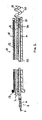
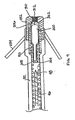
- FIG. 1 is a side, cut-away view of one embodiment of an implantable lead and fluid delivery system in accordance with the present invention.
- the system includes a guide catheter 10 having fluid dispensing capabilities.
- Catheter 10 is provided with a proximal handle 3 and an elongated catheter body 12 having at least two lumens 14 and 16 and is preferably formed from a biocompatible polymer such as polyurethane, silicone, Teflon®, or other acceptable plastic.
- a fluid-delivery lumen 14 is in communication with an active fixation, fluid dispensing member shown as a hollow fixation helix 18 located at the distal end of guide catheter 10.
- An active fixation, fluid dispensing member may alternatively be provided as a hollow "fish hook" type member, stake-like member, or any other type of active fixation member that can be provided as a hollow structure having one or more apertures.
- Hollow fixation helix 18 is provided with one or more apertures 20 through which fluid injected through lumen 14 may exit into a tissue site.
- Fixation helix 18 is preferably formed from a biocompatible metal, such as stainless steel, in which apertures 20 may be formed by laser drilling.
- a hollow fixation helix that may be used for fluid delivery is disclosed in the ⁇ 649 patent issued to Mulier et al and the WO 98/02040 patent issued to Stokes et al .
- a fluid fitting 2 such as a Luer lock fitting, may be inserted or mounted at the proximal end of fluid delivery lumen 14 to allow connection of a syringe for injecting fluid into lumen 14.
- Catheter 10 may be provided as a steerable catheter having a manipulative handle and steering mechanism, such as a pull wire, to aid in maneuvering catheter 10 through body vessels or organs.
- Steering mechanisms included in catheter 10 may be embodied as generally described in U.S. Pat. No. 5,396,902, issued to Brennen, et al. , for example, or U.S. Pat. No. 5,807,249 issued to Qin, et al. .
- a lead-delivery lumen 16 is provided for delivering an implantable lead 22 to a desired implant site.
- the lead-delivery lumen 16 is sized to allow lead 22 to easily pass through guide catheter 10 without undue friction or resistance.
- Lead 22 is shown as an exemplary bipolar lead having a helical tip electrode 24 located at the distal lead end and a ring electrode 26 spaced proximally from tip electrode 24.
- lead 22 may be a unipolar, bipolar, or multipolar lead carrying any combination of tip, ring and/or coil electrodes or other sensors.
- Lead 22 is shown with an active fixation helical electrode 24 but could also be provided with other types of active fixation electrodes or mechanisms, such as a "fish hook" electrode.
- Lead 22 may alternatively be provided with a generally spherical, hemispherical or ring-shaped tip electrode with passive fixation mechanisms, such as tines as generally known in the art.
- a connector assembly 8 is provided at the proximal lead end with a pin connector 4 and ring connector 6 which are electrically coupled to respective conductors that extend to tip electrode 24 and ring electrode 26.
- Conductors extending the length of lead 22 may be coiled conductors or cabled or stranded conductors as is known in the art.
- guide catheter 10 may be passed through a venous pathway into a desired heart chamber until a desired implantation site is reached.
- a guide wire or electrophysiological mapping catheter passed through inner lumen 16, could be used for passage of the catheter through the venous and cardiac anatomy to allow access to the targeted tissue.
- This guide wire or electrophysiological catheter could be steerable and would provide the additional benefit of protecting helix 18 to prevent snagging or entanglement with anatomic structures.
- Fixation helix 18 is advanced into the myocardial wall by rotating catheter 10 at its proximal end.
- Catheter body 12 is therefore provided with torsional stiffness adequate to translate rotational force to the distal fixation helix 18.
- a fluid which may be a pharmacological, genetic, or biologic agent, may then be injected into drug-delivery lumen 14 such that it is dispersed out of apertures 20 into the tissue surrounding fixation helix 18.
- a relatively large volume of tissue may be treated by the relatively large helix 18 on guide catheter 10.
- Lead 22 may then be passed through lead delivery lumen 16 and implanted at the treated tissue site by advancing helical tip electrode 24 into the tissue.
- the position of guide catheter 10 is maintained by helix 18 such that lead 22 may be implanted in the same volume of tissue treated by the injection of fluid through helix 18.
- guide catheter 10 may be removed by rotating catheter 10 in an appropriate direction to remove helix 18 from the tissue site and withdrawing catheter 10 over lead 22.
- Catheter 10 may be provided as a splittable or slittable catheter such that it may be removed from lead 22 without passing it over connector assembly 8.
- connector assembly 8 may be provided as a low profile connector assembly sized to allow catheter 10 to be readily passed over assembly 8.
- FIG 2 is a side, cut away plan view of an alternative embodiment of the guide catheter 10 shown in Figure 1 in which the distal fluid dispensing, fixation member, helix 18, may function as an electrode.
- fixation helix 18 is shown coupled to a conductor 15 that extends the length of catheter body 12 to a proximal terminal 17 enabling connection to a monitoring device, such as an electrocardiogram monitor.
- Helix 18 may thus serve as an electrode allowing electrophysiological signals to be sensed and monitored in order to verify that guide catheter 10 is fixed in a desired location. Monitoring of electrophysiological signals may also aid in verifying a short-term pharmacological effect after delivering a fluid through lumen 14 and helix 18.
- Figures 3A and 3B are cut-away plan views of the distal end of an implantable medical lead and fluid delivery system that includes a guide catheter 200, a fluid delivery device 208, and a medical lead 212.
- Figure 3A shows a guide catheter 200 having an elongated, tubular catheter body 202 with inner lumen 204.
- Guide catheter 200 is provided with a fixation member 206, shown in this embodiment as a helix, that allows catheter 200 to be fixed at a targeted implant site.
- Fixation member 206 may be a solid helix and may function exclusively as a fixation device. Alternatively, fixation member 206 may also function as an electrode as described above with reference to Figure 2 .
- a separate fluid delivery device 208 may be advanced through catheter lumen 204 until device 208 exits the distal end of catheter 200.
- Fluid delivery device 208 which may generally take the form of a hollow needle or stylet, may be tapered at its distal end and is preferably provided with a sharpened or beveled tip 210 such that it may easily pierce the tissue at the targeted implant site.
- the tip 210 may also take the form of a helix or other shape that may penetrate the tissue to a desired depth and dispense a fluid through one or more apertures to treat a volume of tissue.
- Fluid delivery device 208 may also serve as an electrode, alternatively or in addition to helix 206 of catheter 200.
- Fluid delivery device 208 which may be formed from a conductive metal such as stainless steel, may be provided with an insulating coating, such as a coating of ethylene tetrafluoroethylene (ETFE) or Parylene, except for at distal tip 210.
- ETFE ethylene tetrafluoroethylene
- the proximal end of device 208 may be coupled to a monitor such that electrophysiological signals sensed at uninsulated tip 210 may be monitored. Verification that tip 210 is in a desired tissue site, and not in blood or non-excitable tissue, may be made by monitoring electrophysiological signals sensed at tip 210.
- the fluid delivery device 208 may be withdrawn from lumen 204 of guide catheter 200 and replaced with an implantable medical lead 212 as shown in Figure 3B .
- Lead 212 is shown as an exemplary bipolar lead having an active fixation helical tip electrode 214 at its distal end and a ring electrode 216 spaced proximally from tip electrode 214. Lead 212 may be advanced through lumen 204 and implanted at the treated tissue site by advancing helical tip electrode 214 into the tissue. Guide catheter 200 may then be removed, leaving the electrode 214 implanted in the treated tissue.
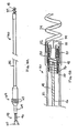
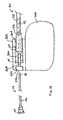
- FIG. 4A is a plan view of an alternative embodiment of an implantable lead and fluid delivery system.
- This system includes a transvenous lead 30 and a fluid delivery device 44.
- the lead 30 has an elongated, tubular lead body 32.
- Lead body 32 may be formed from a resilient, biocompatible polymer, such as silicone or polyurethane.
- Lead 30 is shown as a unipolar lead having an active fixation tip electrode 34 located at its distal end, shown as a helical electrode.
- Lead 30 may alternatively be a bipolar or multipolar lead having, in addition to active fixation tip electrode 32, one or more ring electrodes and/or one or more coil electrodes.
- a connector assembly 62 is provided at the proximal lead end to allow connection of lead 30 to an implantable pulse generator or monitoring device.
- Connector assembly 62 includes a pin terminal 64 that is electrically coupled to tip electrode 48 via a conductor extending the length of lead body 32.
- Pin terminal 64 is provided as a hollow pin that is in communication with a central lumen of lead body 32.
- Sealing rings 63 form a fluid-tight seal with the inner surface of a connector port on an implantable pulse generator or monitoring device.
- Fluid delivery device 44 is shown inserted into the proximal end of hollow pin terminal 44.
- Fluid delivery device 44 may take the form of a hollow needle or stylet as described above in conjunction with Figure 3A .
- Fluid delivery device 44 includes a hollow shaft 46 sized to pass easily through pin terminal 64 and the lumen of lead body 32 such that distal tip 48 of fluid delivery device 44 may exit the distal end of lead 30.
- a fluid fitting 60 which may take the form of a Luer lock fitting, is provided at the proximal end of device 44 to allow connection of a syringe for injecting fluid through shaft 46 to be dispensed from tip 48.
- FIG. 4B is a side cut-away view of the distal end of lead 30 and fluid delivery device 44.
- Helical tip electrode 34 is electrically coupled to a conductive sleeve 50, preferably by laser or resistance welding.
- Conductive sleeve 50 is electrically coupled to a conductor 36.
- Conductor 36 extends to connector assembly 62 at the proximal end of lead 30 and is coupled to pin terminal 64.
- Conductive sleeve 50 may be coupled to conductor 36 by crimping conductive sleeve 50 such that it is compressed against conductor 36, which is supported on its internal diameter by internal sleeve 40. In this way, electrode 34 is electrically coupled to conductor 36 and pin terminal 64.
- Conductor 36 is preferably a coiled conductor provided with insulation 37.
- Insulation 37 may be provided as a coating formed from an appropriate insulating material such as polytetrafluoroethylene (PTFE) or ETFE, preferably surrounding each individual filar included in conductor 36.
- Insulation 37 may alternatively be provided as heat shrink tubing fabricated from PTFE or ETFE as generally described in U.S. Pat. No. 6,052,625 issued to Marshall .
- Conductor 36 may alternatively be provided as an insulated cabled or stranded conductor, such as the conductor generally disclosed in U.S. Pat. No. 5,246,014 issued to Williams .
- Insulation 37 may also be provided as a material having a high Young's modulus, such as a high durometer polyurethane or polyimide, to impart additional lead body stiffness to the small diameter lead as generally described in U.S. Pat. No. 6,366,819 issued to Stokes .
- Insulation 37 electrically isolates conductor 36 from tip 48 and shaft 46 of fluid dispensing device 44 allowing distal tip 48 to function as a sensing electrode for detecting electrophysiological signals at a tissue site.
- fluid delivery device 44 may also be insulated along the entire length of shaft 46, particularly if conductor 36 is not provided with insulation. Distal tip 48 remains uninsulated.
- Insulation on shaft 46 may be provided by an adhesive coating, such as silicone adhesive, or as a tubular sleeve formed from an insulating material such as PTFE, ETFE or Parylene.
- a conductive clamp connected to a monitor such as an ECG monitor, may be coupled to fitting 60 at the proximal end of fluid delivery device 44 for observing electrophysiological signals at the site in which the uninsulated tip 48 is in contact. For example, cardiac P-waves or R-waves could be sensed by tip 48.
- Seal 38 is generally cup shaped and may be formed from a resilient, biocompatible polymer, such as molded silicone rubber. Seal 38 is shown in Figure 4B to be molded onto internal sleeve 40, which is preferably formed from a rigid, insulating material such as Delrin®, available from DuPont. Internal sleeve 40 is provided with an annular, laterally extending flange 52. Seal 38 is retained by the interaction of flange 52 and conductive sleeve 50. Seal 38 may be provided as generally described in U.S. Pat. No. 6,192,280 issued to Sommer et al .
- the seal 38 can be fabricated such that it is entirely contained within a portion of conductor 36 at a point at the distal end of the lead 32 or at a location more proximal.
- Alternative embodiments of a seal at or near the distal end of a medical lead or medical device that may be adapted for use with the present invention are disclosed in U.S. Pat. Application 20020016622 to Janke et al. , and U.S. Pat. Application 20020077685 to Sundquist et al .
- seals for preventing fluid from entering a tubular body may also be used.
- lead 30 may be deployed to a desired implant site.
- Lead 30 deployment may be performed with the aid of a guide wire, stylet, or guide catheter.
- Helical tip electrode 34 may then be fixed in the tissue at the implant site. If a guide wire or stylet is used, it is removed from lumen 42 after lead 30 is positioned so that fluid delivery device 44 may be advanced through lumen 42.
- Fluid delivery device tip 48 is preferably sharpened or beveled such that it can easily pierce through seal 38.
- the fluid delivery device 46 might also be shapeable, allowing it to be used for positioning of the lead 32. Seal 38 may be pre-pierced at line 54 to define a path for the fluid delivery device 44 to pass through. Tip 48 is then further advanced into the implant site.
- Verification that tip 48 is in a desired implant site may be made by monitoring electrophysiological signals sensed by uninsulated tip 48. If no signal is sensed, tip 48 may not be advanced completely through seal 38 or may not be fully inserted into the tissue site. Once tip 48 is adequately advanced into the implant site, a fluid may be injected through device 44 to treat a volume of tissue in which helical tip electrode 34 is implanted. Fluid delivery device 44 may then be withdrawn and removed, leaving lead 30 implanted with helical tip electrode 34 fixed in the treated tissue.
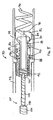
- FIG. 5 is an exploded, cut-away plan view of the distal end of an implantable lead and fluid delivery system wherein the lead 70 is provided with a retractable fixation member.
- a lead 70 is provided with a helical tip electrode 76 that may be retracted into an electrode housing 74.
- Electrode housing 74 is preferably formed from a relatively rigid biocompatible polymer, such as polyurethane. Housing 74 is bonded to an elongated, tubular lead body 72, which may be formed of polyurethane, silicone rubber, or another biocompatible polymer.
- Helical tip electrode 76 is mounted on a conductive sleeve 78, which is electrically coupled to a conductor 92.
- Conductive sleeve 78 which is preferably machined from a conductive metal such as stainless steel, includes a retraction mechanism shown as a threaded barrel 86 that is coaxial with sleeve 78 and located on the outer diameter of sleeve 78. Thread 88, running along the outer surface of barrel 86, acts to engage multiple thread guides 90 mounted on the inner diameter of housing 74.
- Conductor 92 may be rotated relative to lead body 72 by rotating a connector pin to which conductor 92 is coupled at its proximal end. Rotation of a coiled conductor may be achieved as generally described in U.S. Pat. No. 4,106,512, issued to Bisping .
- Rotation of conductor 92 causes rotation of sleeve 80 relative to electrode housing 74.
- Rotation of sleeve 80 causes advancement of helical electrode 76 as threaded barrel 86 is actuated on thread guides 90.
- a stop mechanism 89 may be provided as a ridge or peg near the proximal end of thread 88 that engages a thread guide 90 to prevent over extension of helical electrode 76.
- threaded barrel 86 will interact with housing 74 at lateral face 96 to prevent over-retraction of helix 76.
- a stop mechanism may be provided near the distal end of thread 88 to prevent over-retraction of helix 76.
- a retraction stop mechanism that may be adapted for use in the present invention is disclosed in U.S. Pat. No. 5,837,006, issued to Ocel et al .
- Lead 70 is provided with a seal 82, preferably formed of a resilient biocompatible polymer such as silicone rubber, molded to the distal end of the conductive sleeve 78 to prevent ingress of body fluids.
- Seal 82 may be generally cup shaped and may be pre-pierced at line 94 to guide a fluid delivery device 100 as it passes through seal 82.
- Seal 82 further includes an annular sealing ring 84, coaxial with seal 82 and extending laterally from the outer diameter of seal 82. Sealing ring 84 interacts with the inner surface of housing 74 to complete a fluid-tight seal of the distal end of lead 70. Sealing ring 84 further acts to center helix 76 within housing 74.
- a fluid delivery device 100 is provided which may be generally in the form of a hollow stylet or needle having an elongated shaft 106 extending between a proximal end through which fluid may be injected and a distal tip 102 through which fluid may be dispensed. Distal tip 102 is sharpened or beveled such that it may easily pierce through seal 82 and enter a targeted tissue site.
- a distal segment 104 of fluid delivery device 100 is provided with a reduced diameter allowing it to extend through conductive sleeve 78 such that distal tip 102 may extend out of housing 74 when helix 76 is extended into a tissue site.
- Lateral face 108 may act as a mechanical stop by interacting with the distal end of sleeve 78 and thereby control the maximum depth that fluid delivery device 100 is inserted into the targeted tissue site.
- the outer dimensions of shaft 106 and distal segment 104 and the spacing of lateral face 108 from distal tip 102 may alternatively be dimensioned to provide a stopping interface that interacts with a reduced inner diameter of sleeve 78 or helix 76.
- the tip of helix 76 may be bent to cross the center axis of helix 76 to act as a stop for fluid delivery device 100. Any of these methods for providing a mechanical stop for fluid delivery device 100 allows the tissue depth at which the fluid is injected to be controlled.
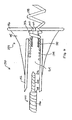
- FIG. 6 is an exploded, cut-away side view of the distal end of an implantable medical lead and fluid delivery system for use on the epicardial surface of the heart.
- a lead 150 is provided with a lead body 152, an insulating electrode head 154 and an active fixation electrode 158.
- Electrode 158 is shown as a helical electrode but may also take the form of a "fish hook" type electrode, or any other active fixation electrode.
- Electrode head 154 includes a tapered body 155 and flange 156, both of which may be formed from silicone rubber and provide a flexible structure for stabilizing the position of lead 150 on the epicardial surface.
- a tool may be used for implanting lead 150 by attaching to and rotating the electrode head 154 to screw the helical electrode 158 into the epicardium as is generally known in the art.
- Epicardial leads and tools for implanting epicardial leads are disclosed in U.S. Pat. No. 3,737,539 issued to Bolduc , U.S. Pat. No. 5,143,090 issued to Dutcher , and U.S. Pat. No. 6,010,526 issued to Sandstrom et al .
- Flange 156 may be reinforced with an embedded netting or mesh material, such as polyester netting. Netting material may optionally be coated with an anti-inflammatory steroid to reduce the inflammatory response at the tissue-lead interface.
- Helical electrode 158 is electrically coupled to a conductive sleeve 170, which is further coupled to a conductor 174, shown as a coiled conductor.
- Conductive sleeve 170 is provided with an annular flange 172.
- a seal 160 is molded to flange 172 to prevent the ingress of bodily fluids into the lead body lumen 164. Seal 160 may be pre-pierced at line 162 to define a path for fluid delivery device 100 to pass through.
- Fluid delivery device 100 may correspond to the fluid delivery device shown in Figure 5 and is shown in Figure 6 with identically labeled components corresponding to those in Figure 5 .
- Lateral face 108 may engage with the proximal end of conductive sleeve 170 to control the depth that fluid delivery device 100 is inserted into the tissue.
- fluid delivery device 100 may be extended through lead body lumen 164 and seal 160 to dispense a fluid into the tissue surrounding helical electrode 158. Fluid delivery device 100 may then be withdrawn from lumen 164 and removed from the patient's body, leaving lead 150 implanted at the treated tissue site.
- FIG 7 is a cut-away, side view of the distal end of an implantable medical lead and fluid delivery system wherein the medical lead is provided as a transvenous lead having a passive fixation mechanism.
- a ring electrode 252 is provided in place of an active fixation electrode at the tip of the lead 250.
- Ring electrode 252 is electrically coupled to conductive sleeve 50, which is further coupled to insulated conductor 36 as previously described with reference to Figure 4B .
- passive fixation members 254 are provided, which may take the form of tines as is generally known in the art.
- Seal 38 may be molded onto internal sleeve 40 as described previously and forms a fluid-tight seal with the inner diameter of ring electrode 252.
- Ring electrode 252 may be provided with an annular lip 256 which may act to retain seal 38.
- Figures 8 and 9 are side, cut-away views of the distal end of an implantable medical lead and fluid delivery system wherein the medical lead is further provided with a fluid reservoir for holding a pharmaceutical, genetic or biologic agent and allowing the agent to elute into adjacent body tissue over time.
- a body implantable lead having a cavity suitable for retaining a drug is disclosed in U.S. Pat. No. 4,506,680 issued to Stokes
- a combined catheter and reservoir, useful for applications involving delivery of genetic material, is disclosed in the previously cited PCT Patent Publication WO 98/02040 .
- the lead shown in Figure 8 corresponds to the lead of Figure 4B having a helical tip electrode 34 electrically coupled to stem 50 which is further coupled to an insulated conductor 36.
- a fluid reservoir 300 is located near the distal end of the lead.
- a fluid delivery device in the form of a hollow stylet or needle, having a shaft 46 and sharpened tip 48, may be used to fill reservoir 300 with a fluid.
- Reservoir 300 preferably includes a seal 304 covering a proximal opening to reservoir 300 and a seal 302 covering a distal opening to reservoir 300.
- Fluid delivery device tip 48 pierces through the proximal seal 304, which may be pre-pierced at line 308 and may be provided with a concave proximal surface to guide tip 48 to reservoir 300 and through seal 302. Fluid may then be injected into reservoir 300, and the fluid delivery device may be removed. The pharmaceutical, genetic, or biologic agent will elute from reservoir 300, through distal seal 302, into the adjacent tissue over time.
- Fluid reservoir 300 may be formed from silicone rubber or alternatively polyurethane or another elastomer.
- the seals 302 and 304 are preferably formed from silicone rubber.
- Seal 304 may be provided as a less permeable material than seal 302 to prevent blood or bodily fluids from entering the lead body lumen 42 while still allowing a pharmaceutical, genetic or biologic material to elute through seal 304.
- the reservoir 300 may be provided as a micro-osmotic pump.
- reservoir 300 may optionally contain a salt-loaded silicone material, which would swell over time as salt is replaced by water, or another polymeric material capable of swelling upon exposure to body fluids. Such swelling would aid in "pumping" a fluid agent out of reservoir 300.
- the fluid delivery device may be further advanced through distal seal 302, which may be pre-pierced at line 306.
- the fluid delivery device may then be inserted into the tissue in which electrode 34 is implanted to deliver a bolus of fluid directly to the tissue site, at a desired depth within the tissue.
- the fluid delivery device may then be withdrawn into reservoir 300 and used to fill reservoir 300 to allow a pharmaceutical, genetic or biologic agent to elute slowly over time into the adjacent tissue.
- local treatment of a volume of tissue may be performed by delivering a bolus of fluid directly into the tissue, or allowing the agent to elute from reservoir 300 over time, or both.
- one or more fluid agents may be delivered directly into the tissue site, and another fluid agent may be used to fill reservoir 300 and elute over time allowing the volume of tissue in which electrode 34 is implanted to be treated by at least two different pharmaceutical, genetic or biologic agents over different time courses.
- FIG. 9 is a cut-away, side view of the distal end of an implantable medical lead and fluid delivery system wherein the medical lead is provided as a transvenous lead having a passive fixation mechanism and a fluid reservoir.
- the system shown in Figure 9 is similar to the system shown in Figure 7 , and identically labeled components correspond to those shown in Figure 7 .
- the transvenous lead is shown having a fluid reservoir 300, similar to the reservoir described above in conjunction with Figure 8 .
- Ring tip electrode 252 is provided with a central bore 310 that may be filled with a porous material through which a pharmaceutical, genetic or biologic agent eluting out of reservoir 300 may pass to reach adjacent body tissue.
- a porous elution path may be formed from sintered metal structures as disclosed in the above incorporated '680 patent.
- central bore 310 may be left open, as shown previously in Figure 7 , to allow a fluid delivery device to be passed through tip electrode 252 to inject fluid directly into the tissue as well as providing an open elution pathway.
- a therapeutic fluid at a time after the lead implantation procedure.
- pharmacological, genetic or biological treatments may need to be repeated at certain intervals over time post-operatively in order to achieve a desired therapeutic effect.
- a situation may also arise requiring a chronically implanted lead to be repositioned due to dislodgment or declining stimulation or sensing performance.
- It may be desirable to treat the tissue at the new implant site at the time the lead is repositioned.
- factors that may be causing poor lead function such as poor tissue conductivity or low membrane potential signals, may be improved by treating the tissue at the chronic lead implant site with a fluid agent, thereby avoiding the need for lead repositioning.
- Figure 10 is a plan view of an implantable lead and fluid delivery system that may be used to deliver a fluid agent to a lead implant site post-operatively.
- lead 30 corresponds generally to that shown in Figure 4A , and all identically labeled components correspond to those illustrated in Figure 4A .
- connector assembly 62 at the proximal end of lead 30 is inserted into a connector bore 264 of a connector block 262 provided on a medical device 260, which may be a pacemaker or implantable cardioverter defibrillator, or other type of implantable pulse generator or electrophysiological monitor.
- Pin terminal 64 is electrically coupled to terminal 266 of connector block 262 to provide electrical connection between lead 30 and device 260.
- Lumen 218 may be accessed through access port 272, which is preferably sealed against body fluids by a grommet 270.
- Fluid delivery device 44 which may generally correspond to the fluid delivery device described in conjunction with Figure 4A , may be inserted through access port 272 and grommet 270 such that it may be passed through lumen 268, hollow pin terminal 64 and lead body lumen 42. Fluid delivery device 44 may then exit the distal end of lead 30 until it penetrates the tissue at the lead 30 implant site, as described previously. Once penetrated to a desired depth, fluid may be delivered through fluid delivery device 44. Fluid delivery device 44 may then be removed. Additionally or alternatively, fluid delivery device 44 may be used to refill a fluid reservoir that may be provided near the distal lead end as described in conjunction with Figures 8 and 9 .
- Access port 272 may be exposed during a minor surgical procedure by making a small skin incision at the site that device 260 is implanted. In this way, a volume of tissue at the lead implant site may advantageously be treated using a fluid delivery device at any time post-operatively without performing major surgery or catheterization procedures.
- the present invention provides a system for treating a volume of tissue concurrently with a lead implant procedure such that the lead may remain implanted at the treated tissue site.
- the present invention further allows tissue at a lead implant site to be treated at any time post-operatively through minimally invasive procedures.
- the various embodiments described herein include a medical lead and fluid delivery system that allow the fluid delivery components to be removed from the patient's body after treating a targeted tissue site so that only the lead remains implanted.
- the inventive system may also be used in procedures for treating a volume of tissue in which chronic implantation of a lead is not required.
- the lead may be used acutely with an associated fluid delivery device to deliver a fluid agent to a targeted tissue site and then removed with the fluid delivery device rather than remaining implanted or implanted at another site.
- inventive therapy modalities that may benefit from the inventive system and may or may not require chronic implantation of a lead may include treatment of myocardial infarction via cell delivery or treatment of coronary artery disease via drugs or biologic agents such as angiogenic factors.
- cardiac leads and the treatment of cardiac tissue aspects of the inventive system may also be used in regard to other types of leads and other types of bodily tissue, such as kidney, brain, pancreas, or other organs or tissues. The described embodiments are therefore exemplary and should not be considered limiting with regard to the following claims.
Abstract
Description
- The present invention relates generally to implantable medical leads and more specifically to an implantable medical lead and fluid delivery system for treating a volume of tissue in which the medical lead may remain implanted.
- Electrical stimulation of excitable body tissue is used as a method for treating various pathological conditions. Therapeutic stimulation generally requires making an electrical contact between excitable tissue and an electrical pulse generator through use of one or more stimulation leads. Various lead systems and various techniques for implanting these lead systems in contact with excitable body tissue, and particularly the heart, have been developed.
- In order to achieve cardiac pacing, sensing, cardioversion and/or defibrillation at different locations in the heart, various types of cardiac leads have been developed including epicardial leads, endocardial leads, and coronary vein leads. A transvenous endocardial lead establishes electrical contact between an electrical pulse generator, such as a pacemaker or implantable cardioverter defibrillator, and the endocardial surface of the heart, typically in a right heart chamber. Endocardial leads, and cardiac leads in general, may be held in place by passive fixation mechanisms, such as tines that interact with the ventricular trabeculae, or active fixation mechanisms, such as a helix. A coronary vein lead may be passed through a venous pathway, into the right atrium, through the coronary sinus ostium and ultimately to a location deep in the cardiac veins. Contact is made with the epicardial surface of the left atrium or left ventricle for delivering stimulation or sensing cardiac signals in the left heart chambers. Epicardial leads are also known in the art and generally require a thoracotomy for placement on the epicardial surface of a heart chamber.
- The safety, efficacy and longevity of an electrical pulse generator depends, in part, on the performance of the associated cardiac lead(s) used in conjunction with the pulse generator. Various properties of the lead, the electrodes and the tissue interfacing with an electrode will result in a characteristic impedance, stimulation threshold and sensing threshold.
- Stimulation threshold is the energy required in a stimulation pulse to depolarize, or "capture," the heart tissue. A relatively high impedance and low threshold is desired to minimize the current drawn from a pulse generator battery in delivering a stimulation pulse. Maximizing the useful life of the pulse generator battery is important since a surgical procedure is required to replace the pulse generator once the battery has reached the end of its useful life.
- One factor that can affect the stimulation threshold, particularly during the first several weeks after implantation of a lead, is the natural immunological response of the body to the lead as a foreign object. The presence of the lead activates the immunologic response, which ultimately results in fibrotic encapsulation of the lead and its electrodes. Since fibrotic tissue is not excitable tissue, an elevated stimulation threshold can persist due to the degraded electrical properties of the electrode-tissue interface.
- To reduce the inflammatory response, medical leads that elute an anti-inflammatory steroid have been developed. Steroid eluting leads are described in
U.S. Pat. No. 4,506,680 issued to Stokes and related MedtronicU.S. Pat. Nos. 4,577,642 , and4,606,118 . Steroid eluting leads may require a monolithic controlled release device (MCRD) to contain the steroid and to thereafter slowly leach out the water soluble steroid into the surrounding tissue. A method for applying a steroid directly to the surface of an electrode is disclosed inU.S. Pat. No. 5,987,746 issued to Williams , Advantages of this method include elimination of additional structures for carrying the steroid and the presentation of the steroid directly at the tissue-electrode interface. - One limitation of a steroid eluting electrode or MCRD, however, is that a relatively limited volume of tissue is treated by the eluting drug since the drug is presented only at the endocardial or epicardial surface. Other devices have been proposed which allow the delivery of a drug to a potentially larger volume of tissue by actually penetrating the tissue rather than relying on diffusion of the drug from the tissue surface. Drug delivery catheters may incorporate a drug dispensing needle or helix that penetrates a targeted tissue for delivering a drug or fluid. Catheters that may be used to deliver a fluid or drug into the myocardium are disclosed in
U.S. Pat. No. 6,102,887 issued to Altman andU.S. Pat. No. 5,431,649 issued to Mulier et al . - Drug delivery catheters may include an electrode to allow sensing or stimulation of the myocardium. An implantable pacing lead having an active fixation electrode with a stylet introduced, anti-inflammatory drug delivery system is disclosed in
U.S. Pat. No. 5,447,533 issued to Vachon et al . A delivery system for delivering a therapeutically effective amount of a genetic material to an identified cardiac location adjacent an atrial or ventricular electrode is disclosed inPCT Patent Publication WO 98/02040 issued to Stokes et al U.S. Pat. No. 4,360,031 to White , andU.S. Pat. No. 5,496,360 to Hoffman . - Advancements in gene therapies and cellular modifications through the delivery of proteins, peptides or even cell delivery, such as stem cell delivery, offer opportunities to alter the properties of tissue to further improve the benefit of a delivered stimulation therapy or improve the ability to sense cardiac signals. Genetic or biologic agents may be used to alter ion channel activity or protein expression at the cellular level. Potential benefits include decreased inflammatory response, increased tissue conductivity for reduction of stimulation thresholds or upregulation of ion channels for increasing membrane potentials to allow better sensing. For example, upregulation of ion channels could enhance cardiac P-waves or R-waves allowing them be more easily sensed by a pacemaker or other cardiac monitor. In particular, cardiac fast sodium channels are responsible for the fast upstroke of the action potential in myocardial cells (Fozzard, et al., Circ. Res. 1995, 56:475-485). A human cardiac voltage-dependent sodium channel, hHl, has been cloned, sequenced, and functionally expressed (Gellens, et al., Proc. Natl. Acad. Sci. USA, 1992, 89:554-558). Alteration of myocardial conductivity may be possible through delivery of proteins that alter cellular electrical coupling. The gap junction protein Connexin43 has been found to play an important role in ventricular conduction (Guerrero PA et al., J. Clin. Invest. 1997, 99:1991-1998).
- Because locally effective doses of a pharmacologic, genetic, or biologic agent may be toxic when given systemically, it is desirable to provide a method for delivering an agent locally at a targeted tissue site. Drug-eluting electrodes may be limited to treating only a relatively small volume of tissue at an electrode-tissue interface. The pharmacological effect is in part limited by the kinetics of the drug leaving the electrode or lead. Furthermore, because biologic and genetic agents may have a limited shelf life, unique storage requirements such as requiring refrigeration, and may not tolerate sterilization procedures, it is not desirable to package a lead having drug eluting capabilities with the biologic or genetic agent already incorporated therein. Other medical leads having drug dispensing capabilities may require additional components that increase the size, stiffness or complexity of the lead. Document
US-5 002 067 discloses a medical lead employing improved penetrating electrode and documentUS-B1-6 346 099 discloses an active fluid delivery catheter without lead delivery lumen. - To take advantage of various genetic or cellular modification therapies, it is desirable to provide an implantable lead and fluid delivery system that allows a pharmaceutical, genetic, or biologic agent to be delivered to a targeted lead implant site at a depth within the myocardium to treat a volume of tissue. Once a fluid agent has been delivered, the fluid delivery components are no longer needed and may be removed from the patient's body. An acutely implanted fluid delivery system eliminates the need to include dispensing components in the medical lead, reducing its complexity, yet still offers the benefit of treating a volume of tissue at a lead implant site, potentially improving lead performance. There is a need, therefore, for a system that allows an acutely implanted fluid delivery device to treat a volume of tissue during a lead implant procedure, or at any time post-operatively, and further allows a lead to be implanted and remain in the location of the treated tissue.
- The present invention is directed toward providing a medical lead and fluid delivery system as defined in claim 1, for treating a volume of tissue with a pharmaceutical, genetic or biologic agent at the time of the medical lead implant and/or at any time post-operatively.
- In one embodiment of the present invention, a guide catheter is provided with a fluid dispensing, fixation member at its distal end. The fixation member communicates with a lumen extending the length of the guide catheter body through which a fluid may be delivered. The fixation member, which may be provided as a hollow helix, is provided with one or more apertures for dispensing a drug into the surrounding tissue in which the helix is fixed. The fixation member may optionally function as an electrode in addition to being a fixation device. After treating a volume of tissue by dispensing a fluid through the fixation member, a medical lead, advanced through a lead-delivery lumen of the guide catheter, may be implanted in the treated tissue. The guide catheter may then be removed leaving the medical lead implanted at the treated tissue site.
- In another embodiment, a system is provided including a guide catheter having a fixation member, a fluid delivery device, and an implantable lead. After fixing the guide catheter at a desired implant site, the fluid delivery device, advanced down a lumen of the guide catheter, may be used to treat a volume of tissue at that site. The fluid delivery device may then be removed and an implantable lead may be advanced through the guide catheter to the treated tissue site. The guide catheter may then be removed, leaving the lead implanted at the treated tissue site.
- In alternative embodiments, a transvenous lead having a distal fixation member is provided with a center lumen through which a fluid delivery device may be advanced to treat a volume of tissue in which the lead is implanted. A seal is preferably provided at the distal end of the lead body to prevent fluid ingress. The distal fixation member may be provided as a passive or active fixation member. In one embodiment, a retractable active fixation member is provided. In another embodiment, the implantable lead may be provided as an epicardial lead.
- The fluid delivery device may take the form of a hollow needle or stylet that may be advanced through a lumen of the lead body, penetrated through the seal and into the targeted tissue site. The fluid delivery device may be provided with a conductive tip to allow sensing of electrophysiological signals. Fluid delivery may be performed once the delivery device is inserted in the targeted tissue location as verified by sensing electrophysiological signals characteristic of the targeted tissue. After delivery of a fluid, which may be a pharmaceutical, genetic or biologic agent carried in a liquid medium, the fluid delivery device may be removed, leaving the transvenous lead implanted at the treated tissue site.
- The medical lead may further include a reservoir that may be filled by the fluid delivery device with a pharmaceutical, genetic or biologic agent. The agent will elute from the reservoir into surrounding tissue over time. Treatment of a volume of tissue with a pharmaceutical, genetic or biologic agent may thus be treated by delivering a bolus injection directly into the tissue or slow elution of the agent over time, or both.
- A fluid may be delivered post-operatively by gaining access to the lumen of an implanted lead through an access port on the implanted device to which the lead is connected. Multiple injections at the lead implant site at various time intervals are possible by inserting a fluid delivery device through the access port and advancing it through the lead lumen. Fluid may be delivered directly to the lead implant site, or a fluid reservoir may be refilled.
- Thus, the present invention provides a medical lead and fluid delivery system that allows a lead to be implanted in a volume of tissue treated concurrently with the lead implant procedure, or at any time post-operatively, by an acutely delivered fluid delivery device.
-
Figure 1 is a side, cut-away view of an implantable lead and fluid delivery system including a guide catheter having fluid dispensing capabilities and an implantable medical lead. -
Figure 2 is a side, cut-away view of an alternative embodiment of the guide catheter shown inFigure 1 in which a fixation member on the guide catheter may also function as an electrode. -
Figures 3A and 3B are side, cut-away views of the distal end of an implantable medical lead and fluid delivery system that includes a guide catheter, a fluid delivery device and a medical lead. -
Figure 4A is a plan view of an alternative embodiment of an implantable lead and fluid delivery system including a transvenous medical lead and a fluid delivery device that may be deployed through a lumen of the lead. -
Figure 4B is a side cut-away, view of the distal end of the system ofFigure 4A . -
Figure 5 is an exploded, side, cut-away view of the distal end of an implantable lead and fluid delivery system in which the lead is provided with a retractable fixation member. -
Figure 6 is an exploded, side, cut-away view of the distal end of an implantable medical lead and fluid delivery system for use on the epicardial surface of the heart. -
Figure 7 is a cut-away, side view of the distal end of an implantable medical lead and fluid delivery system wherein the medical lead is provided as a transvenous lead having a passive fixation mechanism. -
Figures 8 is side, cut-away view of the distal end of an implantable medical lead and fluid delivery system wherein the medical lead is further provided with a fluid reservoir for holding a pharmaceutical, genetic or biologic agent and allowing the agent to elute into adjacent body tissue over time. -
Figure 9 is a side, cut-away view of the distal end of an implantable medical lead and fluid delivery system wherein the medical lead is provided as a transvenous lead having a passive fixation mechanism and a fluid reservoir. -
Figure 10 is a plan view of an implantable lead and fluid delivery system that may be used to deliver a fluid agent to a lead implant site post-operatively. - As described above, the present invention is directed at providing an implantable lead and fluid delivery system in which a fluid delivery device may be used to treat a volume of tissue concurrently with a lead implantation procedure, or at any time post-operatively. After delivering a fluid, the fluid delivery device may be removed leaving the lead implanted at the treated tissue site.
Figure 1 is a side, cut-away view of one embodiment of an implantable lead and fluid delivery system in accordance with the present invention. The system includes a guide catheter 10 having fluid dispensing capabilities. Catheter 10 is provided with a proximal handle 3 and anelongated catheter body 12 having at least twolumens delivery lumen 14 is in communication with an active fixation, fluid dispensing member shown as ahollow fixation helix 18 located at the distal end of guide catheter 10. An active fixation, fluid dispensing member may alternatively be provided as a hollow "fish hook" type member, stake-like member, or any other type of active fixation member that can be provided as a hollow structure having one or more apertures.Hollow fixation helix 18 is provided with one ormore apertures 20 through which fluid injected throughlumen 14 may exit into a tissue site.Fixation helix 18 is preferably formed from a biocompatible metal, such as stainless steel, in which apertures 20 may be formed by laser drilling. A hollow fixation helix that may be used for fluid delivery is disclosed in the `649 patent issued to Mulier et al and theWO 98/02040 patent issued to Stokes et al fluid fitting 2, such as a Luer lock fitting, may be inserted or mounted at the proximal end offluid delivery lumen 14 to allow connection of a syringe for injecting fluid intolumen 14. - Catheter 10 may be provided as a steerable catheter having a manipulative handle and steering mechanism, such as a pull wire, to aid in maneuvering catheter 10 through body vessels or organs. Steering mechanisms included in catheter 10 may be embodied as generally described in
U.S. Pat. No. 5,396,902, issued to Brennen, et al. , for example, orU.S. Pat. No. 5,807,249 issued to Qin, et al. . - A lead-
delivery lumen 16 is provided for delivering animplantable lead 22 to a desired implant site. The lead-delivery lumen 16 is sized to allowlead 22 to easily pass through guide catheter 10 without undue friction or resistance.Lead 22 is shown as an exemplary bipolar lead having ahelical tip electrode 24 located at the distal lead end and aring electrode 26 spaced proximally fromtip electrode 24. In other embodiments, lead 22 may be a unipolar, bipolar, or multipolar lead carrying any combination of tip, ring and/or coil electrodes or other sensors.Lead 22 is shown with an active fixationhelical electrode 24 but could also be provided with other types of active fixation electrodes or mechanisms, such as a "fish hook" electrode.Lead 22 may alternatively be provided with a generally spherical, hemispherical or ring-shaped tip electrode with passive fixation mechanisms, such as tines as generally known in the art. - A
connector assembly 8 is provided at the proximal lead end with a pin connector 4 and ring connector 6 which are electrically coupled to respective conductors that extend to tipelectrode 24 andring electrode 26. Conductors extending the length oflead 22 may be coiled conductors or cabled or stranded conductors as is known in the art. - During a lead implantation procedure, guide catheter 10 may be passed through a venous pathway into a desired heart chamber until a desired implantation site is reached. A guide wire or electrophysiological mapping catheter, passed through
inner lumen 16, could be used for passage of the catheter through the venous and cardiac anatomy to allow access to the targeted tissue. This guide wire or electrophysiological catheter could be steerable and would provide the additional benefit of protectinghelix 18 to prevent snagging or entanglement with anatomic structures.Fixation helix 18 is advanced into the myocardial wall by rotating catheter 10 at its proximal end.Catheter body 12 is therefore provided with torsional stiffness adequate to translate rotational force to thedistal fixation helix 18. A fluid, which may be a pharmacological, genetic, or biologic agent, may then be injected into drug-delivery lumen 14 such that it is dispersed out ofapertures 20 into the tissue surroundingfixation helix 18. A relatively large volume of tissue may be treated by the relativelylarge helix 18 on guide catheter 10. -
Lead 22 may then be passed throughlead delivery lumen 16 and implanted at the treated tissue site by advancinghelical tip electrode 24 into the tissue. The position of guide catheter 10 is maintained byhelix 18 such thatlead 22 may be implanted in the same volume of tissue treated by the injection of fluid throughhelix 18. After implantinglead 22, guide catheter 10 may be removed by rotating catheter 10 in an appropriate direction to removehelix 18 from the tissue site and withdrawing catheter 10 overlead 22. Catheter 10 may be provided as a splittable or slittable catheter such that it may be removed fromlead 22 without passing it overconnector assembly 8. Alternatively,connector assembly 8 may be provided as a low profile connector assembly sized to allow catheter 10 to be readily passed overassembly 8. -
Figure 2 is a side, cut away plan view of an alternative embodiment of the guide catheter 10 shown inFigure 1 in which the distal fluid dispensing, fixation member,helix 18, may function as an electrode. InFigure 2 , all identically labeled components correspond to those illustrated inFigure 1 . InFigure 2 , however,fixation helix 18 is shown coupled to aconductor 15 that extends the length ofcatheter body 12 to aproximal terminal 17 enabling connection to a monitoring device, such as an electrocardiogram monitor.Helix 18 may thus serve as an electrode allowing electrophysiological signals to be sensed and monitored in order to verify that guide catheter 10 is fixed in a desired location. Monitoring of electrophysiological signals may also aid in verifying a short-term pharmacological effect after delivering a fluid throughlumen 14 andhelix 18. -
Figures 3A and 3B are cut-away plan views of the distal end of an implantable medical lead and fluid delivery system that includes aguide catheter 200, afluid delivery device 208, and amedical lead 212.Figure 3A shows aguide catheter 200 having an elongated,tubular catheter body 202 withinner lumen 204.Guide catheter 200 is provided with afixation member 206, shown in this embodiment as a helix, that allowscatheter 200 to be fixed at a targeted implant site.Fixation member 206 may be a solid helix and may function exclusively as a fixation device. Alternatively,fixation member 206 may also function as an electrode as described above with reference toFigure 2 . - A separate
fluid delivery device 208 may be advanced throughcatheter lumen 204 untildevice 208 exits the distal end ofcatheter 200.Fluid delivery device 208, which may generally take the form of a hollow needle or stylet, may be tapered at its distal end and is preferably provided with a sharpened orbeveled tip 210 such that it may easily pierce the tissue at the targeted implant site. Thetip 210 may also take the form of a helix or other shape that may penetrate the tissue to a desired depth and dispense a fluid through one or more apertures to treat a volume of tissue. Oncefluid delivery device 208 is advanced into the tissue, a fluid may be injected in the proximal end offluid delivery device 208 and dispensed into a volume of tissue throughtip 210. -
Fluid delivery device 208 may also serve as an electrode, alternatively or in addition tohelix 206 ofcatheter 200.Fluid delivery device 208, which may be formed from a conductive metal such as stainless steel, may be provided with an insulating coating, such as a coating of ethylene tetrafluoroethylene (ETFE) or Parylene, except for atdistal tip 210. The proximal end ofdevice 208 may be coupled to a monitor such that electrophysiological signals sensed atuninsulated tip 210 may be monitored. Verification that tip 210 is in a desired tissue site, and not in blood or non-excitable tissue, may be made by monitoring electrophysiological signals sensed attip 210. - After dispensing a fluid into the targeted implant site, the
fluid delivery device 208 may be withdrawn fromlumen 204 ofguide catheter 200 and replaced with an implantablemedical lead 212 as shown inFigure 3B .Lead 212 is shown as an exemplary bipolar lead having an active fixationhelical tip electrode 214 at its distal end and aring electrode 216 spaced proximally fromtip electrode 214. Lead 212 may be advanced throughlumen 204 and implanted at the treated tissue site by advancinghelical tip electrode 214 into the tissue.Guide catheter 200 may then be removed, leaving theelectrode 214 implanted in the treated tissue. -
Figure 4A is a plan view of an alternative embodiment of an implantable lead and fluid delivery system. This system includes a transvenous lead 30 and afluid delivery device 44. The lead 30 has an elongated, tubularlead body 32. Leadbody 32 may be formed from a resilient, biocompatible polymer, such as silicone or polyurethane. Lead 30 is shown as a unipolar lead having an activefixation tip electrode 34 located at its distal end, shown as a helical electrode. Lead 30 may alternatively be a bipolar or multipolar lead having, in addition to activefixation tip electrode 32, one or more ring electrodes and/or one or more coil electrodes. - A connector assembly 62 is provided at the proximal lead end to allow connection of lead 30 to an implantable pulse generator or monitoring device. Connector assembly 62 includes a
pin terminal 64 that is electrically coupled to tipelectrode 48 via a conductor extending the length oflead body 32.Pin terminal 64 is provided as a hollow pin that is in communication with a central lumen oflead body 32. Sealing rings 63 form a fluid-tight seal with the inner surface of a connector port on an implantable pulse generator or monitoring device. -
Fluid delivery device 44 is shown inserted into the proximal end ofhollow pin terminal 44.Fluid delivery device 44 may take the form of a hollow needle or stylet as described above in conjunction withFigure 3A .Fluid delivery device 44 includes ahollow shaft 46 sized to pass easily throughpin terminal 64 and the lumen oflead body 32 such thatdistal tip 48 offluid delivery device 44 may exit the distal end of lead 30. Afluid fitting 60, which may take the form of a Luer lock fitting, is provided at the proximal end ofdevice 44 to allow connection of a syringe for injecting fluid throughshaft 46 to be dispensed fromtip 48. -
Figure 4B is a side cut-away view of the distal end of lead 30 andfluid delivery device 44.Helical tip electrode 34 is electrically coupled to aconductive sleeve 50, preferably by laser or resistance welding.Conductive sleeve 50 is electrically coupled to aconductor 36.Conductor 36 extends to connector assembly 62 at the proximal end of lead 30 and is coupled to pin terminal 64.Conductive sleeve 50 may be coupled toconductor 36 by crimpingconductive sleeve 50 such that it is compressed againstconductor 36, which is supported on its internal diameter byinternal sleeve 40. In this way,electrode 34 is electrically coupled toconductor 36 andpin terminal 64. -
Conductor 36 is preferably a coiled conductor provided withinsulation 37.Insulation 37 may be provided as a coating formed from an appropriate insulating material such as polytetrafluoroethylene (PTFE) or ETFE, preferably surrounding each individual filar included inconductor 36.Insulation 37 may alternatively be provided as heat shrink tubing fabricated from PTFE or ETFE as generally described inU.S. Pat. No. 6,052,625 issued to Marshall .Conductor 36 may alternatively be provided as an insulated cabled or stranded conductor, such as the conductor generally disclosed inU.S. Pat. No. 5,246,014 issued to Williams .Insulation 37 may also be provided as a material having a high Young's modulus, such as a high durometer polyurethane or polyimide, to impart additional lead body stiffness to the small diameter lead as generally described inU.S. Pat. No. 6,366,819 issued to Stokes . -
Insulation 37 electrically isolatesconductor 36 fromtip 48 andshaft 46 offluid dispensing device 44 allowingdistal tip 48 to function as a sensing electrode for detecting electrophysiological signals at a tissue site. Whentip 48 is used as a sensing electrode,fluid delivery device 44 may also be insulated along the entire length ofshaft 46, particularly ifconductor 36 is not provided with insulation.Distal tip 48 remains uninsulated. Insulation onshaft 46 may be provided by an adhesive coating, such as silicone adhesive, or as a tubular sleeve formed from an insulating material such as PTFE, ETFE or Parylene. A conductive clamp, connected to a monitor such as an ECG monitor, may be coupled to fitting 60 at the proximal end offluid delivery device 44 for observing electrophysiological signals at the site in which theuninsulated tip 48 is in contact. For example, cardiac P-waves or R-waves could be sensed bytip 48. - Lead 30 is preferably provided with a
seal 38 to prevent the ingress of body fluids.Seal 38 is generally cup shaped and may be formed from a resilient, biocompatible polymer, such as molded silicone rubber.Seal 38 is shown inFigure 4B to be molded ontointernal sleeve 40, which is preferably formed from a rigid, insulating material such as Delrin®, available from DuPont.Internal sleeve 40 is provided with an annular, laterally extendingflange 52.Seal 38 is retained by the interaction offlange 52 andconductive sleeve 50.Seal 38 may be provided as generally described inU.S. Pat. No. 6,192,280 issued to Sommer et al .
Alternatively, theseal 38 can be fabricated such that it is entirely contained within a portion ofconductor 36 at a point at the distal end of thelead 32 or at a location more proximal. Alternative embodiments of a seal at or near the distal end of a medical lead or medical device that may be adapted for use with the present invention are disclosed inU.S. Pat. Application 20020016622 to Janke et al. , andU.S. Pat. Application 20020077685 to Sundquist et al . - Other types of seals for preventing fluid from entering a tubular body may also be used.
- During an implantation procedure, lead 30 may be deployed to a desired implant site. Lead 30 deployment may be performed with the aid of a guide wire, stylet, or guide catheter.
Helical tip electrode 34 may then be fixed in the tissue at the implant site. If a guide wire or stylet is used, it is removed fromlumen 42 after lead 30 is positioned so thatfluid delivery device 44 may be advanced throughlumen 42. Fluiddelivery device tip 48 is preferably sharpened or beveled such that it can easily pierce throughseal 38. Thefluid delivery device 46 might also be shapeable, allowing it to be used for positioning of thelead 32.Seal 38 may be pre-pierced atline 54 to define a path for thefluid delivery device 44 to pass through.Tip 48 is then further advanced into the implant site. Verification that tip 48 is in a desired implant site may be made by monitoring electrophysiological signals sensed byuninsulated tip 48. If no signal is sensed,tip 48 may not be advanced completely throughseal 38 or may not be fully inserted into the tissue site. Oncetip 48 is adequately advanced into the implant site, a fluid may be injected throughdevice 44 to treat a volume of tissue in whichhelical tip electrode 34 is implanted.Fluid delivery device 44 may then be withdrawn and removed, leaving lead 30 implanted withhelical tip electrode 34 fixed in the treated tissue. -
Figure 5 is an exploded, cut-away plan view of the distal end of an implantable lead and fluid delivery system wherein the lead 70 is provided with a retractable fixation member. A lead 70 is provided with ahelical tip electrode 76 that may be retracted into anelectrode housing 74.Electrode housing 74 is preferably formed from a relatively rigid biocompatible polymer, such as polyurethane.Housing 74 is bonded to an elongated, tubularlead body 72, which may be formed of polyurethane, silicone rubber, or another biocompatible polymer. -
Helical tip electrode 76 is mounted on aconductive sleeve 78, which is electrically coupled to aconductor 92.Conductive sleeve 78, which is preferably machined from a conductive metal such as stainless steel, includes a retraction mechanism shown as a threaded barrel 86 that is coaxial withsleeve 78 and located on the outer diameter ofsleeve 78.Thread 88, running along the outer surface of barrel 86, acts to engage multiple thread guides 90 mounted on the inner diameter ofhousing 74.Conductor 92 may be rotated relative to leadbody 72 by rotating a connector pin to whichconductor 92 is coupled at its proximal end. Rotation of a coiled conductor may be achieved as generally described inU.S. Pat. No. 4,106,512, issued to Bisping . - Rotation of
conductor 92 causes rotation ofsleeve 80 relative to electrodehousing 74. Rotation ofsleeve 80 causes advancement ofhelical electrode 76 as threaded barrel 86 is actuated on thread guides 90. A stop mechanism 89 may be provided as a ridge or peg near the proximal end ofthread 88 that engages athread guide 90 to prevent over extension ofhelical electrode 76. During retraction, threaded barrel 86 will interact withhousing 74 at lateral face 96 to prevent over-retraction ofhelix 76. Alternatively, a stop mechanism may be provided near the distal end ofthread 88 to prevent over-retraction ofhelix 76. A retraction stop mechanism that may be adapted for use in the present invention is disclosed inU.S. Pat. No. 5,837,006, issued to Ocel et al . - Lead 70 is provided with a
seal 82, preferably formed of a resilient biocompatible polymer such as silicone rubber, molded to the distal end of theconductive sleeve 78 to prevent ingress of body fluids.Seal 82 may be generally cup shaped and may be pre-pierced atline 94 to guide afluid delivery device 100 as it passes throughseal 82.Seal 82 further includes anannular sealing ring 84, coaxial withseal 82 and extending laterally from the outer diameter ofseal 82. Sealingring 84 interacts with the inner surface ofhousing 74 to complete a fluid-tight seal of the distal end of lead 70. Sealingring 84 further acts to centerhelix 76 withinhousing 74. - A
fluid delivery device 100 is provided which may be generally in the form of a hollow stylet or needle having anelongated shaft 106 extending between a proximal end through which fluid may be injected and adistal tip 102 through which fluid may be dispensed.Distal tip 102 is sharpened or beveled such that it may easily pierce throughseal 82 and enter a targeted tissue site. Adistal segment 104 offluid delivery device 100 is provided with a reduced diameter allowing it to extend throughconductive sleeve 78 such thatdistal tip 102 may extend out ofhousing 74 whenhelix 76 is extended into a tissue site.Lateral face 108 may act as a mechanical stop by interacting with the distal end ofsleeve 78 and thereby control the maximum depth thatfluid delivery device 100 is inserted into the targeted tissue site. The outer dimensions ofshaft 106 anddistal segment 104 and the spacing oflateral face 108 fromdistal tip 102 may alternatively be dimensioned to provide a stopping interface that interacts with a reduced inner diameter ofsleeve 78 orhelix 76. Alternatively, the tip ofhelix 76 may be bent to cross the center axis ofhelix 76 to act as a stop forfluid delivery device 100. Any of these methods for providing a mechanical stop forfluid delivery device 100 allows the tissue depth at which the fluid is injected to be controlled. -
Figure 6 is an exploded, cut-away side view of the distal end of an implantable medical lead and fluid delivery system for use on the epicardial surface of the heart. Alead 150 is provided with alead body 152, an insulatingelectrode head 154 and an active fixation electrode 158. Electrode 158 is shown as a helical electrode but may also take the form of a "fish hook" type electrode, or any other active fixation electrode.Electrode head 154 includes atapered body 155 andflange 156, both of which may be formed from silicone rubber and provide a flexible structure for stabilizing the position oflead 150 on the epicardial surface. A tool may be used for implantinglead 150 by attaching to and rotating theelectrode head 154 to screw the helical electrode 158 into the epicardium as is generally known in the art. Epicardial leads and tools for implanting epicardial leads are disclosed inU.S. Pat. No. 3,737,539 issued to Bolduc ,U.S. Pat. No. 5,143,090 issued to Dutcher , andU.S. Pat. No. 6,010,526 issued to Sandstrom et al . -
Flange 156 may be reinforced with an embedded netting or mesh material, such as polyester netting. Netting material may optionally be coated with an anti-inflammatory steroid to reduce the inflammatory response at the tissue-lead interface. - Helical electrode 158 is electrically coupled to a
conductive sleeve 170, which is further coupled to aconductor 174, shown as a coiled conductor.Conductive sleeve 170 is provided with anannular flange 172. A seal 160 is molded toflange 172 to prevent the ingress of bodily fluids into thelead body lumen 164. Seal 160 may be pre-pierced atline 162 to define a path forfluid delivery device 100 to pass through.Fluid delivery device 100 may correspond to the fluid delivery device shown inFigure 5 and is shown inFigure 6 with identically labeled components corresponding to those inFigure 5 .Lateral face 108 may engage with the proximal end ofconductive sleeve 170 to control the depth thatfluid delivery device 100 is inserted into the tissue. - After implanting
lead 150,fluid delivery device 100 may be extended throughlead body lumen 164 and seal 160 to dispense a fluid into the tissue surrounding helical electrode 158.Fluid delivery device 100 may then be withdrawn fromlumen 164 and removed from the patient's body, leavinglead 150 implanted at the treated tissue site. -
Figure 7 is a cut-away, side view of the distal end of an implantable medical lead and fluid delivery system wherein the medical lead is provided as a transvenous lead having a passive fixation mechanism. In this embodiment, all identically labeled components correspond to those illustrated inFigure 4B , however, in this case, in place of an active fixation electrode at the tip of the lead 250, aring electrode 252 is provided.Ring electrode 252 is electrically coupled toconductive sleeve 50, which is further coupled toinsulated conductor 36 as previously described with reference toFigure 4B . To stabilize the implanted position oflead 252,passive fixation members 254 are provided, which may take the form of tines as is generally known in the art.Seal 38 may be molded ontointernal sleeve 40 as described previously and forms a fluid-tight seal with the inner diameter ofring electrode 252.Ring electrode 252 may be provided with anannular lip 256 which may act to retainseal 38. -
Figures 8 and9 are side, cut-away views of the distal end of an implantable medical lead and fluid delivery system wherein the medical lead is further provided with a fluid reservoir for holding a pharmaceutical, genetic or biologic agent and allowing the agent to elute into adjacent body tissue over time. A body implantable lead having a cavity suitable for retaining a drug is disclosed inU.S. Pat. No. 4,506,680 issued to Stokes A combined catheter and reservoir, useful for applications involving delivery of genetic material, is disclosed in the previously citedPCT Patent Publication WO 98/02040 - The lead shown in
Figure 8 corresponds to the lead ofFigure 4B having ahelical tip electrode 34 electrically coupled to stem 50 which is further coupled to aninsulated conductor 36. In addition to or in place of a seal at or near the distal end of the lead, afluid reservoir 300 is located near the distal end of the lead. A fluid delivery device in the form of a hollow stylet or needle, having ashaft 46 and sharpenedtip 48, may be used to fillreservoir 300 with a fluid.Reservoir 300 preferably includes aseal 304 covering a proximal opening toreservoir 300 and aseal 302 covering a distal opening toreservoir 300. Fluiddelivery device tip 48 pierces through theproximal seal 304, which may be pre-pierced atline 308 and may be provided with a concave proximal surface to guidetip 48 toreservoir 300 and throughseal 302. Fluid may then be injected intoreservoir 300, and the fluid delivery device may be removed. The pharmaceutical, genetic, or biologic agent will elute fromreservoir 300, throughdistal seal 302, into the adjacent tissue over time. -
Fluid reservoir 300 may be formed from silicone rubber or alternatively polyurethane or another elastomer. Theseals Seal 304 may be provided as a less permeable material thanseal 302 to prevent blood or bodily fluids from entering thelead body lumen 42 while still allowing a pharmaceutical, genetic or biologic material to elute throughseal 304. Thereservoir 300 may be provided as a micro-osmotic pump. Forexample reservoir 300 may optionally contain a salt-loaded silicone material, which would swell over time as salt is replaced by water, or another polymeric material capable of swelling upon exposure to body fluids. Such swelling would aid in "pumping" a fluid agent out ofreservoir 300. - Optionally, the fluid delivery device may be further advanced through
distal seal 302, which may be pre-pierced atline 306. The fluid delivery device may then be inserted into the tissue in which electrode 34 is implanted to deliver a bolus of fluid directly to the tissue site, at a desired depth within the tissue. The fluid delivery device may then be withdrawn intoreservoir 300 and used to fillreservoir 300 to allow a pharmaceutical, genetic or biologic agent to elute slowly over time into the adjacent tissue. In this way, local treatment of a volume of tissue may be performed by delivering a bolus of fluid directly into the tissue, or allowing the agent to elute fromreservoir 300 over time, or both. Furthermore, one or more fluid agents may be delivered directly into the tissue site, and another fluid agent may be used to fillreservoir 300 and elute over time allowing the volume of tissue in which electrode 34 is implanted to be treated by at least two different pharmaceutical, genetic or biologic agents over different time courses. - A fluid reservoir for storing a fluid agent that will elute over time may also be included in other embodiments of medical lead and fluid delivery systems.
Figure 9 is a cut-away, side view of the distal end of an implantable medical lead and fluid delivery system wherein the medical lead is provided as a transvenous lead having a passive fixation mechanism and a fluid reservoir. The system shown inFigure 9 is similar to the system shown inFigure 7 , and identically labeled components correspond to those shown inFigure 7 . However, inFigure 9 , the transvenous lead is shown having afluid reservoir 300, similar to the reservoir described above in conjunction withFigure 8 .Ring tip electrode 252 is provided with acentral bore 310 that may be filled with a porous material through which a pharmaceutical, genetic or biologic agent eluting out ofreservoir 300 may pass to reach adjacent body tissue. A porous elution path may be formed from sintered metal structures as disclosed in the above incorporated '680 patent. Alternativelycentral bore 310 may be left open, as shown previously inFigure 7 , to allow a fluid delivery device to be passed throughtip electrode 252 to inject fluid directly into the tissue as well as providing an open elution pathway. - In some cases, it may be desirable to deliver a therapeutic fluid at a time after the lead implantation procedure. For example, pharmacological, genetic or biological treatments may need to be repeated at certain intervals over time post-operatively in order to achieve a desired therapeutic effect. A situation may also arise requiring a chronically implanted lead to be repositioned due to dislodgment or declining stimulation or sensing performance. It may be desirable to treat the tissue at the new implant site at the time the lead is repositioned. On the other hand, factors that may be causing poor lead function, such as poor tissue conductivity or low membrane potential signals, may be improved by treating the tissue at the chronic lead implant site with a fluid agent, thereby avoiding the need for lead repositioning.
-
Figure 10 is a plan view of an implantable lead and fluid delivery system that may be used to deliver a fluid agent to a lead implant site post-operatively. In this embodiment, lead 30 corresponds generally to that shown inFigure 4A , and all identically labeled components correspond to those illustrated inFigure 4A . InFigure 10 , connector assembly 62 at the proximal end of lead 30 is inserted into a connector bore 264 of aconnector block 262 provided on amedical device 260, which may be a pacemaker or implantable cardioverter defibrillator, or other type of implantable pulse generator or electrophysiological monitor.Pin terminal 64 is electrically coupled to terminal 266 ofconnector block 262 to provide electrical connection between lead 30 anddevice 260. The lumen 42 (indicated by dashed line) oflead body 32 that is continuous withhollow pin 64 communicates with alumen 268 withinconnector block 262. Lumen 218 may be accessed throughaccess port 272, which is preferably sealed against body fluids by agrommet 270.Fluid delivery device 44, which may generally correspond to the fluid delivery device described in conjunction withFigure 4A , may be inserted throughaccess port 272 andgrommet 270 such that it may be passed throughlumen 268,hollow pin terminal 64 andlead body lumen 42.Fluid delivery device 44 may then exit the distal end of lead 30 until it penetrates the tissue at the lead 30 implant site, as described previously. Once penetrated to a desired depth, fluid may be delivered throughfluid delivery device 44.Fluid delivery device 44 may then be removed. Additionally or alternatively,fluid delivery device 44 may be used to refill a fluid reservoir that may be provided near the distal lead end as described in conjunction withFigures 8 and9 . -
Access port 272 may be exposed during a minor surgical procedure by making a small skin incision at the site thatdevice 260 is implanted. In this way, a volume of tissue at the lead implant site may advantageously be treated using a fluid delivery device at any time post-operatively without performing major surgery or catheterization procedures. - Thus, the present invention provides a system for treating a volume of tissue concurrently with a lead implant procedure such that the lead may remain implanted at the treated tissue site. The present invention further allows tissue at a lead implant site to be treated at any time post-operatively through minimally invasive procedures. The various embodiments described herein include a medical lead and fluid delivery system that allow the fluid delivery components to be removed from the patient's body after treating a targeted tissue site so that only the lead remains implanted. However, the inventive system may also be used in procedures for treating a volume of tissue in which chronic implantation of a lead is not required. The lead may be used acutely with an associated fluid delivery device to deliver a fluid agent to a targeted tissue site and then removed with the fluid delivery device rather than remaining implanted or implanted at another site. For example, other therapy modalities that may benefit from the inventive system and may or may not require chronic implantation of a lead may include treatment of myocardial infarction via cell delivery or treatment of coronary artery disease via drugs or biologic agents such as angiogenic factors. While the embodiments described herein have been described with regard to cardiac leads and the treatment of cardiac tissue, aspects of the inventive system may also be used in regard to other types of leads and other types of bodily tissue, such as kidney, brain, pancreas, or other organs or tissues. The described embodiments are therefore exemplary and should not be considered limiting with regard to the following claims.
Claims (2)
- A medical system, comprising:an elongated guide catheter (10) including an elongated fluid delivery lumen (14) disposed therein, a hollow, perforated active fixation tip member (18) in fluid communication with the fluid delivery lumen and an elongated lead delivery lumen (16); and said medical system being characterized in that it comprisesan implantable medical electrical lead (22) including a tip electrode (24), said lead removably located in said lead delivery lumen such that the catheter can be removed from a patient leaving the lead in place in the patient; andwherein the active fixation tip member is adapted to fix the guide catheter to a tissue site and to deliver fluid from the fluid delivery lumen into the tissue site; anda distal end of the lead delivery lumen is positioned in proximity to the active fixation tip member such that the tip electrode of the lead, delivered through the lead delivery lumen, exits the lead delivery lumen at the tissue site.
- The medical system of claim 1, wherein the active fixation tip member is further adapted to sense electrophysiological signals at the tissue site.
Applications Claiming Priority (3)
| Application Number | Priority Date | Filing Date | Title |
|---|---|---|---|
| US262046 | 1981-05-11 | ||
| US10/262,046 US7103418B2 (en) | 2002-10-02 | 2002-10-02 | Active fluid delivery catheter |
| PCT/US2003/031372 WO2004030753A1 (en) | 2002-10-02 | 2003-10-02 | Active fluid delivery catheter |
Publications (2)
| Publication Number | Publication Date |
|---|---|
| EP1549388A1 EP1549388A1 (en) | 2005-07-06 |
| EP1549388B1 true EP1549388B1 (en) | 2009-07-15 |
Family
ID=32041844
Family Applications (1)
| Application Number | Title | Priority Date | Filing Date |
|---|---|---|---|
| EP03799404A Expired - Lifetime EP1549388B1 (en) | 2002-10-02 | 2003-10-02 | Active fluid delivery catheter |
Country Status (6)
| Country | Link |
|---|---|
| US (4) | US7103418B2 (en) |
| EP (1) | EP1549388B1 (en) |
| JP (1) | JP2006501894A (en) |
| CA (1) | CA2500726A1 (en) |
| DE (1) | DE60328403D1 (en) |
| WO (1) | WO2004030753A1 (en) |
Families Citing this family (330)
| Publication number | Priority date | Publication date | Assignee | Title |
|---|---|---|---|---|
| US8668737B2 (en) | 1997-10-10 | 2014-03-11 | Senorx, Inc. | Tissue marking implant |
| US6463334B1 (en) | 1998-11-02 | 2002-10-08 | Cardiac Pacemakers, Inc. | Extendable and retractable lead |
| US6701176B1 (en) | 1998-11-04 | 2004-03-02 | Johns Hopkins University School Of Medicine | Magnetic-resonance-guided imaging, electrophysiology, and ablation |
| US8244370B2 (en) * | 2001-04-13 | 2012-08-14 | Greatbatch Ltd. | Band stop filter employing a capacitor and an inductor tank circuit to enhance MRI compatibility of active medical devices |
| US9061139B2 (en) * | 1998-11-04 | 2015-06-23 | Greatbatch Ltd. | Implantable lead with a band stop filter having a capacitor in parallel with an inductor embedded in a dielectric body |
| US8498693B2 (en) | 1999-02-02 | 2013-07-30 | Senorx, Inc. | Intracorporeal marker and marker delivery device |
| US7983734B2 (en) | 2003-05-23 | 2011-07-19 | Senorx, Inc. | Fibrous marker and intracorporeal delivery thereof |
| US20090216118A1 (en) | 2007-07-26 | 2009-08-27 | Senorx, Inc. | Polysaccharide markers |
| US9820824B2 (en) | 1999-02-02 | 2017-11-21 | Senorx, Inc. | Deployment of polysaccharide markers for treating a site within a patent |
| US8361082B2 (en) | 1999-02-02 | 2013-01-29 | Senorx, Inc. | Marker delivery device with releasable plug |
| US6575991B1 (en) | 1999-06-17 | 2003-06-10 | Inrad, Inc. | Apparatus for the percutaneous marking of a lesion |
| US6482145B1 (en) | 2000-02-14 | 2002-11-19 | Obtech Medical Ag | Hydraulic anal incontinence treatment |
| US6461292B1 (en) | 1999-08-12 | 2002-10-08 | Obtech Medical Ag | Anal incontinence treatment with wireless energy supply |
| US6471635B1 (en) | 2000-02-10 | 2002-10-29 | Obtech Medical Ag | Anal incontinence disease treatment with controlled wireless energy supply |
| US6464628B1 (en) | 1999-08-12 | 2002-10-15 | Obtech Medical Ag | Mechanical anal incontinence |
| US7758521B2 (en) * | 1999-10-29 | 2010-07-20 | Medtronic, Inc. | Methods and systems for accessing the pericardial space |
| AU759363B2 (en) * | 2000-02-10 | 2003-04-10 | Implantica Patent Ltd. | Controlled urinary incontinence treatment |
| ATE306233T1 (en) | 2000-02-10 | 2005-10-15 | Potencia Medical Ag | TREATMENT OF URINARY INCONTINENCE WITH WIRELESS ENERGY SUPPLY |
| DE60128971T2 (en) * | 2000-02-10 | 2008-02-07 | Potencia Medical Ag | Mechanical device for impotence treatment |
| CN1400888A (en) * | 2000-02-11 | 2003-03-05 | 波滕西亚医疗公司 | Impotence treatment apparatus with energy transforming means |
| US7931582B2 (en) * | 2000-02-11 | 2011-04-26 | Obtech Medical Ag | Controlled impotence treatment |
| US20030100929A1 (en) | 2000-02-14 | 2003-05-29 | Peter Forsell | Controlled penile prosthesis |
| AU2001232586A1 (en) | 2000-02-14 | 2001-07-09 | Potencia Medical Ag | Penile prosthesis |
| ATE324087T1 (en) * | 2000-02-14 | 2006-05-15 | Potencia Medical Ag | MALE IMPOTENCY PROSTHESIS DEVICE WITH WIRELESS POWER SUPPLY |
| WO2002041786A2 (en) | 2000-11-20 | 2002-05-30 | Senorx, Inc. | Tissue site markers for in vivo imaging |
| US20070088416A1 (en) | 2001-04-13 | 2007-04-19 | Surgi-Vision, Inc. | Mri compatible medical leads |
| US7899551B2 (en) * | 2001-04-13 | 2011-03-01 | Greatbatch Ltd. | Medical lead system utilizing electromagnetic bandstop filters |
| US8509913B2 (en) | 2001-04-13 | 2013-08-13 | Greatbatch Ltd. | Switched diverter circuits for minimizing heating of an implanted lead and/or providing EMI protection in a high power electromagnetic field environment |
| CA2482202C (en) | 2001-04-13 | 2012-07-03 | Surgi-Vision, Inc. | Systems and methods for magnetic-resonance-guided interventional procedures |
| US7787958B2 (en) * | 2001-04-13 | 2010-08-31 | Greatbatch Ltd. | RFID detection and identification system for implantable medical lead systems |
| US8712544B2 (en) | 2001-04-13 | 2014-04-29 | Greatbatch Ltd. | Electromagnetic shield for a passive electronic component in an active medical device implantable lead |
| US8989870B2 (en) | 2001-04-13 | 2015-03-24 | Greatbatch Ltd. | Tuned energy balanced system for minimizing heating and/or to provide EMI protection of implanted leads in a high power electromagnetic field environment |
| US8219208B2 (en) | 2001-04-13 | 2012-07-10 | Greatbatch Ltd. | Frequency selective passive component networks for active implantable medical devices utilizing an energy dissipating surface |
| US8457760B2 (en) | 2001-04-13 | 2013-06-04 | Greatbatch Ltd. | Switched diverter circuits for minimizing heating of an implanted lead and/or providing EMI protection in a high power electromagnetic field environment |
| US9295828B2 (en) | 2001-04-13 | 2016-03-29 | Greatbatch Ltd. | Self-resonant inductor wound portion of an implantable lead for enhanced MRI compatibility of active implantable medical devices |
| US7853325B2 (en) * | 2001-04-13 | 2010-12-14 | Greatbatch Ltd. | Cylindrical bandstop filters for medical lead systems |
| US8977355B2 (en) | 2001-04-13 | 2015-03-10 | Greatbatch Ltd. | EMI filter employing a capacitor and an inductor tank circuit having optimum component values |
| US8845672B2 (en) | 2002-05-09 | 2014-09-30 | Reshape Medical, Inc. | Balloon system and methods for treating obesity |
| AU2003249665B2 (en) | 2002-05-29 | 2008-04-03 | Surgi-Vision, Inc. | Magnetic resonance probes |
| US6923807B2 (en) * | 2002-06-27 | 2005-08-02 | Ethicon, Inc. | Helical device and method for aiding the ablation and assessment of tissue |
| US7400931B2 (en) * | 2002-09-18 | 2008-07-15 | Cardiac Pacemakers, Inc. | Devices and methods to stimulate therapeutic angiogenesis for ischemia and heart failure |
| US8920432B2 (en) | 2002-09-24 | 2014-12-30 | Medtronic, Inc. | Lead delivery device and method |
| US8229572B2 (en) * | 2008-06-27 | 2012-07-24 | Medtronic, Inc. | Lead delivery device and method |
| US9849279B2 (en) * | 2008-06-27 | 2017-12-26 | Medtronic, Inc. | Lead delivery device and method |
| US9636499B2 (en) * | 2002-09-24 | 2017-05-02 | Medtronic, Inc. | Lead delivery device and method |
| US9480839B2 (en) | 2002-09-24 | 2016-11-01 | Medtronic, Inc. | Lead delivery device and method |
| US7103418B2 (en) * | 2002-10-02 | 2006-09-05 | Medtronic, Inc. | Active fluid delivery catheter |
| AU2002952663A0 (en) * | 2002-11-14 | 2002-11-28 | Western Sydney Area Health Service | An intramural needle-tipped surgical device |
| WO2004045675A2 (en) * | 2002-11-15 | 2004-06-03 | Pressure Products Medical Supplies, Inc. | A method and apparatus for anchoring of pacing leads |
| US20060036158A1 (en) | 2003-11-17 | 2006-02-16 | Inrad, Inc. | Self-contained, self-piercing, side-expelling marking apparatus |
| EP1428545B1 (en) * | 2002-12-11 | 2006-04-12 | Abbott Laboratories Vascular Enterprises Limited | Catheter |
| US8103358B2 (en) * | 2003-04-04 | 2012-01-24 | Medtronic, Inc. | Mapping guidelet |
| EP1633320A2 (en) | 2003-05-02 | 2006-03-15 | SurModics, Inc. | Implantable controlled release bioactive agent delivery device |
| US8246974B2 (en) | 2003-05-02 | 2012-08-21 | Surmodics, Inc. | Medical devices and methods for producing the same |
| US7877133B2 (en) | 2003-05-23 | 2011-01-25 | Senorx, Inc. | Marker or filler forming fluid |
| FR2859912B1 (en) * | 2003-09-22 | 2005-11-18 | Ela Medical Sa | NECESSARY FOR DRILLING THE CARDIAC SEPTUM AND PLACING A TRANSSEPTAL DEVICE, IN PARTICULAR A STIMULATION PROBE OF A LEFT CAVITY |
| US20050080472A1 (en) * | 2003-10-10 | 2005-04-14 | Atkinson Robert Emmett | Lead stabilization devices and methods |
| US7657312B2 (en) | 2003-11-03 | 2010-02-02 | Cardiac Pacemakers, Inc. | Multi-site ventricular pacing therapy with parasympathetic stimulation |
| US20050273002A1 (en) | 2004-06-04 | 2005-12-08 | Goosen Ryan L | Multi-mode imaging marker |
| US7197362B2 (en) * | 2003-12-11 | 2007-03-27 | Cardiac Pacemakers, Inc. | Cardiac lead having coated fixation arrangement |
| US7245973B2 (en) * | 2003-12-23 | 2007-07-17 | Cardiac Pacemakers, Inc. | His bundle mapping, pacing, and injection lead |
| US7783353B2 (en) | 2003-12-24 | 2010-08-24 | Cardiac Pacemakers, Inc. | Automatic neural stimulation modulation based on activity and circadian rhythm |
| US8024050B2 (en) | 2003-12-24 | 2011-09-20 | Cardiac Pacemakers, Inc. | Lead for stimulating the baroreceptors in the pulmonary artery |
| US7647114B2 (en) | 2003-12-24 | 2010-01-12 | Cardiac Pacemakers, Inc. | Baroreflex modulation based on monitored cardiovascular parameter |
| US20050149132A1 (en) * | 2003-12-24 | 2005-07-07 | Imad Libbus | Automatic baroreflex modulation based on cardiac activity |
| US20060128647A1 (en) * | 2003-12-24 | 2006-06-15 | Daniel Sigg | Novel oligonucleotides and treating cardiac disorders by using the same |
| US7643875B2 (en) | 2003-12-24 | 2010-01-05 | Cardiac Pacemakers, Inc. | Baroreflex stimulation system to reduce hypertension |
| US8859273B2 (en) * | 2003-12-24 | 2014-10-14 | Medtronic, Inc. | Methods of using HCN genes to treat cardiac arrhythmias |
| US20050244377A1 (en) * | 2003-12-24 | 2005-11-03 | Daniel Sigg | Methods of treating cardiac disorders by suppressing the expression of the potassium inwardly-rectifying channel |
| US8126560B2 (en) * | 2003-12-24 | 2012-02-28 | Cardiac Pacemakers, Inc. | Stimulation lead for stimulating the baroreceptors in the pulmonary artery |
| US8019437B2 (en) * | 2004-02-05 | 2011-09-13 | Medtronic, Inc. | Lead fixation means |
| US20050177199A1 (en) * | 2004-02-09 | 2005-08-11 | Cardiac Pacemakers, Inc. | PSA cable and connector for quadripolar lead terminal |
| US20050245972A1 (en) * | 2004-04-29 | 2005-11-03 | Medtronic, Inc. | Apparatus and methods for treating arrhythmia and lowering defibrillation threshold |
| US20050256541A1 (en) * | 2004-04-30 | 2005-11-17 | Medtronic, Inc. | Catheter with temporary stimulation electrode |
| US9138228B2 (en) | 2004-08-11 | 2015-09-22 | Emory University | Vascular conduit device and system for implanting |
| US20060034891A1 (en) * | 2004-08-12 | 2006-02-16 | Laurie Lawin | Biodegradable controlled release bioactive agent delivery device |
| US7747333B2 (en) * | 2004-08-16 | 2010-06-29 | Cardiac Pacemakers, Inc. | Lead assembly and methods including a push tube |
| US20060041297A1 (en) * | 2004-08-23 | 2006-02-23 | Medtronic, Inc. | Novel electrode assembly for medical electrical leads |
| US8219212B2 (en) * | 2004-08-23 | 2012-07-10 | Medtronic, Inc. | Distal portions for medical electrical leads |
| US7127302B2 (en) * | 2004-10-05 | 2006-10-24 | Biotronik Gmbh & Co. Kg | Electrode lead |
| US8175705B2 (en) | 2004-10-12 | 2012-05-08 | Cardiac Pacemakers, Inc. | System and method for sustained baroreflex stimulation |
| US8423139B2 (en) | 2004-12-20 | 2013-04-16 | Cardiac Pacemakers, Inc. | Methods, devices and systems for cardiac rhythm management using an electrode arrangement |
| US8010191B2 (en) | 2004-12-20 | 2011-08-30 | Cardiac Pacemakers, Inc. | Systems, devices and methods for monitoring efficiency of pacing |
| AR047851A1 (en) | 2004-12-20 | 2006-03-01 | Giniger Alberto German | A NEW MARCAPASOS THAT RESTORES OR PRESERVES THE PHYSIOLOGICAL ELECTRIC DRIVING OF THE HEART AND A METHOD OF APPLICATION |
| US8050756B2 (en) | 2004-12-20 | 2011-11-01 | Cardiac Pacemakers, Inc. | Circuit-based devices and methods for pulse control of endocardial pacing in cardiac rhythm management |
| US8005544B2 (en) | 2004-12-20 | 2011-08-23 | Cardiac Pacemakers, Inc. | Endocardial pacing devices and methods useful for resynchronization and defibrillation |
| US8014861B2 (en) | 2004-12-20 | 2011-09-06 | Cardiac Pacemakers, Inc. | Systems, devices and methods relating to endocardial pacing for resynchronization |
| US8326423B2 (en) | 2004-12-20 | 2012-12-04 | Cardiac Pacemakers, Inc. | Devices and methods for steering electrical stimulation in cardiac rhythm management |
| US8010192B2 (en) | 2004-12-20 | 2011-08-30 | Cardiac Pacemakers, Inc. | Endocardial pacing relating to conduction abnormalities |
| US8290586B2 (en) | 2004-12-20 | 2012-10-16 | Cardiac Pacemakers, Inc. | Methods, devices and systems for single-chamber pacing using a dual-chamber pacing device |
| US8155754B2 (en) * | 2005-01-25 | 2012-04-10 | Medtronic, Inc. | Method for fabrication of low-polarization implantable stimulation electrode |
| US7660628B2 (en) * | 2005-03-23 | 2010-02-09 | Cardiac Pacemakers, Inc. | System to provide myocardial and neural stimulation |
| US20060224118A1 (en) * | 2005-03-31 | 2006-10-05 | Morris Mary M | Medical fluid delivery system |
| US7542800B2 (en) * | 2005-04-05 | 2009-06-02 | Cardiac Pacemakers, Inc. | Method and apparatus for synchronizing neural stimulation to cardiac cycles |
| US10357328B2 (en) | 2005-04-20 | 2019-07-23 | Bard Peripheral Vascular, Inc. and Bard Shannon Limited | Marking device with retractable cannula |
| US20060253181A1 (en) * | 2005-05-05 | 2006-11-09 | Alfred E. Mann Foundation For Scientific Research | Lead insertion tool |
| US20090118612A1 (en) | 2005-05-06 | 2009-05-07 | Sorin Grunwald | Apparatus and Method for Vascular Access |
| US7753696B2 (en) * | 2005-05-12 | 2010-07-13 | Cardiac Pacemakers, Inc. | Lead terminal multi-tool |
| US7765014B2 (en) * | 2005-08-16 | 2010-07-27 | Medtronic, Inc. | Apparatus and methods for delivering transvenous leads |
| US20070055334A1 (en) * | 2005-08-23 | 2007-03-08 | Cardiac Pacemakers, Inc. | Cardiac lead and stylet assembly |
| US20070048289A1 (en) * | 2005-08-31 | 2007-03-01 | Grandjean Pierre A | Cellular intervention to treat damaged myocardium |
| EP2471451A1 (en) | 2005-10-14 | 2012-07-04 | Nanostim, Inc. | Leadless cardiac pacemaker and system |
| US9168383B2 (en) | 2005-10-14 | 2015-10-27 | Pacesetter, Inc. | Leadless cardiac pacemaker with conducted communication |
| US20070100368A1 (en) | 2005-10-31 | 2007-05-03 | Quijano Rodolfo C | Intragastric space filler |
| EP2926847B1 (en) * | 2005-11-02 | 2022-05-25 | MedicalTree Patents Ltd. | Implantable infusion device with advanceable and retractable needle |
| BRPI0618053B8 (en) * | 2005-11-02 | 2021-06-22 | Medicaltree Patent Ltd | artificial valve for implant |
| US7630761B2 (en) * | 2005-11-04 | 2009-12-08 | Cardiac Pacemakers, Inc. | Method and apparatus for modifying tissue to improve electrical stimulation efficacy |
| WO2007102893A2 (en) * | 2005-11-11 | 2007-09-13 | Greatbatch Ltd. | Tank filters placed in series with the lead wires or circuits of active medical devices to enhance mri compatibility |
| US8224462B2 (en) | 2005-11-11 | 2012-07-17 | Greatbatch Ltd. | Medical lead system utilizing electromagnetic bandstop filters |
| EP2392382A1 (en) | 2005-11-11 | 2011-12-07 | Greatbatch Ltd. | Tank filters placed in series with the lead wires or circuits of active medical devices to enhance MRI compatibility |
| EP1957152B1 (en) * | 2005-11-30 | 2011-06-08 | St Jude Medical AB | Endocardial lead |
| EP1968690A4 (en) * | 2005-12-22 | 2009-02-18 | Mayo Foundation | Helical electrodes for intramyocardial pacing and sensing |
| DE102005062658B3 (en) * | 2005-12-28 | 2007-05-31 | Osypka, Peter, Dr.-Ing. | Device for occluding opening between two heart chambers formed by two overlapping tissue lobes in heart, has connecting mechanism which has screw catheter that dilates at its distal end and is movable into feed catheter |
| US8219213B2 (en) * | 2005-12-30 | 2012-07-10 | Medtronic, Inc. | Active fixation cardiac vein medical lead |
| US7657325B2 (en) * | 2005-12-30 | 2010-02-02 | Medtronic, Inc. | Implantable medical lead including a helical fixation member |
| US20070191904A1 (en) * | 2006-02-14 | 2007-08-16 | Imad Libbus | Expandable stimulation electrode with integrated pressure sensor and methods related thereto |
| US7860580B2 (en) * | 2006-04-24 | 2010-12-28 | Medtronic, Inc. | Active fixation medical electrical lead |
| US8116862B2 (en) | 2006-06-08 | 2012-02-14 | Greatbatch Ltd. | Tank filters placed in series with the lead wires or circuits of active medical devices to enhance MRI compatibility |
| US7702387B2 (en) | 2006-06-08 | 2010-04-20 | Greatbatch Ltd. | Tank filters adaptable for placement with a guide wire, in series with the lead wires or circuits of active medical devices to enhance MRI compatibility |
| US8903505B2 (en) | 2006-06-08 | 2014-12-02 | Greatbatch Ltd. | Implantable lead bandstop filter employing an inductive coil with parasitic capacitance to enhance MRI compatibility of active medical devices |
| US9042999B2 (en) * | 2006-06-08 | 2015-05-26 | Greatbatch Ltd. | Low loss band pass filter for RF distance telemetry pin antennas of active implantable medical devices |
| US7945307B2 (en) * | 2006-08-04 | 2011-05-17 | Senorx, Inc. | Marker delivery system with obturator |
| US8457734B2 (en) * | 2006-08-29 | 2013-06-04 | Cardiac Pacemakers, Inc. | System and method for neural stimulation |
| US7917229B2 (en) | 2006-08-31 | 2011-03-29 | Cardiac Pacemakers, Inc. | Lead assembly including a polymer interconnect and methods related thereto |
| AU2007292923B2 (en) * | 2006-09-08 | 2013-10-03 | Cardiopolymers, Inc. | Intramyocardial patterning for global cardiac resizing and reshaping |
| US8036757B2 (en) * | 2006-09-10 | 2011-10-11 | Seth Worley | Pacing lead and method for pacing in the pericardial space |
| US7840283B1 (en) * | 2006-09-21 | 2010-11-23 | Pacesetter, Inc. | Bipolar screw-in lead |
| WO2008043099A2 (en) | 2006-10-06 | 2008-04-10 | Medtronic, Inc. | Hybrid pacing system |
| US20080294229A1 (en) * | 2006-10-17 | 2008-11-27 | Friedman Paul A | Helical Electrodes for Intramyocardial Pacing and Sensing |
| US7778705B2 (en) | 2006-10-31 | 2010-08-17 | Medtronic, Inc. | Electronic and biological pacemaker systems |
| US7881806B2 (en) * | 2006-10-31 | 2011-02-01 | Medtronic, Inc. | Medical lead delivery device |
| US20080172012A1 (en) * | 2006-10-31 | 2008-07-17 | Hiniduma-Lokuge Prasanga D | Injection needle having lateral delivery ports and method for the manufacture thereof |
| US8532733B2 (en) * | 2006-10-31 | 2013-09-10 | Medtronic, Inc. | Mapping guidelet |
| US7657326B2 (en) * | 2006-11-08 | 2010-02-02 | Cardiac Pacemakers, Inc. | Cardiac lead with a retractable helix |
| US9031670B2 (en) | 2006-11-09 | 2015-05-12 | Greatbatch Ltd. | Electromagnetic shield for a passive electronic component in an active medical device implantable lead |
| US9468750B2 (en) | 2006-11-09 | 2016-10-18 | Greatbatch Ltd. | Multilayer planar spiral inductor filter for medical therapeutic or diagnostic applications |
| EP2083910B1 (en) * | 2006-11-13 | 2019-08-14 | Washington University of St. Louis | Cardiac pacing using the inferior nodal extension |
| US8326401B2 (en) * | 2006-11-24 | 2012-12-04 | Senorx, Inc. | MRI detectable obturator |
| US9579077B2 (en) | 2006-12-12 | 2017-02-28 | C.R. Bard, Inc. | Multiple imaging mode tissue marker |
| EP2101670B1 (en) | 2006-12-18 | 2013-07-31 | C.R.Bard, Inc. | Biopsy marker with in situ-generated imaging properties |
| US8996129B2 (en) * | 2007-01-31 | 2015-03-31 | Medtronic, Inc. | Medical electrode including an iridium oxide surface and methods of fabrication |
| WO2008096356A1 (en) * | 2007-02-06 | 2008-08-14 | Mor Research Applications Ltd. | Miniature electrodes for small animals and a method of use thereof |
| US7813811B2 (en) * | 2007-02-08 | 2010-10-12 | Neuropace, Inc. | Refillable reservoir lead systems |
| US7844345B2 (en) | 2007-02-08 | 2010-11-30 | Neuropace, Inc. | Drug eluting lead systems |
| US7890190B1 (en) * | 2007-02-20 | 2011-02-15 | Pacesetter, Inc. | Deflectable hollow stylet guidewire system |
| US10166066B2 (en) * | 2007-03-13 | 2019-01-01 | University Of Virginia Patent Foundation | Epicardial ablation catheter and method of use |
| US9211405B2 (en) * | 2007-03-22 | 2015-12-15 | University Of Virginia Patent Foundation | Electrode catheter for ablation purposes and related method thereof |
| US8644955B2 (en) * | 2007-03-30 | 2014-02-04 | Medtronic, Inc. | Controller for a medical lead delivery device |
| US20080243195A1 (en) * | 2007-03-30 | 2008-10-02 | Sommer John L | Mapping guidelet |
| EP2136706A1 (en) * | 2007-04-18 | 2009-12-30 | Medtronic, Inc. | Chronically-implantable active fixation medical electrical leads and related methods for non-fluoroscopic implantation |
| FR2915087B1 (en) * | 2007-04-20 | 2021-11-26 | Corevalve Inc | IMPLANT FOR TREATMENT OF A HEART VALVE, IN PARTICULAR OF A MITRAL VALVE, EQUIPMENT INCLUDING THIS IMPLANT AND MATERIAL FOR PLACING THIS IMPLANT. |
| US7846123B2 (en) * | 2007-04-24 | 2010-12-07 | Emory University | Conduit device and system for implanting a conduit device in a tissue wall |
| DE202007018527U1 (en) * | 2007-05-11 | 2008-10-30 | Chamalow S.A. | Single-electrode catheter for electrical single- and dual-chamber cardiac stimulation |
| WO2008154033A2 (en) * | 2007-06-11 | 2008-12-18 | Symphony Medical, Inc. | Cardiac patterning for improving diastolic function |
| US8142469B2 (en) * | 2007-06-25 | 2012-03-27 | Reshape Medical, Inc. | Gastric space filler device, delivery system, and related methods |
| WO2009003138A1 (en) | 2007-06-26 | 2008-12-31 | Vasonova, Inc. | Apparatus and method for endovascular device guiding and positioning using physiological parameters |
| WO2009062061A1 (en) * | 2007-11-09 | 2009-05-14 | University Of Virginia Patent Foundation | Steerable epicardial pacing catheter system placed via the subxiphoid process |
| US8112160B2 (en) | 2007-12-14 | 2012-02-07 | Cardiac Pacemakers, Inc. | Fixation helix and multipolar medical electrode |
| US8694128B2 (en) * | 2008-01-25 | 2014-04-08 | Medtronic, Inc. | Medical electrical lead |
| EP4088772A1 (en) * | 2008-01-28 | 2022-11-16 | Implantica Patent Ltd. | A drainage device |
| EP2240138B1 (en) | 2008-01-29 | 2021-07-21 | Implantica Patent Ltd. | Apparatus for treating obesity |
| US9108066B2 (en) | 2008-03-20 | 2015-08-18 | Greatbatch Ltd. | Low impedance oxide resistant grounded capacitor for an AIMD |
| US10080889B2 (en) | 2009-03-19 | 2018-09-25 | Greatbatch Ltd. | Low inductance and low resistance hermetically sealed filtered feedthrough for an AIMD |
| US8801665B2 (en) * | 2008-04-10 | 2014-08-12 | Henry Ford Health System | Apparatus and method for controlled depth of injection into myocardial tissue |
| JP5587864B2 (en) | 2008-04-11 | 2014-09-10 | バル・シール・エンジニアリング・インコーポレイテッド | Connector cartridge laminate for power transfer |
| US8260395B2 (en) | 2008-04-18 | 2012-09-04 | Medtronic, Inc. | Method and apparatus for mapping a structure |
| US8494608B2 (en) * | 2008-04-18 | 2013-07-23 | Medtronic, Inc. | Method and apparatus for mapping a structure |
| US8340751B2 (en) | 2008-04-18 | 2012-12-25 | Medtronic, Inc. | Method and apparatus for determining tracking a virtual point defined relative to a tracked member |
| US8532734B2 (en) | 2008-04-18 | 2013-09-10 | Regents Of The University Of Minnesota | Method and apparatus for mapping a structure |
| US8839798B2 (en) * | 2008-04-18 | 2014-09-23 | Medtronic, Inc. | System and method for determining sheath location |
| US8663120B2 (en) * | 2008-04-18 | 2014-03-04 | Regents Of The University Of Minnesota | Method and apparatus for mapping a structure |
| CA2665215C (en) * | 2008-05-06 | 2015-01-06 | Intertape Polymer Corp. | Edge coatings for tapes |
| US8083720B2 (en) * | 2008-05-13 | 2011-12-27 | Solar Matthew S | Device and method for delivering therapeutic agents to an area of the body |
| DE102008002396A1 (en) | 2008-06-12 | 2009-12-17 | Biotronik Vi Patent Ag | Implantable drug reservoir and device with an implantable drug reservoir |
| US11931523B2 (en) | 2008-06-27 | 2024-03-19 | Medtronic, Inc. | Lead delivery device and method |
| US9775989B2 (en) * | 2008-06-27 | 2017-10-03 | Medtronic, Inc. | Lead delivery device and method |
| US9775990B2 (en) * | 2008-06-27 | 2017-10-03 | Medtronic, Inc. | Lead delivery device and method |
| US8486022B2 (en) * | 2008-07-03 | 2013-07-16 | Abbott Cardiovascular Systems Inc. | Needle catheter with an angled distal tip lumen |
| WO2010002633A1 (en) * | 2008-07-03 | 2010-01-07 | Cardiac Pacemakers, Inc. | Helical fixation member with chemical elution capabilities |
| US8219209B2 (en) * | 2008-08-15 | 2012-07-10 | Cardiac Pacemakers, Inc. | Implantable medical lead having reduced dimension tubing transition |
| US20110202037A1 (en) * | 2008-08-18 | 2011-08-18 | Bolger William E | Fluid delivery catheter apparatus |
| US8945142B2 (en) | 2008-08-27 | 2015-02-03 | Cook Medical Technologies Llc | Delivery system for implanting nasal ventilation tube |
| US20110201996A1 (en) * | 2008-08-27 | 2011-08-18 | Melder Patrick C | Nasal ventilation system and method of using same |
| US9327061B2 (en) | 2008-09-23 | 2016-05-03 | Senorx, Inc. | Porous bioabsorbable implant |
| US8600510B2 (en) | 2008-10-10 | 2013-12-03 | Milux Holding Sa | Apparatus, system and operation method for the treatment of female sexual dysfunction |
| US10219898B2 (en) | 2008-10-10 | 2019-03-05 | Peter Forsell | Artificial valve |
| WO2010042006A1 (en) | 2008-10-10 | 2010-04-15 | Milux Holding S.A. | Heart help method |
| US11123171B2 (en) | 2008-10-10 | 2021-09-21 | Peter Forsell | Fastening means for implantable medical control assembly |
| EP3909559A1 (en) | 2008-10-10 | 2021-11-17 | Medical Tree Patent Ltd | Heart help device and system |
| EP2349025B1 (en) | 2008-10-10 | 2015-09-16 | Kirk Promotion LTD. | A system, an apparatus, and a method for treating a sexual dysfunctional female patient |
| EP2349453A4 (en) * | 2008-10-30 | 2015-07-01 | Greatbatch Ltd | Capacitor and inductor elements physically disposed in series whose lumped parameters are electrically connected in parallel to form a bandstop filter |
| US8870848B2 (en) | 2008-10-31 | 2014-10-28 | Medtronic, Inc. | System and method for delivery of biologic agents |
| US20100114057A1 (en) * | 2008-10-31 | 2010-05-06 | Medtronic, Inc. | System and method for delivery of biologic agents |
| US20100189765A1 (en) * | 2008-11-26 | 2010-07-29 | Erickson Signe R | Implantable ocular drug delivery device and methods |
| US8175681B2 (en) | 2008-12-16 | 2012-05-08 | Medtronic Navigation Inc. | Combination of electromagnetic and electropotential localization |
| US8447414B2 (en) | 2008-12-17 | 2013-05-21 | Greatbatch Ltd. | Switched safety protection circuit for an AIMD system during exposure to high power electromagnetic fields |
| WO2010071849A2 (en) | 2008-12-19 | 2010-06-24 | Action Medical, Inc. | Devices, methods, and systems including cardiac pacing |
| WO2010077244A1 (en) | 2008-12-30 | 2010-07-08 | C.R. Bard Inc. | Marker delivery device for tissue marker placement |
| EP2381989B1 (en) * | 2008-12-30 | 2015-01-21 | Cook Medical Technologies LLC | Corkscrew injection needle |
| US8527068B2 (en) | 2009-02-02 | 2013-09-03 | Nanostim, Inc. | Leadless cardiac pacemaker with secondary fixation capability |
| US8784800B2 (en) * | 2009-03-09 | 2014-07-22 | Medtronic, Inc. | Method of delivering cell therapy to a target site |
| US9174031B2 (en) * | 2009-03-13 | 2015-11-03 | Reshape Medical, Inc. | Device and method for deflation and removal of implantable and inflatable devices |
| EP2405933A1 (en) * | 2009-03-13 | 2012-01-18 | Medtronic, Inc | Method of treating heart failure |
| US8864725B2 (en) | 2009-03-17 | 2014-10-21 | Baxter Corporation Englewood | Hazardous drug handling system, apparatus and method |
| WO2010114429A1 (en) * | 2009-03-31 | 2010-10-07 | St. Jude Medical Ab | A medical implantable lead and a method for manufacturing of such a lead |
| US8328587B2 (en) | 2009-04-20 | 2012-12-11 | Bal Seal Engineering, Inc. | In-line connector stack with testing capability |
| ATE531420T1 (en) * | 2009-04-28 | 2011-11-15 | Sorin Crm Sas | INTRACARDIAL SENSOR FOR STIMULATION OR DEFIBRILLATION WITH RETRACTABLE SCREW |
| WO2010135676A1 (en) | 2009-05-22 | 2010-11-25 | The United States Of America, As Represented By The Secretary, Department Of Health & Human Services | Engineered biological pacemakers |
| US8282667B2 (en) | 2009-06-05 | 2012-10-09 | Entellus Medical, Inc. | Sinus dilation catheter |
| US8834513B2 (en) | 2009-06-05 | 2014-09-16 | Entellus Medical, Inc. | Method and articles for treating the sinus system |
| EP2266657A1 (en) * | 2009-06-23 | 2010-12-29 | Biotronik CRM Patent AG | Electrode probe for medical application |
| WO2011006902A1 (en) | 2009-07-17 | 2011-01-20 | Milux Holding S.A. | Artificial valve for implantation |
| US9949812B2 (en) | 2009-07-17 | 2018-04-24 | Peter Forsell | Vaginal operation method for the treatment of anal incontinence in women |
| US10952836B2 (en) | 2009-07-17 | 2021-03-23 | Peter Forsell | Vaginal operation method for the treatment of urinary incontinence in women |
| EP2456507A4 (en) | 2009-07-22 | 2013-07-03 | Reshape Medical Inc | Retrieval mechanisms for implantable medical devices |
| WO2011011743A2 (en) | 2009-07-23 | 2011-01-27 | Reshape Medical, Inc. | Deflation and removal of implantable medical devices |
| EP2456487A4 (en) | 2009-07-23 | 2017-05-17 | ReShape Medical, Inc. | Inflation and deflation mechanisms for inflatable medical devices |
| US8494613B2 (en) | 2009-08-31 | 2013-07-23 | Medtronic, Inc. | Combination localization system |
| US8446934B2 (en) * | 2009-08-31 | 2013-05-21 | Texas Instruments Incorporated | Frequency diversity and phase rotation |
| US8494614B2 (en) | 2009-08-31 | 2013-07-23 | Regents Of The University Of Minnesota | Combination localization system |
| US9072890B2 (en) | 2009-09-03 | 2015-07-07 | Mayo Foundation For Medical Education And Research | Pacing, sensing or defibrillator leads for implantation into the myocardium |
| US9642534B2 (en) | 2009-09-11 | 2017-05-09 | University Of Virginia Patent Foundation | Systems and methods for determining location of an access needle in a subject |
| US8894568B2 (en) | 2009-09-24 | 2014-11-25 | Reshape Medical, Inc. | Normalization and stabilization of balloon surfaces for deflation |
| US20110106099A1 (en) * | 2009-10-29 | 2011-05-05 | Medtronic, Inc. | Lead extraction device |
| US8355774B2 (en) * | 2009-10-30 | 2013-01-15 | Medtronic, Inc. | System and method to evaluate electrode position and spacing |
| US20110106230A1 (en) * | 2009-11-04 | 2011-05-05 | Erhard Flach | Placement device for inserting medical implants such as electrode lines |
| US8903514B2 (en) * | 2009-11-30 | 2014-12-02 | St. Jude Medical Ab | Medical implantable lead with fixation detection |
| US20110190763A1 (en) * | 2010-01-29 | 2011-08-04 | Medtronic, Inc. | Needle Design for Recording Monophasic Action Potential and Delivery of Therapy |
| US9149611B2 (en) | 2010-02-08 | 2015-10-06 | Reshape Medical, Inc. | Materials and methods for improved intragastric balloon devices |
| EP2533845A4 (en) | 2010-02-08 | 2016-04-06 | Reshape Medical Inc | Improved and enhanced aspiration processes and mechanisms for intragastric devices |
| US8882728B2 (en) * | 2010-02-10 | 2014-11-11 | Apollo Endosurgery, Inc. | Implantable injection port |
| WO2011106637A1 (en) | 2010-02-25 | 2011-09-01 | Reshape Medical, Inc. | Improved and enhanced explant processes and mechanisms for intragastric devices |
| US20110218604A1 (en) * | 2010-03-02 | 2011-09-08 | Sen Ji | Cardiac Lead for Epicardial, Endocardial and Trans-Coronary Sinus Placement |
| WO2011127205A1 (en) | 2010-04-06 | 2011-10-13 | Reshape Medical , Inc. | Inflation devices for intragastric devices with improved attachment and detachment and associated systems and methods |
| US8565880B2 (en) | 2010-04-27 | 2013-10-22 | Cardiac Pacemakers, Inc. | His-bundle capture verification and monitoring |
| US8734780B2 (en) | 2010-06-04 | 2014-05-27 | Medtronic, Inc. | Methods for treating cardiac pacing conditions |
| US8532792B2 (en) | 2010-06-30 | 2013-09-10 | Cardiac Pacemakers, Inc. | Helix retraction assist mechanism |
| US9060692B2 (en) | 2010-10-12 | 2015-06-23 | Pacesetter, Inc. | Temperature sensor for a leadless cardiac pacemaker |
| CN103249452A (en) | 2010-10-12 | 2013-08-14 | 内诺斯蒂姆股份有限公司 | Temperature sensor for a leadless cardiac pacemaker |
| WO2012051235A1 (en) | 2010-10-13 | 2012-04-19 | Nanostim, Inc. | Leadless cardiac pacemaker with anti-unscrewing feature |
| WO2012064881A2 (en) * | 2010-11-09 | 2012-05-18 | Frank Prosl | Hemodialysis access system |
| CN103402578B (en) | 2010-12-13 | 2016-03-02 | 内诺斯蒂姆股份有限公司 | Pacemaker recovery system and method |
| US8615310B2 (en) | 2010-12-13 | 2013-12-24 | Pacesetter, Inc. | Delivery catheter systems and methods |
| US9242102B2 (en) | 2010-12-20 | 2016-01-26 | Pacesetter, Inc. | Leadless pacemaker with radial fixation mechanism |
| JP6130302B2 (en) | 2011-01-28 | 2017-05-17 | アピカ カーディオヴァスキュラー リミテッド | System for sealing tissue wall stings |
| US9320875B2 (en) | 2011-02-01 | 2016-04-26 | Emory University | Systems for implanting and using a conduit within a tissue wall |
| JP2012161496A (en) * | 2011-02-08 | 2012-08-30 | Terumo Corp | Lead assembly, electrical stimulation device, and lead |
| US9931514B2 (en) | 2013-06-30 | 2018-04-03 | Greatbatch Ltd. | Low impedance oxide resistant grounded capacitor for an AIMD |
| US11198014B2 (en) | 2011-03-01 | 2021-12-14 | Greatbatch Ltd. | Hermetically sealed filtered feedthrough assembly having a capacitor with an oxide resistant electrical connection to an active implantable medical device housing |
| US10350421B2 (en) | 2013-06-30 | 2019-07-16 | Greatbatch Ltd. | Metallurgically bonded gold pocket pad for grounding an EMI filter to a hermetic terminal for an active implantable medical device |
| US9427596B2 (en) | 2013-01-16 | 2016-08-30 | Greatbatch Ltd. | Low impedance oxide resistant grounded capacitor for an AIMD |
| US10272252B2 (en) | 2016-11-08 | 2019-04-30 | Greatbatch Ltd. | Hermetic terminal for an AIMD having a composite brazed conductive lead |
| US10596369B2 (en) | 2011-03-01 | 2020-03-24 | Greatbatch Ltd. | Low equivalent series resistance RF filter for an active implantable medical device |
| WO2012125273A2 (en) | 2011-03-14 | 2012-09-20 | Cardiac Pacemakers, Inc. | His capture verification using electro-mechanical delay |
| US8628552B2 (en) * | 2011-03-16 | 2014-01-14 | Pacesetter, Inc. | Apparatus and method for accessing an intrapericardial space |
| US9592398B2 (en) | 2011-05-12 | 2017-03-14 | Medtronic, Inc. | Leadless implantable medical device with osmotic pump |
| US20120330122A1 (en) * | 2011-06-24 | 2012-12-27 | Sen Ji | Cardiac Lead for Epicardial, Endocardial and Trans-Coronary Sinus Placement |
| US9486614B2 (en) | 2011-06-29 | 2016-11-08 | Entellus Medical, Inc. | Sinus dilation catheter |
| US8758365B2 (en) * | 2011-08-03 | 2014-06-24 | Medtronic, Inc. | Implant system including guiding accessory and methods of use |
| EP2747679B1 (en) * | 2011-08-23 | 2016-12-21 | Simcha Milo | Device for creating temporary access and then closure |
| US20140228925A1 (en) * | 2011-10-27 | 2014-08-14 | St. Jude Medical Ab | Implantable medical lead with blood seal |
| WO2013067496A2 (en) | 2011-11-04 | 2013-05-10 | Nanostim, Inc. | Leadless cardiac pacemaker with integral battery and redundant welds |
| US9283360B2 (en) | 2011-11-10 | 2016-03-15 | Entellus Medical, Inc. | Methods and devices for treating sinusitis |
| US9717554B2 (en) * | 2012-03-26 | 2017-08-01 | Biosense Webster (Israel) Ltd. | Catheter with composite construction |
| US20130267833A1 (en) * | 2012-04-09 | 2013-10-10 | General Electric Company, A New York Corporation | Automatic instrument detection for surgical navigation |
| MX351261B (en) | 2012-06-01 | 2017-10-06 | Surmodics Inc | Apparatus and method for coating balloon catheters. |
| US9827401B2 (en) | 2012-06-01 | 2017-11-28 | Surmodics, Inc. | Apparatus and methods for coating medical devices |
| EP2674109A1 (en) * | 2012-06-15 | 2013-12-18 | Endo Tools Therapeutics S.A. | Endoscopic surgical apparatus |
| WO2014022661A1 (en) | 2012-08-01 | 2014-02-06 | Nanostim, Inc. | Biostimulator circuit with flying cell |
| CN102824197B (en) * | 2012-09-21 | 2017-11-17 | 复旦大学附属华山医院 | A kind of stem cell transplantation screw electrode for left ventricle |
| US11090468B2 (en) | 2012-10-25 | 2021-08-17 | Surmodics, Inc. | Apparatus and methods for coating medical devices |
| USRE46699E1 (en) | 2013-01-16 | 2018-02-06 | Greatbatch Ltd. | Low impedance oxide resistant grounded capacitor for an AIMD |
| EP2948104B1 (en) | 2013-01-25 | 2019-07-24 | Apica Cardiovascular Limited | Systems for percutaneous access, stabilization and closure of organs |
| US20170050017A1 (en) * | 2013-02-25 | 2017-02-23 | Cosman Medical, Inc. | Electrosurgical System |
| EP2968717A4 (en) | 2013-03-15 | 2017-02-22 | Apk Advanced Medical Technologies, Inc. | Devices, systems, and methods for implanting and using a connnector in a tissue wall |
| US20150025350A1 (en) * | 2013-07-19 | 2015-01-22 | Biotronik Se & Co. Kg | Electrode With Guide Tunnel for a Cannula, and Kit Comprising Electrode and Cannula |
| USD716450S1 (en) | 2013-09-24 | 2014-10-28 | C. R. Bard, Inc. | Tissue marker for intracorporeal site identification |
| USD715442S1 (en) | 2013-09-24 | 2014-10-14 | C. R. Bard, Inc. | Tissue marker for intracorporeal site identification |
| USD715942S1 (en) | 2013-09-24 | 2014-10-21 | C. R. Bard, Inc. | Tissue marker for intracorporeal site identification |
| USD716451S1 (en) | 2013-09-24 | 2014-10-28 | C. R. Bard, Inc. | Tissue marker for intracorporeal site identification |
| US9956384B2 (en) | 2014-01-24 | 2018-05-01 | Cook Medical Technologies Llc | Articulating balloon catheter and method for using the same |
| EP3212250A4 (en) | 2014-10-31 | 2018-07-11 | Thoratec Corporation | Apical connectors and instruments for use in a heart wall |
| EP3244957B1 (en) * | 2015-01-16 | 2021-09-15 | Boston Scientific Scimed, Inc. | Micro-infusion delivery system |
| US10806922B2 (en) | 2015-02-12 | 2020-10-20 | Medtronic, Inc | Medical access tools, assemblies, and methods |
| WO2016161201A2 (en) * | 2015-03-31 | 2016-10-06 | Oncosec Medical Incorporated | Systems and methods for improved tissue-sensing based electroporation |
| AU2016296866B2 (en) * | 2015-07-20 | 2018-07-19 | Roivios Limited | Ureteral and bladder catheters and methods for inducing negative pressure to increase renal perfusion |
| US10500332B2 (en) * | 2015-11-03 | 2019-12-10 | Clph, Llc | Injection devices and systems and methods for using them |
| KR102527341B1 (en) * | 2016-02-11 | 2023-04-28 | 재단법인 아산사회복지재단 | Apparatus for surgical operation equipped with electrode for intraoperative cortical and subcortical stimulation |
| US10244956B2 (en) | 2016-02-12 | 2019-04-02 | Nuvectra Corporation | Stimulation needle apparatus and method |
| US10376665B2 (en) | 2016-05-05 | 2019-08-13 | Covidien Lp | Fluid dispensing catheter |
| JP6824299B2 (en) * | 2016-06-29 | 2021-02-03 | ピッコロ・メディカル,インコーポレーテッド | Equipment and methods for navigation, evaluation and / or diagnosis of blood vessels |
| US10426951B2 (en) | 2016-08-19 | 2019-10-01 | Pacesetter, Inc. | Medical tool employing a warning mechanism notifying that a rotational limit has been reached |
| JP2019531787A (en) | 2016-08-30 | 2019-11-07 | ザ リージェンツ オブ ザ ユニバーシティ オブ カリフォルニア | Biomedical targeting and delivery method and apparatus and system for performing the same |
| US10249415B2 (en) | 2017-01-06 | 2019-04-02 | Greatbatch Ltd. | Process for manufacturing a leadless feedthrough for an active implantable medical device |
| AU2018302016A1 (en) | 2017-07-17 | 2020-02-06 | The Regents Of The University Of California | Trajectory array guide system |
| CN109464186B (en) | 2017-09-08 | 2023-12-22 | 泽丹医疗股份有限公司 | Device and method for treating lung tumors |
| WO2019067790A2 (en) | 2017-09-27 | 2019-04-04 | Clph, Llc | Injection devices and systems and methods for using them |
| US10639484B2 (en) * | 2017-10-19 | 2020-05-05 | Pacesetter, Inc. | Implantable electronic device employing coated lead retaining setscrews |
| EP3950053B1 (en) | 2017-11-06 | 2023-06-21 | Pacesetter, Inc. | Biostimulator having fixation element |
| US10905888B2 (en) | 2018-03-22 | 2021-02-02 | Greatbatch Ltd. | Electrical connection for an AIMD EMI filter utilizing an anisotropic conductive layer |
| US10912945B2 (en) | 2018-03-22 | 2021-02-09 | Greatbatch Ltd. | Hermetic terminal for an active implantable medical device having a feedthrough capacitor partially overhanging a ferrule for high effective capacitance area |
| CN111902187A (en) | 2018-03-23 | 2020-11-06 | 美敦力公司 | VFA cardiac resynchronization therapy |
| EP3768369A1 (en) | 2018-03-23 | 2021-01-27 | Medtronic, Inc. | Av synchronous vfa cardiac therapy |
| WO2019183514A1 (en) | 2018-03-23 | 2019-09-26 | Medtronic, Inc. | Vfa cardiac therapy for tachycardia |
| DE102018111178A1 (en) * | 2018-05-09 | 2019-11-14 | Biotronik Se & Co. Kg | Electrode lead with fixing device |
| DE102018111741A1 (en) | 2018-05-16 | 2019-11-21 | Biotronik Se & Co. Kg | Passive fixation of an implantable electrode lead by means of compressible anchoring elements |
| US11577086B2 (en) | 2018-08-20 | 2023-02-14 | Pacesetter, Inc. | Fixation mechanisms for a leadless cardiac biostimulator |
| EP3856331A1 (en) | 2018-09-26 | 2021-08-04 | Medtronic, Inc. | Capture in ventricle-from-atrium cardiac therapy |
| EP3866912B1 (en) * | 2018-10-16 | 2023-07-12 | Cardiac Pacemakers, Inc. | His bundle lead with extensible electrode and repositioning features |
| WO2020112816A1 (en) | 2018-11-29 | 2020-06-04 | Surmodics, Inc. | Apparatus and methods for coating medical devices |
| US11071860B2 (en) | 2019-02-06 | 2021-07-27 | Oncosec Medical Incorporated | Systems and methods for detecting fault conditions in electroporation therapy |
| US11679265B2 (en) | 2019-02-14 | 2023-06-20 | Medtronic, Inc. | Lead-in-lead systems and methods for cardiac therapy |
| US11413453B2 (en) * | 2019-02-18 | 2022-08-16 | Pacesetter, Inc. | Biostimulator having resilient scaffold |
| USD894396S1 (en) | 2019-03-08 | 2020-08-25 | Pacesetter, Inc. | Leadless biostimulator attachment feature |
| US11541243B2 (en) * | 2019-03-15 | 2023-01-03 | Pacesetter, Inc. | Biostimulator having coaxial fixation elements |
| US11253699B1 (en) | 2019-03-21 | 2022-02-22 | Terrell M. Williams | Cardiac pacing lead |
| US11697025B2 (en) | 2019-03-29 | 2023-07-11 | Medtronic, Inc. | Cardiac conduction system capture |
| US11213676B2 (en) | 2019-04-01 | 2022-01-04 | Medtronic, Inc. | Delivery systems for VfA cardiac therapy |
| US11712188B2 (en) | 2019-05-07 | 2023-08-01 | Medtronic, Inc. | Posterior left bundle branch engagement |
| US11819590B2 (en) | 2019-05-13 | 2023-11-21 | Surmodics, Inc. | Apparatus and methods for coating medical devices |
| US11305127B2 (en) | 2019-08-26 | 2022-04-19 | Medtronic Inc. | VfA delivery and implant region detection |
| US20210138239A1 (en) | 2019-09-25 | 2021-05-13 | Swift Sync, Llc | Transvenous Intracardiac Pacing Catheter |
| US10980523B1 (en) * | 2019-11-01 | 2021-04-20 | Stephanie Toy | Medical device to access pericardial space with control |
| US20210196370A1 (en) * | 2019-12-30 | 2021-07-01 | Biosense Webster (Israel) Ltd. | Neurosurgery guidewire with integral connector for sensing and applying therapeutic electrical energy |
| US11813466B2 (en) | 2020-01-27 | 2023-11-14 | Medtronic, Inc. | Atrioventricular nodal stimulation |
| US11911168B2 (en) | 2020-04-03 | 2024-02-27 | Medtronic, Inc. | Cardiac conduction system therapy benefit determination |
| US11813464B2 (en) | 2020-07-31 | 2023-11-14 | Medtronic, Inc. | Cardiac conduction system evaluation |
| WO2022182377A1 (en) * | 2021-02-24 | 2022-09-01 | Neuronoff, Inc. | Injectable electrode with helical wire structure and methods for minimally invasive anchoring and removal |
| CN112245802B (en) * | 2020-10-16 | 2023-01-24 | 北京品驰医疗设备有限公司 | Guide wire handle and implantable medical system |
| US11446486B1 (en) * | 2021-06-03 | 2022-09-20 | Gopi Dandamudi | Multielectrode medical lead |
Family Cites Families (55)
| Publication number | Priority date | Publication date | Assignee | Title |
|---|---|---|---|---|
| US191449A (en) * | 1877-05-29 | Hobeison | ||
| US93104A (en) * | 1869-07-27 | Hugh l | ||
| US16622A (en) * | 1857-02-10 | Wool-cleaiang machine | ||
| US1991449A (en) * | 1931-03-07 | 1935-02-19 | Frank H Cornelius | Furnace |
| US3634421A (en) | 1969-07-16 | 1972-01-11 | Geigy Chem Corp | 2-(p-alkoxybenzyl) - 5-chloro-3 2-(tertiary amino)-ethyl - 2 3 - dihydro - 3 - benzo-furanols |
| US4106512A (en) | 1976-12-16 | 1978-08-15 | Medtronic, Inc. | Transvenously implantable lead |
| US4360031A (en) | 1980-09-11 | 1982-11-23 | Medtronic, Inc. | Drug dispensing irrigatable electrode |
| US4506680A (en) | 1983-03-17 | 1985-03-26 | Medtronic, Inc. | Drug dispensing body implantable lead |
| US4606118A (en) | 1985-02-27 | 1986-08-19 | Medtronic, Inc. | Method of making a drug dispensing body |
| US4577642A (en) | 1985-02-27 | 1986-03-25 | Medtronic, Inc. | Drug dispensing body implantable lead employing molecular sieves and methods of fabrication |
| DE3521874A1 (en) | 1985-06-19 | 1987-01-02 | Osypka Peter | HEART PACEMAKER |
| US5002067A (en) | 1989-08-23 | 1991-03-26 | Medtronic, Inc. | Medical electrical lead employing improved penetrating electrode |
| US5143090A (en) | 1989-11-02 | 1992-09-01 | Possis Medical, Inc. | Cardiac lead |
| GB2250080B (en) * | 1990-10-19 | 1994-08-17 | Tokico Ltd | Hydraulic shock absorber |
| DE59008908D1 (en) * | 1990-12-19 | 1995-05-18 | Osypka Peter | Pacemaker lead with an inner channel and with an electrode head. |
| US5246014A (en) | 1991-11-08 | 1993-09-21 | Medtronic, Inc. | Implantable lead system |
| US5447533A (en) | 1992-09-03 | 1995-09-05 | Pacesetter, Inc. | Implantable stimulation lead having an advanceable therapeutic drug delivery system |
| US5353800A (en) | 1992-12-11 | 1994-10-11 | Medtronic, Inc. | Implantable pressure sensor lead |
| US5396902A (en) | 1993-02-03 | 1995-03-14 | Medtronic, Inc. | Steerable stylet and manipulative handle assembly |
| US5415637A (en) * | 1993-04-14 | 1995-05-16 | Advanced Cardiovascular Systems, Inc. | Temporary stenting catheter with drug delivery capabilities |
| US5464650A (en) * | 1993-04-26 | 1995-11-07 | Medtronic, Inc. | Intravascular stent and method |
| AU7404994A (en) * | 1993-07-30 | 1995-02-28 | Regents Of The University Of California, The | Endocardial infusion catheter |
| US5431649A (en) | 1993-08-27 | 1995-07-11 | Medtronic, Inc. | Method and apparatus for R-F ablation |
| US5507743A (en) * | 1993-11-08 | 1996-04-16 | Zomed International | Coiled RF electrode treatment apparatus |
| US5599345A (en) * | 1993-11-08 | 1997-02-04 | Zomed International, Inc. | RF treatment apparatus |
| US5496360A (en) | 1994-04-12 | 1996-03-05 | Ventritex, Inc. | Implantable cardiac electrode with rate controlled drug delivery |
| US5851506A (en) | 1994-04-21 | 1998-12-22 | The United States Of America As Represented By The Secretary Of The Army | Electrode materials from hydrous metal and/or hydrous mixed metal oxides and method of preparing the same |
| US5609151A (en) | 1994-09-08 | 1997-03-11 | Medtronic, Inc. | Method for R-F ablation |
| US5827216A (en) * | 1995-06-07 | 1998-10-27 | Cormedics Corp. | Method and apparatus for accessing the pericardial space |
| US5606118A (en) * | 1995-09-05 | 1997-02-25 | Ford Motor Company | System and method for detecting misfire in an internal combustion engine |
| US5807249A (en) | 1996-02-16 | 1998-09-15 | Medtronic, Inc. | Reduced stiffness, bidirectionally deflecting catheter assembly |
| US5987746A (en) | 1996-02-21 | 1999-11-23 | Medtronic, Inc. | Method of making medical electrical lead |
| EP0928135B1 (en) | 1996-07-17 | 2006-06-14 | Medtronic, Inc. | System for enhancing cardiac signal sensing by cardiac pacemakers through genetic treatment |
| US5837006A (en) | 1996-09-10 | 1998-11-17 | Medtronic, Inc. | Retraction stop for helical medical lead electrode |
| US6416510B1 (en) | 1997-03-13 | 2002-07-09 | Biocardia, Inc. | Drug delivery catheters that attach to tissue and methods for their use |
| US6547787B1 (en) * | 1997-03-13 | 2003-04-15 | Biocardia, Inc. | Drug delivery catheters that attach to tissue and methods for their use |
| US6086582A (en) * | 1997-03-13 | 2000-07-11 | Altman; Peter A. | Cardiac drug delivery system |
| US6144882A (en) * | 1997-07-17 | 2000-11-07 | Medtronic, Inc. | Medical electrical lead |
| US6416490B1 (en) * | 1997-11-04 | 2002-07-09 | Scimed Life Systems, Inc. | PMR device and method |
| US6296630B1 (en) * | 1998-04-08 | 2001-10-02 | Biocardia, Inc. | Device and method to slow or stop the heart temporarily |
| US6102887A (en) | 1998-08-11 | 2000-08-15 | Biocardia, Inc. | Catheter drug delivery system and method for use |
| US6240321B1 (en) | 1998-08-12 | 2001-05-29 | Cardiac Pacemakers, Inc. | Expandable seal for use with medical device and system |
| US6010526A (en) | 1998-09-18 | 2000-01-04 | Medtronic, Inc. | Epicardial lead implant tool and method of use |
| US6052625A (en) | 1998-11-09 | 2000-04-18 | Medtronic, Inc. | Extractable implantable medical lead |
| US6192280B1 (en) | 1999-06-02 | 2001-02-20 | Medtronic, Inc. | Guidewire placed implantable lead with tip seal |
| DE19930266A1 (en) * | 1999-06-25 | 2000-12-28 | Biotronik Mess & Therapieg | catheter |
| US6709427B1 (en) | 1999-08-05 | 2004-03-23 | Kensey Nash Corporation | Systems and methods for delivering agents into targeted tissue of a living being |
| US20030093104A1 (en) | 1999-10-29 | 2003-05-15 | Bonner Matthew D. | Methods and apparatus for providing intra-pericardial access |
| US6613062B1 (en) * | 1999-10-29 | 2003-09-02 | Medtronic, Inc. | Method and apparatus for providing intra-pericardial access |
| US6493591B1 (en) * | 2000-07-19 | 2002-12-10 | Medtronic, Inc. | Implantable active fixation lead with guidewire tip |
| US6684109B1 (en) * | 2000-09-13 | 2004-01-27 | Oscor Inc. | Endocardial lead |
| US6366819B1 (en) | 2000-10-03 | 2002-04-02 | Medtronic, Inc. | Biostable small French lead |
| US6567704B2 (en) | 2000-12-20 | 2003-05-20 | Medtronic, Inc. | Medical electrical lead and method of use |
| US20030032936A1 (en) * | 2001-08-10 | 2003-02-13 | Lederman Robert J. | Side-exit catheter and method for its use |
| US7103418B2 (en) * | 2002-10-02 | 2006-09-05 | Medtronic, Inc. | Active fluid delivery catheter |
-
2002
- 2002-10-02 US US10/262,046 patent/US7103418B2/en not_active Expired - Fee Related
-
2003
- 2003-04-25 US US10/423,116 patent/US6931286B2/en not_active Expired - Fee Related
- 2003-10-02 WO PCT/US2003/031372 patent/WO2004030753A1/en active Application Filing
- 2003-10-02 DE DE60328403T patent/DE60328403D1/de not_active Expired - Lifetime
- 2003-10-02 JP JP2004541658A patent/JP2006501894A/en active Pending
- 2003-10-02 CA CA002500726A patent/CA2500726A1/en not_active Abandoned
- 2003-10-02 EP EP03799404A patent/EP1549388B1/en not_active Expired - Lifetime
-
2004
- 2004-01-08 US US10/754,173 patent/US7274966B2/en not_active Expired - Fee Related
- 2004-01-08 US US10/754,844 patent/US7187971B2/en not_active Expired - Fee Related
Also Published As
| Publication number | Publication date |
|---|---|
| EP1549388A1 (en) | 2005-07-06 |
| US7187971B2 (en) | 2007-03-06 |
| US20040147963A1 (en) | 2004-07-29 |
| US20040143314A1 (en) | 2004-07-22 |
| DE60328403D1 (en) | 2009-08-27 |
| US7274966B2 (en) | 2007-09-25 |
| US20040068299A1 (en) | 2004-04-08 |
| US20040068312A1 (en) | 2004-04-08 |
| US6931286B2 (en) | 2005-08-16 |
| US7103418B2 (en) | 2006-09-05 |
| JP2006501894A (en) | 2006-01-19 |
| WO2004030753A1 (en) | 2004-04-15 |
| CA2500726A1 (en) | 2004-04-15 |
Similar Documents
| Publication | Publication Date | Title |
|---|---|---|
| EP1549388B1 (en) | Active fluid delivery catheter | |
| US8478430B2 (en) | Active fixation medical lead | |
| US8475468B2 (en) | Method and apparatus for providing intra-pericardial access | |
| US8078287B2 (en) | His bundle mapping, pacing, and injection lead | |
| EP2526997B1 (en) | Bundle of His stimulation system | |
| US7104988B2 (en) | Cardiac drug delivery system | |
| US8219213B2 (en) | Active fixation cardiac vein medical lead | |
| US7801622B2 (en) | Medical electrical lead and delivery system | |
| US20030093104A1 (en) | Methods and apparatus for providing intra-pericardial access | |
| US20020077683A1 (en) | Seal for use with medical device and system | |
| US9987484B2 (en) | Method and system for lead delivery | |
| JP2008521569A (en) | Method and system for accessing the pericardial space | |
| US6477428B1 (en) | Endocardial lead with vinylidene fluoride insulation |
Legal Events
| Date | Code | Title | Description |
|---|---|---|---|
| PUAI | Public reference made under article 153(3) epc to a published international application that has entered the european phase |
Free format text: ORIGINAL CODE: 0009012 |
|
| 17P | Request for examination filed |
Effective date: 20050502 |
|
| AK | Designated contracting states |
Kind code of ref document: A1 Designated state(s): AT BE BG CH CY CZ DE DK EE ES FI FR GB GR HU IE IT LI LU MC NL PT RO SE SI SK TR |
|
| RBV | Designated contracting states (corrected) |
Designated state(s): CH DE FR LI NL SE |
|
| 17Q | First examination report despatched |
Effective date: 20071213 |
|
| GRAP | Despatch of communication of intention to grant a patent |
Free format text: ORIGINAL CODE: EPIDOSNIGR1 |
|
| GRAS | Grant fee paid |
Free format text: ORIGINAL CODE: EPIDOSNIGR3 |
|
| GRAA | (expected) grant |
Free format text: ORIGINAL CODE: 0009210 |
|
| AK | Designated contracting states |
Kind code of ref document: B1 Designated state(s): CH DE FR LI NL SE |
|
| REG | Reference to a national code |
Ref country code: CH Ref legal event code: EP |
|
| REF | Corresponds to: |
Ref document number: 60328403 Country of ref document: DE Date of ref document: 20090827 Kind code of ref document: P |
|
| NLV1 | Nl: lapsed or annulled due to failure to fulfill the requirements of art. 29p and 29m of the patents act | ||
| PG25 | Lapsed in a contracting state [announced via postgrant information from national office to epo] |
Ref country code: SE Free format text: LAPSE BECAUSE OF FAILURE TO SUBMIT A TRANSLATION OF THE DESCRIPTION OR TO PAY THE FEE WITHIN THE PRESCRIBED TIME-LIMIT Effective date: 20090715 |
|
| PG25 | Lapsed in a contracting state [announced via postgrant information from national office to epo] |
Ref country code: NL Free format text: LAPSE BECAUSE OF FAILURE TO SUBMIT A TRANSLATION OF THE DESCRIPTION OR TO PAY THE FEE WITHIN THE PRESCRIBED TIME-LIMIT Effective date: 20090715 |
|
| PLBE | No opposition filed within time limit |
Free format text: ORIGINAL CODE: 0009261 |
|
| STAA | Information on the status of an ep patent application or granted ep patent |
Free format text: STATUS: NO OPPOSITION FILED WITHIN TIME LIMIT |
|
| REG | Reference to a national code |
Ref country code: CH Ref legal event code: PL |
|
| 26N | No opposition filed |
Effective date: 20100416 |
|
| PG25 | Lapsed in a contracting state [announced via postgrant information from national office to epo] |
Ref country code: CH Free format text: LAPSE BECAUSE OF NON-PAYMENT OF DUE FEES Effective date: 20091031 Ref country code: LI Free format text: LAPSE BECAUSE OF NON-PAYMENT OF DUE FEES Effective date: 20091031 |
|
| PGFP | Annual fee paid to national office [announced via postgrant information from national office to epo] |
Ref country code: FR Payment date: 20131017 Year of fee payment: 11 Ref country code: DE Payment date: 20131029 Year of fee payment: 11 |
|
| REG | Reference to a national code |
Ref country code: DE Ref legal event code: R119 Ref document number: 60328403 Country of ref document: DE |
|
| PG25 | Lapsed in a contracting state [announced via postgrant information from national office to epo] |
Ref country code: DE Free format text: LAPSE BECAUSE OF NON-PAYMENT OF DUE FEES Effective date: 20150501 |
|
| REG | Reference to a national code |
Ref country code: FR Ref legal event code: ST Effective date: 20150630 |
|
| PG25 | Lapsed in a contracting state [announced via postgrant information from national office to epo] |
Ref country code: FR Free format text: LAPSE BECAUSE OF NON-PAYMENT OF DUE FEES Effective date: 20141031 |