EP1926509B1 - Coatings and articles including natural biodegradable polysaccharides - Google Patents
Coatings and articles including natural biodegradable polysaccharides Download PDFInfo
- Publication number
- EP1926509B1 EP1926509B1 EP05849204A EP05849204A EP1926509B1 EP 1926509 B1 EP1926509 B1 EP 1926509B1 EP 05849204 A EP05849204 A EP 05849204A EP 05849204 A EP05849204 A EP 05849204A EP 1926509 B1 EP1926509 B1 EP 1926509B1
- Authority
- EP
- European Patent Office
- Prior art keywords
- composition
- implant
- acrylate
- amylose
- natural biodegradable
- Prior art date
- Legal status (The legal status is an assumption and is not a legal conclusion. Google has not performed a legal analysis and makes no representation as to the accuracy of the status listed.)
- Not-in-force
Links
Images


Classifications
-
- A—HUMAN NECESSITIES
- A61—MEDICAL OR VETERINARY SCIENCE; HYGIENE
- A61L—METHODS OR APPARATUS FOR STERILISING MATERIALS OR OBJECTS IN GENERAL; DISINFECTION, STERILISATION OR DEODORISATION OF AIR; CHEMICAL ASPECTS OF BANDAGES, DRESSINGS, ABSORBENT PADS OR SURGICAL ARTICLES; MATERIALS FOR BANDAGES, DRESSINGS, ABSORBENT PADS OR SURGICAL ARTICLES
- A61L27/00—Materials for grafts or prostheses or for coating grafts or prostheses
- A61L27/14—Macromolecular materials
- A61L27/20—Polysaccharides
-
- A—HUMAN NECESSITIES
- A61—MEDICAL OR VETERINARY SCIENCE; HYGIENE
- A61K—PREPARATIONS FOR MEDICAL, DENTAL OR TOILETRY PURPOSES
- A61K9/00—Medicinal preparations characterised by special physical form
- A61K9/0012—Galenical forms characterised by the site of application
- A61K9/0019—Injectable compositions; Intramuscular, intravenous, arterial, subcutaneous administration; Compositions to be administered through the skin in an invasive manner
- A61K9/0024—Solid, semi-solid or solidifying implants, which are implanted or injected in body tissue
-
- A—HUMAN NECESSITIES
- A61—MEDICAL OR VETERINARY SCIENCE; HYGIENE
- A61K—PREPARATIONS FOR MEDICAL, DENTAL OR TOILETRY PURPOSES
- A61K9/00—Medicinal preparations characterised by special physical form
- A61K9/0012—Galenical forms characterised by the site of application
- A61K9/0048—Eye, e.g. artificial tears
- A61K9/0051—Ocular inserts, ocular implants
-
- A—HUMAN NECESSITIES
- A61—MEDICAL OR VETERINARY SCIENCE; HYGIENE
- A61K—PREPARATIONS FOR MEDICAL, DENTAL OR TOILETRY PURPOSES
- A61K9/00—Medicinal preparations characterised by special physical form
- A61K9/20—Pills, tablets, discs, rods
- A61K9/2004—Excipients; Inactive ingredients
- A61K9/2022—Organic macromolecular compounds
- A61K9/2027—Organic macromolecular compounds obtained by reactions only involving carbon-to-carbon unsaturated bonds, e.g. polyvinyl pyrrolidone, poly(meth)acrylates
-
- A—HUMAN NECESSITIES
- A61—MEDICAL OR VETERINARY SCIENCE; HYGIENE
- A61K—PREPARATIONS FOR MEDICAL, DENTAL OR TOILETRY PURPOSES
- A61K9/00—Medicinal preparations characterised by special physical form
- A61K9/20—Pills, tablets, discs, rods
- A61K9/2004—Excipients; Inactive ingredients
- A61K9/2022—Organic macromolecular compounds
- A61K9/205—Polysaccharides, e.g. alginate, gums; Cyclodextrin
-
- A—HUMAN NECESSITIES
- A61—MEDICAL OR VETERINARY SCIENCE; HYGIENE
- A61L—METHODS OR APPARATUS FOR STERILISING MATERIALS OR OBJECTS IN GENERAL; DISINFECTION, STERILISATION OR DEODORISATION OF AIR; CHEMICAL ASPECTS OF BANDAGES, DRESSINGS, ABSORBENT PADS OR SURGICAL ARTICLES; MATERIALS FOR BANDAGES, DRESSINGS, ABSORBENT PADS OR SURGICAL ARTICLES
- A61L27/00—Materials for grafts or prostheses or for coating grafts or prostheses
- A61L27/28—Materials for coating prostheses
- A61L27/34—Macromolecular materials
-
- A—HUMAN NECESSITIES
- A61—MEDICAL OR VETERINARY SCIENCE; HYGIENE
- A61L—METHODS OR APPARATUS FOR STERILISING MATERIALS OR OBJECTS IN GENERAL; DISINFECTION, STERILISATION OR DEODORISATION OF AIR; CHEMICAL ASPECTS OF BANDAGES, DRESSINGS, ABSORBENT PADS OR SURGICAL ARTICLES; MATERIALS FOR BANDAGES, DRESSINGS, ABSORBENT PADS OR SURGICAL ARTICLES
- A61L27/00—Materials for grafts or prostheses or for coating grafts or prostheses
- A61L27/50—Materials characterised by their function or physical properties, e.g. injectable or lubricating compositions, shape-memory materials, surface modified materials
- A61L27/54—Biologically active materials, e.g. therapeutic substances
-
- A—HUMAN NECESSITIES
- A61—MEDICAL OR VETERINARY SCIENCE; HYGIENE
- A61L—METHODS OR APPARATUS FOR STERILISING MATERIALS OR OBJECTS IN GENERAL; DISINFECTION, STERILISATION OR DEODORISATION OF AIR; CHEMICAL ASPECTS OF BANDAGES, DRESSINGS, ABSORBENT PADS OR SURGICAL ARTICLES; MATERIALS FOR BANDAGES, DRESSINGS, ABSORBENT PADS OR SURGICAL ARTICLES
- A61L27/00—Materials for grafts or prostheses or for coating grafts or prostheses
- A61L27/50—Materials characterised by their function or physical properties, e.g. injectable or lubricating compositions, shape-memory materials, surface modified materials
- A61L27/58—Materials at least partially resorbable by the body
-
- A—HUMAN NECESSITIES
- A61—MEDICAL OR VETERINARY SCIENCE; HYGIENE
- A61L—METHODS OR APPARATUS FOR STERILISING MATERIALS OR OBJECTS IN GENERAL; DISINFECTION, STERILISATION OR DEODORISATION OF AIR; CHEMICAL ASPECTS OF BANDAGES, DRESSINGS, ABSORBENT PADS OR SURGICAL ARTICLES; MATERIALS FOR BANDAGES, DRESSINGS, ABSORBENT PADS OR SURGICAL ARTICLES
- A61L29/00—Materials for catheters, medical tubing, cannulae, or endoscopes or for coating catheters
- A61L29/08—Materials for coatings
- A61L29/085—Macromolecular materials
-
- A—HUMAN NECESSITIES
- A61—MEDICAL OR VETERINARY SCIENCE; HYGIENE
- A61L—METHODS OR APPARATUS FOR STERILISING MATERIALS OR OBJECTS IN GENERAL; DISINFECTION, STERILISATION OR DEODORISATION OF AIR; CHEMICAL ASPECTS OF BANDAGES, DRESSINGS, ABSORBENT PADS OR SURGICAL ARTICLES; MATERIALS FOR BANDAGES, DRESSINGS, ABSORBENT PADS OR SURGICAL ARTICLES
- A61L29/00—Materials for catheters, medical tubing, cannulae, or endoscopes or for coating catheters
- A61L29/14—Materials characterised by their function or physical properties, e.g. lubricating compositions
- A61L29/16—Biologically active materials, e.g. therapeutic substances
-
- A—HUMAN NECESSITIES
- A61—MEDICAL OR VETERINARY SCIENCE; HYGIENE
- A61L—METHODS OR APPARATUS FOR STERILISING MATERIALS OR OBJECTS IN GENERAL; DISINFECTION, STERILISATION OR DEODORISATION OF AIR; CHEMICAL ASPECTS OF BANDAGES, DRESSINGS, ABSORBENT PADS OR SURGICAL ARTICLES; MATERIALS FOR BANDAGES, DRESSINGS, ABSORBENT PADS OR SURGICAL ARTICLES
- A61L31/00—Materials for other surgical articles, e.g. stents, stent-grafts, shunts, surgical drapes, guide wires, materials for adhesion prevention, occluding devices, surgical gloves, tissue fixation devices
- A61L31/04—Macromolecular materials
- A61L31/042—Polysaccharides
-
- A—HUMAN NECESSITIES
- A61—MEDICAL OR VETERINARY SCIENCE; HYGIENE
- A61L—METHODS OR APPARATUS FOR STERILISING MATERIALS OR OBJECTS IN GENERAL; DISINFECTION, STERILISATION OR DEODORISATION OF AIR; CHEMICAL ASPECTS OF BANDAGES, DRESSINGS, ABSORBENT PADS OR SURGICAL ARTICLES; MATERIALS FOR BANDAGES, DRESSINGS, ABSORBENT PADS OR SURGICAL ARTICLES
- A61L31/00—Materials for other surgical articles, e.g. stents, stent-grafts, shunts, surgical drapes, guide wires, materials for adhesion prevention, occluding devices, surgical gloves, tissue fixation devices
- A61L31/08—Materials for coatings
- A61L31/10—Macromolecular materials
-
- A—HUMAN NECESSITIES
- A61—MEDICAL OR VETERINARY SCIENCE; HYGIENE
- A61L—METHODS OR APPARATUS FOR STERILISING MATERIALS OR OBJECTS IN GENERAL; DISINFECTION, STERILISATION OR DEODORISATION OF AIR; CHEMICAL ASPECTS OF BANDAGES, DRESSINGS, ABSORBENT PADS OR SURGICAL ARTICLES; MATERIALS FOR BANDAGES, DRESSINGS, ABSORBENT PADS OR SURGICAL ARTICLES
- A61L31/00—Materials for other surgical articles, e.g. stents, stent-grafts, shunts, surgical drapes, guide wires, materials for adhesion prevention, occluding devices, surgical gloves, tissue fixation devices
- A61L31/14—Materials characterised by their function or physical properties, e.g. injectable or lubricating compositions, shape-memory materials, surface modified materials
- A61L31/16—Biologically active materials, e.g. therapeutic substances
-
- A—HUMAN NECESSITIES
- A61—MEDICAL OR VETERINARY SCIENCE; HYGIENE
- A61P—SPECIFIC THERAPEUTIC ACTIVITY OF CHEMICAL COMPOUNDS OR MEDICINAL PREPARATIONS
- A61P43/00—Drugs for specific purposes, not provided for in groups A61P1/00-A61P41/00
-
- B—PERFORMING OPERATIONS; TRANSPORTING
- B05—SPRAYING OR ATOMISING IN GENERAL; APPLYING FLUENT MATERIALS TO SURFACES, IN GENERAL
- B05D—PROCESSES FOR APPLYING FLUENT MATERIALS TO SURFACES, IN GENERAL
- B05D1/00—Processes for applying liquids or other fluent materials
- B05D1/36—Successively applying liquids or other fluent materials, e.g. without intermediate treatment
-
- C—CHEMISTRY; METALLURGY
- C08—ORGANIC MACROMOLECULAR COMPOUNDS; THEIR PREPARATION OR CHEMICAL WORKING-UP; COMPOSITIONS BASED THEREON
- C08B—POLYSACCHARIDES; DERIVATIVES THEREOF
- C08B31/00—Preparation of derivatives of starch
- C08B31/003—Crosslinking of starch
- C08B31/006—Crosslinking of derivatives of starch
-
- C—CHEMISTRY; METALLURGY
- C08—ORGANIC MACROMOLECULAR COMPOUNDS; THEIR PREPARATION OR CHEMICAL WORKING-UP; COMPOSITIONS BASED THEREON
- C08B—POLYSACCHARIDES; DERIVATIVES THEREOF
- C08B31/00—Preparation of derivatives of starch
- C08B31/02—Esters
- C08B31/04—Esters of organic acids, e.g. alkenyl-succinated starch
-
- C—CHEMISTRY; METALLURGY
- C08—ORGANIC MACROMOLECULAR COMPOUNDS; THEIR PREPARATION OR CHEMICAL WORKING-UP; COMPOSITIONS BASED THEREON
- C08B—POLYSACCHARIDES; DERIVATIVES THEREOF
- C08B33/00—Preparation of derivatives of amylose
- C08B33/02—Esters
-
- C—CHEMISTRY; METALLURGY
- C08—ORGANIC MACROMOLECULAR COMPOUNDS; THEIR PREPARATION OR CHEMICAL WORKING-UP; COMPOSITIONS BASED THEREON
- C08B—POLYSACCHARIDES; DERIVATIVES THEREOF
- C08B33/00—Preparation of derivatives of amylose
- C08B33/04—Ethers
-
- C—CHEMISTRY; METALLURGY
- C08—ORGANIC MACROMOLECULAR COMPOUNDS; THEIR PREPARATION OR CHEMICAL WORKING-UP; COMPOSITIONS BASED THEREON
- C08L—COMPOSITIONS OF MACROMOLECULAR COMPOUNDS
- C08L3/00—Compositions of starch, amylose or amylopectin or of their derivatives or degradation products
- C08L3/02—Starch; Degradation products thereof, e.g. dextrin
-
- C—CHEMISTRY; METALLURGY
- C08—ORGANIC MACROMOLECULAR COMPOUNDS; THEIR PREPARATION OR CHEMICAL WORKING-UP; COMPOSITIONS BASED THEREON
- C08L—COMPOSITIONS OF MACROMOLECULAR COMPOUNDS
- C08L3/00—Compositions of starch, amylose or amylopectin or of their derivatives or degradation products
- C08L3/12—Amylose; Amylopectin; Degradation products thereof
-
- C—CHEMISTRY; METALLURGY
- C09—DYES; PAINTS; POLISHES; NATURAL RESINS; ADHESIVES; COMPOSITIONS NOT OTHERWISE PROVIDED FOR; APPLICATIONS OF MATERIALS NOT OTHERWISE PROVIDED FOR
- C09D—COATING COMPOSITIONS, e.g. PAINTS, VARNISHES OR LACQUERS; FILLING PASTES; CHEMICAL PAINT OR INK REMOVERS; INKS; CORRECTING FLUIDS; WOODSTAINS; PASTES OR SOLIDS FOR COLOURING OR PRINTING; USE OF MATERIALS THEREFOR
- C09D103/00—Coating compositions based on starch, amylose or amylopectin or on their derivatives or degradation products
- C09D103/02—Starch; Degradation products thereof, e.g. dextrin
-
- C—CHEMISTRY; METALLURGY
- C09—DYES; PAINTS; POLISHES; NATURAL RESINS; ADHESIVES; COMPOSITIONS NOT OTHERWISE PROVIDED FOR; APPLICATIONS OF MATERIALS NOT OTHERWISE PROVIDED FOR
- C09D—COATING COMPOSITIONS, e.g. PAINTS, VARNISHES OR LACQUERS; FILLING PASTES; CHEMICAL PAINT OR INK REMOVERS; INKS; CORRECTING FLUIDS; WOODSTAINS; PASTES OR SOLIDS FOR COLOURING OR PRINTING; USE OF MATERIALS THEREFOR
- C09D103/00—Coating compositions based on starch, amylose or amylopectin or on their derivatives or degradation products
- C09D103/14—Amylose derivatives; Amylopectin derivatives
- C09D103/18—Ethers
-
- A—HUMAN NECESSITIES
- A61—MEDICAL OR VETERINARY SCIENCE; HYGIENE
- A61K—PREPARATIONS FOR MEDICAL, DENTAL OR TOILETRY PURPOSES
- A61K47/00—Medicinal preparations characterised by the non-active ingredients used, e.g. carriers or inert additives; Targeting or modifying agents chemically bound to the active ingredient
- A61K47/30—Macromolecular organic or inorganic compounds, e.g. inorganic polyphosphates
- A61K47/34—Macromolecular compounds obtained otherwise than by reactions only involving carbon-to-carbon unsaturated bonds, e.g. polyesters, polyamino acids, polysiloxanes, polyphosphazines, copolymers of polyalkylene glycol or poloxamers
-
- A—HUMAN NECESSITIES
- A61—MEDICAL OR VETERINARY SCIENCE; HYGIENE
- A61L—METHODS OR APPARATUS FOR STERILISING MATERIALS OR OBJECTS IN GENERAL; DISINFECTION, STERILISATION OR DEODORISATION OF AIR; CHEMICAL ASPECTS OF BANDAGES, DRESSINGS, ABSORBENT PADS OR SURGICAL ARTICLES; MATERIALS FOR BANDAGES, DRESSINGS, ABSORBENT PADS OR SURGICAL ARTICLES
- A61L2300/00—Biologically active materials used in bandages, wound dressings, absorbent pads or medical devices
- A61L2300/20—Biologically active materials used in bandages, wound dressings, absorbent pads or medical devices containing or releasing organic materials
- A61L2300/23—Carbohydrates
- A61L2300/232—Monosaccharides, disaccharides, polysaccharides, lipopolysaccharides
-
- A—HUMAN NECESSITIES
- A61—MEDICAL OR VETERINARY SCIENCE; HYGIENE
- A61L—METHODS OR APPARATUS FOR STERILISING MATERIALS OR OBJECTS IN GENERAL; DISINFECTION, STERILISATION OR DEODORISATION OF AIR; CHEMICAL ASPECTS OF BANDAGES, DRESSINGS, ABSORBENT PADS OR SURGICAL ARTICLES; MATERIALS FOR BANDAGES, DRESSINGS, ABSORBENT PADS OR SURGICAL ARTICLES
- A61L2300/00—Biologically active materials used in bandages, wound dressings, absorbent pads or medical devices
- A61L2300/20—Biologically active materials used in bandages, wound dressings, absorbent pads or medical devices containing or releasing organic materials
- A61L2300/252—Polypeptides, proteins, e.g. glycoproteins, lipoproteins, cytokines
-
- A—HUMAN NECESSITIES
- A61—MEDICAL OR VETERINARY SCIENCE; HYGIENE
- A61L—METHODS OR APPARATUS FOR STERILISING MATERIALS OR OBJECTS IN GENERAL; DISINFECTION, STERILISATION OR DEODORISATION OF AIR; CHEMICAL ASPECTS OF BANDAGES, DRESSINGS, ABSORBENT PADS OR SURGICAL ARTICLES; MATERIALS FOR BANDAGES, DRESSINGS, ABSORBENT PADS OR SURGICAL ARTICLES
- A61L2300/00—Biologically active materials used in bandages, wound dressings, absorbent pads or medical devices
- A61L2300/20—Biologically active materials used in bandages, wound dressings, absorbent pads or medical devices containing or releasing organic materials
- A61L2300/258—Genetic materials, DNA, RNA, genes, vectors, e.g. plasmids
-
- A—HUMAN NECESSITIES
- A61—MEDICAL OR VETERINARY SCIENCE; HYGIENE
- A61L—METHODS OR APPARATUS FOR STERILISING MATERIALS OR OBJECTS IN GENERAL; DISINFECTION, STERILISATION OR DEODORISATION OF AIR; CHEMICAL ASPECTS OF BANDAGES, DRESSINGS, ABSORBENT PADS OR SURGICAL ARTICLES; MATERIALS FOR BANDAGES, DRESSINGS, ABSORBENT PADS OR SURGICAL ARTICLES
- A61L2300/00—Biologically active materials used in bandages, wound dressings, absorbent pads or medical devices
- A61L2300/60—Biologically active materials used in bandages, wound dressings, absorbent pads or medical devices characterised by a special physical form
- A61L2300/602—Type of release, e.g. controlled, sustained, slow
- A61L2300/604—Biodegradation
-
- A—HUMAN NECESSITIES
- A61—MEDICAL OR VETERINARY SCIENCE; HYGIENE
- A61L—METHODS OR APPARATUS FOR STERILISING MATERIALS OR OBJECTS IN GENERAL; DISINFECTION, STERILISATION OR DEODORISATION OF AIR; CHEMICAL ASPECTS OF BANDAGES, DRESSINGS, ABSORBENT PADS OR SURGICAL ARTICLES; MATERIALS FOR BANDAGES, DRESSINGS, ABSORBENT PADS OR SURGICAL ARTICLES
- A61L2300/00—Biologically active materials used in bandages, wound dressings, absorbent pads or medical devices
- A61L2300/60—Biologically active materials used in bandages, wound dressings, absorbent pads or medical devices characterised by a special physical form
- A61L2300/606—Coatings
-
- A—HUMAN NECESSITIES
- A61—MEDICAL OR VETERINARY SCIENCE; HYGIENE
- A61L—METHODS OR APPARATUS FOR STERILISING MATERIALS OR OBJECTS IN GENERAL; DISINFECTION, STERILISATION OR DEODORISATION OF AIR; CHEMICAL ASPECTS OF BANDAGES, DRESSINGS, ABSORBENT PADS OR SURGICAL ARTICLES; MATERIALS FOR BANDAGES, DRESSINGS, ABSORBENT PADS OR SURGICAL ARTICLES
- A61L2420/00—Materials or methods for coatings medical devices
- A61L2420/02—Methods for coating medical devices
-
- B—PERFORMING OPERATIONS; TRANSPORTING
- B05—SPRAYING OR ATOMISING IN GENERAL; APPLYING FLUENT MATERIALS TO SURFACES, IN GENERAL
- B05D—PROCESSES FOR APPLYING FLUENT MATERIALS TO SURFACES, IN GENERAL
- B05D7/00—Processes, other than flocking, specially adapted for applying liquids or other fluent materials to particular surfaces or for applying particular liquids or other fluent materials
- B05D7/50—Multilayers
- B05D7/52—Two layers
Definitions
- the present invention relates to articles formed from natural biodegradable polymers.
- Bioactive agents can be included in the biodegradable articles to provide a therapeutic effect to a patient.
- Biomedical implants comprising modified natural polysaccharides associated via pendent polymerisable groups have been described, for example in WO93/09176 . However, there remains a need for improved implant materials.
- natural biodegradable polysaccharide selected from amylose and maltodextrin are used to prepare an article, such as an article that can be implanted or formed within the body (for example, by in situ formation).
- the article can be amorphous, such as a polymerized mass of natural biodegradable polysaccharides that is formed within or on a portion of the body, by using an in vivo matrix-forming composition.
- the invention provides an article fabricated from natural biodegradable polysaccharide, wherein the article has a defined structure, and wherein the article can be implanted in the body (such as a filament).
- Such articles are referred to herein as "medical implants”.
- a medical implants having a defined structure can be formed by any suitable process, including molding, extruding, shaping, cutting, casting, and the like.
- the article can be used for one or more purposes, such as for releasing or retaining a bioactive agent at a location in the body.
- the article can be a bioactive agent-containing medical implant or depot.
- the article can also provide one or more mechanical or physical properties to a portion the body.
- the natural biodegradable polysaccharides can be included in a composition used for the formation of a biodegradable medical device such as a stent.
- the article such as an in vivo formed matrix
- the article is used in methods for the treatment of any one or more of a variety of medical conditions or indications, including restoring, improving, and/or augmenting tissue growth or function, in particular those for orthopedic, dental, and bone graft applications.
- These functions can be provided by placing a polymerized matrix of biodegradable polysaccharides in contact with a host tissue.
- the matrix can restore or improve tissue growth or function by, for example, promoting or permitting formation of new tissue between and into the matrix.
- the effect on tissue can be caused by the biodegradable polysaccharide itself, or the biodegradable polysaccharide in combination with one or more bioactive agent(s) that can be present in and/or released from the matrix.
- bioactive agents that can affect tissue function include peptides, such as peptides that are involved in tissue repair processes and belonging to the EGF, FGF, PDGF, TGF- ⁇ , VEGF, PD-ECGF or IGF families, and also peptides derived from bone morphogenetic protein 2, or BMP-2.
- the bioactive agent can also be a cell, such as a platelet.
- the article can include a radiopacifying agent.
- a plurality of natural biodegradable polysaccharides are crosslinked to each other via coupling groups that are pendent from the natural biodegradable polysaccharide (i.e., one or more coupling groups are chemically bonded to the polysaccharide).
- the coupling group on the natural biodegradable polysaccharide is a polymerizable group.
- the polymerizable group can crosslink natural biodegradable polysaccharides together in the composition, thereby forming a natural biodegradable polysaccharide matrix, which can be an in-vivo formed matrix, or the body member of a medical implant.
- the natural biodegradable polysaccharides described herein are non-synthetic polysaccharides that can be associated with each other to form a matrix, which can be used as a medical implant or an in-vivo formed matrix.
- the natural biodegradable polysaccharides can also be enzymatically degraded, but offer the advantage of being generally non-enzymatically hydrolytically stable. This is particularly advantageous for bioactive agent delivery, as in some aspects the invention provides articles capable of releasing the bioactive agent under conditions of enzyme-mediated degradation, but not by diffusion. Therefore, the kinetics of bioactive agent release from the coatings or articles of the invention are fundamentally different than those of coatings prepared from synthetic biodegradable materials, such as poly(lactides).
- Natural biodegradable polysaccharides include polysaccharide and/or polysaccharide derivatives that are obtained from natural sources, such as plants or animals.
- Exemplary natural biodegradable polysaccharides include amylose, maltodextrin, amylopectin, starch, dextran, hyaluronic acid, heparin, chondroitin sulfate, dermatan sulfate, heparan sulfate, keratan sulfate, dextran sulfate, pentosan polysulfate, and chitosan.
- Preferred polysaccharides are low molecular weight polymers that have little or no branching, such as those that are derived from and/or found in starch preparations, for example, amylose and maltodextrin.
- natural biodegradable polysaccharides are used that have an average molecular weight of 500,000 Da or less, 250,000 Da or less, 100,000 Da or less, or 50,000 Da or less. In some aspects the natural biodegradable polysaccharides have an average molecular weight of 500 Da or greater. In some aspects the natural biodegradable polysaccharides have an average molecular weight in the range of about 1000 Da to about 10,000 Da. Natural biodegradable polysaccharides of particular molecular weights can be obtained commercially or can be prepared, for example, by acid hydrolysis and/or enzymatic degradation of a natural biodegradable polysaccharide preparation, such as starch.
- the decision of using natural biodegradable polysaccharides of a particular size range may depend on factors such as the physical characteristics of the composition (e.g., viscosity), the desired rate of degradation, the presence of other optional moieties in the composition (for example, bioactive agents, etc.), etc.
- the natural biodegradable polysaccharides that are used in accordance with the methods and compositions of the invention are readily available at a low cost and/or can be prepared easily using established techniques. This allows for a cost effective method of fabricating medical articles.
- natural biodegradable polysaccharides such as maltodextrin or amylose
- Degradation of a natural biodegradable polysaccharide-containing article can result in the release of, for example, naturally occurring mono- or disaccharides, such as glucose, which are common serum components.
- the use of natural biodegradable polysaccharides that degrade into common serum components, such as glucose can be viewed as more acceptable than the use of synthetic biodegradable polysaccharides that degrade into non-natural compounds, or compounds that are found at very low concentrations in the body.
- this advantageous feature is reflected in the use of natural biodegradable polysaccharides which are non-animal derived, such as amylose and maltodextrin, and that degrade into products that present little or no immunogenic or toxic risk to the individual.
- the invention provides improved, cost-efficient, natural biodegradable polysaccharide compositions for articles or coatings that can be used in a variety of medical treatments.
- the invention includes natural biodegradable polysaccharide-containing compositions, articles, and methods of preparing such that have the advantage of providing stability in the presence of an aqueous environment.
- the biodegradable article is formed in situ, for example, by promoting the polymerization of the natural biodegradable polysaccharide within the body.
- one or more bioactive agents and/or microparticles can be added before or after storage.
- the invention also provides the advantage of being able to perform methods wherein the natural biodegradable polysaccharide is subject to exposure to an aqueous solution without risking significant degradation of the natural biodegradable polysaccharide.
- the natural biodegradable polysaccharide may be contacted with an aqueous solution in a synthetic or post-synthetic step, including addition synthesis reactions and purification steps, or a coating that includes the natural biodegradable polysaccharide can be contacted with an aqueous solution in, for example, a sterilization step or a step that involves incorporation of a bioactive agent.
- the invention relates to the stability of an article.
- Degradation of the natural biodegradable polysaccharide-containing article may commence when placed in contact with a body fluid, which may include natural biodegradable polysaccharide-degrading enzymes, such as carbohydrases.
- the invention also provides a useful way to deliver larger hydrophilic bioactive agents, such as polypeptides, nucleic acids, and polysaccharides, as well as viral particles and cells from the biodegradable article, such as a medical device.
- larger hydrophilic bioactive agents such as polypeptides, nucleic acids, and polysaccharides
- viral particles and cells from the biodegradable article, such as a medical device.
- the use of non-degrading drug delivery matrices may not be useful for the delivery of these larger bioactive agents if they are too large to diffuse out of the matrix.
- an article that includes a matrix of the natural biodegradable polysaccharide having a bioactive agent can be placed or formed in the body, and as the matrix degrades the bioactive agent is gradually released from the matrix.
- the bioactive agent has a molecular weight of about 10,000 Da or greater.
- the invention provides a drug-releasing biodegradable article, or composition comprising (i) a natural biodegradable polysaccharide, preferably selected from amylose and maltodextrin, comprising an ethylenically unsaturated group, (ii) an initiator, and (iii) a bioactive agent selected from the group of polypeptides, polynucleotides, and polysaccharides.
- a natural biodegradable polysaccharide preferably selected from amylose and maltodextrin, comprising an ethylenically unsaturated group
- an initiator ethylenically unsaturated group
- a bioactive agent selected from the group of polypeptides, polynucleotides, and polysaccharides.
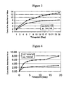
- the methods of the invention can be used to prepare medical implants wherein an amount of bioactive agent in the range of 1% to 17% of the total amount of bioactive agent present in the medical implant is released within a period of 8 days, medical implants wherein an amount of bioactive agent in the range of 1% to 41 % of the total amount ofbioactive agent present in the medical implant is released within a period of 14 days, and medical implants wherein an amount of bioactive agent in the range of 1% to 60% of the total amount of bioactive agent present in the medical implant is released within a period of 21 days.
- the natural biodegradable polysaccharide is modified with a hydrophobic moiety in order to provide a biodegradable matrix having hydrophobic properties. Therefore, a biodegradable article can be formed from natural biodegradable polysaccharide comprising one or more pendent coupling groups and one or more pendent hydrophobic moieties. Exemplary hydrophobic moieties include fatty acids and derivatives thereof, and C 2 -C 18 alkyl chains.
- modification of the natural biodegradable polysaccharide allows for preparation of articles that are biodegradable and that can release a hydrophobic bioactive agent.
- the hydrophobic moiety pendent from the natural biodegradable has properties of a bioactive agent. Upon degradation of the matrix, the hydrophobic moiety can be hydrolyzed from the natural biodegradable polymer and released to provide a therapeutic effect.
- a therapeutically useful hydrophobic moiety is butyric acid.
- the invention provides methods and articles for improving the stability of a bioactive agent that is delivered from an article by utilizing a natural biodegradable non-reducing polysaccharide.
- the non-reducing polysaccharide can provide an inert matrix and thereby improve the stability of sensitive bioactive agents, such as proteins and enzymes.
- the article can include a matrix having a plurality of natural biodegradable non-reducing polysaccharides along with a bioactive agent, such as a polypeptide.
- An exemplary non-reducing polysaccharide comprises polyalditol.
- Biodegradable non-reducing polysaccharides can very useful for formulating articles that release the bioactive agent over a prolonged period of time.
- the present invention demonstrates the preparation of articles that include natural biodegradable polysaccharides that are suitable for in vivo use. These products display excellent physical characteristics and are suitable for use in applications wherein a particular function, such as bioactive agent delivery or a sealant function is desired.
- articles can be prepared having viscoelastic properties.
- the article has an elastic modulus value in the range of 27 kPa to 30 kPa.
- the methods of preparing the compositions for fabrication of articles do not require the use of organic solvents.
- organic solvents can be physically hazardous. Use of organic solvents can potentially destroy the activity of a bioactive agent that can be optionally included in the natural biodegradable polysaccharide-based composition.
- the starting materials in particular the natural biodegradable polysaccharides having pendent coupling groups.
- the natural biodegradable polysaccharides have pendent polymerizable groups, such as ethylenically unsaturated groups.
- the degradable polymerizable polymers are formed by reacting a natural biodegradable polysaccharide with a compound comprising an ethylenically unsaturated group.
- a natural biodegradable polysaccharide is reacted with a compound including an ethylenically unsaturated group and an isocyanate group.
- a natural biodegradable polysaccharide is treated with an oxidizing agent to form a reactive aldehyde species on the polysaccharide and then reacted with a compound comprising an ethylenically unsaturated group and an amine group.
- Polysaccharide macromers were shown to have excellent matrix forming capabilities.
- the invention provides natural biodegradable polysaccharides having an amount of pendent coupling groups of about 0.7 ⁇ moles of coupling group per milligram of natural biodegradable polysaccharide.
- the amount of coupling group per natural biodegradable polysaccharide is in the range of about 0.3 ⁇ moles/mg to about 0.7 ⁇ moles/mg.
- amylose or maltodextrin can be subject to a synthesis reaction with a compound having an ethylenically unsaturated group to provide an amylose or maltodextrin macromer having a ethylenically unsaturated group load level in the range of about 0.3 ⁇ moles/mg to about 0.7 ⁇ moles/mg.
- an initiator is used to promote the formation of the natural biodegradable polysaccharide matrix for article formation.
- the initiator can be an independent compound or a pendent chemical group used to activate the coupling group pendent from the natural biodegradable polymer and promote coupling of a plurality of natural biodegradable polymers.
- the coupling group pendent from the natural biodegradable polysaccharide is a polymerizable group
- the initiator can be used in a free radical polymerization reaction to promote crosslinking of the natural biodegradable polysaccharides together in the composition.
- the invention provides a biodegradable article composition
- a biodegradable article composition comprising (i) a natural biodegradable polysaccharide, preferably selected from amylose and maltodextrin, comprising a coupling group, (ii) an initiator, and (iii) a bioactive agent, wherein the coupling group is able to be activated by the initiator and promote crosslinking of a plurality of natural biodegradable polysaccharides.
- the initiator is independent of the natural biodegradable polysaccharide and in other aspects the initiator is pendent from the natural biodegradable polysaccharide.
- the natural biodegradable polysaccharide comprises an ethylenically unsaturated group.
- the initiator includes an oxidizing agent/reducing agent pair, a "redox pair," to drive polymerization of the biodegradable polysaccharide.
- a redox pair to drive polymerization of the biodegradable polysaccharide.
- the oxidizing agent and reducing agent are combined in the presence of the biodegradable polysaccharide.
- One benefit of using a redox pair is that, when combined, the oxidizing agent and reducing agent can provide a particularly robust initiation system. This is advantageous as it can promote the formation of a matrix, for example, useful for coating or article preparation, from biodegradable polysaccharide compositions having a relatively low viscosity. This can be particularly useful in many applications, especially when the biodegradable polysaccharide composition is used for the formation of an in situ polymerized article. For example, a low viscosity composition can be passed through a small gauge delivery conduit with relative ease to provide the composition that can polymerize in situ.
- the viscosity of the composition is above about 0.005 Pa.s (5 centi Poise (cP)), or about 0.01 Pas.s (10 cP) or greater. In other aspects of the invention the viscosity of the composition is between about 0.005 Pa.s (5 cP) or 0.01 Pas.s (10 cP) and about 0.7 Pa.s (700 cP), and in some aspects between about 0.005 Pa.s (5 cP) or 0.01 Pas.s (10 cP) and about 0.25 Pa.s (250 cP).
- the viscosity of the composition is above about 0.005 Pa.s (5 cP) or 0.01 Pas.s (10 cP) and the biodegradable polysaccharides in the composition have an average molecular weight of 500,000 Da or less, 250,000 Da or less, 100,000 Da or less, or 50,000 Da or less.
- a method for preparing an article can include the steps of (a) providing a first composition that includes a natural biodegradable polysaccharide comprising a coupling group and a first member of a redox pair (for example, the oxidizing agent) and then (b) mixing the first composition with second composition that includes a second member of the redox pair (for example, the reducing agent).
- the second composition includes a natural biodegradable polysaccharide.
- the first composition can include (a) a natural biodegradable polysaccharide having a coupling group and an oxidizing agent and the second composition can include a (b) natural biodegradable polysaccharide having a coupling group and a reducing agent.
- the final composition when the first composition is combined with the second composition, the final composition can be about 5 cP or greater.
- the oxidizing agent can be selected from inorganic or organic oxidizing agents, including enzymes; the reducing agent can be selected from inorganic or organic reducing agents, including enzymes.
- Exemplary oxidizing agents include peroxides, including hydrogen peroxide, metal oxides, and oxidases, such as glucose oxidase.
- Exemplary reducing agents include salts and derivatives of electropositive elemental metals such as Li, Na, Mg, Fe, Zn, Al, and reductases.
- the reducing agent is present in the composition at a concentration of 2.5 mM or greater when mixed with the oxidizing agent.
- Other reagents, such as metal or ammonium salts of persulfate can be present in the composition to promote polymerization of the biodegradable polysaccharide.
- An article formed using redox polymerization can therefore comprise a plurality of natural biodegradable polysaccharides associated via polymerized groups, a reduced oxidizing agent, and an oxidized reducing agent.
- the invention provides methods of preparing biodegradable articles, such as medical implants or in vivo formed matrices.
- the biodegradable articles can also be used for the release of bioactive agents, and in this manner can function as bioactive agent-releasing implants or depots.
- the biodegradable articles of the invention biodegrade within a period that is acceptable for the desired application.
- the biodegradable article is a medical implant that provides mechanical properties at the implantation site and maintains these mechanical properties until they are no longer needed. After this period of time has elapsed, the medical implant is degraded to an extent that the properties are no longer provided by the medical implant, and the biodegradable components can be absorbed and/or excreted by the body. In some embodiments, the medical implant slowly degrades and transfers stress at the appropriate rate to surrounding tissues as these tissues heal and can accommodate the stress once borne by the medical device.
- the biodegradable coating or article includes a natural biodegradable polysaccharide having a coupling group, selected from amylose and maltodextrin.
- a "natural biodegradable polysaccharide” refers to a non-synthetic polysaccharide that is capable of being enzymatically degraded but that is generally non-enzymatically hydrolytically stable.
- Natural biodegradable polysaccharides include polysaccharide and/or polysaccharide derivatives that are obtained from natural sources, such as plants or animals.
- Natural biodegradable polysaccharides include any polysaccharide that has been processed or modified from a natural biodegradable polysaccharide (for example, maltodextrin is a natural biodegradable polysaccharide that is processed from starch).
- Exemplary natural biodegradable polysaccharides include hyaluronic acid, starch, dextran, heparin, chondroitin sulfate, dermatan sulfate, heparan sulfate, keratan sulfate, dextran sulfate, pentosan polysulfate, and chitosan.
- Preferred polysaccharides are low molecular weight polymers that have little or no branching, such as those that are derived from and/or found in starch preparations, for example, amylose and maltodextrin. Therefore, the natural biodegradable polysaccharide can be a substantially non-branched or non-branched poly(glucopyranose) polymer.
- natural biodegradable polysaccharides having an average molecular weight of 500,000 Da or less, 250,000 Da or less, 100,000 Da or less, or 50,000 Da or less. It is also preferred that the natural biodegradable polysaccharides have an average molecular weight of 500 Da or greater.
- a particularly preferred size range for the natural biodegradable polysaccharides is in the range of about 1000 Da to about 10,000 Da. Natural biodegradable polysaccharides of particular molecular weights can be obtained commercially or can be prepared.
- biodegradable polysaccharides of a particular size range may depend on factors such as the physical characteristics of the composition (e.g., viscosity), the desired rate of degradation, the presence of other optional moieties in the composition, for example, bioactive agents, etc.
- amylose or "amylose polymer” refers to a linear polymer having repeating glucopyranose units that are joined by ⁇ -1,4 linkages. Some amylose polymers can have a very small amount of branching via ⁇ -1,6 linkages (about less than 0.5% of the linkages) but still demonstrate the same physical properties as linear (unbranched) amylose polymers do. Generally amylose polymers derived from plant sources have molecular weights of about 1 X 10 6 Da or less. Amylopectin, comparatively, is a branched polymer having repeating glucopyranose units that are joined by ⁇ -1,4 linkages to form linear portions and the linear portions are linked together via ⁇ -1,6 linkages. The branch point linkages are generally greater than 1% of the total linkages and typically 4% - 5% of the total linkages. Generally amylopectin derived from plant sources have molecular weights of 1 X 10 7 Da or greater.
- Amylose can be obtained from, or is present in, a variety of sources. Typically, amylose is obtained from non-animal sources, such as plant sources. In some aspects, a purified preparation of amylose is used as starting material for the preparation of the amylose polymer having coupling groups. In other aspects, as starting material, amylose can be used in a mixture that includes other polysaccharides.
- starch preparations having a high amylose content can be used in the preparation of amylose having the coupling groups.
- amylose is typically present along with amylopectin, which is a branched polysaccharide.
- amylopectin is a branched polysaccharide.
- compositions that include amylose wherein the amylose is present in the composition in an amount greater than amylopectin, if present in the composition.
- starch preparations having high amylose content, purified amylose, synthetically prepared amylose, or enriched amylose preparations can be used in the preparation of amylose polymer having the coupling groups.
- the composition includes a mixture of polysaccharides including amylose wherein the amylose content in the mixture of polysaccharides is 50% or greater, 60% or greater, 70% or greater, 80% or greater, or 85% or greater by weight.
- the composition includes a mixture of polysaccharides including amylose and amylopectin and wherein the amylopectin content in the mixture of polysaccharides is 30% or less, or 15% or less.
- non-retrograding starches such as waxy starch
- the amount of amylopectin present in a starch may also be reduced by treating the starch with amylopectinase, which cleaves ⁇ -1,6 linkages resulting in the debranching of amylopectin into amylose.
- a synthesis reaction can be carried out to prepare an amylose polymer having pendent coupling groups (for example, amylose with pendent ethylenically unsaturated groups) and steps may be performed before, during, and/or after the synthesis to enrich the amount of amylose, or purify the amylose.
- pendent coupling groups for example, amylose with pendent ethylenically unsaturated groups
- Amylose of a particular size, or a combination of particular sizes can be used.
- the choice of amylose in a particular size range may depend on the application, for example, the type of surface coated or the porosity of the surface.
- amylose having an average molecular weight of 500,000 Da or less, 250,000 Da or less, 100,000 Da or less, 50,000 Da or less, preferably greater than 500 Da, or preferably in the range of about 1000 Da to about 10,000 Da is used.
- Amylose of particular molecular weights can be obtained commercially or can be prepared. For example, synthetic amyloses with average molecular masses of 70, 110, 320, and 1,000 kDa can be obtained from Nakano Vinegar Co., Ltd. (Aichi, Japan).
- amylose of a particular size range may depend on factors such as the physical characteristics of the composition (e.g., viscosity), the desired rate of degradation, the presence of other optional moieties in the composition (for example, bioactive agents, etc.), etc.
- Maltodextrin is typically generated by hydrolyzing a starch slurry with heat-stable ⁇ -amylase at temperatures at 85 - 90 °C until the desired degree of hydrolysis is reached and then inactivating the ⁇ -amylase by a second heat treatment.
- the maltodextrin can be purified by filtration and then spray dried to a final product.
- a starch preparation that has been totally hydrolyzed to dextrose (glucose) has a DE of 100, where as starch has a DE of about zero.
- a DE of greater than 0 but less than 100 characterizes the mean-average molecular weight of a starch hydrolysate, and maltodextrins are considered to have a DE of less than 20.
- Maltodextrins of various molecular weights, for example, in the range of about 500 - 5000 Da are commercially available (for example, from CarboMer, San Diego, CA).
- a non-reducing polysaccharide can provide an inert matrix thereby improving the stability of sensitive bioactive agents, such as proteins and enzymes.
- a non-reducing polysaccharide refers to a polymer of non-reducing disaccharides (two monosaccharides linked through their anomeric centers) such as trehalose ( ⁇ -D-glucopyranosyl ⁇ -D-glucopyranoside) and sucrose ( ⁇ -D-fructofuranosyl ⁇ -D-glucopyranoside).
- An exemplary non-reducing polysaccharide comprises polyalditol which is available from GPC (Muscatine, Iowa).
- the polysaccharide is a glucopyranosyl polymer, such as a polymer that includes repeating (1 ⁇ 3)O- ⁇ -D-glucopyranosyl units.
- the compositions can include natural biodegradable polysaccharides that include chemical modifications other than the pendent coupling group.
- modified amylose having esterified hydroxyl groups can be prepared and used in compositions in association with the methods of the invention.
- Other natural biodegradable polysaccharides having hydroxyl groups may be modified in the same manner. These types of modifications can change or improve the properties of the natural biodegradable polysaccharide making for a composition that is particularly suitable for a desired application.
- Many chemically modified amylose polymers, such as chemically modified starch have at least been considered acceptable food additives.
- modified natural biodegradable polysaccharides refers to chemical modifications to the natural biodegradable polysaccharide that are different than those provided by the coupling group or the initiator group.
- Modified amylose polymers having a coupling group (and/or initiator group) can be used in the compositions and methods of the invention.
- modified amylose is described.
- chemically modifying the hydroxyl groups of the amylose the physical properties of the amylose can be altered.
- the hydroxyl groups of amylose allow for extensive hydrogen bonding between amylose polymers in solution and can result in viscous solutions that are observed upon heating and then cooling amylose-containing compositions such as starch in solution (retrograding).
- the hydroxyl groups of amylose can be modified to reduce or eliminate hydrogen bonding between molecules thereby changing the physical properties of amylose in solution.
- the natural biodegradable polysaccharides can include one or more modifications to the hydroxyl groups wherein the modifications are different than those provided by coupling group.
- Modifications include esterification with acetic anhydride (and adipic acid), succinic anhydride, 1-octenylsuccinic anhydride, phosphoryl chloride, sodium trimetaphosphate, sodium tripolyphosphate, and sodium monophosphate; etherification with propylene oxide, acid modification with hydrochloric acid and sulfuric acids; and bleaching or oxidation with hydrogen peroxide, peracetic acid, potassium permanganate, and sodium hypochlorite.
- modified amylose polymers include carboxymethyl amylose, carboxyethyl amylose, ethyl amylose, methyl amylose, hydroxyethyl amylose, hydroxypropyl amylose, acetyl amylose, amino alkyl amylose, allyl amylose, and oxidized amylose.
- modified amylose polymers include succinate amylose and oxtenyl succinate amylose.
- the natural biodegradable polysaccharide is modified with a hydrophobic moiety in order to provide a biodegradable matrix having hydrophobic properties.
- exemplary hydrophobic moieties include those previously listed, fatty acids and derivatives thereof, and C 2 -C 18 alkyl chains.
- a polysaccharide, such as amylose or maltodextrin, can be modified with a compound having a hydrophobic moiety, such as a fatty acid anhydride.
- the hydroxyl group of a polysaccharide can also cause the ring opening of lactones to provide pendent open-chain hydroxy esters.
- the hydrophilic moiety pendent from the natural biodegradable has properties of a bioactive agent.
- the hydrophilic moiety can be hydrolyzed from the natural biodegradable polymer and released from the matrix to provide a therapeutic effect.
- a therapeutically useful hydrophilic moiety is butyric acid, which has been shown to elicit tumor cell differentiation and apoptosis, and is thought to be useful for the treatment of cancer and other blood diseases.
- the hydrophilic moiety that provides a therapeutic effect can also be a natural compound (such as butyric acid). Therefore, degradation of the matrix having a coupled therapeutic agent can result in all natural degradation products.
- a natural biodegradable polysaccharide that includes a coupling group is used to form an article .
- Other polysaccharides can also be present in the composition.
- the two or more natural biodegradable polysaccharides are used to form an article.
- examples include amylose and one or more other natural biodegradable polysaccharide(s), and maltodextrin and one or more other natural biodegradable polysaccharide(s); in one aspect the composition includes a mixture of amylose and maltodextrin, optionally with another natural biodegradable polysaccharide.
- amylose or maltodextrin is the primary polysaccharide.
- the composition includes a mixture of polysaccharides including amylose or maltodextrin and the amylose or maltodextrin content in the mixture of polysaccharides is 50% or greater, 60% or greater, 70% or greater, 80% or greater, or 85% or greater by weight.
- Purified or enriched amylose preparations can be obtained commercially or can be prepared using standard biochemical techniques such as chromatography. In some aspects, high-amylose cornstarch can be used.
- coupling group can include (1) a chemical group that is able to form a reactive species that can react with the same or similar chemical group to form a bond that is able to couple the natural biodegradable polysaccharides together (for example, wherein the formation of a reactive species can be promoted by an initiator); or (2) a pair of two different chemical groups that are able to specifically react to form a bond that is able to couple the natural biodegradable polysaccharides together.
- the coupling group can be attached to any suitable natural biodegradable polysaccharide, including the amylose and maltodextrin polymers as exemplified herein.
- Contemplated reactive pairs include Reactive Group A and corresponding Reactive Group B as shown in the Table 1 below.
- a reactive group from group A can be selected and coupled to a first set of natural biodegradable polysaccharides and a corresponding reactive group B can be selected and coupled to a second set of natural biodegradable polysaccharides.
- Reactive groups A and B can represent first and second coupling groups, respectively. At least one and preferably two, or more than two reactive groups are coupled to an individual natural biodegradable polysaccharide polymer.
- the first and second sets of natural biodegradable polysaccharides can be combined and reacted, for example, thermochemically, if necessary, to promote the coupling of natural biodegradable polysaccharides and the formation of a natural biodegradable polysaccharide matrix.
- Amine also includes hydrazide (R-NH-NH 2 )
- a suitable coupling pair would be a natural biodegradable polysaccharide having an electrophilic group and a natural biodegradable polysaccharide having a nucleophilic group.
- An example of a suitable electrophilic-nucleophilic pair is N-hydroxysuccinimide-amine pair, respectively.
- Another suitable pair would be an oxirane-amine pair.
- the natural biodegradable polysaccharides of the invention include at least one, and more typically more than one, coupling group per natural biodegradable polysaccharide, allowing for a plurality of natural biodegradable polysaccharides to be coupled in linear and/or branched manner.
- the natural biodegradable polysaccharide includes two or more pendent coupling groups.
- the coupling group on the natural biodegradable polysaccharide is a polymerizable group.
- the polymerizable group can couple natural biodegradable polysaccharides together in the composition, thereby forming a biodegradable natural biodegradable polysaccharide matrix.
- a preferred polymerizable group is an ethylenically unsaturated group.
- Suitable ethylenically unsaturated groups include vinyl groups, acrylate groups, methacrylate groups, ethacrylate groups, 2-phenyl acrylate groups, acrylamide groups, methacrylamide groups, itaconate groups, and styrene groups. Combinations of different ethylenically unsaturated groups can be present on a natural biodegradable polysaccharide, such as amylose or maltodextrin.
- any suitable synthesis procedure can be used. Suitable synthetic schemes typically involve reaction of, for example, hydroxyl groups on the natural biodegradable polysaccharide, such as amylose or maltodextrin. Synthetic procedures can be modified to produce a desired number of coupling groups pendent from the natural biodegradable polysaccharide backbone. For example, the hydroxyl groups can be reacted with a coupling group-containing compound or can be modified to be reactive with a coupling group-containing compound. The number and/or density of acrylate groups can be controlled using the present method, for example, by controlling the relative concentration of reactive moiety to saccharide group content.
- the biodegradable polysaccharides have an amount of pendent coupling groups of about 0.7 ⁇ moles of coupling group per milligram of natural biodegradable polysaccharide.
- the amount of coupling group per natural biodegradable polysaccharide is in the range of about 0.3 ⁇ moles/mg to about 0.7 ⁇ moles/mg.
- amylose or maltodextrin can be reacted with an acrylate groups-containing compound to provide an amylose or maltodextrin macromer having a acrylate group load level in the range of about 0.3 ⁇ moles/mg to about 0.7 ⁇ moles/mg.
- an “initiator” refers to a compound, or more than one compound, that is capable of promoting the formation of a reactive species from the coupling group.
- the initiator can promote a free radical reaction of natural biodegradable polysaccharide having a coupling group.
- the initiator can be an "initiator polymer” that includes a polymer having a backbone and one or more initiator groups pendent from the backbone of the polymer.
- Redox initiators can also be used to promote the polymerization of the natural biodegradable polymers having pendent coupling groups.
- combinations of organic and inorganic oxidizers, and organic and inorganic reducing agents are used to generate radicals for polymerization.
- a description of redox initiation can be found in Principles of Polymerization, 2nd Edition, Odian G., John Wiley and Sons, pgs 201-204, (1981 ).
- the polymerization initiator is a polymer that includes an initiator group (herein referred to as an "initiator polymer").
- the polymeric portion of the initiator polymer can be obtained or prepared to have particular properties or features that are desirable for use with a composition .
- the polymeric portion of the initiator polymer can have hydrophilic or amphoteric properties, it can include pendent charged groups, or it can have groups that allow it to interact with a particular surface (this can depend on the type of surface to be coated).
- an initiator can be pendent from a natural biodegradable polysaccharide. Therefore, the natural biodegradable polysaccharide is able to promote activation of polymerizable groups that are pendent from other natural biodegradable polysaccharides and promote the formation of a natural biodegradable polysaccharide matrix.
- the polymeric portion of the initiator polymer can include, for example, acrylamide and methacrylamide monomeric units, or derivatives thereof.
- the composition includes an initiator polymer having a photoreactive group and a polymeric portion selected from the group of acrylamide and methacrylamide polymers and copolymers.
- the initiator includes an oxidizing agent/reducing agent pair, a "redox pair,” to drive polymerization of the biodegradable polysaccharide.
- a redox pair to drive polymerization of the biodegradable polysaccharide.
- polymerization of the biodegradable polysaccharide is carried out upon combining one or more oxidizing agents with one or more reducing agents.
- Other compounds can be included in the composition to promote polymerization of the biodegradable polysaccharides.
- the oxidizing agent and reducing agent can provide a particularly robust initiation system and can drive the formation of a polymerized matrix of polysaccharides from a composition having a low viscosity.
- a polysaccharide composition with a low viscosity may be due to a low concentration of polysaccharide in the composition, a polysaccharide having a low average molecular weight, or combinations thereof.
- Matrix formation from a polysaccharide composition having a low viscosity is particularly advantageous in many applications, especially for in situ polymerization.
- a low viscosity polysaccharide composition is passed through a small gauge delivery conduit, such as a needle, wherein the redox pair causes the polymerization of the polysaccharides in situ.
- the viscosity of the composition is above about 0.005 Pa.s (5 cP), or about 0.01 Pa.s (10 cP) or greater. In other aspects of the invention the viscosity of the composition is between about 0.005 Pa.s (5 cP) or 0.01 Pa.s (10 cP) and about 0.7 Pa.s (700 cP), or between about 0.005 Pa.s (5 cP) or 0.01 Pa.s (10 cP) and about 0.25 Pa.s (250 cP).
- the oxidizing agent is added to the reducing agent in the presence of the one or more biodegradable polysaccharides.
- a composition including a biodegradable polysaccharide and a reducing agent is added to a composition including an oxidizing agent, or a composition including a biodegradable polysaccharide and an oxidizing agent is added to a composition containing a reducing agent.
- One desirable method of preparing a matrix is to combine a composition including a biodegradable polysaccharide and an oxidizing agent with a composition including a biodegradable polysaccharide and a reducing agent.
- the terms "first composition" and "second composition" can be used.
- the viscosities of biodegradable polysaccharide in the first and second compositions can be the same or can be different. Generally, though, it has been observed that good mixing and subsequent matrix formation is obtained when the compositions have the same or similar viscosities. In this regard, if the same biodegradable polymer is used in the first and second composition, the concentration of the biodegradable polymer may be the same or different.
- the oxidizing agent can be selected from inorganic or organic oxidizing agents, including enzymes; the reducing agent can be selected from inorganic or organic reducing agents, including enzymes.
- Exemplary oxidizing agents include peroxides, including hydrogen peroxide, metal oxides, and oxidases, including glucose oxidase.
- Exemplary reducing agents include salts and derivatives of electropositive elemental metals such as Li, Na, Mg, Fe, Zn, Al, and reductases.
- the reducing agent is present at a concentration of about 2.5 mM or greater when the reducing agent is mixed with the oxidizing agent. Prior to mixing, the reducing agent can be present in a composition at a concentration of, for example, 5 mM or greater.
- compositions can be present in the composition to promote polymerization of the biodegradable polysaccharide.
- Other polymerization promoting compounds can be included in the composition, such as metal or ammonium salts of persulfate.
- compositions and methods of the invention can include polymerization accelerants that can improve the efficiency of polymerization.
- useful accelerants include N-vinyl compounds, particularly N-vinyl pyrrolidone and N-vinyl caprolactam.
- Such accelerants can be used, for instance, at a concentration of between about 0.01% and about 5%, and preferably between about 0.05% and about 0.5%, by weight, based on the volume of the coating composition.
- an aqueous composition that includes the natural biodegradable polysaccharide amylose or maltodextrin having pendent coupling groups, and a bioactive agent is obtained and used to form an article.
- This composition can be prepared by mixing a bioactive agent, such as a water-soluble small molecule, a protein, or a nucleic acid, with the natural biodegradable polysaccharide.
- the natural biodegradable polysaccharide that includes a coupling group is used to form an article.
- Other polysaccharides can also be present in the composition.
- the natural biodegradable polysaccharide such as amylose or maltodextrin
- another biodegradable polymer i.e., a secondary polymer
- An additional polymer or polymers can be used to alter the properties of the matrix, or serve as bulk polymers to alter the volume of the matrix.
- biodegradable polysaccharides can be used in combination with the amylose polymer.
- biodegradable polysaccharides include hyaluronic acid, dextran, starch, amylose (for example, non-derivitized), amylopectin, cellulose, xanthan, pullulan, chitosan, pectin, inulin, alginates, and heparin.
- the concentration of the natural biodegradable polysaccharide in the composition can be chosen to provide an article having a desired density of crosslinked natural biodegradable polysaccharide.
- the concentration of natural biodegradable polysaccharide in the composition can depend on the type or nature of the bioactive agent that is included in the composition.
- the natural biodegradable polysaccharide having the coupling groups is present in the composition at a concentration in the range of 5 - 100% (w/v), and 5-50%, and in more specific embodiments in the range of 10-20% and in other embodiments in the range of 20 - 50% (w/v).
- the concentration of the natural biodegradable polysaccharide may be higher to provide a more structurally rigid implant.
- compositions can change or improve the properties of the article that is formed by the natural biodegradable polysaccharide having coupling groups in order to change the elasticity, flexibility or wettability.
- the polymeric compositions can be utilized in connection with an ophthalmic article.
- the ophthalmic article can be configured for placement at an external or internal site of the eye.
- Suitable ophthalmic articles in accordance with these aspects can provide bioactive agent to any desired area of the eye.
- the articles can be utilized to deliver bioactive agent to an anterior segment of the eye (in front of the lens), and/or a posterior segment of the eye (behind the lens).
- Suitable ophthalmic devices can also be utilized to provide bioactive agent to tissues in proximity to the eye, when desired.
- the biodegradable polysaccharide compositions can be used in the construction of an ophthalmic article.
- Suitable external articles can be configured for topical administration of bioactive agent.
- Such external devices can reside on an external surface of the eye, such as the cornea (for example, contact lenses) or bulbar conjunctiva.
- suitable external devices can reside in proximity to an external surface of the eye.
- Articles configured for placement at an internal site of the eye can reside within any desired area of the eye.
- the ophthalmic article can be configured for placement at an intraocular site, such as the vitreous.
- Illustrative intraocular devices include, but are not limited to, those described in U.S. Patent Nos. 6,719,750 B2 ("Devices for Intraocular Drug Delivery,” Varner et al.) and 5,466,233 ("Tack for Intraocular Drug Delivery and Method for Inserting and Removing Same," Weiner et al.); U.S. Publication Nos.
- the ophthalmic article can be configured for placement, or can be formed, at a subretinal area within the eye.
- Illustrative ophthalmic devices for subretinal application include, but are not limited to, those described in U.S. Patent Publication No. 2005/0143363 ("Method for Subretinal Administration of Therapeutics Including Steroids; Method for Localizing Pharmacodynamic Action at the Choroid and the Retina; and Related Methods for Treatment and/or Prevention of Retinal Diseases," de Juan et al.); U.S. Application No. 11/175,850 (“Methods and Devices for the Treatment of Ocular Conditions," de Juan et al.); and related applications.
- the invention provides a biodegradable implant that is formed from the biodegradable polysaccharide and that includes a bioactive agent, such as a high molecular weight bioactive agent useful for treating an ocular condition.
- a bioactive agent such as a high molecular weight bioactive agent useful for treating an ocular condition.
- the invention provides a method for forming an article from the biodegradable polysaccharide, wherein the method includes polymerizing a composition that includes the biodegradable polysaccharide within the eye, such as in a subretinal area or within the vitreous.
- a low viscosity composition including a natural biodegradable polysaccharide and a redox pair to promote polymerization for in situ matrix formation.
- Ophthalmic articles can also be configured for placement within any desired tissues of the eye.
- ophthalmic devices can be configured for placement at a subconjunctival area of the eye, such as devices positioned extrasclerally but under the conjunctiva, such as glaucoma drainage devices and the like.
- the natural biodegradable polysaccharide article includes one or more bioactive agents.
- the bioactive agent can be dispersed within the natural biodegradable polysaccharide article
- bioactive agent refers to a peptide, protein, carbohydrate, nucleic acid, lipid, polysaccharide, synthetic inorganic or organic molecule, viral particle, cell, or combinations thereof, that causes a biological effect when administered in vivo to an animal, including but not limited to birds and mammals, including humans.
- suitable gene therapy agents include (a) therapeutic nucleic acids, including antisense DNA, antisense RNA, and interference RNA, and (b) nucleic acids encoding therapeutic gene products, including plasmid DNA and viral fragments, along with associated promoters and excipients.
- Other molecules that can be incorporated include nucleosides, nucleotides, vitamins, minerals, and steroids.
- the coatings of the invention are particularly useful for delivering bioactive agents that are large hydrophilic molecules, such as polypeptides (including proteins and peptides), nucleic acids (including DNA and RNA), polysaccharides (including heparin), as well as particles, such as viral particles, and cells.
- bioactive agent has a molecular weight of about 10,000 or greater.
- Classes of bioactive agents which can be incorporated into the natural biodegradable matrix of this invention include, but are not limited to: ACE inhibitors, actin inhibitors, analgesics, anesthetics, anti-hypertensives, anti polymerases, antisecretory agents, anti-AIDS substances, antibiotics, anti-cancer substances, anticholinergics, anti-coagulants, anti-convulsants, anti-depressants, anti-emetics, antifungals, anti-glaucoma solutes, antihistamines, antihypertensive agents, anti-inflammatory agents (such as NSAIDs), anti metabolites, antimitotics, antioxidizing agents, anti-parasite and/or anti-Parkinson substances, antiproliferatives (including antiangiogenesis agents), anti-protozoal solutes, anti-psychotic substances, anti-pyretics, antiseptics, anti-spasmodics, antiviral agents, calcium channel blockers, cell response modifiers, chelators, chem
- Antibiotics are art recognized and are substances which inhibit the growth of or kill microorganisms.
- antibiotics include penicillin, tetracycline, chloramphenicol, minocycline, doxycycline, vancomycin, bacitracin, kanamycin, neomycin, gentamycin, erythromycin, cephalosporins, geldanamycin, and analogs thereof.
- cephalosporins examples include cephalothin, cephapirin, cefazolin, cephalexin, cephradine, cefadroxil, cefamandole, cefoxitin, cefaclor, cefuroxime, cefonicid, ceforanide, cefotaxime, moxalactam, ceftizoxime, ceftriaxone, and cefoperazone.
- Antiseptics are recognized as substances that prevent or arrest the growth or action of microorganisms, generally in a nonspecific fashion, e.g., by inhibiting their activity or destroying them.
- antiseptics include silver sulfadiazine, chlorhexidine, glutaraldehyde, peracetic acid, sodium hypochlorite, phenols, phenolic compounds, iodophor compounds, quaternary ammonium compounds, and chlorine compounds.
- Anti-viral agents are substances capable of destroying or suppressing the replication of viruses.
- examples of anti-viral agents include ⁇ -methyl-P-adamantane methylamine, hydroxy-ethoxymethylguanine, adamantanamine, 5-iodo2'-deoxyuridine, trifluorothymidine, interferon, and adenine arabinoside.
- Enzyme inhibitors are substances that inhibit an enzymatic reaction.
- enzyme inhibitors include edrophonium chloride, N-methylphysostigmine, neostigmine bromide, physostigmine sulfate, tacrine HCl, tacrine, 1-hydroxymaleate, iodotubercidin, p-bromotetramisole, 10-( ⁇ -diethylaminopropionyl)-phenothiazine hydrochloride, calmidazolium chloride, hemicholinium-3, 3,5-dinitrocatechol, diacylglycerol kinase inhibitor I, diacylglycerol kinase inhibitor II, 3-phenylpropargylamine, N-monomethyl-L-arginine acetate, carbidopa, 3-hydroxybenzylhydrazine HCl, hydralazine HCl, clorgyline HCl, deprenyl HCl,
- Anti-pyretics are substances capable of relieving or reducing fever.
- Anti-inflammatory agents are substances capable of counteracting or suppressing inflammation. Examples of such agents include aspirin (salicylic acid), indomethacin, sodium indomethacin trihydrate, salicylamide, naproxen, colchicine, fenoprofen, sulindac, diflunisal, diclofenac, indoprofen and sodium salicylamide.
- Local anesthetics are substances that have an anesthetic effect in a localized region. Examples of such anesthetics include procaine, lidocaine, tetracaine and dibucaine.
- Cell response modifiers are chemotactic factors such as platelet-derived growth factor (pDGF).
- Other chemotactic factors include neutrophil-activating protein, monocyte chemoattractant protein, macrophage-inflammatory protein, SIS (small inducible secreted) proteins, platelet factor, platelet basic protein, melanoma growth stimulating activity, epidermal growth factor, transforming growth factor (alpha), fibroblast growth factor, platelet-derived endothelial cell growth factor, insulin-like growth factor, nerve growth factor, and bone growth/cartilage-inducing factor (alpha and beta).
- pDGF platelet-derived growth factor
- Other chemotactic factors include neutrophil-activating protein, monocyte chemoattractant protein, macrophage-inflammatory protein, SIS (small inducible secreted) proteins, platelet factor, platelet basic protein, melanoma growth stimulating activity, epidermal growth factor, transforming growth factor (alpha), fibroblast growth factor, platelet-derived endo
- cell response modifiers are the interleukins, interleukin inhibitors or interleukin receptors, including interleukin 1 through interleukin 10; interferons, including alpha, beta and gamma; hematopoietic factors, including erythropoietin, granulocyte colony stimulating factor, macrophage colony stimulating factor and granulocyte-macrophage colony stimulating factor; tumor necrosis factors, including alpha and beta; transforming growth factors (beta), including beta-1, beta-2, beta-3, inhibin, activin, and DNA that encodes for the production of any of these proteins.
- interleukins interleukin inhibitors or interleukin receptors, including interleukin 1 through interleukin 10
- interferons including alpha, beta and gamma
- hematopoietic factors including erythropoietin, granulocyte colony stimulating factor, macrophage colony stimulating factor and granulocyte-macrophage
- statins examples include lovastatin, pravastatin, simvastatin, fluvastatin, atorvastatin, cerivastatin, rousvastatin, and superstatin.
- Imaging agents are agents capable of imaging a desired site, e.g., tumor, in vivo
- imaging agents include substances having a label which is detectable in vivo, e.g., antibodies attached to fluorescent labels.
- the term antibody includes whole antibodies or fragments thereof.
- Exemplary ligands or receptors include antibodies, antigens, avidin, streptavidin, biotin, heparin, type IV collagen, protein A, and protein G.
- antibiotics include antibiotic peptides.
- the bioactive agent can be selected to improve the compatibility (for example, with blood and/or surrounding tissues) of medical device surfaces.
- biocompatible agents when associated with the medical device surface, can serve to shield the blood from the underlying medical device material.
- Suitable biocompatible agents preferably reduce the likelihood for blood components to adhere to the medical device, thus reducing the formation of thrombus or emboli (blood clots that release and travel downstream).
- the bioactive agent can improve the biocompatibility of the medical article.
- the bioactive agent can provide antirestenotic effects, such as antiproliferative, anti-platelet, and/or antithrombotic effects.
- the bioactive agent can include anti-inflammatory agents, immunosuppressive agents, cell attachment factors, receptors, ligands, growth factors, antibiotics, enzymes, nucleic acids, and the like.
- Compounds having antiproliferative effects include, for example, actinomycin D, angiopeptin, c-myc antisense, paclitaxel, taxane, and the like.
- bioactive agents having antithrombotic effects include heparin, heparin derivatives, sodium heparin, low molecular weight heparin, hirudin, lysine, prostaglandins, argatroban, forskolin, vapiprost, prostacyclin and prostacyclin analogs, D-ph-pr-arg-chloromethylketone (synthetic antithrombin), dipyridamole, glycoprotein IIb/IIIa platelet membrane receptor antibody, coprotein IIb/IIIa platelet membrane receptor antibody, recombinant hirudin, thrombin inhibitor (such as commercially available from Biogen), chondroitin sulfate, modified dextran, albumin, streptokinase, tissue plasminogen activator (TPA), urokinase, nitric oxide inhibitors, and the like.
- heparin heparin derivatives, sodium heparin, low molecular weight heparin,
- the bioactive agent can also be an inhibitor of the GPIIb-IIIa platelet receptor complex, which mediates platelet aggregation.
- GPIIb/IIIa inhibitors can include monoclonal antibody Fab fragment c7E3, also know as abciximab (ReoProTM), and synthetic peptides or peptidomimetics such as eptifibatide (IntegrilinTM) or tirofiban (AgrastatTM).
- the bioactive agent can be an immunosuppressive agent, for example, cyclosporine, CD-34 antibody, everolimus, mycophenolic acid, sirolimus, tacrolimus, and the like.
- immunosuppressive agent for example, cyclosporine, CD-34 antibody, everolimus, mycophenolic acid, sirolimus, tacrolimus, and the like.
- exemplary therapeutic antibodies include trastuzumab (HerceptinTM), a humanized anti-HER2 monoclonal antibody (moAb); alemtuzumab (CampathTM), a humanized anti-CD52 moAb; gemtuzumab (MylotargTM), a humanized anti-CD33 moAb; rituximab (RituxanTM), a chimeric anti-CD20 moAb; ibritumomab (ZevalinTM), a murine moAb conjugated to a beta-emitting radioisotope; tositumomab (BexxarTM), a murine anti-CD20 moAb; edrecolomab (PanorexTM), a murine anti-epithelial cell adhesion molecule moAb; cetuximab (ErbituxTM), a chimeric anti-EGFR moAb; and bevacizumab (AvastinTM), a humanized anti-VEGF moAb.
- the bioactive agent can be a surface adhesion molecule or cell-cell adhesion molecule.
- Exemplary cell adhesion molecules or attachment proteins such as extracellular matrix proteins including fibronectin, laminin, collagen, elastin, vitronectin, tenascin, fibrinogen, thrombospondin, osteopontin, von Willibrand Factor, bone sialoprotein (and active domains thereof), or a hydrophilic polymer such as hyaluronic acid, chitosan or methyl cellulose, and other proteins, carbohydrates, and fatty acids.
- Exemplary cell-cell adhesion molecules include N-cadherin and P-cadherin and active domains thereof.
- Exemplary growth factors include fibroblastic growth factors, epidermal growth factor, platelet-derived growth factors, transforming growth factors, vascular endothelial growth factor, bone morphogenic proteins and other bone growth factors, and neural growth factors.
- the bioactive agent can be also be selected from mono-2-(carboxymethyl) hexadecanamidopoly (ethylene glycol) 200 mono-4-benzoylbenzyl ether, mono-3-carboxyheptadecanamidopoly (ethylene glycol) 200 mono-4-benzoylbenzyl ether, mono-2-(carboxymethyl) hexadecanamidotetra (ethylene glycol) mono-4-benzoylbenzyl ether, mono-3-carboxyheptadecanamidotetra (ethylene glycol) mono-4-benzoylbenzyl ether, N-[2-(4-benzoylbenzyloxy) ethyl]-2-(carboxymethyl) hexadecanamide, N-[2-(4-benzoylbenzyloxy)ethyl]-3-carboxyheptadecanamide, N-[12-(benzoylbenzyloxy) dodecyl]-2-(car
- bioactive agents and/or bioactive agent include analogues of rapamycin ("rapalogs"), ABT-578 from Abbott, dexamethasone, betamethasone, vinblastine, vincristine, vinorelbine, poside, teniposide, daunorubicin, doxorubicin, idarubicin, anthracyclines, mitoxantrone, bleomycins, plicamycin (mithramycin), mitomycin, mechlorethamine, cyclophosphamide and its analogs, melphalan, chlorambucil, ethylenimines and methylmelamines, alkyl sulfonates-busulfan, nitrosoureas, carmustine (BCNU) and analogs, streptozocin, trazenes-dacarbazinine, methotrexate, fluorouracil, floxuridine, cytarabine, mercap
- Viral particles and viruses include those that may be therapeutically useful, such as those used for gene therapy, and also attenuated viral particles and viruses which can promote an immune response and generation of immunity.
- Useful viral particles include both natural and synthetic types.
- Viral particles include, but are not limited to, adenoviruses, baculoviruses, parvoviruses, herpesviruses, poxviruses, adeno-associated viruses, vaccinia viruses, and retroviruses.
- bioactive agents that can be used for altering gene function include plasmids, phages, cosmids, episomes, and integratable DNA fragments, antisense oligonucleotides, antisense DNA and RNA, modified DNA and RNA, iRNA, ribozymes, siRNA, and shRNA.
- bioactive agents include cells such as platelets, stem cells, T lymphocytes, B lymphocytes, acidophils, adipocytes, astrocytes, basophils, hepatocytes, neurons, cardiac muscle cells, chondrocytes, epithelial cells, dendrites, endrocrine cells, endothelial cells, eosinophils, erythrocytes, fibroblasts, follicular cells, ganglion cells, hepatocytes, endothelial cells, Leydig cells, parenchymal cells, lymphocytes, lysozyme-secreting cells, macrophages, mast cells, megakaryocytes, melanocytes, monocytes, myoid cells, neck nerve cells, neutrophils, oligodendrocytes, oocytes, osteoblasts, osteochondroclasts, osteoclasts, osteocytes, plasma cells, spermatocytes, reticulocytes, Schwann cells, Sertoli cells, skeletal
- Additives such as inorganic salts, BSA (bovine serum albumin), and inert organic compounds can be used to alter the profile of bioactive agent release, as known to those skilled in the art.
- BSA bovine serum albumin
- inert organic compounds can be used to alter the profile of bioactive agent release, as known to those skilled in the art.
- the concentration of the bioactive agent or agents dissolved or suspended in the mixture can range from about 0.01 to about 90 percent, by weight, based on the weight of the final composition.
- bioactive agent or combination of bioactive agents, can be selected depending upon one or more of the following factors: the application of the controlled delivery device, the medical condition to be treated, the anticipated duration of treatment, characteristics of the implantation site, the number and type of bioactive agents to be utilized, and the like.
- Bioactive agents are commercially available from Sigma Aldrich Fine Chemicals, Milwaukee, WI.
- the natural biodegradable polysaccharide is selected from the group of amylose and maltodextrin. In other preferred aspects of the following methods, the natural biodegradable polysaccharide has a molecular weight of 500,000 Da or less, 250,000 Da or less, 100,000 Da or less, or 50,000 Da or less. It is also preferred that the natural biodegradable polysaccharides have an average molecular weight of 500 Da or greater. A particularly preferred size range for the natural biodegradable polysaccharides is in the range of about 1000 Da to about 10,000 Da.
- the method includes the steps of (i) disposing a composition comprising (a) a natural biodegradable polysaccharide having a coupling group, (b) an initiator, and (c) a bioactive agent on a surface; and (ii) activating the initiator to provide a medical implant wherein the composition is disposed to form an implant of a desired configuration.
- the composition can be disposed in a mold. This method can also be used to form an in situ formed matrix wherein the composition is disposed within a portion of a subject.
- the method includes the steps of (i) disposing an initiator on a surface, (ii) disposing a composition comprising (a) a natural biodegradable polysaccharide having a coupling group and (b) a bioactive agent on the surface; and (iii) activating the initiator to provide a coated composition having the natural biodegradable polysaccharide and the bioactive agent.
- a composition including the natural biodegradable polysaccharide and the bioactive agent are contacted with the initiator and the initiator is activated to promote the crosslinking of two or more natural biodegradable polysaccharides via their coupling groups.
- the natural biodegradable polysaccharide includes a polymerizable group, such as an ethylenically unsaturated group, and initiator is capable of initiating free radical polymerization of the polymerizable groups.
- the invention provides a method including the steps of (i) disposing a composition comprising (a) a natural biodegradable polysaccharide having a ethylenically unsaturated group, (b) a polymerization initiator, and (c) a bioactive agent on a surface; and (ii) activating the polymerization initiator to cause the polymerization of the amylose compound thereby providing a composition having the natural biodegradable polysaccharide and the bioactive agent on the surface.
- These methods can be used to form medical implants and in situ-formed matrices, wherein the composition is disposed in a mold or in a subject, respectively, rather than on a surface.
- the invention provides a medical device having a composition comprising a plurality of coupled natural biodegradable polysaccharide and a bioactive agent.
- the invention also provides the advantage of being able to perform synthetic and post-synthetic procedures wherein the natural biodegradable polysaccharide is contacted with an aqueous composition, and there is minimal risk of degradation of the polysaccharide.
- the natural biodegradable polysaccharide may be contacted with an aqueous solution for purification without risking significant degradation of the natural biodegradable polysaccharide.
- Amylose having polymerizable vinyl groups was prepared by mixing 0.75g of amylose (A0512; Aldrich) with 100 mL of methylsulfoxide (JT Baker) in a 250 mL amber vial, with stirring. After one hour, 2 mL of triethylamine (TEA; Aldrich) was added and the mixture was allowed to stir for 5 minutes at room temperature. Subsequently, 2 mL of glycidyl acrylate (Polysciences) was added and the amylose and glycidyl acrylate were allowed to react by stirring overnight at room temperature. The mixture containing the amylose-glycidyl acrylate reaction product was dialyzed for 3 days against DI water using continuous flow dialysis. The resultant acrylated-amylose (0.50g; 71.4% yield) was then lyophilized and stored desiccated at room temperature with protection from light.
- TEA triethylamine
- a polymerization initiator was prepared by copolymerizing a methacrylamide having a photoreactive group with acrylamide.
- a methacrylamide-oxothioxanthene monomer (N-[3-(7-Methyl-9-oxothioxanthene-3-carboxamido) propyl]methaerylamide (MTA-APMA)) was first prepared.
- 7-methyl-9-oxothioxanthene-3-carboxylic acid (MTA) was prepared as described in U.S.
- MTA-chloride (MTA-Cl) was made as described in U.S. Patent No. 6,007,833 , Example 1. After cooling the slurry in an ice bath, MTA-Cl (7.69 g; 26.6 mmol) was added as a solid with stirring to the APMA-chloroform suspension. A solution of 7.42 mL (53.2 mmol) of TEA in 20 mL of chloroform was then added over a 1.5 hour time period, followed by a slow warming to room temperature. The mixture was allowed to stir 16 hours at room temperature under a drying tube.
- MTA-APMA was then copolymerized with acrylamide in DMSO in the presence of 2-mercaptoethanol (a chain transfer agent), N,N,N',N'-tetramethylethylenediamine (a co-catalyst), and 2,2'-azobis(2-methyl-propionitrile) (a free radical initiator) at room temperature.
- 2-mercaptoethanol a chain transfer agent
- N,N,N',N'-tetramethylethylenediamine a co-catalyst
- 2,2'-azobis(2-methyl-propionitrile) a free radical initiator
- acrylated-amylose as prepared in Example 1 was placed in an 8 mL amber vial.
- MTA-PAAm lyophilized
- 2-NVP N-vinyl-2-pyrrolidone; accelerant (Bimax)
- 1X phosphate-buffered saline 1X PBS.
- the reagents were then mixed for one hour on a shaker at 37°C.
- the mixture in an amount of 50 ⁇ L was placed onto a glass slide (2991 FI; Esco) and illuminated for 50 seconds with an EFOS 100 SS illumination system equipped with a 400-500 nm filter (50 mW/cm 2 ). After illumination the polymer was found to form a semi-firm gel having elastomeric properties.
- the product was analyzed by gas chromatography and was found to contain 71 % of the desired 4-bromomethylbenzophenone, 8% of the dibromo product, and 20% unreacted 4-methylbenzophenone.
- the reaction mixture was washed with 10 g of sodium bisulfite in 100 mL of water, followed by washing with 3X 200 mL of water.
- the product was dried over sodium sulfate and recrystallized twice from 1:3 toluene:hexane. After drying under vacuum, 635 g of 4-bromomethylbenzophenone was isolated, providing a yield of 60%, having a melting point of 112°C - 114°C.
- N,N,N',N'-Tetramethylethylenediamine (6 g; 51.7 mmol) was dissolved in 225 mL of chloroform with stirring.
- BMBP 29.15 g; 106.0 mmol
- the reaction mixture was stirred at room temperature for 72 hours. After this time, the resulting solid was isolated by filtration and the white solid was rinsed with cold chloroform. The residual solvent was removed under vacuum and 34.4 g of solid was isolated for a 99.7% yield, melting point 218°C - 220°C.
- the PET substrate with the applied amylose mixture was placed in a Dymax Lightweld PC-2 illumination system (Dymax Corp.; light intensity 6.5 mW/cm 2 ), 15 cm from the light source, and illuminated for 60 seconds. After illumination, the applied amylose mixture was found to form a semi-firm gel on the PET substrate, with elastomeric properties evident.
- Dymax Lightweld PC-2 illumination system Dymax Corp.; light intensity 6.5 mW/cm 2
- a maleimide functional acid was prepared in the following manner, and was used in Example 8.
- EACA 6-aminocaproic acid
- Maleic anhydride (78.5 g; 0.801 moles)
- Maleic anhydride was dissolved in 200 mL of acetic acid and added to the EACA solution.
- the mixture was stirred one hour while heating on a boiling water bath, resulting in the formation of a white solid. After cooling overnight at room temperature, the solid was collected by filtration and rinsed two times with 50 mL of hexane each rinse.
- Chlorotrimethyl silane (600 mL; 510 g, 4.69 moles) was then added over two hours. The reaction was brought to reflux and was refluxed overnight ( ⁇ 16 hours). The reaction was cooled and added to a mixture of CHCl 3 (500 mL), water (1.0 liters), ice (300 g), and 12 N hydrochloric acid (240 mL) in a 20 liter container over 15 minutes. After 15 minutes of stirring, the aqueous layer was tested to make sure the pH was less than 5. The organic layer was separated, and the aqueous layer was extracted three times with CHCl 3 (700 mL) each extraction. The organic layers were combined and evaporated on a rotary evaporator.
- the N-(6-oxo-6-azidohexyl)maleimide (Compound 4) solution was further dried by gentle swirling over molecular sieves over night.
- the cold azide solution was filtered and added to refluxing CHCl 3 , 5 mL over a 10 minute period.
- the azide solution was refluxed for 2 hours.
- the weight of Mal-C5-NCO (Compound 5) solution obtained was 55.5 g, which was protected from moisture.
- CEA from Example 9 51 g; ⁇ 0.35 mole
- DMF dimethyl formamide
- 0.2 mL; 0.26 mmole dimethyl formamide
- CH 2 Cl 3 100 mL
- the CEA solution was added slowly (over 2 hours) to a stirred solution of oxalyl chloride (53 mL; 0.61 mole), DMF (0.2 mL; 2.6 mmole), anthraquinone (0.5 g; 2.4 mmole), phenothiazine (0.1 g, 0.5 mmole), and CH 2 Cl 3 (75 mL) in a 500 mL round bottom flask in an ice bath at 200 mm pressure.
- oxalyl chloride 53 mL; 0.61 mole
- DMF 0.2 mL; 2.6 mmole
- anthraquinone 0.5 g; 2.4 mmole
- phenothiazine 0.1 g, 0.5 mmole
- CEA-Cl from Example 10 (109.2 g; 0.671 mole) was dissolved in acetone (135 mL). Sodium azide (57.2 g; 0.806 mole) was dissolved in water (135 mL) and chilled. The CEA-Cl solution was then added to the chilled azide solution with vigorous stirring in an ice bath for 1.5 hours. The reaction mixture was extracted two times with 150 mL of CHCl 3 each extraction. The CHCl 3 solution was passed through a silica gel column 40 mm in diameter by 127 mm. The 3-azido-3-oxopropyl acrylate (Compound 8) solution was gently agitated over dried molecular sieves at 4°C overnight. The dried solution was used in Example 12 without purification.
- Compound 8 3-azido-3-oxopropyl acrylate
- the dried azide solution (from Example 11) was slowly added to refluxing CHCl 3 , 75 mL. After the addition was completed, refluxing was continued 2 hours.
- the EA-NCO (Compound 9) solution (594.3 g) was protected from moisture.
- a sample of the EA-NCO solution (283.4 mg) was mixed with DBB (8.6 mg) and evaporated.
- Maltodextrin (MD; Aldrich; 9.64 g; ⁇ 3.21 mmole; DE (Dextrose Equivalent): 4.0 - 7.0) was dissolved in dimethylsulfoxide (DMSO) 60 mL. The size of the maltodextrin was calculated to be in the range of 2,000 Da - 4,000 Da.
- DMSO dimethylsulfoxide
- a solution of EA-NCO from Example 12 24.73 g; 19.3 mmole was evaporated and dissolved in dried DMSO (7.5 mL). The two DMSO solutions were mixed and heated to 55°C overnight. The DMSO solution was placed in dialysis tubing (1000 MWCO, 45 mm flat width x 50 cm long) and dialyzed against water for 3 days.
- the macromer solution was filtered and lyophilized to give 7.91 g white solid.
- a sample of the macromer (49 mg), and DBB (4.84 mg) was dissolved in 0.8 mL DMSO-d 6 : 1 H NMR (DMSO-d 6 , 400 MHz) ⁇ 7.38 (s, 4H; internal std. integral value of 2.7815), 6.50, 6.19, and 5.93 (doublets, 3H; vinyl protons integral value of 3.0696).
- the calculated acrylate load of macromer was 0.616 ⁇ moles/mg of polymer.
- Example 13 A procedure similar to Example 13 was used to make the MD-Mal macromer.
- a solution of Mal-C5-NCO from Example 8 (0.412 g; 1.98 mmole) was evaporated and dissolved in dried DMSO (2 mL).
- MD (0.991 g; 0.33 mmole) was dissolved in DMSO (5 mL).
- the DMSO solutions were combined and stirred at 55°C for 16 hours. Dialysis and lyophilization gave 0.566 g product.
- a sample of the macromer 44 mg
- DBB (2.74 mg) was dissolved in 00.8 mL DMSO-d 6 : 1 H NMR (DMSO-d 6 , 400 MHz) ⁇ 7.38 (s, 4H; internal std.
- Example 13 250 mg of MD-Acrylate as prepared in Example 13 was placed in an 8 mL amber vial. To the MD-Acrylate was added 3 mg of MTA-PAAm (lyophilized), 2 ⁇ L of 2-NVP, and 1 mL of 1X phosphate-buffered saline (1X PBS), providing a composition having MD-Acrylate at 250 mg/mL. The reagents were then mixed for one hour on a shaker at 37°C. The mixture in an amount of 50 ⁇ L was placed onto a glass slide and illuminated for 40 seconds with an EFOS 100 SS illumination system equipped with a 400-500 nm filter. After illumination the polymer was found to form a semi-firm gel having elastomeric properties.
- MTA-PAAm lyophilized
- 2-NVP 2 ⁇ L
- 1X PBS 1X phosphate-buffered saline
- Example 13 250 mg of MD-acrylate as prepared in Example 13 was placed in an 8mL amber vial. To the MD-Acrylate was added 14 mg of camphorquinone-10-sulfonic acid hydrate (Toronto Research Chemicals, Inc.), 3 ⁇ L of 2-NVP, and 1 mL of distilled water. The reagents were then mixed for one hour on a shaker at 37°C. The mixture in an amount of 50 ⁇ L was placed onto a glass slide and illuminated for 40 seconds with a SmartliteIQ TM LED curing light (Dentsply Caulk). After illumination the polymer was found to form a semi-firm gel having with elastomeric properties.
- Example 14 250 mg of MD-Mal as prepared in Example 14 was placed in an 8 mL amber vial. To the MD-Mal was added 3 mg of MTA-PAAm (lyophilized), 2 ⁇ L of 2-NVP, and 1 mL of 1X phosphate-buffered saline (1X PBS). The reagents were then mixed for one hour on a shaker at 37°C. The mixture in an amount of 50 ⁇ L was placed onto a glass slide and illuminated for 40 seconds with an EFOS 100 SS illumination system equipped with a 400-500 nm filter. After illumination the polymer was found to form a semi-firm gel having elastomeric properties.
- MTA-PAAm lyophilized
- 2-NVP 1 phosphate-buffered saline
- 1X PBS 1X phosphate-buffered saline
- photo-derivatized poly(vinylpyrrolidone) as prepared as described in U.S. Patent No 5,637,460
- photoinitiator tetrakis (4-benzoylbenzyl ether) of pentaerythritol ["tetra-BBE-PET"] 5 mg
- IPA isopropyl alcohol
- PR01 1.8 mL eppendorf tube
- a 1.2 cm PEBAX ® rod (Medical Profiles, Inc) was dipped into the solution for 10 seconds, at a dip rate of 0.1 cm/second, and then removed at the same rate. The rod was allowed to air dry for 5 minutes. The rod was placed into a Dymax Lightweld PC-2 illumination system (Dymax Corp.; light intensity 6.5 mW/cm 2 ), 30 cm from light source, illuminated for 180 seconds, and then removed.
- Dymax Lightweld PC-2 illumination system Dymax Corp.; light intensity 6.5 mW/cm 2
- Example 13 300 mg of MD-Acrylate, as prepared in Example 13, was placed in an 8mL amber vial. To the MD-Acrylate was added 4,5-bis(4-benzoylphenylmethyleneoxy) benzene-1,3-disulfonic acid (5 mg)(DBDS), prepared as described in U.S. Patent No. 6,278,018 (Example 1) and commercially available from SurModics, Inc. (Eden Prairie, MN) as DBDS, and 1 mL of 1X phosphate-buffered saline (1X PBS). The reagents were then mixed for one hour on a shaker at 37°C.
- DBDS 4,5-bis(4-benzoylphenylmethyleneoxy) benzene-1,3-disulfonic acid
- DBDS 4,5-bis(4-benzoylphenylmethyleneoxy) benzene-1,3-disulfonic acid
- DBDS 4,5-bis(4
- the mixture in an amount of 1 mL was placed into a 1.8 mL eppendorf tube (VWR).
- the photo-PVP/tetra-BBE-PET coated PEBAX ® rod was dipped into the mixture for 30 seconds, at a dip rate of 0.3 cm/s, and then removed at the same rate.
- the rod was immediately placed into a Dymax Lightweld PC-2 illumination system (Dymax Corp.; light intensity 6.5 mW/cm 2 ), 30 cm from light source, and illuminated for 180 seconds and then removed.
- the MD-Acrylate coated rod was examined under Scanning Electron Microscope (SEM; LEO Supra 35 VP); the MD-Acrylate coating thickness varied from 2.1 ⁇ m to 2.5 ⁇ m, with an average coating thickness of 2.3 ⁇ m.
- Example 13 500 mg of MD-Acrylate as prepared in Example 13 was placed in an 8 mL amber vial. To the MD-Acrylate was added 3 mg ofMTA-PAAm (lyophilized), 2 ⁇ L of 2-NVP, and 1 mL of 1X phosphate-buffered saline (1X PBS). The reagents were then mixed for one hour on a shaker at 37°C. To this mixture was added either 5 mg 70kD FITC-Dextran or 5mg 10kD FITC-Dextran (Sigma) and vortexed for 30 seconds.
- MTA-PAAm lyophilized
- 2-NVP 2 ⁇ L
- 1X PBS 1X phosphate-buffered saline
- the mixture in an amount of 200 ⁇ L was placed into a Teflon well plate (8mm diameter, 4mm deep) and illuminated for 40 seconds with an EFOS 100 SS illumination system equipped with a 400-500 nm filter.
- the formed matrix was loose, and not as well crosslinked as the formed MD-acrylate matrix in Example 17.
- the matrix was transferred to a 12 well plate (Falcon) and placed in a well containing 0.6 mL PBS.
- 150 ⁇ L of PBS was removed from each well and placed into a 96 well plate. The remaining 850 ⁇ L were removed from the samples, and replaced with 1 mL fresh PBS.
- the 96 well plate was analyzed for FITC-Dextran on a spectrophotometer (Shimadzu) at 490 absorbance. Results showed that at least 70% of the detectable 10kd or 70kD FITC-Dextran was released from the matrix after 2 days. Visual observation showed that an unquantified amount of 10 kD or 70kD FITC-Dextran remained within the matrix after 6 days.
- a MD-Acrylate-coated PEBAX rod (from Example 18) was placed in 5 mL of 1X phosphate-buffered saline (PBS) containing 24 ⁇ g alpha-Amylase (Sigma; catalog # A6814) for 7 days on a rotating plate at 37°C. After 7 days, the rod was removed from the PBS and washed with distilled water. The rod was then examined under a Scanning Electron Microscope (LEO Supra 35 VP); upon examination, no trace of the MD-Acrylate coating was detected.
- PBS 1X phosphate-buffered saline
- alpha-Amylase Sigma; catalog # A6814
- a MD-Acrylate-coated PEBAX was placed in 1X phosphate-buffered saline (PBS) without alpha-Amylase; upon examination, the MD-Acrylate coating was intact and showed no signs of degradation.
- PBS 1X phosphate-buffered saline
- Polyalditol (PA; GPC; 9.64 g; ⁇ 3.21 mmole) was dissolved in dimethylsulfoxide (DMSO) 60 mL. The size of the polyalditol was calculated to be in the range of 2,000 Da - 4,000 Da.
- a solution of EA-NCO from Example 12 (24.73 g; 19.3 mmole) was evaporated and dissolved in dried DMSO (7.5 mL). The two DMSO solutions were mixed and heated to 55°C overnight. The DMSO solution was placed in dialysis tubing (1000 MWCO, 45 mm flat width x 50 cm long) and dialyzed against water for 3 days.
- the polyalditol macromer solution was filtered and lyophilized to give 7.91 g white solid.
- a sample of the macromer (49 mg), and DBB (4.84 mg) was dissolved in 0.8 mL DMSO-d 6 : 1 H NMR (DMSO-d 6 , 400 MHz) ⁇ 7.38 (s, 4H; internal std. integral value of 2.7815), 6.50, 6.19, and 5.93 (doublets, 3H; vinyl protons integral value of 3.0696).
- the calculated acrylate load of macromer was 0.616 ⁇ moles/mg of polymer.
- MD-Acrylate 1,100 milligrams of MD-Acrylate as prepared in Example 13 was placed in an 8 mL amber vial.
- To the MD-Acrylate was added 1 mg of a photoinitiator 4,5-bis(4-benzoylphenyl-methyleneoxy) benzene-1,3-disulfonic acid (5 mg) (DBDS) and 1 mL of 1X phosphate-buffered saline (PBS).
- DBDS photoinitiator 4,5-bis(4-benzoylphenyl-methyleneoxy) benzene-1,3-disulfonic acid
- PBS 1X phosphate-buffered saline
- the reagents were then mixed for one hour on a shaker at 37°C.
- the mixture in an amount of 10 uL was injected, using a 23 gauge needle, into a 22 mm length opaque silicone tube (P/N 10-447-01; Helix Medical, Carpint
- the tubing was placed into a Dymax Lightweld PC-2 illumination system (Dymax Corp.; light intensity 6.5 mW/cm 2 ), 15 cm from light source, illuminated for 270 seconds, and then removed. After illumination, the filament was removed from the silicone tubing by rolling a pencil over the tubing, starting from the back. The filament was firm, which indicated complete polymerization of the MD-Acrylate. No excess liquid was observed. The filament was manipulated with forceps. Maltodextrin filaments were also made from a MD-acrylate solution having concentration of 200 mg/mL. These are physically firm and same as 1,100mg/ml.
- Example 21 1,500 milligrams of polyalditol-acrylate as prepared in Example 21 was placed in an 8ml amber vial. To the polyalditol-acrylate was added 1 mg of DBDS (lyophilized), 15 mg Bovine Serum Albumin, and 200 uL of 1X phosphate-buffered saline ( PBS). The reagents were then mixed for one hour on a shaker at 37°C. The mixture in an amount of 10 uL was injected, using a 23 gauge needle, into a 22 mm length opaque silicone tube (P/N 10-447-01; Helix Medical, Carpinteria, CA).
- DBDS lyophilized
- Bovine Serum Albumin 15 mg
- PBS 1X phosphate-buffered saline
- the mixture in an amount of 10 uL was injected, using a 23 gauge needle, into a 22 mm length opaque silicone tube (P/N 10-447-01; Helix Medical, Carpinteria, CA).
- the tubing was placed into a Dymax Lightweld PC-2 illumination system (Dymax Corp.; light intensity 6.5 mW/cm 2 ), 15 cm from light source, illuminated for 270 seconds, and then removed. After illumination, the filament was removed from the silicone tubing by rolling a pencil over the tubing, starting from the back. The filament was firm, which indicated complete polymerization of the polyalditol-acrylate. No excess liquid was observed. The filament was manipulated with forceps
- Maltodextrin-acrylate filaments were synthesized using 200 mg/mL and 1100 mg/mL MD-acrylate as described in example 22 and were tested for degradation in Amylase solutions. These filaments were placed in microcentrifuge tubes containing 1 mL of either 1X PBS (control), 1X PBS containing alpha-Amylase at 0.121 ⁇ g/mL (Sigma; catalog # A6814), or 1X PBS containing alpha-Amylase at 24 ⁇ g/mL. The tubes were then placed in an incubator at 37°C.
- the 1100 mg/mL filament After 33 days in the 1X PBS containing alpha-Amylase at 0.121 ⁇ g/mL, the 1100 mg/mL filament had lost some of its initial firmness (as noted by the slightly curled appearance of the filament), but was still completely intact. The 1,100 mg/mL filament in the PBS with 24 ug Amylase had completely degraded after 48 hours. The 1,100 mg/ml filament in the PBS showed no signs of degradation.
- MD-Acrylate in an amount of 1,100 milligrams of as prepared in Example 13 was placed in an 8ml amber vial. To the MD-Acrylate was added 1 mg of DBDS (lyophilized), 15 mg Bovine Serum Albumin (representing the bioactive agent; and 1 mL of 1X phosphate-buffered saline (1X PBS). The reagents were then mixed for one hour on a shaker at 37°C. The mixture in an amount of 10 uL was injected, using a 23 gauge needle, into a 22 mm length opaque silicone tube (P/N 10-447-01; Helix Medical, Carpinteria, CA).
- the tubing was placed into a Dymax Lightweld PC-2 illumination system (Dymax Corp.; light intensity 6.5 mW/cm 2 ), 15 cm from light source, illuminated for 270 seconds, and then removed. After illumination, the filament was removed from the silicone tubing by rolling a pencil over the tubing, starting from the back. The filament was firm, which indicated complete polymerization of the MD-Acrylate. No excess liquid was observed.
- Dymax Lightweld PC-2 illumination system Dymax Corp.; light intensity 6.5 mW/cm 2
- the filament was placed in a 1.7 ml microcentrifuge tube with 1 ml 1X PBS. At daily intervals for 6 days, 150 ⁇ L of PBS was removed from each well and placed into a 96 well plate for subsequent analysis. The remaining 850 ⁇ L was removed from the sample, and to the tube was added 1 ml of 1X PBS. After 6 days, the filament was placed in a 1.7 ml microcentrifuge tube with 1X PBS containing alpha-Amylase at 0.121 ⁇ g/mL. At daily intervals for 35 days, 150 ⁇ L of PBS was removed from each well and placed into a 96 well plate for subsequent analysis.
- Timepoint Cumulative BSA release (% of Total BSA) Timepoint Cumulative BSA release (% of Total BSA) 1 4.8 22 25.35 2 5.35 23 26.31 3 5.7 24 26.91 4 5.98 25 27.51 5 6.19 26 28.63 6 6.36 27 29.19 7 9.46 28 29.75 8 10.7 29 30.44 9 11.82 30 31.11 10 12.94 31 31.43 11 14.01 32 31.63 12 15.06 33 31.83 13 16.11 34 32.07 14 17.23 35 32.31 15 18.11 36 32.72 16 19.04 37 32.95 17 19.92 38 33.27 18 21.26 39 33.83 19 22.15 40 34.15 20 23.04 41 34.43 21 24.06 42 34.71
- Polyaldtiol-acrylate in an amount of 1,500 mg of as prepared in Example 21 was placed in an 8ml amber vial.
- To the PA-Acrylate was added 1 mg of DBDS (lyophilized), 15 mg Bovine Serum Albumin, and 1 mL of 1X phosphate-buffered saline (1X PBS).
- the reagents were then mixed for one hour on a shaker at 37°C.
- the mixture in an amount of 10 uL was injected, using a 23 gauge needle, into a 22 mm length opaque silicone tube (P/N 10-447-01; Helix Medical, Carpinteria, CA).
- the tubing was placed into a Dymax Lightweld PC-2 illumination system (Dymax Corp.; light intensity 6.5 mW/cm 2 ), 15 cm from light source, illuminated for 270 seconds, and then removed. After illumination, the filament was removed from the silicone tubing by rolling a pencil over the tubing, starting from the back. The filament was firm, which indicated complete polymerization of the polyalditol-acrylate. No excess liquid was observed. The filament was manipulated with forceps.
- the filament was placed in a 1.7 ml microcentrifuge tube with 1 ml PBS containing alpha-Amylase at 0.121 ⁇ g/mL. At daily intervals for 15 days, 150 ⁇ l of PBS was removed from each well and placed into a 96 well plate for subsequent analysis. The remaining 850 ⁇ L was removed from the sample, and to the tube was added 1 ml of fresh PBS containing alpha-Amylase at 0.121 ⁇ g/mL. The 96-well plate was analyzed for BSA using the Quanitpro Assay Kit (Sigma).
- Maltodextrin filaments were synthesized using a 1,100 mg/mL solution as described in example 25 using an anti-horseradish peroxidase antibody (P7899; Sigma) instead of BSA.
- the filament contained 800 ug of the anti-horseradish peroxidase antibody.
- the filament was placed in a 1.7 ml microcentrifuge tube containing 1 ml of 1X PBS containing alpha-Amylase at 0.121 ⁇ g/mL. At daily intervals for 5 days, 100 ⁇ l of PBS was removed from the sample, placed into a 96 well plate and incubated for 60 minutes at 37°C.
- the remaining 850 ⁇ L was removed from the sample, and replaced with 1 ml fresh 1X PBS containing alpha-Amylase at 0.121 ⁇ g/mL. After 1 hour, the plate was washed three times with 1 ml PBS/Tween (Sigma). 150 ul StabilCoatTM Immunoassay Stabilizer (surModics, Eden Prairie, MN) was added to the well and incubated for 30 minutes at room temperature. After 30 minutes, the 96-well plate was washed three times with PBS/Tween. A solution of 0.5 mg/ml Horseradish Peroxidase (Sigma) in 1X PBS (100 uL) was added to the well and incubated for 60 minutes.
- the 96-well plate was washed six times with PBS/Tween. A chromogenic assay was then performed. After 15 minutes, the 96 well plate was analyzed for HRP conjugate on a spectrophotometer (Tecan) at 560 nm absorbance. Detectable Antibody was found at each time point.
- MD-Acrylate 1000 mg was placed in an 8 mL amber vial.
- To the MD-Acrylate was added 5 mg DBDS, 1 ml of 1X phosphate-buffered saline (PBS), and 100 mg BSA.
- the reagents were then mixed for one hour on a shaker at 37°C.
- the mixture in an amount of 1 ml was placed into a 1.8 ml eppendorf tube (VWR).
- the photo-PVP/tetra-BBE-PET coated PEBAX ® rod was dipped into the mixture for 30 seconds, at a dip rate of 0.3 cm/s, and then removed at the same rate.
- the rod was immediately placed into a Dymax Lightweld PC-2 illumination system (Dymax Corp.; light intensity 6.5 mW/cm 2 ), 30 cm from light source, and illuminated for 180 seconds and then removed.
- Dymax Lightweld PC-2 illumination system Dymax Corp.; light intensity 6.5 mW
- the rod was placed in a 1.7 ml microcentrifuge tube with 1 ml 1X PBS containing alpha-Amylase at 0.121 ⁇ g/mL. At intervals for 9 days, 150 ⁇ l of PBS was removed from each well and placed into a 96 well plate. The remaining 850 ⁇ l were removed from the samples, and replaced with 1 ml fresh 1X PBS containing alpha-Amylase at 0.121 ⁇ g/mL. The 96 well-plate was analyzed for BSA release using the Quanitpro Assay Kit (Sigma). BSA was detected at each timepoint. Results are shown in Table 3 and Figure 2 . Table 3 Timepoint Cumulative BSA release (% of Total BSA) 1 7.0 2 15.0 3 19.1 4 22.7 5 25.6 7 28.6 8 31.5 9 34.2
- the MD-Acrylate-coated PEBAX rod (as prepared in Example 28) was placed in 5 ml of 1X phosphate-buffered saline (PBS) containing alpha-Amylase at 24 ⁇ g/mL for 7 days on a rotating plate at 37°C. After 7 days, the rod was removed from the PBS and washed with distilled water. The rod was then examined under a Scanning Electron Microscope (LEO Supra 35 VP); upon examination, no trace of the MD-Acrylate coating was detected.
- PBS 1X phosphate-buffered saline
- a circumferential dissection of the anterior segment (cornea, aqueous humour, lens) of porcine eye was performed, and the vitreous was squeezed out from the globe into a 20 mL amber vial; approx 10 mL total was retrieved from a total of four eyes.
- 200 mg/mL and 1100 mg/mL Maltodextrin filaments, formed in example 21, were placed into 2 mL of the vitreous solution, and placed at 37°C on a rotator plate. The 200 mg/mL filament had completely dissolved after 24 hours. The 1,100 mg/mL filaments completely degraded after 30 days in the vitreous.
- Solution #1 was prepared as follows: 250 mg of MD-acrylate as prepared in example 13 was placed in an 8 mL vial. To the MD-acrylate was added 15 mg ferrous gluconate hydrate (Sigma), 30 mg Ascorbic Acid (Sigma), 67 uL AMPS (Lubrizol) and 1,000 uL deionized water.
- Solution #2 was prepared as follows: 250 mg of MD-acrylate as prepared in example 13 was placed in a second 8 mL vial. To this MD-acrylate was added 30 uL AMPS, 80 uL Hydrogen Peroxide (Sigma) and 890 uL 0.1 M Acetate buffer (pH 5.5).
- Solution #1 Two solutions were prepared, similar to Example 31, but in this Example Solution #1 different concentrations of ferrous gluconate hydrate (Sigma) and ascorbic acid were used.
- Solution #1 was prepared as follows: 250 mg of MD-acrylate (as prepared in example 13) was placed in an 8 mL vial. To the MD-acrylate was added 5 mg ferrous gluconate hydrate (Sigma), 40 mg ascorbic acid (Sigma), 67 uL AMPS (Lubrizol) and 1,000 uL deionized water.
- Solution #2 was prepared as follows: 250 mg of MD-acrylate as prepared in example 7 was placed in a second 8 mL vial. To this MD-acrylate was added 30 uL AMPS, 80 uL Hydrogen Peroxide (Sigma) and 890 uL 0.1 M Acetate buffer (pH 5.5).
- Solution #1 was prepared as follows: 250 mg of MD-acrylate (as prepared in example 13) was placed in an 8 mL vial. To the MD-acrylate was added 15 mg Iron (II) L-Ascorbate (Sigma), 30 mg Ascorbic Acid (Sigma), 67 uL AMPS (Lubrizol) and 1,000 uL deionized water.
- Solution #2 was prepared as follows: 250 mg of MD-acrylate as prepared in example 7 was placed in a second 8 mL vial. To this MD-acrylate was added 30 uL AMPS, 80 uL hydrogen peroxide (Sigma) and 890 uL 0.1 M Acetate buffer (pH 5.5).
- Solution #1 was prepared as follows: 1,000 mg of Polyalditol-acrylate as prepared in Example 21 was placed in an 8 mL vial. To the Polyalditol-acrylate was added 15 mg Ferrous Sulfate Heptahydrate (Sigma), 30 mg Ascorbic Acid (Sigma), 67 uL AMPS (Lubrizol) and 1,000 uL deionized water; Solution #2 was prepared as follows: 1,000 mg of Polyalditol-acrylate as prepared in example was placed in a second 8 mL vial. To this Polyalditol-acrylate was added 30 uL AMPS, 80 uL Hydrogen Peroxide (Sigma) and 890 uL 0.1 M Acetate buffer (pH 5.5).
- Photo-PVP 100 mg
- the photoinitiator tetra-BBE-PET 5 mg
- the mixture in an amount of 1 mL was placed into a 1.8 mL eppendorf tube (VWR).
- a 1.2 cm PEBAX ® rod (Medical Profiles, Inc) was dipped into the solution for 10 seconds, at a dip rate of 0.1 cm/second, and then removed at the same rate.
- the rod was allowed to air dry for 5 minutes.
- the rod was placed into a Dymax Lightweld PC-2 illumination system (Dymax Corp.; light intensity 6.5 mW/cm 2 ), 30 cm from light source, illuminated for 180 seconds, and then removed.
- Solution #1 was prepared as follows: 250 mg of MD-acrylate (as prepared in example 13) was placed in an 8 ml vial. To the MD-acrylate was added 15 mg Iron (II) Ascorbate (Sigma), 30 mg Ascorbic Acid (Sigma), 67 ul AMPS (Lubrizol) and 1,000 ul deionized water.
- Solution #2 was prepared as follows: 250 mg of MD-acrylate was placed in a second 8 ml vial. To this MD-acrylate was added 30 ul AMPS, 80 ul Hydrogen Peroxide (Sigma) and 890 ul 0.1 M Acetate buffer (pH 5.5).
- Solution #1 in an amount of 1 mL was placed into a 1.8 mL eppendorf tube (VWR).
- the photo-PVP/tetra-BBE-PET coated PEBA ® rod was dipped into the mixture for 30 seconds, at a dip rate of 0.5 cm/s, and then removed at the same rate.
- the PEBAX rod was allowed to air dry for 10 minutes.
- Solution #2 in an amount of 1 mL was placed into a second 1.8 mL eppendorf tube (VWR).
- the photo-PVP/tetra-BBE-PET and Solution #1 coated PEBAX ® rod was dipped into the mixture for 30 seconds, at a dip rate of 0.5 cm/s, and then removed at the same rate.
- the MD-Acrylate coated rod was examined under Scanning Electron Microscope (SEM; LEO Supra 35 VP); the MD-Acrylate coating thickness ranged from 15 to 20 um, with an average thickness of 16.8 um.
- Solution #1 was prepared as follows: 250 mg of MD-acrylate (as prepared in example 13) was placed in an 8 ml vial. To the MD-acrylate was added 15 mg Iron (II) Acetate (Sigma), 30 mg Ascorbic Acid (Sigma), 67 ul AMPS (Lubrizol), 75 mg Bovine Serum Albumin (BSA; representing the bioactive agent) and 1,000 ⁇ L deionized water. Solution #1 was prepared as follows: 250 mg of MD-acrylate was placed in a second 8 ml vial. To this MD-acrylate was added 30 ⁇ L AMPS, 80 ⁇ L Hydrogen Peroxide (Sigma), 75 mg BSA and 890 ⁇ L Acetate buffer (pH 5.5).
- Maltodextrin-acrylate filaments were prepared using the reagents at concentrations as described in Example 31. These filaments were placed in microcentrifuge tubes containing 1 ml either Phosphate Buffered Saline (PBS) or 1X PBS containing alpha-Amylase at 0.121 ⁇ Lg/mL. The tubes were then placed in an incubator at 37°C.
- PBS Phosphate Buffered Saline
- 1X PBS containing alpha-Amylase 0.121 ⁇ Lg/mL.
- Example 13 600 milligrams of MD-Acrylate as prepared in Example 13 was placed in an 8 mL amber vial. To the MD-Acrylate was added 5 mg of DBDS (lyophilized), 10 mg Rabbit Anti-Goat Fragment Antibody (catalog # 300-007-003; Jackson Immunological Research, West Grove, PA) and 1 mL of 1X phosphate-buffered saline (PBS). The reagents were then mixed for one hour on a shaker at 37°C. The mixture in an amount of 10 ⁇ L was pipetted into a 22 mm length opaque silicone tube (P/N 10-447-01; Helix Medical, Carpinteria, CA).
- the tubing was placed into a Dymax Lightweld PC-2 illumination system (Dymax Corp.; light intensity 6.5 mW/cm 2 ), 15 cm from light source, illuminated for 270 seconds, and then removed. After illumination, the filament was removed from the silicone tubing by rolling a pencil over the tubing, starting from the back. The filament was firm and completely crosslinked, with no excess liquid.
- Dymax Lightweld PC-2 illumination system Dymax Corp.; light intensity 6.5 mW/cm 2
- the filament was placed in a 1.7 mL microcentrifuge tube with 0.5 ml 1X PBS containing alpha-Amylase at 0.121 ⁇ g/mL (eluent solution). At predetermined intervals for 17 days, 200 ⁇ L of the eluent solution was removed from each tube, and 100 ⁇ L was placed into two 96 well plates. The remaining 300 ⁇ L were removed from the samples, and replaced with 0.5 mL fresh 1X PBS containing alpha-Amylase at 0.121 ⁇ g/mL. The 96 well plates were analyzed for total FAB molecule release and FAB activity using an Enzyme-Linked Immunosorbent Assay (ELISA).
- ELISA Enzyme-Linked Immunosorbent Assay
- the 100 ⁇ L eluent solution was incubated at 37°C for one hour and then washed 3x with 2 ml PBS/Tween 20 (Sigma).
- the wells were blocked with 100 ⁇ L StabilCoatTM for 1 hour at room temperature and then washed 3x with 2 mL PBS/Tween 20.
- 316V or 304V stainless steel rods (0.035" diameter; Small Parts, Inc.) were cleaned by wiping the rods with isopropyl alcohol (IPA) soaked Alpha 10 clean-wipes (Texwipe; Kernersville, NC) and then sonicating the rods in a solution of 10% Valtron SP2200 detergent (Valtech Corp.) in hot tap water for 10 minutes. The rods were then rinsed 3x with deionized water followed by a 1 minute sonication in hot tap water; the rinse and sonication steps were then repeated.
- IPA isopropyl alcohol
- Alpha 10 clean-wipes Texwipe; Kernersville, NC
- Valtron SP2200 detergent Valtech Corp.
- a 0.5% 1,4-bis(trimethoxysilylethyl)benzene (B2495.6, UCT, Bristol, Pa.) solution in 89.5% IPA and 10% deionized water was prepared (See Example 1 of U.S. Patent No. 6,706,408B2 -) and the cleaned rods were dipped into the silane solution within 2 minutes of coming out of the deionized rinse. The rods were allowed to dwell in the silane solution for 3 minutes and then pulled out at a rate of 1.0 cm/s. They were allowed to air dry for 2 minutes at room temperature and then placed at 110°C for 5 minutes.
- a solution ofphoto-PVP (15 mg/mL), DBDS (1 mg/mL), tetra-BBE-PET 0.075 (mg/mL) and PVP K90 (20 mg/ml; BASF) was prepared in 60% IPA/40% water) (see also Example 1 of U.S. Patent No. 6,706,408B2 ).
- the coated rods were allowed to air dry at room temperature for 5 minutes and then illuminated in a Dymax lamp UV light chamber, with rotation, for 3 minutes. The dipping procedure was then repeated with a dwell time of 120 seconds (all other dipping parameters the same).
- the first solution (#1) was prepared by placing 600 mg of MD-acrylate (as prepared in example 13) into an 8 mL vial and then adding 9 mg iron (II) ascorbate (Sigma), 30 mg ascorbic acid (Sigma), 67 ⁇ L AMPS (Lubrizol), 16 mg Rabbit Anti-HRP antibody (Sigma; catalog # P7899), and 1,000 ⁇ L deionized water.
- the second solution (#2) was prepared by placing 600 mg of MD-acrylate in a second 8 mL vial and then adding 30 ⁇ L AMPS, 16 mg Rabbit Anti-HRP antibody (Sigma; catalog # P7899), 80 ⁇ L hydrogen peroxide (Sigma) and 890 ⁇ L 0.1 M Acetate buffer (pH 5.5).
- Solution #1 in an amount of 1 mL was placed into a 1.8 mL eppendorftube (VWR).
- the (PV01/K90/ DBDS/tetra-BBE-PET)-coated stainless steel rod (20 mm in length) was dipped into the mixture for 20 seconds, at a dip rate of 0.75 cm/s, and then removed at the same rate. The rod was allowed to air dry for 10 minutes.
- Solution #2 in an amount of 1 mL was placed into a second 1.8 mL eppendorf tube (VWR).
- the (PV01/K90/DBDS/tetra-BBE-PET)-coated and Solution #1 coated rod was dipped into the Solution #2 for 20 seconds, at a dip rate of 0.75 cm/s, and then removed at the same rate.
- Contact with Solution #2 initiated the redox reaction and caused formation of an MD-Acrylate coated layer on the PV01/K90/ DBDS/tetra-BBE-PET coated layer; the MD-Acrylate coating was cured within 10 seconds
- the coated rod was examined under Optical Inferometer Microscope (Veeco) which revealed that the MD-Acrylate coating layer had a thickness of 70 ⁇ m.
- the MD-Acrylate coated rod was placed in a 0.6 mL microcentrifuge tube with 0.5 ml 1X PBS containing alpha-Amylase at 0.121 ⁇ g/mL (eluent solution) to assess release of the bioactive agent (antibody) from the coating.
- eluent solution was removed from the tube, which was divided into two 100 ⁇ L aliquots and placed into two 96 well plates. The remaining 300 ⁇ L was removed from the microcentrifuge tube, and 0.5 mL of fresh eluent solution (1X PBS containing alpha-Amylase at 0.121 ⁇ g/mL) was added to the microcentrifuge tube having the MD-Acrylate coated rod. The eluent samples in 96 well plates were analyzed for total Rabbit Antibody molecule release and activity using an Enzyme-Linked Immunosorbent Assay (ELISA).
- ELISA Enzyme-Linked Immunosorbent Assay
- the 100 ⁇ L of eluent solution was incubated at 37°C for one hour and then washed 3x with 2 ml PBS/Tween 20 (Sigma).
- the wells were blocked with 100 ⁇ L StabilCoatTM (SurModics, Eden Prairie, MN) for 1 hour at room temperature and then washed 3x with 2 ml PBS/Tween 20.
- 316V or 304V stainless steel rods (0.035" diameter; Small Parts, Inc., Miami Lakes, FL) were cleaned by wiping the rods with isopropyl alcohol (IPA)-soaked Alpha 10 clean-wipe (Texwipe) and then sonicating the rods in a solution of 10% Valtron SP2200 detergent in hot tap water for 10 minutes. The rods were then rinsed 3x with deionized water followed by a 1 minute sonication in hot tap water; the rinse and sonication step was then repeated.
- IPA isopropyl alcohol
- the cleaned rods were dipped into a 0.5% 1,4-bis(trimethoxysilylethyl) benzene solution (as described in Example 39) within 2 minutes of coming out of the deionized rinse.
- the rods were allowed to dwell in the silane solution for 3 minutes and then pulled out at a rate of 1.0 cm/s.
- the rods were allowed to air dry for 2 minutes at room temperature and then placed at 110°C for 5 minutes.
- a solution of PV01 (15 mg/mL), DBDS (1 mg/mL), tetra-BBE-PET (0.075 mg/ml) and K90 (20 mg/mL; BASF) was prepared in 60% IPA/40% water.
- the coated rods were allowed to air dry at room temperature for 5 minutes and then illuminated in a Dymax lamp UV light chamber, with rotation, for 3 minutes. The dipping procedure was then repeated with a dwell time of 120 seconds (all other dipping parameters the same).
- MD-Acrylate 600 milligrams of MD-Acrylate as prepared in Example 13 was placed in an 8 mL amber vial.
- To the MD-Acrylate was added 5 mg of DBDS (lyophilized), 16 mg Rabbit Anti-HRP antibody (Sigma; catalog # P7899) and 1 ml of 1X phosphate-buffered saline (PBS).
- the reagents were then mixed for one hour on a shaker at 37°C.
- the MD-Acrylate solution in an amount of 1 mL was placed into a 1.8 mL eppendorf tube (VWR).
- the (PV01/K90/ DBDS / tetra-BBE-PET)-coated stainless steel rod (20 mm in length) was dipped into the mixture for 20 seconds, at a dip rate of 0.75 cm/s, and then removed at the same rate. The rod was then immediately illuminated in a Dymax lamp UV light chamber, with rotation, for 3 minutes. The rod was allowed to air dry for 5 minutes, and the dipping and illuminating procedures were repeated one more time.
- the MD-Acrylate coated rod was examined under Optical Inferometer Microscope (Veeco); the MD-Acrylate coating layer had a coating thickness of 20 ⁇ m.
- the MD-Acrylate coated rod was placed in a 0.6 mL microcentrifuge tube with 0.5 mL 1X PBS containing alpha-Amylase at 0.121 ⁇ g/mL (eluent solution) to assess degradation of the coating and release of the bioactive agent (antibody) from the coating.
- eluent solution 0.5 mL 1X PBS containing alpha-Amylase at 0.121 ⁇ g/mL
- the remaining 300 ⁇ L was removed from the microcentrifuge tube, and 0.5 mL of fresh eluent solution (1X PBS containing alpha-Amylase at 0.121 ⁇ g/mL) was added to the microcentrifuge tube having the MD-Acrylate coated rod.
- the 96 well plates were analyzed for total Rabbit Antibody molecule release and activity using an Enzyme-Linked Immunosorbent Assay (ELISA). Briefly, the 100 ⁇ L eluent solution was added to the wells and incubated at 37°C for one hour and then washed 3x with 2 mL PBS/Tween 20 (Sigma). The wells were blocked with 100 ⁇ L StabilCoatTM for 1 hour at room temperature and then washed 3x with 2 mL PBS/Tween 20.
- ELISA Enzyme-Linked Immunosorbent Assay
- the MD-Acrylate coated rod was weighed at various time points to determine the amount of material of the MD-Acrylate coated layer lost from the rods due to amylase digestion. Results are shown in Table 6 and Figure 5 .
- Example 13 600 milligrams of MD-Acrylate as prepared in Example 13 was placed in an 8ml amber vial. To the MD-Acrylate was added 5 mg of DBDS (lyophilized), 16 mg Rabbit Antibody Anti-HRP (Sigma; catalog # P7899) and 1 ml of 1X phosphate-buffered saline (PBS). The reagents were then mixed for one hour on a shaker at 37°C. The mixture in an amount of 10 ⁇ L was pipetted into a 22 mm length opaque silicone tube (P/N 10-447-01; Helix Medical, Carpinteria, CA).
- the tubing was placed into a Dymax Lightweld PC-2 illumination system (Dymax Corp.; light intensity 6.5 mW/cm 2 ), 15 cm from light source, illuminated for 270 seconds, and then removed. After illumination, the filament was removed from the silicone tubing by rolling a pencil over the tubing, starting from the back. The filament was firm and completely crosslinked, with no excess liquid
- the filament was placed in a 1.7 ml microcentrifuge tube with 0.5 ml 1X PBS containing alpha-Amylase at 0.121 ⁇ g/mL (eluent solution). At predetermined intervals for 25 days, 200 ⁇ l of the eluent solution was removed from each tube, and 100 ⁇ L was placed into two 96 well plates. The remaining 300 ⁇ l were removed from the samples, and replaced with 0.5 ml fresh 1X PBS containing alpha-Amylase at 0.121 1 ⁇ g/mL. The 96 wellplates were analyzed for total Rabbit Antibody molecule release and activity using an Enzyme-Linked Immunosorbent Assay (ELISA).
- ELISA Enzyme-Linked Immunosorbent Assay
- the 100 ⁇ L eluent solution was added to the wells and incubated at 37 degrees C for one hour and then washed 3x with 2 ml PBS/Tween 20 (Sigma).
- the wells were blocked with 100 ⁇ L StabilCoatTM (SurModics) for 1 hour at room temperature and then washed 3x with 2 ml PBS/Tween 20.
- MD-acrylate discs formed via redox polymerization of MD-acrylate coating solutions were tested for mechanical properties.
- a first solution (#1) was prepared by placing 300 mg of MD-acrylate as prepared in Example 13 into an 8 ml vial and then adding 9 mg iron (II) ascorbate (Sigma), 30 mg ascorbic acid (Sigma), 67 ⁇ L AMPS (Lubrizol), and 1,000 ⁇ L deionized water.
- Solution #2 was prepared by placing 300 mg of MD-acrylate into a second 8 ml vial and then adding 30 ⁇ L AMPS, 80 ⁇ L if hydrogen peroxide (Sigma) and 890 ⁇ L of 0.1 M Acetate buffer (pH 5.5).
- Viscosity of the first and second solutions were determined on a Brookfield Viscometer. The average viscosity for both solutions was 10.9 cP.
- the modulus of the formed matrix was determined by rheological measurements.
- the first and second solutions were combined on the testing plate in the Rheometer (Rheometric Scientific; model # SR-2000) and the mixture was allowed to polymerize to form a matrix.
- Data recording began before sample was cured in plates. Briefly, 100 ⁇ L of solution #1 and 100 ⁇ L of solution #2 were mixed on the lower testing plate. As the matrix formed, the upper testing plate was lowered to fully contact the mixture of the first and second solutions as the mixture polymerized into a matrix. The sample was cured within 15 seconds. This curing method ensured maximum contact between the two testing plates resulting in more accurate testing compared to preformed matrices being placed between the testing plates.
- the resulting MD-acrylate matrix had properties of an elastic solid with an elastic (storage) modulus ranging from 27 kPa to 30 kPa, and a viscous (loss) modulus of only about 1 kPa. Results are shown in Table 8 and Figure 7 .
- Table 8 (Testing Conditions: Stress: 433 Pa; strain 1.6%; frequency: 1 radian/sec) Time (seconds) G' (Elastic Modulus; Pa) G" (Storage Modulus; Pa) G* (Loss Modulus; Pa) 247 26820.2 1300.5 26851.7 261 26908.5 1294.55 26939.6 274 26872 1299.28 26903.4 288 26943.8 1343.69 26977.3 301 27376.6 1380.43 27411.4 315 27327.7 1373.31 27362.2 329 27319.8 1376.27 27354.5 342 27274.8 1362.35 27308.8 356 27246.6 1369.38 27281 369 27180.6 1373.6 27215.3 383 27174.4 1371.61 27209 397 27119.4 1366.76 27153.9 410 27105.4 1360.49 27139.5 424 27064.1 1358.45 27098.2 437 27019.9 1355.9 2705
- Fricitional testing over the last 40 cycles of a 50 cycle test was evaluated.
- Coated rods were evaluated by a horizontal sled style friction test method (modified ASTM D-1894, as described below).
- Silicone Pads (7mm diameter) were hydrated and then wrapped around a 200 gram stainless steel sled. The silicone pad was clipped together tightly on the opposite side of the sled.
- the sled with rotatable arm was then attached to a 500 gram Chatillon Digital Force Gauge (DGGHS, 500 ⁇ 0.1) with computer interface.
- the testing surface was mounted on a 22.5 inch positioning rail table with micro-stepper motor control (Compumotor SX6 Indexer/Drive).
- MD-coated rods from example 18 were hydrated in deionized water and clamped onto the test surface 1 inch (or approximately 2.5 cm) apart.
- the hydrated Silicone pad (jaw force set at 500 g) moved at 0.5 cm/sec over a 5 cm section for 50 push/pull cycles, and the final force measurements were taken over the last 40 push/pull cycles.
- the MD-acrylate coated rod As shown in Table 9 and Figure 8 , compared to the benchmark synthetic coating (see example 1 of U.S. Patent # 6706408B2) the MD-acrylate coated rod provided a coating having excellent lubricity and durability; the rods coated with a MD-acrylate formulation containing a photoinitiator, the grams of force remained relatively constant for the last 40 cycles, indicating a durable coating.
- Maltodextrin having pendent butyryl groups were prepared by coupling butyric anhydride at varying molar ratios.
- butyrylated-MD (1 butyl/4 glucose units, 1:4 B/GU)
- Maltodextrin (MD; Aldrich; 11.0 g; 3.67 mmole; DE (Dextrose Equivalent): 4.0 - 7.0) was dissolved in dimethylsulfoxide (DMSO) 600 mL with stirring. The size of the maltodextrin was calculated to be in the range of 2,000 Da - 4,000 Da.
- 1-methylimidazole Aldrich; 2.0g, 1.9mls
- butyric anhydride Aldrich; 5.0 g, 5.2 mls
- butyrylated-MD-acrylate (1 butyl/4 glucose units, 1:4 B/GU) the following procedure was performed.
- MD-Acrylate (Example 13; 1.1 g; 0.367 mmoles) was dissolved in dimethylsulfoxide (DMSO) 60 mL with stirring.
- DMSO dimethylsulfoxide
- 1-methylimidazole (0.20g, 0.19mls)
- butyric anhydride (0.50 g, 0.52 mls) was added with stirring.
- the reaction mixture was stirred for four hours at room temperature. After this time, the reaction mixture was quenched with water and dialyzed against DI water using 1,000 MWCO dialysis tubing.
- the butyrylated starch acrylate was isolated via lyophylization to give 821 mg (75% yield, material lost during isolation).
- NMR confirmed a butyrylation of 1:3 B/GU (2.38 mmoles butyl/g sample).
- butyrylated-MD-acrylate To provide butyrylated-MD-acrylate the following procedure is performed. Butyrylated-MD (Example 43; 1.0 g; 0.333 mmole) is dissolved in dimethylsulfoxide (DMSO) 60 mL with stirring. Once the reaction solution is complete, a solution of EA-NCO from Example 12 (353 mg; 2.50 mmole) is evaporated and dissolved in dried DMSO 1.0 mL). The two DMSO solutions are mixed and heated to 55°C overnight. The DMSO solution is placed in dialysis tubing (1000 MWCO) and dialyzed against water for 3 days. The macromer solution is filtered and lyophilized to give a white solid.
- DMSO dimethylsulfoxide
Abstract
Description
- The present invention relates to articles formed from natural biodegradable polymers. Bioactive agents can be included in the biodegradable articles to provide a therapeutic effect to a patient.
- Biomedical implants comprising modified natural polysaccharides associated via pendent polymerisable groups have been described, for example in
WO93/09176 - According to the invention, natural biodegradable polysaccharide selected from amylose and maltodextrin are used to prepare an article, such as an article that can be implanted or formed within the body (for example, by in situ formation). In some aspects, the article can be amorphous, such as a polymerized mass of natural biodegradable polysaccharides that is formed within or on a portion of the body, by using an in vivo matrix-forming composition.
- In other aspects, the invention provides an article fabricated from natural biodegradable polysaccharide, wherein the article has a defined structure, and wherein the article can be implanted in the body (such as a filament). Such articles are referred to herein as "medical implants". A medical implants having a defined structure can be formed by any suitable process, including molding, extruding, shaping, cutting, casting, and the like.
- The article can be used for one or more purposes, such as for releasing or retaining a bioactive agent at a location in the body. For example, the article can be a bioactive agent-containing medical implant or depot. The article can also provide one or more mechanical or physical properties to a portion the body. For example, the natural biodegradable polysaccharides can be included in a composition used for the formation of a biodegradable medical device such as a stent.
- In some aspects, the article, such as an in vivo formed matrix, is used in methods for the treatment of any one or more of a variety of medical conditions or indications, including restoring, improving, and/or augmenting tissue growth or function, in particular those for orthopedic, dental, and bone graft applications. These functions can be provided by placing a polymerized matrix of biodegradable polysaccharides in contact with a host tissue. The matrix can restore or improve tissue growth or function by, for example, promoting or permitting formation of new tissue between and into the matrix. The effect on tissue can be caused by the biodegradable polysaccharide itself, or the biodegradable polysaccharide in combination with one or more bioactive agent(s) that can be present in and/or released from the matrix. Exemplary bioactive agents that can affect tissue function include peptides, such as peptides that are involved in tissue repair processes and belonging to the EGF, FGF, PDGF, TGF-β, VEGF, PD-ECGF or IGF families, and also peptides derived from bone
morphogenetic protein 2, or BMP-2. The bioactive agent can also be a cell, such as a platelet. - In some aspects, the article can include a radiopacifying agent.
- In preparing the articles, a plurality of natural biodegradable polysaccharides are crosslinked to each other via coupling groups that are pendent from the natural biodegradable polysaccharide (i.e., one or more coupling groups are chemically bonded to the polysaccharide). In some aspects, the coupling group on the natural biodegradable polysaccharide is a polymerizable group. In a free radical polymerization reaction the polymerizable group can crosslink natural biodegradable polysaccharides together in the composition, thereby forming a natural biodegradable polysaccharide matrix, which can be an in-vivo formed matrix, or the body member of a medical implant.
- The natural biodegradable polysaccharides described herein are non-synthetic polysaccharides that can be associated with each other to form a matrix, which can be used as a medical implant or an in-vivo formed matrix. The natural biodegradable polysaccharides can also be enzymatically degraded, but offer the advantage of being generally non-enzymatically hydrolytically stable. This is particularly advantageous for bioactive agent delivery, as in some aspects the invention provides articles capable of releasing the bioactive agent under conditions of enzyme-mediated degradation, but not by diffusion. Therefore, the kinetics of bioactive agent release from the coatings or articles of the invention are fundamentally different than those of coatings prepared from synthetic biodegradable materials, such as poly(lactides).
- Natural biodegradable polysaccharides include polysaccharide and/or polysaccharide derivatives that are obtained from natural sources, such as plants or animals. Exemplary natural biodegradable polysaccharides include amylose, maltodextrin, amylopectin, starch, dextran, hyaluronic acid, heparin, chondroitin sulfate, dermatan sulfate, heparan sulfate, keratan sulfate, dextran sulfate, pentosan polysulfate, and chitosan. Preferred polysaccharides are low molecular weight polymers that have little or no branching, such as those that are derived from and/or found in starch preparations, for example, amylose and maltodextrin.
- Because of the particular utility of the amylose and maltodextrin polymers, in some aspects natural biodegradable polysaccharides are used that have an average molecular weight of 500,000 Da or less, 250,000 Da or less, 100,000 Da or less, or 50,000 Da or less. In some aspects the natural biodegradable polysaccharides have an average molecular weight of 500 Da or greater. In some aspects the natural biodegradable polysaccharides have an average molecular weight in the range of about 1000 Da to about 10,000 Da. Natural biodegradable polysaccharides of particular molecular weights can be obtained commercially or can be prepared, for example, by acid hydrolysis and/or enzymatic degradation of a natural biodegradable polysaccharide preparation, such as starch. The decision of using natural biodegradable polysaccharides of a particular size range may depend on factors such as the physical characteristics of the composition (e.g., viscosity), the desired rate of degradation, the presence of other optional moieties in the composition (for example, bioactive agents, etc.), etc.
- The natural biodegradable polysaccharides that are used in accordance with the methods and compositions of the invention are readily available at a low cost and/or can be prepared easily using established techniques. This allows for a cost effective method of fabricating medical articles.
- The use of natural biodegradable polysaccharides, such as maltodextrin or amylose, provides many advantages for the formation of an article, such as one that can be used in vivo. Degradation of a natural biodegradable polysaccharide-containing article, can result in the release of, for example, naturally occurring mono- or disaccharides, such as glucose, which are common serum components. Furthermore, the use of natural biodegradable polysaccharides that degrade into common serum components, such as glucose, can be viewed as more acceptable than the use of synthetic biodegradable polysaccharides that degrade into non-natural compounds, or compounds that are found at very low concentrations in the body.
- In some aspects of the invention, this advantageous feature is reflected in the use of natural biodegradable polysaccharides which are non-animal derived, such as amylose and maltodextrin, and that degrade into products that present little or no immunogenic or toxic risk to the individual. The invention provides improved, cost-efficient, natural biodegradable polysaccharide compositions for articles or coatings that can be used in a variety of medical treatments.
- Therefore, the invention includes natural biodegradable polysaccharide-containing compositions, articles, and methods of preparing such that have the advantage of providing stability in the presence of an aqueous environment. In some aspects, the biodegradable article is formed in situ, for example, by promoting the polymerization of the natural biodegradable polysaccharide within the body. Optionally, one or more bioactive agents and/or microparticles can be added before or after storage.
- In a related aspect, the invention also provides the advantage of being able to perform methods wherein the natural biodegradable polysaccharide is subject to exposure to an aqueous solution without risking significant degradation of the natural biodegradable polysaccharide. For example, the natural biodegradable polysaccharide may be contacted with an aqueous solution in a synthetic or post-synthetic step, including addition synthesis reactions and purification steps, or a coating that includes the natural biodegradable polysaccharide can be contacted with an aqueous solution in, for example, a sterilization step or a step that involves incorporation of a bioactive agent.
- In yet another aspect, the invention relates to the stability of an article.
- Degradation of the natural biodegradable polysaccharide-containing article may commence when placed in contact with a body fluid, which may include natural biodegradable polysaccharide-degrading enzymes, such as carbohydrases.
- The invention also provides a useful way to deliver larger hydrophilic bioactive agents, such as polypeptides, nucleic acids, and polysaccharides, as well as viral particles and cells from the biodegradable article, such as a medical device. Comparatively, the use of non-degrading drug delivery matrices may not be useful for the delivery of these larger bioactive agents if they are too large to diffuse out of the matrix. However, according to some aspects of the invention, an article that includes a matrix of the natural biodegradable polysaccharide having a bioactive agent can be placed or formed in the body, and as the matrix degrades the bioactive agent is gradually released from the matrix. In one aspect of the invention, the bioactive agent has a molecular weight of about 10,000 Da or greater.
- In some aspects, the invention provides a drug-releasing biodegradable article, or composition comprising (i) a natural biodegradable polysaccharide, preferably selected from amylose and maltodextrin, comprising an ethylenically unsaturated group, (ii) an initiator, and (iii) a bioactive agent selected from the group of polypeptides, polynucleotides, and polysaccharides.
- In some aspects, the methods of the invention can be used to prepare medical implants wherein an amount of bioactive agent in the range of 1% to 17% of the total amount of bioactive agent present in the medical implant is released within a period of 8 days, medical implants wherein an amount of bioactive agent in the range of 1% to 41 % of the total amount ofbioactive agent present in the medical implant is released within a period of 14 days, and medical implants wherein an amount of bioactive agent in the range of 1% to 60% of the total amount of bioactive agent present in the medical implant is released within a period of 21 days.
- In another aspect of the invention, the natural biodegradable polysaccharide is modified with a hydrophobic moiety in order to provide a biodegradable matrix having hydrophobic properties. Therefore, a biodegradable article can be formed from natural biodegradable polysaccharide comprising one or more pendent coupling groups and one or more pendent hydrophobic moieties. Exemplary hydrophobic moieties include fatty acids and derivatives thereof, and C2-C18 alkyl chains.
- Therefore, in some aspects of the invention, modification of the natural biodegradable polysaccharide allows for preparation of articles that are biodegradable and that can release a hydrophobic bioactive agent.
- In other aspects, the hydrophobic moiety pendent from the natural biodegradable has properties of a bioactive agent. Upon degradation of the matrix, the hydrophobic moiety can be hydrolyzed from the natural biodegradable polymer and released to provide a therapeutic effect. One example of a therapeutically useful hydrophobic moiety is butyric acid.
- In yet another aspect, the invention provides methods and articles for improving the stability of a bioactive agent that is delivered from an article by utilizing a natural biodegradable non-reducing polysaccharide. The non-reducing polysaccharide can provide an inert matrix and thereby improve the stability of sensitive bioactive agents, such as proteins and enzymes. The article can include a matrix having a plurality of natural biodegradable non-reducing polysaccharides along with a bioactive agent, such as a polypeptide. An exemplary non-reducing polysaccharide comprises polyalditol. Biodegradable non-reducing polysaccharides can very useful for formulating articles that release the bioactive agent over a prolonged period of time.
- While it is desirable to make articles that provide desired properties (for example, bioactive agent release, wettability, etc.), their actual preparation can be challenging. In particular, the use of some polysaccharides for preparing articles may result in products that are unsuitable for use. While these properties may be suitable for pharmaceutical capsules or tablets they are generally undesirable as properties articles, such as bioactive agent releasing medical implants.
- Despite this, the present invention demonstrates the preparation of articles that include natural biodegradable polysaccharides that are suitable for in vivo use. These products display excellent physical characteristics and are suitable for use in applications wherein a particular function, such as bioactive agent delivery or a sealant function is desired. For example, articles can be prepared having viscoelastic properties. In one aspect of the invention, the article has an elastic modulus value in the range of 27 kPa to 30 kPa.
- In some embodiments of the invention, the methods of preparing the compositions for fabrication of articles do not require the use of organic solvents. The use of organic solvents can be physically hazardous. Use of organic solvents can potentially destroy the activity of a bioactive agent that can be optionally included in the natural biodegradable polysaccharide-based composition.
- Many of the advantageous features of the present natural biodegradable polysaccharide-containing articles are thought to be provided by the starting materials, in particular the natural biodegradable polysaccharides having pendent coupling groups. In some aspects the natural biodegradable polysaccharides have pendent polymerizable groups, such as ethylenically unsaturated groups. In a preferred aspect, the degradable polymerizable polymers (macromers) are formed by reacting a natural biodegradable polysaccharide with a compound comprising an ethylenically unsaturated group. For example, in some cases, a natural biodegradable polysaccharide is reacted with a compound including an ethylenically unsaturated group and an isocyanate group. In another example of synthesis, a natural biodegradable polysaccharide is treated with an oxidizing agent to form a reactive aldehyde species on the polysaccharide and then reacted with a compound comprising an ethylenically unsaturated group and an amine group. Polysaccharide macromers were shown to have excellent matrix forming capabilities.
- Synthesis can be carried out to provide the natural biodegradable polysaccharide with a desired quantity of pendent coupling groups. It has been found that use of a natural biodegradable polysaccharide having a predetermined amount of the coupling groups allows for the formation of an article having desirable physical characteristics. Therefore, in some aspects, the invention provides natural biodegradable polysaccharides having an amount of pendent coupling groups of about 0.7 µmoles of coupling group per milligram of natural biodegradable polysaccharide. Preferably the amount of coupling group per natural biodegradable polysaccharide is in the range of about 0.3 µmoles/mg to about 0.7 µmoles/mg. For example, amylose or maltodextrin can be subject to a synthesis reaction with a compound having an ethylenically unsaturated group to provide an amylose or maltodextrin macromer having a ethylenically unsaturated group load level in the range of about 0.3 µmoles/mg to about 0.7 µmoles/mg.
- In some aspects of the invention an initiator is used to promote the formation of the natural biodegradable polysaccharide matrix for article formation. The initiator can be an independent compound or a pendent chemical group used to activate the coupling group pendent from the natural biodegradable polymer and promote coupling of a plurality of natural biodegradable polymers. When the coupling group pendent from the natural biodegradable polysaccharide is a polymerizable group, the initiator can be used in a free radical polymerization reaction to promote crosslinking of the natural biodegradable polysaccharides together in the composition.
- Therefore, in one aspect, the invention provides a biodegradable article composition comprising (i) a natural biodegradable polysaccharide, preferably selected from amylose and maltodextrin, comprising a coupling group, (ii) an initiator, and (iii) a bioactive agent, wherein the coupling group is able to be activated by the initiator and promote crosslinking of a plurality of natural biodegradable polysaccharides. In some aspects of the invention the initiator is independent of the natural biodegradable polysaccharide and in other aspects the initiator is pendent from the natural biodegradable polysaccharide. Preferably, the natural biodegradable polysaccharide comprises an ethylenically unsaturated group.
- The initiator includes an oxidizing agent/reducing agent pair, a "redox pair," to drive polymerization of the biodegradable polysaccharide. In preparing the biodegradable article the oxidizing agent and reducing agent are combined in the presence of the biodegradable polysaccharide. One benefit of using a redox pair is that, when combined, the oxidizing agent and reducing agent can provide a particularly robust initiation system. This is advantageous as it can promote the formation of a matrix, for example, useful for coating or article preparation, from biodegradable polysaccharide compositions having a relatively low viscosity. This can be particularly useful in many applications, especially when the biodegradable polysaccharide composition is used for the formation of an in situ polymerized article. For example, a low viscosity composition can be passed through a small gauge delivery conduit with relative ease to provide the composition that can polymerize in situ.
- In some aspects of the invention, the viscosity of the composition is above about 0.005 Pa.s (5 centi Poise (cP)), or about 0.01 Pas.s (10 cP) or greater. In other aspects of the invention the viscosity of the composition is between about 0.005 Pa.s (5 cP) or 0.01 Pas.s (10 cP) and about 0.7 Pa.s (700 cP), and in some aspects between about 0.005 Pa.s (5 cP) or 0.01 Pas.s (10 cP) and about 0.25 Pa.s (250 cP). In some aspects the viscosity of the composition is above about 0.005 Pa.s (5 cP) or 0.01 Pas.s (10 cP) and the biodegradable polysaccharides in the composition have an average molecular weight of 500,000 Da or less, 250,000 Da or less, 100,000 Da or less, or 50,000 Da or less.
- A method for preparing an article can include the steps of (a) providing a first composition that includes a natural biodegradable polysaccharide comprising a coupling group and a first member of a redox pair (for example, the oxidizing agent) and then (b) mixing the first composition with second composition that includes a second member of the redox pair (for example, the reducing agent). In some aspects the second composition includes a natural biodegradable polysaccharide. For example, the first composition can include (a) a natural biodegradable polysaccharide having a coupling group and an oxidizing agent and the second composition can include a (b) natural biodegradable polysaccharide having a coupling group and a reducing agent. In some aspects, when the first composition is combined with the second composition, the final composition can be about 5 cP or greater.
- The oxidizing agent can be selected from inorganic or organic oxidizing agents, including enzymes; the reducing agent can be selected from inorganic or organic reducing agents, including enzymes. Exemplary oxidizing agents include peroxides, including hydrogen peroxide, metal oxides, and oxidases, such as glucose oxidase. Exemplary reducing agents include salts and derivatives of electropositive elemental metals such as Li, Na, Mg, Fe, Zn, Al, and reductases. In one aspect, the reducing agent is present in the composition at a concentration of 2.5 mM or greater when mixed with the oxidizing agent. Other reagents, such as metal or ammonium salts of persulfate, can be present in the composition to promote polymerization of the biodegradable polysaccharide.
- An article formed using redox polymerization can therefore comprise a plurality of natural biodegradable polysaccharides associated via polymerized groups, a reduced oxidizing agent, and an oxidized reducing agent.
-
-
Figure 1 is a graph of cumulative BSA release from maltodextrin-acrylate filaments treated with amylase, over a period of time. -
Figure 2 is a graph of cumulative BSA release from maltodextrin-acrylate/photo-PVP-coated PEBAX rod treated with amylase, over a period of time. -
Figure 3 is a graph of cumulative absorbance values of active and total IgG Fab fragment release from maltodextrin-acrylate filaments treated with amylase, over a period of time. -
Figure 4 is a graph of cumulative absorbance values of active and total IgG release from maltodextrin-acrylate(redox)/photo-PVP-coated stainless steel rods treated with amylase, over a period of time. -
Figure 5 is a graph of cumulative absorbance values of active and total IgG release from maltodextrin-acrylate(photoinitiation)/photo-PVP-coated stainless steel rods treated with amylase and percent degradation of the maltodextrin-acrylate coating, over a period of time. -
Figure 6 is a graph of cumulative absorbance values of active and total IgG release from a maltodextrin-acrylate filament treated with amylase and percent degradation of the filament, over a period of time. -
Figure 7 is a graph of modulus of a maltodextrin-acrylate matrix formed via REDOX polymerization, over a period of time. -
Figure 8 is a graph of repetitive force testing of maltodextrin-acrylate coated PEBAX rods versus synthetic polymer-coated PEBAX rods. - In another aspect, the invention provides methods of preparing biodegradable articles, such as medical implants or in vivo formed matrices. The biodegradable articles can also be used for the release of bioactive agents, and in this manner can function as bioactive agent-releasing implants or depots. In some aspects, the biodegradable articles of the invention biodegrade within a period that is acceptable for the desired application.
- In some aspects, the biodegradable article is a medical implant that provides mechanical properties at the implantation site and maintains these mechanical properties until they are no longer needed. After this period of time has elapsed, the medical implant is degraded to an extent that the properties are no longer provided by the medical implant, and the biodegradable components can be absorbed and/or excreted by the body. In some embodiments, the medical implant slowly degrades and transfers stress at the appropriate rate to surrounding tissues as these tissues heal and can accommodate the stress once borne by the medical device.
- The biodegradable coating or article includes a natural biodegradable polysaccharide having a coupling group, selected from amylose and maltodextrin.
- As referred to herein, a "natural biodegradable polysaccharide" refers to a non-synthetic polysaccharide that is capable of being enzymatically degraded but that is generally non-enzymatically hydrolytically stable. Natural biodegradable polysaccharides include polysaccharide and/or polysaccharide derivatives that are obtained from natural sources, such as plants or animals. Natural biodegradable polysaccharides include any polysaccharide that has been processed or modified from a natural biodegradable polysaccharide (for example, maltodextrin is a natural biodegradable polysaccharide that is processed from starch). Exemplary natural biodegradable polysaccharides include hyaluronic acid, starch, dextran, heparin, chondroitin sulfate, dermatan sulfate, heparan sulfate, keratan sulfate, dextran sulfate, pentosan polysulfate, and chitosan. Preferred polysaccharides are low molecular weight polymers that have little or no branching, such as those that are derived from and/or found in starch preparations, for example, amylose and maltodextrin. Therefore, the natural biodegradable polysaccharide can be a substantially non-branched or non-branched poly(glucopyranose) polymer.
- Because of the particular utility of the amylose and maltodextrin polymers, it is preferred that natural biodegradable polysaccharides having an average molecular weight of 500,000 Da or less, 250,000 Da or less, 100,000 Da or less, or 50,000 Da or less. It is also preferred that the natural biodegradable polysaccharides have an average molecular weight of 500 Da or greater. A particularly preferred size range for the natural biodegradable polysaccharides is in the range of about 1000 Da to about 10,000 Da. Natural biodegradable polysaccharides of particular molecular weights can be obtained commercially or can be prepared. The decision of using natural biodegradable polysaccharides of a particular size range may depend on factors such as the physical characteristics of the composition (e.g., viscosity), the desired rate of degradation, the presence of other optional moieties in the composition, for example, bioactive agents, etc.
- As used herein, "amylose" or "amylose polymer" refers to a linear polymer having repeating glucopyranose units that are joined by α-1,4 linkages. Some amylose polymers can have a very small amount of branching via α-1,6 linkages (about less than 0.5% of the linkages) but still demonstrate the same physical properties as linear (unbranched) amylose polymers do. Generally amylose polymers derived from plant sources have molecular weights of about 1 X 10⁶ Da or less. Amylopectin, comparatively, is a branched polymer having repeating glucopyranose units that are joined by α-1,4 linkages to form linear portions and the linear portions are linked together via α-1,6 linkages. The branch point linkages are generally greater than 1% of the total linkages and typically 4% - 5% of the total linkages. Generally amylopectin derived from plant sources have molecular weights of 1
X 107 Da or greater. - Amylose can be obtained from, or is present in, a variety of sources. Typically, amylose is obtained from non-animal sources, such as plant sources. In some aspects, a purified preparation of amylose is used as starting material for the preparation of the amylose polymer having coupling groups. In other aspects, as starting material, amylose can be used in a mixture that includes other polysaccharides.
- For example, in some aspects, starch preparations having a high amylose content, purified amylose, synthetically prepared amylose, or enriched amylose preparations can be used in the preparation of amylose having the coupling groups. In starch sources, amylose is typically present along with amylopectin, which is a branched polysaccharide. According to the invention, it is preferred to use compositions that include amylose, wherein the amylose is present in the composition in an amount greater than amylopectin, if present in the composition. For example, in some aspects, starch preparations having high amylose content, purified amylose, synthetically prepared amylose, or enriched amylose preparations can be used in the preparation of amylose polymer having the coupling groups. In some embodiments the composition includes a mixture of polysaccharides including amylose wherein the amylose content in the mixture of polysaccharides is 50% or greater, 60% or greater, 70% or greater, 80% or greater, or 85% or greater by weight. In other embodiments the composition includes a mixture of polysaccharides including amylose and amylopectin and wherein the amylopectin content in the mixture of polysaccharides is 30% or less, or 15% or less.
- In some cases it may be desirable to use non-retrograding starches, such as waxy starch, in the current invention. The amount of amylopectin present in a starch may also be reduced by treating the starch with amylopectinase, which cleaves α-1,6 linkages resulting in the debranching of amylopectin into amylose.
- In some cases a synthesis reaction can be carried out to prepare an amylose polymer having pendent coupling groups (for example, amylose with pendent ethylenically unsaturated groups) and steps may be performed before, during, and/or after the synthesis to enrich the amount of amylose, or purify the amylose.
- Amylose of a particular size, or a combination of particular sizes can be used. The choice of amylose in a particular size range may depend on the application, for example, the type of surface coated or the porosity of the surface. In some embodiments amylose having an average molecular weight of 500,000 Da or less, 250,000 Da or less, 100,000 Da or less, 50,000 Da or less, preferably greater than 500 Da, or preferably in the range of about 1000 Da to about 10,000 Da is used. Amylose of particular molecular weights can be obtained commercially or can be prepared. For example, synthetic amyloses with average molecular masses of 70, 110, 320, and 1,000 kDa can be obtained from Nakano Vinegar Co., Ltd. (Aichi, Japan). The decision of using amylose of a particular size range may depend on factors such as the physical characteristics of the composition (e.g., viscosity), the desired rate of degradation, the presence of other optional moieties in the composition (for example, bioactive agents, etc.), etc.
- Maltodextrin is typically generated by hydrolyzing a starch slurry with heat-stable α-amylase at temperatures at 85 - 90 °C until the desired degree of hydrolysis is reached and then inactivating the α-amylase by a second heat treatment. The maltodextrin can be purified by filtration and then spray dried to a final product. Maltodextrins are typically characterized by their dextrose equivalent (DE) value, which is related to the degree of hydrolysis defined as: DE = MW dextrose/number-averaged MW
starch hydrolysate x 100. - A starch preparation that has been totally hydrolyzed to dextrose (glucose) has a DE of 100, where as starch has a DE of about zero. A DE of greater than 0 but less than 100 characterizes the mean-average molecular weight of a starch hydrolysate, and maltodextrins are considered to have a DE of less than 20. Maltodextrins of various molecular weights, for example, in the range of about 500 - 5000 Da are commercially available (for example, from CarboMer, San Diego, CA).
- Another contemplated class of natural biodegradable polysaccharides is natural biodegradable non-reducing polysaccharides. A non-reducing polysaccharide can provide an inert matrix thereby improving the stability of sensitive bioactive agents, such as proteins and enzymes. A non-reducing polysaccharide refers to a polymer of non-reducing disaccharides (two monosaccharides linked through their anomeric centers) such as trehalose (α-D-glucopyranosyl α-D-glucopyranoside) and sucrose (β-D-fructofuranosyl α-D-glucopyranoside). An exemplary non-reducing polysaccharide comprises polyalditol which is available from GPC (Muscatine, Iowa). In another aspect, the polysaccharide is a glucopyranosyl polymer, such as a polymer that includes repeating (1→3)O-β-D-glucopyranosyl units.
- In some aspects, the compositions can include natural biodegradable polysaccharides that include chemical modifications other than the pendent coupling group. To exemplify this aspect, modified amylose having esterified hydroxyl groups can be prepared and used in compositions in association with the methods of the invention. Other natural biodegradable polysaccharides having hydroxyl groups may be modified in the same manner. These types of modifications can change or improve the properties of the natural biodegradable polysaccharide making for a composition that is particularly suitable for a desired application. Many chemically modified amylose polymers, such as chemically modified starch, have at least been considered acceptable food additives.
- As used herein, "modified natural biodegradable polysaccharides" refers to chemical modifications to the natural biodegradable polysaccharide that are different than those provided by the coupling group or the initiator group. Modified amylose polymers having a coupling group (and/or initiator group) can be used in the compositions and methods of the invention.
- To exemplify this aspect, modified amylose is described. By chemically modifying the hydroxyl groups of the amylose, the physical properties of the amylose can be altered. The hydroxyl groups of amylose allow for extensive hydrogen bonding between amylose polymers in solution and can result in viscous solutions that are observed upon heating and then cooling amylose-containing compositions such as starch in solution (retrograding). The hydroxyl groups of amylose can be modified to reduce or eliminate hydrogen bonding between molecules thereby changing the physical properties of amylose in solution.
- Therefore, in some embodiments the natural biodegradable polysaccharides, such as amylose, can include one or more modifications to the hydroxyl groups wherein the modifications are different than those provided by coupling group. Modifications include esterification with acetic anhydride (and adipic acid), succinic anhydride, 1-octenylsuccinic anhydride, phosphoryl chloride, sodium trimetaphosphate, sodium tripolyphosphate, and sodium monophosphate; etherification with propylene oxide, acid modification with hydrochloric acid and sulfuric acids; and bleaching or oxidation with hydrogen peroxide, peracetic acid, potassium permanganate, and sodium hypochlorite.
- Examples of modified amylose polymers include carboxymethyl amylose, carboxyethyl amylose, ethyl amylose, methyl amylose, hydroxyethyl amylose, hydroxypropyl amylose, acetyl amylose, amino alkyl amylose, allyl amylose, and oxidized amylose. Other modified amylose polymers include succinate amylose and oxtenyl succinate amylose.
- In another aspect of the invention, the natural biodegradable polysaccharide is modified with a hydrophobic moiety in order to provide a biodegradable matrix having hydrophobic properties. Exemplary hydrophobic moieties include those previously listed, fatty acids and derivatives thereof, and C2-C18 alkyl chains. A polysaccharide, such as amylose or maltodextrin, can be modified with a compound having a hydrophobic moiety, such as a fatty acid anhydride. The hydroxyl group of a polysaccharide can also cause the ring opening of lactones to provide pendent open-chain hydroxy esters.
- In some aspects, the hydrophilic moiety pendent from the natural biodegradable has properties of a bioactive agent. The hydrophilic moiety can be hydrolyzed from the natural biodegradable polymer and released from the matrix to provide a therapeutic effect. One example of a therapeutically useful hydrophilic moiety is butyric acid, which has been shown to elicit tumor cell differentiation and apoptosis, and is thought to be useful for the treatment of cancer and other blood diseases. The hydrophilic moiety that provides a therapeutic effect can also be a natural compound (such as butyric acid). Therefore, degradation of the matrix having a coupled therapeutic agent can result in all natural degradation products.
- According to the invention, a natural biodegradable polysaccharide that includes a coupling group is used to form an article . Other polysaccharides can also be present in the composition. For example, the two or more natural biodegradable polysaccharides are used to form an article. Examples include amylose and one or more other natural biodegradable polysaccharide(s), and maltodextrin and one or more other natural biodegradable polysaccharide(s); in one aspect the composition includes a mixture of amylose and maltodextrin, optionally with another natural biodegradable polysaccharide.
- In one preferred embodiment, amylose or maltodextrin is the primary polysaccharide. In some embodiments, the composition includes a mixture of polysaccharides including amylose or maltodextrin and the amylose or maltodextrin content in the mixture of polysaccharides is 50% or greater, 60% or greater, 70% or greater, 80% or greater, or 85% or greater by weight.
- Purified or enriched amylose preparations can be obtained commercially or can be prepared using standard biochemical techniques such as chromatography. In some aspects, high-amylose cornstarch can be used.
- As used herein, "coupling group" can include (1) a chemical group that is able to form a reactive species that can react with the same or similar chemical group to form a bond that is able to couple the natural biodegradable polysaccharides together (for example, wherein the formation of a reactive species can be promoted by an initiator); or (2) a pair of two different chemical groups that are able to specifically react to form a bond that is able to couple the natural biodegradable polysaccharides together. The coupling group can be attached to any suitable natural biodegradable polysaccharide, including the amylose and maltodextrin polymers as exemplified herein.
- Contemplated reactive pairs include Reactive Group A and corresponding Reactive Group B as shown in the Table 1 below. For the preparation of a coating composition, a reactive group from group A can be selected and coupled to a first set of natural biodegradable polysaccharides and a corresponding reactive group B can be selected and coupled to a second set of natural biodegradable polysaccharides. Reactive groups A and B can represent first and second coupling groups, respectively. At least one and preferably two, or more than two reactive groups are coupled to an individual natural biodegradable polysaccharide polymer. The first and second sets of natural biodegradable polysaccharides can be combined and reacted, for example, thermochemically, if necessary, to promote the coupling of natural biodegradable polysaccharides and the formation of a natural biodegradable polysaccharide matrix.
Table 1 Reactive group A Reactive group B amine, hydroxyl, sulfhydryl N-oxysuccinimide ("NOS") amine .Aldehyde amine Isothiocyanate amine, sulfhydryl Bromoacetyl amine, sulfhydryl Chloroacetyl amine, sulfhydryl Iodoacetyl amine, hydroxyl Anhydride aldehyde Hydrazide amine, hydroxyl, carboxylic acid Isocyanate amine, sulfhydryl Maleimide sulfhydryl Vinylsulfone - Amine also includes hydrazide (R-NH-NH2)
- For example, a suitable coupling pair would be a natural biodegradable polysaccharide having an electrophilic group and a natural biodegradable polysaccharide having a nucleophilic group. An example of a suitable electrophilic-nucleophilic pair is N-hydroxysuccinimide-amine pair, respectively. Another suitable pair would be an oxirane-amine pair.
- In some aspects, the natural biodegradable polysaccharides of the invention include at least one, and more typically more than one, coupling group per natural biodegradable polysaccharide, allowing for a plurality of natural biodegradable polysaccharides to be coupled in linear and/or branched manner. In some preferred embodiments, the natural biodegradable polysaccharide includes two or more pendent coupling groups.
- In some aspects, the coupling group on the natural biodegradable polysaccharide is a polymerizable group. In a free radical polymerization reaction the polymerizable group can couple natural biodegradable polysaccharides together in the composition, thereby forming a biodegradable natural biodegradable polysaccharide matrix.
- A preferred polymerizable group is an ethylenically unsaturated group. Suitable ethylenically unsaturated groups include vinyl groups, acrylate groups, methacrylate groups, ethacrylate groups, 2-phenyl acrylate groups, acrylamide groups, methacrylamide groups, itaconate groups, and styrene groups. Combinations of different ethylenically unsaturated groups can be present on a natural biodegradable polysaccharide, such as amylose or maltodextrin.
- In preparing the natural biodegradable polysaccharide having pendent coupling groups any suitable synthesis procedure can be used. Suitable synthetic schemes typically involve reaction of, for example, hydroxyl groups on the natural biodegradable polysaccharide, such as amylose or maltodextrin. Synthetic procedures can be modified to produce a desired number of coupling groups pendent from the natural biodegradable polysaccharide backbone. For example, the hydroxyl groups can be reacted with a coupling group-containing compound or can be modified to be reactive with a coupling group-containing compound. The number and/or density of acrylate groups can be controlled using the present method, for example, by controlling the relative concentration of reactive moiety to saccharide group content.
- In some modes of practice, the biodegradable polysaccharides have an amount of pendent coupling groups of about 0.7 µmoles of coupling group per milligram of natural biodegradable polysaccharide. In a preferred aspect, the amount of coupling group per natural biodegradable polysaccharide is in the range of about 0.3 µmoles/mg to about 0.7 µmoles/mg. For example, amylose or maltodextrin can be reacted with an acrylate groups-containing compound to provide an amylose or maltodextrin macromer having a acrylate group load level in the range of about 0.3 µmoles/mg to about 0.7 µmoles/mg.
- As used herein, an "initiator" refers to a compound, or more than one compound, that is capable of promoting the formation of a reactive species from the coupling group. For example, the initiator can promote a free radical reaction of natural biodegradable polysaccharide having a coupling group. In some embodiments, the initiator can be an "initiator polymer" that includes a polymer having a backbone and one or more initiator groups pendent from the backbone of the polymer.
- Redox initiators can also be used to promote the polymerization of the natural biodegradable polymers having pendent coupling groups. In general, combinations of organic and inorganic oxidizers, and organic and inorganic reducing agents are used to generate radicals for polymerization. A description of redox initiation can be found in Principles of Polymerization, 2nd Edition, Odian G., John Wiley and Sons, pgs 201-204, (1981).
- In some aspects the polymerization initiator is a polymer that includes an initiator group (herein referred to as an "initiator polymer"). The polymeric portion of the initiator polymer can be obtained or prepared to have particular properties or features that are desirable for use with a composition . For example, the polymeric portion of the initiator polymer can have hydrophilic or amphoteric properties, it can include pendent charged groups, or it can have groups that allow it to interact with a particular surface (this can depend on the type of surface to be coated).
- For example, in some aspects an initiator can be pendent from a natural biodegradable polysaccharide. Therefore, the natural biodegradable polysaccharide is able to promote activation of polymerizable groups that are pendent from other natural biodegradable polysaccharides and promote the formation of a natural biodegradable polysaccharide matrix.
- In other cases, the polymeric portion of the initiator polymer can include, for example, acrylamide and methacrylamide monomeric units, or derivatives thereof. In some embodiments, the composition includes an initiator polymer having a photoreactive group and a polymeric portion selected from the group of acrylamide and methacrylamide polymers and copolymers.
- The initiator includes an oxidizing agent/reducing agent pair, a "redox pair," to drive polymerization of the biodegradable polysaccharide. In this case, polymerization of the biodegradable polysaccharide is carried out upon combining one or more oxidizing agents with one or more reducing agents. Other compounds can be included in the composition to promote polymerization of the biodegradable polysaccharides.
- When combined, the oxidizing agent and reducing agent can provide a particularly robust initiation system and can drive the formation of a polymerized matrix of polysaccharides from a composition having a low viscosity. A polysaccharide composition with a low viscosity may be due to a low concentration of polysaccharide in the composition, a polysaccharide having a low average molecular weight, or combinations thereof. Matrix formation from a polysaccharide composition having a low viscosity is particularly advantageous in many applications, especially for in situ polymerization. In some aspects of the invention, a low viscosity polysaccharide composition is passed through a small gauge delivery conduit, such as a needle, wherein the redox pair causes the polymerization of the polysaccharides in situ.
- In some aspects of the invention, the viscosity of the composition is above about 0.005 Pa.s (5 cP), or about 0.01 Pa.s (10 cP) or greater. In other aspects of the invention the viscosity of the composition is between about 0.005 Pa.s (5 cP) or 0.01 Pa.s (10 cP) and about 0.7 Pa.s (700 cP), or between about 0.005 Pa.s (5 cP) or 0.01 Pa.s (10 cP) and about 0.25 Pa.s (250 cP).
- In order to promote polymerization of the biodegradable polysaccharides in a composition to form a matrix, the oxidizing agent is added to the reducing agent in the presence of the one or more biodegradable polysaccharides. For example, a composition including a biodegradable polysaccharide and a reducing agent is added to a composition including an oxidizing agent, or a composition including a biodegradable polysaccharide and an oxidizing agent is added to a composition containing a reducing agent. One desirable method of preparing a matrix is to combine a composition including a biodegradable polysaccharide and an oxidizing agent with a composition including a biodegradable polysaccharide and a reducing agent. For purposes of describing this method, the terms "first composition" and "second composition" can be used.
- The viscosities of biodegradable polysaccharide in the first and second compositions can be the same or can be different. Generally, though, it has been observed that good mixing and subsequent matrix formation is obtained when the compositions have the same or similar viscosities. In this regard, if the same biodegradable polymer is used in the first and second composition, the concentration of the biodegradable polymer may be the same or different.
- The oxidizing agent can be selected from inorganic or organic oxidizing agents, including enzymes; the reducing agent can be selected from inorganic or organic reducing agents, including enzymes. Exemplary oxidizing agents include peroxides, including hydrogen peroxide, metal oxides, and oxidases, including glucose oxidase. Exemplary reducing agents include salts and derivatives of electropositive elemental metals such as Li, Na, Mg, Fe, Zn, Al, and reductases. In one mode of practice, the reducing agent is present at a concentration of about 2.5 mM or greater when the reducing agent is mixed with the oxidizing agent. Prior to mixing, the reducing agent can be present in a composition at a concentration of, for example, 5 mM or greater.
- Other reagents can be present in the composition to promote polymerization of the biodegradable polysaccharide. Other polymerization promoting compounds can be included in the composition, such as metal or ammonium salts of persulfate.
- Optionally, the compositions and methods of the invention can include polymerization accelerants that can improve the efficiency of polymerization. Examples of useful accelerants include N-vinyl compounds, particularly N-vinyl pyrrolidone and N-vinyl caprolactam. Such accelerants can be used, for instance, at a concentration of between about 0.01% and about 5%, and preferably between about 0.05% and about 0.5%, by weight, based on the volume of the coating composition.
- In some aspects, an aqueous composition that includes the natural biodegradable polysaccharide amylose or maltodextrin having pendent coupling groups, and a bioactive agent is obtained and used to form an article. This composition can be prepared by mixing a bioactive agent, such as a water-soluble small molecule, a protein, or a nucleic acid, with the natural biodegradable polysaccharide.
- According to the invention, the natural biodegradable polysaccharide that includes a coupling group is used to form an article. Other polysaccharides can also be present in the composition. For example, two different natural biodegradable polysaccharides, or more than two different natural biodegradable polysaccharides. For example, in some cases the natural biodegradable polysaccharide (such as amylose or maltodextrin) can be present in the article composition along with another biodegradable polymer (i.e., a secondary polymer), or more than one other biodegradable polymer. An additional polymer or polymers can be used to alter the properties of the matrix, or serve as bulk polymers to alter the volume of the matrix. For example, other biodegradable polysaccharides can be used in combination with the amylose polymer. These include hyaluronic acid, dextran, starch, amylose (for example, non-derivitized), amylopectin, cellulose, xanthan, pullulan, chitosan, pectin, inulin, alginates, and heparin.
- The concentration of the natural biodegradable polysaccharide in the composition can be chosen to provide an article having a desired density of crosslinked natural biodegradable polysaccharide. In some embodiments, the concentration of natural biodegradable polysaccharide in the composition can depend on the type or nature of the bioactive agent that is included in the composition. In some embodiments the natural biodegradable polysaccharide having the coupling groups is present in the composition at a concentration in the range of 5 - 100% (w/v), and 5-50%, and in more specific embodiments in the range of 10-20% and in other embodiments in the range of 20 - 50% (w/v).
- For example, in forming a medical implant, the concentration of the natural biodegradable polysaccharide may be higher to provide a more structurally rigid implant.
- Other polymers or non-polymeric compounds can be included in the composition that can change or improve the properties of the article that is formed by the natural biodegradable polysaccharide having coupling groups in order to change the elasticity, flexibility or wettability.
- In some aspects, the polymeric compositions can be utilized in connection with an ophthalmic article. The ophthalmic article can be configured for placement at an external or internal site of the eye. Suitable ophthalmic articles in accordance with these aspects can provide bioactive agent to any desired area of the eye. In some aspects, the articles can be utilized to deliver bioactive agent to an anterior segment of the eye (in front of the lens), and/or a posterior segment of the eye (behind the lens). Suitable ophthalmic devices can also be utilized to provide bioactive agent to tissues in proximity to the eye, when desired. The biodegradable polysaccharide compositions can be used in the construction of an ophthalmic article.
- Suitable external articles can be configured for topical administration of bioactive agent. Such external devices can reside on an external surface of the eye, such as the cornea (for example, contact lenses) or bulbar conjunctiva. In some embodiments, suitable external devices can reside in proximity to an external surface of the eye.
- Articles configured for placement at an internal site of the eye can reside within any desired area of the eye. In some aspects, the ophthalmic article can be configured for placement at an intraocular site, such as the vitreous. Illustrative intraocular devices include, but are not limited to, those described in
U.S. Patent Nos. 6,719,750 B2 ("Devices for Intraocular Drug Delivery," Varner et al.) and5,466,233 ("Tack for Intraocular Drug Delivery and Method for Inserting and Removing Same," Weiner et al.);U.S. Publication Nos. 2005/0019371 A1 ("Controlled Release Bioactive Agent Delivery Device," Anderson et al.),2004/0133155 A1 ("Devices for Intraocular Drug Delivery," Varner et al.),2005/0059956 A1 ("Devices for Intraocular Drug Delivery," Varner et al.), andU.S. Application Nos. 11/204,195 (filed August 15, 2005, Anderson et al. 11/204,271 (filed August 15, 2005, Anderson et al. 11/203,981 (filed August 15, 2005, Anderson et al. 11/203,879 (filed August 15, 2005, Anderson et al. 11/203,931 (filed August 15, 2005, Anderson et al. - In some aspects, the ophthalmic article can be configured for placement, or can be formed, at a subretinal area within the eye. Illustrative ophthalmic devices for subretinal application include, but are not limited to, those described in
U.S. Patent Publication No. 2005/0143363 ("Method for Subretinal Administration of Therapeutics Including Steroids; Method for Localizing Pharmacodynamic Action at the Choroid and the Retina; and Related Methods for Treatment and/or Prevention of Retinal Diseases," de Juan et al.);U.S. Application No. 11/175,850 ("Methods and Devices for the Treatment of Ocular Conditions," de Juan et al.); and related applications. - In some aspects, the invention provides a biodegradable implant that is formed from the biodegradable polysaccharide and that includes a bioactive agent, such as a high molecular weight bioactive agent useful for treating an ocular condition.
- In some aspects, the invention provides a method for forming an article from the biodegradable polysaccharide, wherein the method includes polymerizing a composition that includes the biodegradable polysaccharide within the eye, such as in a subretinal area or within the vitreous. For example, a low viscosity composition including a natural biodegradable polysaccharide and a redox pair to promote polymerization for in situ matrix formation.
- Ophthalmic articles can also be configured for placement within any desired tissues of the eye. For example, ophthalmic devices can be configured for placement at a subconjunctival area of the eye, such as devices positioned extrasclerally but under the conjunctiva, such as glaucoma drainage devices and the like.
- In many aspects of the invention, the natural biodegradable polysaccharide article includes one or more bioactive agents. The bioactive agent can be dispersed within the natural biodegradable polysaccharide article
- The term "bioactive agent" refers to a peptide, protein, carbohydrate, nucleic acid, lipid, polysaccharide, synthetic inorganic or organic molecule, viral particle, cell, or combinations thereof, that causes a biological effect when administered in vivo to an animal, including but not limited to birds and mammals, including humans. Nonlimiting examples are antigens, enzymes, hormones, receptors, peptides, and gene therapy agents. Examples of suitable gene therapy agents include (a) therapeutic nucleic acids, including antisense DNA, antisense RNA, and interference RNA, and (b) nucleic acids encoding therapeutic gene products, including plasmid DNA and viral fragments, along with associated promoters and excipients. Examples of other molecules that can be incorporated include nucleosides, nucleotides, vitamins, minerals, and steroids.
- Although not limited to such, the coatings of the invention are particularly useful for delivering bioactive agents that are large hydrophilic molecules, such as polypeptides (including proteins and peptides), nucleic acids (including DNA and RNA), polysaccharides (including heparin), as well as particles, such as viral particles, and cells. In one aspect, the bioactive agent has a molecular weight of about 10,000 or greater.
- Classes of bioactive agents which can be incorporated into the natural biodegradable matrix of this invention include, but are not limited to: ACE inhibitors, actin inhibitors, analgesics, anesthetics, anti-hypertensives, anti polymerases, antisecretory agents, anti-AIDS substances, antibiotics, anti-cancer substances, anticholinergics, anti-coagulants, anti-convulsants, anti-depressants, anti-emetics, antifungals, anti-glaucoma solutes, antihistamines, antihypertensive agents, anti-inflammatory agents (such as NSAIDs), anti metabolites, antimitotics, antioxidizing agents, anti-parasite and/or anti-Parkinson substances, antiproliferatives (including antiangiogenesis agents), anti-protozoal solutes, anti-psychotic substances, anti-pyretics, antiseptics, anti-spasmodics, antiviral agents, calcium channel blockers, cell response modifiers, chelators, chemotherapeutic agents, dopamine agonists, extracellular matrix components, fibrinolytic agents, free radical scavengers, growth hormone antagonists, hypnotics, immunosuppressive agents, immunotoxins, inhibitors of surface glycoprotein receptors, microtubule inhibitors, miotics, muscle contractants, muscle relaxants, neurotoxins, neurotransmitters, opioids, photodynamic therapy agents, prostaglandins, remodeling inhibitors, statins, steroids, thrombolytic agents, tranquilizers, vasodilators, and vasospasm inhibitors.
- Antibiotics are art recognized and are substances which inhibit the growth of or kill microorganisms. Examples of antibiotics include penicillin, tetracycline, chloramphenicol, minocycline, doxycycline, vancomycin, bacitracin, kanamycin, neomycin, gentamycin, erythromycin, cephalosporins, geldanamycin, and analogs thereof. Examples of cephalosporins include cephalothin, cephapirin, cefazolin, cephalexin, cephradine, cefadroxil, cefamandole, cefoxitin, cefaclor, cefuroxime, cefonicid, ceforanide, cefotaxime, moxalactam, ceftizoxime, ceftriaxone, and cefoperazone.
- Antiseptics are recognized as substances that prevent or arrest the growth or action of microorganisms, generally in a nonspecific fashion, e.g., by inhibiting their activity or destroying them. Examples of antiseptics include silver sulfadiazine, chlorhexidine, glutaraldehyde, peracetic acid, sodium hypochlorite, phenols, phenolic compounds, iodophor compounds, quaternary ammonium compounds, and chlorine compounds.
- Anti-viral agents are substances capable of destroying or suppressing the replication of viruses. Examples of anti-viral agents include α-methyl-P-adamantane methylamine, hydroxy-ethoxymethylguanine, adamantanamine, 5-iodo2'-deoxyuridine, trifluorothymidine, interferon, and adenine arabinoside.
- Enzyme inhibitors are substances that inhibit an enzymatic reaction. Examples of enzyme inhibitors include edrophonium chloride, N-methylphysostigmine, neostigmine bromide, physostigmine sulfate, tacrine HCl, tacrine, 1-hydroxymaleate, iodotubercidin, p-bromotetramisole, 10-(α-diethylaminopropionyl)-phenothiazine hydrochloride, calmidazolium chloride, hemicholinium-3, 3,5-dinitrocatechol, diacylglycerol kinase inhibitor I, diacylglycerol kinase inhibitor II, 3-phenylpropargylamine, N-monomethyl-L-arginine acetate, carbidopa, 3-hydroxybenzylhydrazine HCl, hydralazine HCl, clorgyline HCl, deprenyl HCl, L(-), deprenyl HCl, D(+), hydroxylamine HCl, iproniazid phosphate, 6-MeO-tetrahydro-9H-pyrido-indole, nialamide, pargyline HCl, quinacrine HCl, semicarbazide HCl, tranylcypromine HCl, N,N-diethylaminoethyl-2,2-diphenylvalerate hydrochloride, 3-isobutyl-1-methylxanthine, papaverine HCl, indomethacin, 2-cyclooctyl-2-hydroxyethylamine hydrochloride, 2, 3-dichloro-α-methylbenzylamine (DCMB), 8,9-dichloro-2,3,4,5-tetrahydro-1H-2-benzazepine hydrochloride, p-aminoglutethimide, p-aminoglutethimide tartrate, R(+), p-aminoglutethimide tartrate, S(-), 3-iodotyrosine, alpha-methyltyrosine, L(-) alpha-methyltyrosine, D L(-), cetazolamide, dichlorphenamide, 6-hydroxy-2-benzothiazolesulfonamide, and allopurinol.
- Anti-pyretics are substances capable of relieving or reducing fever. Anti-inflammatory agents are substances capable of counteracting or suppressing inflammation. Examples of such agents include aspirin (salicylic acid), indomethacin, sodium indomethacin trihydrate, salicylamide, naproxen, colchicine, fenoprofen, sulindac, diflunisal, diclofenac, indoprofen and sodium salicylamide. Local anesthetics are substances that have an anesthetic effect in a localized region. Examples of such anesthetics include procaine, lidocaine, tetracaine and dibucaine.
- Cell response modifiers are chemotactic factors such as platelet-derived growth factor (pDGF). Other chemotactic factors include neutrophil-activating protein, monocyte chemoattractant protein, macrophage-inflammatory protein, SIS (small inducible secreted) proteins, platelet factor, platelet basic protein, melanoma growth stimulating activity, epidermal growth factor, transforming growth factor (alpha), fibroblast growth factor, platelet-derived endothelial cell growth factor, insulin-like growth factor, nerve growth factor, and bone growth/cartilage-inducing factor (alpha and beta). Other cell response modifiers are the interleukins, interleukin inhibitors or interleukin receptors, including
interleukin 1 throughinterleukin 10; interferons, including alpha, beta and gamma; hematopoietic factors, including erythropoietin, granulocyte colony stimulating factor, macrophage colony stimulating factor and granulocyte-macrophage colony stimulating factor; tumor necrosis factors, including alpha and beta; transforming growth factors (beta), including beta-1, beta-2, beta-3, inhibin, activin, and DNA that encodes for the production of any of these proteins. - Examples of statins include lovastatin, pravastatin, simvastatin, fluvastatin, atorvastatin, cerivastatin, rousvastatin, and superstatin.
- Imaging agents are agents capable of imaging a desired site, e.g., tumor, in vivo Examples of imaging agents include substances having a label which is detectable in vivo, e.g., antibodies attached to fluorescent labels. The term antibody includes whole antibodies or fragments thereof.
- Exemplary ligands or receptors include antibodies, antigens, avidin, streptavidin, biotin, heparin, type IV collagen, protein A, and protein G.
- Exemplary antibiotics include antibiotic peptides.
- In some aspects the bioactive agent can be selected to improve the compatibility (for example, with blood and/or surrounding tissues) of medical device surfaces. These agents, referred to herein as "biocompatible agents," when associated with the medical device surface, can serve to shield the blood from the underlying medical device material. Suitable biocompatible agents preferably reduce the likelihood for blood components to adhere to the medical device, thus reducing the formation of thrombus or emboli (blood clots that release and travel downstream).
- The bioactive agent can improve the biocompatibility of the medical article. The bioactive agent can provide antirestenotic effects, such as antiproliferative, anti-platelet, and/or antithrombotic effects. In some embodiments, the bioactive agent can include anti-inflammatory agents, immunosuppressive agents, cell attachment factors, receptors, ligands, growth factors, antibiotics, enzymes, nucleic acids, and the like. Compounds having antiproliferative effects include, for example, actinomycin D, angiopeptin, c-myc antisense, paclitaxel, taxane, and the like.
- Representative examples of bioactive agents having antithrombotic effects include heparin, heparin derivatives, sodium heparin, low molecular weight heparin, hirudin, lysine, prostaglandins, argatroban, forskolin, vapiprost, prostacyclin and prostacyclin analogs, D-ph-pr-arg-chloromethylketone (synthetic antithrombin), dipyridamole, glycoprotein IIb/IIIa platelet membrane receptor antibody, coprotein IIb/IIIa platelet membrane receptor antibody, recombinant hirudin, thrombin inhibitor (such as commercially available from Biogen), chondroitin sulfate, modified dextran, albumin, streptokinase, tissue plasminogen activator (TPA), urokinase, nitric oxide inhibitors, and the like.
- The bioactive agent can also be an inhibitor of the GPIIb-IIIa platelet receptor complex, which mediates platelet aggregation. GPIIb/IIIa inhibitors can include monoclonal antibody Fab fragment c7E3, also know as abciximab (ReoPro™), and synthetic peptides or peptidomimetics such as eptifibatide (Integrilin™) or tirofiban (Agrastat™).
- The bioactive agent can be an immunosuppressive agent, for example, cyclosporine, CD-34 antibody, everolimus, mycophenolic acid, sirolimus, tacrolimus, and the like.
- Other exemplary therapeutic antibodies include trastuzumab (Herceptin™), a humanized anti-HER2 monoclonal antibody (moAb); alemtuzumab (Campath™), a humanized anti-CD52 moAb; gemtuzumab (Mylotarg™), a humanized anti-CD33 moAb; rituximab (Rituxan™), a chimeric anti-CD20 moAb; ibritumomab (Zevalin™), a murine moAb conjugated to a beta-emitting radioisotope; tositumomab (Bexxar™), a murine anti-CD20 moAb; edrecolomab (Panorex™), a murine anti-epithelial cell adhesion molecule moAb; cetuximab (Erbitux™), a chimeric anti-EGFR moAb; and bevacizumab (Avastin™), a humanized anti-VEGF moAb.
- Additionally, the bioactive agent can be a surface adhesion molecule or cell-cell adhesion molecule. Exemplary cell adhesion molecules or attachment proteins (such as extracellular matrix proteins including fibronectin, laminin, collagen, elastin, vitronectin, tenascin, fibrinogen, thrombospondin, osteopontin, von Willibrand Factor, bone sialoprotein (and active domains thereof), or a hydrophilic polymer such as hyaluronic acid, chitosan or methyl cellulose, and other proteins, carbohydrates, and fatty acids. Exemplary cell-cell adhesion molecules include N-cadherin and P-cadherin and active domains thereof.
- Exemplary growth factors include fibroblastic growth factors, epidermal growth factor, platelet-derived growth factors, transforming growth factors, vascular endothelial growth factor, bone morphogenic proteins and other bone growth factors, and neural growth factors.
- The bioactive agent can be also be selected from mono-2-(carboxymethyl) hexadecanamidopoly (ethylene glycol)200 mono-4-benzoylbenzyl ether, mono-3-carboxyheptadecanamidopoly (ethylene glycol)200 mono-4-benzoylbenzyl ether, mono-2-(carboxymethyl) hexadecanamidotetra (ethylene glycol) mono-4-benzoylbenzyl ether, mono-3-carboxyheptadecanamidotetra (ethylene glycol) mono-4-benzoylbenzyl ether, N-[2-(4-benzoylbenzyloxy) ethyl]-2-(carboxymethyl) hexadecanamide, N-[2-(4-benzoylbenzyloxy)ethyl]-3-carboxyheptadecanamide, N-[12-(benzoylbenzyloxy) dodecyl]-2-(carboxymethyl) hexadecanamide, N-[12-(benzoylbenzyloxy) dodecyl]-3-carboxy-heptadecanamide, N-[3-(4-benzoylbenzamido) propyl]-2-(carboxymethyl) hexadecanamide, N-[3-(4-benzoylbenzamido) propyl]-3-carboxyheptadecanamide, N-(3-benzoylphenyl)-2-(carboxymethyl) hexadecanamide, N-(3-benzoylphenyl)-3-carboxyheptadecanamide, N-(4-benzoylphenyl)-2-(carboxymethyl) hexadecanamide, poly(ethylene glycol)200 mono-15-carboxypentadecyl mono-4-benzoylbenzyl ether, and mono-15-carboxypentadecanamidopoly (ethylene glycol)200 mono-4-benzoylbenzyl ether.
- Additional examples of contemplated bioactive agents and/or bioactive agent include analogues of rapamycin ("rapalogs"), ABT-578 from Abbott, dexamethasone, betamethasone, vinblastine, vincristine, vinorelbine, poside, teniposide, daunorubicin, doxorubicin, idarubicin, anthracyclines, mitoxantrone, bleomycins, plicamycin (mithramycin), mitomycin, mechlorethamine, cyclophosphamide and its analogs, melphalan, chlorambucil, ethylenimines and methylmelamines, alkyl sulfonates-busulfan, nitrosoureas, carmustine (BCNU) and analogs, streptozocin, trazenes-dacarbazinine, methotrexate, fluorouracil, floxuridine, cytarabine, mercaptopurine, thioguanine, pentostatin, 2-chlorodeoxyadenosine, cisplatin, carboplatin, procarbazine, hydroxyurea, mitotane, estrogen, ticlopidine, clopidogrel, abciximab, breveldin, cortisol, cortisone, fludrocortisone, prednisone, prednisolone, 6U-methylprednisolone, triamcinolone, acetaminophen, etodalac, tolmetin, ketorolac, ibuprofen and derivatives, mefenamic acid, meclofenamic acid, piroxicam, tenoxicam, phenylbutazone, oxyphenthatrazone, nabumetone, auranofin, aurothioglucose, gold sodium thiomalate, azathioprine, mycophenolate mofetil; angiotensin receptor blockers; nitric oxide donors; and mTOR inhibitors.
- Viral particles and viruses include those that may be therapeutically useful, such as those used for gene therapy, and also attenuated viral particles and viruses which can promote an immune response and generation of immunity. Useful viral particles include both natural and synthetic types. Viral particles include, but are not limited to, adenoviruses, baculoviruses, parvoviruses, herpesviruses, poxviruses, adeno-associated viruses, vaccinia viruses, and retroviruses.
- Other bioactive agents that can be used for altering gene function include plasmids, phages, cosmids, episomes, and integratable DNA fragments, antisense oligonucleotides, antisense DNA and RNA, modified DNA and RNA, iRNA, ribozymes, siRNA, and shRNA.
- Other bioactive agents include cells such as platelets, stem cells, T lymphocytes, B lymphocytes, acidophils, adipocytes, astrocytes, basophils, hepatocytes, neurons, cardiac muscle cells, chondrocytes, epithelial cells, dendrites, endrocrine cells, endothelial cells, eosinophils, erythrocytes, fibroblasts, follicular cells, ganglion cells, hepatocytes, endothelial cells, Leydig cells, parenchymal cells, lymphocytes, lysozyme-secreting cells, macrophages, mast cells, megakaryocytes, melanocytes, monocytes, myoid cells, neck nerve cells, neutrophils, oligodendrocytes, oocytes, osteoblasts, osteochondroclasts, osteoclasts, osteocytes, plasma cells, spermatocytes, reticulocytes, Schwann cells, Sertoli cells, skeletal muscle cells, and smooth muscle cells. Bioactive agents can also include genetically modified, recombinant, hybrid, mutated cells, and cells with other alterations.
- Additives such as inorganic salts, BSA (bovine serum albumin), and inert organic compounds can be used to alter the profile of bioactive agent release, as known to those skilled in the art.
- The concentration of the bioactive agent or agents dissolved or suspended in the mixture can range from about 0.01 to about 90 percent, by weight, based on the weight of the final composition.
- The particular bioactive agent, or combination of bioactive agents, can be selected depending upon one or more of the following factors: the application of the controlled delivery device, the medical condition to be treated, the anticipated duration of treatment, characteristics of the implantation site, the number and type of bioactive agents to be utilized, and the like.
- A comprehensive listing of bioactive agents can be found in The Merck Index, Thirteenth Edition, Merck & Co. (2001). Bioactive agents are commercially available from Sigma Aldrich Fine Chemicals, Milwaukee, WI.
- The natural biodegradable polysaccharide is selected from the group of amylose and maltodextrin. In other preferred aspects of the following methods, the natural biodegradable polysaccharide has a molecular weight of 500,000 Da or less, 250,000 Da or less, 100,000 Da or less, or 50,000 Da or less. It is also preferred that the natural biodegradable polysaccharides have an average molecular weight of 500 Da or greater. A particularly preferred size range for the natural biodegradable polysaccharides is in the range of about 1000 Da to about 10,000 Da.
- For example, in some aspects the method includes the steps of (i) disposing a composition comprising (a) a natural biodegradable polysaccharide having a coupling group, (b) an initiator, and (c) a bioactive agent on a surface; and (ii) activating the initiator to provide a medical implant wherein the composition is disposed to form an implant of a desired configuration. For example, the composition can be disposed in a mold. This method can also be used to form an in situ formed matrix wherein the composition is disposed within a portion of a subject.
- In other aspects, the method includes the steps of (i) disposing an initiator on a surface, (ii) disposing a composition comprising (a) a natural biodegradable polysaccharide having a coupling group and (b) a bioactive agent on the surface; and (iii) activating the initiator to provide a coated composition having the natural biodegradable polysaccharide and the bioactive agent.
- During the step of activating, a composition including the natural biodegradable polysaccharide and the bioactive agent are contacted with the initiator and the initiator is activated to promote the crosslinking of two or more natural biodegradable polysaccharides via their coupling groups. In preferred aspects the natural biodegradable polysaccharide includes a polymerizable group, such as an ethylenically unsaturated group, and initiator is capable of initiating free radical polymerization of the polymerizable groups. Therefore, in another embodiment, the invention provides a method including the steps of (i) disposing a composition comprising (a) a natural biodegradable polysaccharide having a ethylenically unsaturated group, (b) a polymerization initiator, and (c) a bioactive agent on a surface; and (ii) activating the polymerization initiator to cause the polymerization of the amylose compound thereby providing a composition having the natural biodegradable polysaccharide and the bioactive agent on the surface. These methods can be used to form medical implants and in situ-formed matrices, wherein the composition is disposed in a mold or in a subject, respectively, rather than on a surface.
- In yet another aspect the invention provides a medical device having a composition comprising a plurality of coupled natural biodegradable polysaccharide and a bioactive agent.
- In a related aspect, the invention also provides the advantage of being able to perform synthetic and post-synthetic procedures wherein the natural biodegradable polysaccharide is contacted with an aqueous composition, and there is minimal risk of degradation of the polysaccharide. For example, the natural biodegradable polysaccharide may be contacted with an aqueous solution for purification without risking significant degradation of the natural biodegradable polysaccharide.
- The invention will be further described with reference to the following nonlimiting Examples. It will be apparent to those skilled in the art that many changes can be made in the embodiments described without departing from the scope of the present invention. Thus the scope of the present invention should not be limited to the embodiments described in this application, but only by embodiments described by the language of the claims and the equivalents of those embodiments. Unless otherwise indicated, all percentages are by weight.
- Amylose having polymerizable vinyl groups was prepared by mixing 0.75g of amylose (A0512; Aldrich) with 100 mL of methylsulfoxide (JT Baker) in a 250 mL amber vial, with stirring. After one hour, 2 mL of triethylamine (TEA; Aldrich) was added and the mixture was allowed to stir for 5 minutes at room temperature. Subsequently, 2 mL of glycidyl acrylate (Polysciences) was added and the amylose and glycidyl acrylate were allowed to react by stirring overnight at room temperature. The mixture containing the amylose-glycidyl acrylate reaction product was dialyzed for 3 days against DI water using continuous flow dialysis. The resultant acrylated-amylose (0.50g; 71.4% yield) was then lyophilized and stored desiccated at room temperature with protection from light.
- A polymerization initiator was prepared by copolymerizing a methacrylamide having a photoreactive group with acrylamide.
- A methacrylamide-oxothioxanthene monomer (N-[3-(7-Methyl-9-oxothioxanthene-3-carboxamido) propyl]methaerylamide (MTA-APMA)) was first prepared. N-(3-aminopropyl)methacrylamide hydrochloride (APMA), 4.53 g (25.4 mmol), prepared as described in
U.S. Patent No. 5,858,653 , Example 2, was suspended in 100 mL of anhydrous chloroform in a 250 mL round bottom flask equipped with a drying tube. 7-methyl-9-oxothioxanthene-3-carboxylic acid (MTA) was prepared as described inU.S. Patent No. 4,506,083 , Example D. MTA-chloride (MTA-Cl) was made as described inU.S. Patent No. 6,007,833 , Example 1. After cooling the slurry in an ice bath, MTA-Cl (7.69 g; 26.6 mmol) was added as a solid with stirring to the APMA-chloroform suspension. A solution of 7.42 mL (53.2 mmol) of TEA in 20 mL of chloroform was then added over a 1.5 hour time period, followed by a slow warming to room temperature. The mixture was allowed to stir 16 hours at room temperature under a drying tube. After this time, the reaction was washed with 0.1 N HCl and the solvent was removed under vacuum after adding a small amount of phenothiazine as an inhibitor. The resulting product was recrystallized from tetrahydrofuran (THF)/toluene (3/1) and gave 8.87 g (88.7% yield) of product after air drying. The structure of MTA-APMA was confirmed by NMR analysis. - MTA-APMA was then copolymerized with acrylamide in DMSO in the presence of 2-mercaptoethanol (a chain transfer agent), N,N,N',N'-tetramethylethylenediamine (a co-catalyst), and 2,2'-azobis(2-methyl-propionitrile) (a free radical initiator) at room temperature. The solution was sparged with nitrogen for 20 minutes, sealed tightly, and incubated at 55°C for 20 hours. The solution was dialyzed for 3 days against DI water using continuous flow dialysis. The resultant MTA-PAAm was lyophilized, stored desiccated, and protected from light at room temperature.
- 100 mg of acrylated-amylose as prepared in Example 1 was placed in an 8 mL amber vial. To the acrylated-amylose was added 3 mg of MTA-PAAm (lyophilized), 2 µL of 2-NVP (N-vinyl-2-pyrrolidone; accelerant (Bimax)) and 1 mL of 1X phosphate-buffered saline (1X PBS). The reagents were then mixed for one hour on a shaker at 37°C. The mixture in an amount of 50 µL was placed onto a glass slide (2991 FI; Esco) and illuminated for 50 seconds with an
EFOS 100 SS illumination system equipped with a 400-500 nm filter (50 mW/cm2). After illumination the polymer was found to form a semi-firm gel having elastomeric properties. - 4-Methylbenzophenone (750 g; 3.82 moles) was added to a 5 liter Morton flask equipped with an overhead stirrer and dissolved in 2850 mL of benzene. The solution was then heated to reflux, followed by the dropwise addition of 610 g (3.82 moles) of bromine in 330 mL of benzene. The addition rate was approximately 1.5 mL/min and the flask was illuminated with a 90 watt (90 joule/sec) halogen spotlight to initiate the reaction. A timer was used with the lamp to provide a 10% duty cycle (on 5 seconds, off 40 seconds), followed in one hour by a 20% duty cycle (on 10 seconds, off 40 seconds). At the end of the addition, the product was analyzed by gas chromatography and was found to contain 71 % of the desired 4-bromomethylbenzophenone, 8% of the dibromo product, and 20% unreacted 4-methylbenzophenone. After cooling, the reaction mixture was washed with 10 g of sodium bisulfite in 100 mL of water, followed by washing with
3X 200 mL of water. The product was dried over sodium sulfate and recrystallized twice from 1:3 toluene:hexane. After drying under vacuum, 635 g of 4-bromomethylbenzophenone was isolated, providing a yield of 60%, having a melting point of 112°C - 114°C. Nuclear magnetic resonance ("NMR") analysis (1H NMR (CDCl3)) was consistent with the desired product: aromatic protons 7.20-7.80 (m, 9H) and methylene protons 4.48 (s, 2H). All chemical shift values are in ppm downfield from a tetramethylsilane internal standard. - N,N,N',N'-Tetramethylethylenediamine (6 g; 51.7 mmol) was dissolved in 225 mL of chloroform with stirring. BMBP (29.15 g; 106.0 mmol), as described in Example 4, was added as a solid and the reaction mixture was stirred at room temperature for 72 hours. After this time, the resulting solid was isolated by filtration and the white solid was rinsed with cold chloroform. The residual solvent was removed under vacuum and 34.4 g of solid was isolated for a 99.7% yield, melting point 218°C - 220°C. Analysis on an NMR spectrometer was consistent with the desired product: 1H NMR (DMSO-d6) aromatic protons 7.20-7.80 (m, 18H), benzylic methylenes 4.80 (br. s, 4H), amine methylenes 4.15 (br. s, 4H), and methyls 3.15 (br. s, 12H).
- Acrylated-amylose (100 mg), as described in Example 1, was placed in an 8mL amber vial. Ethylenebis(4-benzoylbenzyldimethylammonium) dibromide (3 mg), as described in Example 5,2 µl of 2-NVP, and 1 mL of 1X phosphate buffered saline (1X PBS) was added to the acrylated-amylose and mixed for two hours on a shaker at 37°C. The mixture (250 µl) was spread onto a 3 cm x 2 cm polyethylene terephthalate (PET) mesh substrate (41 µm monofil diameter; Goodfellow Cambridge Ltd., UK). The PET substrate with the applied amylose mixture was placed in a Dymax Lightweld PC-2 illumination system (Dymax Corp.; light intensity 6.5 mW/cm2), 15 cm from the light source, and illuminated for 60 seconds. After illumination, the applied amylose mixture was found to form a semi-firm gel on the PET substrate, with elastomeric properties evident.
- A maleimide functional acid was prepared in the following manner, and was used in Example 8. EACA (6-aminocaproic acid), (100 g; 0.762 moles), was dissolved in 300 mL of acetic acid in a three-neck, three liter flask equipped with an overhead stirrer and drying tube. Maleic anhydride, (78.5 g; 0.801 moles), was dissolved in 200 mL of acetic acid and added to the EACA solution. The mixture was stirred one hour while heating on a boiling water bath, resulting in the formation of a white solid. After cooling overnight at room temperature, the solid was collected by filtration and rinsed two times with 50 mL of hexane each rinse. After drying, the yield of the (z)-4-oxo-5-aza-undec-2-endioic acid (Compound 1) was in the range of 158-165 g (90-95%) with a melting point of 160-165°C. Analysis on an NMR spectrometer was consistent with the desired product: 1H NMR (DMSO-d6, 400 MHz) δ 6.41, 6.24 (d, 2H, J =12.6 Hz; vinyl protons), 3.6-3.2 (b, 1H; amide proton), 3.20-3.14 (m, 2H: methylene adjacent to nitrogen), 2.20 (t, 2H, J = 7.3; methylene adjacent to carbonyl), 1.53-1.44 (m, 4H; methylenes adjacent to the central methylene), and 1.32-1.26 (m, 2H; the central methylene).
- (z)-4-oxo-5-aza-undec-2-endioic acid, (160 g; 0.698 moles), zinc chloride, 280 g (2.05 moles), and phenothiazine, 0.15 g were added to a two liter round bottom flask fitted with an overhead stirrer, condenser, thermocouple, addition funnel, an inert gas inlet, and heating mantle. Chloroform (CHCl3), 320 mL was added to the 2 liter reaction flask, and stirring of the mixture was started. Triethylamine (480 mL; 348 g, 3.44 moles (TEA)) was added over one hour. Chlorotrimethyl silane (600 mL; 510 g, 4.69 moles) was then added over two hours. The reaction was brought to reflux and was refluxed overnight (~16 hours). The reaction was cooled and added to a mixture of CHCl3 (500 mL), water (1.0 liters), ice (300 g), and 12 N hydrochloric acid (240 mL) in a 20 liter container over 15 minutes. After 15 minutes of stirring, the aqueous layer was tested to make sure the pH was less than 5. The organic layer was separated, and the aqueous layer was extracted three times with CHCl3 (700 mL) each extraction. The organic layers were combined and evaporated on a rotary evaporator. The residue was then placed in a 20 liter container. A solution of sodium bicarbonate (192 g) in water (2.4 liters) was added to the residue. The bicarbonate solution was stirred until the solids were dissolved. The bicarbonate solution was treated with a solution of hydrochloric acid, (26 liters of 1.1 N) over 5 minutes to a pH of below 2. The acidified mixture was then extracted with two portions of CHCl3, (1.2 liters and 0.8 liters) each extraction. The combined extracts were dried over sodium sulfate and evaporated. The residue was recrystallized from toluene and hexane. The crystalline product was then isolated by filtration and dried which produced 85.6 g of white N-(6-oxo-6-hydroxyhexyl)maleimide (Mal-EACA; Compound 2). Analysis on an NMR spectrometer was consistent with the desired product: 1H NMR (CDCl3, 400 MHz) δ 6.72 (s, 2H; maleimide protons), 3.52 (t, 2H, J = 7.2 Hz; methylene next to maleimide), 2.35 (t, 2H, J = 7.4; methylene next to carbonyl), 1.69 -1.57 (m, 4H; methylenes adjacent to central methylene), and 1.39 -1.30 (m, 2H; the central methylene). The product had a DSC (differential scanning calorimator) melting point peak at 89.9°C.


- Mal-EACA from Example 7 (5.0 g; 23.5 mmole) and CHCl3 (25 mL) were placed in a 100 mL round bottom flask and stirred using a magnetic bar with cooling in an ice bath. Oxalyl chloride (10.3 mL; ~15 g; 118 mmole) was added and the reaction was brought to room temperature with stirring overnight. The volatiles were removed on a rotary evaporator, and the residue was azetroped with three times with 10 mL CHCl3 each time. The intermediate Mal-EAC-Cl [N-(6-oxo-6-chlorohexyl)maleimide] (Compound 3) was dissolved in acetone (10 mL) and added to a cold (ice bath) stirred solution of sodium azide (2.23 g; 34.3 mmole) in water (10 mL). The mixture was stirred one hour using an ice bath. The organic layer was set aside in an ice bath, and the aqueous layer was extracted three times with 10 mL CHCl3. All operations of the acylazide were done at ice bath temperatures. The combined organic solutions of the azide reaction were dried for an hour over anhydrous sodium sulfate. The N-(6-oxo-6-azidohexyl)maleimide (Compound 4) solution was further dried by gentle swirling over molecular sieves over night. The cold azide solution was filtered and added to refluxing CHCl3, 5 mL over a 10 minute period. The azide solution was refluxed for 2 hours. The weight of Mal-C5-NCO (Compound 5) solution obtained was 55.5 g, which was protected from moisture. A sample of the isocyanate solution, 136 mg was evaporated and treated with DBB (1,4-dibromobenzene), 7.54 mg and chloroform-d, 0.9 mL: 1H NMR (CDCl3, 400MHz) δ 6.72 (s,2H), 3.55 (t, 2H, J = 7.2 Hz), 3.32 (t, 2H, J = 6.6 Hz), 1.70-1.59 (m, 4H), 1.44-1.35 (m, 2H). The NMR spectra was consistent with desired product. The DBB internal standard δ at 7.38 (integral value was 2.0, 4H; per mole of product) was used to estimate the moles of Mal-C5-NCO in solution. The calculated amount of product in solution was 23.2 mmole for a yield of 98% of theory. NCO reagent (concentration was 0.42 mmole/g) was used to prepare a macromer in Example 14.


- Acrylic acid (100 g; 1.39 mole) and phenothiazine (0.1 g) were placed in a 500 mL round bottom flask. The reaction was stirred at 92°C for 14 hours. The excess acrylic acid was removed on a rotary evaporator at 25°C using a mechanical vacuum pump. The amount of residue obtained was 51.3 g. The CEA (Compound 6) was used in Example 10 without purification.

- CEA from Example 9 (51 g;~ 0.35 mole) and dimethyl formamide (DMF; 0.2 mL; 0.26 mmole) were dissolved in CH2Cl3 (100 mL). The CEA solution was added slowly (over 2 hours) to a stirred solution of oxalyl chloride (53 mL; 0.61 mole), DMF (0.2 mL; 2.6 mmole), anthraquinone (0.5 g; 2.4 mmole), phenothiazine (0.1 g, 0.5 mmole), and CH2Cl3 (75 mL) in a 500 mL round bottom flask in an ice bath at 200 mm pressure. A dry ice condenser was used to retain the CH2Cl3 in the reaction flask. After the addition was complete the reaction was stirred at room temperature overnight. The weight of reaction solution was 369 g. A sample of the CEA-Cl (Compound 7) reaction solution (124 mg) was treated with 1,4-dibromobenzene (DBB, 6.85 mg) evaporated and dissolved in CDCl3: 1H NMR (CDCl3, 400 MHz) δ 7.38 (s, 4H; DBB internal std.), 6.45 (d, 1H, J = 17.4 Hz), 6.13 (dd, 1H, J = 17.4, 10.4 Hz), 5.90 (d, 1H, J =10.4 Hz), 4.47 (t, 2H, J = 5.9 Hz), 3.28 (t, 2H, J = 5.9). The spectra was consistent with the desired product. There was 0.394 mole DBB for 1.0 mole CEA-Cl by integration, which gave a calculated yield of 61 %. Commercially available CEA (426 g; Aldrich) was reacted with oxalyl chloride (532 mL) in a procedure similar to the one listed above. The residue CEA-Cl (490 g) was distilled using an oil bath at 140°C at a pressure of 18 mm Hg. The distillate temperature reached 98°C and 150 g of distillate was collected. The distillate was redistilled at 18 mm Hg at a maximum bath temperature of 120°C. The temperature range for the distillate was 30°C to 70°C which gave 11 g of material. The distillate appeared to be 3-chloro-3-oxopropyl 3-chloropropanoate. The residue of the second distillation (125 g; 26 % of theory) was used in Example 11.

- CEA-Cl from Example 10 (109.2 g; 0.671 mole) was dissolved in acetone (135 mL). Sodium azide (57.2 g; 0.806 mole) was dissolved in water (135 mL) and chilled. The CEA-Cl solution was then added to the chilled azide solution with vigorous stirring in an ice bath for 1.5 hours. The reaction mixture was extracted two times with 150 mL of CHCl3 each extraction. The CHCl3 solution was passed through a
silica gel column 40 mm in diameter by 127 mm. The 3-azido-3-oxopropyl acrylate (Compound 8) solution was gently agitated over dried molecular sieves at 4°C overnight. The dried solution was used in Example 12 without purification.
- The dried azide solution (from Example 11) was slowly added to refluxing CHCl3, 75 mL. After the addition was completed, refluxing was continued 2 hours. The EA-NCO (Compound 9) solution (594.3 g) was protected from moisture. A sample of the EA-NCO solution (283.4 mg) was mixed with DBB (8.6 mg) and evaporated. The residue was dissolved in CDCl3: 1H NMR (CDCl3, 400 MHz) δ 7.38 (s, 4H; DBB internal std.), 6.50 (d, 1H, J = 17.3 Hz), 6.19 (dd, 1H, J = 17.3, 10.5 Hz), 5.93 (d, 1H, J = 10.5 Hz), 4.32 (t, 2H, J = 5.3 Hz), 3.59 (t, 2H, J = 5.3). The spectra was consistent with the desired EA-NCO. There was 0.165 mole DBB for 1.0 mole EA-NCO by integration, which gave a calculated concentration of 110 mg EA-NCO/g of solution. The EA-NCO solution was used to prepare a macromer in Example 13.

- Maltodextrin (MD; Aldrich; 9.64 g; ~3.21 mmole; DE (Dextrose Equivalent): 4.0 - 7.0) was dissolved in dimethylsulfoxide (DMSO) 60 mL. The size of the maltodextrin was calculated to be in the range of 2,000 Da - 4,000 Da. A solution of EA-NCO from Example 12 (24.73 g; 19.3 mmole) was evaporated and dissolved in dried DMSO (7.5 mL). The two DMSO solutions were mixed and heated to 55°C overnight. The DMSO solution was placed in dialysis tubing (1000 MWCO, 45 mm flat width x 50 cm long) and dialyzed against water for 3 days. The macromer solution was filtered and lyophilized to give 7.91 g white solid. A sample of the macromer (49 mg), and DBB (4.84 mg) was dissolved in 0.8 mL DMSO-d6: 1H NMR (DMSO-d6, 400 MHz) δ 7.38 (s, 4H; internal std. integral value of 2.7815), 6.50, 6.19, and 5.93 (doublets, 3H; vinyl protons integral value of 3.0696). The calculated acrylate load of macromer was 0.616 µmoles/mg of polymer.
- A procedure similar to Example 13 was used to make the MD-Mal macromer. A solution of Mal-C5-NCO from Example 8 (0.412 g; 1.98 mmole) was evaporated and dissolved in dried DMSO (2 mL). MD (0.991 g; 0.33 mmole) was dissolved in DMSO (5 mL). The DMSO solutions were combined and stirred at 55°C for 16 hours. Dialysis and lyophilization gave 0.566 g product. A sample of the macromer (44 mg), and DBB (2.74 mg) was dissolved in 00.8 mL DMSO-d6: 1H NMR (DMSO-d6, 400 MHz) δ 7.38 (s, 4H; internal std. integral value of 2.3832), 6.9 (s, 2H; Maleimide protons integral value of 1.000). The calculated acrylate load of macromer was 0.222 µmoles/mg of polymer. The macromer was tested for its ability to make a matrix (see Example 17)
- 250 mg of MD-Acrylate as prepared in Example 13 was placed in an 8 mL amber vial. To the MD-Acrylate was added 3 mg of MTA-PAAm (lyophilized), 2 µL of 2-NVP, and 1 mL of 1X phosphate-buffered saline (1X PBS), providing a composition having MD-Acrylate at 250 mg/mL. The reagents were then mixed for one hour on a shaker at 37°C. The mixture in an amount of 50 µL was placed onto a glass slide and illuminated for 40 seconds with an
EFOS 100 SS illumination system equipped with a 400-500 nm filter. After illumination the polymer was found to form a semi-firm gel having elastomeric properties. - 250 mg of MD-acrylate as prepared in Example 13 was placed in an 8mL amber vial. To the MD-Acrylate was added 14 mg of camphorquinone-10-sulfonic acid hydrate (Toronto Research Chemicals, Inc.), 3 µL of 2-NVP, and 1 mL of distilled water. The reagents were then mixed for one hour on a shaker at 37°C. The mixture in an amount of 50 µL was placed onto a glass slide and illuminated for 40 seconds with a SmartliteIQ™ LED curing light (Dentsply Caulk). After illumination the polymer was found to form a semi-firm gel having with elastomeric properties.
- 250 mg of MD-Mal as prepared in Example 14 was placed in an 8 mL amber vial. To the MD-Mal was added 3 mg of MTA-PAAm (lyophilized), 2 µL of 2-NVP, and 1 mL of 1X phosphate-buffered saline (1X PBS). The reagents were then mixed for one hour on a shaker at 37°C. The mixture in an amount of 50 µL was placed onto a glass slide and illuminated for 40 seconds with an
EFOS 100 SS illumination system equipped with a 400-500 nm filter. After illumination the polymer was found to form a semi-firm gel having elastomeric properties. - 100 mg photo-derivatized poly(vinylpyrrolidone) (photo-PVP) as prepared as described in
U.S. Patent No 5,637,460 , and the photoinitiator tetrakis (4-benzoylbenzyl ether) of pentaerythritol ["tetra-BBE-PET"] (5 mg), prepared as described inU.S. Patent No 5,414,075 (Example 1) and commercially available from SurModics, Inc. (Eden Prairie, MN) as PR01, were mixed with 10 mL isopropyl alcohol (IPA; Fisher) for 1 minute. The mixture in an amount of 1 mL was placed into a 1.8 mL eppendorf tube (VWR). A 1.2 cm PEBAX® rod (Medical Profiles, Inc) was dipped into the solution for 10 seconds, at a dip rate of 0.1 cm/second, and then removed at the same rate. The rod was allowed to air dry for 5 minutes. The rod was placed into a Dymax Lightweld PC-2 illumination system (Dymax Corp.; light intensity 6.5 mW/cm2), 30 cm from light source, illuminated for 180 seconds, and then removed. - 300 mg of MD-Acrylate, as prepared in Example 13, was placed in an 8mL amber vial. To the MD-Acrylate was added 4,5-bis(4-benzoylphenylmethyleneoxy) benzene-1,3-disulfonic acid (5 mg)(DBDS), prepared as described in
U.S. Patent No. 6,278,018 (Example 1) and commercially available from SurModics, Inc. (Eden Prairie, MN) as DBDS, and 1 mL of 1X phosphate-buffered saline (1X PBS). The reagents were then mixed for one hour on a shaker at 37°C. The mixture in an amount of 1 mL was placed into a 1.8 mL eppendorf tube (VWR). The photo-PVP/tetra-BBE-PET coated PEBAX® rod was dipped into the mixture for 30 seconds, at a dip rate of 0.3 cm/s, and then removed at the same rate. The rod was immediately placed into a Dymax Lightweld PC-2 illumination system (Dymax Corp.; light intensity 6.5 mW/cm2), 30 cm from light source, and illuminated for 180 seconds and then removed. - The MD-Acrylate coated rod was examined under Scanning Electron Microscope (SEM;
LEO Supra 35 VP); the MD-Acrylate coating thickness varied from 2.1 µm to 2.5 µm, with an average coating thickness of 2.3 µm. - 500 mg of MD-Acrylate as prepared in Example 13 was placed in an 8 mL amber vial. To the MD-Acrylate was added 3 mg ofMTA-PAAm (lyophilized), 2 µL of 2-NVP, and 1 mL of 1X phosphate-buffered saline (1X PBS). The reagents were then mixed for one hour on a shaker at 37°C. To this mixture was added either 5 mg 70kD FITC-Dextran or 5mg 10kD FITC-Dextran (Sigma) and vortexed for 30 seconds. The mixture in an amount of 200 µL was placed into a Teflon well plate (8mm diameter, 4mm deep) and illuminated for 40 seconds with an
EFOS 100 SS illumination system equipped with a 400-500 nm filter. The formed matrix was loose, and not as well crosslinked as the formed MD-acrylate matrix in Example 17. After illumination, the matrix was transferred to a 12 well plate (Falcon) and placed in a well containing 0.6 mL PBS. At daily intervals for 6 days, 150 µL of PBS was removed from each well and placed into a 96 well plate. The remaining 850 µL were removed from the samples, and replaced with 1 mL fresh PBS. The 96 well plate was analyzed for FITC-Dextran on a spectrophotometer (Shimadzu) at 490 absorbance. Results showed that at least 70% of the detectable 10kd or 70kD FITC-Dextran was released from the matrix after 2 days. Visual observation showed that an unquantified amount of 10 kD or 70kD FITC-Dextran remained within the matrix after 6 days. - A MD-Acrylate-coated PEBAX rod (from Example 18) was placed in 5 mL of 1X phosphate-buffered saline (PBS) containing 24 µg alpha-Amylase (Sigma; catalog # A6814) for 7 days on a rotating plate at 37°C. After 7 days, the rod was removed from the PBS and washed with distilled water. The rod was then examined under a Scanning Electron Microscope (
LEO Supra 35 VP); upon examination, no trace of the MD-Acrylate coating was detected. As a control, a MD-Acrylate-coated PEBAX was placed in 1X phosphate-buffered saline (PBS) without alpha-Amylase; upon examination, the MD-Acrylate coating was intact and showed no signs of degradation. - Polyalditol (PA; GPC; 9.64 g; ~3.21 mmole) was dissolved in dimethylsulfoxide (DMSO) 60 mL. The size of the polyalditol was calculated to be in the range of 2,000 Da - 4,000 Da. A solution of EA-NCO from Example 12 (24.73 g; 19.3 mmole) was evaporated and dissolved in dried DMSO (7.5 mL). The two DMSO solutions were mixed and heated to 55°C overnight. The DMSO solution was placed in dialysis tubing (1000 MWCO, 45 mm flat width x 50 cm long) and dialyzed against water for 3 days. The polyalditol macromer solution was filtered and lyophilized to give 7.91 g white solid. A sample of the macromer (49 mg), and DBB (4.84 mg) was dissolved in 0.8 mL DMSO-d6: 1H NMR (DMSO-d6, 400 MHz) δ 7.38 (s, 4H; internal std. integral value of 2.7815), 6.50, 6.19, and 5.93 (doublets, 3H; vinyl protons integral value of 3.0696). The calculated acrylate load of macromer was 0.616 µmoles/mg of polymer.
- 1,100 milligrams of MD-Acrylate as prepared in Example 13 was placed in an 8 mL amber vial. To the MD-Acrylate was added 1 mg of a
photoinitiator 4,5-bis(4-benzoylphenyl-methyleneoxy) benzene-1,3-disulfonic acid (5 mg) (DBDS) and 1 mL of 1X phosphate-buffered saline (PBS). The reagents were then mixed for one hour on a shaker at 37°C. The mixture in an amount of 10 uL was injected, using a 23 gauge needle, into a 22 mm length opaque silicone tube (P/N 10-447-01; Helix Medical, Carpinteria, CA). The tubing was placed into a Dymax Lightweld PC-2 illumination system (Dymax Corp.; light intensity 6.5 mW/cm2), 15 cm from light source, illuminated for 270 seconds, and then removed. After illumination, the filament was removed from the silicone tubing by rolling a pencil over the tubing, starting from the back. The filament was firm, which indicated complete polymerization of the MD-Acrylate. No excess liquid was observed. The filament was manipulated with forceps. Maltodextrin filaments were also made from a MD-acrylate solution having concentration of 200 mg/mL. These are physically firm and same as 1,100mg/ml. - 1,500 milligrams of polyalditol-acrylate as prepared in Example 21 was placed in an 8ml amber vial. To the polyalditol-acrylate was added 1 mg of DBDS (lyophilized), 15 mg Bovine Serum Albumin, and 200 uL of 1X phosphate-buffered saline ( PBS). The reagents were then mixed for one hour on a shaker at 37°C. The mixture in an amount of 10 uL was injected, using a 23 gauge needle, into a 22 mm length opaque silicone tube (P/N 10-447-01; Helix Medical, Carpinteria, CA). The tubing was placed into a Dymax Lightweld PC-2 illumination system (Dymax Corp.; light intensity 6.5 mW/cm2), 15 cm from light source, illuminated for 270 seconds, and then removed. After illumination, the filament was removed from the silicone tubing by rolling a pencil over the tubing, starting from the back. The filament was firm, which indicated complete polymerization of the polyalditol-acrylate. No excess liquid was observed. The filament was manipulated with forceps
- Maltodextrin-acrylate filaments were synthesized using 200 mg/mL and 1100 mg/mL MD-acrylate as described in example 22 and were tested for degradation in Amylase solutions. These filaments were placed in microcentrifuge tubes containing 1 mL of either 1X PBS (control), 1X PBS containing alpha-Amylase at 0.121 µg/mL (Sigma; catalog # A6814), or 1X PBS containing alpha-Amylase at 24 µg/mL. The tubes were then placed in an incubator at 37°C.
- After 2 days in the PBS with the 0.121 µg/mL alpha-Amylase solution the 200 mg/mL filament was completely degraded, and no trace of the filament was observable. The 200 mg/mL filament in PBS (control) showed no signs of degradation.
- After 33 days in the 1X PBS containing alpha-Amylase at 0.121 µg/mL, the 1100 mg/mL filament had lost some of its initial firmness (as noted by the slightly curled appearance of the filament), but was still completely intact. The 1,100 mg/mL filament in the PBS with 24 ug Amylase had completely degraded after 48 hours. The 1,100 mg/ml filament in the PBS showed no signs of degradation.
- Example 25
- MD-Acrylate in an amount of 1,100 milligrams of as prepared in Example 13 was placed in an 8ml amber vial. To the MD-Acrylate was added 1 mg of DBDS (lyophilized), 15 mg Bovine Serum Albumin (representing the bioactive agent; and 1 mL of 1X phosphate-buffered saline (1X PBS). The reagents were then mixed for one hour on a shaker at 37°C. The mixture in an amount of 10 uL was injected, using a 23 gauge needle, into a 22 mm length opaque silicone tube (P/N 10-447-01; Helix Medical, Carpinteria, CA). The tubing was placed into a Dymax Lightweld PC-2 illumination system (Dymax Corp.; light intensity 6.5 mW/cm2), 15 cm from light source, illuminated for 270 seconds, and then removed. After illumination, the filament was removed from the silicone tubing by rolling a pencil over the tubing, starting from the back. The filament was firm, which indicated complete polymerization of the MD-Acrylate. No excess liquid was observed.
- The filament was placed in a 1.7 ml microcentrifuge tube with 1 ml 1X PBS. At daily intervals for 6 days, 150 µL of PBS was removed from each well and placed into a 96 well plate for subsequent analysis. The remaining 850 µL was removed from the sample, and to the tube was added 1 ml of 1X PBS. After 6 days, the filament was placed in a 1.7 ml microcentrifuge tube with 1X PBS containing alpha-Amylase at 0.121 µg/mL. At daily intervals for 35 days, 150 µL of PBS was removed from each well and placed into a 96 well plate for subsequent analysis. The remaining 850 µL was removed from the sample, and to the tube was added 1 ml of fresh 1X PBS containing alpha-Amylase at 0.121 µg/mL. The 96-well plate was analyzed for BSA using the Quanitpro Assay Kit (Sigma). For the first 6 days, there was an initial burst of BSA, followed by a very slow release. After the addition of PBS + Amylase, the rate of BSA release significantly increased, and was relatively constant over the next 35 days. Results are shown in Table 2 and
Figure 1 .Table 2 Timepoint Cumulative BSA release (% of Total BSA) Timepoint Cumulative BSA release (% of Total BSA) 1 4.8 22 25.35 2 5.35 23 26.31 3 5.7 24 26.91 4 5.98 25 27.51 5 6.19 26 28.63 6 6.36 27 29.19 7 9.46 28 29.75 8 10.7 29 30.44 9 11.82 30 31.11 10 12.94 31 31.43 11 14.01 32 31.63 12 15.06 33 31.83 13 16.11 34 32.07 14 17.23 35 32.31 15 18.11 36 32.72 16 19.04 37 32.95 17 19.92 38 33.27 18 21.26 39 33.83 19 22.15 40 34.15 20 23.04 41 34.43 21 24.06 42 34.71 - Polyaldtiol-acrylate in an amount of 1,500 mg of as prepared in Example 21 was placed in an 8ml amber vial. To the PA-Acrylate was added 1 mg of DBDS (lyophilized), 15 mg Bovine Serum Albumin, and 1 mL of 1X phosphate-buffered saline (1X PBS). The reagents were then mixed for one hour on a shaker at 37°C. The mixture in an amount of 10 uL was injected, using a 23 gauge needle, into a 22 mm length opaque silicone tube (P/N 10-447-01; Helix Medical, Carpinteria, CA). The tubing was placed into a Dymax Lightweld PC-2 illumination system (Dymax Corp.; light intensity 6.5 mW/cm2), 15 cm from light source, illuminated for 270 seconds, and then removed. After illumination, the filament was removed from the silicone tubing by rolling a pencil over the tubing, starting from the back. The filament was firm, which indicated complete polymerization of the polyalditol-acrylate. No excess liquid was observed. The filament was manipulated with forceps.
- The filament was placed in a 1.7 ml microcentrifuge tube with 1 ml PBS containing alpha-Amylase at 0.121 µg/mL. At daily intervals for 15 days, 150 µl of PBS was removed from each well and placed into a 96 well plate for subsequent analysis. The remaining 850 µL was removed from the sample, and to the tube was added 1 ml of fresh PBS containing alpha-Amylase at 0.121 µg/mL. The 96-well plate was analyzed for BSA using the Quanitpro Assay Kit (Sigma).
- Maltodextrin filaments were synthesized using a 1,100 mg/mL solution as described in example 25 using an anti-horseradish peroxidase antibody (P7899; Sigma) instead of BSA. The filament contained 800 ug of the anti-horseradish peroxidase antibody. The filament was placed in a 1.7 ml microcentrifuge tube containing 1 ml of 1X PBS containing alpha-Amylase at 0.121 µg/mL. At daily intervals for 5 days, 100 µl of PBS was removed from the sample, placed into a 96 well plate and incubated for 60 minutes at 37°C. The remaining 850 µL was removed from the sample, and replaced with 1 ml fresh 1X PBS containing alpha-Amylase at 0.121 µg/mL. After 1 hour, the plate was washed three times with 1 ml PBS/Tween (Sigma). 150 ul StabilCoat™ Immunoassay Stabilizer (surModics, Eden Prairie, MN) was added to the well and incubated for 30 minutes at room temperature. After 30 minutes, the 96-well plate was washed three times with PBS/Tween. A solution of 0.5 mg/ml Horseradish Peroxidase (Sigma) in 1X PBS (100 uL) was added to the well and incubated for 60 minutes. After 60 minutes, the 96-well plate was washed six times with PBS/Tween. A chromogenic assay was then performed. After 15 minutes, the 96 well plate was analyzed for HRP conjugate on a spectrophotometer (Tecan) at 560 nm absorbance. Detectable Antibody was found at each time point.
- 100 mg photo-PVP and 5 mg the photoinitiator tetra-BBE-PET were mixed with 10 ml Isopropyl alcohol (IPA; Fisher) for 1 minute. The mixture in an amount of 1 ml was placed into a 1.7 ml eppendorf tube (VWR). A 1.2 cm PEBAX® rod (Medical Profiles, Inc) was dipped into the solution for 10 seconds, at a dip rate of 0.75 cm/second, and then removed at the same rate. The rod was allowed to air dry for 10 minutes. The rod was placed into a Dymax Lightweld PC-2 illumination system (Dymax Corp.; light intensity 6.5 mW/cm2), 30 cm from light source, illuminated for 180 seconds, and then removed.
- 1000 mg of MD-Acrylate, as prepared in Example 13 was placed in an 8 mL amber vial. To the MD-Acrylate was added 5 mg DBDS, 1 ml of 1X phosphate-buffered saline (PBS), and 100 mg BSA. The reagents were then mixed for one hour on a shaker at 37°C. The mixture in an amount of 1 ml was placed into a 1.8 ml eppendorf tube (VWR). The photo-PVP/tetra-BBE-PET coated PEBAX® rod was dipped into the mixture for 30 seconds, at a dip rate of 0.3 cm/s, and then removed at the same rate. The rod was immediately placed into a Dymax Lightweld PC-2 illumination system (Dymax Corp.; light intensity 6.5 mW/cm2), 30 cm from light source, and illuminated for 180 seconds and then removed.
- The rod was placed in a 1.7 ml microcentrifuge tube with 1 ml 1X PBS containing alpha-Amylase at 0.121 µg/mL. At intervals for 9 days, 150 µl of PBS was removed from each well and placed into a 96 well plate. The remaining 850 µl were removed from the samples, and replaced with 1 ml fresh 1X PBS containing alpha-Amylase at 0.121 µg/mL. The 96 well-plate was analyzed for BSA release using the Quanitpro Assay Kit (Sigma). BSA was detected at each timepoint. Results are shown in Table 3 and
Figure 2 .Table 3 Timepoint Cumulative BSA release (% of Total BSA) 1 7.0 2 15.0 3 19.1 4 22.7 5 25.6 7 28.6 8 31.5 9 34.2 - The MD-Acrylate-coated PEBAX rod (as prepared in Example 28) was placed in 5 ml of 1X phosphate-buffered saline (PBS) containing alpha-Amylase at 24 µg/mL for 7 days on a rotating plate at 37°C. After 7 days, the rod was removed from the PBS and washed with distilled water. The rod was then examined under a Scanning Electron Microscope (
LEO Supra 35 VP); upon examination, no trace of the MD-Acrylate coating was detected. - A circumferential dissection of the anterior segment (cornea, aqueous humour, lens) of porcine eye was performed, and the vitreous was squeezed out from the globe into a 20 mL amber vial; approx 10 mL total was retrieved from a total of four eyes. 200 mg/mL and 1100 mg/mL Maltodextrin filaments, formed in example 21, were placed into 2 mL of the vitreous solution, and placed at 37°C on a rotator plate. The 200 mg/mL filament had completely dissolved after 24 hours. The 1,100 mg/mL filaments completely degraded after 30 days in the vitreous.
- Two solutions were prepared.
Solution # 1 was prepared as follows: 250 mg of MD-acrylate as prepared in example 13 was placed in an 8 mL vial. To the MD-acrylate was added 15 mg ferrous gluconate hydrate (Sigma), 30 mg Ascorbic Acid (Sigma), 67 uL AMPS (Lubrizol) and 1,000 uL deionized water.Solution # 2 was prepared as follows: 250 mg of MD-acrylate as prepared in example 13 was placed in a second 8 mL vial. To this MD-acrylate was added 30 uL AMPS, 80 uL Hydrogen Peroxide (Sigma) and 890 uL 0.1 M Acetate buffer (pH 5.5). - 50 uL of
Solution # 1 was added to a glass slide. 50 uL ofsolution # 2 was added toSolution # 1 with slight vortexing. After mixing for 2 seconds, the mixture polymerized and formed a semi-firm gel having elastomeric properties. - Two solutions were prepared, similar to Example 31, but in this
Example Solution # 1 different concentrations of ferrous gluconate hydrate (Sigma) and ascorbic acid were used.Solution # 1 was prepared as follows: 250 mg of MD-acrylate (as prepared in example 13) was placed in an 8 mL vial. To the MD-acrylate was added 5 mg ferrous gluconate hydrate (Sigma), 40 mg ascorbic acid (Sigma), 67 uL AMPS (Lubrizol) and 1,000 uL deionized water.Solution # 2 was prepared as follows: 250 mg of MD-acrylate as prepared in example 7 was placed in a second 8 mL vial. To this MD-acrylate was added 30 uL AMPS, 80 uL Hydrogen Peroxide (Sigma) and 890 uL 0.1 M Acetate buffer (pH 5.5). - 50 uL of
Solution # 1 was added to a glass slide. 50 uL ofsolution # 2 was added toSolution # 1 with slight vortexing. After mixing for 8 seconds, the mixture polymerized and formed a semi-firm gel having elastomeric properties. - Two solutions were prepared.
Solution # 1 was prepared as follows: 250 mg of MD-acrylate (as prepared in example 13) was placed in an 8 mL vial. To the MD-acrylate was added 15 mg Iron (II) L-Ascorbate (Sigma), 30 mg Ascorbic Acid (Sigma), 67 uL AMPS (Lubrizol) and 1,000 uL deionized water.Solution # 2 was prepared as follows: 250 mg of MD-acrylate as prepared in example 7 was placed in a second 8 mL vial. To this MD-acrylate was added 30 uL AMPS, 80 uL hydrogen peroxide (Sigma) and 890 uL 0.1 M Acetate buffer (pH 5.5). - 50 uL of
Solution # 1 was added to a glass slide. 50 uL ofsolution # 2 was added toSolution # 1 with slight vortexing. After mixing for 2 seconds, the mixture polymerized and formed a semi-firm gel having elastomeric properties. - Two solutions were prepared.
Solution # 1 was prepared as follows: 1,000 mg of Polyalditol-acrylate as prepared in Example 21 was placed in an 8 mL vial. To the Polyalditol-acrylate was added 15 mg Ferrous Sulfate Heptahydrate (Sigma), 30 mg Ascorbic Acid (Sigma), 67 uL AMPS (Lubrizol) and 1,000 uL deionized water;Solution # 2 was prepared as follows: 1,000 mg of Polyalditol-acrylate as prepared in example was placed in a second 8 mL vial. To this Polyalditol-acrylate was added 30 uL AMPS, 80 uL Hydrogen Peroxide (Sigma) and 890 uL 0.1 M Acetate buffer (pH 5.5). - 50 uL of
Solution # 1 was added to a glass slide. 50 uL ofsolution # 2 was added toSolution # 1 with slight vortexing. After mixing for 2 seconds, the mixture polymerized and formed a semi-firm gel having elastomeric properties. - Photo-PVP (100 mg) and the photoinitiator tetra-BBE-PET (5 mg), were mixed with 10 mL isopropyl alcohol (IPA; Fisher) for 1 minute. The mixture in an amount of 1 mL was placed into a 1.8 mL eppendorf tube (VWR). A 1.2 cm PEBAX® rod (Medical Profiles, Inc) was dipped into the solution for 10 seconds, at a dip rate of 0.1 cm/second, and then removed at the same rate. The rod was allowed to air dry for 5 minutes. The rod was placed into a Dymax Lightweld PC-2 illumination system (Dymax Corp.; light intensity 6.5 mW/cm2), 30 cm from light source, illuminated for 180 seconds, and then removed.
- Two solutions were prepared.
Solution # 1 was prepared as follows: 250 mg of MD-acrylate (as prepared in example 13) was placed in an 8 ml vial. To the MD-acrylate was added 15 mg Iron (II) Ascorbate (Sigma), 30 mg Ascorbic Acid (Sigma), 67 ul AMPS (Lubrizol) and 1,000 ul deionized water.Solution # 2 was prepared as follows: 250 mg of MD-acrylate was placed in a second 8 ml vial. To this MD-acrylate was added 30 ul AMPS, 80 ul Hydrogen Peroxide (Sigma) and 890 ul 0.1 M Acetate buffer (pH 5.5). -
Solution # 1 in an amount of 1 mL was placed into a 1.8 mL eppendorf tube (VWR). The photo-PVP/tetra-BBE-PET coated PEBA® rod was dipped into the mixture for 30 seconds, at a dip rate of 0.5 cm/s, and then removed at the same rate. The PEBAX rod was allowed to air dry for 10 minutes.Solution # 2 in an amount of 1 mL was placed into a second 1.8 mL eppendorf tube (VWR). The photo-PVP/tetra-BBE-PET andSolution # 1 coated PEBAX® rod was dipped into the mixture for 30 seconds, at a dip rate of 0.5 cm/s, and then removed at the same rate. - The MD-Acrylate coated rod was examined under Scanning Electron Microscope (SEM;
LEO Supra 35 VP); the MD-Acrylate coating thickness ranged from 15 to 20 um, with an average thickness of 16.8 um. - Two solutions were prepared.
Solution # 1 was prepared as follows: 250 mg of MD-acrylate (as prepared in example 13) was placed in an 8 ml vial. To the MD-acrylate was added 15 mg Iron (II) Acetate (Sigma), 30 mg Ascorbic Acid (Sigma), 67 ul AMPS (Lubrizol), 75 mg Bovine Serum Albumin (BSA; representing the bioactive agent) and 1,000 µL deionized water.Solution # 1 was prepared as follows: 250 mg of MD-acrylate was placed in a second 8 ml vial. To this MD-acrylate was added 30 µL AMPS, 80 µL Hydrogen Peroxide (Sigma), 75 mg BSA and 890 µL Acetate buffer (pH 5.5). - 50 µL of
Solution # 1 was added to a glass slide. 50 µL ofsolution # 2 was added toSolution # 1 with slight vortexing. After mixing for 2 seconds, the mixture polymerized and formed a semi-firm gel having elastomeric properties. - Maltodextrin-acrylate filaments were prepared using the reagents at concentrations as described in Example 31. These filaments were placed in microcentrifuge tubes containing 1 ml either Phosphate Buffered Saline (PBS) or 1X PBS containing alpha-Amylase at 0.121 µLg/mL. The tubes were then placed in an incubator at 37°C.
- After 4 days in the 1X PBS containing alpha-Amylase at 0.121 µg/mL, the 250 mg/mL filament had completely degraded, leaving no trace of the matrix. The matrix in PBS showed no signs of degradation.
- 600 milligrams of MD-Acrylate as prepared in Example 13 was placed in an 8 mL amber vial. To the MD-Acrylate was added 5 mg of DBDS (lyophilized), 10 mg Rabbit Anti-Goat Fragment Antibody (catalog # 300-007-003; Jackson Immunological Research, West Grove, PA) and 1 mL of 1X phosphate-buffered saline (PBS). The reagents were then mixed for one hour on a shaker at 37°C. The mixture in an amount of 10 µL was pipetted into a 22 mm length opaque silicone tube (P/N 10-447-01; Helix Medical, Carpinteria, CA). The tubing was placed into a Dymax Lightweld PC-2 illumination system (Dymax Corp.; light intensity 6.5 mW/cm2), 15 cm from light source, illuminated for 270 seconds, and then removed. After illumination, the filament was removed from the silicone tubing by rolling a pencil over the tubing, starting from the back. The filament was firm and completely crosslinked, with no excess liquid.
- The filament was placed in a 1.7 mL microcentrifuge tube with 0.5 ml 1X PBS containing alpha-Amylase at 0.121 µg/mL (eluent solution). At predetermined intervals for 17 days, 200 µL of the eluent solution was removed from each tube, and 100 µL was placed into two 96 well plates. The remaining 300 µL were removed from the samples, and replaced with 0.5 mL fresh 1X PBS containing alpha-Amylase at 0.121 µg/mL. The 96 well plates were analyzed for total FAB molecule release and FAB activity using an Enzyme-Linked Immunosorbent Assay (ELISA). Briefly, the 100 µL eluent solution was incubated at 37°C for one hour and then washed 3x with 2 ml PBS/Tween 20 (Sigma). The wells were blocked with 100 µL StabilCoat™ for 1 hour at room temperature and then washed 3x with 2 mL PBS/
Tween 20. 100 uL of either 0.1 ug/mL (in PBS/Tween) HRP-labeled Goat IgG (Jackson Immunological; catalog #005-030-003) for molecule activity or 0.08 ug/mL (in PBS/Tween) HRP-labeled Goat anti-Rabbit IgG (Jackson Immunological; catalog #111-305-003) was incubated for 1 hour at 37°C. The wells were washed 6x with 2 mL PBS/Tween 20. 100 µL of TMB Microwell Peroxidase Substrate System (KPL, Catalog #50-76-00; Gaithersburg, MD) as added to each well. After 15 minutes, the 96 well plate was analyzed for HRP conjugate on a spectrophotometer (Tecan) at 650 nm absorbance. Detectable Antibody was found at each timepoint. Results are shown in Table 4 andFigure 3 .Table 4: Fab Fragment release ABS values Timepoint (Day) Cumulative Active FAB Abs at 650 nm Cumulative Total Fab Abs at 650 nm 1 1.37 1.97 3 3.12 4.07 4 4.54 5.87 6 5.69 7.54 7 6.12 8.60 8 6.53 9.01 10 6.94 9.79 13 7.34 10.64 15 7.54 11.18 17 7.71 11.62 19 7.81 11.92 21 7.90 12.28 23 8.00 12.68 26 8.09 13.11 - 316V or 304V stainless steel rods (0.035" diameter; Small Parts, Inc.) were cleaned by wiping the rods with isopropyl alcohol (IPA) soaked
Alpha 10 clean-wipes (Texwipe; Kernersville, NC) and then sonicating the rods in a solution of 10% Valtron SP2200 detergent (Valtech Corp.) in hot tap water for 10 minutes. The rods were then rinsed 3x with deionized water followed by a 1 minute sonication in hot tap water; the rinse and sonication steps were then repeated. - A 0.5% 1,4-bis(trimethoxysilylethyl)benzene (B2495.6, UCT, Bristol, Pa.) solution in 89.5% IPA and 10% deionized water was prepared (See Example 1 of
U.S. Patent No. 6,706,408B2 -) and the cleaned rods were dipped into the silane solution within 2 minutes of coming out of the deionized rinse. The rods were allowed to dwell in the silane solution for 3 minutes and then pulled out at a rate of 1.0 cm/s. They were allowed to air dry for 2 minutes at room temperature and then placed at 110°C for 5 minutes. - A solution ofphoto-PVP (15 mg/mL), DBDS (1 mg/mL), tetra-BBE-PET 0.075 (mg/mL) and PVP K90 (20 mg/ml; BASF) was prepared in 60% IPA/40% water) (see also Example 1 of
U.S. Patent No. 6,706,408B2 ). The silane treated rods were dip coated into the above solution using the following parameters:
The coated rods were allowed to air dry at room temperature for 5 minutes and then illuminated in a Dymax lamp UV light chamber, with rotation, for 3 minutes. The dipping procedure was then repeated with a dwell time of 120 seconds (all other dipping parameters the same). - Two solutions were prepared. The first solution (#1) was prepared by placing 600 mg of MD-acrylate (as prepared in example 13) into an 8 mL vial and then adding 9 mg iron (II) ascorbate (Sigma), 30 mg ascorbic acid (Sigma), 67 µL AMPS (Lubrizol), 16 mg Rabbit Anti-HRP antibody (Sigma; catalog # P7899), and 1,000 µL deionized water. The second solution (#2) was prepared by placing 600 mg of MD-acrylate in a second 8 mL vial and then adding 30 µL AMPS, 16 mg Rabbit Anti-HRP antibody (Sigma; catalog # P7899), 80 µL hydrogen peroxide (Sigma) and 890 µL 0.1 M Acetate buffer (pH 5.5).
-
Solution # 1 in an amount of 1 mL was placed into a 1.8 mL eppendorftube (VWR). The (PV01/K90/ DBDS/tetra-BBE-PET)-coated stainless steel rod (20 mm in length) was dipped into the mixture for 20 seconds, at a dip rate of 0.75 cm/s, and then removed at the same rate. The rod was allowed to air dry for 10 minutes.Solution # 2 in an amount of 1 mL was placed into a second 1.8 mL eppendorf tube (VWR). The (PV01/K90/DBDS/tetra-BBE-PET)-coated andSolution # 1 coated rod was dipped into theSolution # 2 for 20 seconds, at a dip rate of 0.75 cm/s, and then removed at the same rate. Contact withSolution # 2 initiated the redox reaction and caused formation of an MD-Acrylate coated layer on the PV01/K90/ DBDS/tetra-BBE-PET coated layer; the MD-Acrylate coating was cured within 10 seconds - The coated rod was examined under Optical Inferometer Microscope (Veeco) which revealed that the MD-Acrylate coating layer had a thickness of 70 µm.
- The MD-Acrylate coated rod was placed in a 0.6 mL microcentrifuge tube with 0.5 ml 1X PBS containing alpha-Amylase at 0.121 µg/mL (eluent solution) to assess release of the bioactive agent (antibody) from the coating.
- At predetermined intervals for 19 days, 200 µL of the eluent solution was removed from the tube, which was divided into two 100 µL aliquots and placed into two 96 well plates. The remaining 300 µL was removed from the microcentrifuge tube, and 0.5 mL of fresh eluent solution (1X PBS containing alpha-Amylase at 0.121 µg/mL) was added to the microcentrifuge tube having the MD-Acrylate coated rod.
The eluent samples in 96 well plates were analyzed for total Rabbit Antibody molecule release and activity using an Enzyme-Linked Immunosorbent Assay (ELISA). Briefly, the 100 µL of eluent solution was incubated at 37°C for one hour and then washed 3x with 2 ml PBS/Tween 20 (Sigma). The wells were blocked with 100 µL StabilCoat™ (SurModics, Eden Prairie, MN) for 1 hour at room temperature and then washed 3x with 2 ml PBS/Tween 20. 100 µL of either 0.1 ug/mL (in PBS/Tween) HRP (Sigma; catalog # P8375) for molecule activity or 0.08 ug/mL (in PBS/Tween) HRP-labeled Goat anti-Rabbit IgG (Jackson Immunological; catalog # 111-305-003) was incubated for 1 hour at 37°C. The wells were washed 6x with 2 ml PBS/Tween 20. 100 µL of TMB Microwell Peroxidase Substrate System (KPL, Catalog # 50-76-00; Gaithersburg, MD) was added to each well. After 15 minutes, the 96 well plate was analyzed for HRP conjugate on a spectrophotometer (Tecan) at 650 nm absorbance. Detectable antibody was found in the eluate samples at each timepoint. Results are shown in Table 5 andFigure 4 .Table 5: Timepoint (Day) Cumulative Active IgG Abs at 650 nm Cumulative Total IgG Abs at 650 nm 1 1.08 2.01 2 1.82 3.78 4 2.01 5.11 6 2.41 5.57 8 2.48 5.75 10 2.54 5.88 12 2.59 5.97 19 2.87 7.88 - 316V or 304V stainless steel rods (0.035" diameter; Small Parts, Inc., Miami Lakes, FL) were cleaned by wiping the rods with isopropyl alcohol (IPA)-soaked
Alpha 10 clean-wipe (Texwipe) and then sonicating the rods in a solution of 10% Valtron SP2200 detergent in hot tap water for 10 minutes. The rods were then rinsed 3x with deionized water followed by a 1 minute sonication in hot tap water; the rinse and sonication step was then repeated. - The cleaned rods were dipped into a 0.5% 1,4-bis(trimethoxysilylethyl) benzene solution (as described in Example 39) within 2 minutes of coming out of the deionized rinse. The rods were allowed to dwell in the silane solution for 3 minutes and then pulled out at a rate of 1.0 cm/s. The rods were allowed to air dry for 2 minutes at room temperature and then placed at 110°C for 5 minutes.
- A solution of PV01 (15 mg/mL), DBDS (1 mg/mL), tetra-BBE-PET (0.075 mg/ml) and K90 (20 mg/mL; BASF) was prepared in 60% IPA/40% water. The silane treated rods were dip coated into the above solution using the following parameters:

The coated rods were allowed to air dry at room temperature for 5 minutes and then illuminated in a Dymax lamp UV light chamber, with rotation, for 3 minutes. The dipping procedure was then repeated with a dwell time of 120 seconds (all other dipping parameters the same). - 600 milligrams of MD-Acrylate as prepared in Example 13 was placed in an 8 mL amber vial. To the MD-Acrylate was added 5 mg of DBDS (lyophilized), 16 mg Rabbit Anti-HRP antibody (Sigma; catalog # P7899) and 1 ml of 1X phosphate-buffered saline (PBS). The reagents were then mixed for one hour on a shaker at 37°C. The MD-Acrylate solution in an amount of 1 mL was placed into a 1.8 mL eppendorf tube (VWR). The (PV01/K90/ DBDS / tetra-BBE-PET)-coated stainless steel rod (20 mm in length) was dipped into the mixture for 20 seconds, at a dip rate of 0.75 cm/s, and then removed at the same rate. The rod was then immediately illuminated in a Dymax lamp UV light chamber, with rotation, for 3 minutes. The rod was allowed to air dry for 5 minutes, and the dipping and illuminating procedures were repeated one more time.
- The MD-Acrylate coated rod was examined under Optical Inferometer Microscope (Veeco); the MD-Acrylate coating layer had a coating thickness of 20 µm.
- The MD-Acrylate coated rod was placed in a 0.6 mL microcentrifuge tube with 0.5 mL 1X PBS containing alpha-Amylase at 0.121 µg/mL (eluent solution) to assess degradation of the coating and release of the bioactive agent (antibody) from the coating. At predetermined intervals for 25 days, 200 µL of the eluent solution was removed from each tube, which was divided into two 100 µL aliquots and placed into two 96 well plates. The remaining 300 µL was removed from the microcentrifuge tube, and 0.5 mL of fresh eluent solution (1X PBS containing alpha-Amylase at 0.121 µg/mL) was added to the microcentrifuge tube having the MD-Acrylate coated rod.
- The 96 well plates were analyzed for total Rabbit Antibody molecule release and activity using an Enzyme-Linked Immunosorbent Assay (ELISA). Briefly, the 100 µL eluent solution was added to the wells and incubated at 37°C for one hour and then washed 3x with 2 mL PBS/Tween 20 (Sigma). The wells were blocked with 100 µL StabilCoat™ for 1 hour at room temperature and then washed 3x with 2 mL PBS/
Tween 20. 100 µL of either 0.1 ug/mL (in PBS/Tween) HRP (Sigma; catalog # P8375) for molecule activity or 0.08 ug/mL (in PBS/Tween) HRP-labeled Goat anti-Rabbit IgG (Jackson Immunological; catalog # 111-305-003) was incubated for 1 hour at 37°C. The wells were washed 6x with 2 mL PBS/Tween 20. 100 µL of TMB Microwell Peroxidase Substrate System (KPL, Catalog # 50-76-00; Gaithersburg, MD) was added to each well. After 15 minutes, the 96 well plate was analyzed for HRP conjugate on a spectrophotometer (Tecan) at 650 nm absorbance. Detectable antibody was found in the eluate samples at each time point. - Based on the initial weight of the coating before degradation, a theoretical maximum concentration of the antibody was calculated. In addition to measurements performed by ELISA, the MD-Acrylate coated rod was weighed at various time points to determine the amount of material of the MD-Acrylate coated layer lost from the rods due to amylase digestion. Results are shown in Table 6 and
Figure 5 .Table 6: Timepoint (Day) Cumulative Active IgG release (%) (ELISA) Cumulative Total IgG release (%) (ELISA) MD-acrylate coating remaining (%) Maximum theoretical total IgG release (%) 1 7.14 9.29 2 8.14 10.14 83 17 4 8.49 10.29 6 8.93 10.49 7 80 20 8 9.27 10.76 10 9.63 10.90 12 10.06 11.19 14 10.19 11.36 80 20 17 10.65 11.91 19 11.19 12.68 22 12.62 13.38 25 14.76 14.35 - 600 milligrams of MD-Acrylate as prepared in Example 13 was placed in an 8ml amber vial. To the MD-Acrylate was added 5 mg of DBDS (lyophilized), 16 mg Rabbit Antibody Anti-HRP (Sigma; catalog # P7899) and 1 ml of 1X phosphate-buffered saline (PBS). The reagents were then mixed for one hour on a shaker at 37°C. The mixture in an amount of 10 µL was pipetted into a 22 mm length opaque silicone tube (P/N 10-447-01; Helix Medical, Carpinteria, CA). The tubing was placed into a Dymax Lightweld PC-2 illumination system (Dymax Corp.; light intensity 6.5 mW/cm2), 15 cm from light source, illuminated for 270 seconds, and then removed. After illumination, the filament was removed from the silicone tubing by rolling a pencil over the tubing, starting from the back. The filament was firm and completely crosslinked, with no excess liquid
- The filament was placed in a 1.7 ml microcentrifuge tube with 0.5 ml 1X PBS containing alpha-Amylase at 0.121 µg/mL (eluent solution). At predetermined intervals for 25 days, 200 µl of the eluent solution was removed from each tube, and 100 µL was placed into two 96 well plates. The remaining 300 µl were removed from the samples, and replaced with 0.5 ml fresh 1X PBS containing alpha-Amylase at 0.121 1 µg/mL. The 96 wellplates were analyzed for total Rabbit Antibody molecule release and activity using an Enzyme-Linked Immunosorbent Assay (ELISA). Briefly, the 100 µL eluent solution was added to the wells and incubated at 37 degrees C for one hour and then washed 3x with 2 ml PBS/Tween 20 (Sigma). The wells were blocked with 100 µL StabilCoat™ (SurModics) for 1 hour at room temperature and then washed 3x with 2 ml PBS/
Tween 20. 100 µL of either 0.1 ug/ml (in PBS/Tween) HRP (Sigma; catalog # P8375) for molecule activity or 0.08 ug/ml (in PBS/Tween) HRP-labeled Goat anti-Rabbit IgG (Jackson Immunological; catalog # 111-305-003) was incubated for 1 hour at 37 degrees C. The wells were washed 6x with 2 ml PBS/Tween 20. 100 µL of TMB Microwell Peroxidase Substrate System (KPL, Catalog # 50-76-00; Gaithersburg, MD) was added to each well. After 15 minutes, the 96 well plate was analyzed for HRP conjugate on a spectrophotometer (Tecan) at 650 nm absorbance. Detectable Antibody was found at each time point.
Results are shown in Table 7 andFigure 6 .Table 7 Timepoint (Day) Cumulative Active IgG release (%) (ELISA) Cumulative Total IgG release (%) (ELISA) MD-acrylate coating remaining (%) Maximum theoretical total IgG release (%) 1 5.56 5.31 2 12.13 11.94 4 18.38 19.13 6 27.75 22.88 7 83 17 8 33.50 25.44 10 37.63 27.44 12 39.50 28.31 14 40.75 28.57 59 31 17 41.75 28.76 19 42.75 28.98 21 40 60 22 43.44 29.67 25 44.31 30.67 - MD-acrylate discs formed via redox polymerization of MD-acrylate coating solutions were tested for mechanical properties.
- A first solution (#1) was prepared by placing 300 mg of MD-acrylate as prepared in Example 13 into an 8 ml vial and then adding 9 mg iron (II) ascorbate (Sigma), 30 mg ascorbic acid (Sigma), 67 µL AMPS (Lubrizol), and 1,000 µL deionized water.
Solution # 2 was prepared by placing 300 mg of MD-acrylate into a second 8 ml vial and then adding 30 µL AMPS, 80 µL if hydrogen peroxide (Sigma) and 890 µL of 0.1 M Acetate buffer (pH 5.5). - Viscosity of the first and second solutions were determined on a Brookfield Viscometer. The average viscosity for both solutions was 10.9 cP.
- The modulus of the formed matrix was determined by rheological measurements. In order to perform rheological measurements, the first and second solutions were combined on the testing plate in the Rheometer (Rheometric Scientific; model # SR-2000) and the mixture was allowed to polymerize to form a matrix. Data recording began before sample was cured in plates. Briefly, 100 µL of
solution # solution # 2 were mixed on the lower testing plate. As the matrix formed, the upper testing plate was lowered to fully contact the mixture of the first and second solutions as the mixture polymerized into a matrix. The sample was cured within 15 seconds. This curing method ensured maximum contact between the two testing plates resulting in more accurate testing compared to preformed matrices being placed between the testing plates.
The resulting MD-acrylate matrix had properties of an elastic solid with an elastic (storage) modulus ranging from 27 kPa to 30 kPa, and a viscous (loss) modulus of only about 1 kPa. Results are shown in Table 8 andFigure 7 .Table 8: (Testing Conditions: Stress: 433 Pa; strain 1.6%; frequency: 1 radian/sec) Time (seconds) G' (Elastic Modulus; Pa) G" (Storage Modulus; Pa) G* (Loss Modulus; Pa) 247 26820.2 1300.5 26851.7 261 26908.5 1294.55 26939.6 274 26872 1299.28 26903.4 288 26943.8 1343.69 26977.3 301 27376.6 1380.43 27411.4 315 27327.7 1373.31 27362.2 329 27319.8 1376.27 27354.5 342 27274.8 1362.35 27308.8 356 27246.6 1369.38 27281 369 27180.6 1373.6 27215.3 383 27174.4 1371.61 27209 397 27119.4 1366.76 27153.9 410 27105.4 1360.49 27139.5 424 27064.1 1358.45 27098.2 437 27019.9 1355.9 27053.9 451 27019.8 1355.39 27053.8 465 26972.3 1355.85 27006.4 478 26956.2 1361.11 26990.5 492 26918.2 1352.58 26952.2 505 26880.7 1355.85 26914.9 519 26840.4 1360.47 26874.9 - To assess lubricity and tenacity of MD-acrylate coated parts, frictional force testing was performed.
- Fricitional testing over the last 40 cycles of a 50 cycle test was evaluated. Coated rods were evaluated by a horizontal sled style friction test method (modified ASTM D-1894, as described below). Silicone Pads (7mm diameter) were hydrated and then wrapped around a 200 gram stainless steel sled. The silicone pad was clipped together tightly on the opposite side of the sled. The sled with rotatable arm was then attached to a 500 gram Chatillon Digital Force Gauge (DGGHS, 500×0.1) with computer interface. The testing surface was mounted on a 22.5 inch positioning rail table with micro-stepper motor control (Compumotor SX6 Indexer/Drive).
- MD-coated rods (from example 18) were hydrated in deionized water and clamped onto the
test surface 1 inch (or approximately 2.5 cm) apart. The hydrated Silicone pad (jaw force set at 500 g) moved at 0.5 cm/sec over a 5 cm section for 50 push/pull cycles, and the final force measurements were taken over the last 40 push/pull cycles. - As shown in Table 9 and
Figure 8 , compared to the benchmark synthetic coating (see example 1 of U.S. Patent # 6706408B2) the MD-acrylate coated rod provided a coating having excellent lubricity and durability; the rods coated with a MD-acrylate formulation containing a photoinitiator, the grams of force remained relatively constant for the last 40 cycles, indicating a durable coating.Table 9: MD-acrylate photo coated rod Synthetic MD-acrylate photo coated rod Synthetic 1 9.67 12.53 21 8.85 11.93 2 9.53 12.43 22 8.85 11.86 3 9.45 12.26 23 8.88 11.83 4 9.18 12.40 24 8.81 12.01 5 9.12 12.33 25 8.90 11.94 6 9.13 12.17 26 8.96 11.94 7 9.11 12.15 27 9.04 11.87 8 9.06 12.12 28 9.04 11.71 9 8.92 12.25 29 8.97 12.00 10 8.96 12.19 30 9.03 11.94 11 8.91 12.11 31 9.05 11.79 12 8.98 12.02 32 9.20 11.83 13 8.95 12.02 33 9.32 11.75 14 8.82 12.25 34 9.23 11.90 15 8.79 12.10 35 9.22 11.82 16 8.84 12.08 36 9.39 11.79 17 8.85 12.02 37 9.46 11.68 18 8.89 11.92 38 9.52 11.74 19 8.76 12.10 39 9.53 11.91 20 8.81 12.08 40 9.56 11.87 - Maltodextrin having pendent butyryl groups were prepared by coupling butyric anhydride at varying molar ratios.
- To provide butyrylated-MD (1 butyl/4 glucose units, 1:4 B/GU) the following procedure was performed. Maltodextrin (MD; Aldrich; 11.0 g; 3.67 mmole; DE (Dextrose Equivalent): 4.0 - 7.0) was dissolved in dimethylsulfoxide (DMSO) 600 mL with stirring. The size of the maltodextrin was calculated to be in the range of 2,000 Da - 4,000 Da. Once the reaction solution was complete, 1-methylimidazole (Aldrich; 2.0g, 1.9mls) and butyric anhydride (Aldrich; 5.0 g, 5.2 mls) was added with stirring. The reaction mixture was stirred for four hours at room temperature. After this time, the reaction mixture was quenched with water and dialyzed against DI water using 1,000 MWCO dialysis tubing. The butyrylated starch was isolated via lyophylization to give 9.315 g (85 % yield). NMR confirmed a butyrylation of 1:3 B/GU (1.99mmoles butyl/g sample).
- To provide butyrylated-MD (1:8 B/GU), 2.5g (2.6 mL) butyric anhydride was substituted for the amount of butyric anhydride described above. A yield of 79% (8.741 g) was obtained. NMR confirmed a butyrylation of 1:5 B/GU (1.31 mmoles butyl/g sample).
- To provide butyrylated-MD (1:2B/GU), 10.0g (10.4 mL) butyric anhydride was substituted for the amount of butyric anhydride described above. A yield of 96% (10.536 g) was obtained. NMR confirmed a butyrylation of 1:2 B/GU (3.42 mmoles butyl/g sample).
- Preparation of an acylated maltodextrin macromer having pendent butyryl and acrylate groups prepared by coupling butyric anhydride at varying molar ratios.
- To provide butyrylated-MD-acrylate (1 butyl/4 glucose units, 1:4 B/GU) the following procedure was performed. MD-Acrylate (Example 13; 1.1 g; 0.367 mmoles) was dissolved in dimethylsulfoxide (DMSO) 60 mL with stirring. Once the reaction solution was complete, 1-methylimidazole (0.20g, 0.19mls) and butyric anhydride (0.50 g, 0.52 mls) was added with stirring. The reaction mixture was stirred for four hours at room temperature. After this time, the reaction mixture was quenched with water and dialyzed against DI water using 1,000 MWCO dialysis tubing. The butyrylated starch acrylate was isolated via lyophylization to give 821 mg (75% yield, material lost during isolation). NMR confirmed a butyrylation of 1:3 B/GU (2.38 mmoles butyl/g sample).
- Maltodextrin having pendent butyryl and acrylate groups prepared by coupling butyric anhydride at varying molar ratios.
- To provide butyrylated-MD-acrylate the following procedure is performed. Butyrylated-MD (Example 43; 1.0 g; 0.333 mmole) is dissolved in dimethylsulfoxide (DMSO) 60 mL with stirring. Once the reaction solution is complete, a solution of EA-NCO from Example 12 (353 mg; 2.50 mmole) is evaporated and dissolved in dried DMSO 1.0 mL). The two DMSO solutions are mixed and heated to 55°C overnight. The DMSO solution is placed in dialysis tubing (1000 MWCO) and dialyzed against water for 3 days. The macromer solution is filtered and lyophilized to give a white solid.
Claims (21)
- A medical implant comprising a biodegradable body member comprising a plurality of natural biodegradable polysaccharides selected from amylose and maltodextrin associated via pendent polymerised groups, wherein the biodegradable polysaccharides have a molecular weight of 500,000 Da or less, and wherein the implant is formed using a redox pair comprising a first member and a second member.
- An in vivo or in situ matrix-forming composition comprising a natural biodegradable polysaccharide selected from amylose and maltodextrin, comprising a pendent polymerisable group, a first member of a redox pair and a second member of a redox pair.
- A kit for preparing an in vivo or in situ matrix-forming composition, the kit comprising:- a natural biodegradable polysaccharide selected from amylose and maltodextrin, comprising a pendent polymerizable group,- a first member of a redox pair, and- a second member of a redox pair.
- An implant, composition or kit according to any one of the preceding claims, wherein the biodegradable polysaccharide has a molecular weight of 100,000 Da or less.
- An implant, composition or kit according to any one of claims 1 to 3, wherein the biodegradable polysaccharide has a molecular weight of 50,000 Da or less.
- An implant, composition or kit according to any one of claims 1 to 3, wherein the biodegradable polysaccharide has a molecular weight in the range of 1000 Da to 10,000 Da.
- A composition or kit according to any one of claims 2 to 6, wherein the biodegradable polysaccharide comprises 0.3 µmoles to 0.7 µmoles ethylenically unsaturated groups per mg of biodegradable polysaccharide.
- An implant, composition or kit according to any one of the preceding claims, wherein the first member of the redox pair comprises an oxidizing agent selected from peroxides, metal oxides, and oxidases, and the second member of the redox pair comprises a reducing agent selected from salts and derivatives of electropositive elemental metals and reductases.
- An implant, composition or kit according to any one of the preceding claims, further comprising a bioactive agent.
- An implant, composition or kit according to claim 9, wherein the bioactive agent is selected from polypeptides, nucleic acids, and polysaccharides.
- An implant, composition or kit according to claim 10, wherein the bioactive agent has a molecular weight of 10,000 Da or greater.
- An implant, composition or kit according to any one of the preceding claims, wherein the biodegradable polysaccharides further comprise a pendent hydrophobic moiety.
- An implant, composition or kit according to claim 11, wherein the pendent hydrophobic moiety comprises a fatty acid or derivative thereof.
- An implant, composition or kit according to claim 13, wherein the pendent hydrophobic moiety comprises a C2-C18 alkyl chain.
- An implant according to any one of the preceding claims, having a dry mass of 2.5g or less.
- An implant according to any one of the preceding claims, adapted for placement into at least one portion of the eye.
- An implant according to any one of the preceding claims, having an intravitreal lifetime of 24 hours or less.
- An implant according to any one of the preceding claims, having an intravitreal lifetime of one month or less.
- An implant according to any one of the preceding claims, wherein the body member is formed from a composition comprising the biodegradable polysaccharide at a concentration in the range of 200 mg/mL to 2000 mg/mL.
- A method of forming a medical implant according to any one of the preceding claims, the method comprising providing a first composition including a natural biodegradable polysaccharide selected from amylose and maltodextrin comprising a pendent polymerisable group and a first member of a redox pair, and then mixing the first composition with a second composition comprising a second member of a redox pair.
- An implant of any one of claims 1 to 19, or a composition or kit according to any one of claims 1 to 14, for treatment of a human or animal subject by therapy.
Applications Claiming Priority (2)
| Application Number | Priority Date | Filing Date | Title |
|---|---|---|---|
| US71946605P | 2005-09-21 | 2005-09-21 | |
| PCT/US2005/040927 WO2007040557A1 (en) | 2005-09-21 | 2005-11-11 | Coatings and articles including natural biodegradable polysaccharides |
Publications (2)
| Publication Number | Publication Date |
|---|---|
| EP1926509A1 EP1926509A1 (en) | 2008-06-04 |
| EP1926509B1 true EP1926509B1 (en) | 2012-01-11 |
Family
ID=36958998
Family Applications (2)
| Application Number | Title | Priority Date | Filing Date |
|---|---|---|---|
| EP05849204A Not-in-force EP1926509B1 (en) | 2005-09-21 | 2005-11-11 | Coatings and articles including natural biodegradable polysaccharides |
| EP06815123A Not-in-force EP1937327B1 (en) | 2005-09-21 | 2006-09-21 | Natural biodegradable matrices and uses thereof |
Family Applications After (1)
| Application Number | Title | Priority Date | Filing Date |
|---|---|---|---|
| EP06815123A Not-in-force EP1937327B1 (en) | 2005-09-21 | 2006-09-21 | Natural biodegradable matrices and uses thereof |
Country Status (7)
| Country | Link |
|---|---|
| US (4) | US20070065483A1 (en) |
| EP (2) | EP1926509B1 (en) |
| JP (2) | JP5139301B2 (en) |
| CN (3) | CN101309709A (en) |
| AT (2) | ATE540705T1 (en) |
| CA (2) | CA2621595A1 (en) |
| WO (2) | WO2007040557A1 (en) |
Families Citing this family (148)
| Publication number | Priority date | Publication date | Assignee | Title |
|---|---|---|---|---|
| DK1311269T3 (en) | 2000-08-04 | 2012-03-26 | Dmi Biosciences Inc | PROCEDURE FOR USING DIKETOPIPERAZINES AND COMPOSITION CONTAINING THEM |
| US7431710B2 (en) | 2002-04-08 | 2008-10-07 | Glaukos Corporation | Ocular implants with anchors and methods thereof |
| US8313760B2 (en) * | 2002-05-24 | 2012-11-20 | Angiotech International Ag | Compositions and methods for coating medical implants |
| EP1509256B1 (en) * | 2002-05-24 | 2009-07-22 | Angiotech International Ag | Compositions and methods for coating medical implants |
| EP2537524B1 (en) | 2003-05-15 | 2016-07-06 | Ampio Pharmaceuticals, Inc. | Treatment of T-cell mediated diseases |
| JP5026956B2 (en) * | 2004-05-12 | 2012-09-19 | サーモディクス,インコーポレイティド | Natural biodegradable polysaccharide coating for medical devices |
| US9452001B2 (en) * | 2005-02-22 | 2016-09-27 | Tecres S.P.A. | Disposable device for treatment of infections of human limbs |
| US8153435B1 (en) | 2005-03-30 | 2012-04-10 | Tracer Detection Technology Corp. | Methods and articles for identifying objects using encapsulated perfluorocarbon tracers |
| JP5139301B2 (en) * | 2005-09-21 | 2013-02-06 | サーモディクス,インコーポレイティド | Coatings and devices containing biodegradable natural polysaccharides |
| EP1926504B1 (en) * | 2005-09-21 | 2010-01-13 | SurModics, Inc. | In situ occluding compositions ncluding natural biodegradable polysaccharides |
| WO2007059144A1 (en) * | 2005-11-15 | 2007-05-24 | Surmodics, Inc. | Ultrasonic nozzles for applying two-component coatings |
| EP1948230A4 (en) * | 2005-11-17 | 2010-03-10 | Revance Therapeutics Inc | Compositions and methods of topical application and transdermal delivery of botulinum toxins with reduced non-toxin proteins |
| US7943242B2 (en) | 2006-03-30 | 2011-05-17 | Becton, Dickinson And Company | Sealing members, articles using the same and methods of reducing sticktion |
| US8075995B2 (en) * | 2006-03-30 | 2011-12-13 | Becton, Dickinson And Company | Coating system, articles and assembly using the same and methods of reducing sticktion |
| US8486135B2 (en) | 2006-06-01 | 2013-07-16 | Abbott Cardiovascular Systems Inc. | Implantable medical devices fabricated from branched polymers |
| US8968782B2 (en) * | 2006-06-28 | 2015-03-03 | Surmodics, Inc. | Combination degradable and non-degradable matrices for active agent delivery |
| US20090305089A1 (en) * | 2006-07-14 | 2009-12-10 | Akermin, Inc. | Organelles in bioanodes, biocathodes, and biofuel cells |
| ES2540532T3 (en) * | 2006-07-28 | 2015-07-10 | Biograde (Hong Kong) Pty Ltd. | Stock mix and polymer composition |
| US20080171087A1 (en) * | 2006-08-16 | 2008-07-17 | Chappa Ralph A | Methods and materials for increasing the adhesion of elution control matrices to substrates |
| WO2008039749A2 (en) * | 2006-09-25 | 2008-04-03 | Surmodics, Inc. | Multi-layered coatings and methods for controlling elution of active agents |
| WO2008088593A2 (en) * | 2006-09-27 | 2008-07-24 | Surmodics, Inc. | Additives and methods for enhancing active agent elution kinetics |
| US20080114096A1 (en) * | 2006-11-09 | 2008-05-15 | Hydromer, Inc. | Lubricious biopolymeric network compositions and methods of making same |
| JP5241517B2 (en) * | 2007-02-06 | 2013-07-17 | 久光製薬株式会社 | Microneedle device for allergy diagnosis |
| WO2009005718A1 (en) * | 2007-06-28 | 2009-01-08 | Surmodics, Inc. | Polypeptide microparticles having sustained release characteristics, methods and uses |
| US20110137243A1 (en) * | 2007-09-06 | 2011-06-09 | Abbott Cardiovascular Systems Inc. | Coating On A Balloon Device |
| EP2185637B1 (en) * | 2007-09-19 | 2013-04-03 | SurModics, Inc. | Biocompatible foams, systems, and methods |
| CN101809446B (en) | 2007-09-27 | 2013-11-27 | 株式会社三养生物制药 | Sol-gel phase-reversible hydrogel templates and uses thereof |
| US20090214601A1 (en) * | 2007-09-28 | 2009-08-27 | Chappa Ralph A | Porous Drug Delivery Devices and Related Methods |
| US20110104228A1 (en) * | 2007-10-19 | 2011-05-05 | Interface Biologics, Inc | Self-eliminating coatings |
| WO2009064957A1 (en) * | 2007-11-14 | 2009-05-22 | Osteosphere, Llc | Development of a human colloidal bone graft material |
| US20110091862A1 (en) * | 2007-11-14 | 2011-04-21 | Osteosphere, Llc | Generation of an hla-negative osteogenic precursor cell line |
| EP2067494A1 (en) * | 2007-12-04 | 2009-06-10 | Charité-Universitätsmedizin Berlin | Sheet or tubular structure consisting of elastic biocompatible material and its use |
| US20090186059A1 (en) * | 2008-01-14 | 2009-07-23 | Johnson Elizabeth E | Devices and methods for elution of nucleic acid delivery complexes |
| US8109908B1 (en) | 2008-01-22 | 2012-02-07 | Greatbatch Ltd. | Biodegradable shroud for a dilator/sheath assembly |
| US8173222B2 (en) * | 2008-03-14 | 2012-05-08 | Spencer Brody | Controlled degradability of organic materials |
| WO2009126830A2 (en) * | 2008-04-09 | 2009-10-15 | Surmodics, Inc. | Delivery of nucleic acid complexes from materials including negatively charged groups |
| CA2722425A1 (en) * | 2008-04-28 | 2009-11-05 | Surmodics, Inc. | Poly-.alpha.(1-4)glucopyranose-based matrices with hydrazide crosslinking |
| DE102008021894A1 (en) * | 2008-05-02 | 2009-11-05 | Biotronik Vi Patent Ag | Implant comprising a surface with reduced thrombogenicity |
| US8936811B2 (en) * | 2008-05-07 | 2015-01-20 | Surmodics, Inc. | Device coated with glycogen particles comprising nucleic acid complexes |
| US8217047B2 (en) | 2008-05-27 | 2012-07-10 | Dmi Acquisition Corp. | Therapeutic methods and compounds |
| JP5340823B2 (en) * | 2008-06-24 | 2013-11-13 | ナガセ医薬品株式会社 | Medical lubricant |
| EP2306929A1 (en) * | 2008-07-14 | 2011-04-13 | SurModics, Inc. | Medical devices and methods for delivery of nucleic acids |
| US9198968B2 (en) | 2008-09-15 | 2015-12-01 | The Spectranetics Corporation | Local delivery of water-soluble or water-insoluble therapeutic agents to the surface of body lumens |
| US8114429B2 (en) | 2008-09-15 | 2012-02-14 | Cv Ingenuity Corp. | Local delivery of water-soluble or water-insoluble therapeutic agents to the surface of body lumens |
| US8257722B2 (en) | 2008-09-15 | 2012-09-04 | Cv Ingenuity Corp. | Local delivery of water-soluble or water-insoluble therapeutic agents to the surface of body lumens |
| US8128951B2 (en) | 2008-09-15 | 2012-03-06 | Cv Ingenuity Corp. | Local delivery of water-soluble or water-insoluble therapeutic agents to the surface of body lumens |
| US20100124568A1 (en) * | 2008-11-20 | 2010-05-20 | Med-Eez, Inc | Pharmaceutical articles coated with lubricious coatings |
| US8883208B2 (en) * | 2009-04-08 | 2014-11-11 | Surmodics, Inc. | Particles for delivery of nucleic acids and related devices and methods |
| WO2010118238A2 (en) * | 2009-04-08 | 2010-10-14 | Surmodics, Inc. | Controlled release devices and methods for delivery of nucleic acids |
| US9155815B2 (en) | 2009-04-17 | 2015-10-13 | Tenaxis Medical, Inc. | Biocompatible phase invertible proteinaceous compositions and methods for making and using the same |
| US20100280594A1 (en) * | 2009-05-01 | 2010-11-04 | Medi-Solve, Llc | Antithrombotic Neurovascular Device Containing a Glycoprotein IIB/IIIA Receptor Inhibitor for The Treatment of Brain Aneurysms and/or Acute Ischemic Stroke, and Methods Related Thereto |
| US10206813B2 (en) | 2009-05-18 | 2019-02-19 | Dose Medical Corporation | Implants with controlled drug delivery features and methods of using same |
| US8246576B2 (en) | 2009-05-18 | 2012-08-21 | Surmodics, Inc. | Method and apparatus for delivery of a therapeutic agent with an expandable medical device |
| DE102009022805B4 (en) * | 2009-05-27 | 2013-04-25 | Fraunhofer-Gesellschaft zur Förderung der angewandten Forschung e.V. | Process for the preparation of polysaccharide esters or polysaccharide mixed esters |
| US20100303878A1 (en) * | 2009-06-02 | 2010-12-02 | Joram Slager | Biodegradable bioactive agent releasing matrices with particulates |
| US8586731B2 (en) * | 2009-06-11 | 2013-11-19 | Surmodics, Inc. | Hydrophobic polysaccharides with diester- or carbonate ester-containing linkages having enhanced degradation |
| US8986377B2 (en) | 2009-07-21 | 2015-03-24 | Lifecell Corporation | Graft materials for surgical breast procedures |
| US20110046721A1 (en) * | 2009-08-10 | 2011-02-24 | Surmodics, Inc. | Biodegradable Metal-Polymer Composite Constructs For Implantable Medical Devices |
| DK2301595T3 (en) | 2009-09-23 | 2014-04-07 | Dentsply Ih Ab | Translucent catheter and method for making such a catheter |
| EP2482863A1 (en) * | 2009-09-30 | 2012-08-08 | SurModics, Inc. | Hydrophobic polysaccharides with silyl ether linkages having enhanced degradation and medical articles made therefrom |
| US8900603B2 (en) | 2009-12-18 | 2014-12-02 | Interface Biologics, Inc. | Local delivery of drugs from self assembled coatings |
| US8568760B2 (en) * | 2009-12-30 | 2013-10-29 | Surmodics, Inc. | Hydrophobic polysaccharides with pendent groups having terminal reactive functionalities and medical uses thereof |
| CA2794958A1 (en) * | 2010-03-29 | 2011-10-06 | Surmodics, Inc. | Injectable drug delivery formulation |
| US8871819B2 (en) | 2010-05-10 | 2014-10-28 | Surmodics, Inc. | Glycerol ester active agent delivery systems and methods |
| US20110319473A1 (en) | 2010-06-29 | 2011-12-29 | Surmodics, Inc. | Compositions and methods for enhancement of nucleic acid delivery |
| WO2012003293A1 (en) * | 2010-06-30 | 2012-01-05 | Surmodics, Inc. | Lipid coating for medical devices delivering bioactive agent |
| US9295663B2 (en) | 2010-07-14 | 2016-03-29 | Abbott Cardiovascular Systems Inc. | Drug coated balloon with in-situ formed drug containing microspheres |
| US20120100187A1 (en) | 2010-10-26 | 2012-04-26 | Surmodics, Inc. | Coatings and methods for controlled elution of hydrophilic active agents |
| JP6034795B2 (en) * | 2010-12-04 | 2016-11-30 | アレクサンダー リュベンAlexander Ruebben | COATING FOR COATING BALLOON OF BALLOON CATHETER AND COATING METHOD |
| US8901092B2 (en) | 2010-12-29 | 2014-12-02 | Surmodics, Inc. | Functionalized polysaccharides for active agent delivery |
| US20120171769A1 (en) * | 2010-12-30 | 2012-07-05 | Mcgonigle Joseph S | Cell attachment coatings and methods |
| US9861727B2 (en) | 2011-05-20 | 2018-01-09 | Surmodics, Inc. | Delivery of hydrophobic active agent particles |
| US10213529B2 (en) | 2011-05-20 | 2019-02-26 | Surmodics, Inc. | Delivery of coated hydrophobic active agent particles |
| US10245178B1 (en) | 2011-06-07 | 2019-04-02 | Glaukos Corporation | Anterior chamber drug-eluting ocular implant |
| GB201112407D0 (en) * | 2011-07-19 | 2011-08-31 | Neoss Ltd | Surface treatment process for implantable medical device |
| US8980834B2 (en) | 2011-10-10 | 2015-03-17 | Ampio Pharmaceuticals, Inc. | Treatment of degenerative joint disease |
| CN103841934A (en) | 2011-10-10 | 2014-06-04 | 安皮奥制药股份有限公司 | Implantable medical devices with increased immune tolerance, and methods for making and implanting |
| BR112014008036A2 (en) | 2011-10-28 | 2017-04-11 | Ampio Pharmaceuticals Inc | rhinitis treatment |
| ES2596522T3 (en) | 2012-01-13 | 2017-01-10 | Lifecell Corporation | Breast prostheses and methods of manufacturing breast prostheses |
| WO2013146376A1 (en) | 2012-03-27 | 2013-10-03 | テルモ株式会社 | Coating composition and medical device |
| US9827401B2 (en) | 2012-06-01 | 2017-11-28 | Surmodics, Inc. | Apparatus and methods for coating medical devices |
| JP6549482B2 (en) | 2012-06-01 | 2019-07-24 | サーモディクス,インコーポレイテッド | Device and method for coating a balloon catheter |
| DE102012010800A1 (en) * | 2012-06-01 | 2013-12-05 | Alexander Rübben | Coating of balloon catheters |
| US9956385B2 (en) | 2012-06-28 | 2018-05-01 | The Spectranetics Corporation | Post-processing of a medical device to control morphology and mechanical properties |
| WO2014004997A1 (en) | 2012-06-29 | 2014-01-03 | Surmodics, Inc. | Cell attachment coatings and methods using phosphorous-containing photoreagent |
| US8759075B2 (en) * | 2012-07-13 | 2014-06-24 | Diomics Corporation | Biologic sample collection devices and methods of production and use thereof |
| US9708600B2 (en) * | 2012-07-13 | 2017-07-18 | Diomics Corporation | Biologic sample collection devices and methods of production and use thereof |
| US10519434B2 (en) | 2012-07-13 | 2019-12-31 | Diomics Corporation | Biologic sample collection devices and methods of production and use thereof |
| CN102827770A (en) * | 2012-08-20 | 2012-12-19 | 三峡大学 | Color developing method for screening amylase producing strain |
| US20140105956A1 (en) * | 2012-10-11 | 2014-04-17 | Rupak BANERJEE | Biodegradable polymer based microimplant for ocular drug delivery |
| EP2908870B1 (en) | 2012-10-16 | 2018-05-23 | SurModics, Inc. | Wound packing device and methods |
| US11090468B2 (en) | 2012-10-25 | 2021-08-17 | Surmodics, Inc. | Apparatus and methods for coating medical devices |
| US11246963B2 (en) | 2012-11-05 | 2022-02-15 | Surmodics, Inc. | Compositions and methods for delivery of hydrophobic active agents |
| WO2014071387A1 (en) | 2012-11-05 | 2014-05-08 | Surmodics, Inc. | Composition and method for delivery of hydrophobic active agents |
| EP2916901B1 (en) | 2012-11-12 | 2020-06-24 | Hollister Incorporated | Intermittent catheter assembly |
| WO2014077886A1 (en) | 2012-11-14 | 2014-05-22 | Hollister Incorporated | Disposable catheter with selectively degradable inner core |
| NL2010040C2 (en) * | 2012-12-21 | 2014-06-24 | Internat Inst For Diagnostic And Analitical Affairs B V | Cleavable coating material having microbial functionality. |
| US9808454B2 (en) | 2013-03-15 | 2017-11-07 | Ampio Pharmaceuticals, Inc. | Compositions for the mobilization, homing, expansion and differentiation of stem cells and methods of using the same |
| GB2514592A (en) * | 2013-05-30 | 2014-12-03 | Medtrade Products Ltd | Degradable haemostat composition |
| US9446431B2 (en) | 2013-08-15 | 2016-09-20 | Cook Medical Technologies Llc | Hydrophilic coatings and methods for coating medical devices with hydrophilic coatings |
| EP3862031B1 (en) | 2013-11-08 | 2023-08-09 | Hollister Incorporated | Oleophilic lubricated catheters |
| DK3079748T3 (en) | 2013-12-12 | 2020-08-17 | Hollister Inc | EXCLUSIVE DECOMPOSITION CATHETER |
| CA2923676C (en) | 2013-12-12 | 2020-10-13 | Hollister Incorporated | Flushable catheters |
| US10420859B2 (en) | 2013-12-12 | 2019-09-24 | Hollister Incorporated | Flushable catheters |
| EP3079752B1 (en) | 2013-12-12 | 2020-04-01 | Hollister Incorporated | Flushable catheters |
| US10525171B2 (en) | 2014-01-24 | 2020-01-07 | The Spectranetics Corporation | Coatings for medical devices |
| US9662096B2 (en) | 2014-05-01 | 2017-05-30 | Diomics Corporation | Devices and kits for collection, storage and analysis of samples and methods of production and use thereof |
| WO2015184173A1 (en) | 2014-05-29 | 2015-12-03 | Dose Medical Corporation | Implants with controlled drug delivery features and methods of using same |
| CN206887114U (en) * | 2014-06-19 | 2018-01-16 | 丹尼尔·西普勒 | Biomarker analyte detection identifying system and device |
| WO2016014455A1 (en) | 2014-07-22 | 2016-01-28 | Diomics Corporation | Airborne agent collectors, methods, systems and devices for monitoring airborne agents |
| US10201457B2 (en) | 2014-08-01 | 2019-02-12 | Surmodics, Inc. | Wound packing device with nanotextured surface |
| WO2016025021A1 (en) | 2014-08-15 | 2016-02-18 | Diomics Corporation | Films for biologic analyte collection and analysis and methods of production and use thereof |
| KR20170045274A (en) | 2014-08-18 | 2017-04-26 | 앰피오 파마슈티컬스 인코퍼레이티드 | Treatment of joint conditions |
| WO2016164861A1 (en) * | 2015-04-10 | 2016-10-13 | The Regents Of The University Of California | Microfluidic assisted perfusion devices |
| WO2016183145A1 (en) * | 2015-05-11 | 2016-11-17 | Access For Life, Inc. | Vascular access device |
| CA2985537A1 (en) | 2015-05-15 | 2016-11-24 | Lifecell Corporation | Tissue matrices for plastic surgery |
| WO2016205018A1 (en) | 2015-06-17 | 2016-12-22 | Hollister Incorporated | Water disintegrable flushable catheter |
| EP3310404B1 (en) | 2015-06-17 | 2024-03-13 | Hollister Incorporated | Selectively water disintegrable materials and catheters made of such materials |
| WO2016209969A1 (en) | 2015-06-22 | 2016-12-29 | Ampio Pharmaceuticals, Inc. | Use of low molecular weight fractions of human serum albumin in treating diseases |
| CA2996287A1 (en) | 2015-08-21 | 2017-03-02 | Lifecell Corporation | Breast treatment device |
| WO2017040853A1 (en) | 2015-09-02 | 2017-03-09 | Glaukos Corporation | Drug delivery implants with bi-directional delivery capacity |
| JP6823875B2 (en) | 2015-09-15 | 2021-02-03 | サベージ メディカル, インコーポレイテッド | Devices and methods for mooring sheaths in tissue cavities |
| US11564833B2 (en) | 2015-09-25 | 2023-01-31 | Glaukos Corporation | Punctal implants with controlled drug delivery features and methods of using same |
| EP3181700A1 (en) * | 2015-12-18 | 2017-06-21 | Safeguard Biosystems Holdings Ltd. | Three-dimensional polymer networks with channels situated therein |
| US11420174B2 (en) | 2015-12-18 | 2022-08-23 | Safeguard Biosystems Holdings Ltd. | Three-dimensional polymer networks with channels situated therein |
| PL3623390T3 (en) | 2015-12-29 | 2024-04-02 | Galderma Holding SA | Carbohydrate crosslinker |
| CN105606556B (en) * | 2016-01-28 | 2018-09-21 | 广西壮族自治区中医药研究院 | The ultraviolet spectrophotometry of polyoses content in fokien angiopteris rhizome medicinal material |
| US10792477B2 (en) * | 2016-02-08 | 2020-10-06 | Orbusneich Medical Pte. Ltd. | Drug eluting balloon |
| WO2017184881A1 (en) | 2016-04-20 | 2017-10-26 | Harold Alexander Heitzmann | Bioresorbable ocular drug delivery device |
| HUE058354T2 (en) | 2016-07-14 | 2022-07-28 | Hollister Inc | Hygienic medical devices having hydrophilic coating and methods of forming the same |
| ES2832673T3 (en) | 2016-08-31 | 2021-06-10 | Lifecell Corp | Device for breast treatment |
| CN106474565A (en) * | 2016-12-09 | 2017-03-08 | 泰州学院 | A kind of tussah silk fibroin is combined the preparation method of maltodextrin active scaffold material |
| US10898446B2 (en) | 2016-12-20 | 2021-01-26 | Surmodics, Inc. | Delivery of hydrophobic active agents from hydrophilic polyether block amide copolymer surfaces |
| US20180221021A1 (en) * | 2017-02-08 | 2018-08-09 | Ethicon, Inc. | Braided suture with filament containing a medicant |
| JP2018153296A (en) * | 2017-03-16 | 2018-10-04 | Jsr株式会社 | Composition containing unsaturated compound |
| DE102017111486A1 (en) * | 2017-05-17 | 2018-11-22 | Phenox Gmbh | Coating for medical devices |
| WO2019222843A1 (en) | 2018-05-22 | 2019-11-28 | Interface Biologics, Inc. | Compositions and methods for delivering drugs to a vessel wall |
| US11628466B2 (en) | 2018-11-29 | 2023-04-18 | Surmodics, Inc. | Apparatus and methods for coating medical devices |
| FR3089753B1 (en) * | 2018-12-13 | 2022-05-13 | Franc Cecile | Bi-component gels for the controlled application of an oxidizing treatment on surfaces |
| WO2020210647A2 (en) * | 2019-04-12 | 2020-10-15 | The Trustees Of Columbia University In The City Of New York | Methods and compositions for protection of bioprosthetic heart valve tissue from glycation and associated protein incorporation |
| WO2020227095A1 (en) | 2019-05-03 | 2020-11-12 | Lifecell Corporation | Breast treatment device |
| US11819590B2 (en) | 2019-05-13 | 2023-11-21 | Surmodics, Inc. | Apparatus and methods for coating medical devices |
| TW202128120A (en) * | 2019-10-28 | 2021-08-01 | 瑞士商葛德瑪控股公司 | Ready-to-use esthetic compositions |
| CA3160575A1 (en) | 2019-12-02 | 2021-06-10 | Johan Olsson | High molecular weight esthetic compositions |
| US11739166B2 (en) | 2020-07-02 | 2023-08-29 | Davol Inc. | Reactive polysaccharide-based hemostatic agent |
| CN112618802B (en) * | 2020-12-08 | 2021-12-24 | 南方医科大学口腔医院 | Fluorine-containing antibacterial invisible appliance for improving enamel demineralization and preparation method thereof |
| CN114452442A (en) * | 2022-01-30 | 2022-05-10 | 上海松力生物技术有限公司 | Implant for in-situ induction of uterine wall tissue regeneration |
| WO2024030418A2 (en) * | 2022-08-02 | 2024-02-08 | The Secant Group, Llc | Biodegradable, elastomeric anastomotic tissue junction stabilizer |
Family Cites Families (74)
| Publication number | Priority date | Publication date | Assignee | Title |
|---|---|---|---|---|
| US627899A (en) * | 1897-07-03 | 1899-06-27 | George Martin Brill | Car-truck. |
| US2668156A (en) * | 1950-04-06 | 1954-02-02 | Nat Starch Products Inc | Unsaturated starch compounds and insoluble derivatives thereof |
| US4060506A (en) * | 1976-04-27 | 1977-11-29 | A. E. Staley Manufacturing Company | Starch acrylamides and the method for preparing the same |
| US4079025A (en) | 1976-04-27 | 1978-03-14 | A. E. Staley Manufacturing Company | Copolymerized starch composition |
| US4320221A (en) * | 1980-12-12 | 1982-03-16 | The Dow Chemical Company | Addition polymerizable isocyanate-polyol anaerobic adhesives |
| US5160745A (en) * | 1986-05-16 | 1992-11-03 | The University Of Kentucky Research Foundation | Biodegradable microspheres as a carrier for macromolecules |
| US5116927A (en) * | 1989-06-27 | 1992-05-26 | Sequa Chemicals, Inc. | Starch polymer graft composition and method of preparation |
| IL98087A (en) * | 1990-05-04 | 1996-11-14 | Perio Prod Ltd | Colonic drug delivery system |
| US5626863A (en) * | 1992-02-28 | 1997-05-06 | Board Of Regents, The University Of Texas System | Photopolymerizable biodegradable hydrogels as tissue contacting materials and controlled-release carriers |
| US5736371A (en) * | 1991-06-04 | 1998-04-07 | A Et S Biovecteurs | Biodegradable particulate vector for transporting molecules having biological activity |
| JPH07503943A (en) * | 1991-10-29 | 1995-04-27 | クローバー コンソリデイテッド,リミテッド | Cross-linked polysaccharides, polycations and lipids useful for encapsulation and drug release |
| US5318780A (en) * | 1991-10-30 | 1994-06-07 | Mediventures Inc. | Medical uses of in situ formed gels |
| JP3455217B2 (en) * | 1992-02-13 | 2003-10-14 | バイオ−メトリック システムズ インコーポレイテッド | Immobilization of chemical species in crosslinked matrix |
| DE4210334A1 (en) * | 1992-03-30 | 1993-10-07 | Stoess & Co Gelatine | Biodegradable, water-resistant polymer material |
| US6277899B1 (en) * | 1992-08-03 | 2001-08-21 | Novamont S.P.A. | Biodegradable polymeric composition |
| US5514379A (en) * | 1992-08-07 | 1996-05-07 | The General Hospital Corporation | Hydrogel compositions and methods of use |
| US5414075A (en) | 1992-11-06 | 1995-05-09 | Bsi Corporation | Restrained multifunctional reagent for surface modification |
| US5272181A (en) * | 1992-11-18 | 1993-12-21 | Evergreen Solutions, Inc. | Biodegradable expanded foam material |
| WO1994012540A1 (en) * | 1992-11-30 | 1994-06-09 | Ciba-Geigy Ag | Polymerizable carbohydrate esters, polymers made from such esters and the use of such polymers |
| SE500964C2 (en) * | 1993-01-19 | 1994-10-10 | Medicarb Ab | Solid surface modified carrier wherein the modification is effected by a primer containing a polysaccharide and process for producing such a carrier |
| US5589577A (en) * | 1993-04-07 | 1996-12-31 | Alko Group Ltd. | Applications and methods for the preparation of fatty acid esters of polysaccharides |
| US5773021A (en) * | 1994-03-14 | 1998-06-30 | Vetoquinol S.A. | Bioadhesive ophthalmic insert |
| US5466233A (en) | 1994-04-25 | 1995-11-14 | Escalon Ophthalmics, Inc. | Tack for intraocular drug delivery and method for inserting and removing same |
| DE69530553T2 (en) * | 1994-05-13 | 2004-03-25 | KURARAY CO., LTD, Kurashiki | MEDICAL POLYMER GEL |
| GB9514285D0 (en) * | 1995-07-13 | 1995-09-13 | Univ Nottingham | Polymeric lamellar substrate particles for drug delivery |
| TW362100B (en) | 1995-07-21 | 1999-06-21 | Novartis Ag | The preparation and use of photochemically cross-linked polysaccharide derivatives having no photopolymerisable functional groups |
| JP2997995B2 (en) * | 1995-09-13 | 2000-01-11 | 日本コーンスターチ株式会社 | Aqueous dispersion of biodegradable resin composition |
| US5714360A (en) * | 1995-11-03 | 1998-02-03 | Bsi Corporation | Photoactivatable water soluble cross-linking agents containing an onium group |
| US5749848A (en) | 1995-11-13 | 1998-05-12 | Cardiovascular Imaging Systems, Inc. | Catheter system having imaging, balloon angioplasty, and stent deployment capabilities, and method of use for guided stent deployment |
| CA2173818A1 (en) * | 1996-04-10 | 1997-10-11 | Francois Chouinard | Time-released pharmaceutical compound containing a cured amylose-based support and hydroxypropylmethylcellulose |
| ZA978537B (en) * | 1996-09-23 | 1998-05-12 | Focal Inc | Polymerizable biodegradable polymers including carbonate or dioxanone linkages. |
| FR2753902B1 (en) * | 1996-09-27 | 1999-04-02 | Ioualalen Karim | NEW TYPE OF BIODEGRADABLE ION MATRIX OF MODULATED INTERNAL POLARITY WITH GRAFT POLYMER |
| US5879707A (en) * | 1996-10-30 | 1999-03-09 | Universite De Montreal | Substituted amylose as a matrix for sustained drug release |
| EP0842657A1 (en) | 1996-11-19 | 1998-05-20 | OctoPlus B.V. | Microspheres for controlled release and processes to prepare these microspheres |
| IT1286510B1 (en) * | 1996-11-29 | 1998-07-15 | Cooperativa Centro Ricerche Po | BUTYRIC ESTERS WITH ANTI-PROLIFERATIVE ACTIVITY AND PHARMACEUTICAL COMPOSITIONS THAT CONTAIN THEM |
| AU5596898A (en) * | 1996-12-03 | 1998-06-29 | Osteobiologics, Inc. | Biodegradable polymeric film |
| US5866165A (en) * | 1997-01-15 | 1999-02-02 | Orquest, Inc. | Collagen-polysaccharide matrix for bone and cartilage repair |
| US20020039594A1 (en) * | 1997-05-13 | 2002-04-04 | Evan C. Unger | Solid porous matrices and methods of making and using the same |
| US6121027A (en) * | 1997-08-15 | 2000-09-19 | Surmodics, Inc. | Polybifunctional reagent having a polymeric backbone and photoreactive moieties and bioactive groups |
| US6316522B1 (en) * | 1997-08-18 | 2001-11-13 | Scimed Life Systems, Inc. | Bioresorbable hydrogel compositions for implantable prostheses |
| DE19737481A1 (en) * | 1997-08-28 | 1999-03-04 | Hoechst Ag | Microparticles containing spherical linear polysaccharides |
| US6284273B1 (en) * | 1998-02-24 | 2001-09-04 | Vincent Lenaerts | Cross-linked high amylose starch resistant to amylase as a matrix for the slow release of biologically active compounds |
| US6294644B1 (en) * | 1998-03-06 | 2001-09-25 | Ube Industries, Ltd. | Nylon 12, nylon composition, method for producing nylon 12, and tubular molded product using nylon 12 |
| US6007833A (en) * | 1998-03-19 | 1999-12-28 | Surmodics, Inc. | Crosslinkable macromers bearing initiator groups |
| US6410044B1 (en) | 1998-03-19 | 2002-06-25 | Surmodics, Inc. | Crosslinkable macromers |
| US6559257B2 (en) * | 1998-06-12 | 2003-05-06 | Lord Corporation | Adhesive formulations |
| FR2780901B1 (en) * | 1998-07-09 | 2000-09-29 | Coletica | PARTICLES, IN PARTICULAR MICRO- OR NANOPARTICLES OF CROSSLINKED MONOSACCHARIDES AND OLIGOSACCHARIDES, THEIR PREPARATION METHODS AND COSMETIC, PHARMACEUTICAL OR FOOD COMPOSITIONS CONTAINING THE SAME |
| SI1109873T1 (en) * | 1998-07-23 | 2005-12-31 | Avebe Coop Verkoop Prod | Adhesive composition |
| US6514534B1 (en) * | 1998-08-14 | 2003-02-04 | Incept Llc | Methods for forming regional tissue adherent barriers and drug delivery systems |
| US6303047B1 (en) * | 1999-03-22 | 2001-10-16 | Lsi Logic Corporation | Low dielectric constant multiple carbon-containing silicon oxide dielectric material for use in integrated circuit structures, and method of making same |
| US6716445B2 (en) * | 1999-04-12 | 2004-04-06 | Cornell Research Foundation, Inc. | Hydrogel entrapping therapeutic agent and stent with coating comprising this |
| DE60038010T2 (en) * | 1999-04-12 | 2009-03-05 | Cornell Research Foundation, Inc. | HYDROGEL-FORMING SYSTEM WITH HYDROPHOBIC AND HYDROPHILIC COMPONENTS |
| US6668935B1 (en) * | 1999-09-24 | 2003-12-30 | Schlumberger Technology Corporation | Valve for use in wells |
| ATE458063T1 (en) * | 2000-07-17 | 2010-03-15 | Ezaki Glico Co | BIODEGRADABLE ITEMS MADE FROM ENZYMATICALLY PRODUCED AMYLOSE |
| DE60135352D1 (en) | 2000-08-30 | 2008-09-25 | Univ Johns Hopkins | DEVICE FOR INTRA-OCCULAR ACTIVE AGGREGATION |
| SE517421C2 (en) * | 2000-10-06 | 2002-06-04 | Bioglan Ab | New production of microparticles involves use of aqueous solution of purified amylopectin-based starch of reduced molecular weight |
| US6748954B2 (en) * | 2000-10-27 | 2004-06-15 | The Regents Of The University Of Michigan | Drug release from polymer matrices through mechanical stimulation |
| US7595392B2 (en) * | 2000-12-29 | 2009-09-29 | University Of Iowa Research Foundation | Biodegradable oxidized cellulose esters |
| US6586493B1 (en) * | 2001-03-07 | 2003-07-01 | Arizona Board Of Regents Arizona State University | Polysaccharide-based hydrogels and pre-gel blends for the same |
| EP1395301A1 (en) * | 2001-06-07 | 2004-03-10 | SurModics, Inc. | Crosslinkable macromers |
| US7501157B2 (en) * | 2001-06-26 | 2009-03-10 | Accelr8 Technology Corporation | Hydroxyl functional surface coating |
| US20030218130A1 (en) * | 2002-05-02 | 2003-11-27 | Ciphergen Biosystems, Inc. | Biochips with surfaces coated with polysaccharide-based hydrogels |
| DE10223310A1 (en) * | 2002-05-24 | 2003-12-11 | Biotronik Mess & Therapieg | Process for coating implants with a polysaccharide layer |
| US7438925B2 (en) * | 2002-08-26 | 2008-10-21 | Biovention Holdings Ltd. | Drug eluting coatings for medical implants |
| US7862831B2 (en) * | 2002-10-09 | 2011-01-04 | Synthasome, Inc. | Method and material for enhanced tissue-biomaterial integration |
| US20040062778A1 (en) * | 2002-09-26 | 2004-04-01 | Adi Shefer | Surface dissolution and/or bulk erosion controlled release compositions and devices |
| CA2498489C (en) | 2002-09-29 | 2010-02-23 | Surmodics, Inc. | Method for subretinal administration of therapeutics including steroids;method for localizing pharmacodynamic action at the choroid and the retina; and related methods for treatment and/or prevention of retinal diseases |
| US7531191B2 (en) * | 2002-12-17 | 2009-05-12 | Massachusetts Institute Of Technology | Stimuli-responsive systems for controlled drug delivery |
| US20050025797A1 (en) * | 2003-04-08 | 2005-02-03 | Xingwu Wang | Medical device with low magnetic susceptibility |
| ATE476960T1 (en) | 2003-05-02 | 2010-08-15 | Surmodics Inc | SYSTEM FOR THE CONTROLLED RELEASE OF A BIOACTIVE INGREDIENT IN THE BACK OF THE EYE |
| JP5026956B2 (en) * | 2004-05-12 | 2012-09-19 | サーモディクス,インコーポレイティド | Natural biodegradable polysaccharide coating for medical devices |
| WO2006020818A2 (en) * | 2004-08-12 | 2006-02-23 | The Sherwin-Williams Company | Carbohydrate based alkyds |
| JP5139301B2 (en) * | 2005-09-21 | 2013-02-06 | サーモディクス,インコーポレイティド | Coatings and devices containing biodegradable natural polysaccharides |
| EP1926504B1 (en) * | 2005-09-21 | 2010-01-13 | SurModics, Inc. | In situ occluding compositions ncluding natural biodegradable polysaccharides |
-
2005
- 2005-11-11 JP JP2008532203A patent/JP5139301B2/en not_active Expired - Fee Related
- 2005-11-11 US US11/271,238 patent/US20070065483A1/en not_active Abandoned
- 2005-11-11 AT AT05849204T patent/ATE540705T1/en active
- 2005-11-11 CA CA002621595A patent/CA2621595A1/en not_active Abandoned
- 2005-11-11 US US11/271,237 patent/US8241656B2/en not_active Expired - Fee Related
- 2005-11-11 US US11/271,213 patent/US8512736B2/en not_active Expired - Fee Related
- 2005-11-11 CN CNA2005800520005A patent/CN101309709A/en active Pending
- 2005-11-11 WO PCT/US2005/040927 patent/WO2007040557A1/en active Application Filing
- 2005-11-11 EP EP05849204A patent/EP1926509B1/en not_active Not-in-force
-
2006
- 2006-09-21 CA CA002622401A patent/CA2622401A1/en not_active Abandoned
- 2006-09-21 EP EP06815123A patent/EP1937327B1/en not_active Not-in-force
- 2006-09-21 JP JP2008532392A patent/JP5226518B2/en not_active Expired - Fee Related
- 2006-09-21 AT AT06815123T patent/ATE554801T1/en active
- 2006-09-21 CN CNA2006800412325A patent/CN101300036A/en active Pending
- 2006-09-21 US US11/524,926 patent/US20100226960A1/en not_active Abandoned
- 2006-09-21 WO PCT/US2006/036861 patent/WO2007035886A1/en active Application Filing
- 2006-09-21 CN CNA2006800433482A patent/CN101316619A/en active Pending
Also Published As
| Publication number | Publication date |
|---|---|
| US20070065483A1 (en) | 2007-03-22 |
| JP5139301B2 (en) | 2013-02-06 |
| CN101309709A (en) | 2008-11-19 |
| US20070065482A1 (en) | 2007-03-22 |
| JP5226518B2 (en) | 2013-07-03 |
| EP1937327A1 (en) | 2008-07-02 |
| CA2621595A1 (en) | 2007-04-12 |
| WO2007040557A1 (en) | 2007-04-12 |
| ATE554801T1 (en) | 2012-05-15 |
| ATE540705T1 (en) | 2012-01-15 |
| US8241656B2 (en) | 2012-08-14 |
| JP2009508658A (en) | 2009-03-05 |
| CN101300036A (en) | 2008-11-05 |
| JP2009508626A (en) | 2009-03-05 |
| US8512736B2 (en) | 2013-08-20 |
| CN101316619A (en) | 2008-12-03 |
| US20070065481A1 (en) | 2007-03-22 |
| US20100226960A1 (en) | 2010-09-09 |
| EP1937327B1 (en) | 2012-04-25 |
| EP1926509A1 (en) | 2008-06-04 |
| WO2007035886A1 (en) | 2007-03-29 |
| CA2622401A1 (en) | 2007-03-29 |
Similar Documents
| Publication | Publication Date | Title |
|---|---|---|
| EP1926509B1 (en) | Coatings and articles including natural biodegradable polysaccharides | |
| EP1926504B1 (en) | In situ occluding compositions ncluding natural biodegradable polysaccharides | |
| CA2664879C (en) | Biodegradable ocular implants and methods for treating ocular conditions | |
| US8241655B2 (en) | Coatings for medical articles including natural biodegradable polysaccharides | |
| US20080154241A1 (en) | Latent stabilization of bioactive agents releasable from implantable medical articles | |
| US8663674B2 (en) | Microparticle containing matrices for drug delivery | |
| US20070260054A1 (en) | Hydrophobic derivatives of natural biodegradable polysaccharides and uses thereof |
Legal Events
| Date | Code | Title | Description |
|---|---|---|---|
| PUAI | Public reference made under article 153(3) epc to a published international application that has entered the european phase |
Free format text: ORIGINAL CODE: 0009012 |
|
| 17P | Request for examination filed |
Effective date: 20080328 |
|
| AK | Designated contracting states |
Kind code of ref document: A1 Designated state(s): AT BE BG CH CY CZ DE DK EE ES FI FR GB GR HU IE IS IT LI LT LU LV MC NL PL PT RO SE SI SK TR |
|
| 17Q | First examination report despatched |
Effective date: 20080814 |
|
| REG | Reference to a national code |
Ref country code: DE Ref legal event code: R079 Ref document number: 602005032213 Country of ref document: DE Free format text: PREVIOUS MAIN CLASS: A61L0031100000 Ipc: A61L0027200000 |
|
| GRAP | Despatch of communication of intention to grant a patent |
Free format text: ORIGINAL CODE: EPIDOSNIGR1 |
|
| DAX | Request for extension of the european patent (deleted) | ||
| RIC1 | Information provided on ipc code assigned before grant |
Ipc: A61L 27/20 20060101AFI20110610BHEP |
|
| GRAS | Grant fee paid |
Free format text: ORIGINAL CODE: EPIDOSNIGR3 |
|
| GRAA | (expected) grant |
Free format text: ORIGINAL CODE: 0009210 |
|
| AK | Designated contracting states |
Kind code of ref document: B1 Designated state(s): AT BE BG CH CY CZ DE DK EE ES FI FR GB GR HU IE IS IT LI LT LU LV MC NL PL PT RO SE SI SK TR |
|
| REG | Reference to a national code |
Ref country code: GB Ref legal event code: FG4D |
|
| REG | Reference to a national code |
Ref country code: CH Ref legal event code: EP |
|
| REG | Reference to a national code |
Ref country code: AT Ref legal event code: REF Ref document number: 540705 Country of ref document: AT Kind code of ref document: T Effective date: 20120115 |
|
| REG | Reference to a national code |
Ref country code: IE Ref legal event code: FG4D |
|
| REG | Reference to a national code |
Ref country code: DE Ref legal event code: R096 Ref document number: 602005032213 Country of ref document: DE Effective date: 20120308 |
|
| REG | Reference to a national code |
Ref country code: NL Ref legal event code: VDEP Effective date: 20120111 |
|
| PG25 | Lapsed in a contracting state [announced via postgrant information from national office to epo] |
Ref country code: SI Free format text: LAPSE BECAUSE OF FAILURE TO SUBMIT A TRANSLATION OF THE DESCRIPTION OR TO PAY THE FEE WITHIN THE PRESCRIBED TIME-LIMIT Effective date: 20120111 |
|
| LTIE | Lt: invalidation of european patent or patent extension |
Effective date: 20120111 |
|
| PG25 | Lapsed in a contracting state [announced via postgrant information from national office to epo] |
Ref country code: NL Free format text: LAPSE BECAUSE OF FAILURE TO SUBMIT A TRANSLATION OF THE DESCRIPTION OR TO PAY THE FEE WITHIN THE PRESCRIBED TIME-LIMIT Effective date: 20120111 Ref country code: IS Free format text: LAPSE BECAUSE OF FAILURE TO SUBMIT A TRANSLATION OF THE DESCRIPTION OR TO PAY THE FEE WITHIN THE PRESCRIBED TIME-LIMIT Effective date: 20120511 Ref country code: BE Free format text: LAPSE BECAUSE OF FAILURE TO SUBMIT A TRANSLATION OF THE DESCRIPTION OR TO PAY THE FEE WITHIN THE PRESCRIBED TIME-LIMIT Effective date: 20120111 Ref country code: BG Free format text: LAPSE BECAUSE OF FAILURE TO SUBMIT A TRANSLATION OF THE DESCRIPTION OR TO PAY THE FEE WITHIN THE PRESCRIBED TIME-LIMIT Effective date: 20120411 Ref country code: LT Free format text: LAPSE BECAUSE OF FAILURE TO SUBMIT A TRANSLATION OF THE DESCRIPTION OR TO PAY THE FEE WITHIN THE PRESCRIBED TIME-LIMIT Effective date: 20120111 |
|
| PG25 | Lapsed in a contracting state [announced via postgrant information from national office to epo] |
Ref country code: GR Free format text: LAPSE BECAUSE OF FAILURE TO SUBMIT A TRANSLATION OF THE DESCRIPTION OR TO PAY THE FEE WITHIN THE PRESCRIBED TIME-LIMIT Effective date: 20120412 Ref country code: PT Free format text: LAPSE BECAUSE OF FAILURE TO SUBMIT A TRANSLATION OF THE DESCRIPTION OR TO PAY THE FEE WITHIN THE PRESCRIBED TIME-LIMIT Effective date: 20120511 Ref country code: LV Free format text: LAPSE BECAUSE OF FAILURE TO SUBMIT A TRANSLATION OF THE DESCRIPTION OR TO PAY THE FEE WITHIN THE PRESCRIBED TIME-LIMIT Effective date: 20120111 Ref country code: FI Free format text: LAPSE BECAUSE OF FAILURE TO SUBMIT A TRANSLATION OF THE DESCRIPTION OR TO PAY THE FEE WITHIN THE PRESCRIBED TIME-LIMIT Effective date: 20120111 Ref country code: PL Free format text: LAPSE BECAUSE OF FAILURE TO SUBMIT A TRANSLATION OF THE DESCRIPTION OR TO PAY THE FEE WITHIN THE PRESCRIBED TIME-LIMIT Effective date: 20120111 |
|
| REG | Reference to a national code |
Ref country code: AT Ref legal event code: MK05 Ref document number: 540705 Country of ref document: AT Kind code of ref document: T Effective date: 20120111 |
|
| PG25 | Lapsed in a contracting state [announced via postgrant information from national office to epo] |
Ref country code: CY Free format text: LAPSE BECAUSE OF FAILURE TO SUBMIT A TRANSLATION OF THE DESCRIPTION OR TO PAY THE FEE WITHIN THE PRESCRIBED TIME-LIMIT Effective date: 20120111 |
|
| PG25 | Lapsed in a contracting state [announced via postgrant information from national office to epo] |
Ref country code: CZ Free format text: LAPSE BECAUSE OF FAILURE TO SUBMIT A TRANSLATION OF THE DESCRIPTION OR TO PAY THE FEE WITHIN THE PRESCRIBED TIME-LIMIT Effective date: 20120111 Ref country code: RO Free format text: LAPSE BECAUSE OF FAILURE TO SUBMIT A TRANSLATION OF THE DESCRIPTION OR TO PAY THE FEE WITHIN THE PRESCRIBED TIME-LIMIT Effective date: 20120111 Ref country code: SE Free format text: LAPSE BECAUSE OF FAILURE TO SUBMIT A TRANSLATION OF THE DESCRIPTION OR TO PAY THE FEE WITHIN THE PRESCRIBED TIME-LIMIT Effective date: 20120111 Ref country code: DK Free format text: LAPSE BECAUSE OF FAILURE TO SUBMIT A TRANSLATION OF THE DESCRIPTION OR TO PAY THE FEE WITHIN THE PRESCRIBED TIME-LIMIT Effective date: 20120111 Ref country code: EE Free format text: LAPSE BECAUSE OF FAILURE TO SUBMIT A TRANSLATION OF THE DESCRIPTION OR TO PAY THE FEE WITHIN THE PRESCRIBED TIME-LIMIT Effective date: 20120111 |
|
| PLBE | No opposition filed within time limit |
Free format text: ORIGINAL CODE: 0009261 |
|
| STAA | Information on the status of an ep patent application or granted ep patent |
Free format text: STATUS: NO OPPOSITION FILED WITHIN TIME LIMIT |
|
| PG25 | Lapsed in a contracting state [announced via postgrant information from national office to epo] |
Ref country code: SK Free format text: LAPSE BECAUSE OF FAILURE TO SUBMIT A TRANSLATION OF THE DESCRIPTION OR TO PAY THE FEE WITHIN THE PRESCRIBED TIME-LIMIT Effective date: 20120111 |
|
| 26N | No opposition filed |
Effective date: 20121012 |
|
| PG25 | Lapsed in a contracting state [announced via postgrant information from national office to epo] |
Ref country code: AT Free format text: LAPSE BECAUSE OF FAILURE TO SUBMIT A TRANSLATION OF THE DESCRIPTION OR TO PAY THE FEE WITHIN THE PRESCRIBED TIME-LIMIT Effective date: 20120111 |
|
| REG | Reference to a national code |
Ref country code: DE Ref legal event code: R097 Ref document number: 602005032213 Country of ref document: DE Effective date: 20121012 |
|
| PG25 | Lapsed in a contracting state [announced via postgrant information from national office to epo] |
Ref country code: ES Free format text: LAPSE BECAUSE OF FAILURE TO SUBMIT A TRANSLATION OF THE DESCRIPTION OR TO PAY THE FEE WITHIN THE PRESCRIBED TIME-LIMIT Effective date: 20120422 |
|
| REG | Reference to a national code |
Ref country code: CH Ref legal event code: PL |
|
| PG25 | Lapsed in a contracting state [announced via postgrant information from national office to epo] |
Ref country code: LI Free format text: LAPSE BECAUSE OF NON-PAYMENT OF DUE FEES Effective date: 20121130 Ref country code: CH Free format text: LAPSE BECAUSE OF NON-PAYMENT OF DUE FEES Effective date: 20121130 |
|
| PGFP | Annual fee paid to national office [announced via postgrant information from national office to epo] |
Ref country code: GB Payment date: 20131127 Year of fee payment: 9 Ref country code: DE Payment date: 20131127 Year of fee payment: 9 Ref country code: IE Payment date: 20131125 Year of fee payment: 9 Ref country code: FR Payment date: 20131118 Year of fee payment: 9 |
|
| PGFP | Annual fee paid to national office [announced via postgrant information from national office to epo] |
Ref country code: IT Payment date: 20131125 Year of fee payment: 9 |
|
| PG25 | Lapsed in a contracting state [announced via postgrant information from national office to epo] |
Ref country code: MC Free format text: LAPSE BECAUSE OF NON-PAYMENT OF DUE FEES Effective date: 20121130 Ref country code: TR Free format text: LAPSE BECAUSE OF FAILURE TO SUBMIT A TRANSLATION OF THE DESCRIPTION OR TO PAY THE FEE WITHIN THE PRESCRIBED TIME-LIMIT Effective date: 20120111 |
|
| PG25 | Lapsed in a contracting state [announced via postgrant information from national office to epo] |
Ref country code: LU Free format text: LAPSE BECAUSE OF NON-PAYMENT OF DUE FEES Effective date: 20121111 |
|
| PG25 | Lapsed in a contracting state [announced via postgrant information from national office to epo] |
Ref country code: HU Free format text: LAPSE BECAUSE OF FAILURE TO SUBMIT A TRANSLATION OF THE DESCRIPTION OR TO PAY THE FEE WITHIN THE PRESCRIBED TIME-LIMIT Effective date: 20051111 |
|
| REG | Reference to a national code |
Ref country code: DE Ref legal event code: R119 Ref document number: 602005032213 Country of ref document: DE |
|
| GBPC | Gb: european patent ceased through non-payment of renewal fee |
Effective date: 20141111 |
|
| REG | Reference to a national code |
Ref country code: IE Ref legal event code: MM4A |
|
| REG | Reference to a national code |
Ref country code: FR Ref legal event code: ST Effective date: 20150731 |
|
| PG25 | Lapsed in a contracting state [announced via postgrant information from national office to epo] |
Ref country code: IE Free format text: LAPSE BECAUSE OF NON-PAYMENT OF DUE FEES Effective date: 20141111 Ref country code: DE Free format text: LAPSE BECAUSE OF NON-PAYMENT OF DUE FEES Effective date: 20150602 Ref country code: GB Free format text: LAPSE BECAUSE OF NON-PAYMENT OF DUE FEES Effective date: 20141111 |
|
| PG25 | Lapsed in a contracting state [announced via postgrant information from national office to epo] |
Ref country code: FR Free format text: LAPSE BECAUSE OF NON-PAYMENT OF DUE FEES Effective date: 20141201 |
|
| PG25 | Lapsed in a contracting state [announced via postgrant information from national office to epo] |
Ref country code: IT Free format text: LAPSE BECAUSE OF NON-PAYMENT OF DUE FEES Effective date: 20141111 |