EP2010920B1 - Blood group antigens of different types for diagnostic and therapeutic applications - Google Patents
Blood group antigens of different types for diagnostic and therapeutic applications Download PDFInfo
- Publication number
- EP2010920B1 EP2010920B1 EP07804873A EP07804873A EP2010920B1 EP 2010920 B1 EP2010920 B1 EP 2010920B1 EP 07804873 A EP07804873 A EP 07804873A EP 07804873 A EP07804873 A EP 07804873A EP 2010920 B1 EP2010920 B1 EP 2010920B1
- Authority
- EP
- European Patent Office
- Prior art keywords
- blood group
- type
- galβ1
- fucα1
- microbeads
- Prior art date
- Legal status (The legal status is an assumption and is not a legal conclusion. Google has not performed a legal analysis and makes no representation as to the accuracy of the status listed.)
- Not-in-force
Links
- 239000008280 blood Substances 0.000 title claims description 147
- 210000004369 blood Anatomy 0.000 title claims description 115
- 239000000427 antigen Substances 0.000 title claims description 88
- 108091007433 antigens Proteins 0.000 title claims description 88
- 102000036639 antigens Human genes 0.000 title claims description 88
- 230000001225 therapeutic effect Effects 0.000 title 1
- 238000000034 method Methods 0.000 claims abstract description 49
- 239000000203 mixture Substances 0.000 claims abstract description 23
- 239000011325 microbead Substances 0.000 claims description 57
- 210000002966 serum Anatomy 0.000 claims description 31
- 230000027455 binding Effects 0.000 claims description 23
- 239000011324 bead Substances 0.000 claims description 17
- 239000003446 ligand Substances 0.000 claims description 17
- 238000001514 detection method Methods 0.000 claims description 15
- 239000007787 solid Substances 0.000 claims description 14
- 239000012634 fragment Substances 0.000 claims description 13
- 102000008394 Immunoglobulin Fragments Human genes 0.000 claims description 12
- 108010021625 Immunoglobulin Fragments Proteins 0.000 claims description 12
- UTKJZGYXPDLBQS-AARMIUGASA-N alpha-D-Galp-(1->3)-[alpha-L-Fucp-(1->2)]-beta-D-Galp-(1->3)-beta-D-GalpNAc Chemical compound O[C@H]1[C@H](O)[C@H](O)[C@H](C)O[C@H]1O[C@@H]1[C@@H](O[C@@H]2[C@@H]([C@@H](O)[C@@H](O)[C@@H](CO)O2)O)[C@@H](O)[C@@H](CO)O[C@H]1O[C@@H]1[C@@H](NC(C)=O)[C@H](O)O[C@H](CO)[C@@H]1O UTKJZGYXPDLBQS-AARMIUGASA-N 0.000 claims description 9
- 238000000684 flow cytometry Methods 0.000 claims description 8
- 239000004816 latex Substances 0.000 claims description 5
- 229920000126 latex Polymers 0.000 claims description 5
- 239000003086 colorant Substances 0.000 claims description 4
- 206010052779 Transplant rejections Diseases 0.000 abstract description 8
- 238000009582 blood typing Methods 0.000 abstract description 8
- 230000001404 mediated effect Effects 0.000 abstract description 6
- 150000002482 oligosaccharides Chemical class 0.000 description 34
- 229920001542 oligosaccharide Polymers 0.000 description 31
- 210000004027 cell Anatomy 0.000 description 30
- 150000001720 carbohydrates Chemical group 0.000 description 27
- 102100034925 P-selectin glycoprotein ligand 1 Human genes 0.000 description 25
- 101710137390 P-selectin glycoprotein ligand 1 Proteins 0.000 description 24
- 239000000523 sample Substances 0.000 description 23
- 239000003153 chemical reaction reagent Substances 0.000 description 17
- 239000013604 expression vector Substances 0.000 description 17
- 108090000623 proteins and genes Proteins 0.000 description 17
- 210000002381 plasma Anatomy 0.000 description 14
- 150000001875 compounds Chemical class 0.000 description 13
- 108090000765 processed proteins & peptides Proteins 0.000 description 13
- 238000002054 transplantation Methods 0.000 description 12
- 102000004196 processed proteins & peptides Human genes 0.000 description 11
- 239000012472 biological sample Substances 0.000 description 10
- 230000003993 interaction Effects 0.000 description 10
- 235000014633 carbohydrates Nutrition 0.000 description 9
- LOKCTEFSRHRXRJ-UHFFFAOYSA-I dipotassium trisodium dihydrogen phosphate hydrogen phosphate dichloride Chemical compound P(=O)(O)(O)[O-].[K+].P(=O)(O)([O-])[O-].[Na+].[Na+].[Cl-].[K+].[Cl-].[Na+] LOKCTEFSRHRXRJ-UHFFFAOYSA-I 0.000 description 9
- 230000004927 fusion Effects 0.000 description 9
- 238000002372 labelling Methods 0.000 description 9
- 239000002609 medium Substances 0.000 description 9
- 239000002953 phosphate buffered saline Substances 0.000 description 9
- 229920001184 polypeptide Polymers 0.000 description 9
- 239000000126 substance Substances 0.000 description 9
- 102000004190 Enzymes Human genes 0.000 description 8
- 108090000790 Enzymes Proteins 0.000 description 8
- 102000015728 Mucins Human genes 0.000 description 8
- 108010063954 Mucins Proteins 0.000 description 8
- 238000001890 transfection Methods 0.000 description 8
- 241000283707 Capra Species 0.000 description 7
- 238000002965 ELISA Methods 0.000 description 7
- 230000000903 blocking effect Effects 0.000 description 7
- 229940051875 mucins Drugs 0.000 description 7
- 102000004169 proteins and genes Human genes 0.000 description 7
- 108020001507 fusion proteins Proteins 0.000 description 6
- 102000037865 fusion proteins Human genes 0.000 description 6
- 210000000056 organ Anatomy 0.000 description 6
- 239000000047 product Substances 0.000 description 6
- 230000004083 survival effect Effects 0.000 description 6
- 101150076489 B gene Proteins 0.000 description 5
- 108091003079 Bovine Serum Albumin Proteins 0.000 description 5
- 102100029727 Enteropeptidase Human genes 0.000 description 5
- 108010013369 Enteropeptidase Proteins 0.000 description 5
- 108060003951 Immunoglobulin Proteins 0.000 description 5
- 239000006096 absorbing agent Substances 0.000 description 5
- 229940098773 bovine serum albumin Drugs 0.000 description 5
- 239000000872 buffer Substances 0.000 description 5
- 102000018358 immunoglobulin Human genes 0.000 description 5
- 239000012528 membrane Substances 0.000 description 5
- 101150058734 A gene Proteins 0.000 description 4
- 108700023372 Glycosyltransferases Proteins 0.000 description 4
- VYPSYNLAJGMNEJ-UHFFFAOYSA-N Silicium dioxide Chemical compound O=[Si]=O VYPSYNLAJGMNEJ-UHFFFAOYSA-N 0.000 description 4
- 230000015572 biosynthetic process Effects 0.000 description 4
- 238000004113 cell culture Methods 0.000 description 4
- 239000012228 culture supernatant Substances 0.000 description 4
- 238000010494 dissociation reaction Methods 0.000 description 4
- 230000005593 dissociations Effects 0.000 description 4
- 239000007850 fluorescent dye Substances 0.000 description 4
- 229930182830 galactose Natural products 0.000 description 4
- 230000001900 immune effect Effects 0.000 description 4
- 210000003734 kidney Anatomy 0.000 description 4
- 229920000642 polymer Polymers 0.000 description 4
- RXWNCPJZOCPEPQ-NVWDDTSBSA-N puromycin Chemical compound C1=CC(OC)=CC=C1C[C@H](N)C(=O)N[C@H]1[C@@H](O)[C@H](N2C3=NC=NC(=C3N=C2)N(C)C)O[C@@H]1CO RXWNCPJZOCPEPQ-NVWDDTSBSA-N 0.000 description 4
- 238000000926 separation method Methods 0.000 description 4
- 238000001179 sorption measurement Methods 0.000 description 4
- 238000006467 substitution reaction Methods 0.000 description 4
- 108700026220 vif Genes Proteins 0.000 description 4
- 108010001336 Horseradish Peroxidase Proteins 0.000 description 3
- OKKJLVBELUTLKV-UHFFFAOYSA-N Methanol Chemical compound OC OKKJLVBELUTLKV-UHFFFAOYSA-N 0.000 description 3
- 229920002684 Sepharose Polymers 0.000 description 3
- 239000002299 complementary DNA Substances 0.000 description 3
- MHMNJMPURVTYEJ-UHFFFAOYSA-N fluorescein-5-isothiocyanate Chemical compound O1C(=O)C2=CC(N=C=S)=CC=C2C21C1=CC=C(O)C=C1OC1=CC(O)=CC=C21 MHMNJMPURVTYEJ-UHFFFAOYSA-N 0.000 description 3
- 125000002887 hydroxy group Chemical group [H]O* 0.000 description 3
- 238000010166 immunofluorescence Methods 0.000 description 3
- 239000000463 material Substances 0.000 description 3
- 239000002245 particle Substances 0.000 description 3
- 102000013415 peroxidase activity proteins Human genes 0.000 description 3
- 108040007629 peroxidase activity proteins Proteins 0.000 description 3
- 238000003752 polymerase chain reaction Methods 0.000 description 3
- 108020003175 receptors Proteins 0.000 description 3
- 102000005962 receptors Human genes 0.000 description 3
- 239000012679 serum free medium Substances 0.000 description 3
- 210000001519 tissue Anatomy 0.000 description 3
- 239000013598 vector Substances 0.000 description 3
- 238000001262 western blot Methods 0.000 description 3
- YBJHBAHKTGYVGT-ZKWXMUAHSA-N (+)-Biotin Chemical compound N1C(=O)N[C@@H]2[C@H](CCCCC(=O)O)SC[C@@H]21 YBJHBAHKTGYVGT-ZKWXMUAHSA-N 0.000 description 2
- IANQTJSKSUMEQM-UHFFFAOYSA-N 1-benzofuran Chemical compound C1=CC=C2OC=CC2=C1 IANQTJSKSUMEQM-UHFFFAOYSA-N 0.000 description 2
- BUOYTFVLNZIELF-UHFFFAOYSA-N 2-phenyl-1h-indole-4,6-dicarboximidamide Chemical compound N1C2=CC(C(=N)N)=CC(C(N)=N)=C2C=C1C1=CC=CC=C1 BUOYTFVLNZIELF-UHFFFAOYSA-N 0.000 description 2
- HJCUTNIGJHJGCF-UHFFFAOYSA-N 9,10-dihydroacridine Chemical compound C1=CC=C2CC3=CC=CC=C3NC2=C1 HJCUTNIGJHJGCF-UHFFFAOYSA-N 0.000 description 2
- CSCPPACGZOOCGX-UHFFFAOYSA-N Acetone Chemical compound CC(C)=O CSCPPACGZOOCGX-UHFFFAOYSA-N 0.000 description 2
- 229920000936 Agarose Polymers 0.000 description 2
- 108091026890 Coding region Proteins 0.000 description 2
- 102100039835 Galactoside alpha-(1,2)-fucosyltransferase 1 Human genes 0.000 description 2
- 102100040837 Galactoside alpha-(1,2)-fucosyltransferase 2 Human genes 0.000 description 2
- 101000885616 Homo sapiens Galactoside alpha-(1,2)-fucosyltransferase 1 Proteins 0.000 description 2
- 101000893710 Homo sapiens Galactoside alpha-(1,2)-fucosyltransferase 2 Proteins 0.000 description 2
- SIKJAQJRHWYJAI-UHFFFAOYSA-N Indole Chemical compound C1=CC=C2NC=CC2=C1 SIKJAQJRHWYJAI-UHFFFAOYSA-N 0.000 description 2
- 241001465754 Metazoa Species 0.000 description 2
- OVRNDRQMDRJTHS-CBQIKETKSA-N N-Acetyl-D-Galactosamine Chemical compound CC(=O)N[C@H]1[C@@H](O)O[C@H](CO)[C@H](O)[C@@H]1O OVRNDRQMDRJTHS-CBQIKETKSA-N 0.000 description 2
- MBLBDJOUHNCFQT-UHFFFAOYSA-N N-acetyl-D-galactosamine Natural products CC(=O)NC(C=O)C(O)C(O)C(O)CO MBLBDJOUHNCFQT-UHFFFAOYSA-N 0.000 description 2
- UFWIBTONFRDIAS-UHFFFAOYSA-N Naphthalene Chemical compound C1=CC=CC2=CC=CC=C21 UFWIBTONFRDIAS-UHFFFAOYSA-N 0.000 description 2
- 229930193140 Neomycin Natural products 0.000 description 2
- 229920001213 Polysorbate 20 Polymers 0.000 description 2
- JUJWROOIHBZHMG-UHFFFAOYSA-N Pyridine Chemical compound C1=CC=NC=C1 JUJWROOIHBZHMG-UHFFFAOYSA-N 0.000 description 2
- SMWDFEZZVXVKRB-UHFFFAOYSA-N Quinoline Chemical compound N1=CC=CC2=CC=CC=C21 SMWDFEZZVXVKRB-UHFFFAOYSA-N 0.000 description 2
- 108091034057 RNA (poly(A)) Proteins 0.000 description 2
- 108010084455 Zeocin Proteins 0.000 description 2
- DZBUGLKDJFMEHC-UHFFFAOYSA-N acridine Chemical compound C1=CC=CC2=CC3=CC=CC=C3N=C21 DZBUGLKDJFMEHC-UHFFFAOYSA-N 0.000 description 2
- 125000003277 amino group Chemical group 0.000 description 2
- MWPLVEDNUUSJAV-UHFFFAOYSA-N anthracene Chemical compound C1=CC=CC2=CC3=CC=CC=C3C=C21 MWPLVEDNUUSJAV-UHFFFAOYSA-N 0.000 description 2
- 230000000890 antigenic effect Effects 0.000 description 2
- CUFNKYGDVFVPHO-UHFFFAOYSA-N azulene Chemical compound C1=CC=CC2=CC=CC2=C1 CUFNKYGDVFVPHO-UHFFFAOYSA-N 0.000 description 2
- 230000004888 barrier function Effects 0.000 description 2
- IOJUPLGTWVMSFF-UHFFFAOYSA-N benzothiazole Chemical compound C1=CC=C2SC=NC2=C1 IOJUPLGTWVMSFF-UHFFFAOYSA-N 0.000 description 2
- 230000002457 bidirectional effect Effects 0.000 description 2
- 125000003178 carboxy group Chemical group [H]OC(*)=O 0.000 description 2
- 210000003855 cell nucleus Anatomy 0.000 description 2
- 230000030570 cellular localization Effects 0.000 description 2
- 238000005119 centrifugation Methods 0.000 description 2
- 238000006243 chemical reaction Methods 0.000 description 2
- 230000009918 complex formation Effects 0.000 description 2
- ZYGHJZDHTFUPRJ-UHFFFAOYSA-N coumarin Chemical compound C1=CC=C2OC(=O)C=CC2=C1 ZYGHJZDHTFUPRJ-UHFFFAOYSA-N 0.000 description 2
- 230000008878 coupling Effects 0.000 description 2
- 238000010168 coupling process Methods 0.000 description 2
- 238000005859 coupling reaction Methods 0.000 description 2
- 239000000975 dye Substances 0.000 description 2
- -1 e.g. Substances 0.000 description 2
- 230000000694 effects Effects 0.000 description 2
- 238000005516 engineering process Methods 0.000 description 2
- 230000005284 excitation Effects 0.000 description 2
- 125000000524 functional group Chemical group 0.000 description 2
- 150000004676 glycans Chemical class 0.000 description 2
- 108700014210 glycosyltransferase activity proteins Proteins 0.000 description 2
- 102000045442 glycosyltransferase activity proteins Human genes 0.000 description 2
- PCHJSUWPFVWCPO-UHFFFAOYSA-N gold Chemical compound [Au] PCHJSUWPFVWCPO-UHFFFAOYSA-N 0.000 description 2
- 101150118163 h gene Proteins 0.000 description 2
- 239000003112 inhibitor Substances 0.000 description 2
- 150000002632 lipids Chemical class 0.000 description 2
- 239000011159 matrix material Substances 0.000 description 2
- 239000002184 metal Substances 0.000 description 2
- 229910052751 metal Inorganic materials 0.000 description 2
- 150000002736 metal compounds Chemical class 0.000 description 2
- 239000003068 molecular probe Substances 0.000 description 2
- 150000002772 monosaccharides Chemical group 0.000 description 2
- 229960004927 neomycin Drugs 0.000 description 2
- CWCMIVBLVUHDHK-ZSNHEYEWSA-N phleomycin D1 Chemical compound N([C@H](C(=O)N[C@H](C)[C@@H](O)[C@H](C)C(=O)N[C@@H]([C@H](O)C)C(=O)NCCC=1SC[C@@H](N=1)C=1SC=C(N=1)C(=O)NCCCCNC(N)=N)[C@@H](O[C@H]1[C@H]([C@@H](O)[C@H](O)[C@H](CO)O1)O[C@@H]1[C@H]([C@@H](OC(N)=O)[C@H](O)[C@@H](CO)O1)O)C=1N=CNC=1)C(=O)C1=NC([C@H](CC(N)=O)NC[C@H](N)C(N)=O)=NC(N)=C1C CWCMIVBLVUHDHK-ZSNHEYEWSA-N 0.000 description 2
- 238000002616 plasmapheresis Methods 0.000 description 2
- 239000000256 polyoxyethylene sorbitan monolaurate Substances 0.000 description 2
- 235000010486 polyoxyethylene sorbitan monolaurate Nutrition 0.000 description 2
- 239000002243 precursor Substances 0.000 description 2
- 229950010131 puromycin Drugs 0.000 description 2
- BBEAQIROQSPTKN-UHFFFAOYSA-N pyrene Chemical compound C1=CC=C2C=CC3=CC=CC4=CC=C1C2=C43 BBEAQIROQSPTKN-UHFFFAOYSA-N 0.000 description 2
- 238000011002 quantification Methods 0.000 description 2
- 239000000377 silicon dioxide Substances 0.000 description 2
- 210000003491 skin Anatomy 0.000 description 2
- 238000001228 spectrum Methods 0.000 description 2
- 239000000758 substrate Substances 0.000 description 2
- 239000006228 supernatant Substances 0.000 description 2
- 238000012360 testing method Methods 0.000 description 2
- 238000013518 transcription Methods 0.000 description 2
- 230000035897 transcription Effects 0.000 description 2
- 238000005406 washing Methods 0.000 description 2
- 238000002689 xenotransplantation Methods 0.000 description 2
- AUTOLBMXDDTRRT-JGVFFNPUSA-N (4R,5S)-dethiobiotin Chemical compound C[C@@H]1NC(=O)N[C@@H]1CCCCCC(O)=O AUTOLBMXDDTRRT-JGVFFNPUSA-N 0.000 description 1
- BCMCBBGGLRIHSE-UHFFFAOYSA-N 1,3-benzoxazole Chemical compound C1=CC=C2OC=NC2=C1 BCMCBBGGLRIHSE-UHFFFAOYSA-N 0.000 description 1
- PKYCWFICOKSIHZ-UHFFFAOYSA-N 1-(3,7-dihydroxyphenoxazin-10-yl)ethanone Chemical compound OC1=CC=C2N(C(=O)C)C3=CC=C(O)C=C3OC2=C1 PKYCWFICOKSIHZ-UHFFFAOYSA-N 0.000 description 1
- HIYWOHBEPVGIQN-UHFFFAOYSA-N 1h-benzo[g]indole Chemical compound C1=CC=CC2=C(NC=C3)C3=CC=C21 HIYWOHBEPVGIQN-UHFFFAOYSA-N 0.000 description 1
- SMZOUWXMTYCWNB-UHFFFAOYSA-N 2-(2-methoxy-5-methylphenyl)ethanamine Chemical compound COC1=CC=C(C)C=C1CCN SMZOUWXMTYCWNB-UHFFFAOYSA-N 0.000 description 1
- NIXOWILDQLNWCW-UHFFFAOYSA-N 2-Propenoic acid Natural products OC(=O)C=C NIXOWILDQLNWCW-UHFFFAOYSA-N 0.000 description 1
- CMLFRMDBDNHMRA-UHFFFAOYSA-N 2h-1,2-benzoxazine Chemical compound C1=CC=C2C=CNOC2=C1 CMLFRMDBDNHMRA-UHFFFAOYSA-N 0.000 description 1
- BCHZICNRHXRCHY-UHFFFAOYSA-N 2h-oxazine Chemical compound N1OC=CC=C1 BCHZICNRHXRCHY-UHFFFAOYSA-N 0.000 description 1
- WCICUBWFIOLNSV-UHFFFAOYSA-N 2h-oxazine-3,4-diamine Chemical class NC1=C(N)C=CON1 WCICUBWFIOLNSV-UHFFFAOYSA-N 0.000 description 1
- YRNWIFYIFSBPAU-UHFFFAOYSA-N 4-[4-(dimethylamino)phenyl]-n,n-dimethylaniline Chemical compound C1=CC(N(C)C)=CC=C1C1=CC=C(N(C)C)C=C1 YRNWIFYIFSBPAU-UHFFFAOYSA-N 0.000 description 1
- BHOXPDZNQUGRRH-UHFFFAOYSA-N 4-amino-4h-oxazin-3-one Chemical class NC1C=CONC1=O BHOXPDZNQUGRRH-UHFFFAOYSA-N 0.000 description 1
- DEQPBRIACBATHE-FXQIFTODSA-N 5-[(3as,4s,6ar)-2-oxo-1,3,3a,4,6,6a-hexahydrothieno[3,4-d]imidazol-4-yl]-2-iminopentanoic acid Chemical compound N1C(=O)N[C@@H]2[C@H](CCCC(=N)C(=O)O)SC[C@@H]21 DEQPBRIACBATHE-FXQIFTODSA-N 0.000 description 1
- GJCOSYZMQJWQCA-UHFFFAOYSA-N 9H-xanthene Chemical compound C1=CC=C2CC3=CC=CC=C3OC2=C1 GJCOSYZMQJWQCA-UHFFFAOYSA-N 0.000 description 1
- 108700028369 Alleles Proteins 0.000 description 1
- 101710098620 Alpha-1,2-fucosyltransferase Proteins 0.000 description 1
- 108090001008 Avidin Proteins 0.000 description 1
- OZIOWHWTZWIMCZ-MPUPURDASA-N B-Trisaccharide Chemical compound O[C@H]1[C@H](O)[C@H](O)[C@H](C)O[C@H]1O[C@@H](C=O)[C@H]([C@@H](O)[C@H](O)CO)O[C@@H]1[C@H](O)[C@@H](O)[C@@H](O)[C@@H](CO)O1 OZIOWHWTZWIMCZ-MPUPURDASA-N 0.000 description 1
- 241000894006 Bacteria Species 0.000 description 1
- 231100000699 Bacterial toxin Toxicity 0.000 description 1
- 108010039209 Blood Coagulation Factors Proteins 0.000 description 1
- 102000015081 Blood Coagulation Factors Human genes 0.000 description 1
- 102000004506 Blood Proteins Human genes 0.000 description 1
- 108010017384 Blood Proteins Proteins 0.000 description 1
- 241000283690 Bos taurus Species 0.000 description 1
- 102000014914 Carrier Proteins Human genes 0.000 description 1
- 108010078791 Carrier Proteins Proteins 0.000 description 1
- 102100026735 Coagulation factor VIII Human genes 0.000 description 1
- 108020004635 Complementary DNA Proteins 0.000 description 1
- 229920000858 Cyclodextrin Polymers 0.000 description 1
- SHZGCJCMOBCMKK-UHFFFAOYSA-N D-mannomethylose Natural products CC1OC(O)C(O)C(O)C1O SHZGCJCMOBCMKK-UHFFFAOYSA-N 0.000 description 1
- 108020004414 DNA Proteins 0.000 description 1
- SHIBSTMRCDJXLN-UHFFFAOYSA-N Digoxigenin Natural products C1CC(C2C(C3(C)CCC(O)CC3CC2)CC2O)(O)C2(C)C1C1=CC(=O)OC1 SHIBSTMRCDJXLN-UHFFFAOYSA-N 0.000 description 1
- 241000282326 Felis catus Species 0.000 description 1
- 108010058861 Fibrin Fibrinogen Degradation Products Proteins 0.000 description 1
- 108010049003 Fibrinogen Proteins 0.000 description 1
- 102000008946 Fibrinogen Human genes 0.000 description 1
- PNNNRSAQSRJVSB-SLPGGIOYSA-N Fucose Natural products C[C@H](O)[C@@H](O)[C@H](O)[C@H](O)C=O PNNNRSAQSRJVSB-SLPGGIOYSA-N 0.000 description 1
- 102000006395 Globulins Human genes 0.000 description 1
- 108010044091 Globulins Proteins 0.000 description 1
- 229930186217 Glycolipid Natural products 0.000 description 1
- 102000051366 Glycosyltransferases Human genes 0.000 description 1
- 101000911390 Homo sapiens Coagulation factor VIII Proteins 0.000 description 1
- 108091006905 Human Serum Albumin Proteins 0.000 description 1
- 102000008100 Human Serum Albumin Human genes 0.000 description 1
- 102000018071 Immunoglobulin Fc Fragments Human genes 0.000 description 1
- 108010091135 Immunoglobulin Fc Fragments Proteins 0.000 description 1
- SHZGCJCMOBCMKK-DHVFOXMCSA-N L-fucopyranose Chemical compound C[C@@H]1OC(O)[C@@H](O)[C@H](O)[C@@H]1O SHZGCJCMOBCMKK-DHVFOXMCSA-N 0.000 description 1
- 239000012097 Lipofectamine 2000 Substances 0.000 description 1
- 239000007987 MES buffer Substances 0.000 description 1
- 241000124008 Mammalia Species 0.000 description 1
- CERQOIWHTDAKMF-UHFFFAOYSA-N Methacrylic acid Chemical compound CC(=C)C(O)=O CERQOIWHTDAKMF-UHFFFAOYSA-N 0.000 description 1
- 239000000020 Nitrocellulose Substances 0.000 description 1
- 108091028043 Nucleic acid sequence Proteins 0.000 description 1
- ZCQWOFVYLHDMMC-UHFFFAOYSA-N Oxazole Chemical compound C1=COC=N1 ZCQWOFVYLHDMMC-UHFFFAOYSA-N 0.000 description 1
- 108010054395 P-selectin ligand protein Proteins 0.000 description 1
- OAICVXFJPJFONN-UHFFFAOYSA-N Phosphorus Chemical compound [P] OAICVXFJPJFONN-UHFFFAOYSA-N 0.000 description 1
- 241000288906 Primates Species 0.000 description 1
- 108010094028 Prothrombin Proteins 0.000 description 1
- 102100027378 Prothrombin Human genes 0.000 description 1
- 239000012506 Sephacryl® Substances 0.000 description 1
- PJANXHGTPQOBST-VAWYXSNFSA-N Stilbene Natural products C=1C=CC=CC=1/C=C/C1=CC=CC=C1 PJANXHGTPQOBST-VAWYXSNFSA-N 0.000 description 1
- 108010090804 Streptavidin Proteins 0.000 description 1
- 101000874347 Streptococcus agalactiae IgA FC receptor Proteins 0.000 description 1
- QAOWNCQODCNURD-UHFFFAOYSA-N Sulfuric acid Chemical compound OS(O)(=O)=O QAOWNCQODCNURD-UHFFFAOYSA-N 0.000 description 1
- FZWLAAWBMGSTSO-UHFFFAOYSA-N Thiazole Chemical compound C1=CSC=N1 FZWLAAWBMGSTSO-UHFFFAOYSA-N 0.000 description 1
- LFTYTUAZOPRMMI-NESSUJCYSA-N UDP-N-acetyl-alpha-D-galactosamine Chemical compound O1[C@H](CO)[C@H](O)[C@H](O)[C@@H](NC(=O)C)[C@H]1O[P@](O)(=O)O[P@](O)(=O)OC[C@@H]1[C@@H](O)[C@@H](O)[C@H](N2C(NC(=O)C=C2)=O)O1 LFTYTUAZOPRMMI-NESSUJCYSA-N 0.000 description 1
- LFTYTUAZOPRMMI-UHFFFAOYSA-N UNPD164450 Natural products O1C(CO)C(O)C(O)C(NC(=O)C)C1OP(O)(=O)OP(O)(=O)OCC1C(O)C(O)C(N2C(NC(=O)C=C2)=O)O1 LFTYTUAZOPRMMI-UHFFFAOYSA-N 0.000 description 1
- HSCJRCZFDFQWRP-UHFFFAOYSA-N Uridindiphosphoglukose Natural products OC1C(O)C(O)C(CO)OC1OP(O)(=O)OP(O)(=O)OCC1C(O)C(O)C(N2C(NC(=O)C=C2)=O)O1 HSCJRCZFDFQWRP-UHFFFAOYSA-N 0.000 description 1
- 241000700605 Viruses Species 0.000 description 1
- 108010027570 Xanthine phosphoribosyltransferase Proteins 0.000 description 1
- NYNRGZULARUZCC-UHFFFAOYSA-N [4-(4-azaniumyl-3,5-dimethylphenyl)-2,6-dimethylphenyl]azanium;dichloride Chemical compound Cl.Cl.CC1=C(N)C(C)=CC(C=2C=C(C)C(N)=C(C)C=2)=C1 NYNRGZULARUZCC-UHFFFAOYSA-N 0.000 description 1
- USAZACJQJDHAJH-KDEXOMDGSA-N [[(2r,3s,4r,5s)-5-(2,4-dioxo-1h-pyrimidin-6-yl)-3,4-dihydroxyoxolan-2-yl]methoxy-hydroxyphosphoryl] [(2r,3r,4s,5r,6r)-3,4,5-trihydroxy-6-(hydroxymethyl)oxan-2-yl] hydrogen phosphate Chemical compound O[C@@H]1[C@@H](O)[C@@H](O)[C@@H](CO)O[C@@H]1OP(O)(=O)OP(O)(=O)OC[C@@H]1[C@@H](O)[C@@H](O)[C@H](C=2NC(=O)NC(=O)C=2)O1 USAZACJQJDHAJH-KDEXOMDGSA-N 0.000 description 1
- 238000010521 absorption reaction Methods 0.000 description 1
- 102000005421 acetyltransferase Human genes 0.000 description 1
- 108020002494 acetyltransferase Proteins 0.000 description 1
- 230000006978 adaptation Effects 0.000 description 1
- 150000001299 aldehydes Chemical group 0.000 description 1
- 238000011316 allogeneic transplantation Methods 0.000 description 1
- HUMHYXGDUOGHTG-NCTIFWBYSA-N alpha-D-GalpNAc-(1->3)-[alpha-L-Fucp-(1->2)]-beta-D-Galp Chemical compound O[C@H]1[C@H](O)[C@H](O)[C@H](C)O[C@H]1O[C@@H]1[C@@H](O[C@@H]2[C@@H]([C@@H](O)[C@@H](O)[C@@H](CO)O2)NC(C)=O)[C@@H](O)[C@@H](CO)O[C@H]1O HUMHYXGDUOGHTG-NCTIFWBYSA-N 0.000 description 1
- WQZGKKKJIJFFOK-PHYPRBDBSA-N alpha-D-galactose Chemical group OC[C@H]1O[C@H](O)[C@H](O)[C@@H](O)[C@H]1O WQZGKKKJIJFFOK-PHYPRBDBSA-N 0.000 description 1
- QIGJYVCQYDKYDW-SDOYDPJRSA-N alpha-D-galactosyl-(1->3)-D-galactose Chemical compound O[C@@H]1[C@@H](O)[C@@H](O)[C@@H](CO)O[C@@H]1O[C@H]1[C@@H](O)[C@@H](CO)OC(O)[C@@H]1O QIGJYVCQYDKYDW-SDOYDPJRSA-N 0.000 description 1
- PHTAQVMXYWFMHF-QVPNGJTFSA-N alpha-L-Fucp-(1->2)-beta-D-Galp-(1->4)-beta-D-GlcpNAc Chemical compound O[C@H]1[C@H](O)[C@H](O)[C@H](C)O[C@H]1O[C@H]1[C@H](O[C@@H]2[C@H](O[C@@H](O)[C@H](NC(C)=O)[C@H]2O)CO)O[C@H](CO)[C@H](O)[C@@H]1O PHTAQVMXYWFMHF-QVPNGJTFSA-N 0.000 description 1
- 125000000539 amino acid group Chemical group 0.000 description 1
- 150000001413 amino acids Chemical class 0.000 description 1
- RWZYAGGXGHYGMB-UHFFFAOYSA-N anthranilic acid Chemical compound NC1=CC=CC=C1C(O)=O RWZYAGGXGHYGMB-UHFFFAOYSA-N 0.000 description 1
- 230000000781 anti-lymphocytic effect Effects 0.000 description 1
- 230000001494 anti-thymocyte effect Effects 0.000 description 1
- 238000013459 approach Methods 0.000 description 1
- 238000003556 assay Methods 0.000 description 1
- 239000000688 bacterial toxin Substances 0.000 description 1
- SJCPQBRQOOJBFM-UHFFFAOYSA-N benzo[a]phenalen-1-one Chemical compound C1=CC=C2C(C(=O)C=C3)=C4C3=CC=CC4=CC2=C1 SJCPQBRQOOJBFM-UHFFFAOYSA-N 0.000 description 1
- KFEUJDWYNGMDBV-RPHKZZMBSA-N beta-D-Galp-(1->4)-D-GlcpNAc Chemical compound O[C@@H]1[C@@H](NC(=O)C)C(O)O[C@H](CO)[C@H]1O[C@H]1[C@H](O)[C@@H](O)[C@@H](O)[C@@H](CO)O1 KFEUJDWYNGMDBV-RPHKZZMBSA-N 0.000 description 1
- 230000000975 bioactive effect Effects 0.000 description 1
- 239000013060 biological fluid Substances 0.000 description 1
- 229960002685 biotin Drugs 0.000 description 1
- 235000020958 biotin Nutrition 0.000 description 1
- 239000011616 biotin Substances 0.000 description 1
- 239000003114 blood coagulation factor Substances 0.000 description 1
- 210000001124 body fluid Anatomy 0.000 description 1
- 239000010839 body fluid Substances 0.000 description 1
- 238000010322 bone marrow transplantation Methods 0.000 description 1
- 238000004364 calculation method Methods 0.000 description 1
- 239000000298 carbocyanine Substances 0.000 description 1
- 102000023852 carbohydrate binding proteins Human genes 0.000 description 1
- 108091008400 carbohydrate binding proteins Proteins 0.000 description 1
- 229920002678 cellulose Chemical class 0.000 description 1
- 239000001913 cellulose Chemical class 0.000 description 1
- ZAIPMKNFIOOWCQ-UEKVPHQBSA-N cephalexin Chemical compound C1([C@@H](N)C(=O)N[C@H]2[C@@H]3N(C2=O)C(=C(CS3)C)C(O)=O)=CC=CC=C1 ZAIPMKNFIOOWCQ-UEKVPHQBSA-N 0.000 description 1
- 238000007385 chemical modification Methods 0.000 description 1
- 239000003593 chromogenic compound Substances 0.000 description 1
- 238000003776 cleavage reaction Methods 0.000 description 1
- 238000000576 coating method Methods 0.000 description 1
- 230000000295 complement effect Effects 0.000 description 1
- 238000010276 construction Methods 0.000 description 1
- 229920001577 copolymer Polymers 0.000 description 1
- 210000004087 cornea Anatomy 0.000 description 1
- 229960000956 coumarin Drugs 0.000 description 1
- 235000001671 coumarin Nutrition 0.000 description 1
- 239000013078 crystal Substances 0.000 description 1
- 229940097362 cyclodextrins Drugs 0.000 description 1
- 239000000412 dendrimer Substances 0.000 description 1
- 229920000736 dendritic polymer Polymers 0.000 description 1
- 230000001419 dependent effect Effects 0.000 description 1
- 230000000779 depleting effect Effects 0.000 description 1
- FHIVAFMUCKRCQO-UHFFFAOYSA-N diazinon Chemical compound CCOP(=S)(OCC)OC1=CC(C)=NC(C(C)C)=N1 FHIVAFMUCKRCQO-UHFFFAOYSA-N 0.000 description 1
- QONQRTHLHBTMGP-UHFFFAOYSA-N digitoxigenin Natural products CC12CCC(C3(CCC(O)CC3CC3)C)C3C11OC1CC2C1=CC(=O)OC1 QONQRTHLHBTMGP-UHFFFAOYSA-N 0.000 description 1
- SHIBSTMRCDJXLN-KCZCNTNESA-N digoxigenin Chemical compound C1([C@@H]2[C@@]3([C@@](CC2)(O)[C@H]2[C@@H]([C@@]4(C)CC[C@H](O)C[C@H]4CC2)C[C@H]3O)C)=CC(=O)OC1 SHIBSTMRCDJXLN-KCZCNTNESA-N 0.000 description 1
- 238000010790 dilution Methods 0.000 description 1
- 239000012895 dilution Substances 0.000 description 1
- 208000037265 diseases, disorders, signs and symptoms Diseases 0.000 description 1
- 208000035475 disorder Diseases 0.000 description 1
- 239000012153 distilled water Substances 0.000 description 1
- 238000009826 distribution Methods 0.000 description 1
- 238000000295 emission spectrum Methods 0.000 description 1
- 210000002919 epithelial cell Anatomy 0.000 description 1
- 210000003743 erythrocyte Anatomy 0.000 description 1
- 239000000208 fibrin degradation product Substances 0.000 description 1
- 229940012952 fibrinogen Drugs 0.000 description 1
- 238000009093 first-line therapy Methods 0.000 description 1
- GVEPBJHOBDJJJI-UHFFFAOYSA-N fluoranthrene Natural products C1=CC(C2=CC=CC=C22)=C3C2=CC=CC3=C1 GVEPBJHOBDJJJI-UHFFFAOYSA-N 0.000 description 1
- 238000009472 formulation Methods 0.000 description 1
- 231100000221 frame shift mutation induction Toxicity 0.000 description 1
- 230000037433 frameshift Effects 0.000 description 1
- 230000002496 gastric effect Effects 0.000 description 1
- 238000002523 gelfiltration Methods 0.000 description 1
- 239000010931 gold Substances 0.000 description 1
- 229910052737 gold Inorganic materials 0.000 description 1
- 239000011544 gradient gel Substances 0.000 description 1
- 230000035931 haemagglutination Effects 0.000 description 1
- 210000002216 heart Anatomy 0.000 description 1
- 210000005003 heart tissue Anatomy 0.000 description 1
- 235000020256 human milk Nutrition 0.000 description 1
- 210000004251 human milk Anatomy 0.000 description 1
- 230000002209 hydrophobic effect Effects 0.000 description 1
- 238000003384 imaging method Methods 0.000 description 1
- RAXXELZNTBOGNW-UHFFFAOYSA-N imidazole Substances C1=CNC=N1 RAXXELZNTBOGNW-UHFFFAOYSA-N 0.000 description 1
- 210000000987 immune system Anatomy 0.000 description 1
- 229940072221 immunoglobulins Drugs 0.000 description 1
- 230000001506 immunosuppresive effect Effects 0.000 description 1
- PZOUSPYUWWUPPK-UHFFFAOYSA-N indole Natural products CC1=CC=CC2=C1C=CN2 PZOUSPYUWWUPPK-UHFFFAOYSA-N 0.000 description 1
- RKJUIXBNRJVNHR-UHFFFAOYSA-N indolenine Natural products C1=CC=C2CC=NC2=C1 RKJUIXBNRJVNHR-UHFFFAOYSA-N 0.000 description 1
- 230000005764 inhibitory process Effects 0.000 description 1
- 238000002347 injection Methods 0.000 description 1
- 239000007924 injection Substances 0.000 description 1
- QDLAGTHXVHQKRE-UHFFFAOYSA-N lichenxanthone Natural products COC1=CC(O)=C2C(=O)C3=C(C)C=C(OC)C=C3OC2=C1 QDLAGTHXVHQKRE-UHFFFAOYSA-N 0.000 description 1
- 239000002502 liposome Substances 0.000 description 1
- 210000004185 liver Anatomy 0.000 description 1
- 210000005228 liver tissue Anatomy 0.000 description 1
- 239000013541 low molecular weight contaminant Substances 0.000 description 1
- 238000004519 manufacturing process Methods 0.000 description 1
- 239000004005 microsphere Substances 0.000 description 1
- 238000012986 modification Methods 0.000 description 1
- 230000004048 modification Effects 0.000 description 1
- 239000012120 mounting media Substances 0.000 description 1
- 230000035772 mutation Effects 0.000 description 1
- UPSFMJHZUCSEHU-JYGUBCOQSA-N n-[(2s,3r,4r,5s,6r)-2-[(2r,3s,4r,5r,6s)-5-acetamido-4-hydroxy-2-(hydroxymethyl)-6-(4-methyl-2-oxochromen-7-yl)oxyoxan-3-yl]oxy-4,5-dihydroxy-6-(hydroxymethyl)oxan-3-yl]acetamide Chemical compound CC(=O)N[C@@H]1[C@@H](O)[C@H](O)[C@@H](CO)O[C@H]1O[C@H]1[C@H](O)[C@@H](NC(C)=O)[C@H](OC=2C=C3OC(=O)C=C(C)C3=CC=2)O[C@@H]1CO UPSFMJHZUCSEHU-JYGUBCOQSA-N 0.000 description 1
- 239000002159 nanocrystal Substances 0.000 description 1
- 239000013642 negative control Substances 0.000 description 1
- 229920001220 nitrocellulos Polymers 0.000 description 1
- 239000002773 nucleotide Substances 0.000 description 1
- 125000003729 nucleotide group Chemical group 0.000 description 1
- 210000004940 nucleus Anatomy 0.000 description 1
- 150000004893 oxazines Chemical class 0.000 description 1
- 238000012856 packing Methods 0.000 description 1
- 210000000496 pancreas Anatomy 0.000 description 1
- 125000002080 perylenyl group Chemical group C1(=CC=C2C=CC=C3C4=CC=CC5=CC=CC(C1=C23)=C45)* 0.000 description 1
- CSHWQDPOILHKBI-UHFFFAOYSA-N peryrene Natural products C1=CC(C2=CC=CC=3C2=C2C=CC=3)=C3C2=CC=CC3=C1 CSHWQDPOILHKBI-UHFFFAOYSA-N 0.000 description 1
- WWBGWPHHLRSTFI-UHFFFAOYSA-N phenalen-1-one Chemical compound C1=CC(C(=O)C=C2)=C3C2=CC=CC3=C1 WWBGWPHHLRSTFI-UHFFFAOYSA-N 0.000 description 1
- 230000004962 physiological condition Effects 0.000 description 1
- 239000000049 pigment Substances 0.000 description 1
- QWYZFXLSWMXLDM-UHFFFAOYSA-M pinacyanol iodide Chemical compound [I-].C1=CC2=CC=CC=C2N(CC)C1=CC=CC1=CC=C(C=CC=C2)C2=[N+]1CC QWYZFXLSWMXLDM-UHFFFAOYSA-M 0.000 description 1
- 239000004033 plastic Substances 0.000 description 1
- 229920003023 plastic Polymers 0.000 description 1
- 229920002401 polyacrylamide Polymers 0.000 description 1
- 229920000151 polyglycol Polymers 0.000 description 1
- 239000010695 polyglycol Substances 0.000 description 1
- 229920002635 polyurethane Polymers 0.000 description 1
- 239000004814 polyurethane Substances 0.000 description 1
- 150000004032 porphyrins Chemical class 0.000 description 1
- 239000013641 positive control Substances 0.000 description 1
- 229960004618 prednisone Drugs 0.000 description 1
- 238000002203 pretreatment Methods 0.000 description 1
- 230000002265 prevention Effects 0.000 description 1
- 229940039716 prothrombin Drugs 0.000 description 1
- 230000002685 pulmonary effect Effects 0.000 description 1
- 238000000746 purification Methods 0.000 description 1
- UMJSCPRVCHMLSP-UHFFFAOYSA-N pyridine Natural products COC1=CC=CN=C1 UMJSCPRVCHMLSP-UHFFFAOYSA-N 0.000 description 1
- 238000012797 qualification Methods 0.000 description 1
- 239000002096 quantum dot Substances 0.000 description 1
- 210000005084 renal tissue Anatomy 0.000 description 1
- 230000001850 reproductive effect Effects 0.000 description 1
- 238000011160 research Methods 0.000 description 1
- 239000011347 resin Substances 0.000 description 1
- 229920005989 resin Polymers 0.000 description 1
- 229960004641 rituximab Drugs 0.000 description 1
- YGSDEFSMJLZEOE-UHFFFAOYSA-M salicylate Chemical compound OC1=CC=CC=C1C([O-])=O YGSDEFSMJLZEOE-UHFFFAOYSA-M 0.000 description 1
- 229960001860 salicylate Drugs 0.000 description 1
- 210000003079 salivary gland Anatomy 0.000 description 1
- 230000007017 scission Effects 0.000 description 1
- 238000012163 sequencing technique Methods 0.000 description 1
- 238000002415 sodium dodecyl sulfate polyacrylamide gel electrophoresis Methods 0.000 description 1
- VGTPCRGMBIAPIM-UHFFFAOYSA-M sodium thiocyanate Chemical compound [Na+].[S-]C#N VGTPCRGMBIAPIM-UHFFFAOYSA-M 0.000 description 1
- 125000006850 spacer group Chemical group 0.000 description 1
- 230000003595 spectral effect Effects 0.000 description 1
- 238000010911 splenectomy Methods 0.000 description 1
- 238000010561 standard procedure Methods 0.000 description 1
- PJANXHGTPQOBST-UHFFFAOYSA-N stilbene Chemical compound C=1C=CC=CC=1C=CC1=CC=CC=C1 PJANXHGTPQOBST-UHFFFAOYSA-N 0.000 description 1
- 235000021286 stilbenes Nutrition 0.000 description 1
- 210000002784 stomach Anatomy 0.000 description 1
- 238000004809 thin layer chromatography Methods 0.000 description 1
- ANRHNWWPFJCPAZ-UHFFFAOYSA-M thionine Chemical compound [Cl-].C1=CC(N)=CC2=[S+]C3=CC(N)=CC=C3N=C21 ANRHNWWPFJCPAZ-UHFFFAOYSA-M 0.000 description 1
- 150000004043 trisaccharides Chemical group 0.000 description 1
- 238000011144 upstream manufacturing Methods 0.000 description 1
- XLYOFNOQVPJJNP-UHFFFAOYSA-N water Chemical compound O XLYOFNOQVPJJNP-UHFFFAOYSA-N 0.000 description 1
Images


Classifications
-
- G—PHYSICS
- G01—MEASURING; TESTING
- G01N—INVESTIGATING OR ANALYSING MATERIALS BY DETERMINING THEIR CHEMICAL OR PHYSICAL PROPERTIES
- G01N33/00—Investigating or analysing materials by specific methods not covered by groups G01N1/00 - G01N31/00
- G01N33/48—Biological material, e.g. blood, urine; Haemocytometers
- G01N33/50—Chemical analysis of biological material, e.g. blood, urine; Testing involving biospecific ligand binding methods; Immunological testing
- G01N33/58—Chemical analysis of biological material, e.g. blood, urine; Testing involving biospecific ligand binding methods; Immunological testing involving labelled substances
- G01N33/585—Chemical analysis of biological material, e.g. blood, urine; Testing involving biospecific ligand binding methods; Immunological testing involving labelled substances with a particulate label, e.g. coloured latex
-
- A—HUMAN NECESSITIES
- A61—MEDICAL OR VETERINARY SCIENCE; HYGIENE
- A61P—SPECIFIC THERAPEUTIC ACTIVITY OF CHEMICAL COMPOUNDS OR MEDICINAL PREPARATIONS
- A61P37/00—Drugs for immunological or allergic disorders
- A61P37/02—Immunomodulators
- A61P37/06—Immunosuppressants, e.g. drugs for graft rejection
-
- A—HUMAN NECESSITIES
- A61—MEDICAL OR VETERINARY SCIENCE; HYGIENE
- A61P—SPECIFIC THERAPEUTIC ACTIVITY OF CHEMICAL COMPOUNDS OR MEDICINAL PREPARATIONS
- A61P7/00—Drugs for disorders of the blood or the extracellular fluid
- A61P7/08—Plasma substitutes; Perfusion solutions; Dialytics or haemodialytics; Drugs for electrolytic or acid-base disorders, e.g. hypovolemic shock
-
- C—CHEMISTRY; METALLURGY
- C07—ORGANIC CHEMISTRY
- C07K—PEPTIDES
- C07K14/00—Peptides having more than 20 amino acids; Gastrins; Somatostatins; Melanotropins; Derivatives thereof
- C07K14/435—Peptides having more than 20 amino acids; Gastrins; Somatostatins; Melanotropins; Derivatives thereof from animals; from humans
- C07K14/705—Receptors; Cell surface antigens; Cell surface determinants
- C07K14/70596—Molecules with a "CD"-designation not provided for elsewhere
-
- G—PHYSICS
- G01—MEASURING; TESTING
- G01N—INVESTIGATING OR ANALYSING MATERIALS BY DETERMINING THEIR CHEMICAL OR PHYSICAL PROPERTIES
- G01N33/00—Investigating or analysing materials by specific methods not covered by groups G01N1/00 - G01N31/00
- G01N33/48—Biological material, e.g. blood, urine; Haemocytometers
- G01N33/50—Chemical analysis of biological material, e.g. blood, urine; Testing involving biospecific ligand binding methods; Immunological testing
- G01N33/53—Immunoassay; Biospecific binding assay; Materials therefor
- G01N33/543—Immunoassay; Biospecific binding assay; Materials therefor with an insoluble carrier for immobilising immunochemicals
- G01N33/54313—Immunoassay; Biospecific binding assay; Materials therefor with an insoluble carrier for immobilising immunochemicals the carrier being characterised by its particulate form
-
- G—PHYSICS
- G01—MEASURING; TESTING
- G01N—INVESTIGATING OR ANALYSING MATERIALS BY DETERMINING THEIR CHEMICAL OR PHYSICAL PROPERTIES
- G01N33/00—Investigating or analysing materials by specific methods not covered by groups G01N1/00 - G01N31/00
- G01N33/48—Biological material, e.g. blood, urine; Haemocytometers
- G01N33/50—Chemical analysis of biological material, e.g. blood, urine; Testing involving biospecific ligand binding methods; Immunological testing
- G01N33/80—Chemical analysis of biological material, e.g. blood, urine; Testing involving biospecific ligand binding methods; Immunological testing involving blood groups or blood types or red blood cells
-
- C—CHEMISTRY; METALLURGY
- C07—ORGANIC CHEMISTRY
- C07K—PEPTIDES
- C07K2319/00—Fusion polypeptide
- C07K2319/30—Non-immunoglobulin-derived peptide or protein having an immunoglobulin constant or Fc region, or a fragment thereof, attached thereto
-
- G—PHYSICS
- G01—MEASURING; TESTING
- G01N—INVESTIGATING OR ANALYSING MATERIALS BY DETERMINING THEIR CHEMICAL OR PHYSICAL PROPERTIES
- G01N2333/00—Assays involving biological materials from specific organisms or of a specific nature
- G01N2333/435—Assays involving biological materials from specific organisms or of a specific nature from animals; from humans
- G01N2333/46—Assays involving biological materials from specific organisms or of a specific nature from animals; from humans from vertebrates
- G01N2333/47—Assays involving proteins of known structure or function as defined in the subgroups
- G01N2333/4701—Details
- G01N2333/4725—Mucins, e.g. human intestinal mucin
-
- G—PHYSICS
- G01—MEASURING; TESTING
- G01N—INVESTIGATING OR ANALYSING MATERIALS BY DETERMINING THEIR CHEMICAL OR PHYSICAL PROPERTIES
- G01N2400/00—Assays, e.g. immunoassays or enzyme assays, involving carbohydrates
- G01N2400/10—Polysaccharides, i.e. having more than five saccharide radicals attached to each other by glycosidic linkages; Derivatives thereof, e.g. ethers, esters
- G01N2400/38—Heteroglycans, i.e. polysaccharides having more than one sugar residue in the main chain in either alternating or less regular sequence, e.g. gluco- or galactomannans, e.g. Konjac gum, Locust bean gum, Guar gum
- G01N2400/40—Glycosaminoglycans, i.e. GAG or mucopolysaccharides, e.g. chondroitin sulfate, dermatan sulfate, hyaluronic acid, heparin, heparan sulfate, and related sulfated polysaccharides
-
- G—PHYSICS
- G01—MEASURING; TESTING
- G01N—INVESTIGATING OR ANALYSING MATERIALS BY DETERMINING THEIR CHEMICAL OR PHYSICAL PROPERTIES
- G01N2800/00—Detection or diagnosis of diseases
- G01N2800/24—Immunology or allergic disorders
- G01N2800/245—Transplantation related diseases, e.g. graft versus host disease
-
- Y—GENERAL TAGGING OF NEW TECHNOLOGICAL DEVELOPMENTS; GENERAL TAGGING OF CROSS-SECTIONAL TECHNOLOGIES SPANNING OVER SEVERAL SECTIONS OF THE IPC; TECHNICAL SUBJECTS COVERED BY FORMER USPC CROSS-REFERENCE ART COLLECTIONS [XRACs] AND DIGESTS
- Y10—TECHNICAL SUBJECTS COVERED BY FORMER USPC
- Y10T—TECHNICAL SUBJECTS COVERED BY FORMER US CLASSIFICATION
- Y10T436/00—Chemistry: analytical and immunological testing
- Y10T436/10—Composition for standardization, calibration, simulation, stabilization, preparation or preservation; processes of use in preparation for chemical testing
- Y10T436/101666—Particle count or volume standard or control [e.g., platelet count standards, etc.]
-
- Y—GENERAL TAGGING OF NEW TECHNOLOGICAL DEVELOPMENTS; GENERAL TAGGING OF CROSS-SECTIONAL TECHNOLOGIES SPANNING OVER SEVERAL SECTIONS OF THE IPC; TECHNICAL SUBJECTS COVERED BY FORMER USPC CROSS-REFERENCE ART COLLECTIONS [XRACs] AND DIGESTS
- Y10—TECHNICAL SUBJECTS COVERED BY FORMER USPC
- Y10T—TECHNICAL SUBJECTS COVERED BY FORMER US CLASSIFICATION
- Y10T436/00—Chemistry: analytical and immunological testing
- Y10T436/10—Composition for standardization, calibration, simulation, stabilization, preparation or preservation; processes of use in preparation for chemical testing
- Y10T436/105831—Protein or peptide standard or control [e.g., hemoglobin, etc.]
-
- Y—GENERAL TAGGING OF NEW TECHNOLOGICAL DEVELOPMENTS; GENERAL TAGGING OF CROSS-SECTIONAL TECHNOLOGIES SPANNING OVER SEVERAL SECTIONS OF THE IPC; TECHNICAL SUBJECTS COVERED BY FORMER USPC CROSS-REFERENCE ART COLLECTIONS [XRACs] AND DIGESTS
- Y10—TECHNICAL SUBJECTS COVERED BY FORMER USPC
- Y10T—TECHNICAL SUBJECTS COVERED BY FORMER US CLASSIFICATION
- Y10T436/00—Chemistry: analytical and immunological testing
- Y10T436/25—Chemistry: analytical and immunological testing including sample preparation
- Y10T436/25125—Digestion or removing interfering materials
-
- Y—GENERAL TAGGING OF NEW TECHNOLOGICAL DEVELOPMENTS; GENERAL TAGGING OF CROSS-SECTIONAL TECHNOLOGIES SPANNING OVER SEVERAL SECTIONS OF THE IPC; TECHNICAL SUBJECTS COVERED BY FORMER USPC CROSS-REFERENCE ART COLLECTIONS [XRACs] AND DIGESTS
- Y10—TECHNICAL SUBJECTS COVERED BY FORMER USPC
- Y10T—TECHNICAL SUBJECTS COVERED BY FORMER US CLASSIFICATION
- Y10T436/00—Chemistry: analytical and immunological testing
- Y10T436/25—Chemistry: analytical and immunological testing including sample preparation
- Y10T436/25375—Liberation or purification of sample or separation of material from a sample [e.g., filtering, centrifuging, etc.]
Definitions
- the invention relates to generally to compositions for treating or preventing antibody-mediated graft rejection and more particularly to compositions including blood group determinants useful for removing anti-blood group antigen antibodies.
- Renal transplantation across the ABO barriers was found, already in the early days of transplantation, to result in a high incidence of transplants that never functional, and it was therefore regarded as a prerequisite in allotransplantation to comply with the traditional Landsteiner rules used for blood transfusion.
- the recipient's preformed anti-A/B isoagglutinins are responsible for hyperacute rejection of ABO-incompatible grafts. This hyperacute rejection is similar to that seen in alloimmunized patients with donor-reactive HLA-antibodies.
- the first trial to cross the ABO barrier in transplantation was started in the early 1970's grafting blood group A 2 cadaveric kidneys to O recipients.
- US 2003/0073822 relates to fusion polypeptides, and their use in the treatment of antibody-mediated graft rejection.
- the fusions comprise blood group epitopes expressed on mucin-type protein backbones.
- the invention is based in part on improved compositions and method for removing blood group antigen antibodies form plasma and a method of blood typing.
- the invention provides a composition comprising blood group antigens including:
- the invention provides a method for determining the presence of blood group antibodies in a sample from a subject (e.g. blood typing), said method comprising:
- the ligand is for example an antibody or fragment thereof.
- the antibody is a monovalent antibody fragment such as Fab or Fab' fragment.
- the Fab or Fab' fragment is an anti-Fc antibody fragment, an anti -kappa light chain antibody fragment, an anti-lambda light chain antibody fragment, and a single chain antibody fragment.
- the label is for example a fluorescent label. Labeled ligand is detected by methods known in the art such as flow cytometry or luminex.
- the invention provides a method for removing blood group reactive antibodies in serum, said method comprising:
- ABO oligosaccharide or ABO fusion proteins are linked, e.g. covalently or non-covalently to a solid support such as microbeads.
- the microbeads are for example latex.
- microbeads of a different subtype is meant that the microbeads differ from one another by size, color or both. The range in diameter from about 2 ⁇ m to about 15 ⁇ m.
- the microbead is approximately 5 ⁇ m in diameter. Most preferably, the microbeads are approximately 5 ⁇ m in diameter.
- the sample is for example, whole blood, serum or plasma.
- compositions are formulated as an absorber for the removal of blood group antibodies from whole blood or plasma.
- a collection of microbeads of different subtypes wherein each subtype is coated with different blood group antigen.
- the collection contains as microbeads of at least 2, 3, 4, 5, 6, 7, 8, 9, 10, 20, 25, 30, 40, 50 or more different subtypes.
- the invention is based in part in the discovery that blood group epitopes can be specifically expressed at high density and by different core saccharides chains (e.g. type 1, type 2, type 3 or type 4) as either free saccharides or on mucin-type protein backbones. It has been discovered that using blood group antigens carried by the different core saccharides chains when used in combination are more efficient at removing anti-blood group antibodies from blood prior to transplantation. Additionally, the compositions of the invention are useful diagnostically and prognostically to determine the presence of blood group reactive antibodies in a subject.
- different core saccharides chains e.g. type 1, type 2, type 3 or type 4
- the invention provides a composition comprising blood group antigens including.:
- oligosaccharides are referred to herein as "ABO oligosaccharides".
- the blood group antigens are free saccharide.
- the ABO oligosaccharides are linked to a solid support to allow for separation of blood group antigens from blood.
- the ABO oligosaccharides are useful in eliminating recipient anti-blood group ABO antibodies from blood or plasma prior to for example, an ABO incompatible organ, bone marrow transplantation or in order to make universal donor plasma.
- the ABO oligosaccharides absorb 50%, 60%, 70%, 80%, 90%, 95%, 98% or 100% of anti[tau]blood group ABO antibodies from recipient blood or plasma.
- the ABO oligosaccharide is more efficient on a carbohydrate molar basis in removing or binding anti-blood group antibodies as compared blood group oligosaccharide expressed on a single core saccharide chain.
- the ABO oligosaccharide binds 2, 4, 10, 20, 50, 80, 100 or more-fold greater number of anti-blood group antibodies as compared to an equivalent amount of free saccharides expressed on a single core saccharide chain.
- the ABO oligosaccharides are also useful in a method of blood typing.
- the methods of the invention are superior to current blood typing methods as it allows quantification of blood group antibodies of different classes and subclasses. In particular it allows for the detection of chain-type specific antibodies. This is clinically relevant as the immune system can respond specifically to blood group antigens on different core chains. Moreover blood group antigens on different core chains are expressed in a cell and tissue specific manner. Thus, by allowing the detection of chain specific antibodies will allow for better donor-reciepent cross matching in ABO incompatible organ allografts, which will decrease hyperacute rejection
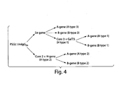
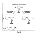
- the ABH antigen are found on almost all cells in the human body, but their physiological role, if any, remains an unresolved issue. They are of carbohydrate nature and are built up by different glycosyltransferases, i.e. enzymes adding monosaccharide units in a sequential manner to the non-reducing end of the growing oligosaccharide chain. Oligosaccharides can be carried by proteins or lipids, 9 or can be found free in body fluids (e.g. breast-milk). 9
- the ABH antigens are divided into subgroups, depending on the inner core saccharide chain. 9 As an example, both A, B and H antigens are expressed on type 1 (Gal ⁇ 1,3GlcNAc), type 2 (Gal ⁇ 1,4GlcNAc), type 3 (Gal ⁇ 1,3GalNAc ⁇ ) and type 4 (Gal ⁇ 1, 3GalNAc ⁇ ) chains. Type 4 chain ABH antigens are only found lipid-bound. H antigens are produced by the addition of a fucose in an ⁇ 1,2 linkage to the different core chains containing a terminal galactose.
- Both A and the B antigens are produced from subtypes of H by addition of an N-acetylgalactosamine (A) or a galactose (B) in an ⁇ 1,3 linkage to the terminal galactose.
- the glycosyltransferases responsible for the biosynthesis of A and B antigens require the presence of the ⁇ 1,2-fucose for addition of the terminal N-acetylgalactosamine and galactose, respectively.
- FUT-I H locus
- Se secretor locus
- the FUT-I gene product is responsible for erythrocyte H antigen expression and acts predominantly on type 2 chains, but activity towards type 1 chains has also been shown.
- the FUT-II gene product is expressed by salivary gland acinar cells as well as epithelial cells lining the gastrointestinal, reproductive and pulmonary tracts, and acts mainly on type 1 chains but probably also on type 3 and 4 chains.
- the gene products responsible for A and B antigen expression have been shown to have a common origin. 11 Mutations leading to the substitution of four amino acid residues in the A as compared to the B encoded gene product result in a shift in the donor sugar nucleotide preference from UDP-N-acetylgalactosamine to UDP-galactose.
- the serologically named O phenotype lacks expression of both of these gene products due to a frame shift mutation in the original A allele, and hence do not express A or B determinants.
- the blood group A has been subdivided into serologically distinct groups, the most frequent subgroups being A 1 and A 2 .
- a subtypes are produced by two different ⁇ 1,3-N-acetylgalactosaminyltransferases, one with a preference for H type 3 and 4 antigens (A 1 ) and the other with a preference for H type 1 and 2 (A 2 ) antigens.
- Protein-carbohydrate interactions are generally characterized by a low affinity of binding. Although this may seem irrational, it provides the basis for a fast on/off rate that is essential under physiological conditions as it allows for fast, highly changeable interactions to occur between cell receptors, antibodies and other carbohydrate binding proteins and their glycosylated ligands. Higher affinities, when needed, are in nature accomplished by the use of multivalency. The binding of several receptors on one biological entity to several carbohydrate ligands on another, can result in a 10-10 000 fold increase of the affinity. Examples of polyvalent interactions include the binding of microbes (viruses, bacteria or bacterial toxins) to a cell surface, cell-cell binding, and binding of polyvalent molecules, such as antibodies, to a cell surface.
- microbes viruses, bacteria or bacterial toxins
- the nanomolar activities most often characterizing physiological protein-carbohydrate interactions has been achieved in a few cases 13 by optimising ligand presentation (i.e . ligand structure, degree of valency, backbone structure and intra/inter-ligand distance), i.e . nature's way of presenting the carbohydrate was mimicked in as much detail as possible.
- ligand presentation i.e . ligand structure, degree of valency, backbone structure and intra/inter-ligand distance
- Mucin-type proteins are normally found at mucosal surfaces and are characterized by their abundant O-glycan substitution (every second to third amino acid). Up to 50% of their molecular weight is due to the carbohydrate substitution.
- O-glycan substitution every second to third amino acid.
- mucin-Ig By co-expressing various glycosyltransferase cDNAs with the mucin-Ig they can determine the structures of its O-glycans such that it carries several copies of biologically significant carbohydrate determinants. In this way, mucins carrying blood group A determinants have been made and shown to bind anti-blood group A Abs with high efficacy 1 .
- mucin-based absorbers of anti-blood group A antibodies were shown to be approximately a 100 times more efficient (as calculated on the number of blood group A determinants) in binding anti- A antibodies than the blood group A trisaccharide linked via a spacer directly to agarose beads (this is the arrangement of Glucosorb ® ). This, is explained by the fact that several copies of the A determinant is expressed with for Ab-binding optimal spacing on one mucin-type carrier protein'.
- mucins carrying the carbohydrate epitope, Gal ⁇ 1,3Gal( ⁇ ,- Gal), have been shown to be very efficient absorbers of anti-Gal antibodies; the main hurdle preventing successful pig-to-man xenotransplantation 15-17 .
- mucins are very efficient scaffolds for optimal presentation of bioactive carbohydrates.
- blood group ABH determinants can be carried by different core saccharide chains. These have a cell- and tissue-specific distribution in the human body, and the importance of the individual ABH antigens as target molecules for anti-blood group ABH antibodies is not known. Similarly, it is not known whether the anti-blood group ABH antibody repertoire is heterogeneous, i.e. whether subpopulations of these antibodies can distinguish between the different subtypes of ABH antigens and actually require more of the carbohydrate chain than the terminal trisaccharide for binding. Some data suggests that it is sufficient to use only the A or B trisaccharide for adsorption of all antibodies specific for blood group A or B antigens, respectively, indicating that this would be true also for detection of these antibodies 19-20 . However the data of the present invention show that it is possible to obtain more efficient antibody adsorption and detection using different subtypes (i.e. carried by different core saccharide chains) of the ABH blood group antigens.
- Oligosaccharides with tailored core saccharide chains as efficient absorbers of anti- carbohydrate antibodies are provided.
- the invention provides a mixture of oligosaccharides carrying the ABH determinants on different core saccharide chains (to obtain structural heterogeneity) will adsorb a broader repertoire of anti-A/B antibodies.
- Corresponding mixtures of mucin-type fusion proteins would also absorb a broader range of antibodies.
- the mucinimmunoglobulin fusion protein can be engineered to carry several copies of O-linked glycans with defined inner and outer core saccharide chains. These can be further elongated with the ABH determinants.
- recombinant technology can be used to make structurally diverse blood group A or B mucins, which can be used to adsorb a broad repertoire of anti-A or -B antibodies.
- Blood group antigens include (A, B, and O (H).
- the blood group antigens are specific for all the blood group subtypes.
- blood group subtypes it is meant that the blood group antigens are expressed on different core saccharide chain types.
- Core saccharides chain types include type- , type-2, type 3 and type-4 glycan precursors.
- Exemplary blood group antigen oligosaccharides include the following:
- R represents the point of attachment when the oligosaccharide is linked to a solid support, or nothing when the oligosaccharide is a free saccharide
- compositions comprising blood group antigens including:
- the ABO oligosaccharides are linked to a solid support.
- the linkage is covalent.
- the linkage is non-covalent.
- the solid support can be, for example, a bead, resin, membrane or disk, or any solid support material suitable for methods of the invention.
- the solid support is a bead, e.g., microbead.
- the size of the bead is not critical. Typically, the bead is at least 1 to 50 ⁇ m in diameter with a particle size of 1 to 10 ⁇ m being preferred. Most preferably the beads had a diameter of about 4-10 ⁇ m .For example, 2, 3, 4, 5, 10, 15, 20, 25, 30, 40 or 50 ⁇ m diameter.
- the bead may be made of metal compounds, silica, latex, polymeric material, or a silica, latex or polymer nuclei coated with a metal or metal compound.
- the solid support may carry functional groups such as hydroxyl, carboxyl, aldehyde or amino groups.
- the support may be positively charge, negatively charged or hydrophobic.
- Functionalized coated supports for use in the present invention may be prepared by modification of the support. For example uncoated supports may be treated with a polymer carrying one or such functional groups, such as polyurethane together with a polyglycol to provide hydroxyl groups or a cellulose derivative to provide hydroxyl groups, a polymer or copolymer of acrylic acid or methacrylic acid to provide carboxyl groups or an aminoalkylated polymer to provide amino groups.
- US Pat No. 4,654,267 describes the introduction of many surface coatings.
- each different ABO oligosaccharide is on a microbead of a different subtype.
- subtype is meant that each microbead is detectably distinguishable such as by being of different sizes or having distinguishable labels.
- Such a use of differently sized microbeads or microbeads labeled such as with fluorophores allows the identification and/or separation of different beads by, for example, flow cytometry.
- Kits for the separation of identification of blood group reactive antibodies from a sample according to the methods of the invention may contain a collection of microbeads of different subtypes each subtype is coated with a different ABO oligosaccharide or ABO fusion protein.
- the kit contains at least open labeled ligand capable of specifically binding an anti-blood group antigen antibody.
- the ligand is an antibody or fragment thereof.
- the antibody or fragment thereof is a monoclonal antibody.
- the antibody or fragment thereof is a polyclonal antibody.
- the antibody is a recombinant antibody.
- the antibody is an antibody fragment such as F ab , F ab' - and F (ab')2 fragments.
- the antibody is a monovalent.
- the label is any substance used to facilitate identification and/or quantitation of a target. Labels are directly observed or measured or indirectly observed or measured. Labels include, but are not limited to, radiolabels that can be measured with radiation-counting devices; pigments, dyes or other chromogens that can be visually observed or measured with a spectrophotometer; spin labels that can be measured with a spin label analyzer; and fluorescent moieties, where the output signal is generated by the excitation of a suitable molecular adduct and that can be visualized by excitation with light that is absorbed by the dye or can be measured with standard fluorometers or imaging systems, for example.
- the label can be a luminescent substance such as a phosphor or fluorogen; a bioluminescent substance; a chemiluminescent substance, where the output signal is generated by chemical modification of the signal compound; a metal-containing substance; or an enzyme, where there occurs an enzyme-dependent secondary generation of signal, such as the formation of a colored product from a colorless substrate.
- the label may also take the form of a chemical or biochemical, or an inert particle, including but not limited to colloidal gold, microspheres, quantum dots, or inorganic crystals such as nanocrystals or phosphors (see, e.g., Beverloo, et ah, Anal. Biochem. 203, 326-34 (1992 )).
- label can also refer to a "tag" or hapten that can bind selectively to a labeled molecule such that the labeled molecule, when added subsequently, is used to generate a detectable signal.
- a tag or hapten that can bind selectively to a labeled molecule such that the labeled molecule, when added subsequently, is used to generate a detectable signal.
- HRP horseradish peroxidase
- a chromogenic substrate e.g., tetramethylbenzidine
- fluorogenic substrate such as Amplex Red or Amplex Gold (Molecular Probes, Inc.
- the tag can be a hapten or antigen (e.g., digoxigenin), and an enzymatically, fluorescently, or radioactively labeled antibody can be used to bind to the tag.
- hapten or antigen e.g., digoxigenin
- an enzymatically, fluorescently, or radioactively labeled antibody can be used to bind to the tag.
- labels include, but are not limited to, particles, fluorescent dyes, haptens, enzymes and their chromogenic, fluorogenic, and chemiluminescent substrates, and other labels that are described in the Molecular Probes Handbook Of Fluorescent Probes And Research Chemicals by Richard P. Haugland, 6th Ed., (1996 ), and its subsequent 7th edition and 8th edition updates issued on CD Rom in November 1999 and May 2001, respectively, and in other published sources.
- a fluorophore is any chemical moiety that exhibits an absorption maximum beyond 280 nm, and when covalently attached to a labeling reagent retains its spectral properties.
- Fluorophores include, without limitation; a pyrene (including any of the corresponding derivative compounds disclosed in U.S. Pat. No.
- oxazines include resorufins (including any corresponding compounds disclosed in Pat. No. 5,242,805), aminooxazinones, diaminooxazines, and their benzo-substituted analogs.
- kits may contain a positive or negative control or both, instruction for using the kit (e.g., written, tape, VCR, CD-ROM, etc.), sample collection means.
- Sample collection means are well known to those skilled in the art.
- the sample collection means is a CPT vacutainer tube.
- the reagents are packaged in separate containers, e.g., ABO oligosaccharide (either bound to a solid matrix or packaged separately with reagents for binding them to the matrix), a control reagent (positive and/or negative), and or a labeled ligand.
- AMR antibody mediated graft rejection
- transplants include but are not limited to kidney, liver, skin, pancreas, cornea, or heart.
- AMR is meant to include any antibody mediated graft rejection by the recipient.
- the method includes contacting a biological sample from a subject with the ABO oligosaccharide of the invention or an ABO fusion peptide.
- the biological sample is for example, blood, i.e., whole blood or plasma.
- the sample is known to or suspected of comprising an antibody, e.g., an anti-blood group antibody.
- the biological sample is withdrawn from the subject prior to contacting the sample with the ABO oligosaccharide or ABO fusion polypeptide.
- the biological sample is contacted with the ABO oligosaccharide of the invention or ABO fusion peptide under conditions to allow formation of an ABO oligosaccharide of the invention or ABO fusion peptide- anti- blood group antibody complex.
- the ABO oligosaccharide of the invention or ABO fusion peptide-complex, if present is separated from the biological sample to eliminate the anti-blood group antibodies and the biological sample is reinfused into the subject.
- AMR may also be treated or prevented by administering to a subject an ABO fusion polypeptide.
- the subject can be e.g., any mammal, e.g., a human, a primate, mouse, rat, dog, cat, cow, horse, pig.
- the treatment is administered prior to the subject receiving an ABO-incompatible transplant.
- treatment is administered after a subject receives an ABO incompatible transplant.
- the biological sample is contacted with the ABO oligosaccharide of the invention or ABO fusion protein by methods known to those skilled in the art. For example, plasmapheresis or extracorporeal immunoabsorption.
- AMR is treated or prevent when the survival rate of the organ transplant is greater than an organ transplant not treated by the method of the invention.
- survival rate of the transplant is meant the time before the transplant is rejected by the recipient
- AMR is treated or prevent when the transplant survives at least 1, 2, 4 or 8 weeks after transplantion.
- the transplant survives 3, 6, 13 months. More preferably, the transplant survives 2, 3, 5 or more years.
- the invention provides methods of removing or depleting anti-blood group antibodies from a sample, said method comprising a method for removing blood group reactive antibodies in serum, said method comprising:
- the sample is a biological fluid such as blood or plasma.
- the sample is a biological tissue, such as heart tissue, liver tissue, skin, or kidney tissue.
- This method is useful to produce universal donor plasma.
- a subject is blood typed by contacting a sample, e.g., plasma or whole blood from a subject with a collection of microbeads of different subtypes carrying the oligosaccharides of the invention. Each subtype contains a different blood group antigen.
- the blood group antigens are expressed on different core saccharide chain types
- the microbeads and sample are contacted, e.g. incubated, for a sufficient amount a time to allow the anti-blood group antibodies present in the sample to bind, e.g. form an blood group antigen-antibody complex, to the blood group antigens on the microbeads.
- the sample is optionally washed one or more times to remove unbound plasma components.
- unbound plasma components are separated from the micobeads by performing a separation step in which the microbead are removed from the sample. Seperation is performed by methods known in the art such as centrifugation.
- the microbeads are further contacted with a labeling reagent that specifically binds the anti-blood group antibodies that is bound to microbead.
- the microbeads are optionally washed one or more times to remove unbound labeling reagent.
- the presence or absence of the anti-blood group antibdies in the sample is then determined by detecting the labeling reagent. Detection is done my methods known in the art such as by flow cytometry or luminex.
- the invention also provides for the detection of multiple anti-blood group antibodies in a sample.
- multiple anti-blood group antibodies it is meant not only antibodies specific for each of the blood groups (i.e, ABH) but antibodies specific fro blood group antigens of different oligosaccharide core chain types.
- Multiple targets are identified by contacting the biological sample with additional detection reagents followed by additional labeling reagent specific for the additional detection reagents using the method described above. For example, subsets of microbeads are prepared with distinct blood group antigens, e.g., blood group antigens that are distinguished by core oligosaccharide chain type.. The microbead subsets are then added to the biological sample containing in a controlled ratio.
- subsets of labeling reagent are prepared with distinct labels, e.g., fluorophores that are distinguished by their emission spectra, e.g., one that emits in the green spectra and one that emits in the red spectra.
- the labeling reagent subsets are then added to the biological sample containing detection reagent- target complexes in a controlled ratio, e.g., two parts one labeling reagent (e.g., green emission) and one part the other labeling reagent (e.g., red emission) per target binding antibody.
- a controlled ratio e.g., two parts one labeling reagent (e.g., green emission) and one part the other labeling reagent (e.g., red emission) per target binding antibody.
- the immuno- labeled complexes can be used to detect a target. If another immuno-labeled complex were added to the sample the original target could be distinguished from the subsequently detected target.
- the sample is defined to include any material that may contain a blood group antigen Typically the sample is whole blood, sera or plasma.
- the methods of the invention provides significant advantages over existing technology for blood typing. Specifically it allows for the detection of blood group antigen subtypes. Moreover, the methods allow for the qualification of the different blood group antigen subtypes in a sample to be determined.
- the detection reagent is a compound that is capable of specifically binding the blood group antibodies bound to the microbeadd.
- the detection reagent is selected based on the desired target.
- the detection reagent is for example a polypeptide such as a target specific antibody or fragment thereof.
- the term "antibody” refers to immunoglobulin molecules and immunologically active portions of immunoglobulin (Ig) molecules, i.e., molecules that contain an antigen binding site that specifically binds (immunoreacts with) an antigen.
- Such antibodies include, polyclonal, monoclonal, chimeric, single chain, F ab , F ab' and F (ab')2 fragments, and an F ab expression library.
- Monoclonal antibodies are particularly advantageous in practicing the methods of the present invention. Generally, monoclonal antibodies are more sensitive and specific than polyclonal antibodies. In addition, unlike polyclonal antibodies, which depend upon the longevity of the animal producing the antibody, the supply of monoclonal antibodies is indefinite. Polyclonal antibodies however, are useful when it is necessary to use antibodies with multiple isotypes, as generally most monoclonal antibodies are of the IgG1 subclass.
- immunological binding refers to the non-covalent interactions of the type that occur between an immunoglobulin molecule and an antigen for which the immunoglobulin is specific.
- the strength, or affinity of immunological binding interactions can be expressed in terms of the dissociation constant (K d ) of the interaction, wherein a smaller K d represents a greater affinity.
- Immunological binding properties of selected polypeptides are quantified using methods well known in the art. One such method entails measuring the rates of antigen-binding site/antigen complex formation and dissociation, wherein those rates depend on the concentrations of the complex partners, the affinity of the interaction, and geometric parameters that equally influence the rate in both directions.
- both the "on rate constant” (K on ) and the “off rate constant” (K off ) can be determined by calculation of the concentrations and the actual rates of association and dissociation. (See Nature 361:186-87 (1993 )).
- the ratio of K off /K on enables the cancellation of all parameters not related to affinity, and is equal to the dissociation constant K d . (See, generally, Davies et al. (1990) Annual Rev Biochem 59:439-473 ).
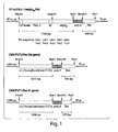
- the expression vector carrying P-selectin glycoprotein ligand-1/mouse IgG 2b (PSGL-1/mIgG 2b ) cDNA was modified to contain an enterokinase (EK) cleavage site. This site can be used for down-stream release of the mouse IgG 2b part.
- EK enterokinase
- the human blood group A gene was polymerase chain reaction (PCR) amplified off cDNA made from total RNA isolated from the MKN-45 cell line
- the blood group B gene was PCR amplified off cDNA made from total RNA isolated from the HuTu80 cell line.
- the expression vectors used to generate stable transfectants are bidirectional, see schematic figures.
- the PSGL-1/mIgG 2b expression vector has the EF1 ⁇ promoter upstream of a polylinker, a splice donor and acceptor site, and the bidirectional poly(A) additional signal of SV40. Opposite in orientation to this transcription unit, using the poly(A) signals from the opposite direction, is a second transcription unit consisting of the HSV TK promoter followed by the coding sequence for puromycin acetyltransferase (PAC).
- the ⁇ 1,6GlcNAcT (core 2 enzyme) expression vector contains the EF1 ⁇ promoter and the coding sequence for the neomycin resistance gene (Neo).
- the FUT2 (Se-gene) expression vector contains the same vector backbone, but with the CMV promoter and the neomycin resistance gene.
- the GalNAcT (A-gene) and the GalT (B-gene) expression vectors contain the CMV promoter and the blasictidin resistance gene (Bsd)
- the FUT1 (H-gene) and the ⁇ 1,3GlcNAcT6 (core 3 enzyme) contain the CMV promoter and the zeocin resistance gene
- the GalT5 expression vector contain the CMV promoter and guanosine phosphoribosyl transferase gene (GPT).
- the DNA sequences of the expression vectors were verified by automated sequencing.
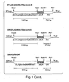
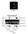
- EXAMPLE 2 DETERMINATION OF PSGL-1/MIGG2B EXPRESSION USING WESTERN BLOTTING AND INDIRECT IMMUNOFLUORESCENCE
- PSGL-1/mIgG 2b The cellular localization of PSGL-1/mIgG 2b was determined by indirect immunofluorescence.
- CHO-K1 cells seeded on cover slips in six-well plates were transiently transfected with the PSGL-1/mIgG 2b expression vector using Lipofectamine 2000 (Invitrogen), according to the manufacturer's instructions. Forty eight hours after transfection, cells were washed with PBS and fixed in 30% acetone/MeOH. Following blocking for 30 minutes in 1% BSA in PBS, PSGL-1/mIgG 2b protein was detected using a FITC-conjugated goat anti-mouse IgG Fc antibody (Sigma) diluted 1:200 in blocking buffer.
- FITC-conjugated goat anti-mouse IgG Fc antibody Sigma
- DAPI 4,6-diamidino2-phenylindole
- the cover slips were mounted on slides with Vectashield Mounting Medium (Vector Laboratories). Slides were examined using a DMRXA microscope (Leica Corp.), and digitally imaged using a Hamamatsu C4880-40 CCD camera (Hamamatsu Photonics Norden AB), the Openlab software package (Improvision), and Adobe Photoshop software (see figure).
- the CHO-K1 cell line (ATCC CCL-61) was adapted to serum-free medium, Ex-cell 302 (JHR Bioscience, Inc), according to the manufacturer's instructions.
- CHO-K1 cells adapted to serum-free medium were seeded in 75 cm 2 flasks and were transfected with the PSGL-1/mIgG 2b expression vector using Lipofectamine 200 CD (Invitrogen), an animal origin-free formulation, according to the manufacturer's instructions. Forty eight hours after transfection, cells were incubated in puromycin-containing selection medium (6 ⁇ g/mL). The selection medium was changed every third day. After approx. 2 weeks, dead cells were removed using Dead Cell Removal MicroBeads (Miltenyi Biotech), according to the manufacturer's instructions. Live cells were single cell-cloned in 96-well plates, and expanded in selection medium for approx. 2 weeks.
- hPSGL-1/mIgG 2b concentration of hPSGL-1/mIgG 2b was assessed by enzyme-linked immunosorbent assay (ELISA), see protocol below, using a goat anti-mouse IgG Fc antibody (Sigma).
- ELISA enzyme-linked immunosorbent assay
- EXAMPLE 6 GENERATION OF PSGL-1/MIGG 2B SUBSTITUTED WITH BLOOD GROUP A/B TYPE 3 ANTIGENS
- the stable CHO-K1 cell line with the highest PSGL-1/mIgG 2b expression was transfected with the FUT2 (Se-gene) expression vector, as described above, and selected in G418-containing medium (400 ⁇ g/mL).
- the cell clone with the highest relative number of blood group H type 3 antigens on PSGL-1/mIgG 2b will be transfected with either the GalNAcT (A-gene) or GAlT (B-gene) expression vectors, and selected in blasticidine-containing medium, to generate cell lines that produce blood group A and B, respectively, type 3 antigens on PSGL-1/mIgG 2b (see transfection scheme).
- EXAMPLE 7 GENERATION OF PSGL-1/MIGG 2B SUBSTITUTED WITH BLOOD GROUP A/B TYPE 2 ANTIGENS
- the stable CHO-K1 cell line with the highest PSGL-1/mIgG 2b expression was transfected with the ⁇ 1,6GlcNAcT1 (core 2 enzyme) and FUT1 (H-gene) expression vectors, and selected in medium containing G418 (400 ⁇ g/mL).
- the cell clone with the highest relative number of H type 2 antigens on PSGL-1/mIgG 2b will be transfected with either the ⁇ 1, 3 GalNAcT (A-gene) or ⁇ 1, 3 GalT (B-gene) expression vectors, and selected in blasticidine-containing medium, to generate cell lines that produce blood group A and B, respectively, type 2 antigens on PSGL-1/mIgG 2b (see transfection scheme).
- EXAMPLE 8 GENERATION OF PSGL-1/MIGG 2B SUBSTITUTED WITH BLOOD GROUP A/B TYPE 1 ANTIGENS
- the cell clone with the highest relative number of blood group H type 3 antigens on PSGL-1/mIgG 2b will be transfected with the ⁇ 1,3GlcNAcT (core 3 enzyme) and GalT5 expression vectors, and selected in medium containing zeocin.
- the clone with the highest relative number of blood group H type 1 antigens on PSGL-1/mIgG 2b will be transfected with either the ⁇ 1, 3 GalNAcT (A-gene) or ⁇ 1, 3 GalT (B-gene) expression vectors, and selected in blasticidine-containing medium, to generate cell lines that produce blood group A and B, respectively, type 1 antigens on PSGL-1/mIgG 2b (see transfection scheme).
- hPSGL-1/EK/mIgG 2b may be further purified by gel filtration on a HiPrep 16/60 Sephacryl S-200 HR column (Amersham Bioscience).
- Purified blood group A and B mucins will be covalent coupled to sepharose fast flow beads using standard bioconjugation chemistry. Following coupling, the sepharose will be packed in the plastic container constituting the actual column.
- the titres of anti-blood group ABO antibodies prior to and after adsorption of pooled blood group O plasma will be assessed by standard blood banking techniques including hemagglutination and the indirect anti-globulin test.
- Plasma proteins including immunoglobulins (IgG, IgM and IgA), complement factors/fragments (C3, C3a, C3d, C4 and sC5b-9), immune complexes, and coagulation factors (FVIII, prothrombin, fibrinogen, fibrin degradation products) will all be measured by standard techniques.
- the specificities of adsorbed and eluted, and nonadsorbed ABO antibodies will be determined by ELISA using recombinant mucins carrying blood group A/B antigens on defined core saccharide chains or by an thin-layer chromatography-based overlay assay in which purified and structurally defined blood group A and B glycolipids are used.
- EXAMPLE 10 PRODUCTION OF BEADS OF DIFFERENT SIZES AND COLOURS FOR DETERMINATION OF ANTIBODY TITERS
- Beads of different sizes and colours were produced micromod Pelletechnologie GmbH in Rostock, Germany and conjugated to blood group antigens of different types. Beads were analyzed by flow cytometry and showed good resolution both regarding size and colour (see figure 5 ). Using this method it is possible to use a mixture of a large number of beads conjugated were each size-colour intensity combination represents a specific blood group antigen expressed on a specific core structure.
- Serum samples are diluted 1:10 in PBS with 0.5% human albumin and mixed with latex microbead (4.6 ⁇ m; 18 ⁇ g dry weight) carrying blood group antigen oligosaccharides. Serum and microbeads are incubated at room temperature for 30 minutes and then washed with 0.5% HAS/PBS. FITC labeled anti-human IgM and PE labeled IgG is added to the washed microbeads and analyzed by flow cytometry.
Abstract
Description
- The invention relates to generally to compositions for treating or preventing antibody-mediated graft rejection and more particularly to compositions including blood group determinants useful for removing anti-blood group antigen antibodies.
- Renal transplantation across the ABO barriers was found, already in the early days of transplantation, to result in a high incidence of transplants that never functional, and it was therefore regarded as a prerequisite in allotransplantation to comply with the traditional Landsteiner rules used for blood transfusion. The recipient's preformed anti-A/B isoagglutinins are responsible for hyperacute rejection of ABO-incompatible grafts. This hyperacute rejection is similar to that seen in alloimmunized patients with donor-reactive HLA-antibodies. The first trial to cross the ABO barrier in transplantation was started in the early 1970's grafting blood group A2 cadaveric kidneys to O recipients.1 In the 1980's, using A1 and B donors Alexandre performed the first series of ABO incompatible living donor (LD) renal transplantations and obtained graft survival similar to those of ABO compatible cases. The immunosuppressive protocol encompassed pre-operative plasmapheresis to remove anti-A/B antibodies, donor platelet transfusion, splenectomy and induction therapy with anti-lymphocyte/thymocyte globulin, injection of blood group A or B substances extracted from porcine stomach and Cyclosporine-Azathioprine-prednisone. Since then, more than 500 cases of ABO incompatible LD renal transplantations have been reported worldwide, mainly from Japan (reviewed in 1), and the importance of reducing recipient anti-A/B antibody levels before grafting to avoid rejection has been well documented.2,3 The graft survival in these series is good (1 year graft survival of about 85% for A1 and B donors) but slightly inferior to that of ABO compatible grafts due to single cases with severe anti-A/B antibody mediated rejection.4,5 Recent data on ABO incompatible renal transplantations using an anti-CD20 antibody (Rituximab) combined with antibody removal were shown to be even better with regard to graft survival 6,7.
-
US 2003/0073822 relates to fusion polypeptides, and their use in the treatment of antibody-mediated graft rejection. The fusions comprise blood group epitopes expressed on mucin-type protein backbones. - In times of severe organ shortage, an increased use of grafts from ABO incompatible donors will allow more LD kidney transplantations to be performed. In addition, the experience gained in this field will also be applicable on the pre-treatment and post-transplant management of HLA-sensitized patients.8
- The invention is based in part on improved compositions and method for removing blood group antigen antibodies form plasma and a method of blood typing.
- In one aspect the invention provides a composition comprising blood group antigens including:
- A
type 1 - GalNAcα1,3(Fucα1,2)Galβ1, 3 GlcNAcβ1-R,
- A
type 2 - GalNAcα1,3(Fucα1,2)Galβ1, 4 GlcNAcβ1-R,
- A
type 3 - GalNAcα1,3(Fucα1,2)Galβ1,3GalNAcα1-R,
- A
type 4 - GalNAcα1,3(Fucα1,2)Galβ1, 3 GalNAcβ1-R,
- B type I
- Galα1,3(Fucα1,2)Galβ1, 3 GlcNAcβ1-R,
-
B type 2 - Galα1,3(Fucα1,2)Galβ1, 4 GlcNAcβ1-R,
-
B type 3 - Galα1,3(Fucα1,2)Galβ1,3GalNAcα1-R, and
-
B type 4 - Galα1,3(Fucα1,2)Galβ1,3GalNAcβ1-R
- In another aspect, the invention provides a method for determining the presence of blood group antibodies in a sample from a subject (e.g. blood typing), said method comprising:
- a) providing a collection of microbeads of different subtypes wherein each subtype is coated with a different blood group antigen, including:
- A
type 1 - GalNAcα1,3(Fucα1,2)Galβ1, 3 GlcNAcβ1-R,
- A
type 2 - GalNAcα1,3(Fucα1,2)Galβ1, 4 GlcNAcβ1-R,
- A
type 3 - GalNAcα1,3(Fucα1,2)Galβ1,3GalNAcα1-R,
- A
type 4 - GalNAcα1,3(Fucα1,2)Galβ1,3GalNAcβ1-R,
-
B type 1 - Galα1,3(Fucα1,2)Galβ1, 3 GlcNAcβ1-R,
-
B type 2 - Galα1,3(Fucα1,2)Galβ1,4GlcNAcβ1-R,
-
B type 3 - Galα1,3(Fucα1,2)Galβ1,3GalNAcα1-R, and
-
B type 4 - Galα1,3(Fucα1,2)Galβ1,3GalNAcβ1-R
- A
- b) adding said serum from said subject to said collection of microbeads;
- c) incubating said serum and microbeads for sufficient time for anti-blood group antibodies in said serum to bind to said blood group antigens;
- d) incubating said microbeads with at least one labelled ligand capable of specifically binding with said anti-blood group antibodies bound to said blood group antigens; and
- e) detecting the presence of labelled ligand bound to said anti-blood group antibodies to determine the presence or absence or said reactive antibodies.
- The ligand is for example an antibody or fragment thereof. The antibody is a monovalent antibody fragment such as Fab or Fab' fragment. For example the Fab or Fab' fragment is an anti-Fc antibody fragment, an anti -kappa light chain antibody fragment, an anti-lambda light chain antibody fragment, and a single chain antibody fragment. The label is for example a fluorescent label. Labeled ligand is detected by methods known in the art such as flow cytometry or luminex.
- In a further aspect, the invention provides a method for removing blood group reactive antibodies in serum, said method comprising:
- a) providing a collection of microbeads of different subtypes, wherein each subtype is coated with a different blood group antigen, including:
- A
type 1 - GalNAcα1,3(Fucα1,2)Galβ1, 3 GlcNAcβ1-R,
- A
type 2 - GalNAcα1,3(Fucα1,2)Galβ1, 4 GlcNAcβ1-R,
- A
type 3 - GalNAcα1,3(Fucα1,2)Galβ1,3GalNAcα1-R,
- A
type 4 - GalNAcα1,3(Fucα1,2)Galβ1, 3 GalNAcβ1-R,
-
B type 1 - Galα1,3(Fucα1,2)Galβ1, 3 GlcNAcβ1-R,
-
B type 2 - Galα1,3(Fucα1,2)Galβ1, 4 GlcNAcβ1-R,
-
B type 3 - Galα1,3(Fucα1,2)Galβ1,3GalNAcα1-R, and
-
B type 4 - Galα1,3(Fucα1,2)Galβ1,3GalNAcβ1-R
- A
- b) contacting said serum with said collection of microbeads;
- c) incubating said serum and microbeads for sufficient time for anti-blood group antibodies in said serum to bind to said blood group antigens;
- d) separating said microbeads from said serum
- Optionally, ABO oligosaccharide or ABO fusion proteins are linked, e.g. covalently or non-covalently to a solid support such as microbeads. The microbeads are for example latex. By microbeads of a different subtype is meant that the microbeads differ from one another by size, color or both. The range in diameter from about 2 µm to about 15 µm.
- Preferably, the microbead is approximately 5 µm in diameter. Most preferably, the microbeads are approximately 5 µm in diameter.
The sample is for example, whole blood, serum or plasma. - In some aspects, the compositions are formulated as an absorber for the removal of blood group antibodies from whole blood or plasma. Also provided by the invention is a collection of microbeads of different subtypes, wherein each subtype is coated with different blood group antigen. For example. The collection contains as microbeads of at least 2, 3, 4, 5, 6, 7, 8, 9, 10, 20, 25, 30, 40, 50 or more different subtypes.
- Unless otherwise defined, all technical and scientific terms used herein have the same meaning as commonly understood by one of ordinary skill in the art to which this invention belongs.
- Other features and advantages of the invention will be apparent from the following detailed description, and from the claims.
-
-
Figure 1 is a schematic representation of the vectors used to produce the fusion proteins carrying blood group antigens. -
Figure 2 is a photograph showing the cellular localization of PSGL- 1/mIgG2b was determined by indirect immunofluorescence. PSGL- 1/mIgG2b protein was detected using a FITC-conjugated goat anti-mouse IgGFc antibody (Sigma) diluted 1:200 in blocking buffer. Cell nuclei were stained with 4, 6-diamidino2-phenylindole (DAPI). -
Figure 3 is a photograph of a Western Blot showing blood group A determinants carried different outer core chains. -
Figure 4 is a schematic representation of the ABH transfection scheme to produce ABO fusion peptides on different glycan precursors. -
Figure 5 are graphs showing flow cytometry results from beads of 5 different sizes and colour intensities clearly showing that it is possible to use beads of many size-colour combinations. Using combinations of the above shown sizes and colours it would be possible to make a mixture of up to 25 different beads each expressing a unique blood group antigen. -
Figure 6 is a schematic representation of outer core structures of blood group antigen. -
Figure 7 is a schematic representation of blood group ABH antigens. -
Figure 8 is a graphical representation of flow cytometry results identifying anti-blood group antibodies in serum -
Figure 9 is a series of scatter plots showing IgG and IgM blood group A antibodies in serum from A, B and O individuals. - The invention is based in part in the discovery that blood group epitopes can be specifically expressed at high density and by different core saccharides chains (e.g.
type 1,type 2,type 3 or type 4) as either free saccharides or on mucin-type protein backbones. It has been discovered that using blood group antigens carried by the different core saccharides chains when used in combination are more efficient at removing anti-blood group antibodies from blood prior to transplantation. Additionally, the compositions of the invention are useful diagnostically and prognostically to determine the presence of blood group reactive antibodies in a subject. - In one aspect the invention provides a composition comprising blood group antigens including.:
- A
type 1 - GalNAcα1,3(Fucα1,2)Galβ1, 3 GlcNAcβ1-R,
- A
type 2 - GalNAcα1,3(Fucα1,2)Galβ1, 4 GlcNAcβ1-R,
- A
type 3 - GalNAcα1,3(Fucα1,2)Galβ1,3GalNAcα1-R,
- A
type 4 - GalNAcα1,3(Fucα1,2)Galβ1,3GalNAcβ1-R,
-
B type 1 - Galα1,3(Fucα1,2)Galβ1, 3 GlcNAcβ1-R,
-
B type 2 - Galα1,3(Fucα1,2)Galβ1, 4 GlcNAcβ1-R,
-
B type 3 - Galα1,3(Fucα1,2)Galβ1,3GalNAcα1-R, and
-
B type 4 - Galα1,3(Fucα1,2)Galβ1,3GalNAcβ1-R
- These oligosaccharides are referred to herein as "ABO oligosaccharides". The blood group antigens are free saccharide.
- Optionally, the ABO oligosaccharides are linked to a solid support to allow for separation of blood group antigens from blood.
- Accordingly, the ABO oligosaccharides are useful in eliminating recipient anti-blood group ABO antibodies from blood or plasma prior to for example, an ABO incompatible organ, bone marrow transplantation or in order to make universal donor plasma. The ABO oligosaccharides absorb 50%, 60%, 70%, 80%, 90%, 95%, 98% or 100% of anti[tau]blood group ABO antibodies from recipient blood or plasma.
- The ABO oligosaccharide is more efficient on a carbohydrate molar basis in removing or binding anti-blood group antibodies as compared blood group oligosaccharide expressed on a single core saccharide chain. The ABO oligosaccharide binds 2, 4, 10, 20, 50, 80, 100 or more-fold greater number of anti-blood group antibodies as compared to an equivalent amount of free saccharides expressed on a single core saccharide chain.
- The ABO oligosaccharides are also useful in a method of blood typing. The methods of the invention are superior to current blood typing methods as it allows quantification of blood group antibodies of different classes and subclasses. In particular it allows for the detection of chain-type specific antibodies. This is clinically relevant as the immune system can respond specifically to blood group antigens on different core chains. Moreover blood group antigens on different core chains are expressed in a cell and tissue specific manner. Thus, by allowing the detection of chain specific antibodies will allow for better donor-reciepent cross matching in ABO incompatible organ allografts, which will decrease hyperacute rejection
- The ABH antigen are found on almost all cells in the human body, but their physiological role, if any, remains an unresolved issue. They are of carbohydrate nature and are built up by different glycosyltransferases, i.e. enzymes adding monosaccharide units in a sequential manner to the non-reducing end of the growing oligosaccharide chain. Oligosaccharides can be carried by proteins or lipids,9 or can be found free in body fluids (e.g. breast-milk).9
- The ABH antigens are divided into subgroups, depending on the inner core saccharide chain.9 As an example, both A, B and H antigens are expressed on type 1 (Galβ1,3GlcNAc), type 2 (Galβ1,4GlcNAc), type 3 (Galβ1,3GalNAcα) and type 4 (Galβ1, 3GalNAcβ) chains.
Type 4 chain ABH antigens are only found lipid-bound. H antigens are produced by the addition of a fucose in an α1,2 linkage to the different core chains containing a terminal galactose. Both A and the B antigens are produced from subtypes of H by addition of an N-acetylgalactosamine (A) or a galactose (B) in an α1,3 linkage to the terminal galactose. The glycosyltransferases responsible for the biosynthesis of A and B antigens require the presence of the α1,2-fucose for addition of the terminal N-acetylgalactosamine and galactose, respectively. - Two structurally distinct α1,2-fucosyltransferases enables the biosynthesis of the H antigen as initially described by Oriol.10 One is encoded by the H locus (FUT-I) and the other by the secretor (Se) locus (FUT-II). The FUT-I gene product is responsible for erythrocyte H antigen expression and acts predominantly on
type 2 chains, but activity towardstype 1 chains has also been shown. In contrast, the FUT-II gene product is expressed by salivary gland acinar cells as well as epithelial cells lining the gastrointestinal, reproductive and pulmonary tracts, and acts mainly ontype 1 chains but probably also ontype - The gene products responsible for A and B antigen expression have been shown to have a common origin.11 Mutations leading to the substitution of four amino acid residues in the A as compared to the B encoded gene product result in a shift in the donor sugar nucleotide preference from UDP-N-acetylgalactosamine to UDP-galactose. The serologically named O phenotype lacks expression of both of these gene products due to a frame shift mutation in the original A allele, and hence do not express A or B determinants.11 The blood group A has been subdivided into serologically distinct groups, the most frequent subgroups being A1 and A2.12 These A subtypes are produced by two different α1,3-N-acetylgalactosaminyltransferases, one with a preference for
H type H type 1 and 2 (A2) antigens. - Protein-carbohydrate interactions are generally characterized by a low affinity of binding. Although this may seem irrational, it provides the basis for a fast on/off rate that is essential under physiological conditions as it allows for fast, highly changeable interactions to occur between cell receptors, antibodies and other carbohydrate binding proteins and their glycosylated ligands. Higher affinities, when needed, are in nature accomplished by the use of multivalency. The binding of several receptors on one biological entity to several carbohydrate ligands on another, can result in a 10-10 000 fold increase of the affinity. Examples of polyvalent interactions include the binding of microbes (viruses, bacteria or bacterial toxins) to a cell surface, cell-cell binding, and binding of polyvalent molecules, such as antibodies, to a cell surface.13 Inhibition of polyvalent interactions with monovalent inhibitors is usually ineffective even if the binding activity of the inhibitor has been structurally optimized. Accordingly, a number of different molecules (e.g. polyacrylamide, peptides, bovine serum albumin, dendrimers and cyclodextrins) have been used as backbones for multiple presentations of mono- and oligosaccharides attempting to create multivalent binding of the corresponding receptors.13 An alternative approach involves non-covalent association of the ligand with the head groups in liposomes, membranes or other surfaces.13 The success of these glycoconjugates varies, but in general the affinity is enhanced 10- to 1000-fold as compared to the monovalent interactions. The nanomolar activities most often characterizing physiological protein-carbohydrate interactions has been achieved in a few cases13 by optimising ligand presentation (i.e. ligand structure, degree of valency, backbone structure and intra/inter-ligand distance), i.e. nature's way of presenting the carbohydrate was mimicked in as much detail as possible.
- Recombinant mucins with tailored glycan-substitution as efficient absorbers of anti-carbohydrate antibodies.
- Mucin-type proteins are normally found at mucosal surfaces and are characterized by their abundant O-glycan substitution (every second to third amino acid). Up to 50% of their molecular weight is due to the carbohydrate substitution. By co-expressing various glycosyltransferase cDNAs with the mucin-Ig they can determine the structures of its O-glycans such that it carries several copies of biologically significant carbohydrate determinants. In this way, mucins carrying blood group A determinants have been made and shown to bind anti-blood group A Abs with high efficacy1. In fact, mucin-based absorbers of anti-blood group A antibodies were shown to be approximately a 100 times more efficient (as calculated on the number of blood group A determinants) in binding anti- A antibodies than the blood group A trisaccharide linked via a spacer directly to agarose beads (this is the arrangement of Glucosorb®). This, is explained by the fact that several copies of the A determinant is expressed with for Ab-binding optimal spacing on one mucin-type carrier protein'. Likewise, recombinant mucins carrying the carbohydrate epitope, Galα1,3Gal(α,- Gal), have been shown to be very efficient absorbers of anti-Gal antibodies; the main hurdle preventing successful pig-to-man xenotransplantation15-17. Thus, mucins are very efficient scaffolds for optimal presentation of bioactive carbohydrates.
- As described above, blood group ABH determinants can be carried by different core saccharide chains. These have a cell- and tissue-specific distribution in the human body, and the importance of the individual ABH antigens as target molecules for anti-blood group ABH antibodies is not known. Similarly, it is not known whether the anti-blood group ABH antibody repertoire is heterogeneous, i.e. whether subpopulations of these antibodies can distinguish between the different subtypes of ABH antigens and actually require more of the carbohydrate chain than the terminal trisaccharide for binding. Some data suggests that it is sufficient to use only the A or B trisaccharide for adsorption of all antibodies specific for blood group A or B antigens, respectively, indicating that this would be true also for detection of these antibodies 19-20. However the data of the present invention show that it is possible to obtain more efficient antibody adsorption and detection using different subtypes (i.e. carried by different core saccharide chains) of the ABH blood group antigens.
- Following from the discussion above, the invention provides a mixture of oligosaccharides carrying the ABH determinants on different core saccharide chains (to obtain structural heterogeneity) will adsorb a broader repertoire of anti-A/B antibodies. Corresponding mixtures of mucin-type fusion proteins would also absorb a broader range of antibodies. Using different combinations of glycosyltransferase genes, the mucinimmunoglobulin fusion protein can be engineered to carry several copies of O-linked glycans with defined inner and outer core saccharide chains. These can be further elongated with the ABH determinants. Thus, recombinant technology can be used to make structurally diverse blood group A or B mucins, which can be used to adsorb a broad repertoire of anti-A or -B antibodies.
- The invention provides blood group antigen oligosaccharides. Blood group antigens include (A, B, and O (H). The blood group antigens are specific for all the blood group subtypes. By blood group subtypes it is meant that the blood group antigens are expressed on different core saccharide chain types. Core saccharides chain types include type- , type-2,
type 3 and type-4 glycan precursors. - Exemplary blood group antigen oligosaccharides include the following:
- Blood group A antigens on types 1-4
- A
type 1 - GalNAcα1,3(Fucα1,2)Galβ1, 3 GlcNAcβ1-R
- A
type 2 - GalNAcα1,3(Fucα1,2)Galβ1, 4 GlcNAcβ1-R
- A
type 3 - GalNAcα1,3(Fucα1,2)Galβ1,3GalNAcα1-R
- A
type 4 - GalNAcα1,3(Fucα1,2)Galβ1,3GalNAcβ1-R
- A
- Blood group B antigens on types 1-4
-
B type 1 - Galα1,3(Fucα1,2)Galβ1, 3 GlcNAcβ1-R
-
B type 2 - Galα1,3(Fucα1,2)Galβ1, 4 GlcNAcβ1-R
-
B type 3 - Galα1,3(Fucα1,2)Galβ1,3GalNAcα1-R
-
B type 4 - Galα1,3(Fucα1,2)Galβ1,3GalNAcβ1-R
-
- Wherein R represents the point of attachment when the oligosaccharide is linked to a solid support, or nothing when the oligosaccharide is a free saccharide
- The invention provides compositions comprising blood group antigens including:
- A
type 1 - GalNAcα1,3(Fucα1,2)Galβ1, 3 GlcNAcβ1-R,
- A
type 2 - GalNAcα1,3(Fucα1,2)Galβ1, 4 GlcNAcβ1-R,
- A
type 3 - GalNAcα1,3(Fucα1,2)Galβ1,3GalNAcα1-R,
- A
type 4 - GalNAcα1,3(Fucα1,2)Galβ1, 3 GalNAcβ1-R,
-
B type 1 - Galα1,3(Fucα1,2)Galβ1, 3 GlcNAcβ1-R,
-
B type 2 - Galα1,3(Fucα1,2)Galβ1, 4 GlcNAcβ1-R,
-
B type 3 - Galα1,3(Fucα1,2)Galβ1,3GalNAcα1-R, and
-
B type 4 - Galα1,3(Fucα1,2)Galβ1,3GalNAcβ1-R
- Optionally, the ABO oligosaccharides are linked to a solid support. The linkage is covalent. Alternatively, the linkage is non-covalent.
- The solid support can be, for example, a bead, resin, membrane or disk, or any solid support material suitable for methods of the invention. Preferably, the solid support is a bead, e.g., microbead. The size of the bead is not critical. Typically, the bead is at least 1 to 50µm in diameter with a particle size of 1 to 10µm being preferred. Most preferably the beads had a diameter of about 4-10 µm .For example, 2, 3, 4, 5, 10, 15, 20, 25, 30, 40 or 50 µm diameter.
- The bead may be made of metal compounds, silica, latex, polymeric material, or a silica, latex or polymer nuclei coated with a metal or metal compound.
- The solid support may carry functional groups such as hydroxyl, carboxyl, aldehyde or amino groups. The support may be positively charge, negatively charged or hydrophobic. Functionalized coated supports for use in the present invention may be prepared by modification of the support. For example uncoated supports may be treated with a polymer carrying one or such functional groups, such as polyurethane together with a polyglycol to provide hydroxyl groups or a cellulose derivative to provide hydroxyl groups, a polymer or copolymer of acrylic acid or methacrylic acid to provide carboxyl groups or an aminoalkylated polymer to provide amino groups.
US Pat No. 4,654,267 describes the introduction of many surface coatings. - Preferably each different ABO oligosaccharide is on a microbead of a different subtype. By subtype is meant that each microbead is detectably distinguishable such as by being of different sizes or having distinguishable labels. Such a use of differently sized microbeads or microbeads labeled such as with fluorophores allows the identification and/or separation of different beads by, for example, flow cytometry.
- Kits for the separation of identification of blood group reactive antibodies from a sample according to the methods of the invention may contain a collection of microbeads of different subtypes each subtype is coated with a different ABO oligosaccharide or ABO fusion protein. Optionally the kit contains at least open labeled ligand capable of specifically binding an anti-blood group antigen antibody. For example, the ligand is an antibody or fragment thereof. The antibody or fragment thereof is a monoclonal antibody. Alternatively, the antibody or fragment thereof is a polyclonal antibody. Optionally, the antibody is a recombinant antibody. The antibody is an antibody fragment such as Fab, Fab'- and F(ab')2 fragments. By "specifically bind" or "immunoreacts with" is meant that the antibody reacts with one or more antigenic determinants of the desired antigen and does not react (i.e., bind) with other polypeptides or binds at much lower affinity (Kd > 10-6) with other polypeptides.
- The antibody is a monovalent.
- The label, is any substance used to facilitate identification and/or quantitation of a target. Labels are directly observed or measured or indirectly observed or measured. Labels include, but are not limited to, radiolabels that can be measured with radiation-counting devices; pigments, dyes or other chromogens that can be visually observed or measured with a spectrophotometer; spin labels that can be measured with a spin label analyzer; and fluorescent moieties, where the output signal is generated by the excitation of a suitable molecular adduct and that can be visualized by excitation with light that is absorbed by the dye or can be measured with standard fluorometers or imaging systems, for example. The label can be a luminescent substance such as a phosphor or fluorogen; a bioluminescent substance; a chemiluminescent substance, where the output signal is generated by chemical modification of the signal compound; a metal-containing substance; or an enzyme, where there occurs an enzyme-dependent secondary generation of signal, such as the formation of a colored product from a colorless substrate. The label may also take the form of a chemical or biochemical, or an inert particle, including but not limited to colloidal gold, microspheres, quantum dots, or inorganic crystals such as nanocrystals or phosphors (see, e.g., Beverloo, et ah, Anal. Biochem. 203, 326-34 (1992)). The term label can also refer to a "tag" or hapten that can bind selectively to a labeled molecule such that the labeled molecule, when added subsequently, is used to generate a detectable signal. For instance, one can use biotin, iminobiotin or desthiobiotin as a tag and then use an avidin or streptavidin conjugate of horseradish peroxidase (HRP) to bind to the tag, and then use a chromogenic substrate (e.g., tetramethylbenzidine) or a fluorogenic substrate such as Amplex Red or Amplex Gold (Molecular Probes, Inc.) to detect the presence of HRP. Similarly, the tag can be a hapten or antigen (e.g., digoxigenin), and an enzymatically, fluorescently, or radioactively labeled antibody can be used to bind to the tag. Numerous labels are known by those of skill in the art and include, but are not limited to, particles, fluorescent dyes, haptens, enzymes and their chromogenic, fluorogenic, and chemiluminescent substrates, and other labels that are described in the Molecular Probes Handbook Of Fluorescent Probes And Research Chemicals by Richard P. Haugland, 6th Ed., (1996), and its subsequent 7th edition and 8th edition updates issued on CD Rom in November 1999 and May 2001, respectively, and in other published sources.
- A fluorophore is any chemical moiety that exhibits an absorption maximum beyond 280 nm, and when covalently attached to a labeling reagent retains its spectral properties. Fluorophores include, without limitation; a pyrene (including any of the corresponding derivative compounds disclosed in
U.S. Pat. No. 5,132,432 ), an anthracene, a naphthalene, an acridine, a stilbene, an indole or benzindole, an oxazole or benzoxazole, a thiazole or benzothiazole, a 4-amino-7-nitrobenz-2-oxa-1,3-diazole (NBD), a cyanine (including any corresponding compounds inU.S. Ser. Nos. 09/968,401 09/969,853 U.S. Ser. Nos. 09/557,275 09/969,853 09/968,401 U.S. Pat. Nos. 4,981,977 ;5,268,486 ;5,569,587 ;5,569,766 ;5,486,616 ;5,627,027 ;5,808,044 ;5,877,310 ;6,002,003 ;6,004,536 ;6,008,373 ;6,043,025 ;6,127,134 ;6, 130,094 ;6,133,445 ; and publicationsWO 02/26891 WO 97/40104 WO 99/51702 WO 01/21624 EP 1 065 250 AlU.S. Pat. Nos. 4,774,339 ;5,187,288 ;5,248,782 ;5,274,1 13 ; and5,433,896 ), a xanthene (including any corresponding compounds disclosed inU.S. Pat. No. 6, 162,931 ;6,130,101 ;6,229,055 ;6,339,392 ;5,451,343 andU.S. Ser. No. 09/922,333 U.S. Pat. No. 4,714,763 ) or a benzoxazine, a carbazine (including any corresponding compounds disclosed inU.S. Pat. No. 4,810,636 ), a phenalenone, a coumarin (including an corresponding compounds disclosed inU.S. Pat. Nos. 5,696,157 ;5,459,276 ;5,501,980 and5,830,912 ), a benzofuran (including an corresponding compounds disclosed inU.S. Pat. Nos. 4,603,209 and4,849,362 ) and benzphenalenone (including any corresponding compounds disclosed inU.S. Pat. No. 4,812,409 ) and derivatives thereof. As used herein, oxazines include resorufins (including any corresponding compounds disclosed in Pat. No. 5,242,805), aminooxazinones, diaminooxazines, and their benzo-substituted analogs. - Optionally, kits may contain a positive or negative control or both, instruction for using the kit (e.g., written, tape, VCR, CD-ROM, etc.), sample collection means. Sample collection means are well known to those skilled in the art. For example, the sample collection means is a CPT vacutainer tube.
- The reagents are packaged in separate containers, e.g., ABO oligosaccharide (either bound to a solid matrix or packaged separately with reagents for binding them to the matrix), a control reagent (positive and/or negative), and or a labeled ligand.
- Also disclosed are methods of treating or preventing antibody mediated graft rejection (AMR), e.g., organ transplant rejection. Such transplants include but are not limited to kidney, liver, skin, pancreas, cornea, or heart. AMR is meant to include any antibody mediated graft rejection by the recipient. The method includes contacting a biological sample from a subject with the ABO oligosaccharide of the invention or an ABO fusion peptide. The biological sample is for example, blood, i.e., whole blood or plasma. The sample is known to or suspected of comprising an antibody, e.g., an anti-blood group antibody. In some aspects, the biological sample is withdrawn from the subject prior to contacting the sample with the ABO oligosaccharide or ABO fusion polypeptide. The biological sample is contacted with the ABO oligosaccharide of the invention or ABO fusion peptide under conditions to allow formation of an ABO oligosaccharide of the invention or ABO fusion peptide- anti- blood group antibody complex. The ABO oligosaccharide of the invention or ABO fusion peptide-complex, if present is separated from the biological sample to eliminate the anti-blood group antibodies and the biological sample is reinfused into the subject.
- AMR may also be treated or prevented by administering to a subject an ABO fusion polypeptide.
- The subject can be e.g., any mammal, e.g., a human, a primate, mouse, rat, dog, cat, cow, horse, pig. The treatment is administered prior to the subject receiving an ABO-incompatible transplant. Alternatively, treatment is administered after a subject receives an ABO incompatible transplant.
- The biological sample is contacted with the ABO oligosaccharide of the invention or ABO fusion protein by methods known to those skilled in the art. For example, plasmapheresis or extracorporeal immunoabsorption.
- Essentially, any disorder, which is etiologically linked to an antibody mediated reaction is considered amenable to prevention or to treatment. AMR is treated or prevent when the survival rate of the organ transplant is greater than an organ transplant not treated by the method of the invention. By survival rate of the transplant is meant the time before the transplant is rejected by the recipient For example, AMR is treated or prevent when the transplant survives at least 1, 2, 4 or 8 weeks after transplantion. Preferably, the transplant survives 3, 6, 13 months. More preferably, the transplant survives 2, 3, 5 or more years.
- The invention provides methods of removing or depleting anti-blood group antibodies from a sample, said method comprising a method for removing blood group reactive antibodies in serum, said method comprising:
- a) providing a collection of microbeads of different subtypes, wherein each subtype is coated with a different blood group antigen, including:
- A
type 1 - GalNAcα1,3(Fucα1,2)Galβ1, 3 GlcNAcβ1-R,
- A
type 2 - GalNAcα1,3(Fucα1,2)Galβ1, 4 GlcNAcβ1-R,
- A
type 3 - GalNAcα1,3(Fucα1,2)Galβ1,3GalNAcα1-R,
- A
type 4 - GalNAcα1,3(Fucα1,2)Galβ1,3GalNAcβ1-R,
-
B type 1 - Galα1,3(Fucα1,2)Galβ1, 3 GlcNAcβ1-R,
-
B type 2 - Galα1,3(Fucα1,2)Galβ1, 4 GlcNAcβ1-R,
-
B type 3 - Galα1,3(Fucα1,2)Galβ1,3GalNAcα1-R, and
-
B type 4 - Galα1,3(Fucα1,2)Galβ1,3GalNAcβ1-R
- A
- b) contacting said serum with said collection of microbeads;
- c) incubating said serum and microbeads for sufficient time for anti-blood group antibodies in said serum to bind to said blood group antigens;
- d) separating said microbeads from said serum
- The sample is a biological fluid such as blood or plasma. Alternatively, the sample is a biological tissue, such as heart tissue, liver tissue, skin, or kidney tissue.
- This method is useful to produce universal donor plasma.
- Also included in the invention are methods of blood typing a subject. A subject is blood typed by contacting a sample, e.g., plasma or whole blood from a subject with a collection of microbeads of different subtypes carrying the oligosaccharides of the invention. Each subtype contains a different blood group antigen. The blood group antigens are expressed on different core saccharide chain types The microbeads and sample are contacted, e.g. incubated, for a sufficient amount a time to allow the anti-blood group antibodies present in the sample to bind, e.g. form an blood group antigen-antibody complex, to the blood group antigens on the microbeads.
- After complex formation the sample is optionally washed one or more times to remove unbound plasma components. Alternatively unbound plasma components are separated from the micobeads by performing a separation step in which the microbead are removed from the sample. Seperation is performed by methods known in the art such as centrifugation. The microbeads are further contacted with a labeling reagent that specifically binds the anti-blood group antibodies that is bound to microbead. The microbeads are optionally washed one or more times to remove unbound labeling reagent. The presence or absence of the anti-blood group antibdies in the sample is then determined by detecting the labeling reagent. Detection is done my methods known in the art such as by flow cytometry or luminex.
- The invention also provides for the detection of multiple anti-blood group antibodies in a sample. By multiple anti-blood group antibodies it is meant not only antibodies specific for each of the blood groups (i.e, ABH) but antibodies specific fro blood group antigens of different oligosaccharide core chain types. Multiple targets are identified by contacting the biological sample with additional detection reagents followed by additional labeling reagent specific for the additional detection reagents using the method described above. For example, subsets of microbeads are prepared with distinct blood group antigens, e.g., blood group antigens that are distinguished by core oligosaccharide chain type.. The microbead subsets are then added to the biological sample containing in a controlled ratio. In alternative methods, subsets of labeling reagent are prepared with distinct labels, e.g., fluorophores that are distinguished by their emission spectra, e.g., one that emits in the green spectra and one that emits in the red spectra. The labeling reagent subsets are then added to the biological sample containing detection reagent- target complexes in a controlled ratio, e.g., two parts one labeling reagent (e.g., green emission) and one part the other labeling reagent (e.g., red emission) per target binding antibody. In this way the immuno- labeled complexes can be used to detect a target. If another immuno-labeled complex were added to the sample the original target could be distinguished from the subsequently detected target.
- The sample is defined to include any material that may contain a blood group antigen Typically the sample is whole blood, sera or plasma.
- The methods of the invention provides significant advantages over existing technology for blood typing. Specifically it allows for the detection of blood group antigen subtypes. Moreover, the methods allow for the qualification of the different blood group antigen subtypes in a sample to be determined.
- The detection reagent is a compound that is capable of specifically binding the blood group antibodies bound to the microbeadd. The detection reagent is selected based on the desired target. The detection reagent is for example a polypeptide such as a target specific antibody or fragment thereof. As used herein, the term "antibody" refers to immunoglobulin molecules and immunologically active portions of immunoglobulin (Ig) molecules, i.e., molecules that contain an antigen binding site that specifically binds (immunoreacts with) an antigen. Such antibodies include, polyclonal, monoclonal, chimeric, single chain, Fab, Fab' and F(ab')2 fragments, and an Fab expression library. By "specifically bind" or "immunoreacts with" is meant that the antibody reacts with one or more antigenic determinants of the desired antigen and does not react (i.e., bind) with other polypeptides or binds at much lower affinity (Kd > 10-6) with other polypeptides.
- Monoclonal antibodies are particularly advantageous in practicing the methods of the present invention. Generally, monoclonal antibodies are more sensitive and specific than polyclonal antibodies. In addition, unlike polyclonal antibodies, which depend upon the longevity of the animal producing the antibody, the supply of monoclonal antibodies is indefinite. Polyclonal antibodies however, are useful when it is necessary to use antibodies with multiple isotypes, as generally most monoclonal antibodies are of the IgG1 subclass.
- As used herein, the terms "immunological binding," and "immunological binding properties" refer to the non-covalent interactions of the type that occur between an immunoglobulin molecule and an antigen for which the immunoglobulin is specific. The strength, or affinity of immunological binding interactions can be expressed in terms of the dissociation constant (Kd) of the interaction, wherein a smaller Kd represents a greater affinity. Immunological binding properties of selected polypeptides are quantified using methods well known in the art. One such method entails measuring the rates of antigen-binding site/antigen complex formation and dissociation, wherein those rates depend on the concentrations of the complex partners, the affinity of the interaction, and geometric parameters that equally influence the rate in both directions. Thus, both the "on rate constant" (Kon) and the "off rate constant" (Koff) can be determined by calculation of the concentrations and the actual rates of association and dissociation. (See Nature 361:186-87 (1993)). The ratio of Koff /Kon enables the cancellation of all parameters not related to affinity, and is equal to the dissociation constant Kd. (See, generally, Davies et al. (1990) Annual Rev Biochem 59:439-473).
- The invention will be further illustrated in the following non-limiting examples.
- The expression vector carrying P-selectin glycoprotein ligand-1/mouse IgG2b (PSGL-1/mIgG2b) cDNA was modified to contain an enterokinase (EK) cleavage site. This site can be used for down-stream release of the mouse IgG2b part.
- The human blood group A gene was polymerase chain reaction (PCR) amplified off cDNA made from total RNA isolated from the MKN-45 cell line, and the blood group B gene was PCR amplified off cDNA made from total RNA isolated from the HuTu80 cell line.
- The expression vectors used to generate stable transfectants are bidirectional, see schematic figures. The PSGL-1/mIgG2b expression vector has the EF1α promoter upstream of a polylinker, a splice donor and acceptor site, and the bidirectional poly(A) additional signal of SV40. Opposite in orientation to this transcription unit, using the poly(A) signals from the opposite direction, is a second transcription unit consisting of the HSV TK promoter followed by the coding sequence for puromycin acetyltransferase (PAC). Similarly, the β1,6GlcNAcT (
core 2 enzyme) expression vector contains the EF1α promoter and the coding sequence for the neomycin resistance gene (Neo). The FUT2 (Se-gene) expression vector contains the same vector backbone, but with the CMV promoter and the neomycin resistance gene. The GalNAcT (A-gene) and the GalT (B-gene) expression vectors contain the CMV promoter and the blasictidin resistance gene (Bsd), the FUT1 (H-gene) and the β1,3GlcNAcT6 (core 3 enzyme) contain the CMV promoter and the zeocin resistance gene, and the GalT5 expression vector contain the CMV promoter and guanosine phosphoribosyl transferase gene (GPT). - The DNA sequences of the expression vectors were verified by automated sequencing.
- Expression of PSGL-1/mIgG2b in cell culture supernatants was determined using SDS-PAGE and Western blotting. Samples were run on 4-12% gradient gels (Invitrogen) in MES buffer (Invitrogen), and separated proteins were electrophoretically blotted onto nitrocellulose membranes (Invitrogen). Following blocking for 1 hour in 3% bovine serum albumin (BSA) in phosphate buffered saline (PBS) with 0.2% Tween 20, the membranes were probed for 1 hour at room temperature with a peroxidase-conjugated goat anti-mouse IgG Fc antibody (Sigma) diluted 1:4.000 in blocking buffer. The membranes were washed three times with PBS containing 0.2% Tween 20, and bound antibodies were visualized by chemiluminescence using the ECL kit (Amerham Biosciences), according to the manufacturer's instructions.
- The cellular localization of PSGL-1/mIgG2b was determined by indirect immunofluorescence. CHO-K1 cells seeded on cover slips in six-well plates were transiently transfected with the PSGL-1/mIgG2b expression vector using Lipofectamine 2000 (Invitrogen), according to the manufacturer's instructions. Forty eight hours after transfection, cells were washed with PBS and fixed in 30% acetone/MeOH. Following blocking for 30 minutes in 1% BSA in PBS, PSGL-1/mIgG2b protein was detected using a FITC-conjugated goat anti-mouse IgG Fc antibody (Sigma) diluted 1:200 in blocking buffer. Cell nuclei were stained with 4,6-diamidino2-phenylindole (DAPI). The cover slips were mounted on slides with Vectashield Mounting Medium (Vector Laboratories). Slides were examined using a DMRXA microscope (Leica Corp.), and digitally imaged using a Hamamatsu C4880-40 CCD camera (Hamamatsu Photonics Norden AB), the Openlab software package (Improvision), and Adobe Photoshop software (see figure).
- The CHO-K1 cell line (ATCC CCL-61) was adapted to serum-free medium, Ex-cell 302 (JHR Bioscience, Inc), according to the manufacturer's instructions.
- CHO-K1 cells adapted to serum-free medium were seeded in 75 cm2 flasks and were transfected with the PSGL-1/mIgG2b expression
vector using Lipofectamine 200 CD (Invitrogen), an animal origin-free formulation, according to the manufacturer's instructions. Forty eight hours after transfection, cells were incubated in puromycin-containing selection medium (6µg/mL). The selection medium was changed every third day. After approx. 2 weeks, dead cells were removed using Dead Cell Removal MicroBeads (Miltenyi Biotech), according to the manufacturer's instructions. Live cells were single cell-cloned in 96-well plates, and expanded in selection medium for approx. 2 weeks. Cell culture supernatants were harvested and the concentration of hPSGL-1/mIgG2b was assessed by enzyme-linked immunosorbent assay (ELISA), see protocol below, using a goat anti-mouse IgG Fc antibody (Sigma). The cell clones with the highest hPSGL-1/mIgG2b expression were chosen for expansion. - Cells were seeded in 25 cm2 flasks (∼1x10⁶ cells/mL). Cell culture supernatants were harvested after 4 days. The concentration of PSGL-1/mIgG2b produced by the cell clones was assessed by enzyme-linked immunosorbent assay (ELISA). Ninety-six-well ELISA plates (Costar 3590, Coming) were coated for 2 hours with an affinity-purified goat anti-mIgG Fc antibody (Sigma) at a concentration of 10 µg/mL. The plates were blocked with 1% BSA in PBS for 2 hours. The supernatants containing PSGL-1/mIgG2b were serially diluted in blocking buffer and incubated for 2 hours. Following washing, the plates were incubated for 2 hours with a peroxidase-conjugated goat anti-mouse IgG Fc antibody (Sigma) diluted 1:4.000 in blocking buffer. Bound peroxidase-conjugated antibody was visualized with 3, 3', 5, 5'-tetramethylbenzidine dihydrochloride (Sigma). The reaction was stopped with 2 M H2SO4 and the plates were read at 450 nm in an automated microplate reader (Bio-Tek Instruments). The PSGL-1/mIgG2b concentration was estimated using as a standard a dilution series of purified mIgG Fc fragments (Sigma). The highest producing cell clone (∼25µg/mL) was chosen for further transfections, as described below.
- The stable CHO-K1 cell line with the highest PSGL-1/mIgG2b expression was transfected with the FUT2 (Se-gene) expression vector, as described above, and selected in G418-containing medium (400µg/mL). The cell clone with the highest relative number of blood
group H type 3 antigens on PSGL-1/mIgG2b will be transfected with either the GalNAcT (A-gene) or GAlT (B-gene) expression vectors, and selected in blasticidine-containing medium, to generate cell lines that produce blood group A and B, respectively,type 3 antigens on PSGL-1/mIgG2b (see transfection scheme). - The stable CHO-K1 cell line with the highest PSGL-1/mIgG2b expression was transfected with the β1,6GlcNAcT1 (
core 2 enzyme) and FUT1 (H-gene) expression vectors, and selected in medium containing G418 (400µg/mL). The cell clone with the highest relative number ofH type 2 antigens on PSGL-1/mIgG2b will be transfected with either the α1, 3 GalNAcT (A-gene) or α1, 3 GalT (B-gene) expression vectors, and selected in blasticidine-containing medium, to generate cell lines that produce blood group A and B, respectively,type 2 antigens on PSGL-1/mIgG2b (see transfection scheme). - The cell clone with the highest relative number of blood
group H type 3 antigens on PSGL-1/mIgG2b will be transfected with the β1,3GlcNAcT (core 3 enzyme) and GalT5 expression vectors, and selected in medium containing zeocin. The clone with the highest relative number of bloodgroup H type 1 antigens on PSGL-1/mIgG2b will be transfected with either the α1, 3 GalNAcT (A-gene) or α1, 3 GalT (B-gene) expression vectors, and selected in blasticidine-containing medium, to generate cell lines that produce blood group A and B, respectively,type 1 antigens on PSGL-1/mIgG2b (see transfection scheme). - Cell culture supernatants will be cleared from debris by centrifugation, and passed through a column containing goat anti-mouse IgG (whole molecule) agarose (Sigma). After washing with PBS, bound hPSGL-1/EK/mIgG2b will be eluted with 3M NaSCN and dialyzed against distilled water. To remove low-molecular weight contaminants, hPSGL-1/EK/mIgG2b may be further purified by gel filtration on a HiPrep 16/60 Sephacryl S-200 HR column (Amersham Bioscience).
- Purified blood group A and B mucins will be covalent coupled to sepharose fast flow beads using standard bioconjugation chemistry. Following coupling, the sepharose will be packed in the plastic container constituting the actual column.
- The titres of anti-blood group ABO antibodies prior to and after adsorption of pooled blood group O plasma will be assessed by standard blood banking techniques including hemagglutination and the indirect anti-globulin test. Plasma proteins, including immunoglobulins (IgG, IgM and IgA), complement factors/fragments (C3, C3a, C3d, C4 and sC5b-9), immune complexes, and coagulation factors (FVIII, prothrombin, fibrinogen, fibrin degradation products) will all be measured by standard techniques. The specificities of adsorbed and eluted, and nonadsorbed ABO antibodies will be determined by ELISA using recombinant mucins carrying blood group A/B antigens on defined core saccharide chains or by an thin-layer chromatography-based overlay assay in which purified and structurally defined blood group A and B glycolipids are used.
- Beads of different sizes and colours were produced micromod Partikeltechnologie GmbH in Rostock, Germany and conjugated to blood group antigens of different types. Beads were analyzed by flow cytometry and showed good resolution both regarding size and colour (see
figure 5 ). Using this method it is possible to use a mixture of a large number of beads conjugated were each size-colour intensity combination represents a specific blood group antigen expressed on a specific core structure. - Serum samples are diluted 1:10 in PBS with 0.5% human albumin and mixed with latex microbead (4.6 µm; 18 µg dry weight) carrying blood group antigen oligosaccharides. Serum and microbeads are incubated at room temperature for 30 minutes and then washed with 0.5% HAS/PBS. FITC labeled anti-human IgM and PE labeled IgG is added to the washed microbeads and analyzed by flow cytometry.
-
- 1) L Rydberg Transfus Med 2001;11:325-342
- 2) KI Welsh, M van Dam, CG Koffman Transplant Proc 1987; 19:4565-4567
- 3) K Tanabe, K Takahashi, T Agishi, H Toma, K Ota Transfus Sci 1996;17: 455-462
- 4) K Takahashi Acomodation in ABO-incompatible kidney transplantation: Elsevier, Amsterdam; 2004.
- 5) MD Stegall, PG Dean, JM Gloor Transplantation 2004; 78: 635-640
- 6) CJ Sonnenday, DS Warren, M Cooper, et al Am J Transplant 2004; 4: 1315-1322
- 7) G Tyden, G Kumlien, H Genberg, J Sandberg, T Lundgren, I Fehrman Am J Transplant 2005; 5: 145-148
- 8) DS Warren, AA Zachary, CJ Sonnenday, et al Am J Transplant 2004; 4: 561-568
- 9) H Clausen, S Hakomori Vox Sang 1989; 56: 1-20
- 10) R Oriol, J Danilovs, HR Hawkins A J Hum Genet 1981; 33
- 11) F Yamamoto Immunohematol 2004; 20: 3-22
- 12) K Furukawa, MJ Mattes, KO Lloyd J Immunol 1995; 135: 4090-4094
- 13) M Mammen, S Choi, G Whitesides Angew Chem Int Ed 1998; 37: 2754-2794
- 14) JC Löfling, E Hauzenberger, J Holgersson Glycobiology 2002; 12: 173-182
- 15) J Liu, A Gustafsson, ME Breimer, A Kussak, J Holgersson Glycobiology 2005; 15: 571-583
- 16) J Liu, A Weintraub, J Holgersson Xenotransplantation 2003; 10:149-163
- 17) J Liu, Y Qian, J Holgersson Transplantation 1997;63: 1673-1682
- 18) L Rydberg, A Bengtsson, O Samuelsson, K Nilsson, ME Breimer Transpl Int 2005; 17: 666-672
- 19) G Tyden, G Kumlien, I Fehrman Transplantation 2003; 76: 730-1
- 20) G Tyden, G Kumlien, H Genberg, J Sandberg, T Lundgren, I Fehrman Am J Transplant. 2005; 5: 145-8.
Claims (14)
- A composition comprising blood group antigens including.:A type 1 GalNAcα1,3(Fucα1,2)Galβ1, 3 GlcNAcβ1-R,A type 2 GalNAcα1,3(Fucα1,2)Galβ1, 4 GlcNAcβ1-R,A type 3 GalNAcα1,3(Fucα1,2)Galβ1,3GalNAcα1-R,A type 4 GalNAcα1,3(Fucα1,2)Galβ1, 3 GalNAcβ1-R,B type 1 Galα1,3(Fucα1,2)Galβ1, 3 GlcNAcβ1-R,B type 2 Galα1,3(Fucα1,2)Galβ1, 4 GlcNAcβ1-R,B type 3 Galα1,3(Fucα1,2)Galβ1,3GalNAcα1-R, andB type 4 Galα1,3(Fucα1,2)Galβ1,3GalNAcβ1-Rwherein R represents a point suitable for attachment to a solid support
- An adsorber comprising the composition of claim 1.
- The composition of claim 1 or the adsorber of claim 2 further comprising a solid support to which said blood group antigens are attached and wherein the solid support is a bead, preferably a microbead.
- The composition of claim 1 further comprising a collection of microbeads of different subtypes, wherein each subtype is coated with different blood group antigen, including:A type 1 GalNAcα1,3(Fucα1,2)Galβ1, 3 GlcNAcβ1-R,A type 2 GalNAcα1,3(Fucα1,2)Galβ1, 4 GlcNAcβ1-R,A type 3 GalNAcα1,3(Fucα1,2)Galβ1,3GalNAcα1-R,A type 4 GalNAcα1,3(Fucα1,2)Galβ1, 3 GalNAcβ1-R,B type I Galα1,3(Fucα1,2)Galβ1, 3 GlcNAcβ1-R,B type 2 Galα1,3(Fucα1,2)Galβ1, 4 GlcNAcβ1-R,B type 3 Galα1,3(Fucα1,2)Galβ1,3GalNAcα1-R, andB type 4 Galα1,3(Fucα1,2)Galβ1,3GalNAcβ1-R.
- A method for determining the presence of blood group reactive antibodies in serum of a subject, said method comprising:a) providing a collection of microbeads of different subtypes as defined in claim 4;b) adding said serum from said subject to said collection of microbeads;c) incubating said serum and microbeads for sufficient time for anti-blood group antibodies in said serum to bind to said blood group antigens;d) incubating said microbeads with at least one labelled ligand capable of specifically binding with said anti-blood group antibodies bound to said blood group antigens; ande) detecting the presence of labelled ligand bound to said anti-blood group antibodies to determine the presence or absence or said reactive antibodies.
- The method of claim 5, wherein said detection is by flow cytometry or luminex.
- The method of claim 5, further comprising the step of removing said serum components which do not specifically bind with said blood group antigens presented on said microbeads prior to step (d), or further comprising the step of removing labelled ligand that is not bound to said anti-blood group antibodies prior to step (e).
- The method of claim 5, wherein said ligand is an antibody or fragment thereof, and optionally wherein said antibody is a monovalent antibody fragment, and further optionally wherein said monovalent antibody fragment is a Fab or Fab' fragment.
- The method of claim 8, wherein said Fab or Fab' fragment is selected from the group consisting of an anti-Fc antibody fragment, an anti-kappa light chain antibody fragment, an anti-lambda light chain antibody fragment, and a single chain antibody fragment.
- A method for removing blood group reactive antibodies in serum, said method comprising:a) providing a collection of microbeads of different subtypes as defined in claim 4;b) contacting said serum with said collection of microbeads;c) incubating said serum and microbeads for sufficient time for anti-blood group antibodies in said serum to bind to said blood group antigens;d) separating said microbeads from said serumthereby removing said reactive antibodies from said serum.
- The composition of claim 3 or 4, or the method of claims 5 or 10, wherein the microbeads are latex.
- The composition of claim 3 or 4, or the method of claims 5 or 10, wherein microbeads of at least one blood group antigen subtype differ from microbeads of at least one other blood group subtype by being selected to have different diameters or different colors.
- The composition of claim 3 or 4, or the method of claims 5 or 10, wherein microbeads of at least one blood group subtype differ from microbeads of at least one other blood group subtype by being labelled with different labels, and optionally wherein the labels are fluorescent labels.
- The composition of claim 3 or 4, or the method of claims 5 or 10, wherein the microbeads are approximately 5 µm in diameter, or wherein the microbeads range in diameter from 2 µm to 15 µm and preferably 4 µm to 10 µm.
Priority Applications (2)
| Application Number | Priority Date | Filing Date | Title |
|---|---|---|---|
| EP11155714A EP2362224A3 (en) | 2006-03-23 | 2007-03-23 | Blood group antigens of different types for diagnostic and therapeutic applications |
| EP11155711A EP2362223A3 (en) | 2006-03-23 | 2007-03-23 | Blood group antigens of different types for diagnostic and therapeutic applications |
Applications Claiming Priority (2)
| Application Number | Priority Date | Filing Date | Title |
|---|---|---|---|
| US78570006P | 2006-03-23 | 2006-03-23 | |
| PCT/IB2007/002530 WO2007135571A2 (en) | 2006-03-23 | 2007-03-23 | Blood group antigens of different types for diagnostic and therapeutic applications |
Related Child Applications (2)
| Application Number | Title | Priority Date | Filing Date |
|---|---|---|---|
| EP11155711.2 Division-Into | 2011-02-23 | ||
| EP11155714.6 Division-Into | 2011-02-23 |
Publications (2)
| Publication Number | Publication Date |
|---|---|
| EP2010920A2 EP2010920A2 (en) | 2009-01-07 |
| EP2010920B1 true EP2010920B1 (en) | 2011-07-06 |
Family
ID=38617489
Family Applications (3)
| Application Number | Title | Priority Date | Filing Date |
|---|---|---|---|
| EP11155711A Withdrawn EP2362223A3 (en) | 2006-03-23 | 2007-03-23 | Blood group antigens of different types for diagnostic and therapeutic applications |
| EP07804873A Not-in-force EP2010920B1 (en) | 2006-03-23 | 2007-03-23 | Blood group antigens of different types for diagnostic and therapeutic applications |
| EP11155714A Withdrawn EP2362224A3 (en) | 2006-03-23 | 2007-03-23 | Blood group antigens of different types for diagnostic and therapeutic applications |
Family Applications Before (1)
| Application Number | Title | Priority Date | Filing Date |
|---|---|---|---|
| EP11155711A Withdrawn EP2362223A3 (en) | 2006-03-23 | 2007-03-23 | Blood group antigens of different types for diagnostic and therapeutic applications |
Family Applications After (1)
| Application Number | Title | Priority Date | Filing Date |
|---|---|---|---|
| EP11155714A Withdrawn EP2362224A3 (en) | 2006-03-23 | 2007-03-23 | Blood group antigens of different types for diagnostic and therapeutic applications |
Country Status (14)
| Country | Link |
|---|---|
| US (2) | US7897328B2 (en) |
| EP (3) | EP2362223A3 (en) |
| JP (2) | JP2009536150A (en) |
| CN (1) | CN101405603A (en) |
| AT (1) | ATE515700T1 (en) |
| AU (1) | AU2007252926A1 (en) |
| BR (1) | BRPI0709102A2 (en) |
| CA (1) | CA2643991A1 (en) |
| ES (1) | ES2371039T3 (en) |
| IL (2) | IL213524A0 (en) |
| MX (1) | MX2008012167A (en) |
| NZ (1) | NZ572194A (en) |
| SG (2) | SG171598A1 (en) |
| WO (1) | WO2007135571A2 (en) |
Cited By (1)
| Publication number | Priority date | Publication date | Assignee | Title |
|---|---|---|---|---|
| EP4245764A1 (en) | 2022-03-18 | 2023-09-20 | RemAb Therapeutics SL | New carbohydrate derivatives as mimetics of blood group a and b antigens |
Families Citing this family (14)
| Publication number | Priority date | Publication date | Assignee | Title |
|---|---|---|---|---|
| GB0706558D0 (en) * | 2007-04-03 | 2007-05-09 | Common Services Agency For The | Diagnostic assay |
| EP2734841B1 (en) | 2011-07-20 | 2017-07-05 | University Of Washington Through Its Center For Commercialization | Photonic blood typing |
| AU2012304181C1 (en) * | 2011-08-30 | 2017-04-27 | The Governors Of The University Of Alberta | Method and system for ABO antibody detection and characterization |
| WO2013062479A1 (en) * | 2011-10-28 | 2013-05-02 | Glycorex Transplantation Ab | A method for extracorporeal elimination of one or more components from blood |
| US10031138B2 (en) | 2012-01-20 | 2018-07-24 | University Of Washington Through Its Center For Commercialization | Hierarchical films having ultra low fouling and high recognition element loading properties |
| CN103926415B (en) * | 2014-03-24 | 2015-11-25 | 上海市血液中心 | Blood group antibody associating rapid detection system in human blood |
| EP3062109A1 (en) * | 2015-02-25 | 2016-08-31 | DRK-Blutspendedienst Baden-Württemberg-Hessen gGmbH | Antibody detection method and system |
| EP3341738A4 (en) | 2015-08-24 | 2019-02-27 | University of Cincinnati | Methods and compositions for the detection of fc receptor binding activity of antibodies |
| US10697983B2 (en) * | 2015-09-08 | 2020-06-30 | Merck Patent Gmbh | Methods of evaluating quality of media suitable for removing anti-A or anti-B antibodies |
| JP2021514060A (en) * | 2018-02-16 | 2021-06-03 | ディアガストDiagast | In vitro diagnostic device containing beads and its use |
| CN108375572A (en) * | 2018-02-26 | 2018-08-07 | 深圳市生强科技有限公司 | Detection method and device based on chemoluminescence method |
| CN111257556A (en) * | 2018-11-30 | 2020-06-09 | 深圳市帝迈生物技术有限公司 | Immunoassay analyzer and sample analysis method |
| CN115343485B (en) * | 2022-10-18 | 2023-01-06 | 天津德祥生物技术股份有限公司 | Application of blood group antigen-trisaccharide conjugate in blood group antibody detection |
| CN117269514A (en) * | 2022-10-18 | 2023-12-22 | 天津德祥生物技术股份有限公司 | Blood group antigen trisaccharide A analogue protein conjugate and application thereof |
Family Cites Families (50)
| Publication number | Priority date | Publication date | Assignee | Title |
|---|---|---|---|---|
| NO155316C (en) | 1982-04-23 | 1987-03-11 | Sintef | PROCEDURE FOR MAKING MAGNETIC POLYMER PARTICLES. |
| US4603209A (en) | 1984-09-07 | 1986-07-29 | The Regents Of The University Of California | Fluorescent indicator dyes for calcium ions |
| US4710761A (en) | 1985-07-09 | 1987-12-01 | American Telephone And Telegraph Company, At&T Bell Laboratories | Window border generation in a bitmapped graphics workstation |
| US4714763A (en) | 1985-07-11 | 1987-12-22 | Viomedics Inc. | Novel oxazine-ureas and thiazine urea chromophors as fluorescent labels |
| US4812409A (en) | 1986-01-31 | 1989-03-14 | Eastman Kodak Company | Hydrolyzable fluorescent substrates and analytical determinations using same |
| US5268486A (en) | 1986-04-18 | 1993-12-07 | Carnegie-Mellon Unversity | Method for labeling and detecting materials employing arylsulfonate cyanine dyes |
| US5569587A (en) | 1986-04-18 | 1996-10-29 | Carnegie Mellon University | Method for labeling and detecting materials employing luminescent arysulfonate cyanine dyes |
| US5627027A (en) | 1986-04-18 | 1997-05-06 | Carnegie Mellon University | Cyanine dyes as labeling reagents for detection of biological and other materials by luminescence methods |
| US4810636A (en) | 1986-12-09 | 1989-03-07 | Miles Inc. | Chromogenic acridinone enzyme substrates |
| US4857639A (en) * | 1987-01-12 | 1989-08-15 | The Biomembrane Institute | A-associated H-antigens, monoclonal antibodies specific thereto and methods for employing the same in blood typing |
| US4774339A (en) | 1987-08-10 | 1988-09-27 | Molecular Probes, Inc. | Chemically reactive dipyrrometheneboron difluoride dyes |
| US4849362A (en) | 1988-05-19 | 1989-07-18 | Smithkline Beckman Corporation | Fluorescent intracellular calcium indicators |
| US5225538A (en) | 1989-02-23 | 1993-07-06 | Genentech, Inc. | Lymphocyte homing receptor/immunoglobulin fusion proteins |
| US4981977A (en) | 1989-06-09 | 1991-01-01 | Carnegie-Mellon University | Intermediate for and fluorescent cyanine dyes containing carboxylic acid groups |
| US5132432A (en) | 1989-09-22 | 1992-07-21 | Molecular Probes, Inc. | Chemically reactive pyrenyloxy sulfonic acid dyes |
| US5501980A (en) | 1994-05-20 | 1996-03-26 | Molecular Probes, Inc. | Benzazolylcoumarin-based ion indicators |
| US5459276A (en) | 1994-05-20 | 1995-10-17 | Molecular Probes, Inc. | Benzazolylcoumarin-based ion indicators for heavy metals |
| US5274113A (en) | 1991-11-01 | 1993-12-28 | Molecular Probes, Inc. | Long wavelength chemically reactive dipyrrometheneboron difluoride dyes and conjugates |
| US5433896A (en) | 1994-05-20 | 1995-07-18 | Molecular Probes, Inc. | Dibenzopyrrometheneboron difluoride dyes |
| CA2082265A1 (en) * | 1990-05-07 | 1991-11-08 | Chien-Hsing Chang | Method for radiolabeling monovalent antibody fragments |
| US5248782A (en) | 1990-12-18 | 1993-09-28 | Molecular Probes, Inc. | Long wavelength heteroaryl-substituted dipyrrometheneboron difluoride dyes |
| US5451343A (en) | 1991-05-20 | 1995-09-19 | Spectra Group Limited, Inc. | Fluorone and pyronin y derivatives |
| US5187288A (en) | 1991-05-22 | 1993-02-16 | Molecular Probes, Inc. | Ethenyl-substituted dipyrrometheneboron difluoride dyes and their synthesis |
| US6136310A (en) | 1991-07-25 | 2000-10-24 | Idec Pharmaceuticals Corporation | Recombinant anti-CD4 antibodies for human therapy |
| US5242805A (en) | 1991-08-23 | 1993-09-07 | Molecular Probes, Inc. | Long wavelength lipophilic fluorogenic glycosidase substrates |
| US5808044A (en) | 1993-01-22 | 1998-09-15 | Pharmacia Biotech Inc. | Indocarbocyanine and benzindocarbocyanine phosphoramidites |
| US5516964A (en) | 1994-01-21 | 1996-05-14 | Sun Company, Inc. (R&M) | Hydrocarbon isomerization using solid superacid catalysts comprising platinum metal |
| US5420368A (en) | 1994-06-29 | 1995-05-30 | E. I. Du Pont De Nemours And Company | Production CF3 CH2 CF3 and/or CF3 CH═CF2 by the conversion of fluorinated ethers |
| US6127134A (en) | 1995-04-20 | 2000-10-03 | Carnegie Mellon University | Difference gel electrophoresis using matched multiple dyes |
| US6008373A (en) | 1995-06-07 | 1999-12-28 | Carnegie Mellon University | Fluorescent labeling complexes with large stokes shift formed by coupling together cyanine and other fluorochromes capable of resonance energy transfer |
| US6004536A (en) | 1995-11-14 | 1999-12-21 | Molecular Probes, Inc. | Lipophilic cyanine dyes with enchanced aqueous solubilty |
| US6162931A (en) | 1996-04-12 | 2000-12-19 | Molecular Probes, Inc. | Fluorinated xanthene derivatives |
| DE69706417T2 (en) | 1996-04-19 | 2002-06-27 | Amersham Pharm Biotech Uk Ltd | SQUARE DYES AND THEIR USE IN A FLUORESCENCE SEQUENCING PROCESS |
| US5776711A (en) | 1996-11-12 | 1998-07-07 | The Regents Of The University Of California | Simultaneous human ABO and RH(D) blood typing or antibody screening by flow cytometry |
| US5696157A (en) | 1996-11-15 | 1997-12-09 | Molecular Probes, Inc. | Sulfonated derivatives of 7-aminocoumarin |
| US5830912A (en) | 1996-11-15 | 1998-11-03 | Molecular Probes, Inc. | Derivatives of 6,8-difluoro-7-hydroxycoumarin |
| US6686457B1 (en) * | 1996-12-23 | 2004-02-03 | Kurt Nilsson | Material |
| US5877310A (en) | 1997-04-25 | 1999-03-02 | Carnegie Mellon University | Glycoconjugated fluorescent labeling reagents |
| US5948627A (en) * | 1997-05-30 | 1999-09-07 | One Lambda | Immunobead flow cytometric detection of anti-HLA panel-reactive antibody |
| US6130101A (en) | 1997-09-23 | 2000-10-10 | Molecular Probes, Inc. | Sulfonated xanthene derivatives |
| US6133445A (en) | 1997-12-17 | 2000-10-17 | Carnegie Mellon University | Rigidized trimethine cyanine dyes |
| WO1999051702A1 (en) | 1998-04-08 | 1999-10-14 | Terpetschnig Ewald A | Luminescent compounds |
| US6002003A (en) | 1998-04-14 | 1999-12-14 | Beckman Instruments, Inc. | Cyanine dye activating group with improved coupling selectivity |
| JP3983404B2 (en) | 1999-01-13 | 2007-09-26 | 本田技研工業株式会社 | Radar equipped vehicle gate |
| US6664047B1 (en) | 1999-04-30 | 2003-12-16 | Molecular Probes, Inc. | Aza-benzazolium containing cyanine dyes |
| ATE435262T1 (en) | 1999-07-02 | 2009-07-15 | Visen Medical Inc | FLUORESCENT CYANINE LABELS WITH A SULPHAMIDO BRIDGE |
| ATE296828T1 (en) | 1999-09-20 | 2005-06-15 | Fuji Photo Film Co Ltd | COMPOUNDS FOR FLUORESCENCE LABELING |
| EP2045252B1 (en) | 2000-08-04 | 2013-05-01 | Life Technologies Corporation | Derivatives of 1,2-dihydro-7-hydroxyquinolines containing fused rings |
| DE60126297T2 (en) | 2000-09-29 | 2007-11-15 | Molecular Probes, Inc., Eugene | MODIFIED CARBOCYANINE DYES AND THEIR CONJUGATES |
| CA2454469A1 (en) * | 2001-07-20 | 2003-02-06 | Absorber, Ab | Blood group antigen fusion polypeptides and methods of use thereof |
-
2007
- 2007-03-23 AT AT07804873T patent/ATE515700T1/en not_active IP Right Cessation
- 2007-03-23 NZ NZ572194A patent/NZ572194A/en not_active IP Right Cessation
- 2007-03-23 EP EP11155711A patent/EP2362223A3/en not_active Withdrawn
- 2007-03-23 US US11/690,616 patent/US7897328B2/en not_active Expired - Fee Related
- 2007-03-23 CN CNA2007800103345A patent/CN101405603A/en active Pending
- 2007-03-23 EP EP07804873A patent/EP2010920B1/en not_active Not-in-force
- 2007-03-23 EP EP11155714A patent/EP2362224A3/en not_active Withdrawn
- 2007-03-23 JP JP2009500966A patent/JP2009536150A/en active Pending
- 2007-03-23 SG SG201102871-9A patent/SG171598A1/en unknown
- 2007-03-23 MX MX2008012167A patent/MX2008012167A/en active IP Right Grant
- 2007-03-23 WO PCT/IB2007/002530 patent/WO2007135571A2/en active Application Filing
- 2007-03-23 SG SG201102872-7A patent/SG171599A1/en unknown
- 2007-03-23 CA CA002643991A patent/CA2643991A1/en not_active Abandoned
- 2007-03-23 BR BRPI0709102-8A patent/BRPI0709102A2/en not_active IP Right Cessation
- 2007-03-23 AU AU2007252926A patent/AU2007252926A1/en not_active Abandoned
- 2007-03-23 ES ES07804873T patent/ES2371039T3/en active Active
-
2011
- 2011-02-28 US US13/036,446 patent/US8404456B2/en not_active Expired - Fee Related
- 2011-06-13 IL IL213524A patent/IL213524A0/en unknown
- 2011-06-13 IL IL213523A patent/IL213523A0/en unknown
-
2012
- 2012-10-12 JP JP2012226703A patent/JP2013011626A/en active Pending
Cited By (2)
| Publication number | Priority date | Publication date | Assignee | Title |
|---|---|---|---|---|
| EP4245764A1 (en) | 2022-03-18 | 2023-09-20 | RemAb Therapeutics SL | New carbohydrate derivatives as mimetics of blood group a and b antigens |
| WO2023175140A1 (en) | 2022-03-18 | 2023-09-21 | Remab Therapeutics Sl | New carbohydrate derivatives as mimetics of blood group a and b antigens |
Also Published As
| Publication number | Publication date |
|---|---|
| EP2362224A2 (en) | 2011-08-31 |
| IL213523A0 (en) | 2011-07-31 |
| US8404456B2 (en) | 2013-03-26 |
| JP2009536150A (en) | 2009-10-08 |
| US20110287556A1 (en) | 2011-11-24 |
| CA2643991A1 (en) | 2007-11-29 |
| WO2007135571A3 (en) | 2008-06-19 |
| ES2371039T3 (en) | 2011-12-26 |
| EP2362223A3 (en) | 2012-03-07 |
| SG171598A1 (en) | 2011-06-29 |
| AU2007252926A1 (en) | 2007-11-29 |
| MX2008012167A (en) | 2009-01-07 |
| SG171599A1 (en) | 2011-06-29 |
| BRPI0709102A2 (en) | 2011-06-28 |
| EP2362223A2 (en) | 2011-08-31 |
| JP2013011626A (en) | 2013-01-17 |
| US20070224652A1 (en) | 2007-09-27 |
| IL213524A0 (en) | 2011-07-31 |
| EP2362224A3 (en) | 2012-03-14 |
| ATE515700T1 (en) | 2011-07-15 |
| WO2007135571A2 (en) | 2007-11-29 |
| EP2010920A2 (en) | 2009-01-07 |
| US7897328B2 (en) | 2011-03-01 |
| CN101405603A (en) | 2009-04-08 |
| NZ572194A (en) | 2012-02-24 |
Similar Documents
| Publication | Publication Date | Title |
|---|---|---|
| EP2010920B1 (en) | Blood group antigens of different types for diagnostic and therapeutic applications | |
| JP3022930B2 (en) | Improved assessment of graft acceptance | |
| JPWO2010058860A1 (en) | Method and reagent for measuring C-reactive protein in sample | |
| US20110229908A1 (en) | Methods and products for measuring free immunoglobulin light chain molecules | |
| JP2011510291A (en) | Methods and kits for detecting antibodies against therapeutic antibodies | |
| CN108508200A (en) | Detect the method and its application of the cell of CD19 CAR | |
| CA2304961A1 (en) | Methods and compositions for binding hematopoietic stem cells | |
| US20060073536A1 (en) | Immunoassays for determining vitamin B12, and reagents and kits therefor | |
| JP5984670B2 (en) | Reagent for FDP measurement, reagent kit, and measurement method | |
| KR20170118858A (en) | The immunological measurement method of L-FABP and the measurement reagent used in the method | |
| EP0968429B1 (en) | Method and kit for determining the phenotype of a haptoglobin and use thereof | |
| US5776700A (en) | Thrombine receptor activation assay for use in identifying thrombin receptor antagonists | |
| JPH08248029A (en) | Competitive immunoassy using derivative of complex material under inspection | |
| WO2005116057A1 (en) | Monoclonal antibody against abca1 | |
| CN111983213A (en) | Protein A/G microsphere, latex antibody and kit | |
| AMIRAL et al. | Standardization of HIT Diagnostic Assays, with K070, a chimeric human-mouse antibody, mimicking heparin dependent pathogenic antibodies | |
| WO2021193763A1 (en) | Measuring method for fragment including 7s-domain of human type-iv collagen, and kit to be used therefor | |
| CA2110019C (en) | Immunoassays for determining vitamin b12, and reagents and kits therefor | |
| JP2020186175A (en) | Immunoglobulin a-bound periostin and antibodies that bind to immunoglobulin a-bound periostin, methods for measuring periostin, reagents for measuring periostin and methods for improving accuracy of periostin measurement | |
| Stroncek et al. | The chemical and immunoglobulin structural features necessary for reactions of quinine‐dependent antibodies to neutrophils | |
| WO2019039557A1 (en) | Method and kit for detecting zika virus | |
| WO2001044499A9 (en) | Modified labeled complement components for immunoassays | |
| DD230938A1 (en) | PROCESS FOR DETERMINING KAPPA CHAINS | |
| Oliver et al. | TRANSFUSION COMPLICATIONS | |
| WO2010043113A1 (en) | Method for separating red blood cells from blood samples and use thereof |
Legal Events
| Date | Code | Title | Description |
|---|---|---|---|
| PUAI | Public reference made under article 153(3) epc to a published international application that has entered the european phase |
Free format text: ORIGINAL CODE: 0009012 |
|
| 17P | Request for examination filed |
Effective date: 20081022 |
|
| AK | Designated contracting states |
Kind code of ref document: A2 Designated state(s): AT BE BG CH CY CZ DE DK EE ES FI FR GB GR HU IE IS IT LI LT LU LV MC MT NL PL PT RO SE SI SK TR |
|
| AX | Request for extension of the european patent |
Extension state: AL BA HR MK RS |
|
| RIN1 | Information on inventor provided before grant (corrected) |
Inventor name: HOLGERSSON, JAN Inventor name: LIU, JINING Inventor name: BJORNSTROM, LINDA Inventor name: GRUFMAN, PER |
|
| 17Q | First examination report despatched |
Effective date: 20090223 |
|
| RIN1 | Information on inventor provided before grant (corrected) |
Inventor name: GRUFMAN, PER Inventor name: BJORNSTROM, LINDA Inventor name: LIU, JINING Inventor name: HOLGERSSON, JAN |
|
| GRAP | Despatch of communication of intention to grant a patent |
Free format text: ORIGINAL CODE: EPIDOSNIGR1 |
|
| DAX | Request for extension of the european patent (deleted) | ||
| GRAS | Grant fee paid |
Free format text: ORIGINAL CODE: EPIDOSNIGR3 |
|
| GRAA | (expected) grant |
Free format text: ORIGINAL CODE: 0009210 |
|
| AK | Designated contracting states |
Kind code of ref document: B1 Designated state(s): AT BE BG CH CY CZ DE DK EE ES FI FR GB GR HU IE IS IT LI LT LU LV MC MT NL PL PT RO SE SI SK TR |
|
| REG | Reference to a national code |
Ref country code: GB Ref legal event code: FG4D |
|
| REG | Reference to a national code |
Ref country code: CH Ref legal event code: EP |
|
| REG | Reference to a national code |
Ref country code: IE Ref legal event code: FG4D |
|
| REG | Reference to a national code |
Ref country code: DE Ref legal event code: R096 Ref document number: 602007015678 Country of ref document: DE Effective date: 20110825 |
|
| REG | Reference to a national code |
Ref country code: SE Ref legal event code: TRGR |
|
| REG | Reference to a national code |
Ref country code: NL Ref legal event code: VDEP Effective date: 20110706 |
|
| PG25 | Lapsed in a contracting state [announced via postgrant information from national office to epo] |
Ref country code: SI Free format text: LAPSE BECAUSE OF FAILURE TO SUBMIT A TRANSLATION OF THE DESCRIPTION OR TO PAY THE FEE WITHIN THE PRESCRIBED TIME-LIMIT Effective date: 20110706 |
|
| REG | Reference to a national code |
Ref country code: ES Ref legal event code: FG2A Ref document number: 2371039 Country of ref document: ES Kind code of ref document: T3 Effective date: 20111226 |
|
| REG | Reference to a national code |
Ref country code: AT Ref legal event code: MK05 Ref document number: 515700 Country of ref document: AT Kind code of ref document: T Effective date: 20110706 |
|
| PG25 | Lapsed in a contracting state [announced via postgrant information from national office to epo] |
Ref country code: FI Free format text: LAPSE BECAUSE OF FAILURE TO SUBMIT A TRANSLATION OF THE DESCRIPTION OR TO PAY THE FEE WITHIN THE PRESCRIBED TIME-LIMIT Effective date: 20110706 Ref country code: NL Free format text: LAPSE BECAUSE OF FAILURE TO SUBMIT A TRANSLATION OF THE DESCRIPTION OR TO PAY THE FEE WITHIN THE PRESCRIBED TIME-LIMIT Effective date: 20110706 Ref country code: IS Free format text: LAPSE BECAUSE OF FAILURE TO SUBMIT A TRANSLATION OF THE DESCRIPTION OR TO PAY THE FEE WITHIN THE PRESCRIBED TIME-LIMIT Effective date: 20111106 Ref country code: LT Free format text: LAPSE BECAUSE OF FAILURE TO SUBMIT A TRANSLATION OF THE DESCRIPTION OR TO PAY THE FEE WITHIN THE PRESCRIBED TIME-LIMIT Effective date: 20110706 Ref country code: BE Free format text: LAPSE BECAUSE OF FAILURE TO SUBMIT A TRANSLATION OF THE DESCRIPTION OR TO PAY THE FEE WITHIN THE PRESCRIBED TIME-LIMIT Effective date: 20110706 Ref country code: PT Free format text: LAPSE BECAUSE OF FAILURE TO SUBMIT A TRANSLATION OF THE DESCRIPTION OR TO PAY THE FEE WITHIN THE PRESCRIBED TIME-LIMIT Effective date: 20111107 |
|
| PG25 | Lapsed in a contracting state [announced via postgrant information from national office to epo] |
Ref country code: CY Free format text: LAPSE BECAUSE OF FAILURE TO SUBMIT A TRANSLATION OF THE DESCRIPTION OR TO PAY THE FEE WITHIN THE PRESCRIBED TIME-LIMIT Effective date: 20110706 Ref country code: AT Free format text: LAPSE BECAUSE OF FAILURE TO SUBMIT A TRANSLATION OF THE DESCRIPTION OR TO PAY THE FEE WITHIN THE PRESCRIBED TIME-LIMIT Effective date: 20110706 Ref country code: LV Free format text: LAPSE BECAUSE OF FAILURE TO SUBMIT A TRANSLATION OF THE DESCRIPTION OR TO PAY THE FEE WITHIN THE PRESCRIBED TIME-LIMIT Effective date: 20110706 Ref country code: PL Free format text: LAPSE BECAUSE OF FAILURE TO SUBMIT A TRANSLATION OF THE DESCRIPTION OR TO PAY THE FEE WITHIN THE PRESCRIBED TIME-LIMIT Effective date: 20110706 Ref country code: GR Free format text: LAPSE BECAUSE OF FAILURE TO SUBMIT A TRANSLATION OF THE DESCRIPTION OR TO PAY THE FEE WITHIN THE PRESCRIBED TIME-LIMIT Effective date: 20111007 |
|
| PG25 | Lapsed in a contracting state [announced via postgrant information from national office to epo] |
Ref country code: SK Free format text: LAPSE BECAUSE OF FAILURE TO SUBMIT A TRANSLATION OF THE DESCRIPTION OR TO PAY THE FEE WITHIN THE PRESCRIBED TIME-LIMIT Effective date: 20110706 Ref country code: CZ Free format text: LAPSE BECAUSE OF FAILURE TO SUBMIT A TRANSLATION OF THE DESCRIPTION OR TO PAY THE FEE WITHIN THE PRESCRIBED TIME-LIMIT Effective date: 20110706 |
|
| PLBE | No opposition filed within time limit |
Free format text: ORIGINAL CODE: 0009261 |
|
| STAA | Information on the status of an ep patent application or granted ep patent |
Free format text: STATUS: NO OPPOSITION FILED WITHIN TIME LIMIT |
|
| PG25 | Lapsed in a contracting state [announced via postgrant information from national office to epo] |
Ref country code: EE Free format text: LAPSE BECAUSE OF FAILURE TO SUBMIT A TRANSLATION OF THE DESCRIPTION OR TO PAY THE FEE WITHIN THE PRESCRIBED TIME-LIMIT Effective date: 20110706 Ref country code: RO Free format text: LAPSE BECAUSE OF FAILURE TO SUBMIT A TRANSLATION OF THE DESCRIPTION OR TO PAY THE FEE WITHIN THE PRESCRIBED TIME-LIMIT Effective date: 20110706 |
|
| 26N | No opposition filed |
Effective date: 20120411 |
|
| PG25 | Lapsed in a contracting state [announced via postgrant information from national office to epo] |
Ref country code: DK Free format text: LAPSE BECAUSE OF FAILURE TO SUBMIT A TRANSLATION OF THE DESCRIPTION OR TO PAY THE FEE WITHIN THE PRESCRIBED TIME-LIMIT Effective date: 20110706 |
|
| REG | Reference to a national code |
Ref country code: DE Ref legal event code: R097 Ref document number: 602007015678 Country of ref document: DE Effective date: 20120411 |
|
| PG25 | Lapsed in a contracting state [announced via postgrant information from national office to epo] |
Ref country code: MC Free format text: LAPSE BECAUSE OF NON-PAYMENT OF DUE FEES Effective date: 20120331 |
|
| REG | Reference to a national code |
Ref country code: CH Ref legal event code: PL |
|
| REG | Reference to a national code |
Ref country code: IE Ref legal event code: MM4A |
|
| PG25 | Lapsed in a contracting state [announced via postgrant information from national office to epo] |
Ref country code: LI Free format text: LAPSE BECAUSE OF NON-PAYMENT OF DUE FEES Effective date: 20120331 Ref country code: IE Free format text: LAPSE BECAUSE OF NON-PAYMENT OF DUE FEES Effective date: 20120323 Ref country code: CH Free format text: LAPSE BECAUSE OF NON-PAYMENT OF DUE FEES Effective date: 20120331 |
|
| PG25 | Lapsed in a contracting state [announced via postgrant information from national office to epo] |
Ref country code: BG Free format text: LAPSE BECAUSE OF FAILURE TO SUBMIT A TRANSLATION OF THE DESCRIPTION OR TO PAY THE FEE WITHIN THE PRESCRIBED TIME-LIMIT Effective date: 20111006 |
|
| PG25 | Lapsed in a contracting state [announced via postgrant information from national office to epo] |
Ref country code: MT Free format text: LAPSE BECAUSE OF FAILURE TO SUBMIT A TRANSLATION OF THE DESCRIPTION OR TO PAY THE FEE WITHIN THE PRESCRIBED TIME-LIMIT Effective date: 20110706 |
|
| PG25 | Lapsed in a contracting state [announced via postgrant information from national office to epo] |
Ref country code: TR Free format text: LAPSE BECAUSE OF FAILURE TO SUBMIT A TRANSLATION OF THE DESCRIPTION OR TO PAY THE FEE WITHIN THE PRESCRIBED TIME-LIMIT Effective date: 20110706 |
|
| PG25 | Lapsed in a contracting state [announced via postgrant information from national office to epo] |
Ref country code: LU Free format text: LAPSE BECAUSE OF NON-PAYMENT OF DUE FEES Effective date: 20120323 |
|
| PG25 | Lapsed in a contracting state [announced via postgrant information from national office to epo] |
Ref country code: HU Free format text: LAPSE BECAUSE OF FAILURE TO SUBMIT A TRANSLATION OF THE DESCRIPTION OR TO PAY THE FEE WITHIN THE PRESCRIBED TIME-LIMIT Effective date: 20070323 |
|
| REG | Reference to a national code |
Ref country code: FR Ref legal event code: PLFP Year of fee payment: 10 |
|
| REG | Reference to a national code |
Ref country code: FR Ref legal event code: PLFP Year of fee payment: 11 |
|
| PGFP | Annual fee paid to national office [announced via postgrant information from national office to epo] |
Ref country code: DE Payment date: 20170320 Year of fee payment: 11 Ref country code: FR Payment date: 20170314 Year of fee payment: 11 |
|
| PGFP | Annual fee paid to national office [announced via postgrant information from national office to epo] |
Ref country code: GB Payment date: 20170322 Year of fee payment: 11 |
|
| PGFP | Annual fee paid to national office [announced via postgrant information from national office to epo] |
Ref country code: IT Payment date: 20170321 Year of fee payment: 11 |
|
| REG | Reference to a national code |
Ref country code: DE Ref legal event code: R082 Ref document number: 602007015678 Country of ref document: DE Representative=s name: SCHMITT-NILSON SCHRAUD WAIBEL WOHLFROM PATENTA, DE |
|
| PGFP | Annual fee paid to national office [announced via postgrant information from national office to epo] |
Ref country code: ES Payment date: 20170328 Year of fee payment: 11 |
|
| PGFP | Annual fee paid to national office [announced via postgrant information from national office to epo] |
Ref country code: SE Payment date: 20180206 Year of fee payment: 12 |
|
| REG | Reference to a national code |
Ref country code: DE Ref legal event code: R119 Ref document number: 602007015678 Country of ref document: DE |
|
| GBPC | Gb: european patent ceased through non-payment of renewal fee |
Effective date: 20180323 |
|
| PG25 | Lapsed in a contracting state [announced via postgrant information from national office to epo] |
Ref country code: DE Free format text: LAPSE BECAUSE OF NON-PAYMENT OF DUE FEES Effective date: 20181002 |
|
| PG25 | Lapsed in a contracting state [announced via postgrant information from national office to epo] |
Ref country code: GB Free format text: LAPSE BECAUSE OF NON-PAYMENT OF DUE FEES Effective date: 20180323 Ref country code: IT Free format text: LAPSE BECAUSE OF NON-PAYMENT OF DUE FEES Effective date: 20180323 |
|
| PG25 | Lapsed in a contracting state [announced via postgrant information from national office to epo] |
Ref country code: FR Free format text: LAPSE BECAUSE OF NON-PAYMENT OF DUE FEES Effective date: 20180331 |
|
| REG | Reference to a national code |
Ref country code: ES Ref legal event code: FD2A Effective date: 20190911 |
|
| REG | Reference to a national code |
Ref country code: SE Ref legal event code: EUG |
|
| PG25 | Lapsed in a contracting state [announced via postgrant information from national office to epo] |
Ref country code: SE Free format text: LAPSE BECAUSE OF NON-PAYMENT OF DUE FEES Effective date: 20190324 Ref country code: ES Free format text: LAPSE BECAUSE OF NON-PAYMENT OF DUE FEES Effective date: 20180324 |