EP2270159A1 - Markers for pre-cancer and cancer cells and the method to interfere with cell proliferation therein - Google Patents
Markers for pre-cancer and cancer cells and the method to interfere with cell proliferation therein Download PDFInfo
- Publication number
- EP2270159A1 EP2270159A1 EP20100171926 EP10171926A EP2270159A1 EP 2270159 A1 EP2270159 A1 EP 2270159A1 EP 20100171926 EP20100171926 EP 20100171926 EP 10171926 A EP10171926 A EP 10171926A EP 2270159 A1 EP2270159 A1 EP 2270159A1
- Authority
- EP
- European Patent Office
- Prior art keywords
- cells
- oligonucleotides
- seq
- rna
- chimeric rna
- Prior art date
- Legal status (The legal status is an assumption and is not a legal conclusion. Google has not performed a legal analysis and makes no representation as to the accuracy of the status listed.)
- Granted
Links
- 206010028980 Neoplasm Diseases 0.000 title claims abstract description 71
- 201000011510 cancer Diseases 0.000 title claims abstract description 53
- 238000000034 method Methods 0.000 title description 26
- 230000004663 cell proliferation Effects 0.000 title description 6
- 230000002438 mitochondrial effect Effects 0.000 claims abstract description 199
- 108091092236 Chimeric RNA Proteins 0.000 claims abstract description 198
- 108091034117 Oligonucleotide Proteins 0.000 claims abstract description 116
- JLCPHMBAVCMARE-UHFFFAOYSA-N [3-[[3-[[3-[[3-[[3-[[3-[[3-[[3-[[3-[[3-[[3-[[5-(2-amino-6-oxo-1H-purin-9-yl)-3-[[3-[[3-[[3-[[3-[[3-[[5-(2-amino-6-oxo-1H-purin-9-yl)-3-[[5-(2-amino-6-oxo-1H-purin-9-yl)-3-hydroxyoxolan-2-yl]methoxy-hydroxyphosphoryl]oxyoxolan-2-yl]methoxy-hydroxyphosphoryl]oxy-5-(5-methyl-2,4-dioxopyrimidin-1-yl)oxolan-2-yl]methoxy-hydroxyphosphoryl]oxy-5-(6-aminopurin-9-yl)oxolan-2-yl]methoxy-hydroxyphosphoryl]oxy-5-(6-aminopurin-9-yl)oxolan-2-yl]methoxy-hydroxyphosphoryl]oxy-5-(6-aminopurin-9-yl)oxolan-2-yl]methoxy-hydroxyphosphoryl]oxy-5-(6-aminopurin-9-yl)oxolan-2-yl]methoxy-hydroxyphosphoryl]oxyoxolan-2-yl]methoxy-hydroxyphosphoryl]oxy-5-(5-methyl-2,4-dioxopyrimidin-1-yl)oxolan-2-yl]methoxy-hydroxyphosphoryl]oxy-5-(4-amino-2-oxopyrimidin-1-yl)oxolan-2-yl]methoxy-hydroxyphosphoryl]oxy-5-(5-methyl-2,4-dioxopyrimidin-1-yl)oxolan-2-yl]methoxy-hydroxyphosphoryl]oxy-5-(5-methyl-2,4-dioxopyrimidin-1-yl)oxolan-2-yl]methoxy-hydroxyphosphoryl]oxy-5-(6-aminopurin-9-yl)oxolan-2-yl]methoxy-hydroxyphosphoryl]oxy-5-(6-aminopurin-9-yl)oxolan-2-yl]methoxy-hydroxyphosphoryl]oxy-5-(4-amino-2-oxopyrimidin-1-yl)oxolan-2-yl]methoxy-hydroxyphosphoryl]oxy-5-(4-amino-2-oxopyrimidin-1-yl)oxolan-2-yl]methoxy-hydroxyphosphoryl]oxy-5-(4-amino-2-oxopyrimidin-1-yl)oxolan-2-yl]methoxy-hydroxyphosphoryl]oxy-5-(6-aminopurin-9-yl)oxolan-2-yl]methoxy-hydroxyphosphoryl]oxy-5-(4-amino-2-oxopyrimidin-1-yl)oxolan-2-yl]methyl [5-(6-aminopurin-9-yl)-2-(hydroxymethyl)oxolan-3-yl] hydrogen phosphate Polymers Cc1cn(C2CC(OP(O)(=O)OCC3OC(CC3OP(O)(=O)OCC3OC(CC3O)n3cnc4c3nc(N)[nH]c4=O)n3cnc4c3nc(N)[nH]c4=O)C(COP(O)(=O)OC3CC(OC3COP(O)(=O)OC3CC(OC3COP(O)(=O)OC3CC(OC3COP(O)(=O)OC3CC(OC3COP(O)(=O)OC3CC(OC3COP(O)(=O)OC3CC(OC3COP(O)(=O)OC3CC(OC3COP(O)(=O)OC3CC(OC3COP(O)(=O)OC3CC(OC3COP(O)(=O)OC3CC(OC3COP(O)(=O)OC3CC(OC3COP(O)(=O)OC3CC(OC3COP(O)(=O)OC3CC(OC3COP(O)(=O)OC3CC(OC3COP(O)(=O)OC3CC(OC3COP(O)(=O)OC3CC(OC3COP(O)(=O)OC3CC(OC3CO)n3cnc4c(N)ncnc34)n3ccc(N)nc3=O)n3cnc4c(N)ncnc34)n3ccc(N)nc3=O)n3ccc(N)nc3=O)n3ccc(N)nc3=O)n3cnc4c(N)ncnc34)n3cnc4c(N)ncnc34)n3cc(C)c(=O)[nH]c3=O)n3cc(C)c(=O)[nH]c3=O)n3ccc(N)nc3=O)n3cc(C)c(=O)[nH]c3=O)n3cnc4c3nc(N)[nH]c4=O)n3cnc4c(N)ncnc34)n3cnc4c(N)ncnc34)n3cnc4c(N)ncnc34)n3cnc4c(N)ncnc34)O2)c(=O)[nH]c1=O JLCPHMBAVCMARE-UHFFFAOYSA-N 0.000 claims abstract description 97
- 230000004611 cancer cell death Effects 0.000 claims abstract description 7
- 210000004027 cell Anatomy 0.000 claims description 171
- 230000000295 complement effect Effects 0.000 claims description 65
- 239000002773 nucleotide Substances 0.000 claims description 58
- 125000003729 nucleotide group Chemical group 0.000 claims description 58
- 108091032973 (ribonucleotides)n+m Proteins 0.000 claims description 47
- 238000011282 treatment Methods 0.000 claims description 27
- 150000001875 compounds Chemical class 0.000 claims description 16
- 239000003814 drug Substances 0.000 claims description 14
- 102000040650 (ribonucleotides)n+m Human genes 0.000 claims description 12
- 239000002246 antineoplastic agent Substances 0.000 claims description 9
- 210000001519 tissue Anatomy 0.000 claims description 8
- 108020004418 ribosomal RNA Proteins 0.000 claims description 6
- 108020004459 Small interfering RNA Proteins 0.000 claims description 5
- 229940044683 chemotherapy drug Drugs 0.000 claims description 5
- 230000001939 inductive effect Effects 0.000 claims description 4
- 238000004519 manufacturing process Methods 0.000 claims description 4
- 102000040430 polynucleotide Human genes 0.000 claims description 3
- 108091033319 polynucleotide Proteins 0.000 claims description 3
- 239000002157 polynucleotide Substances 0.000 claims description 3
- 239000000463 material Substances 0.000 claims description 2
- 239000004055 small Interfering RNA Substances 0.000 claims description 2
- 231100000405 induce cancer Toxicity 0.000 claims 1
- 230000000692 anti-sense effect Effects 0.000 abstract description 77
- 108091064355 mitochondrial RNA Proteins 0.000 abstract description 49
- 238000009396 hybridization Methods 0.000 abstract description 21
- 238000011275 oncology therapy Methods 0.000 abstract description 8
- 238000011160 research Methods 0.000 abstract description 4
- 230000000694 effects Effects 0.000 abstract description 3
- 210000004698 lymphocyte Anatomy 0.000 description 51
- 210000004881 tumor cell Anatomy 0.000 description 49
- 230000006907 apoptotic process Effects 0.000 description 43
- 230000002062 proliferating effect Effects 0.000 description 30
- 108091093088 Amplicon Proteins 0.000 description 24
- 108090000623 proteins and genes Proteins 0.000 description 22
- 238000007901 in situ hybridization Methods 0.000 description 19
- 102000011727 Caspases Human genes 0.000 description 18
- 108010076667 Caspases Proteins 0.000 description 18
- 230000003321 amplification Effects 0.000 description 18
- 230000030833 cell death Effects 0.000 description 18
- 239000000203 mixture Substances 0.000 description 18
- 238000003199 nucleic acid amplification method Methods 0.000 description 18
- 108020005187 Oligonucleotide Probes Proteins 0.000 description 17
- 239000002751 oligonucleotide probe Substances 0.000 description 17
- 108020005196 Mitochondrial DNA Proteins 0.000 description 16
- 230000014509 gene expression Effects 0.000 description 16
- 230000000284 resting effect Effects 0.000 description 16
- 102000004169 proteins and genes Human genes 0.000 description 15
- 108020004414 DNA Proteins 0.000 description 14
- 108091060210 Heavy strand Proteins 0.000 description 13
- 238000013467 fragmentation Methods 0.000 description 13
- 238000006062 fragmentation reaction Methods 0.000 description 13
- 102000039446 nucleic acids Human genes 0.000 description 13
- 108020004707 nucleic acids Proteins 0.000 description 13
- 150000007523 nucleic acids Chemical class 0.000 description 13
- SHIBSTMRCDJXLN-UHFFFAOYSA-N Digoxigenin Natural products C1CC(C2C(C3(C)CCC(O)CC3CC2)CC2O)(O)C2(C)C1C1=CC(=O)OC1 SHIBSTMRCDJXLN-UHFFFAOYSA-N 0.000 description 11
- 241000714260 Human T-lymphotropic virus 1 Species 0.000 description 11
- QONQRTHLHBTMGP-UHFFFAOYSA-N digitoxigenin Natural products CC12CCC(C3(CCC(O)CC3CC3)C)C3C11OC1CC2C1=CC(=O)OC1 QONQRTHLHBTMGP-UHFFFAOYSA-N 0.000 description 11
- SHIBSTMRCDJXLN-KCZCNTNESA-N digoxigenin Chemical compound C1([C@@H]2[C@@]3([C@@](CC2)(O)[C@H]2[C@@H]([C@@]4(C)CC[C@H](O)C[C@H]4CC2)C[C@H]3O)C)=CC(=O)OC1 SHIBSTMRCDJXLN-KCZCNTNESA-N 0.000 description 11
- 108020004999 messenger RNA Proteins 0.000 description 11
- 230000004913 activation Effects 0.000 description 10
- 230000001640 apoptogenic effect Effects 0.000 description 10
- 239000002299 complementary DNA Substances 0.000 description 10
- 108090000994 Catalytic RNA Proteins 0.000 description 9
- 102000053642 Catalytic RNA Human genes 0.000 description 9
- 210000002510 keratinocyte Anatomy 0.000 description 9
- 210000003470 mitochondria Anatomy 0.000 description 9
- 210000004940 nucleus Anatomy 0.000 description 9
- 108091092562 ribozyme Proteins 0.000 description 9
- 239000000523 sample Substances 0.000 description 9
- 108050006400 Cyclin Proteins 0.000 description 8
- 102000009339 Proliferating Cell Nuclear Antigen Human genes 0.000 description 8
- HKSZLNNOFSGOKW-UHFFFAOYSA-N ent-staurosporine Natural products C12=C3N4C5=CC=CC=C5C3=C3CNC(=O)C3=C2C2=CC=CC=C2N1C1CC(NC)C(OC)C4(C)O1 HKSZLNNOFSGOKW-UHFFFAOYSA-N 0.000 description 8
- 238000003757 reverse transcription PCR Methods 0.000 description 8
- HKSZLNNOFSGOKW-FYTWVXJKSA-N staurosporine Chemical compound C12=C3N4C5=CC=CC=C5C3=C3CNC(=O)C3=C2C2=CC=CC=C2N1[C@H]1C[C@@H](NC)[C@@H](OC)[C@]4(C)O1 HKSZLNNOFSGOKW-FYTWVXJKSA-N 0.000 description 8
- CGPUWJWCVCFERF-UHFFFAOYSA-N staurosporine Natural products C12=C3N4C5=CC=CC=C5C3=C3CNC(=O)C3=C2C2=CC=CC=C2N1C1CC(NC)C(OC)C4(OC)O1 CGPUWJWCVCFERF-UHFFFAOYSA-N 0.000 description 8
- 102000007272 Apoptosis Inducing Factor Human genes 0.000 description 7
- 108010033604 Apoptosis Inducing Factor Proteins 0.000 description 7
- 108091007065 BIRCs Proteins 0.000 description 7
- 108020002230 Pancreatic Ribonuclease Proteins 0.000 description 7
- 102000005891 Pancreatic ribonuclease Human genes 0.000 description 7
- 108020004566 Transfer RNA Proteins 0.000 description 7
- 239000012634 fragment Substances 0.000 description 7
- 230000006870 function Effects 0.000 description 7
- 230000004807 localization Effects 0.000 description 7
- -1 ND 5 Proteins 0.000 description 6
- 239000000427 antigen Substances 0.000 description 6
- 102000036639 antigens Human genes 0.000 description 6
- 108091007433 antigens Proteins 0.000 description 6
- 230000008045 co-localization Effects 0.000 description 6
- 229940079593 drug Drugs 0.000 description 6
- 230000006882 induction of apoptosis Effects 0.000 description 6
- 230000005855 radiation Effects 0.000 description 6
- 230000002441 reversible effect Effects 0.000 description 6
- 238000010186 staining Methods 0.000 description 6
- 238000013518 transcription Methods 0.000 description 6
- 230000035897 transcription Effects 0.000 description 6
- 239000013598 vector Substances 0.000 description 6
- 108020004465 16S ribosomal RNA Proteins 0.000 description 5
- 102100024319 Intestinal-type alkaline phosphatase Human genes 0.000 description 5
- 238000004458 analytical method Methods 0.000 description 5
- 239000000074 antisense oligonucleotide Substances 0.000 description 5
- 238000012230 antisense oligonucleotides Methods 0.000 description 5
- 238000013459 approach Methods 0.000 description 5
- 238000001574 biopsy Methods 0.000 description 5
- 230000015572 biosynthetic process Effects 0.000 description 5
- 244000309466 calf Species 0.000 description 5
- 230000029087 digestion Effects 0.000 description 5
- 230000001605 fetal effect Effects 0.000 description 5
- 210000005260 human cell Anatomy 0.000 description 5
- 230000003211 malignant effect Effects 0.000 description 5
- 230000035772 mutation Effects 0.000 description 5
- 231100000590 oncogenic Toxicity 0.000 description 5
- 230000002246 oncogenic effect Effects 0.000 description 5
- 230000008569 process Effects 0.000 description 5
- 230000001105 regulatory effect Effects 0.000 description 5
- 210000002966 serum Anatomy 0.000 description 5
- 238000003786 synthesis reaction Methods 0.000 description 5
- 208000003200 Adenoma Diseases 0.000 description 4
- 108020000948 Antisense Oligonucleotides Proteins 0.000 description 4
- 208000005623 Carcinogenesis Diseases 0.000 description 4
- 102000004190 Enzymes Human genes 0.000 description 4
- 108090000790 Enzymes Proteins 0.000 description 4
- 241000713321 Intracisternal A-particles Species 0.000 description 4
- 102000035195 Peptidases Human genes 0.000 description 4
- 108091005804 Peptidases Proteins 0.000 description 4
- ISAKRJDGNUQOIC-UHFFFAOYSA-N Uracil Chemical compound O=C1C=CNC(=O)N1 ISAKRJDGNUQOIC-UHFFFAOYSA-N 0.000 description 4
- 230000027455 binding Effects 0.000 description 4
- 230000036952 cancer formation Effects 0.000 description 4
- 231100000504 carcinogenesis Toxicity 0.000 description 4
- 238000003776 cleavage reaction Methods 0.000 description 4
- OPTASPLRGRRNAP-UHFFFAOYSA-N cytosine Chemical compound NC=1C=CNC(=O)N=1 OPTASPLRGRRNAP-UHFFFAOYSA-N 0.000 description 4
- 229940088598 enzyme Drugs 0.000 description 4
- 238000002474 experimental method Methods 0.000 description 4
- 210000003953 foreskin Anatomy 0.000 description 4
- 230000009368 gene silencing by RNA Effects 0.000 description 4
- 238000011534 incubation Methods 0.000 description 4
- 230000006698 induction Effects 0.000 description 4
- 239000003550 marker Substances 0.000 description 4
- 239000003226 mitogen Substances 0.000 description 4
- 239000008194 pharmaceutical composition Substances 0.000 description 4
- 230000007017 scission Effects 0.000 description 4
- 230000001225 therapeutic effect Effects 0.000 description 4
- RWQNBRDOKXIBIV-UHFFFAOYSA-N thymine Chemical compound CC1=CNC(=O)NC1=O RWQNBRDOKXIBIV-UHFFFAOYSA-N 0.000 description 4
- 231100000588 tumorigenic Toxicity 0.000 description 4
- 230000000381 tumorigenic effect Effects 0.000 description 4
- YSAJFXWTVFGPAX-UHFFFAOYSA-N 2-[(2,4-dioxo-1h-pyrimidin-5-yl)oxy]acetic acid Chemical compound OC(=O)COC1=CNC(=O)NC1=O YSAJFXWTVFGPAX-UHFFFAOYSA-N 0.000 description 3
- ZKHQWZAMYRWXGA-KQYNXXCUSA-J ATP(4-) Chemical compound C1=NC=2C(N)=NC=NC=2N1[C@@H]1O[C@H](COP([O-])(=O)OP([O-])(=O)OP([O-])([O-])=O)[C@@H](O)[C@H]1O ZKHQWZAMYRWXGA-KQYNXXCUSA-J 0.000 description 3
- ZKHQWZAMYRWXGA-UHFFFAOYSA-N Adenosine triphosphate Natural products C1=NC=2C(N)=NC=NC=2N1C1OC(COP(O)(=O)OP(O)(=O)OP(O)(O)=O)C(O)C1O ZKHQWZAMYRWXGA-UHFFFAOYSA-N 0.000 description 3
- 208000026310 Breast neoplasm Diseases 0.000 description 3
- 102000004039 Caspase-9 Human genes 0.000 description 3
- 108090000566 Caspase-9 Proteins 0.000 description 3
- 102100030497 Cytochrome c Human genes 0.000 description 3
- 108010075031 Cytochromes c Proteins 0.000 description 3
- 238000000116 DAPI staining Methods 0.000 description 3
- 102000004163 DNA-directed RNA polymerases Human genes 0.000 description 3
- 101710101225 Diablo IAP-binding mitochondrial protein Proteins 0.000 description 3
- 102100033189 Diablo IAP-binding mitochondrial protein Human genes 0.000 description 3
- 241001465754 Metazoa Species 0.000 description 3
- 239000004365 Protease Substances 0.000 description 3
- 238000012228 RNA interference-mediated gene silencing Methods 0.000 description 3
- FAPWRFPIFSIZLT-UHFFFAOYSA-M Sodium chloride Chemical compound [Na+].[Cl-] FAPWRFPIFSIZLT-UHFFFAOYSA-M 0.000 description 3
- GLNADSQYFUSGOU-GPTZEZBUSA-J Trypan blue Chemical compound [Na+].[Na+].[Na+].[Na+].C1=C(S([O-])(=O)=O)C=C2C=C(S([O-])(=O)=O)C(/N=N/C3=CC=C(C=C3C)C=3C=C(C(=CC=3)\N=N\C=3C(=CC4=CC(=CC(N)=C4C=3O)S([O-])(=O)=O)S([O-])(=O)=O)C)=C(O)C2=C1N GLNADSQYFUSGOU-GPTZEZBUSA-J 0.000 description 3
- 241000700605 Viruses Species 0.000 description 3
- 230000004075 alteration Effects 0.000 description 3
- 230000010428 chromatin condensation Effects 0.000 description 3
- 239000003184 complementary RNA Substances 0.000 description 3
- 210000000172 cytosol Anatomy 0.000 description 3
- 238000001962 electrophoresis Methods 0.000 description 3
- GNBHRKFJIUUOQI-UHFFFAOYSA-N fluorescein Chemical compound O1C(=O)C2=CC=CC=C2C21C1=CC=C(O)C=C1OC1=CC(O)=CC=C21 GNBHRKFJIUUOQI-UHFFFAOYSA-N 0.000 description 3
- 238000000338 in vitro Methods 0.000 description 3
- 208000015181 infectious disease Diseases 0.000 description 3
- 239000003112 inhibitor Substances 0.000 description 3
- 238000005304 joining Methods 0.000 description 3
- 210000003712 lysosome Anatomy 0.000 description 3
- 230000001868 lysosomic effect Effects 0.000 description 3
- 201000001441 melanoma Diseases 0.000 description 3
- 230000037361 pathway Effects 0.000 description 3
- 230000002797 proteolythic effect Effects 0.000 description 3
- 238000001959 radiotherapy Methods 0.000 description 3
- 210000000952 spleen Anatomy 0.000 description 3
- 230000009466 transformation Effects 0.000 description 3
- FWBHETKCLVMNFS-UHFFFAOYSA-N 4',6-Diamino-2-phenylindol Chemical compound C1=CC(C(=N)N)=CC=C1C1=CC2=CC=C(C(N)=N)C=C2N1 FWBHETKCLVMNFS-UHFFFAOYSA-N 0.000 description 2
- OVONXEQGWXGFJD-UHFFFAOYSA-N 4-sulfanylidene-1h-pyrimidin-2-one Chemical compound SC=1C=CNC(=O)N=1 OVONXEQGWXGFJD-UHFFFAOYSA-N 0.000 description 2
- OIVLITBTBDPEFK-UHFFFAOYSA-N 5,6-dihydrouracil Chemical compound O=C1CCNC(=O)N1 OIVLITBTBDPEFK-UHFFFAOYSA-N 0.000 description 2
- ZLAQATDNGLKIEV-UHFFFAOYSA-N 5-methyl-2-sulfanylidene-1h-pyrimidin-4-one Chemical compound CC1=CNC(=S)NC1=O ZLAQATDNGLKIEV-UHFFFAOYSA-N 0.000 description 2
- LRFVTYWOQMYALW-UHFFFAOYSA-N 9H-xanthine Chemical compound O=C1NC(=O)NC2=C1NC=N2 LRFVTYWOQMYALW-UHFFFAOYSA-N 0.000 description 2
- 108020005544 Antisense RNA Proteins 0.000 description 2
- 208000003174 Brain Neoplasms Diseases 0.000 description 2
- 206010006187 Breast cancer Diseases 0.000 description 2
- 208000003170 Bronchiolo-Alveolar Adenocarcinoma Diseases 0.000 description 2
- 201000009030 Carcinoma Diseases 0.000 description 2
- 208000001333 Colorectal Neoplasms Diseases 0.000 description 2
- 108020004394 Complementary RNA Proteins 0.000 description 2
- 102100038023 DNA fragmentation factor subunit beta Human genes 0.000 description 2
- AOJJSUZBOXZQNB-TZSSRYMLSA-N Doxorubicin Chemical compound O([C@H]1C[C@@](O)(CC=2C(O)=C3C(=O)C=4C=CC=C(C=4C(=O)C3=C(O)C=21)OC)C(=O)CO)[C@H]1C[C@H](N)[C@H](O)[C@H](C)O1 AOJJSUZBOXZQNB-TZSSRYMLSA-N 0.000 description 2
- 102100031780 Endonuclease Human genes 0.000 description 2
- 241000282412 Homo Species 0.000 description 2
- 241000341655 Human papillomavirus type 16 Species 0.000 description 2
- 102000055031 Inhibitor of Apoptosis Proteins Human genes 0.000 description 2
- 229930010555 Inosine Natural products 0.000 description 2
- UGQMRVRMYYASKQ-KQYNXXCUSA-N Inosine Chemical compound O[C@@H]1[C@H](O)[C@@H](CO)O[C@H]1N1C2=NC=NC(O)=C2N=C1 UGQMRVRMYYASKQ-KQYNXXCUSA-N 0.000 description 2
- 101150117895 LAMP2 gene Proteins 0.000 description 2
- 206010025323 Lymphomas Diseases 0.000 description 2
- HYVABZIGRDEKCD-UHFFFAOYSA-N N(6)-dimethylallyladenine Chemical compound CC(C)=CCNC1=NC=NC2=C1N=CN2 HYVABZIGRDEKCD-UHFFFAOYSA-N 0.000 description 2
- 238000000636 Northern blotting Methods 0.000 description 2
- 101710163270 Nuclease Proteins 0.000 description 2
- 108700026244 Open Reading Frames Proteins 0.000 description 2
- 206010033128 Ovarian cancer Diseases 0.000 description 2
- 241001631646 Papillomaviridae Species 0.000 description 2
- ISWSIDIOOBJBQZ-UHFFFAOYSA-N Phenol Chemical compound OC1=CC=CC=C1 ISWSIDIOOBJBQZ-UHFFFAOYSA-N 0.000 description 2
- 206010035226 Plasma cell myeloma Diseases 0.000 description 2
- 208000006664 Precursor Cell Lymphoblastic Leukemia-Lymphoma Diseases 0.000 description 2
- 108010092799 RNA-directed DNA polymerase Proteins 0.000 description 2
- 239000012980 RPMI-1640 medium Substances 0.000 description 2
- 108020004511 Recombinant DNA Proteins 0.000 description 2
- 108091023040 Transcription factor Proteins 0.000 description 2
- 102000040945 Transcription factor Human genes 0.000 description 2
- RJURFGZVJUQBHK-UHFFFAOYSA-N actinomycin D Natural products CC1OC(=O)C(C(C)C)N(C)C(=O)CN(C)C(=O)C2CCCN2C(=O)C(C(C)C)NC(=O)C1NC(=O)C1=C(N)C(=O)C(C)=C2OC(C(C)=CC=C3C(=O)NC4C(=O)NC(C(N5CCCC5C(=O)N(C)CC(=O)N(C)C(C(C)C)C(=O)OC4C)=O)C(C)C)=C3N=C21 RJURFGZVJUQBHK-UHFFFAOYSA-N 0.000 description 2
- 230000001154 acute effect Effects 0.000 description 2
- 229960000643 adenine Drugs 0.000 description 2
- 229940041181 antineoplastic drug Drugs 0.000 description 2
- 238000003782 apoptosis assay Methods 0.000 description 2
- 230000005775 apoptotic pathway Effects 0.000 description 2
- 210000004369 blood Anatomy 0.000 description 2
- 239000008280 blood Substances 0.000 description 2
- 108010042238 caspase-activated deoxyribonuclease Proteins 0.000 description 2
- 230000015556 catabolic process Effects 0.000 description 2
- 230000022131 cell cycle Effects 0.000 description 2
- 210000000170 cell membrane Anatomy 0.000 description 2
- 230000036755 cellular response Effects 0.000 description 2
- 238000005119 centrifugation Methods 0.000 description 2
- 239000003153 chemical reaction reagent Substances 0.000 description 2
- 230000001684 chronic effect Effects 0.000 description 2
- 208000024207 chronic leukemia Diseases 0.000 description 2
- 210000000805 cytoplasm Anatomy 0.000 description 2
- 238000006731 degradation reaction Methods 0.000 description 2
- 238000013461 design Methods 0.000 description 2
- 230000002616 endonucleolytic effect Effects 0.000 description 2
- 230000006624 extrinsic pathway Effects 0.000 description 2
- 206010017758 gastric cancer Diseases 0.000 description 2
- 230000002068 genetic effect Effects 0.000 description 2
- UYTPUPDQBNUYGX-UHFFFAOYSA-N guanine Chemical compound O=C1NC(N)=NC2=C1N=CN2 UYTPUPDQBNUYGX-UHFFFAOYSA-N 0.000 description 2
- 206010073071 hepatocellular carcinoma Diseases 0.000 description 2
- 231100000844 hepatocellular carcinoma Toxicity 0.000 description 2
- FDGQSTZJBFJUBT-UHFFFAOYSA-N hypoxanthine Chemical compound O=C1NC=NC2=C1NC=N2 FDGQSTZJBFJUBT-UHFFFAOYSA-N 0.000 description 2
- 238000003365 immunocytochemistry Methods 0.000 description 2
- 238000012296 in situ hybridization assay Methods 0.000 description 2
- 238000001727 in vivo Methods 0.000 description 2
- 230000002401 inhibitory effect Effects 0.000 description 2
- 230000005764 inhibitory process Effects 0.000 description 2
- 229960003786 inosine Drugs 0.000 description 2
- 230000002452 interceptive effect Effects 0.000 description 2
- 230000006623 intrinsic pathway Effects 0.000 description 2
- DRAVOWXCEBXPTN-UHFFFAOYSA-N isoguanine Chemical compound NC1=NC(=O)NC2=C1NC=N2 DRAVOWXCEBXPTN-UHFFFAOYSA-N 0.000 description 2
- 210000003734 kidney Anatomy 0.000 description 2
- 208000032839 leukemia Diseases 0.000 description 2
- 230000002132 lysosomal effect Effects 0.000 description 2
- 230000036210 malignancy Effects 0.000 description 2
- 230000007246 mechanism Effects 0.000 description 2
- 239000002609 medium Substances 0.000 description 2
- 238000000386 microscopy Methods 0.000 description 2
- 210000001700 mitochondrial membrane Anatomy 0.000 description 2
- 239000003068 molecular probe Substances 0.000 description 2
- 238000006384 oligomerization reaction Methods 0.000 description 2
- 230000002018 overexpression Effects 0.000 description 2
- 239000002245 particle Substances 0.000 description 2
- 230000035699 permeability Effects 0.000 description 2
- 230000008823 permeabilization Effects 0.000 description 2
- 229920001184 polypeptide Polymers 0.000 description 2
- 239000002243 precursor Substances 0.000 description 2
- 230000002265 prevention Effects 0.000 description 2
- 230000000861 pro-apoptotic effect Effects 0.000 description 2
- 108090000765 processed proteins & peptides Proteins 0.000 description 2
- 102000004196 processed proteins & peptides Human genes 0.000 description 2
- 238000012545 processing Methods 0.000 description 2
- 230000005522 programmed cell death Effects 0.000 description 2
- 230000035945 sensitivity Effects 0.000 description 2
- 238000011895 specific detection Methods 0.000 description 2
- 230000002269 spontaneous effect Effects 0.000 description 2
- 230000000638 stimulation Effects 0.000 description 2
- UCSJYZPVAKXKNQ-HZYVHMACSA-N streptomycin Chemical compound CN[C@H]1[C@H](O)[C@@H](O)[C@H](CO)O[C@H]1O[C@@H]1[C@](C=O)(O)[C@H](C)O[C@H]1O[C@@H]1[C@@H](NC(N)=N)[C@H](O)[C@@H](NC(N)=N)[C@H](O)[C@H]1O UCSJYZPVAKXKNQ-HZYVHMACSA-N 0.000 description 2
- 230000004083 survival effect Effects 0.000 description 2
- RYYWUUFWQRZTIU-UHFFFAOYSA-K thiophosphate Chemical compound [O-]P([O-])([O-])=S RYYWUUFWQRZTIU-UHFFFAOYSA-K 0.000 description 2
- 231100000331 toxic Toxicity 0.000 description 2
- 230000002588 toxic effect Effects 0.000 description 2
- 241001430294 unidentified retrovirus Species 0.000 description 2
- 229940035893 uracil Drugs 0.000 description 2
- 239000013603 viral vector Substances 0.000 description 2
- CADQNXRGRFJSQY-UOWFLXDJSA-N (2r,3r,4r)-2-fluoro-2,3,4,5-tetrahydroxypentanal Chemical compound OC[C@@H](O)[C@@H](O)[C@@](O)(F)C=O CADQNXRGRFJSQY-UOWFLXDJSA-N 0.000 description 1
- TZCPCKNHXULUIY-RGULYWFUSA-N 1,2-distearoyl-sn-glycero-3-phosphoserine Chemical compound CCCCCCCCCCCCCCCCCC(=O)OC[C@H](COP(O)(=O)OC[C@H](N)C(O)=O)OC(=O)CCCCCCCCCCCCCCCCC TZCPCKNHXULUIY-RGULYWFUSA-N 0.000 description 1
- WJNGQIYEQLPJMN-IOSLPCCCSA-N 1-methylinosine Chemical compound C1=NC=2C(=O)N(C)C=NC=2N1[C@@H]1O[C@H](CO)[C@@H](O)[C@H]1O WJNGQIYEQLPJMN-IOSLPCCCSA-N 0.000 description 1
- HLYBTPMYFWWNJN-UHFFFAOYSA-N 2-(2,4-dioxo-1h-pyrimidin-5-yl)-2-hydroxyacetic acid Chemical compound OC(=O)C(O)C1=CNC(=O)NC1=O HLYBTPMYFWWNJN-UHFFFAOYSA-N 0.000 description 1
- SGAKLDIYNFXTCK-UHFFFAOYSA-N 2-[(2,4-dioxo-1h-pyrimidin-5-yl)methylamino]acetic acid Chemical compound OC(=O)CNCC1=CNC(=O)NC1=O SGAKLDIYNFXTCK-UHFFFAOYSA-N 0.000 description 1
- XQCZBXHVTFVIFE-UHFFFAOYSA-N 2-amino-4-hydroxypyrimidine Chemical compound NC1=NC=CC(O)=N1 XQCZBXHVTFVIFE-UHFFFAOYSA-N 0.000 description 1
- XMSMHKMPBNTBOD-UHFFFAOYSA-N 2-dimethylamino-6-hydroxypurine Chemical compound N1C(N(C)C)=NC(=O)C2=C1N=CN2 XMSMHKMPBNTBOD-UHFFFAOYSA-N 0.000 description 1
- SMADWRYCYBUIKH-UHFFFAOYSA-N 2-methyl-7h-purin-6-amine Chemical compound CC1=NC(N)=C2NC=NC2=N1 SMADWRYCYBUIKH-UHFFFAOYSA-N 0.000 description 1
- KOLPWZCZXAMXKS-UHFFFAOYSA-N 3-methylcytosine Chemical compound CN1C(N)=CC=NC1=O KOLPWZCZXAMXKS-UHFFFAOYSA-N 0.000 description 1
- GJAKJCICANKRFD-UHFFFAOYSA-N 4-acetyl-4-amino-1,3-dihydropyrimidin-2-one Chemical compound CC(=O)C1(N)NC(=O)NC=C1 GJAKJCICANKRFD-UHFFFAOYSA-N 0.000 description 1
- MQJSSLBGAQJNER-UHFFFAOYSA-N 5-(methylaminomethyl)-1h-pyrimidine-2,4-dione Chemical compound CNCC1=CNC(=O)NC1=O MQJSSLBGAQJNER-UHFFFAOYSA-N 0.000 description 1
- WPYRHVXCOQLYLY-UHFFFAOYSA-N 5-[(methoxyamino)methyl]-2-sulfanylidene-1h-pyrimidin-4-one Chemical compound CONCC1=CNC(=S)NC1=O WPYRHVXCOQLYLY-UHFFFAOYSA-N 0.000 description 1
- WOVKYSAHUYNSMH-RRKCRQDMSA-N 5-bromodeoxyuridine Chemical compound C1[C@H](O)[C@@H](CO)O[C@H]1N1C(=O)NC(=O)C(Br)=C1 WOVKYSAHUYNSMH-RRKCRQDMSA-N 0.000 description 1
- LQLQRFGHAALLLE-UHFFFAOYSA-N 5-bromouracil Chemical compound BrC1=CNC(=O)NC1=O LQLQRFGHAALLLE-UHFFFAOYSA-N 0.000 description 1
- VKLFQTYNHLDMDP-PNHWDRBUSA-N 5-carboxymethylaminomethyl-2-thiouridine Chemical compound O[C@@H]1[C@H](O)[C@@H](CO)O[C@H]1N1C(=S)NC(=O)C(CNCC(O)=O)=C1 VKLFQTYNHLDMDP-PNHWDRBUSA-N 0.000 description 1
- ZFTBZKVVGZNMJR-UHFFFAOYSA-N 5-chlorouracil Chemical compound ClC1=CNC(=O)NC1=O ZFTBZKVVGZNMJR-UHFFFAOYSA-N 0.000 description 1
- KSNXJLQDQOIRIP-UHFFFAOYSA-N 5-iodouracil Chemical compound IC1=CNC(=O)NC1=O KSNXJLQDQOIRIP-UHFFFAOYSA-N 0.000 description 1
- KELXHQACBIUYSE-UHFFFAOYSA-N 5-methoxy-1h-pyrimidine-2,4-dione Chemical compound COC1=CNC(=O)NC1=O KELXHQACBIUYSE-UHFFFAOYSA-N 0.000 description 1
- LRSASMSXMSNRBT-UHFFFAOYSA-N 5-methylcytosine Chemical compound CC1=CNC(=O)N=C1N LRSASMSXMSNRBT-UHFFFAOYSA-N 0.000 description 1
- DCPSTSVLRXOYGS-UHFFFAOYSA-N 6-amino-1h-pyrimidine-2-thione Chemical compound NC1=CC=NC(S)=N1 DCPSTSVLRXOYGS-UHFFFAOYSA-N 0.000 description 1
- MSSXOMSJDRHRMC-UHFFFAOYSA-N 9H-purine-2,6-diamine Chemical compound NC1=NC(N)=C2NC=NC2=N1 MSSXOMSJDRHRMC-UHFFFAOYSA-N 0.000 description 1
- 102100029344 ATP synthase protein 8 Human genes 0.000 description 1
- 102100021921 ATP synthase subunit a Human genes 0.000 description 1
- 208000030090 Acute Disease Diseases 0.000 description 1
- 208000024893 Acute lymphoblastic leukemia Diseases 0.000 description 1
- 208000014697 Acute lymphocytic leukaemia Diseases 0.000 description 1
- 229930024421 Adenine Natural products 0.000 description 1
- GFFGJBXGBJISGV-UHFFFAOYSA-N Adenine Chemical compound NC1=NC=NC2=C1N=CN2 GFFGJBXGBJISGV-UHFFFAOYSA-N 0.000 description 1
- 206010001233 Adenoma benign Diseases 0.000 description 1
- 241000388169 Alphapapillomavirus 7 Species 0.000 description 1
- 201000003076 Angiosarcoma Diseases 0.000 description 1
- 102000010565 Apoptosis Regulatory Proteins Human genes 0.000 description 1
- 108010063104 Apoptosis Regulatory Proteins Proteins 0.000 description 1
- 108010089941 Apoptosomes Proteins 0.000 description 1
- 108700003785 Baculoviral IAP Repeat-Containing 3 Proteins 0.000 description 1
- 102100021662 Baculoviral IAP repeat-containing protein 3 Human genes 0.000 description 1
- 206010004146 Basal cell carcinoma Diseases 0.000 description 1
- 206010005003 Bladder cancer Diseases 0.000 description 1
- 208000017897 Carcinoma of esophagus Diseases 0.000 description 1
- 102000014914 Carrier Proteins Human genes 0.000 description 1
- 108090000397 Caspase 3 Proteins 0.000 description 1
- 102100029855 Caspase-3 Human genes 0.000 description 1
- 206010008805 Chromosomal abnormalities Diseases 0.000 description 1
- 208000031404 Chromosome Aberrations Diseases 0.000 description 1
- 208000037088 Chromosome Breakage Diseases 0.000 description 1
- 108091026890 Coding region Proteins 0.000 description 1
- 206010009944 Colon cancer Diseases 0.000 description 1
- CMSMOCZEIVJLDB-UHFFFAOYSA-N Cyclophosphamide Chemical compound ClCCN(CCCl)P1(=O)NCCCO1 CMSMOCZEIVJLDB-UHFFFAOYSA-N 0.000 description 1
- ZAQJHHRNXZUBTE-WUJLRWPWSA-N D-xylulose Chemical compound OC[C@@H](O)[C@H](O)C(=O)CO ZAQJHHRNXZUBTE-WUJLRWPWSA-N 0.000 description 1
- 230000006820 DNA synthesis Effects 0.000 description 1
- 102000052510 DNA-Binding Proteins Human genes 0.000 description 1
- 108700020911 DNA-Binding Proteins Proteins 0.000 description 1
- 108090000626 DNA-directed RNA polymerases Proteins 0.000 description 1
- 108010092160 Dactinomycin Proteins 0.000 description 1
- 102000009058 Death Domain Receptors Human genes 0.000 description 1
- 108010049207 Death Domain Receptors Proteins 0.000 description 1
- 101710088194 Dehydrogenase Proteins 0.000 description 1
- 241000702421 Dependoparvovirus Species 0.000 description 1
- 102100039178 Dimethyladenosine transferase 1, mitochondrial Human genes 0.000 description 1
- 102100039147 Dimethyladenosine transferase 2, mitochondrial Human genes 0.000 description 1
- 239000006144 Dulbecco’s modified Eagle's medium Substances 0.000 description 1
- 102100037024 E3 ubiquitin-protein ligase XIAP Human genes 0.000 description 1
- KCXVZYZYPLLWCC-UHFFFAOYSA-N EDTA Chemical compound OC(=O)CN(CC(O)=O)CCN(CC(O)=O)CC(O)=O KCXVZYZYPLLWCC-UHFFFAOYSA-N 0.000 description 1
- 206010014733 Endometrial cancer Diseases 0.000 description 1
- 206010014759 Endometrial neoplasm Diseases 0.000 description 1
- 102100021008 Endonuclease G, mitochondrial Human genes 0.000 description 1
- 102000002494 Endoribonucleases Human genes 0.000 description 1
- 108010093099 Endoribonucleases Proteins 0.000 description 1
- 208000000461 Esophageal Neoplasms Diseases 0.000 description 1
- 201000008808 Fibrosarcoma Diseases 0.000 description 1
- GHASVSINZRGABV-UHFFFAOYSA-N Fluorouracil Chemical compound FC1=CNC(=O)NC1=O GHASVSINZRGABV-UHFFFAOYSA-N 0.000 description 1
- 108700039691 Genetic Promoter Regions Proteins 0.000 description 1
- 206010018404 Glucagonoma Diseases 0.000 description 1
- ZWZWYGMENQVNFU-UHFFFAOYSA-N Glycerophosphorylserin Natural products OC(=O)C(N)COP(O)(=O)OCC(O)CO ZWZWYGMENQVNFU-UHFFFAOYSA-N 0.000 description 1
- 108090001102 Hammerhead ribozyme Proteins 0.000 description 1
- 208000001258 Hemangiosarcoma Diseases 0.000 description 1
- 208000002250 Hematologic Neoplasms Diseases 0.000 description 1
- 206010019629 Hepatic adenoma Diseases 0.000 description 1
- 102100033636 Histone H3.2 Human genes 0.000 description 1
- 108010033040 Histones Proteins 0.000 description 1
- 208000017604 Hodgkin disease Diseases 0.000 description 1
- 208000010747 Hodgkins lymphoma Diseases 0.000 description 1
- 101000700892 Homo sapiens ATP synthase protein 8 Proteins 0.000 description 1
- 101000753741 Homo sapiens ATP synthase subunit a Proteins 0.000 description 1
- 101000889412 Homo sapiens Dimethyladenosine transferase 1, mitochondrial Proteins 0.000 description 1
- 101000889470 Homo sapiens Dimethyladenosine transferase 2, mitochondrial Proteins 0.000 description 1
- 101001014059 Homo sapiens Metallothionein-2 Proteins 0.000 description 1
- 101001109060 Homo sapiens NADH-ubiquinone oxidoreductase chain 4L Proteins 0.000 description 1
- 101000835023 Homo sapiens Transcription factor A, mitochondrial Proteins 0.000 description 1
- 206010020751 Hypersensitivity Diseases 0.000 description 1
- UGQMRVRMYYASKQ-UHFFFAOYSA-N Hypoxanthine nucleoside Natural products OC1C(O)C(CO)OC1N1C(NC=NC2=O)=C2N=C1 UGQMRVRMYYASKQ-UHFFFAOYSA-N 0.000 description 1
- XQFRJNBWHJMXHO-RRKCRQDMSA-N IDUR Chemical compound C1[C@H](O)[C@@H](CO)O[C@H]1N1C(=O)NC(=O)C(I)=C1 XQFRJNBWHJMXHO-RRKCRQDMSA-N 0.000 description 1
- 108010020437 Ki-67 Antigen Proteins 0.000 description 1
- 102000009875 Ki-67 Antigen Human genes 0.000 description 1
- 208000008839 Kidney Neoplasms Diseases 0.000 description 1
- FBOZXECLQNJBKD-ZDUSSCGKSA-N L-methotrexate Chemical compound C=1N=C2N=C(N)N=C(N)C2=NC=1CN(C)C1=CC=C(C(=O)N[C@@H](CCC(O)=O)C(O)=O)C=C1 FBOZXECLQNJBKD-ZDUSSCGKSA-N 0.000 description 1
- COLNVLDHVKWLRT-QMMMGPOBSA-N L-phenylalanine Chemical compound OC(=O)[C@@H](N)CC1=CC=CC=C1 COLNVLDHVKWLRT-QMMMGPOBSA-N 0.000 description 1
- 108091026898 Leader sequence (mRNA) Proteins 0.000 description 1
- 208000002404 Liver Cell Adenoma Diseases 0.000 description 1
- 206010058467 Lung neoplasm malignant Diseases 0.000 description 1
- 101150076419 MT-CO3 gene Proteins 0.000 description 1
- 108090000157 Metallothionein Proteins 0.000 description 1
- 108010058682 Mitochondrial Proteins Proteins 0.000 description 1
- 102000006404 Mitochondrial Proteins Human genes 0.000 description 1
- 229930192392 Mitomycin Natural products 0.000 description 1
- 208000034578 Multiple myelomas Diseases 0.000 description 1
- NWIBSHFKIJFRCO-WUDYKRTCSA-N Mytomycin Chemical compound C1N2C(C(C(C)=C(N)C3=O)=O)=C3[C@@H](COC(N)=O)[C@@]2(OC)[C@@H]2[C@H]1N2 NWIBSHFKIJFRCO-WUDYKRTCSA-N 0.000 description 1
- 102100021452 NADH-ubiquinone oxidoreductase chain 4L Human genes 0.000 description 1
- 101100329869 Nitrosomonas europaea (strain ATCC 19718 / CIP 103999 / KCTC 2705 / NBRC 14298) cyt gene Proteins 0.000 description 1
- 208000015914 Non-Hodgkin lymphomas Diseases 0.000 description 1
- 102000007999 Nuclear Proteins Human genes 0.000 description 1
- 108010089610 Nuclear Proteins Proteins 0.000 description 1
- 206010030155 Oesophageal carcinoma Diseases 0.000 description 1
- 108700020796 Oncogene Proteins 0.000 description 1
- 206010061535 Ovarian neoplasm Diseases 0.000 description 1
- 206010061902 Pancreatic neoplasm Diseases 0.000 description 1
- 229930182555 Penicillin Natural products 0.000 description 1
- JGSARLDLIJGVTE-MBNYWOFBSA-N Penicillin G Chemical compound N([C@H]1[C@H]2SC([C@@H](N2C1=O)C(O)=O)(C)C)C(=O)CC1=CC=CC=C1 JGSARLDLIJGVTE-MBNYWOFBSA-N 0.000 description 1
- 108091093037 Peptide nucleic acid Proteins 0.000 description 1
- ONIBWKKTOPOVIA-UHFFFAOYSA-N Proline Natural products OC(=O)C1CCCN1 ONIBWKKTOPOVIA-UHFFFAOYSA-N 0.000 description 1
- 206010060862 Prostate cancer Diseases 0.000 description 1
- 208000000236 Prostatic Neoplasms Diseases 0.000 description 1
- 102000009572 RNA Polymerase II Human genes 0.000 description 1
- 108010009460 RNA Polymerase II Proteins 0.000 description 1
- 102000014450 RNA Polymerase III Human genes 0.000 description 1
- 108010078067 RNA Polymerase III Proteins 0.000 description 1
- 206010038389 Renal cancer Diseases 0.000 description 1
- 102000006382 Ribonucleases Human genes 0.000 description 1
- 108010083644 Ribonucleases Proteins 0.000 description 1
- 108091028664 Ribonucleotide Proteins 0.000 description 1
- 241000714474 Rous sarcoma virus Species 0.000 description 1
- 230000018199 S phase Effects 0.000 description 1
- 240000004808 Saccharomyces cerevisiae Species 0.000 description 1
- 206010039491 Sarcoma Diseases 0.000 description 1
- 201000010208 Seminoma Diseases 0.000 description 1
- 206010070834 Sensitisation Diseases 0.000 description 1
- 206010040070 Septic Shock Diseases 0.000 description 1
- 102100021117 Serine protease HTRA2, mitochondrial Human genes 0.000 description 1
- 101710146118 Serine protease HTRA2, mitochondrial Proteins 0.000 description 1
- 241000700584 Simplexvirus Species 0.000 description 1
- 241000251131 Sphyrna Species 0.000 description 1
- 108091081024 Start codon Proteins 0.000 description 1
- 208000005718 Stomach Neoplasms Diseases 0.000 description 1
- 208000006011 Stroke Diseases 0.000 description 1
- 208000000389 T-cell leukemia Diseases 0.000 description 1
- 208000028530 T-cell lymphoblastic leukemia/lymphoma Diseases 0.000 description 1
- 238000012288 TUNEL assay Methods 0.000 description 1
- 206010043276 Teratoma Diseases 0.000 description 1
- 208000024313 Testicular Neoplasms Diseases 0.000 description 1
- 206010057644 Testis cancer Diseases 0.000 description 1
- 108091036066 Three prime untranslated region Proteins 0.000 description 1
- 102000006601 Thymidine Kinase Human genes 0.000 description 1
- 108020004440 Thymidine kinase Proteins 0.000 description 1
- 208000024770 Thyroid neoplasm Diseases 0.000 description 1
- 102100026155 Transcription factor A, mitochondrial Human genes 0.000 description 1
- 241000700618 Vaccinia virus Species 0.000 description 1
- 201000003761 Vaginal carcinoma Diseases 0.000 description 1
- 208000008383 Wilms tumor Diseases 0.000 description 1
- 108700031544 X-Linked Inhibitor of Apoptosis Proteins 0.000 description 1
- 230000002159 abnormal effect Effects 0.000 description 1
- RJURFGZVJUQBHK-IIXSONLDSA-N actinomycin D Chemical compound C[C@H]1OC(=O)[C@H](C(C)C)N(C)C(=O)CN(C)C(=O)[C@@H]2CCCN2C(=O)[C@@H](C(C)C)NC(=O)[C@H]1NC(=O)C1=C(N)C(=O)C(C)=C2OC(C(C)=CC=C3C(=O)N[C@@H]4C(=O)N[C@@H](C(N5CCC[C@H]5C(=O)N(C)CC(=O)N(C)[C@@H](C(C)C)C(=O)O[C@@H]4C)=O)C(C)C)=C3N=C21 RJURFGZVJUQBHK-IIXSONLDSA-N 0.000 description 1
- 230000009471 action Effects 0.000 description 1
- 230000003213 activating effect Effects 0.000 description 1
- 239000012190 activator Substances 0.000 description 1
- 208000009956 adenocarcinoma Diseases 0.000 description 1
- 238000000246 agarose gel electrophoresis Methods 0.000 description 1
- 229940100198 alkylating agent Drugs 0.000 description 1
- 239000002168 alkylating agent Substances 0.000 description 1
- 208000026935 allergic disease Diseases 0.000 description 1
- 229940024606 amino acid Drugs 0.000 description 1
- 150000001413 amino acids Chemical class 0.000 description 1
- 210000004102 animal cell Anatomy 0.000 description 1
- 230000002547 anomalous effect Effects 0.000 description 1
- 239000003242 anti bacterial agent Substances 0.000 description 1
- 230000002424 anti-apoptotic effect Effects 0.000 description 1
- 230000009949 anti-apoptotic pathway Effects 0.000 description 1
- 230000000340 anti-metabolite Effects 0.000 description 1
- 229940044684 anti-microtubule agent Drugs 0.000 description 1
- 229940088710 antibiotic agent Drugs 0.000 description 1
- 229940100197 antimetabolite Drugs 0.000 description 1
- 239000002256 antimetabolite Substances 0.000 description 1
- 108010091987 aposome Proteins 0.000 description 1
- PYMYPHUHKUWMLA-WDCZJNDASA-N arabinose Chemical compound OC[C@@H](O)[C@@H](O)[C@H](O)C=O PYMYPHUHKUWMLA-WDCZJNDASA-N 0.000 description 1
- PYMYPHUHKUWMLA-UHFFFAOYSA-N arabinose Natural products OCC(O)C(O)C(O)C=O PYMYPHUHKUWMLA-UHFFFAOYSA-N 0.000 description 1
- 210000003719 b-lymphocyte Anatomy 0.000 description 1
- 208000003373 basosquamous carcinoma Diseases 0.000 description 1
- 102000055104 bcl-X Human genes 0.000 description 1
- 108700000711 bcl-X Proteins 0.000 description 1
- 230000008901 benefit Effects 0.000 description 1
- SRBFZHDQGSBBOR-UHFFFAOYSA-N beta-D-Pyranose-Lyxose Natural products OC1COC(O)C(O)C1O SRBFZHDQGSBBOR-UHFFFAOYSA-N 0.000 description 1
- 108091008324 binding proteins Proteins 0.000 description 1
- 230000004071 biological effect Effects 0.000 description 1
- 230000031018 biological processes and functions Effects 0.000 description 1
- 201000001531 bladder carcinoma Diseases 0.000 description 1
- 210000001185 bone marrow Anatomy 0.000 description 1
- 201000008275 breast carcinoma Diseases 0.000 description 1
- 239000000872 buffer Substances 0.000 description 1
- AIYUHDOJVYHVIT-UHFFFAOYSA-M caesium chloride Chemical compound [Cl-].[Cs+] AIYUHDOJVYHVIT-UHFFFAOYSA-M 0.000 description 1
- 239000001639 calcium acetate Substances 0.000 description 1
- 208000002458 carcinoid tumor Diseases 0.000 description 1
- 208000022033 carcinoma of urethra Diseases 0.000 description 1
- 239000000969 carrier Substances 0.000 description 1
- 230000003197 catalytic effect Effects 0.000 description 1
- 230000010307 cell transformation Effects 0.000 description 1
- 230000001413 cellular effect Effects 0.000 description 1
- 208000019065 cervical carcinoma Diseases 0.000 description 1
- 230000008859 change Effects 0.000 description 1
- 238000006243 chemical reaction Methods 0.000 description 1
- 238000002512 chemotherapy Methods 0.000 description 1
- 230000005886 chromosome breakage Effects 0.000 description 1
- DQLATGHUWYMOKM-UHFFFAOYSA-L cisplatin Chemical compound N[Pt](N)(Cl)Cl DQLATGHUWYMOKM-UHFFFAOYSA-L 0.000 description 1
- 229960004316 cisplatin Drugs 0.000 description 1
- 238000010276 construction Methods 0.000 description 1
- DMSZORWOGDLWGN-UHFFFAOYSA-N ctk1a3526 Chemical compound NP(N)(N)=O DMSZORWOGDLWGN-UHFFFAOYSA-N 0.000 description 1
- 238000012258 culturing Methods 0.000 description 1
- 208000035250 cutaneous malignant susceptibility to 1 melanoma Diseases 0.000 description 1
- 229960004397 cyclophosphamide Drugs 0.000 description 1
- 229940104302 cytosine Drugs 0.000 description 1
- 230000001086 cytosolic effect Effects 0.000 description 1
- SUYVUBYJARFZHO-RRKCRQDMSA-N dATP Chemical compound C1=NC=2C(N)=NC=NC=2N1[C@H]1C[C@H](O)[C@@H](COP(O)(=O)OP(O)(=O)OP(O)(O)=O)O1 SUYVUBYJARFZHO-RRKCRQDMSA-N 0.000 description 1
- SUYVUBYJARFZHO-UHFFFAOYSA-N dATP Natural products C1=NC=2C(N)=NC=NC=2N1C1CC(O)C(COP(O)(=O)OP(O)(=O)OP(O)(O)=O)O1 SUYVUBYJARFZHO-UHFFFAOYSA-N 0.000 description 1
- 229960000640 dactinomycin Drugs 0.000 description 1
- 230000006378 damage Effects 0.000 description 1
- 238000012217 deletion Methods 0.000 description 1
- 230000037430 deletion Effects 0.000 description 1
- 230000003297 denaturating effect Effects 0.000 description 1
- 238000011161 development Methods 0.000 description 1
- 230000004069 differentiation Effects 0.000 description 1
- 239000003085 diluting agent Substances 0.000 description 1
- 230000003292 diminished effect Effects 0.000 description 1
- NAGJZTKCGNOGPW-UHFFFAOYSA-K dioxido-sulfanylidene-sulfido-$l^{5}-phosphane Chemical compound [O-]P([O-])([S-])=S NAGJZTKCGNOGPW-UHFFFAOYSA-K 0.000 description 1
- 238000006073 displacement reaction Methods 0.000 description 1
- 230000003828 downregulation Effects 0.000 description 1
- 229960004679 doxorubicin Drugs 0.000 description 1
- 239000003937 drug carrier Substances 0.000 description 1
- 238000010894 electron beam technology Methods 0.000 description 1
- 238000001493 electron microscopy Methods 0.000 description 1
- 210000002308 embryonic cell Anatomy 0.000 description 1
- 210000002257 embryonic structure Anatomy 0.000 description 1
- 201000003914 endometrial carcinoma Diseases 0.000 description 1
- 108010047964 endonuclease G Proteins 0.000 description 1
- 210000001163 endosome Anatomy 0.000 description 1
- 230000002255 enzymatic effect Effects 0.000 description 1
- 208000028653 esophageal adenocarcinoma Diseases 0.000 description 1
- 201000004101 esophageal cancer Diseases 0.000 description 1
- 201000007550 esophagus adenocarcinoma Diseases 0.000 description 1
- 239000003797 essential amino acid Substances 0.000 description 1
- 235000020776 essential amino acid Nutrition 0.000 description 1
- 238000012869 ethanol precipitation Methods 0.000 description 1
- 125000001495 ethyl group Chemical group [H]C([H])([H])C([H])([H])* 0.000 description 1
- 238000000605 extraction Methods 0.000 description 1
- 238000000684 flow cytometry Methods 0.000 description 1
- 229960002949 fluorouracil Drugs 0.000 description 1
- 238000009472 formulation Methods 0.000 description 1
- 208000010749 gastric carcinoma Diseases 0.000 description 1
- 208000015419 gastrin-producing neuroendocrine tumor Diseases 0.000 description 1
- 201000000052 gastrinoma Diseases 0.000 description 1
- 230000030279 gene silencing Effects 0.000 description 1
- 238000012226 gene silencing method Methods 0.000 description 1
- 238000001415 gene therapy Methods 0.000 description 1
- 210000001280 germinal center Anatomy 0.000 description 1
- ZDXPYRJPNDTMRX-UHFFFAOYSA-N glutamine Natural products OC(=O)C(N)CCC(N)=O ZDXPYRJPNDTMRX-UHFFFAOYSA-N 0.000 description 1
- 230000012010 growth Effects 0.000 description 1
- 230000003394 haemopoietic effect Effects 0.000 description 1
- 201000002735 hepatocellular adenoma Diseases 0.000 description 1
- 150000002402 hexoses Chemical class 0.000 description 1
- 230000013632 homeostatic process Effects 0.000 description 1
- 230000009610 hypersensitivity Effects 0.000 description 1
- 208000026278 immune system disease Diseases 0.000 description 1
- 238000010348 incorporation Methods 0.000 description 1
- 239000012678 infectious agent Substances 0.000 description 1
- 238000013383 initial experiment Methods 0.000 description 1
- 239000003999 initiator Substances 0.000 description 1
- 230000000977 initiatory effect Effects 0.000 description 1
- 206010022498 insulinoma Diseases 0.000 description 1
- 230000016507 interphase Effects 0.000 description 1
- 210000002570 interstitial cell Anatomy 0.000 description 1
- 230000000968 intestinal effect Effects 0.000 description 1
- 239000007927 intramuscular injection Substances 0.000 description 1
- 238000010255 intramuscular injection Methods 0.000 description 1
- 238000007912 intraperitoneal administration Methods 0.000 description 1
- 239000007928 intraperitoneal injection Substances 0.000 description 1
- 238000001990 intravenous administration Methods 0.000 description 1
- 230000005865 ionizing radiation Effects 0.000 description 1
- 238000002955 isolation Methods 0.000 description 1
- 210000003125 jurkat cell Anatomy 0.000 description 1
- 201000010982 kidney cancer Diseases 0.000 description 1
- 239000003446 ligand Substances 0.000 description 1
- 150000002632 lipids Chemical class 0.000 description 1
- 239000002502 liposome Substances 0.000 description 1
- 210000004185 liver Anatomy 0.000 description 1
- 210000004072 lung Anatomy 0.000 description 1
- 201000005296 lung carcinoma Diseases 0.000 description 1
- 208000003747 lymphoid leukemia Diseases 0.000 description 1
- 239000006166 lysate Substances 0.000 description 1
- 208000015486 malignant pancreatic neoplasm Diseases 0.000 description 1
- 210000004962 mammalian cell Anatomy 0.000 description 1
- 239000011159 matrix material Substances 0.000 description 1
- 230000035800 maturation Effects 0.000 description 1
- 238000002844 melting Methods 0.000 description 1
- 230000008018 melting Effects 0.000 description 1
- 239000012528 membrane Substances 0.000 description 1
- 230000001394 metastastic effect Effects 0.000 description 1
- 206010061289 metastatic neoplasm Diseases 0.000 description 1
- 229960000485 methotrexate Drugs 0.000 description 1
- 150000004702 methyl esters Chemical class 0.000 description 1
- 208000012268 mitochondrial disease Diseases 0.000 description 1
- 230000004065 mitochondrial dysfunction Effects 0.000 description 1
- 108010045576 mitochondrial transcription factor A Proteins 0.000 description 1
- 229960004857 mitomycin Drugs 0.000 description 1
- 125000004573 morpholin-4-yl group Chemical group N1(CCOCC1)* 0.000 description 1
- 210000000663 muscle cell Anatomy 0.000 description 1
- 208000025113 myeloid leukemia Diseases 0.000 description 1
- 201000000050 myeloid neoplasm Diseases 0.000 description 1
- XJVXMWNLQRTRGH-UHFFFAOYSA-N n-(3-methylbut-3-enyl)-2-methylsulfanyl-7h-purin-6-amine Chemical compound CSC1=NC(NCCC(C)=C)=C2NC=NC2=N1 XJVXMWNLQRTRGH-UHFFFAOYSA-N 0.000 description 1
- 230000006654 negative regulation of apoptotic process Effects 0.000 description 1
- 230000009826 neoplastic cell growth Effects 0.000 description 1
- 208000015122 neurodegenerative disease Diseases 0.000 description 1
- 108091027963 non-coding RNA Proteins 0.000 description 1
- 102000042567 non-coding RNA Human genes 0.000 description 1
- 231100000252 nontoxic Toxicity 0.000 description 1
- 230000003000 nontoxic effect Effects 0.000 description 1
- 239000002777 nucleoside Substances 0.000 description 1
- 150000003833 nucleoside derivatives Chemical class 0.000 description 1
- 102000027450 oncoproteins Human genes 0.000 description 1
- 108091008819 oncoproteins Proteins 0.000 description 1
- 210000003463 organelle Anatomy 0.000 description 1
- 210000003101 oviduct Anatomy 0.000 description 1
- 230000010627 oxidative phosphorylation Effects 0.000 description 1
- 201000002528 pancreatic cancer Diseases 0.000 description 1
- 208000008443 pancreatic carcinoma Diseases 0.000 description 1
- 201000008129 pancreatic ductal adenocarcinoma Diseases 0.000 description 1
- 208000021255 pancreatic insulinoma Diseases 0.000 description 1
- 238000007911 parenteral administration Methods 0.000 description 1
- 230000007170 pathology Effects 0.000 description 1
- 239000008188 pellet Substances 0.000 description 1
- 229940049954 penicillin Drugs 0.000 description 1
- 230000002093 peripheral effect Effects 0.000 description 1
- COLNVLDHVKWLRT-UHFFFAOYSA-N phenylalanine Natural products OC(=O)C(N)CC1=CC=CC=C1 COLNVLDHVKWLRT-UHFFFAOYSA-N 0.000 description 1
- 239000008363 phosphate buffer Substances 0.000 description 1
- 230000001817 pituitary effect Effects 0.000 description 1
- 239000003755 preservative agent Substances 0.000 description 1
- 230000035755 proliferation Effects 0.000 description 1
- 238000011321 prophylaxis Methods 0.000 description 1
- 238000012342 propidium iodide staining Methods 0.000 description 1
- 201000005825 prostate adenocarcinoma Diseases 0.000 description 1
- 230000017854 proteolysis Effects 0.000 description 1
- 229940024999 proteolytic enzymes for treatment of wounds and ulcers Drugs 0.000 description 1
- 230000002685 pulmonary effect Effects 0.000 description 1
- 230000007115 recruitment Effects 0.000 description 1
- 230000022983 regulation of cell cycle Effects 0.000 description 1
- 102000037983 regulatory factors Human genes 0.000 description 1
- 108091008025 regulatory factors Proteins 0.000 description 1
- 230000010076 replication Effects 0.000 description 1
- 230000001177 retroviral effect Effects 0.000 description 1
- 201000009410 rhabdomyosarcoma Diseases 0.000 description 1
- PYWVYCXTNDRMGF-UHFFFAOYSA-N rhodamine B Chemical compound [Cl-].C=12C=CC(=[N+](CC)CC)C=C2OC2=CC(N(CC)CC)=CC=C2C=1C1=CC=CC=C1C(O)=O PYWVYCXTNDRMGF-UHFFFAOYSA-N 0.000 description 1
- 239000002336 ribonucleotide Substances 0.000 description 1
- 125000002652 ribonucleotide group Chemical group 0.000 description 1
- 239000013049 sediment Substances 0.000 description 1
- 230000008313 sensitization Effects 0.000 description 1
- 230000036303 septic shock Effects 0.000 description 1
- 206010040560 shock Diseases 0.000 description 1
- 210000003491 skin Anatomy 0.000 description 1
- 206010073373 small intestine adenocarcinoma Diseases 0.000 description 1
- 239000011780 sodium chloride Substances 0.000 description 1
- 230000000392 somatic effect Effects 0.000 description 1
- 238000010561 standard procedure Methods 0.000 description 1
- 201000011549 stomach cancer Diseases 0.000 description 1
- 201000000498 stomach carcinoma Diseases 0.000 description 1
- 229960005322 streptomycin Drugs 0.000 description 1
- 238000007920 subcutaneous administration Methods 0.000 description 1
- 239000007929 subcutaneous injection Substances 0.000 description 1
- 239000000126 substance Substances 0.000 description 1
- 230000001629 suppression Effects 0.000 description 1
- 239000004094 surface-active agent Substances 0.000 description 1
- 208000024891 symptom Diseases 0.000 description 1
- 208000001608 teratocarcinoma Diseases 0.000 description 1
- 201000003120 testicular cancer Diseases 0.000 description 1
- 229940124597 therapeutic agent Drugs 0.000 description 1
- 229940126585 therapeutic drug Drugs 0.000 description 1
- 238000002560 therapeutic procedure Methods 0.000 description 1
- 229940104230 thymidine Drugs 0.000 description 1
- 229940113082 thymine Drugs 0.000 description 1
- 201000002510 thyroid cancer Diseases 0.000 description 1
- 230000000699 topical effect Effects 0.000 description 1
- 238000000844 transformation Methods 0.000 description 1
- 230000007704 transition Effects 0.000 description 1
- 238000013519 translation Methods 0.000 description 1
- 230000001173 tumoral effect Effects 0.000 description 1
- 241000701161 unidentified adenovirus Species 0.000 description 1
- 241001515965 unidentified phage Species 0.000 description 1
- 208000010570 urinary bladder carcinoma Diseases 0.000 description 1
- 210000004291 uterus Anatomy 0.000 description 1
- 210000003905 vulva Anatomy 0.000 description 1
- 201000004916 vulva carcinoma Diseases 0.000 description 1
- 208000013013 vulvar carcinoma Diseases 0.000 description 1
- 238000005406 washing Methods 0.000 description 1
- 238000001262 western blot Methods 0.000 description 1
- WCNMEQDMUYVWMJ-JPZHCBQBSA-N wybutoxosine Chemical compound C1=NC=2C(=O)N3C(CC([C@H](NC(=O)OC)C(=O)OC)OO)=C(C)N=C3N(C)C=2N1[C@@H]1O[C@H](CO)[C@@H](O)[C@H]1O WCNMEQDMUYVWMJ-JPZHCBQBSA-N 0.000 description 1
- 229940075420 xanthine Drugs 0.000 description 1
Images


Classifications
-
- C—CHEMISTRY; METALLURGY
- C12—BIOCHEMISTRY; BEER; SPIRITS; WINE; VINEGAR; MICROBIOLOGY; ENZYMOLOGY; MUTATION OR GENETIC ENGINEERING
- C12Q—MEASURING OR TESTING PROCESSES INVOLVING ENZYMES, NUCLEIC ACIDS OR MICROORGANISMS; COMPOSITIONS OR TEST PAPERS THEREFOR; PROCESSES OF PREPARING SUCH COMPOSITIONS; CONDITION-RESPONSIVE CONTROL IN MICROBIOLOGICAL OR ENZYMOLOGICAL PROCESSES
- C12Q1/00—Measuring or testing processes involving enzymes, nucleic acids or microorganisms; Compositions therefor; Processes of preparing such compositions
- C12Q1/68—Measuring or testing processes involving enzymes, nucleic acids or microorganisms; Compositions therefor; Processes of preparing such compositions involving nucleic acids
- C12Q1/6876—Nucleic acid products used in the analysis of nucleic acids, e.g. primers or probes
- C12Q1/6883—Nucleic acid products used in the analysis of nucleic acids, e.g. primers or probes for diseases caused by alterations of genetic material
- C12Q1/6886—Nucleic acid products used in the analysis of nucleic acids, e.g. primers or probes for diseases caused by alterations of genetic material for cancer
-
- A—HUMAN NECESSITIES
- A61—MEDICAL OR VETERINARY SCIENCE; HYGIENE
- A61P—SPECIFIC THERAPEUTIC ACTIVITY OF CHEMICAL COMPOUNDS OR MEDICINAL PREPARATIONS
- A61P35/00—Antineoplastic agents
-
- A—HUMAN NECESSITIES
- A61—MEDICAL OR VETERINARY SCIENCE; HYGIENE
- A61P—SPECIFIC THERAPEUTIC ACTIVITY OF CHEMICAL COMPOUNDS OR MEDICINAL PREPARATIONS
- A61P43/00—Drugs for specific purposes, not provided for in groups A61P1/00-A61P41/00
-
- C—CHEMISTRY; METALLURGY
- C07—ORGANIC CHEMISTRY
- C07F—ACYCLIC, CARBOCYCLIC OR HETEROCYCLIC COMPOUNDS CONTAINING ELEMENTS OTHER THAN CARBON, HYDROGEN, HALOGEN, OXYGEN, NITROGEN, SULFUR, SELENIUM OR TELLURIUM
- C07F9/00—Compounds containing elements of Groups 5 or 15 of the Periodic System
- C07F9/02—Phosphorus compounds
- C07F9/547—Heterocyclic compounds, e.g. containing phosphorus as a ring hetero atom
- C07F9/6561—Heterocyclic compounds, e.g. containing phosphorus as a ring hetero atom containing systems of two or more relevant hetero rings condensed among themselves or condensed with a common carbocyclic ring or ring system, with or without other non-condensed hetero rings
- C07F9/65616—Heterocyclic compounds, e.g. containing phosphorus as a ring hetero atom containing systems of two or more relevant hetero rings condensed among themselves or condensed with a common carbocyclic ring or ring system, with or without other non-condensed hetero rings containing the ring system having three or more than three double bonds between ring members or between ring members and non-ring members, e.g. purine or analogs
-
- C—CHEMISTRY; METALLURGY
- C12—BIOCHEMISTRY; BEER; SPIRITS; WINE; VINEGAR; MICROBIOLOGY; ENZYMOLOGY; MUTATION OR GENETIC ENGINEERING
- C12N—MICROORGANISMS OR ENZYMES; COMPOSITIONS THEREOF; PROPAGATING, PRESERVING, OR MAINTAINING MICROORGANISMS; MUTATION OR GENETIC ENGINEERING; CULTURE MEDIA
- C12N15/00—Mutation or genetic engineering; DNA or RNA concerning genetic engineering, vectors, e.g. plasmids, or their isolation, preparation or purification; Use of hosts therefor
- C12N15/09—Recombinant DNA-technology
- C12N15/11—DNA or RNA fragments; Modified forms thereof; Non-coding nucleic acids having a biological activity
- C12N15/113—Non-coding nucleic acids modulating the expression of genes, e.g. antisense oligonucleotides; Antisense DNA or RNA; Triplex- forming oligonucleotides; Catalytic nucleic acids, e.g. ribozymes; Nucleic acids used in co-suppression or gene silencing
-
- C—CHEMISTRY; METALLURGY
- C12—BIOCHEMISTRY; BEER; SPIRITS; WINE; VINEGAR; MICROBIOLOGY; ENZYMOLOGY; MUTATION OR GENETIC ENGINEERING
- C12N—MICROORGANISMS OR ENZYMES; COMPOSITIONS THEREOF; PROPAGATING, PRESERVING, OR MAINTAINING MICROORGANISMS; MUTATION OR GENETIC ENGINEERING; CULTURE MEDIA
- C12N5/00—Undifferentiated human, animal or plant cells, e.g. cell lines; Tissues; Cultivation or maintenance thereof; Culture media therefor
- C12N5/06—Animal cells or tissues; Human cells or tissues
- C12N5/0602—Vertebrate cells
- C12N5/0693—Tumour cells; Cancer cells
-
- C—CHEMISTRY; METALLURGY
- C12—BIOCHEMISTRY; BEER; SPIRITS; WINE; VINEGAR; MICROBIOLOGY; ENZYMOLOGY; MUTATION OR GENETIC ENGINEERING
- C12Q—MEASURING OR TESTING PROCESSES INVOLVING ENZYMES, NUCLEIC ACIDS OR MICROORGANISMS; COMPOSITIONS OR TEST PAPERS THEREFOR; PROCESSES OF PREPARING SUCH COMPOSITIONS; CONDITION-RESPONSIVE CONTROL IN MICROBIOLOGICAL OR ENZYMOLOGICAL PROCESSES
- C12Q1/00—Measuring or testing processes involving enzymes, nucleic acids or microorganisms; Compositions therefor; Processes of preparing such compositions
- C12Q1/68—Measuring or testing processes involving enzymes, nucleic acids or microorganisms; Compositions therefor; Processes of preparing such compositions involving nucleic acids
- C12Q1/6804—Nucleic acid analysis using immunogens
-
- C—CHEMISTRY; METALLURGY
- C12—BIOCHEMISTRY; BEER; SPIRITS; WINE; VINEGAR; MICROBIOLOGY; ENZYMOLOGY; MUTATION OR GENETIC ENGINEERING
- C12Q—MEASURING OR TESTING PROCESSES INVOLVING ENZYMES, NUCLEIC ACIDS OR MICROORGANISMS; COMPOSITIONS OR TEST PAPERS THEREFOR; PROCESSES OF PREPARING SUCH COMPOSITIONS; CONDITION-RESPONSIVE CONTROL IN MICROBIOLOGICAL OR ENZYMOLOGICAL PROCESSES
- C12Q1/00—Measuring or testing processes involving enzymes, nucleic acids or microorganisms; Compositions therefor; Processes of preparing such compositions
- C12Q1/70—Measuring or testing processes involving enzymes, nucleic acids or microorganisms; Compositions therefor; Processes of preparing such compositions involving virus or bacteriophage
- C12Q1/701—Specific hybridization probes
- C12Q1/708—Specific hybridization probes for papilloma
-
- C—CHEMISTRY; METALLURGY
- C12—BIOCHEMISTRY; BEER; SPIRITS; WINE; VINEGAR; MICROBIOLOGY; ENZYMOLOGY; MUTATION OR GENETIC ENGINEERING
- C12N—MICROORGANISMS OR ENZYMES; COMPOSITIONS THEREOF; PROPAGATING, PRESERVING, OR MAINTAINING MICROORGANISMS; MUTATION OR GENETIC ENGINEERING; CULTURE MEDIA
- C12N2310/00—Structure or type of the nucleic acid
- C12N2310/10—Type of nucleic acid
- C12N2310/11—Antisense
-
- C—CHEMISTRY; METALLURGY
- C12—BIOCHEMISTRY; BEER; SPIRITS; WINE; VINEGAR; MICROBIOLOGY; ENZYMOLOGY; MUTATION OR GENETIC ENGINEERING
- C12N—MICROORGANISMS OR ENZYMES; COMPOSITIONS THEREOF; PROPAGATING, PRESERVING, OR MAINTAINING MICROORGANISMS; MUTATION OR GENETIC ENGINEERING; CULTURE MEDIA
- C12N2310/00—Structure or type of the nucleic acid
- C12N2310/50—Physical structure
- C12N2310/53—Physical structure partially self-complementary or closed
-
- C—CHEMISTRY; METALLURGY
- C12—BIOCHEMISTRY; BEER; SPIRITS; WINE; VINEGAR; MICROBIOLOGY; ENZYMOLOGY; MUTATION OR GENETIC ENGINEERING
- C12N—MICROORGANISMS OR ENZYMES; COMPOSITIONS THEREOF; PROPAGATING, PRESERVING, OR MAINTAINING MICROORGANISMS; MUTATION OR GENETIC ENGINEERING; CULTURE MEDIA
- C12N2330/00—Production
- C12N2330/10—Production naturally occurring
-
- C—CHEMISTRY; METALLURGY
- C12—BIOCHEMISTRY; BEER; SPIRITS; WINE; VINEGAR; MICROBIOLOGY; ENZYMOLOGY; MUTATION OR GENETIC ENGINEERING
- C12N—MICROORGANISMS OR ENZYMES; COMPOSITIONS THEREOF; PROPAGATING, PRESERVING, OR MAINTAINING MICROORGANISMS; MUTATION OR GENETIC ENGINEERING; CULTURE MEDIA
- C12N2501/00—Active agents used in cell culture processes, e.g. differentation
- C12N2501/06—Anti-neoplasic drugs, anti-retroviral drugs, e.g. azacytidine, cyclophosphamide
-
- C—CHEMISTRY; METALLURGY
- C12—BIOCHEMISTRY; BEER; SPIRITS; WINE; VINEGAR; MICROBIOLOGY; ENZYMOLOGY; MUTATION OR GENETIC ENGINEERING
- C12Q—MEASURING OR TESTING PROCESSES INVOLVING ENZYMES, NUCLEIC ACIDS OR MICROORGANISMS; COMPOSITIONS OR TEST PAPERS THEREFOR; PROCESSES OF PREPARING SUCH COMPOSITIONS; CONDITION-RESPONSIVE CONTROL IN MICROBIOLOGICAL OR ENZYMOLOGICAL PROCESSES
- C12Q2600/00—Oligonucleotides characterized by their use
- C12Q2600/112—Disease subtyping, staging or classification
-
- C—CHEMISTRY; METALLURGY
- C12—BIOCHEMISTRY; BEER; SPIRITS; WINE; VINEGAR; MICROBIOLOGY; ENZYMOLOGY; MUTATION OR GENETIC ENGINEERING
- C12Q—MEASURING OR TESTING PROCESSES INVOLVING ENZYMES, NUCLEIC ACIDS OR MICROORGANISMS; COMPOSITIONS OR TEST PAPERS THEREFOR; PROCESSES OF PREPARING SUCH COMPOSITIONS; CONDITION-RESPONSIVE CONTROL IN MICROBIOLOGICAL OR ENZYMOLOGICAL PROCESSES
- C12Q2600/00—Oligonucleotides characterized by their use
- C12Q2600/158—Expression markers
Definitions
- the invention relates to cancer therapy and research reagents in connection with a novel family of human mitochondrial RNAs referred to as human mitochondrial chimeric RNAs.
- this invention relates to oligonucleotides targeted to the human mitochondrial chimeric RNAs.
- the oligonucleotides of the invention hybridize to the chimeric RNAs inducing cancer cell death.
- the composition and methods provided in the invention are useful as a new cancer therapy.
- Mitochondria are subcellular organelles that manufacture the essential molecule adenosine triphosphate (ATP) by oxidative phosphorylation.
- the human mitochondrial DNA (mtDNA) of 16,654 base pairs encodes two ribosomal RNAs, 22 transfer RNAs (tRNAs) and 13 open reading frames (ORF) that encode a similar number of polypeptides ( Clayton, Hum Reprod., Suppl. 2:11-17, 2000 ; Taanman, Biochim. Biophys. Acta, 1410:103-123, 1999 ).
- the two strands of the mtDNA differ in buoyant density and can be separated in denaturating cesium chloride gradients.
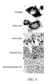
- the heavy strand or H-strand encodes the two ribosomal RNAs (12S and 16S), 14 tRNAs and 12 polypeptides corresponding to ND 1, ND 2, ND 3, ND4L, ND4, ND 5, COX I, COX II, COX III, ATP6, ATP8 and Cyt b.
- the light strand or L-strand codes for 8 tRNAs and the subunit of the complex NAD dehydrogenase ND6 ( Clayton, Hum Reprod., Suppl. 2:11-17, 2000 ; Taanman, Biochim. Biophys. Acta, 1410:103-123, 1999 ).
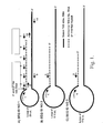
- a large proportion of the mtDNA contains a short three-stranded structure called the displacement loop or D-loop.
- This region that in humans is 1,006 base pairs, is flanked by the genes for tRNA of phenylalanine (tRNA Phe ) and the tRNA of proline (tRNA Pro ) and contains a short nucleic acid strand complementary to the L-strand and displacing the H-strand ( Clayton, Hum Reprod., Suppl. 2:11 -17, 2000 ; Taanman, Biochim. Biophys. Acta, 1410:103-123, 1999 ).
- This region has evolved as the major control site for mtDNA expression and contains the leading-strand or H-strand origin of replication and the major promoters for transcription of the H- (HSP) and L-strand (LSP).
- HSP H-
- LSP L-strand
- these regulatory elements are functionally independent in vitro ( Shuey and Attardi, J. Biol. Chem., 260:1952-1958, 1985 ; Taanman, Biochim. Biophys. Acta, 1410:103-123, 1999 ) as well as in vivo , utilizing model patients with mitochondrial diseases ( Chinnery and Turnbull, Mol. Med. Today, 6:425432, 2000 ).
- RNA polymerase is a protein of 1,230 amino acids with significant homology with the sequence of yeast mitochondrial RNA polymerase and with the RNA polymerases of several bacteriophages ( Tiranti et al., Hum Mol Genet., 6:615-625, 1997 ).
- Mitochondria play a central role in apoptosis, a fundamental biological process by which ceils die in a well-controlled or programmed manner. This cell suicide program is essential during development and for adult homeostasis of all metazoan animals. Apoptosis is activated to eradicate superfluous, damaged, mutated and aged cells ( Meter et al., Nature, 407:796-801, 2000 ). Disregulation of apoptosis is implicated in the appearance of several pathologies. Thus, abnormal inhibition of apoptosis is a hallmark of neoplasia, whereas massive apoptosis has been linked with acute diseases such as stroke, septic shock and neurodegenerative disorders.
- the extrinsic pathway is a process that is initiated at the cell membrane by the binding of different ligands to the death receptors ( Krammer, Nature, 407:789-795, 2000 ; Zornig et al., Biochim. Biophys. Acta, 1551:F1-f37, 2001 ).
- Caspases are responsible for the proteolytic cascade in apoptosis. Caspases are synthesized as inactive precursor proteins that undergo proteolytic maturation or processing upon apoptosis induction ( Zornig et al., Biochim. Biophys. Acta, 1551:F1 - F37, 2001 ). However, more recently several experimental studies indicate that lysosomal proteases constitute an alternative pathway of proteolysis after apoptotic insults ( Guicciardi et al., Oncogene, 23:2881-2890, 2004 ). On the other hand, anti-apoptotic proteins homologous to the human oncoprotein Bc1-2 have been described.
- This protein belongs to a family of proteins that are either anti-apoptotic (Bc1-2, Bcl-XL, Bcl-w) or pro-apoptotic (Bax, Bak, Bim, Bid, etc.) ( Zorning et al., Biochim. Biophys. Acta, 1551:F1-F37, 2001 ).
- Mitochondria are particularly affected early during the apoptotic process and at the present time they are recognized as the central coordinators of cell death ( Boya et al., Biochem. Biophys. Res. Commun., 304:575-581, 2003 ; Ferri and Kroemer, Nature Cell Biol., 3:E255-E263, 2001 ; Zornig et al., Biochim. Biophys. Acta, 1551:F1-F37, 2001 ).
- Several pro-apoptotic signal and damage pathways converge on mitochondria to induce mitochondrial membrane permeabilization, a phenomenon that is under the control of Bc1-2 proteins ( Boya et al., Biochem. Biophys. Res.
- cytochrome c Liu et al., Apoptosis, 6:453-462, 2001 .
- Cytochrome c binds to Apaf-1 (apoptotic protease activation factor-1) facilitating the binding of dATP/ATP to the complex and the oligomerization of Apaf-1 ( Adrain et al., J. Biol. Chem., 274:20855-20860, 1999 ; Benedict et al., J. Biol. Chem., 275:8461-8468, 2000 ). Oligomerization of Apaf-1 allows the recruitment of pro-caspase-9 which catalyzes the proteolytic activation of the precursor and generation of active caspase-9 ( Adrain et al., J. Biol. Chem., 274:20855-20860, 1999 ; Benedict et al., J. Biol. Chem., 275:8461-8468, 2000 ).
- Apaf-1 apoptotic protease activation factor-1
- a family of cytosolic inhibitor of apoptosis proteins has been described and is known as XIAP, c-IAP1 and c-IAP2. These proteins bind to and inhibit processed caspase-3 and caspase-9 and consequently stop the cascade of degradation. However, the cell also contains countermine mechanisms to bypass this anti-apoptotic pathway. In cells undergoing apoptosis, caspases are liberated of this inhibitory effect by binding to IAPs of a protein known as Smac (Second Mitochondrial Activator of Caspases) or DIABLO (Direct IAP Binding protein with Low pl) ( Verhagen et al., Apoptosis, 7:163-166, 2002 ).
- Smac Synd Mitochondrial Activator of Caspases
- DIABLO Direct IAP Binding protein with Low pl
- HtrA2 Another protein, known as HtrA2, is released from the mitochondria into the cytosol after apoptotic insult where the protein binds to IAPs in a similar fashion as does Smac/DIABLO and thereby facilitating caspases activation ( Verhagen et al., Apoptosis, 7:163-166, 2002 ; Martins et al., 2001; Suzuki et al., Mol. Cell, 8:613- 621, 2001 ; Hedge et al., Apoptosis, 7:123-132, 2002 ).
- the apoptosis inducing factor is another component of the apoptotic cascade. After induction of apoptosis, AIF translocates to the cytosol and to the nucleus. In the nucleus, AIF induces peripheral chromatin condensation and DNA fragmentation. AIF also induces several hallmarks of apoptosis like ⁇ m dissipation and phosphatidylserine exposure ( Zornig et al., Biochim. Biophys. Acta, 1551:F1-F37, 2001 ).
- a factor that seems to regulate the apoptotic activity of AIF is the heat schock protein 70 ( Ravagnan et al., Nature Cell Biol., 3:839-843, 2001 ).
- Another mitochondrial factor that exits the mitochondria and translocates into the nucleus like AIF is endonuclease G or Endo G.
- Endo G generates DNA fragmentation even in the presence of caspase inhibitors ( Li et al., Nature, 412:95- 99, 2001 ).
- Endo G may act in similar fashion as CAD (caspase-activated DNAse), a nuclease whose activation critically relies on caspases ( Samejima et al., J. Biol. Chem., 276:45427-45432, 2001 ).
- Cancer is a cellular malignancy whose unique trait, loss of normal control of cell cycle, results in unregulated growth, lack of differentiation, and ability to invade other tissues and metastasize.
- Carcinogenesis is the process by which a normal cell is transformed in a malignant cell.
- Carcinogenesis is a multiple step process beginning with the genetic event of initiation followed by selective expansion of altered cells during promotion to form early adenomas. In the absence of continuous promotion, the adenoma regresses and disappears. With a second genetic event, a small number of promoted adenomas progress to form late adenomas some of which may then undergo malignant conversion ( McKinnell et al., "The Biology Basis of Cancer", Ch. 3, 1998 ).
- the etiology of cancer is complex and includes alteration of the cell cycle regulation, chromosomal abnormalities and chromosome breakage. Infectious agents such several types of oncogenic viruses, chemicals, radiation (ultraviolet or ionizing radiation) and immunological disorders are thought to be the major causes of carcinogenesis ( McKinnell et al., "The Biological Basis of Cancer", Ch. 3, 1998 ).
- mitochondrial mutation might be the primary cause of cell transformation and cancer (Warburg, 1956; Carew and Huang, Mol. Cancer, 1:1-12, 2002 ). Alterations of the mitochondrial DNA (mtDNA) were reported in hematologic malignancies ( Clayton and Vinograd, Nature, 216:652-657, 1967 ) and in breast cancer (Tan et al., 2002; Parrella et al., 2001).
- Mutations of several regions of the mtDNA and deletions have been also identified in patients with colorectal cancer, prostate cancer, ovarian cancer, gastric cancer, pancreatic cancer, hepatocellular carcinoma, esophageal cancer, kidney cancer, thyroid cancer and brain tumors (reviewed by Carew and Huang, Mol. Cancer, 1:1- 12, 2002 ).
- colorectal cancer prostate cancer, ovarian cancer, gastric cancer, pancreatic cancer, hepatocellular carcinoma, esophageal cancer, kidney cancer, thyroid cancer and brain tumors
- the majority of the mutations are base transitions from T to C and G to A.
- the D-loop seems to be the most frequent somatic mutated region of the mtDNA in most tumor types.
- Pre-cancer cells are defined here as a transformed cell which can evolve or differentiate into a malignant cell. Some examples are cells transformed by DNA or RNA oncoviruses.
- the present invention is related to a novel family of mitochondrial RNAs and the use herein of these RNAs for cancer therapy.
- composition and methods are provided to induce massive and selective tumor cell death. Therefore, the present invention provides compositions and methods which may be used in cancer and pre-cancer therapy as well as for research.
- the present invention provides compositions of isolated human mitochondrial chimeric RNA molecules comprising the sense 16S mitochondrial ribosomal RNA covalently linked at its 5' end to the 3' end of a polynucleotide with an inverted repeat sequence.
- a sense mitochondrial chimeric RNA is comprised of an inverted repeat of 815 nucleotides joined covalently to the 5' end of the 16S mitochondrial ribosomal RNA (SEQ ID NO 1).
- the inverted repeat corresponds to a fragment of 815 nucleotides of the RNA transcribed from the L-strand of the 16S gene of the mtDNA.
- this novel RNA requires the transcription of the L-strand and the H-strand of the 16S gene of the mtDNA. Since transcription of both strands of the mtDNA is regulated by different promoters, we refer to this novel RNA in the present invention as the mitochondrial chimeric RNA (SEQ ID NO 1). In addition, since the inverted repeat of 815 nucleotides is joined to the "sense" 16S RNA (transcribed from the H-strand) we refer to this novel RNA as the "sense mitochondrial chimeric RNA.”
- In situ hybridization assays revealed that the sense mitochondrial chimeric RNA is expressed in normal proliferating cells, in tumor cells in culture as well as in tumor cells present in human biopsies of different tumor types.
- the sense mitochondrial chimeric RNA is not expressed in normal resting cells.
- sense mitochondrial chimeric RNA in cells transformed with papilloma virus 16 or 18 Heusen, Biochim. Biophys. Acta, 1288:F55-F78, 1996
- SEQ ID NO 2 a new sense mitochondrial chimeric RNA comprising of an inverted repeat of 754 nucleotides joined covalently to the 5' end of the 16S mitochondrial RNA is expressed (SEQ ID NO 2). This RNA is not present in normal proliferating cells or in tumor cells.
- a third sense mitochondrial chimeric RNA comprising an inverted repeat of 694 nucleotides joined covalently to the 5' end of the 16S mitochondrial RNA (SEQ ID NO 3), is present in cells transformed with HTLV-1.
- methods and compositions are provided to interfere with the sense mitochondrial chimeric RNAs.
- One preferred embodiment is to interfere with the antisense mitochondrial chimeric RNA in tumor cells which contains a low copy number of this transcript.
- the interference is carried out with oligonucleotides or oligonucleotide analogs, whose sequences are complementary to the sequences of the sense mitochondrial chimeric RNA (SEQ ID NO 1 and/or SEQ ID NO 2 and/or SEQ ID NO 3). Treatment of tumor cells of different types with one or more of these complementary oligonucleotides induces cell death or apoptosis.
- oligonucleotides 10 to 50 nucleobases in length which are sufficiently complementary to one human mitochondrial chimeric RNA molecule comprising a sense 16S mitochondrial ribosomal RNA covalently linked at its 5' end to the 3' end of a polynucleotide with an inverted repeat sequence to be able to hybridize with that human mitochondrial chimeric RNA molecule to form a stable duplex.
- the oligonucleotides are preferably compounds of 15 to 50 nucleotides where at least 15 nucleobases are complementary to SEQ ID NO 1 and/or SEQ ID NO 2 and/or SEQ ID NO 3.
- Examples of these complementary oligonucleotides are shown in SEQ ID NOS 98 to 196.
- the induction of apoptosis is selective since treatment of human lymphocytes (normal resting cell) or human lymphocytes stimulated with phytohaemagglutinin (normal proliferating cells) do not undergo apoptosis after treatment with oligonucleotides complementary to the sequences of the antisense mitochondrial chimeric RNA under the same conditions. If the tumor cells are treated with oligonucleotides targeted or complementary to the sense mitochondrial chimeric RNA (SEQ ID NO 1 and/or SEQ ID NO 2 and/or SEQ ID NO 3) a diminished induction of cell death or apoptosis is obtained.
- FIGS. 1A , B and C Line drawings showing the structure of sense mitochondrial chimeric RNAs corresponding to SEQ ID NO 1, SEQ ID NO 2 and SEQ ID NO 3.
- the arrows indicate the relative position of the primers used to amplify the RNA by pieces.
- the arrows below the lines are reverse primers, and the arrows on top of the lines are forward primers.
- Primer 1 is positioned close to the 5' end of the 16S mitochondrial RNA.
- the black lines correspond to the sense 16S mitochondrial RNA, while the dotted lines correspond to the antisense 16S mitochondrial RNA.
- Fig. 2 Agarose gel electrophoresis to show the amplification product obtained between primer 1 and primer 3 indicated in FIG. 1A .
- the amplification was carried out by RT-PCR using as a template a tumor cell (SiHa), human keratinocytes transformed with HPV 16 (HFK698) or B-lymphocytes transformed with HTLV-1.
- SiHa tumor cell
- HFK698 human keratinocytes transformed with HPV 16
- B-lymphocytes transformed with HTLV-1 B-lymphocytes transformed with HTLV-1.
- RNA from SiHa cells only one single amplicon of 210 bp was obtained and corresponds to a segment of SEQ ID NO 1.
- a second amplicon of 150 bp was obtained and corresponds to a segment of SEQ ID NO 2.
- RNA from cells transformed with HTLV-1 besides the amplicons of 210 bp and 150 bp, a third amplicon was obtained and corresponds to
- FIG. 3 Line drawings showing the structure of the antisense mitochondrial chimeric RNAs corresponding to SEQ ID NO 4, SEQ ID NO 5 and SEQ ID NO 6.
- the arrows represent the primer used for amplification, and primer 1 is positioned close to the 5' end of the antisense 16S mitochondrial RNA.
- the strategy to obtain the sequence of these transcripts is similar to that described in Fig. 1 .
- FIGS. 4A and 4B In situ hybridization assays carried out with several tumor cell lines in culture. The cells were hybridized with oligonucleotide probes complementary to the sense mitochondrial chimeric RNA, and labeled with digoxigenin (left panels). In addition, the cells were also hybridized in parallel with oligonucleotide probes complementary to the antisense mitochondrial chimeric RNA labeled with digoxigenin (right panels). The cells lines are identified at the left.
- FIG. 5A In situ hybridization of several sections of human biopsies corresponding to different tumor types. The tumor sections were hybridized with oligonucleotide probes complementary to the sense mitochondrial chimeric RNA, and labeled with digoxigenin (left panels). In addition, parallel tumor sections were hybridized with oligonucleotide probes complementary to the antisense mitochondrial chimeric RNA labeled with digoxigenin (right panels).
- Fig. 5B shows in situ hybridization of different human tumors carried out with oligonucleotide probes complementary to the sense mitochondrial chimeric RNA labeled with digoxigenin.
- FIG. 6 shows in situ hybridization of normal proliferating cells.
- the samples were hybridized with probes targeted to the sense or the antisense mitochondrial chimeric RNA and labeled with digoxigenin.
- a strong hybridization signal was obtained with both probes, one complementary to the sense mitochondrial chimeric RNA (left panels) as well as to the antisense mitochondrial chimeric RNA (right panels).
- the tissues or cells are identified at the left.
- FIG. 7 Immunocytochemistry and in situ hybridization to show expression changes in human lymphocytes stimulated to proliferate with the mitogen PHA. After 48 hours of stimulation with PHA, the lymphocytes express the antigens Ki-67 and PCNA (right panels). These antigens are not expressed in the control or resting lymphocytes (left panels).
- the in situ hybridization was carried out with oligonucleotide probes targeted to the sense and the antisense mitochondrial chimeric RNA and labeled with digoxigenin. The stimulated lymphocytes over express the sense as well as the antisense mitochondrial chimeric RNA (right panels).
- Fig. 8 In situ hybridization of tumor cells showing localization of the sense mitochondrial chimeric RNA in the nucleolus. The cells or tumor sections are indicated at the left.
- FIG. 9 Fluorescent microscopy to reveal changes occurring in tumoral HL- 60 cells treated with oligonucleotide probes targeted to the antisense mitochondrial chimeric RNA.
- A, B. C and D show staining with a compound (VAD-fmok) that binds with high affinity to activated caspases. This compound is labeled with fluorescein.
- the oligonucleotide probes targeted to the antisense mitochondrial chimeric RNA induce activation of caspases in similar manner than the drug staurosporine (compare B and C).
- Activated caspases are not detected in control untreated cells (A) or in cells treated with oligonucleotide probes targeted to the 12S mitochondrial RNA (D), as control.
- E and F show staining of HL-60 cells with DAPI.
- the control cells (untreated) show homogeneous staining of the nucleus (E), while cells treated with the oligonucleotide probes targeted to the antisense mitochondrial chimeric RNA show massive fragmentation of the nucleus (F).
- FIG. 10 Percent of apoptotic cells after different treatment conditions of resting and proliferating lymphocytes. Apoptosis was measured in resting lymphocytes or PHA-stimulated lymphocytes by DAPI staining. The bars 1 and 2 correspond to untreated cells. A low spontaneous apoptosis of control (1) or PHA-stimulated lymphocytes (2) was observed. A similar low level of apoptosis was observed in resting lymphocytes (3) or PHA-stimulated lymphocytes (4) treated with 15 uM oligonucleotide probes targeted to the antisense mitochondrial chimeric RNA for 15 hours, showing that apoptosis is not induced in normal cells. As a control, resting lymphocytes and PHA-stimulated lymphocyte were treated with staurosporine. Under these conditions, around 90% of resting lymphocytes (5) or PHA-stimulated lymphocytes (6) undergo apoptosis.
- the human mitochondrial chimeric RNA family The human mitochondrial chimeric RNA family
- the present invention is based on the surprising discovery that human cells express a family of novel mitochondrial RNAs, referred to as the human mitochondrial chimeric RNAs.
- One of these transcripts contains a long inverted repeat of 815 nucleotides covalently joined to the 5' end of the mitochondrial 16S ribosomal RNA, named sense mitochondrial chimeric RNA.
- the long inverted repeat is fully complementary to the 16S ribosomal RNA from positions 51 to 866, forming a long double stranded stem and a loop of 50 nucleotides.
- the stem of 815 base pairs represents a significant problem for any reverse transcriptase to synthesize the corresponding cDNA. Therefore a new strategy was used to amplify this RNA by RT-PCR which is illustrated in Fig. 1A . After obtaining the sequence of each overlapping fragment, they were assembled as contigs to obtain the complete sequence of the sense mitochondrial chimeric RNA shown in SEQ ID NO 1 ( Fig. 1A ).
- Another aspect of this invention is the discovery of other novel sense mitochondrial chimeric RNAs which are expressed in cells transformed with the oncogenic human papilloma virus 16 or 18.
- Human foreskin keratinocytes HFK
- HPV 16 or 18 Heusen, Biochim. Biophys. Acta, 1 288:F55-F78, 1996 .
- the infection induces transformation or immortalization of the HFK.
- these cells are not tumorigenic such as the related SiHa cells (infected with HPV 16) or HeLa cells (infected with HPV 18).
- SEQ ID NO 1 the sense mitochondrial chimeric RNA
- the transformed cells also express another second sense mitochondrial chimeric RNA which contains an inverted repeat of 754 nucleotides joined to the 16S ribosomal RNA ( Fig. 1B ) (SEQ ID NO 2).
- This new sense mitochondrial chimeric RNA is down regulated or is not expressed in normal human cells (HFK) or in tumorigenic cells (SiHa or HeLa cells).
- a third sense mitochondrial chimeric RNA in cells transformed with HTLV-1 ( Kobayashi et al., EMBO J., 3:1339-1343, 1984 ).
- MT-2 cells infected with HTLV-1 express the sense mitochondrial chimeric RNA (SEQ ID NO 1) and the sense mitochondrial chimeric RNA expressed in cells transformed with HPV 16 or 18 (SEQ ID NO 2).
- SEQ ID NO 2 the sense mitochondrial chimeric RNA expressed in cells transformed with HPV 16 or 18
- the cell infected with HTLV-1 expresses a third sense mitochondrial chimeric RNA containing an inverted repeat of 694 nucleotides joined to the 5' end of the 16S ribosomal RNA.
- This novel RNA ( Fig. 1C ) (SEQ ID NO 3) is not expressed in normal proliferating cells, in tumor cells or in HFK transformed with HPV 16 or 18.
- normal proliferating cells such as human foreskin keratinocytes (HFK) as described in the previous section also over express the sense mitochondrial chimeric RNA ( Fig. 6 ) (SEQ ID NO 1).
- Human lymphocytes stimulated with mitogens such as phytohaemagglutinin (PHA) enter into the S phase of the cell cycle and begin the synthesis of DNA ( Yu et al., J. Biol. Chem., 266:7588-7595, 1991 ).
- the lymphocytes also express antigens related to proliferation such as Ki-67 and proliferating cell nuclear antigen or PCNA ( Bantis et al., Cytopathology, 15:25- 31, 2004 ).
- the stimulated lymphocytes also over express the sense mitochondrial chimeric RNA (SEQ ID NO 1).
- Other proliferating cells such as lymphocytes in the germinal center of the spleen, spermatogonia, and embryonic cells also over express the sense mitochondrial chimeric RNA (SEQ ID NO 1) ( Fig. 4 ).
- non-proliferating cells such as non-stimulated lymphocytes, or muscle cells do not express the sense mitochondrial chimeric RNA ( Fig. 7 ).
- the present invention is also based on the surprising discovery that normal proliferating cells also over express an antisense mitochondrial chimeric RNA.
- the expression of the antisense mitochondrial chimeric RNA was confirmed in human lymphocytes stimulated with PHA ( Fig. 7 ), in normal HFK and in other normal proliferating cells ( Fig. 6 ).
- Another surprising discovery of the present invention is that different to normal proliferating cells, tumor cells do not express the antisense mitochondrial chimeric RNA or markedly down regulated the production (compare Fig. 4 with Fig. 6 and Fig. 7 ).
- HeLa cells infected with HPV 18 or SiHa cells (infected with HPV 16) over express the sense mitochondrial chimeric RNA but down regulate the expression of the antisense mitochondrial chimeric RNAs.
- HFK as normal proliferating cells, over express both the sense as well as the antisense mitochondrial chimeric RNAs. After transformation of HFK with HPV 16 or HPV 18, the cells acquire the tumor phenotype: they over express the sense mitochondrial chimeric RNA and down regulate the expression of the antisense mitochondrial chimeric RNA.
- the over expression of the sense mitochondrial chimeric RNA and down regulation of the antisense mitochondrial chimeric RNA can be determined by in situ hybridization, amplification of the RNA by RT-PCR or by using other methods to determine a RNA by ways well known to the person skilled in the art. These methods and compositions can be used also to determine the change in the expression of the chimeric RNA family in cells transformed with other oncogenic virus or by compounds that induce transformations or carcinogenesis ( McKinnell et al., "The biological basis of Cancer", Cambridge University Press1998 ).
- Chemotherapeutic drugs can induce a series of cellular responses that impact on tumor cell proliferation and survival. The best studied of these cellular responses is apoptosis and is evident at the present time that anti-cancer drugs can kill tumor cells by activating common apoptotic pathways. Unfortunately, these drugs also affect rapidly dividing normal cells of the bone marrow, normal hematopoietic and intestinal cells and hair matrix keratinocytes ( McKinnell et al., The biological Basis of cancer, 1998 ; Komarov et al., Science, 285:1733-1737, 1999 ; Johnstone et al., Cell, 108:153-164, 2002 ).
- a paradigm of a cancer therapy treatment is a procedure that selectively triggers apoptosis of tumor cells, that does not alter normal proliferating cells and that bypasses the altered or mutated factors of the different apoptotic pathways.
- compositions and methods of the present invention are based on the discovery that tumor and pre-tumor cells over express the sense mitochondrial chimeric RNA at similar levels of the normal proliferating cells. However, and in contrast with normal proliferating cells, tumor and pre-tumor cells down regulate the expression of the antisense mitochondrial chimeric RNA.
- pre-tumor and tumor cells over express the sense mitochondrial chimeric RNA and down regulate the expression of the antisense mitochondrial chimeric RNA.
- the suppression or inhibition of the synthesis of the antisense mitochondrial chimeric RNA in pre-tumor and tumor cells constitutes a novel difference in phenotype between a cancer cell and a normal proliferating cell, which is considered as one of the major embodiments of the present invention.
- tumor cells in human biopsies of different cancer types also exhibit the same phenotype of cancer cells in culture ( Fig. 5A and 5B ).
- the function of the sense mitochondrial chimeric RNA and the antisense mitochondrial chimeric RNA is not clear, a close correlation exists between the expression of these RNAs and cell proliferation.
- normal proliferating cells in tissues like liver, kidney and spleen and defined as such by the expression of the antigens such Ki-67, PCNA or phosphorylated histone H3, over express the sense mitochondrial chimeric RNA as well as the antisense mitochondrial chimeric RNA.
- the non-proliferating cells of the same tissues which do not express Ki-67 or PCNA, the sense mitochondrial chimeric RNA and the antisense mitochondrial chimeric RNA are not expressed.
- resting lymphocytes or non-stimulated lymphocytes do not express neither the sense mitochondrial chimeric RNA nor the antisense mitochondrial chimeric RNA.
- oligonucleotides complementary to the sense or antisense mitochondrial chimeric RNAs undergo apoptosis (Table 2).
- the changes observed in the treated cells were chromatin condensation, nuclear fragmentation, DNA fragmentation, activation of caspases and altered process of the cell membrane.
- Control oligonucleotides with 4 or more mismatches or scrambled oligonucleotides did not induce apoptosis.
- cells were not affected if treated with oligonucleotides targeted to the sense or antisense 12S mitochondrial RNA or targeted to the mRNA or the antisense transcript of the mitochondrial ND1 subunit.
- oligonucleotides targeted to the antisense mitochondrial chimeric RNA were much more effective, at the same concentration, than oligonucleotides targeted to the sense mitochondrial chimeric RNA. This was an expected result since the tumor cells over express the sense mitochondrial chimeric RNA and therefore it is more difficult to reach a concentration of oligonucleotides inside the cell to interfere with all the copies of this transcript. On the other hand, since tumor cells down regulate the antisense mitochondrial chimeric RNA, it should be easier to interfere with this RNA since there are a lower number of copies per cell.
- apoptosis is also selective for tumor cells. Resting human lymphocytes or human lymphocytes stimulated for 48 hours with PHA are not affected by the treatment with oligonucleotides complementary to the antisense mitochondrial chimeric RNAs or targeted to the sense mitochondrial chimeric RNA even after overnight treatment and with a high dose of complementary oligonucleotides (15 uM).
- Apoptosis induction by treatment with complementary oligonucleotides targeted to the antisense mitochondrial chimeric RNA has been achieved by, but is not limited to, promielocytic leukemia cell HL-60, acute lymphoblastic leukemia MOLT- 4, T-lymphocitic leukemia cells, Jurkat, a T-cell leukemia, Devernelle or B-lymphoma, NSO/2 or myeloma, HeLa cells, DU145, PC- 3, Caco-2, Hep-2 and HepG2.
- MCF/7 breast carcinoma
- melanoma Two cells, MCF/7 (breast carcinoma) and melanoma, that can be considered as a paradigm of treatment-resistant (chemotherapy or radiotherapy) tumor cells undergo apoptosis at a rate of over 80% when treated for 15 hours with complementary oligonucleotides targeted to the antisense mitochondrial chimeric RNA (SEQ ID NO 4, SEQ ID NO 5, SEQ ID NO 6).
- SEQ ID NO 4 SEQ ID NO 5
- SEQ ID NO 6 A lower apoptotic effect was obtained with oligonucleotides complementary to the sense chimeric RNA (SEQ ID NO 1).
- SEQ ID NO 1 sense chimeric RNA
- Described below are methods and compositions for treating cancer using the sense chimeric RNAs as a therapeutic target.
- the preferred embodiments are methods and compositions for treating cancer using oligonucleotides complementary to the chimeric RNAs.
- the outcome of this treatment is to at least produce in a treated subject a healthful benefit, which in the case of cancer, includes, but is not limited to, remission of the cancer, palliation of the symptoms of the cancer, and control of metastatic spread of the cancer. All such methods involve the induction of apoptosis in the tumor cells and with minor effect in normal cells.
- Complementary oligonucleotides that target specific RNAs have been used to diminish or abrogate the expression of a large variety of mRNA or the synthesis of the corresponding proteins (e.g. Vickers et al., J. Biol.
- one or more oligonucleotides targeted to the mitochondrial chimeric RNA can be used.
- the use of two or more complementary oligonucleotides is more effective and shows some degree of synergism.
- the oligonucleotide of the invention may be complementary to the sense mitochondrial chimeric RNA.
- the complementary oligonucleotides will bind to the sense mitochondrial chimeric RNAs and interfere with their functions. Absolute complementarily, although preferred, is not required.
- An oligonucleotide sequence "complementary" to a portion of an RNA, as referred to herein, means a sequence having sufficient complementarily to be able to hybridize with the RNA, forming a stable duplex. The ability to hybridize will depend on both the degree of complementarily and the length of the oligonucleotide.
- the longer the hybridizing nucleic acid the more base mismatches with an RNA it may contain and still form a stable duplex.
- Those skilled in the art can ascertain a tolerable degree of mismatch by use of standard procedures to determine the melting point of the hybridized complex.
- complementary oligonucleotides to hybridize with mRNAs for different proteins are targeted to the 5' untranslated region of the mRNA including the complement of the AUG start codon, or the 3' untranslated region to be more effective.
- Oligonucleotides complementary to mRNA coding regions are less efficient inhibitors of translation (see previous references).
- the sense mitochondrial chimeric RNAs are non-coding RNA and therefore the target region of the oligonucleotides can be complementary to any region of these transcripts.
- the most effective regions are located around the single-stranded segments of the antisense mitochondrial chimeric RNA determined by scanning the sequences of the sense mitochondrial chimeric RNA with complementary oligonucleotides designed every 30 nucleotides.
- SEQ ID NO 1 SEQ ID NO 2 and SEQ ID NO 3
- SEQ ID NO 3 are targets to design alternative complementary oligonucleotides.
- the complementary oligonucleotides targeted to the sense mitochondrial chimeric RNA resulting in the induction of tumor cell death according to the present invention will generally contain backbones different to the natural phosphodiester bonds.
- the oligonucleotides can have alternate inter nucleoside linkages, comprising, but not limited to, phosphorothioate ( Mag et al., Nucleic Acids Res., 19:1437-1441, 1991 ; and U.S. Pat. No. 5, 644,048 ), peptide nucleic acid or PNA ( Egholm, Nature, 365:566-568, 1993 ; and U.S. Pat. No. 6,656,687 ), phosphoramide ( Beaucage, Methods Mol.
- oligonucleotide analogs include, but are not limited to, morpholino ( Summerton, Biochim. Biophys. Acta, 1489:141-158, 1999 ), locked oligonucleotides ( Wahlestedt wt al., Proc. Natl. Acad.
- the nucleic acids may contain any combination of deoxyribo- and ribonucleotides, and any combination of bases, including uracil, adenine, thymine, cytosine, guanine, inosine, xathanine hypoxathanine, isocytosine, isoguanine, etc.
- the complementary oligonucleotides according to the invention may comprise at least one modified base moiety which is selected from the group including, but not limited, to 5- fluorouracil, 5-bromouracil, 5-chlorouracil, 5-iodouracil, hypoxanthine, xanthine, 4-acetylcytosine, 5-(carboxyhydroxylmethyl) uracil, 5-carboxymethylaminomethyl-2-thiouridine, 5-carboxymethylaminomethyluracil, dihydrouracil, beta-D-galactosylqueosine, inosine, N6-isopentenyladenine, 1- methylquanine, 1-methylinosine, 2,2-dimethylguanine, 2-methyladenine, 2- methylquanine, 3-methylcytosine, 5-methylcytosine, N6-adenine, 7- methylquanine, 5-methylaminomethyluracil, 5-methoxyaminomethyl-2- thiouracil, beta-
- methylester uracil-5-oxyacetic acid (v), 5-methyl-2- thiouracil, 3-(3-amino-3-N-2-carboxypropyl) uracil, (acp3)w, and 2,6-diaminopurine.
- the complementary oligonucleotides may also comprise at least one modified sugar moiety selected from the group including but not limited to arabinose, 2-fluoroarabinose, xylulose, and hexose.
- the complementary oligonucleotides are designed to hybridize with any region of the sense mitochondrial chimeric RNA.
- the complementary oligonucleotides should be at least ten nucleotides in length, and are preferably complementary oligonucleotides ranging from 10 to about 50 nucleotides in length.
- the complementary oligonucleotide is at least 12 nucleotides, at least 18 nucleotides, at least 22 nucleotides, at least 30 nucleotides, at least 50 nucleotides.
- the complementary oligonucleotides targeted to the sense mitochondrial chimeric RNAs are used as therapeutic agents to animals or to patients having cancer can induce sensitivity to anti-cancer therapeutic drugs and radiation. Induced sensitivity, also known as sensitization or hypersensitivity, can be measured in tumor cells showing tolerance to anti-cancer therapeutic or radiation.
- the anti-cancer drugs comprise those already known in the art and in use or as-yet undiscovered drugs.
- the conventional chemotherapeutic drugs are alkylating agents, anti-metabolite, antibiotics and anti-microtubule agents. Some examples of these drugs are cisplatin, methotrexate, doxorubicin dactinomycin, mitomycin, cyclophosphamide, etc.
- the patient can be treated with radiotherapy, wherein said radiotherapy includes ultraviolet radiation, gamma radiation, alpha particles, beta particles, X-ray and electron beams.
- RNA interference In another aspect of this invention, interference with the function of the sense mitochondrial chimeric RNA to induce tumor cell death can be achieved by RNA interference or RNA silencing.
- RNA interference RNA interference
- Synthetically synthesized double stranded RNA molecules of about 19 to 21 nucleotides in length hybridize specifically to their complementary target mRNA, leading to degradation of the mRNA and subsequent protein knockdown.
- RNA or siRNA small interfering RNA or siRNA
- small interfering RNA or siRNA Lu et al., Curr. Opin. Mol. Ther., 5:225-234, 2003 .; Wacheck et al., Oligonucleotides, 13:393-400, 2003 ). Therefore, synthetic double stranded RNA of about 19 to 21 nucleotides targeted to the antisense mitochondrial chimeric RNA or to the sense mitochondrial chimeric RNA can be used to degrade these transcripts and induce tumor cell death.
- siRNA has to be complementary to any region of the antisense mitochondrial chimeric RNAs or to the sense mitochondrial chimeric RNAs (SEQ ID NO 1, SEQ ID NO 2 and SEQ ID NO 3).
- ribozymes can be used to interfere with the sense mitochondrial chimeric RNA to induce tumor cell death.
- the sequence of the ribozyme has to be designed according to the sequence of the sense mitochondrial chimeric RNA (SEQ ID NO 1, SEQ ID NO 2, SEQ ID NO 3) to cleave specific regions of the transcript that are more efficient to trigger tumor cell death.
- Ribozymes are enzymatic RNA molecules capable of catalyzing the specific cleavage of RNA ( Ross), Curr. Biology, 4:469-471, 1994 ).
- the mechanism of ribozyme action involves sequence specific hybridization of the ribozyme molecule to complementary target RNA, followed by an endonucleolytic cleavage.
- composition of ribozyme molecules must include one or more sequences complementary to the target gene mRNA, and must include the well known catalytic sequence responsible for mRNA cleavage, and described in U.S. Pat. No. 5,093,246 .
- hammerhead ribozyme molecules are engineered that specifically and efficiently catalyze endonucleolytic cleavage of the antisense mitochondrial chimeric RNAs or the sense mitochondrial chimeric RNAs.
- the construction and production of hammerhead ribozymes is well known in the art and it was described ( Haseloff et al., Gene, 82:43-52, 1989 ).
- Ribozymes of the present invention also include RNA endoribonucleases ( Zaug et al., Science, 224:574-578, 1984 ).
- Gene therapy refers to treatment or prevention of cancer performed by the administration of a nucleic acid to a patient who has cancer or in whom prevention or inhibition of cancer is desirable.
- the therapeutic nucleic acid produced intracellularly is a complementary RNA targeted to the sense mitochondrial chimeric RNA that mediates the therapeutic effect by interfering or inhibiting the function of these mitochondrial transcripts, inducing tumor cell death. Therefore, one preferred approach is to utilize a recombinant DNA construct in which the transcription of the antisense RNA is placed under the control of strong promoters of RNA polymerase II or III. Expression of the sequence encoding the complementary RNA can be by any promoter known in the art to act in mammalian, preferably human cells.
- Such promoters include but are not limited to SV40 early promoter region ( Benoist and Chambon, Nature, 290:304-310, 1981 ), the herpes thymidine kinase promoter ( Wagner et al., Proc. Natl. Acad. Sci. U.S.A., 78:1441- 1445, 1981 ), the regulatory sequences of the metallothionein gene ( Brinster et al., Nature, 296:39-42, 1982 ), the promoter of Rous sarcoma virus ( Yamamoto et al., Cell, 22:787-797, 1980 ), etc.
- the recombinant DNA construct to produce the complimentary RNA can be a viral vector which includes, but is not limited to adenovirus vector, adeno-associated virus vector, herpes simplex virus vector, vaccinia virus vector and retrovirus vectors.
- the vector is introduced in the target tumor cells, in a pharmaceutical composition, using methods familiar to those skilled in the art.
- compositions of the invention comprising an effective amount of a complementary nucleic acid (complementary oligonucleotides, siRNA, ribozymes or viral vectors) in a pharmaceutically acceptable carrier, that can be administered to a patient having cancer to interfere with the function of the sense mitochondrial chimeric RNA and induce apoptosis of the tumor cells.
- the complementary nucleic acids may be formulated in a pharmaceutical composition, which may include carriers, diluents, buffers, preservatives, surface active agents, polyethylenimide (PEI), liposomes or other lipid formulation known in the art.
- the pharmaceutical composition may be administered by topical application, oral, parenteral or rectal administration. Parenteral administration includes intravenous, subcutaneous, intraperitoneal or intramuscular injection or pulmonary administration by inhalation or insufflation.
- compositions of the present invention can be utilized for therapeutics, prophylaxis and as research reagents and kits.
- cancer includes cells afflicted by any one of the following identified anomalous conditions. These include myeloid leukemia acute or chronic, lymphoblastic leukemia acute or chronic, multiple myeloma, Hodgkin's disease, non-Hodgkin's lymphoma or malignant lymphoma; stomach carcinoma, esophagus carcinoma or adenocarcinoma, pancreas ductal adenocarcinoma, insulinoma, glucagonoma, gastrinoma, small bowel adenocarcinoma, colorectal carcinomas; hepatocellular carcinoma, hepatocellular adenoma; carcinoids, genitourinary tract such as kidney adenocarcinoma, Wilm's tumor, bladder and urethra carcinoma and prostate adenocarcinoma, testis cancer like seminoma, teratoma,
- cancerous cell includes a cell afflicted by any one of the above-identified conditions.
- the segment of the antisense 16S mitochondrial RNA was fully complementary to the sense 16S mitochondrial RNA and therefore corresponded to an inverted repeat joined to the 5' end of the sense 16S transcript. Based on this structure, primers were designed to amplify this putative transcript by RT-PCR.
- One reverse primer was at position 11 to 31 from the 5' end of the human sense 16S mitochondrial RNA or at the beginning of the theoretical loop (primer 1, Fig. 1A ) (SEQ ID NO 139).
- the sequence of the forward primer used was that of positions 213-234 of the sense 16S mitochondrial RNA, and corresponds to primer 3 in Fig. 1A .
- RNA from several human tissues and cells including HeLa, HL-60, Du145, MCF/7 and human lymphocytes stimulated with PHA see Example 7
- RT-PCR was carried out as described before ( Villegas et al., DNA & Cell Biol.,19.579-588, 2000 ; Villegas et al., Nucleic Acids Res., 30:1895-1901, 2002 ).
- the amplicons from each human tissue or cells were cloned and both strands were sequenced.
- human cDNA was amplified by PCR between the forward primer 2 positioned at the 3' end of the inverted repeat and primer 3, which is also reverse at positions 213-234 of the sense 16S mitochondrial RNA ( Fig.1A ).
- An amplicon of approximately 240 bp was obtained and the sequence showed that the first 234 nucleotides of the sense 16S mitochondrial RNA were joined to the last 25 nucleotides of the 3' end of the inverted repeat.
- the sequence of the 25 nucleotides of the inverted repeat was fully complementary to the sense 16S mitochondrial RNA from positions 51 to 75 ( Fig. 1A ).
- the emerging structure of the human sense mitochondrial chimeric RNA confirmed a loop of 50 nucleotides and a double stranded structure of at least 769 bp ( Fig. 1A ) (see also SEQ ID NO 1).
- RNA from HeLa or other cells was digested with RNase A (50 ug per ml), followed by phenol extraction, and the nuclease-resistant material was recovered by ethanol precipitation. The cDNA from the digested RNA was then amplified by PCR using the primers showed in Fig. 1A .
- the amplicon of about 800 bp obtained with primers 1 and 6 was not amplified after RNase A digestion indicating that the loop was digested by the enzyme. The same was true with the amplicon of 360 bp obtained with primers 10 and 11 as indicated in Fig. 1A . Altogether, these results indicated that the loop as well as the 3' region of the sense mitochondrial chimeric RNA that extends beyond the stem, were digested by the enzyme. On the other hand, amplification of the 750 bp amplicon, corresponding to the double stranded structure of the sense mitochondrial chimeric RNA and obtained with primers 8 and 6, was not affected by the RNase A digestion. The sequence of the double strand fragment resistant to ribonuclease digestion was identical with the expected sequence of the stem. The same results were obtained after digestion of total RNA from HL- 60 cells or other human cells.
- the stem of the transcript obtained after RNase A digestion was used for 5' RACE analysis.
- the 5' end determination of the inverted repeat was carried out according to the manufacturer's instructions (Invitrogen). The results indicated that the inverted repeat extends for 46 additional nucleotides from the 5' end of the amplicon obtained after amplification of the sense mitochondrial chimeric RNA with primers 1 and 6.
- the inverted repeat of 815 nucleotides is joined to the 5' end of the first 865 nucleotides of the 16S mitochondrial RNA.
- the sequence of this transcript showed 99.8% identity with the human 16S mitochondrial gene (H and L strand) (SEQ ID NO 1). The 5' ends of both extremes of the double stranded stem were confirmed by 5' RACE.
- the sense mitochondrial chimeric RNA contained a stem or double stranded structure of 815 base pair and a loop of 50 nucleotides.
- these results do not prove that the inverted repeat is joined to the complete 16S sense mitochondrial RNA.
- the use of conventional approaches such as synthesis of the complete cDNA from the 3' end is useless, since the double stranded structure of the transcript represents an insurmountable problem to reverse transcriptases, including Tth ( Myers and Gelfand, Biochemistry, 30:7661- 7666, 1991 ). If the inverted repeat of 815 nucleotides is joined to the 1559 nucleotides of the 16S mitochondrial RNA one would expect a transcript of 2.3 Kb.
- oligonucleotide probe It is possible to specifically detect the junction region between the inverted repeat and the sense 16S mitochondrial RNA, using an oligonucleotide probe.
- the probe has to include 7 to 10 nucleotides at each side of the joining point between the 3' end of the inverted repeat and the beginning of the sense 16S mitochondrial RNA.
- This oligonucleotide can be used for in situ hybridization or amplification by RT- PCR or any other methods familiar to those skilled in the art to detect this novel RNA.
- the sense mitochondrial chimeric RNA is present in normal proliferating cells (human foreskin keratinocytes, spleen, lymphocytes stimulated with PHA, mouse embryos), in pre-cancer cells (keratinocytes transformed with HPV 16 or 18, MT-2 cells transformed with HTLV-1) and in tumor cells. It is not present in normal resting cells. A summary of these results is presented in Table 1 (in Example 4).
- Fig. 1B Human keratinocytes transformed with papilloma virus synthesize a novel sense mitochondrial chimeric RNA (Fig. 1B, SEQ ID NO 2).
- HFK Human foreskin keratinocytes
- HPV 16 human papilloma virus 16
- the cells were cultured with 3 parts of K-SFM, one part of DMEM medium (Invitrogen), 5 ng/ml of EGF, 50 ug/ml of pituitary extract and 10% calf fetal serum.
- the culture conditions were 37°C and 5% CO 2 .
- the transformed HFK were transferred to a new flask and grown under the same conditions. After this time the cells (HFK698) were successively transferred to new culture flasks every 3 days using a split ratio of 1:3 to 1:4.
- This amplification protocol yielded the expected amplicon of 210 bp where the first 31 nucleotides of the sense 16S mitochondrial RNA are joined to the inverted repeat of 184 nucleotides as described before in Example 1. Electrophoresis analysis of the amplification products revealed the presence of the amplicon of 210 base pairs corresponding to the sense mitochondrial chimeric RNA, plus another amplification fragment of about 150 base pairs as shown in Fig. 2 .
- an oligonucleotide probe can be used for the specific detection of this transcript.
- the probe has to include 7 to 10 nucleotides at each side of the joining point between the 3' end of the inverted repeat and the beginning of the sense 16S mitochondrial RNA, such as the oligonucleotide of SEQ ID NO 7.
- This oligonucleotide can be used for in situ hybridization or amplification by RT-PCR or any other methods familiar to those skilled in the art to detect this novel and specific marker of pre-cancer cells.
- Cells transformed with HTLV-1 induce the expression of a third novel sense mitochondrial chimeric RNAs (Fig. 1C, SEQ ID NO 3).
- Human MT-2 cells transformed with HTLV-1 were cultured as described ( Kobayashi et al., EMBO J., 3:1339-1343, 1984 ). The cells were harvested, centrifuged at 300 x g for 10 min and washed twice with PBS. The final cell pellet was extracted with Trizol as described in Example 1. The cDNA was synthesized with random hexamers using the RNA as template and the cDNA was amplified by PCR using the reverse primer 1 and the forward primer 3 as described in Fig. 1A .
- this amplification protocol yields an amplicon of 210 base pairs that contains the first 31 nucleotides of the sense 16S mitochondrial RNA joined to an inverted repeat of 184 nucleotides which corresponds to the sense mitochondrial chimeric RNA as described in Example 1.
- Electrophoresis analysis of the amplification products revealed, besides the presence of the already discussed amplicon of 210 base pairs, a band of about 150 base pairs (see Fig. 2 ).
- the sequence of the amplicon of 150 base pairs is identical to the sequence of the amplicon described in Example 2, corresponding to a second sense mitochondrial chimeric RNA expressed in cells transformed with HPV 16 or HPV 18 (SEQ ID NO 2).
- a new amplification product was found of about 100 bp ( Fig. 2 ).
- the sequence of this third amplicon revealed an inverted repeat of 61 nucleotides joined to the 5' end of the sense 16S mitochondrial RNA and generating a loop of 167 nucleotides ( Fig. 1C ; SEQ ID NO 3).
- This novel amplicon was not present in normal cells, in tumor cells and in cells transformed with HPV 16 or 18. Therefore, this new sense mitochondrial chimeric RNA is a potential marker of cells transformed with the oncogenic retrovirus HTLV-1.
- an oligonucleotide probe can be used for the specific detection of this transcript.
- the probe has to span between 7 to 10 nucleotides at each side of the joining point between the 3' end of the inverted repeat and the beginning of the sense 16S mitochondrial RNA, such as an oligonucleotide of SEQ ID NO 8.
- This oligonucleotide can be used for in situ hybridization or amplification by RT-PCR or any other methods familiar to those skilled in the art to detect this specific marker of cells transformed with a retroviral oncogenic virus.
- PHA phytohaemagglutinin
- the blood was diluted with one volume of 0.9% NaCl and the mixture was applied on 5 ml of Histopaque-1077 (Sigma) in a centrifuge tube.
- the tubes were centrifuged at 800 x g for 20 minutes at room temperature.
- the white cells at the interphase were collected, diluted with 2 volumes of 0.9% NaCl and centrifuged at 250 x g for 10 minutes at room temperature.
- the collected cells were suspended and washed twice with RPMI 1640 medium supplemented with 200 mM glutamine, 10mM non-essential amino acids, penicillin and streptomycin devoid of calf fetal serum.
- the final sediment was re-suspended in the same medium with 10% calf fetal serum and the number of human lymphocytes per ml was determined by counting under the microscope in a Neubauer chamber.
- Human lymphocytes were cultured in 96-wells microtiter plates with the RPMI 1640 medium supplemented as described plus 10% calf fetal serum at 37°C and with 5% CO 2 . About 30,000 lymphocytes per well were cultured with or without 10 ug per ml of the mitogen PHA, which induce cell proliferation ( Yu et al., J. Biol. Chem., 266:7588-7595, 1991 ). After 48 to 72 hours of treatment with PHA, the cells are actively engaged in DNA synthesis as demonstrated by the incorporation of H3-thymidine or BrdU ( Yu et al., J. Biol. Chem., 266:7588-7595, 1991 ).
- lymphocytes overexpressed other markers of cell proliferation such as the proliferating cell nuclear antigen or PCNA and Ki-67 ( Bantis et al., Cytopathology, 15:25-31, 2004 ) ( Fig. 7 ).
- PCNA proliferating cell nuclear antigen
- Ki-67 Ki-67
- the cells were subjected to in situ hybridization with oligonucleotide probes labeled with digoxigenin and targeted to the sense mitochondrial chimeric RNA.
- the in situ hybridization protocol employed was described in Example 5.
- a strong hybridization signal was obtained indicating overexpression of this transcript ( Fig. 7 ).
- the hybridization signal was similar in intensity to that observed on tumor cells or other normal proliferating cells (compare Fig. 7 with Fig. 4A and B . Fig. 5A and B ). No hybridization signal was observed on the control lymphocytes incubated without PHA ( Fig. 7 ).
- the sense mitochondrial chimeric RNA exhibits different localizations in normal and tumor cells.
- HeLa cells were subjected to in situ hybridization with oligonucleotide probes labeled with digoxigenin as described in Example 5. After post-hybridization and the washing procedures, the cells were incubated with an anti-digoxigenin antibody labeled with rhodamine (Roche) and an anti- Lamp-2 antibody labeled with fluorescein (BD Pharmingen). After incubation at room temperature for 3 hours in the dark, the slides were washed, mounted and analyzed with a Zeiss confocal microscope. A clear co-localization of the hybridization signal with the localization of Lamp-2 was obtained.
- RNA specially a mitochondrial transcript
- Determination of the localization of the sense mitochondrial chimeric RNA in tumor cells may have important prognostic value for patients with cancer.
- the sense and the antisense mitochondrial chimeric RNAs are mainly localized in the nucleus.
- HL-60 cells were cultured under the optimal conditions recommended by ATCC. About 30,000 cells were cultured in 96-well microtiter plates. Oligonucleotides (2 uM) targeted to the sense or to the antisense mitochondrial chimeric RNA were added. To enhance the permeability of the cells, the oligonucleotides were added in a mixture with lipofectamin or oligofectamin (Invitrogen) or with polyethylenimide (PEI) (Exgen TM500, Fermentas). PEI was preferred because is practically non-toxic to the cells. The cells were incubated with the oligonucleotides for 6 hours and the percentage of cell survival was determined by permeability to trypan blue.
- oligonucleotides targeted to the antisense mitochondrial chimeric RNA were more effective to induce cell death (about 90% versus 15% of cell death).
- no apoptosis was induced when the cells were treated with oligonucleotides targeted to the sense or antisense 12S mitochondrial RNA or the mRNA of ND1 subunit or with scrambled oligonucleotides or oligonucleotides with four mismatches, all of which were used as controls.
- the oligonucleotides used in these studies contain phosphorothioate linkage in the first 5 nucleotides at the 5' end and the last five nucleotides at the 3' end. On the average, the 10 central nucleotides contain phosphodiester bonds.
- HL-60 cells were incubated under the same conditions described before with oligonucleotides for 6 hours.
- About 30,000 HL-60 cells were cultured in 200 ul of IDMEM plus 10% calf fetal serum in 96-wells microtiter plate together with 1 uM of oligonucleotides targeted to the sense mitochondrial chimeric RNA or targeted to the antisense mitochondrial chimeric RNA.
- the chemistry of the oligonucleotides added in mixture with PEI was the same described in the previous section.
- the cells were assayed for DNA fragmentation using the TUNEL assay (DeadEnd Colorimetric TUNEL System, Promega). As shown in Table 2, about 96% of the cells showed DNA fragmentation after treatment with the oligonucleotide targeted to the antisense mitochondrial chimeric RNA. A similar rate of DNA fragmentation was obtained with the drug staurosporine. Scrambled oligonucleotides or oligonucleotides with mismatches showed no effect. In contrast, only about 20% of the cells died when treated with oligonucleotide targeted to the sense mitochondrial chimeric RNA (Table 2).
- oligonucleotides targeted to the antisense mitochondrial chimeric RNA induced caspases activation.
- Caspases are proteolytic enzymes, actively involved in programmed cell death or apoptosis.
- HL-60 were incubated with oligonucleotides targeted to the antisense mitochondrial chimeric RNA or with staurosporine for 6 hours under the culturing conditions described before.
- VAD-fmk CaspaCe FITC-VAD-FMK, Promega conjugated with fluorescein was added to the culture and incubated for 30 minutes at 37°C.
- VAD-fmok is strong inhibitor of caspases and binds to the proteases with very high affinity ( Gracia-Calvo et al., J. Biol. Chem., 273:32608-32613, 1998 ).
- the cells were washed by centrifugation, mounted and observed with a fluorescence microscope.
- HL-60 cells treated with the oligonucleotide targeted to the antisense mitochondrial chimeric RNA induced activation of caspases, at a similar level to the activation achieved with staurosporine. No activation of caspases was obtained with antisense oligonucleotides targeted to the 12S mitochondrial RNA used as control.
- the cells treated with oligoncleotides targeted to the antisense mitochondrial chimeric RNA also exhibit other changes that are congruent with apoptosis. Electron microscopy analysis showed nuclear fragmentation and chromatin condensation. Nuclear fragmentation was also demonstrated by staining of the nuclei with DAPI. After treatment with these oligonucleotides targeted to the antisense mitochondrial chimeric RNA, the cells undergo nuclear fragmentation as revealed by DAPI staining ( Fig. 9E and 9F ).
- Apoptosis can also be determined by trypan blue staining, propidium iodide staining, anexine immunochemistry. In these techniques, the cells can be analyzed by fluorescent microscopy or by flow cytometry. DNA fragmentation can be measured by TUNEL or by electrophoresis to reveal the ladder of DNA. Western blot analysis can also be used to determine the processing of proteins such as caspases, poly(ADP-Rib) synthase, etc.
- oligonucleotides complementary to the antisense mitochondrial chimeric RNA are refractory to apoptosis.
- Human lymphocytes were stimulated with 10 ug per ml of PHA for 48 hours as described in Example 8. In parallel, control lymphocytes were incubated also for 48 hours but without PHA. At 48 hours of culture, 15 uM of oligonucleotide mixed with PEI (see Example 10) was added to the stimulated and control lymphocytes and further incubated with 15 hours. The concentration of the oligonucleotide was 10 fold higher than the concentration used in previous experiments (1-2 uM). Other samples of stimulated or control lymphocytes were treated with 0.4 uM staurosporine for the same period of time. At the end of the experiment, cell death was measured by either trypan blue staining or DAPI staining. As shown in Fig.
- control lymphocytes or lymphocytes stimulated with PHA incubated for 15 hours without oligonucleotide showed a similar level of spontaneous apoptosis that varied between 7 to 10% in different experiments. A similar result was obtained with a lower (1-2 uM) concentration of oligonucleotide.
- control and stimulated lymphocytes incubated with 15 uM antisense oligonucleotide for 15 hours showed a similar low level of apoptosis (around 10%) ( Fig. 10 ).
- control lymphocytes or lymphocytes stimulated with PHA and also incubated with staurosporine for 15 hours showed that over 80% of the cells undergo apoptosis ( Fig. 10 ).
Abstract
Description
- The invention relates to cancer therapy and research reagents in connection with a novel family of human mitochondrial RNAs referred to as human mitochondrial chimeric RNAs. In particular, this invention relates to oligonucleotides targeted to the human mitochondrial chimeric RNAs. The oligonucleotides of the invention hybridize to the chimeric RNAs inducing cancer cell death. The composition and methods provided in the invention are useful as a new cancer therapy.
- Mitochondria are subcellular organelles that manufacture the essential molecule adenosine triphosphate (ATP) by oxidative phosphorylation. The human mitochondrial DNA (mtDNA) of 16,654 base pairs encodes two ribosomal RNAs, 22 transfer RNAs (tRNAs) and 13 open reading frames (ORF) that encode a similar number of polypeptides (Clayton, Hum Reprod., Suppl. 2:11-17, 2000; Taanman, Biochim. Biophys. Acta, 1410:103-123, 1999). On the basis of the content of G+T base composition, the two strands of the mtDNA differ in buoyant density and can be separated in denaturating cesium chloride gradients. The heavy strand or H-strand encodes the two ribosomal RNAs (12S and 16S), 14 tRNAs and 12 polypeptides corresponding to
ND 1,ND 2,ND 3, ND4L, ND4,ND 5, COX I, COX II, COX III, ATP6, ATP8 and Cyt b. The light strand or L-strand codes for 8 tRNAs and the subunit of the complex NAD dehydrogenase ND6 (Clayton, Hum Reprod., Suppl. 2:11-17, 2000; Taanman, Biochim. Biophys. Acta, 1410:103-123, 1999). - A large proportion of the mtDNA contains a short three-stranded structure called the displacement loop or D-loop. This region, that in humans is 1,006 base pairs, is flanked by the genes for tRNA of phenylalanine (tRNAPhe) and the tRNA of proline (tRNAPro) and contains a short nucleic acid strand complementary to the L-strand and displacing the H-strand (Clayton, Hum Reprod., Suppl. 2:11 -17, 2000; Taanman, Biochim. Biophys. Acta, 1410:103-123, 1999). This region has evolved as the major control site for mtDNA expression and contains the leading-strand or H-strand origin of replication and the major promoters for transcription of the H- (HSP) and L-strand (LSP). Despite the close proximity of the HSP and LSP (about 150 bp), these regulatory elements are functionally independent in vitro (Shuey and Attardi, J. Biol. Chem., 260:1952-1958, 1985; Taanman, Biochim. Biophys. Acta, 1410:103-123, 1999) as well as in vivo, utilizing model patients with mitochondrial diseases (Chinnery and Turnbull, Mol. Med. Today, 6:425432, 2000).
- Both strands are transcribed as polycistronic RNAs which are then processed to release the individual mRNAs, tRNAs and the rRNAs (Taanman, Biochim. Biophys. Acta, 1410:103-123, 1999). In humans, the mitochondrial RNA polymerase is a protein of 1,230 amino acids with significant homology with the sequence of yeast mitochondrial RNA polymerase and with the RNA polymerases of several bacteriophages (Tiranti et al., Hum Mol Genet., 6:615-625, 1997). In addition, a family of transcription factors has been characterized such as the mitochondrial transcription factor A or TFAM which is essential for mammalian mtDNA transcription and is a member of the high mobility group (HMG)-box family of DNA-binding proteins (Paris) and Clayton, Science, 252:965-969, 1991). Recently, two independent reports described the characteristics of new transcription factors, TFB1M and TFB2M, in human and mouse (McCulloch et al., Mol. Cell Biol., 22,1116-1125, 2002; Falkenberg et al., Nat. Genet., 31:289-294, 2002; Rantanen et al., Mamm. Genome, 14:1-6, 2003). In spite of the considerable progress achieved on the cis- and trans-acting elements involved in mtDNA transcription, the functional details are not fully understood.
- Mitochondria play a central role in apoptosis, a fundamental biological process by which ceils die in a well-controlled or programmed manner. This cell suicide program is essential during development and for adult homeostasis of all metazoan animals. Apoptosis is activated to eradicate superfluous, damaged, mutated and aged cells (Meter et al., Nature, 407:796-801, 2000). Disregulation of apoptosis is implicated in the appearance of several pathologies. Thus, abnormal inhibition of apoptosis is a hallmark of neoplasia, whereas massive apoptosis has been linked with acute diseases such as stroke, septic shock and neurodegenerative disorders. At present the process of apoptosis is described as two major pathways known as the extrinsic and the intrinsic pathways (Zornig et al., Biochim. Biophys. Acta, 1551:F1-F37, 2001). The extrinsic pathway is a process that is initiated at the cell membrane by the binding of different ligands to the death receptors (Krammer, Nature, 407:789-795, 2000; Zornig et al., Biochim. Biophys. Acta, 1551:F1-f37, 2001).
- Caspases are responsible for the proteolytic cascade in apoptosis. Caspases are synthesized as inactive precursor proteins that undergo proteolytic maturation or processing upon apoptosis induction (Zornig et al., Biochim. Biophys. Acta, 1551:F1 - F37, 2001). However, more recently several experimental studies indicate that lysosomal proteases constitute an alternative pathway of proteolysis after apoptotic insults (Guicciardi et al., Oncogene, 23:2881-2890, 2004). On the other hand, anti-apoptotic proteins homologous to the human oncoprotein Bc1-2 have been described. This protein belongs to a family of proteins that are either anti-apoptotic (Bc1-2, Bcl-XL, Bcl-w) or pro-apoptotic (Bax, Bak, Bim, Bid, etc.) (Zorning et al., Biochim. Biophys. Acta, 1551:F1-F37, 2001).
- Mitochondria are particularly affected early during the apoptotic process and at the present time they are recognized as the central coordinators of cell death (Boya et al., Biochem. Biophys. Res. Commun., 304:575-581, 2003; Ferri and Kroemer, Nature Cell Biol., 3:E255-E263, 2001; Zornig et al., Biochim. Biophys. Acta, 1551:F1-F37, 2001). Several pro-apoptotic signal and damage pathways converge on mitochondria to induce mitochondrial membrane permeabilization, a phenomenon that is under the control of Bc1-2 proteins (Boya et al., Biochem. Biophys. Res. Commun., 304:575-581, 2003; Zornig et al., Biochim. Biophys. Acta, 1551:F1-F37, 2001). After cells receive apoptotic insults, the trans-membrane potential (Δψm) dissipates resulting in the complete permeabilization of the outer mitochondrial membrane and the consequent leakage of toxic mitochondrial intermembrane proteins. The first example of the leakage of a mitochondrial protein was the liberation of cytochrome c (Liu et al., Apoptosis, 6:453-462, 2001). When cytochrome c is present in the cytosol, it drives the assembly of the caspase activating complex termed the apoptosome. Cytochrome c binds to Apaf-1 (apoptotic protease activation factor-1) facilitating the binding of dATP/ATP to the complex and the oligomerization of Apaf-1 (Adrain et al., J. Biol. Chem., 274:20855-20860, 1999; Benedict et al., J. Biol. Chem., 275:8461-8468, 2000). Oligomerization of Apaf-1 allows the recruitment of pro-caspase-9 which catalyzes the proteolytic activation of the precursor and generation of active caspase-9 (Adrain et al., J. Biol. Chem., 274:20855-20860, 1999; Benedict et al., J. Biol. Chem., 275:8461-8468, 2000).
- A family of cytosolic inhibitor of apoptosis proteins has been described and is known as XIAP, c-IAP1 and c-IAP2. These proteins bind to and inhibit processed caspase-3 and caspase-9 and consequently stop the cascade of degradation. However, the cell also contains countermine mechanisms to bypass this anti-apoptotic pathway. In cells undergoing apoptosis, caspases are liberated of this inhibitory effect by binding to IAPs of a protein known as Smac (Second Mitochondrial Activator of Caspases) or DIABLO (Direct IAP Binding protein with Low pl) (Verhagen et al., Apoptosis, 7:163-166, 2002). By binding to IAPs, Smac/DIABLO displace active caspases from IAPs and thus promote cell death. Another protein, known as HtrA2, is released from the mitochondria into the cytosol after apoptotic insult where the protein binds to IAPs in a similar fashion as does Smac/DIABLO and thereby facilitating caspases activation (Verhagen et al., Apoptosis, 7:163-166, 2002; Martins et al., 2001; Suzuki et al., Mol. Cell, 8:613- 621, 2001; Hedge et al., Apoptosis, 7:123-132, 2002).
- The apoptosis inducing factor (AIF) is another component of the apoptotic cascade. After induction of apoptosis, AIF translocates to the cytosol and to the nucleus. In the nucleus, AIF induces peripheral chromatin condensation and DNA fragmentation. AIF also induces several hallmarks of apoptosis like Δψm dissipation and phosphatidylserine exposure (Zornig et al., Biochim. Biophys. Acta, 1551:F1-F37, 2001). A factor that seems to regulate the apoptotic activity of AIF is the heat schock protein 70 (Ravagnan et al., Nature Cell Biol., 3:839-843, 2001). Another mitochondrial factor that exits the mitochondria and translocates into the nucleus like AIF is endonuclease G or Endo G. In the nucleus, Endo G generates DNA fragmentation even in the presence of caspase inhibitors (Li et al., Nature, 412:95- 99, 2001). Endo G may act in similar fashion as CAD (caspase-activated DNAse), a nuclease whose activation critically relies on caspases (Samejima et al., J. Biol. Chem., 276:45427-45432, 2001).
- Cancer is a cellular malignancy whose unique trait, loss of normal control of cell cycle, results in unregulated growth, lack of differentiation, and ability to invade other tissues and metastasize. Carcinogenesis is the process by which a normal cell is transformed in a malignant cell. Carcinogenesis is a multiple step process beginning with the genetic event of initiation followed by selective expansion of altered cells during promotion to form early adenomas. In the absence of continuous promotion, the adenoma regresses and disappears. With a second genetic event, a small number of promoted adenomas progress to form late adenomas some of which may then undergo malignant conversion (McKinnell et al., "The Biology Basis of Cancer", Ch. 3, 1998).
- The etiology of cancer is complex and includes alteration of the cell cycle regulation, chromosomal abnormalities and chromosome breakage. Infectious agents such several types of oncogenic viruses, chemicals, radiation (ultraviolet or ionizing radiation) and immunological disorders are thought to be the major causes of carcinogenesis (McKinnell et al., "The Biological Basis of Cancer", Ch. 3, 1998).
- It has been proposed for a long time that cancer is also related to mitochondrial dysfunction. One of these theories suggests that mitochondrial mutation might be the primary cause of cell transformation and cancer (Warburg, 1956; Carew and Huang, Mol. Cancer, 1:1-12, 2002). Alterations of the mitochondrial DNA (mtDNA) were reported in hematologic malignancies (Clayton and Vinograd, Nature, 216:652-657, 1967) and in breast cancer (Tan et al., 2002; Parrella et al., 2001). Mutations of several regions of the mtDNA and deletions have been also identified in patients with colorectal cancer, prostate cancer, ovarian cancer, gastric cancer, pancreatic cancer, hepatocellular carcinoma, esophageal cancer, kidney cancer, thyroid cancer and brain tumors (reviewed by Carew and Huang, Mol. Cancer, 1:1- 12, 2002). In general, there appear to be two major features of mtDNA alterations in cancer irrespective of tumor type. The majority of the mutations are base transitions from T to C and G to A. Second, while there is diversity in the particular genes in which mutations occur, the D-loop seems to be the most frequent somatic mutated region of the mtDNA in most tumor types.
- Pre-cancer cells are defined here as a transformed cell which can evolve or differentiate into a malignant cell. Some examples are cells transformed by DNA or RNA oncoviruses.
- The present invention is related to a novel family of mitochondrial RNAs and the use herein of these RNAs for cancer therapy. In another embodiment of the invention, composition and methods are provided to induce massive and selective tumor cell death. Therefore, the present invention provides compositions and methods which may be used in cancer and pre-cancer therapy as well as for research.
- In one aspect, the present invention provides compositions of isolated human mitochondrial chimeric RNA molecules comprising the
sense 16S mitochondrial ribosomal RNA covalently linked at its 5' end to the 3' end of a polynucleotide with an inverted repeat sequence.A sense mitochondrial chimeric RNA is comprised of an inverted repeat of 815 nucleotides joined covalently to the 5' end of the 16S mitochondrial ribosomal RNA (SEQ ID NO 1). The inverted repeat corresponds to a fragment of 815 nucleotides of the RNA transcribed from the L-strand of the 16S gene of the mtDNA. Thus, the synthesis of this novel RNA requires the transcription of the L-strand and the H-strand of the 16S gene of the mtDNA. Since transcription of both strands of the mtDNA is regulated by different promoters, we refer to this novel RNA in the present invention as the mitochondrial chimeric RNA (SEQ ID NO 1). In addition, since the inverted repeat of 815 nucleotides is joined to the "sense" 16S RNA (transcribed from the H-strand) we refer to this novel RNA as the "sense mitochondrial chimeric RNA." - In situ hybridization assays revealed that the sense mitochondrial chimeric RNA is expressed in normal proliferating cells, in tumor cells in culture as well as in tumor cells present in human biopsies of different tumor types. The sense mitochondrial chimeric RNA is not expressed in normal resting cells. In sense mitochondrial chimeric RNA in cells transformed with papilloma virus 16 or 18 (Heusen, Biochim. Biophys. Acta, 1288:F55-F78, 1996), a new sense mitochondrial chimeric RNA comprising of an inverted repeat of 754 nucleotides joined covalently to the 5' end of the 16S mitochondrial RNA is expressed (SEQ ID NO 2). This RNA is not present in normal proliferating cells or in tumor cells. The methods and compositions also demonstrated that a third sense mitochondrial chimeric RNA, comprising an inverted repeat of 694 nucleotides joined covalently to the 5' end of the 16S mitochondrial RNA (SEQ ID NO 3), is present in cells transformed with HTLV-1.
- In another aspect of this invention, methods and compositions are provided to interfere with the sense mitochondrial chimeric RNAs. One preferred embodiment is to interfere with the antisense mitochondrial chimeric RNA in tumor cells which contains a low copy number of this transcript. The interference is carried out with oligonucleotides or oligonucleotide analogs, whose sequences are complementary to the sequences of the sense mitochondrial chimeric RNA (
SEQ ID NO 1 and/orSEQ ID NO 2 and/or SEQ ID NO 3). Treatment of tumor cells of different types with one or more of these complementary oligonucleotides induces cell death or apoptosis. The treatment of pre-cancer or cancer is carried out with compounds oroligonucleotides 10 to 50 nucleobases in length which are sufficiently complementary to one human mitochondrial chimeric RNA molecule comprising asense 16S mitochondrial ribosomal RNA covalently linked at its 5' end to the 3' end of a polynucleotide with an inverted repeat sequence to be able to hybridize with that human mitochondrial chimeric RNA molecule to form a stable duplex. The oligonucleotides are preferably compounds of 15 to 50 nucleotides where at least 15 nucleobases are complementary toSEQ ID NO 1 and/orSEQ ID NO 2 and/orSEQ ID NO 3. Examples of these complementary oligonucleotides are shown in SEQ ID NOS 98 to 196. The induction of apoptosis is selective since treatment of human lymphocytes (normal resting cell) or human lymphocytes stimulated with phytohaemagglutinin (normal proliferating cells) do not undergo apoptosis after treatment with oligonucleotides complementary to the sequences of the antisense mitochondrial chimeric RNA under the same conditions. If the tumor cells are treated with oligonucleotides targeted or complementary to the sense mitochondrial chimeric RNA (SEQ ID NO 1 and/orSEQ ID NO 2 and/or SEQ ID NO 3) a diminished induction of cell death or apoptosis is obtained. -
FIGS. 1A , B and C. Line drawings showing the structure of sense mitochondrial chimeric RNAs corresponding toSEQ ID NO 1,SEQ ID NO 2 andSEQ ID NO 3. The arrows indicate the relative position of the primers used to amplify the RNA by pieces. The arrows below the lines are reverse primers, and the arrows on top of the lines are forward primers.Primer 1 is positioned close to the 5' end of the 16S mitochondrial RNA. The black lines correspond to thesense 16S mitochondrial RNA, while the dotted lines correspond to the antisense 16S mitochondrial RNA. -
Fig. 2 . Agarose gel electrophoresis to show the amplification product obtained betweenprimer 1 andprimer 3 indicated inFIG. 1A . The amplification was carried out by RT-PCR using as a template a tumor cell (SiHa), human keratinocytes transformed with HPV 16 (HFK698) or B-lymphocytes transformed with HTLV-1. With RNA from SiHa cells only one single amplicon of 210 bp was obtained and corresponds to a segment ofSEQ ID NO 1. In total RNA of keratinocytes transformed with HPV 16, besides the amplicon of 210 bp, a second amplicon of 150 bp was obtained and corresponds to a segment ofSEQ ID NO 2. With RNA from cells transformed with HTLV-1, besides the amplicons of 210 bp and 150 bp, a third amplicon was obtained and corresponds to a segment ofSEQ ID NO 3. -
FIG. 3 . Line drawings showing the structure of the antisense mitochondrial chimeric RNAs corresponding toSEQ ID NO 4,SEQ ID NO 5 andSEQ ID NO 6. The arrows represent the primer used for amplification, andprimer 1 is positioned close to the 5' end of the antisense 16S mitochondrial RNA. The strategy to obtain the sequence of these transcripts is similar to that described inFig. 1 . -
FIGS. 4A and4B . In situ hybridization assays carried out with several tumor cell lines in culture. The cells were hybridized with oligonucleotide probes complementary to the sense mitochondrial chimeric RNA, and labeled with digoxigenin (left panels). In addition, the cells were also hybridized in parallel with oligonucleotide probes complementary to the antisense mitochondrial chimeric RNA labeled with digoxigenin (right panels). The cells lines are identified at the left. -
FIG. 5A . In situ hybridization of several sections of human biopsies corresponding to different tumor types. The tumor sections were hybridized with oligonucleotide probes complementary to the sense mitochondrial chimeric RNA, and labeled with digoxigenin (left panels). In addition, parallel tumor sections were hybridized with oligonucleotide probes complementary to the antisense mitochondrial chimeric RNA labeled with digoxigenin (right panels). -
Fig. 5B shows in situ hybridization of different human tumors carried out with oligonucleotide probes complementary to the sense mitochondrial chimeric RNA labeled with digoxigenin. -
FIG. 6 shows in situ hybridization of normal proliferating cells. The samples were hybridized with probes targeted to the sense or the antisense mitochondrial chimeric RNA and labeled with digoxigenin. A strong hybridization signal was obtained with both probes, one complementary to the sense mitochondrial chimeric RNA (left panels) as well as to the antisense mitochondrial chimeric RNA (right panels). The tissues or cells are identified at the left. -
FIG. 7 . Immunocytochemistry and in situ hybridization to show expression changes in human lymphocytes stimulated to proliferate with the mitogen PHA. After 48 hours of stimulation with PHA, the lymphocytes express the antigens Ki-67 and PCNA (right panels). These antigens are not expressed in the control or resting lymphocytes (left panels). The in situ hybridization was carried out with oligonucleotide probes targeted to the sense and the antisense mitochondrial chimeric RNA and labeled with digoxigenin. The stimulated lymphocytes over express the sense as well as the antisense mitochondrial chimeric RNA (right panels). -
Fig. 8 . In situ hybridization of tumor cells showing localization of the sense mitochondrial chimeric RNA in the nucleolus. The cells or tumor sections are indicated at the left. -
FIG. 9 . Fluorescent microscopy to reveal changes occurring in tumoral HL- 60 cells treated with oligonucleotide probes targeted to the antisense mitochondrial chimeric RNA. A, B. C and D show staining with a compound (VAD-fmok) that binds with high affinity to activated caspases. This compound is labeled with fluorescein. The oligonucleotide probes targeted to the antisense mitochondrial chimeric RNA induce activation of caspases in similar manner than the drug staurosporine (compare B and C). Activated caspases are not detected in control untreated cells (A) or in cells treated with oligonucleotide probes targeted to the 12S mitochondrial RNA (D), as control. E and F show staining of HL-60 cells with DAPI. The control cells (untreated) show homogeneous staining of the nucleus (E), while cells treated with the oligonucleotide probes targeted to the antisense mitochondrial chimeric RNA show massive fragmentation of the nucleus (F). -
FIG. 10 . Percent of apoptotic cells after different treatment conditions of resting and proliferating lymphocytes. Apoptosis was measured in resting lymphocytes or PHA-stimulated lymphocytes by DAPI staining. Thebars - The present invention is based on the surprising discovery that human cells express a family of novel mitochondrial RNAs, referred to as the human mitochondrial chimeric RNAs.
- One of these transcripts contains a long inverted repeat of 815 nucleotides covalently joined to the 5' end of the mitochondrial 16S ribosomal RNA, named sense mitochondrial chimeric RNA. The long inverted repeat is fully complementary to the 16S ribosomal RNA from positions 51 to 866, forming a long double stranded stem and a loop of 50 nucleotides. As shown in
Fig. 1A , the stem of 815 base pairs represents a significant problem for any reverse transcriptase to synthesize the corresponding cDNA. Therefore a new strategy was used to amplify this RNA by RT-PCR which is illustrated inFig. 1A . After obtaining the sequence of each overlapping fragment, they were assembled as contigs to obtain the complete sequence of the sense mitochondrial chimeric RNA shown in SEQ ID NO 1 (Fig. 1A ). - Another aspect of this invention is the discovery of other novel sense mitochondrial chimeric RNAs which are expressed in cells transformed with the oncogenic human papilloma virus 16 or 18. Human foreskin keratinocytes (HFK) where infected with HPV 16 or 18 (Heusen, Biochim. Biophys. Acta, 1 288:F55-F78, 1996). The infection induces transformation or immortalization of the HFK. However, these cells are not tumorigenic such as the related SiHa cells (infected with HPV 16) or HeLa cells (infected with HPV 18). These cells express the sense mitochondrial chimeric RNA (SEQ ID NO 1) similar to SiHa and HeLa cells. However, the transformed cells also express another second sense mitochondrial chimeric RNA which contains an inverted repeat of 754 nucleotides joined to the 16S ribosomal RNA (
Fig. 1B ) (SEQ ID NO 2). This new sense mitochondrial chimeric RNA is down regulated or is not expressed in normal human cells (HFK) or in tumorigenic cells (SiHa or HeLa cells). - In another embodiment of this invention we determined the expression of a third sense mitochondrial chimeric RNA in cells transformed with HTLV-1 (Kobayashi et al., EMBO J., 3:1339-1343, 1984). MT-2 cells infected with HTLV-1 express the sense mitochondrial chimeric RNA (SEQ ID NO 1) and the sense mitochondrial chimeric RNA expressed in cells transformed with HPV 16 or 18 (SEQ ID NO 2). Besides these transcripts, the cell infected with HTLV-1 expresses a third sense mitochondrial chimeric RNA containing an inverted repeat of 694 nucleotides joined to the 5' end of the 16S ribosomal RNA. This novel RNA (
Fig. 1C ) (SEQ ID NO 3) is not expressed in normal proliferating cells, in tumor cells or in HFK transformed with HPV 16 or 18. - Normal proliferating cells such as human foreskin keratinocytes (HFK) as described in the previous section also over express the sense mitochondrial chimeric RNA (
Fig. 6 ) (SEQ ID NO 1). Human lymphocytes stimulated with mitogens such as phytohaemagglutinin (PHA) enter into the S phase of the cell cycle and begin the synthesis of DNA (Yu et al., J. Biol. Chem., 266:7588-7595, 1991). As proliferating cells, the lymphocytes also express antigens related to proliferation such as Ki-67 and proliferating cell nuclear antigen or PCNA (Bantis et al., Cytopathology, 15:25- 31, 2004). The stimulated lymphocytes also over express the sense mitochondrial chimeric RNA (SEQ ID NO 1). Other proliferating cells such as lymphocytes in the germinal center of the spleen, spermatogonia, and embryonic cells also over express the sense mitochondrial chimeric RNA (SEQ ID NO 1) (Fig. 4 ). In contrast, non-proliferating cells such as non-stimulated lymphocytes, or muscle cells do not express the sense mitochondrial chimeric RNA (Fig. 7 ). - As described before, tumor and normal proliferating cells over express the sense mitochondrial chimeric RNA described in
SEQ ID No 1. In addition, in specific situations of infection with HPV and HTLV-1, additional chimeric RNA are found (SEQ ID NO 2 and SEQ ID NO 3). However, the present invention is also based on the surprising discovery that normal proliferating cells also over express an antisense mitochondrial chimeric RNA. The expression of the antisense mitochondrial chimeric RNA was confirmed in human lymphocytes stimulated with PHA (Fig. 7 ), in normal HFK and in other normal proliferating cells (Fig. 6 ). Another surprising discovery of the present invention is that different to normal proliferating cells, tumor cells do not express the antisense mitochondrial chimeric RNA or markedly down regulated the production (compareFig. 4 withFig. 6 andFig. 7 ). - HeLa cells (infected with HPV 18) or SiHa cells (infected with HPV 16) over express the sense mitochondrial chimeric RNA but down regulate the expression of the antisense mitochondrial chimeric RNAs. On the other hand, HFK as normal proliferating cells, over express both the sense as well as the antisense mitochondrial chimeric RNAs. After transformation of HFK with HPV 16 or HPV 18, the cells acquire the tumor phenotype: they over express the sense mitochondrial chimeric RNA and down regulate the expression of the antisense mitochondrial chimeric RNA. The over expression of the sense mitochondrial chimeric RNA and down regulation of the antisense mitochondrial chimeric RNA can be determined by in situ hybridization, amplification of the RNA by RT-PCR or by using other methods to determine a RNA by ways well known to the person skilled in the art. These methods and compositions can be used also to determine the change in the expression of the chimeric RNA family in cells transformed with other oncogenic virus or by compounds that induce transformations or carcinogenesis (McKinnell et al., "The biological basis of Cancer", Cambridge University Press1998).
- Chemotherapeutic drugs can induce a series of cellular responses that impact on tumor cell proliferation and survival. The best studied of these cellular responses is apoptosis and is evident at the present time that anti-cancer drugs can kill tumor cells by activating common apoptotic pathways. Unfortunately, these drugs also affect rapidly dividing normal cells of the bone marrow, normal hematopoietic and intestinal cells and hair matrix keratinocytes (McKinnell et al., The biological Basis of cancer, 1998; Komarov et al., Science, 285:1733-1737, 1999; Johnstone et al., Cell, 108:153-164, 2002).
- On the other hand, many tumor cells have mutated apoptotic initiator factors, regulatory factors and execution factors of apoptosis, which explain why tumor cells of different cancer types become resistant to a variety of chemotherapeutic drugs and radiation. Mutations have been reported of factors of the intrinsic pathway, post mitochondrial events and the extrinsic pathway of apoptosis (Ramping et al., Science, 275:967-969, 1997; Vogelstein et al., Nature, 408: 307-310, 2000; Teitz et al., Nature Med., 6:529-535, 2000; Reed, J. Clin. Oncol., 17:2941- 2953, 1999; Johnston et al., Cell, 108:153-164, 2002). Therefore, a paradigm of a cancer therapy treatment is a procedure that selectively triggers apoptosis of tumor cells, that does not alter normal proliferating cells and that bypasses the altered or mutated factors of the different apoptotic pathways.
- The compositions and methods of the present invention are based on the discovery that tumor and pre-tumor cells over express the sense mitochondrial chimeric RNA at similar levels of the normal proliferating cells. However, and in contrast with normal proliferating cells, tumor and pre-tumor cells down regulate the expression of the antisense mitochondrial chimeric RNA.
- The structures of these transcripts are shown in
Fig. 1 andFig. 3 , and the corresponding sequences inSEQ ID NO 1,SEQ ID NO 2,SEQ ID NO 3,SEQ ID NO 4,SEQ ID NO 5 andSEQ ID NO 6. - In contrast, and constituting another surprising discovery, pre-tumor and tumor cells over express the sense mitochondrial chimeric RNA and down regulate the expression of the antisense mitochondrial chimeric RNA. The suppression or inhibition of the synthesis of the antisense mitochondrial chimeric RNA in pre-tumor and tumor cells constitutes a novel difference in phenotype between a cancer cell and a normal proliferating cell, which is considered as one of the major embodiments of the present invention. Moreover, tumor cells in human biopsies of different cancer types, also exhibit the same phenotype of cancer cells in culture (
Fig. 5A and5B ). - Although the function of the sense mitochondrial chimeric RNA and the antisense mitochondrial chimeric RNA is not clear, a close correlation exists between the expression of these RNAs and cell proliferation. For example, normal proliferating cells in tissues like liver, kidney and spleen, and defined as such by the expression of the antigens such Ki-67, PCNA or phosphorylated histone H3, over express the sense mitochondrial chimeric RNA as well as the antisense mitochondrial chimeric RNA. In the non-proliferating cells of the same tissues, which do not express Ki-67 or PCNA, the sense mitochondrial chimeric RNA and the antisense mitochondrial chimeric RNA are not expressed. Furthermore, and as illustrated in
Fig. 7 , human lymphocytes stimulated with the mitogen PHA synthesize DNA and express the proliferating antigens Ki-67 and PCNA. The stimulated lymphocytes also over express the sense mitochondrial chimeric RNA as well as the antisense mitochondrial chimeric RNA (Fig. 7 ). In contrast, resting lymphocytes or non-stimulated lymphocytes do not express neither the sense mitochondrial chimeric RNA nor the antisense mitochondrial chimeric RNA. - The previous finding, which is one fundamental part of the present invention, shows that while normal proliferating cells express the sense and antisense mitochondrial chimeric RNAs, tumor cells express the sense mitochondrial chimeric RNA and down regulate the expression of the antisense mitochondrial chimeric RNA. To understand the function of these RNAs in cell proliferation, cancer cells in culture were treated with antisense oligonucleotides targeted to the sense mitochondrial chimeric RNAs (
SEQ ID NO 1,SEQ ID NO 2, SEQ ID NO 3) or to the antisense mitochondrial chimeric RNA (SEQ ID NO 4,SEQ ID NO 5, SEQ ID NO 6). The results, constituting another surprising discovery, showed that under these conditions the cells undergo programmed cell death or apoptosis. After treatment with the oligonucleotides complementary to the sense or antisense mitochondrial chimeric RNAs for 6 to 15 hours, between 75 to 96% of the cells undergo apoptosis (Table 2). The changes observed in the treated cells were chromatin condensation, nuclear fragmentation, DNA fragmentation, activation of caspases and altered process of the cell membrane. Control oligonucleotides with 4 or more mismatches or scrambled oligonucleotides did not induce apoptosis. Also, cells were not affected if treated with oligonucleotides targeted to the sense or antisense 12S mitochondrial RNA or targeted to the mRNA or the antisense transcript of the mitochondrial ND1 subunit. In general, oligonucleotides targeted to the antisense mitochondrial chimeric RNA were much more effective, at the same concentration, than oligonucleotides targeted to the sense mitochondrial chimeric RNA. This was an expected result since the tumor cells over express the sense mitochondrial chimeric RNA and therefore it is more difficult to reach a concentration of oligonucleotides inside the cell to interfere with all the copies of this transcript. On the other hand, since tumor cells down regulate the antisense mitochondrial chimeric RNA, it should be easier to interfere with this RNA since there are a lower number of copies per cell. - The induction of apoptosis is also selective for tumor cells. Resting human lymphocytes or human lymphocytes stimulated for 48 hours with PHA are not affected by the treatment with oligonucleotides complementary to the antisense mitochondrial chimeric RNAs or targeted to the sense mitochondrial chimeric RNA even after overnight treatment and with a high dose of complementary oligonucleotides (15 uM).
- Apoptosis induction by treatment with complementary oligonucleotides targeted to the antisense mitochondrial chimeric RNA (
SEQ ID NO 4,SEQ ID NO 5, SEQ ID NO 6) has been achieved by, but is not limited to, promielocytic leukemia cell HL-60, acute lymphoblastic leukemia MOLT- 4, T-lymphocitic leukemia cells, Jurkat, a T-cell leukemia, Devernelle or B-lymphoma, NSO/2 or myeloma, HeLa cells, DU145, PC- 3, Caco-2, Hep-2 and HepG2. Two cells, MCF/7 (breast carcinoma) and melanoma, that can be considered as a paradigm of treatment-resistant (chemotherapy or radiotherapy) tumor cells undergo apoptosis at a rate of over 80% when treated for 15 hours with complementary oligonucleotides targeted to the antisense mitochondrial chimeric RNA (SEQ ID NO 4,SEQ ID NO 5, SEQ ID NO 6). A lower apoptotic effect was obtained with oligonucleotides complementary to the sense chimeric RNA (SEQ ID NO 1). As reported before, oligonucleotides with 4 mismatches or scrambled oligonucleotides do not induce cell death. - Described below are methods and compositions for treating cancer using the sense chimeric RNAs as a therapeutic target.
- The preferred embodiments are methods and compositions for treating cancer using oligonucleotides complementary to the chimeric RNAs. The outcome of this treatment is to at least produce in a treated subject a healthful benefit, which in the case of cancer, includes, but is not limited to, remission of the cancer, palliation of the symptoms of the cancer, and control of metastatic spread of the cancer. All such methods involve the induction of apoptosis in the tumor cells and with minor effect in normal cells. Complementary oligonucleotides that target specific RNAs have been used to diminish or abrogate the expression of a large variety of mRNA or the synthesis of the corresponding proteins (e.g. Vickers et al., J. Biol. Chem., 278:7108-7118, 2003). At present, about 42 antisense oligonucleotides with different chemistries are currently being screened as potential drugs (Stephens and Rivers, Burr. Opin. Mol. Therapeut., 5:118-122, 2003) (see also as examples
U.S. Pat. Nos. 5,801,154 ;6,576,759 ;6,720,413 ;6,573,050 and6,673,917 ). - In one aspect of this invention, one or more oligonucleotides targeted to the mitochondrial chimeric RNA can be used. The use of two or more complementary oligonucleotides is more effective and shows some degree of synergism.
- The oligonucleotide of the invention may be complementary to the sense mitochondrial chimeric RNA. The complementary oligonucleotides will bind to the sense mitochondrial chimeric RNAs and interfere with their functions. Absolute complementarily, although preferred, is not required. An oligonucleotide sequence "complementary" to a portion of an RNA, as referred to herein, means a sequence having sufficient complementarily to be able to hybridize with the RNA, forming a stable duplex. The ability to hybridize will depend on both the degree of complementarily and the length of the oligonucleotide. Generally, the longer the hybridizing nucleic acid, the more base mismatches with an RNA it may contain and still form a stable duplex. Those skilled in the art can ascertain a tolerable degree of mismatch by use of standard procedures to determine the melting point of the hybridized complex.
- In general, complementary oligonucleotides to hybridize with mRNAs for different proteins are targeted to the 5' untranslated region of the mRNA including the complement of the AUG start codon, or the 3' untranslated region to be more effective. Oligonucleotides complementary to mRNA coding regions are less efficient inhibitors of translation (see previous references). The sense mitochondrial chimeric RNAs are non-coding RNA and therefore the target region of the oligonucleotides can be complementary to any region of these transcripts. The most effective regions are located around the single-stranded segments of the antisense mitochondrial chimeric RNA determined by scanning the sequences of the sense mitochondrial chimeric RNA with complementary oligonucleotides designed every 30 nucleotides. Those skilled in the art will understand that other sequences within the complete sequences of the transcripts of
SEQ ID NO 1,SEQ ID NO 2 andSEQ ID NO 3, are targets to design alternative complementary oligonucleotides. - The complementary oligonucleotides targeted to the sense mitochondrial chimeric RNA resulting in the induction of tumor cell death according to the present invention will generally contain backbones different to the natural phosphodiester bonds. The oligonucleotides can have alternate inter nucleoside linkages, comprising, but not limited to, phosphorothioate (Mag et al., Nucleic Acids Res., 19:1437-1441, 1991; and
U.S. Pat. No. 5, 644,048 ), peptide nucleic acid or PNA (Egholm, Nature, 365:566-568, 1993; andU.S. Pat. No. 6,656,687 ), phosphoramide (Beaucage, Methods Mol. Biol., 20:33-61, 1993), phosphorodithioate (Capaldi et al., Nucleic Acids Res., 28:E40, 2000). Other oligonucleotide analogs include, but are not limited to, morpholino (Summerton, Biochim. Biophys. Acta, 1489:141-158, 1999), locked oligonucleotides (Wahlestedt wt al., Proc. Natl. Acad. Sci., US, 97:5633-5638, 2000), peptidic nucleic acids or PNA (Nielsen et al., 1993; Hyrup and Nielsen, 1996) or 2-o-(2-methoxy) ethyl modified 5' and 3' end oligonucleotides (McKay et al., J. Biol. Chem., 274:1715-1722, 1999). The nucleic acids may contain any combination of deoxyribo- and ribonucleotides, and any combination of bases, including uracil, adenine, thymine, cytosine, guanine, inosine, xathanine hypoxathanine, isocytosine, isoguanine, etc. The complementary oligonucleotides according to the invention may comprise at least one modified base moiety which is selected from the group including, but not limited, to 5- fluorouracil, 5-bromouracil, 5-chlorouracil, 5-iodouracil, hypoxanthine, xanthine, 4-acetylcytosine, 5-(carboxyhydroxylmethyl) uracil, 5-carboxymethylaminomethyl-2-thiouridine, 5-carboxymethylaminomethyluracil, dihydrouracil, beta-D-galactosylqueosine, inosine, N6-isopentenyladenine, 1- methylquanine, 1-methylinosine, 2,2-dimethylguanine, 2-methyladenine, 2- methylquanine, 3-methylcytosine, 5-methylcytosine, N6-adenine, 7- methylquanine, 5-methylaminomethyluracil, 5-methoxyaminomethyl-2- thiouracil, beta-D-mannosylqueosine, 5'-methoxycarboxymethyluracil, 5- methoxyuracil, 2-methylthio-N6-isopentenyladenine, uracil-5-oxyacetic acid (v), wybutoxosine, pseudouracil, queosine, 2-thiocytosine, 5-methyl- 2-thiouracil, 2-thiouracil, 4-thiouracil, 5-methyluracil, uracil-5- oxyacetic acid. methylester, uracil-5-oxyacetic acid (v), 5-methyl-2- thiouracil, 3-(3-amino-3-N-2-carboxypropyl) uracil, (acp3)w, and 2,6-diaminopurine. - The complementary oligonucleotides may also comprise at least one modified sugar moiety selected from the group including but not limited to arabinose, 2-fluoroarabinose, xylulose, and hexose.
- In another embodiment of the present invention, the complementary oligonucleotides are designed to hybridize with any region of the sense mitochondrial chimeric RNA. The complementary oligonucleotides should be at least ten nucleotides in length, and are preferably complementary oligonucleotides ranging from 10 to about 50 nucleotides in length. In specific aspects, the complementary oligonucleotide is at least 12 nucleotides, at least 18 nucleotides, at least 22 nucleotides, at least 30 nucleotides, at least 50 nucleotides.
- It is important to consider, for in vitro as well as for in vivo experiments, utilizing controls that distinguish between antisense interference with the function of the sense mitochondrial chimeric RNA with nonspecific biological effects of antisense or complementary oligonucleotides. Therefore, the design of the oligonucleotides has to avoid the presence in the sequence of CpG tracks, 5' GGGG tracks and other sequences that have toxic effect in animal cells as reported in
US Pat. No. 6,673,917 . Also the presence of the sequence 5' CGTTA was avoided for the non-antisense effect that was reported (Tidd et al., Nucleic Acids Res., 28:2242-2250, 2000). - In another embodiment of the present invention, the complementary oligonucleotides targeted to the sense mitochondrial chimeric RNAs are used as therapeutic agents to animals or to patients having cancer can induce sensitivity to anti-cancer therapeutic drugs and radiation. Induced sensitivity, also known as sensitization or hypersensitivity, can be measured in tumor cells showing tolerance to anti-cancer therapeutic or radiation. The anti-cancer drugs comprise those already known in the art and in use or as-yet undiscovered drugs. Among the conventional chemotherapeutic drugs are alkylating agents, anti-metabolite, antibiotics and anti-microtubule agents. Some examples of these drugs are cisplatin, methotrexate, doxorubicin dactinomycin, mitomycin, cyclophosphamide, etc.
- In another aspect of the invention, together with or after the treatment of an animal or a patient having cancer with complementary oligonucleotides targeted to the sense mitochondrial chimeric RNA, the patient can be treated with radiotherapy, wherein said radiotherapy includes ultraviolet radiation, gamma radiation, alpha particles, beta particles, X-ray and electron beams.
- In another aspect of this invention, interference with the function of the sense mitochondrial chimeric RNA to induce tumor cell death can be achieved by RNA interference or RNA silencing. Over the last six years RNA interference (RNAi) has emerged as a novel and promising approach for gene silencing in mammalian cells (Elbashir et al., Nature, 411:494- 498, 2001; McManus et al., Nature Rev. Genet., 3:737-747, 2002). Synthetically synthesized double stranded RNA molecules of about 19 to 21 nucleotides in length hybridize specifically to their complementary target mRNA, leading to degradation of the mRNA and subsequent protein knockdown. Several different genes have been silenced successfully by small interfering RNA or siRNA (Lu et al., Curr. Opin. Mol. Ther., 5:225-234, 2003.; Wacheck et al., Oligonucleotides, 13:393-400, 2003). Therefore, synthetic double stranded RNA of about 19 to 21 nucleotides targeted to the antisense mitochondrial chimeric RNA or to the sense mitochondrial chimeric RNA can be used to degrade these transcripts and induce tumor cell death. Those familiar with the art will understand that the sequence of the siRNA has to be complementary to any region of the antisense mitochondrial chimeric RNAs or to the sense mitochondrial chimeric RNAs (
SEQ ID NO 1,SEQ ID NO 2 and SEQ ID NO 3). - In another embodiment of the invention, ribozymes can be used to interfere with the sense mitochondrial chimeric RNA to induce tumor cell death. The sequence of the ribozyme has to be designed according to the sequence of the sense mitochondrial chimeric RNA (
SEQ ID NO 1,SEQ ID NO 2, SEQ ID NO 3) to cleave specific regions of the transcript that are more efficient to trigger tumor cell death. Ribozymes are enzymatic RNA molecules capable of catalyzing the specific cleavage of RNA (Ross), Curr. Biology, 4:469-471, 1994). The mechanism of ribozyme action involves sequence specific hybridization of the ribozyme molecule to complementary target RNA, followed by an endonucleolytic cleavage. The composition of ribozyme molecules must include one or more sequences complementary to the target gene mRNA, and must include the well known catalytic sequence responsible for mRNA cleavage, and described inU.S. Pat. No. 5,093,246 . As such, within the scope of the invention hammerhead ribozyme molecules are engineered that specifically and efficiently catalyze endonucleolytic cleavage of the antisense mitochondrial chimeric RNAs or the sense mitochondrial chimeric RNAs. The construction and production of hammerhead ribozymes is well known in the art and it was described (Haseloff et al., Gene, 82:43-52, 1989). Ribozymes of the present invention also include RNA endoribonucleases (Zaug et al., Science, 224:574-578, 1984). - Gene therapy refers to treatment or prevention of cancer performed by the administration of a nucleic acid to a patient who has cancer or in whom prevention or inhibition of cancer is desirable. In this embodiment of the present invention, the therapeutic nucleic acid produced intracellularly is a complementary RNA targeted to the sense mitochondrial chimeric RNA that mediates the therapeutic effect by interfering or inhibiting the function of these mitochondrial transcripts, inducing tumor cell death. Therefore, one preferred approach is to utilize a recombinant DNA construct in which the transcription of the antisense RNA is placed under the control of strong promoters of RNA polymerase II or III. Expression of the sequence encoding the complementary RNA can be by any promoter known in the art to act in mammalian, preferably human cells. Such promoters include but are not limited to SV40 early promoter region (Benoist and Chambon, Nature, 290:304-310, 1981), the herpes thymidine kinase promoter (Wagner et al., Proc. Natl. Acad. Sci. U.S.A., 78:1441- 1445, 1981), the regulatory sequences of the metallothionein gene (Brinster et al., Nature, 296:39-42, 1982), the promoter of Rous sarcoma virus (Yamamoto et al., Cell, 22:787-797, 1980), etc. The recombinant DNA construct to produce the complimentary RNA can be a viral vector which includes, but is not limited to adenovirus vector, adeno-associated virus vector, herpes simplex virus vector, vaccinia virus vector and retrovirus vectors. The vector is introduced in the target tumor cells, in a pharmaceutical composition, using methods familiar to those skilled in the art.
- Pharmaceutical compositions of the invention comprising an effective amount of a complementary nucleic acid (complementary oligonucleotides, siRNA, ribozymes or viral vectors) in a pharmaceutically acceptable carrier, that can be administered to a patient having cancer to interfere with the function of the sense mitochondrial chimeric RNA and induce apoptosis of the tumor cells. The complementary nucleic acids may be formulated in a pharmaceutical composition, which may include carriers, diluents, buffers, preservatives, surface active agents, polyethylenimide (PEI), liposomes or other lipid formulation known in the art. The pharmaceutical composition may be administered by topical application, oral, parenteral or rectal administration. Parenteral administration includes intravenous, subcutaneous, intraperitoneal or intramuscular injection or pulmonary administration by inhalation or insufflation.
- The compositions of the present invention can be utilized for therapeutics, prophylaxis and as research reagents and kits.
- The compositions and methods provided herein are deemed particularly useful for the treatment of cancer. The term cancer as used herein includes cells afflicted by any one of the following identified anomalous conditions. These include myeloid leukemia acute or chronic, lymphoblastic leukemia acute or chronic, multiple myeloma, Hodgkin's disease, non-Hodgkin's lymphoma or malignant lymphoma; stomach carcinoma, esophagus carcinoma or adenocarcinoma, pancreas ductal adenocarcinoma, insulinoma, glucagonoma, gastrinoma, small bowel adenocarcinoma, colorectal carcinomas; hepatocellular carcinoma, hepatocellular adenoma; carcinoids, genitourinary tract such as kidney adenocarcinoma, Wilm's tumor, bladder and urethra carcinoma and prostate adenocarcinoma, testis cancer like seminoma, teratoma, teratocarcinoma, interstitial cell carcinoma; uterus endometrial carcinoma, cervical carcinoma, ovarian carcinoma, vulva and vagina carcinoma, Sertoli-L-eydig cell tumors, melanoma, and fallopian tubes carcinoma; lung, alveolar and bronchiolar carcinomas; brain tumors; skin malignant melanoma, basal cell carcinoma, squamous cell carcinoma and Karposi's sarcoma. Also fibrosarcoma, angiosarcoma and rhabdomyosarcoma of the heart and other malignancies familiar to those skilled in the art. Thus, the term "cancerous cell" as provided herein, includes a cell afflicted by any one of the above-identified conditions.
- The following examples serve to describe the manner of using the above-described invention as well as to set forth the best manner for carrying out various aspects of the invention. It is understood that in no way these examples meant to limit the scope of this invention, but rather they are presented for illustrative purposes.
- Initial experiments indicated that the putative human sense mitochondrial chimeric RNA contained a more complex and stable secondary structure than the mouse chimeric RNA (Villegas et al., DNA & Cell Biol., 19:579-588, 2000; Villegas et al., Nucleic Acids Res., 30:1895-1901, 2002). Therefore, and based in the mouse mitochondrial chimeric RNA secondary structure, a theoretical human sense mitochondrial chimeric RNA secondary structure was deduced (
Fig. 1A ). The theoretical human transcript contained the complete sequence of thesense 16S mitochondrial RNA joined by the 5' end to a fragment of the antisense 16S mitochondrial RNA forming a loop of unknown length (Fig. 1A ). The segment of the antisense 16S mitochondrial RNA was fully complementary to thesense 16S mitochondrial RNA and therefore corresponded to an inverted repeat joined to the 5' end of thesense 16S transcript. Based on this structure, primers were designed to amplify this putative transcript by RT-PCR. One reverse primer was atposition 11 to 31 from the 5' end of thehuman sense 16S mitochondrial RNA or at the beginning of the theoretical loop (primer 1,Fig. 1A ) (SEQ ID NO 139). The sequence of the forward primer used was that of positions 213-234 of thesense 16S mitochondrial RNA, and corresponds toprimer 3 inFig. 1A . Amplification of RNA from several human tissues and cells including HeLa, HL-60, Du145, MCF/7 and human lymphocytes stimulated with PHA (see Example 7) by RT-PCR using primers 1 and 3 (Fig. 1A ), yielded a unique amplicon of about 210 bp (Figure 2 ). RT-PCR was carried out as described before (Villegas et al., DNA & Cell Biol.,19.579-588, 2000; Villegas et al., Nucleic Acids Res., 30:1895-1901, 2002). The amplicons from each human tissue or cells were cloned and both strands were sequenced. In all cases, an identical sequence of 216 bp was obtained, containing an inverted repeat of 184 nucleotides joined to the first 31 nucleotides of the 5' end of thesense 16S mitochondrial RNA. Then, we determined if the inverted repeat was longer than 184 nucleotides and extended further toward the 5' end of the antisense 16S mitochondrial RNA (Fig.1A ). The cDNA from HeLa or other cells described before was amplified between thereverse primer 1 positioned at the loop as described before, andprimers 4 to 7 to walk toward the 5' end of the putative longer inverted repeat (Fig. 1A ). By using this approach, amplification fragments of approximately 500, 700 and 800 bp were obtained whenprimer 1 was used in combination withprimers primer 1 andprimer 7 no amplification product was obtained, suggesting that the 5' end of the inverted repeat was betweenprimers 7 and 8 (see below). The complete sequence of the amplicon of 800 bp reveals an inverted repeat of 769 nucleotides joined to the first 31 nucleotides of thesense 16S mitochondrial RNA (SEQ ID NO 1) (Fig.1A ). The sequence at the 3' end of the inverted repeat joined to thesense 16S mitochondrial RNA was identical to that found in the same region of the amplicon of 216 bp. This is important because it indicates that in both cases we were amplifying the same RNA. In addition, the sequence showed that 50 nucleotides of the 3' end of the antisense 16S mitochondrial RNA was missing in the inverted repeat of the sense mitochondrial chimeric RNA. Altogether, these results suggest that the double stranded structure formed between the inverted repeat and thesense 16S mitochondrial RNA begins at position 51 of the latter, and forms a putative loop of nucleotides. - To confirm the size of the loop, human cDNA was amplified by PCR between the
forward primer 2 positioned at the 3' end of the inverted repeat andprimer 3, which is also reverse at positions 213-234 of thesense 16S mitochondrial RNA (Fig.1A ). An amplicon of approximately 240 bp was obtained and the sequence showed that the first 234 nucleotides of thesense 16S mitochondrial RNA were joined to the last 25 nucleotides of the 3' end of the inverted repeat. The sequence of the 25 nucleotides of the inverted repeat was fully complementary to thesense 16S mitochondrial RNA from positions 51 to 75 (Fig. 1A ). - If the sequence of the amplicon obtained with
primers primers Fig. 1A ) (see also SEQ ID NO 1). - Since double stranded RNA is not digested by RNase A, the stem of the human sense mitochondrial chimeric RNA should be resistant to this enzyme. On the other hand, the loop or the 3' region of the
sense 16S mitochondrial RNA strand that extended beyond the double stranded structure should be digested by the enzyme. RNA from HeLa or other cells was digested with RNase A (50 ug per ml), followed by phenol extraction, and the nuclease-resistant material was recovered by ethanol precipitation. The cDNA from the digested RNA was then amplified by PCR using the primers showed inFig. 1A . The amplicon of about 800 bp obtained withprimers primers Fig. 1A . Altogether, these results indicated that the loop as well as the 3' region of the sense mitochondrial chimeric RNA that extends beyond the stem, were digested by the enzyme. On the other hand, amplification of the 750 bp amplicon, corresponding to the double stranded structure of the sense mitochondrial chimeric RNA and obtained withprimers - To determine the 5' end of the inverted repeat of the sense mitochondrial chimeric RNA, the stem of the transcript obtained after RNase A digestion was used for 5' RACE analysis. The 5' end determination of the inverted repeat was carried out according to the manufacturer's instructions (Invitrogen). The results indicated that the inverted repeat extends for 46 additional nucleotides from the 5' end of the amplicon obtained after amplification of the sense mitochondrial chimeric RNA with
primers - The above results indicated that the sense mitochondrial chimeric RNA contained a stem or double stranded structure of 815 base pair and a loop of 50 nucleotides. However, these results do not prove that the inverted repeat is joined to the complete 16S sense mitochondrial RNA. The use of conventional approaches such as synthesis of the complete cDNA from the 3' end is useless, since the double stranded structure of the transcript represents an insurmountable problem to reverse transcriptases, including Tth (Myers and Gelfand, Biochemistry, 30:7661- 7666, 1991). If the inverted repeat of 815 nucleotides is joined to the 1559 nucleotides of the 16S mitochondrial RNA one would expect a transcript of 2.3 Kb. Northern blot analysis of total RNA from HeLa, HL-60 and MCF/7 cells were carried out with a probe labeled with 32P and targeted to the double stranded structure of the sense mitochondrial chimeric RNA. The results revealed a band of about 2.4 Kb, besides a band of 1.6 Kb, corresponding to the sense mitochondrial chimeric RNA and the
sense 16S mitochondrial RNA, respectively. If the RNA was digested with RNase A prior to the Northern blot, a single hybridization band of approximately 0.8 Kb was obtained, which corresponds to the size of the stem of the sense mitochondrial chimeric RNA. Altogether, these results strongly demonstrated that the sense mitochondrial chimeric RNA contained an inverted repeat of 815 nucleotides joined to the 5' end of thecomplete sense 16S mitochondrial RNA, and corresponding toSEQ ID NO 1. - It is possible to specifically detect the junction region between the inverted repeat and the
sense 16S mitochondrial RNA, using an oligonucleotide probe. The probe has to include 7 to 10 nucleotides at each side of the joining point between the 3' end of the inverted repeat and the beginning of thesense 16S mitochondrial RNA. This oligonucleotide can be used for in situ hybridization or amplification by RT- PCR or any other methods familiar to those skilled in the art to detect this novel RNA. - The sense mitochondrial chimeric RNA is present in normal proliferating cells (human foreskin keratinocytes, spleen, lymphocytes stimulated with PHA, mouse embryos), in pre-cancer cells (keratinocytes transformed with HPV 16 or 18, MT-2 cells transformed with HTLV-1) and in tumor cells. It is not present in normal resting cells. A summary of these results is presented in Table 1 (in Example 4).
- Human foreskin keratinocytes (HFK) were transformed by incubation with a lysate of cells previously infected with the human papilloma virus 16 (HPV 16). The cells were cultured with 3 parts of K-SFM, one part of DMEM medium (Invitrogen), 5 ng/ml of EGF, 50 ug/ml of pituitary extract and 10% calf fetal serum. The culture conditions were 37°C and 5% CO2. After 24 hours of infection, the transformed HFK were transferred to a new flask and grown under the same conditions. After this time the cells (HFK698) were successively transferred to new culture flasks every 3 days using a split ratio of 1:3 to 1:4. After passage 19th the cells (HFK698 transformed with HPV 16) were harvested as described (Heusen, Biochim. Biophys. Acta, 1288:F55-F78, 1996), collected by centrifugation at 300x g for 10 min and washed twice with saline phosphate buffer (PBS). Total RNA was extracted from the washed cells with Trizol (Invitrogen). About 200 nanograms of RNA were used to synthesize the cDNA with random hexamers as described in Example 1. The cDNA was amplified by PCR using the
reverse primer 1 and theforward primer 3 as described inFig. 1A . This amplification protocol yielded the expected amplicon of 210 bp where the first 31 nucleotides of thesense 16S mitochondrial RNA are joined to the inverted repeat of 184 nucleotides as described before in Example 1. Electrophoresis analysis of the amplification products revealed the presence of the amplicon of 210 base pairs corresponding to the sense mitochondrial chimeric RNA, plus another amplification fragment of about 150 base pairs as shown inFig. 2 . The complete sequence of this new fragment (SEQ ID NO 2) showed that the initial 31 nucleotides from the 5' end of thesense 16S mitochondrial RNA are joined to an inverted repeat of 121 nucleotides, which is shorter by 63 nucleotides if compared with the inverted repeat of the sense mitochondrial chimeric RNA ofSEQ ID NO 1. This shorter inverted repeat generates a longer loop of 96 nucleotides (Fig. 1B ) in the structure of the mitochondrial chimeric RNA. The remainder of the sequence is identical toSEQ ID NO 1. This novel sense mitochondrial chimeric RNA is not present in SiHa cells (Fig. 4A ), which are tumorigenic cells transformed with HPV 16, nor in normal proliferating human cells like human lymphocytes stimulated with PHA (see Example). Similar results were obtained with HFK transformed with HPV 18 or 18Nco cells. The cells transformed or immortalized (but not tumorigenic) with HPV 16 or HPV 18 are considered as pre-malignant cells and therefore the novel sense mitochondrial chimeric RNA is a new potential marker for pre-malignant cells. - Since the sequence of the 3' end of the inverted repeat of
SEQ ID NO 2 joined to the 16S mitochondrial RNA is different from the same region ofSEQ ID NO 1, an oligonucleotide probe can be used for the specific detection of this transcript. The probe has to include 7 to 10 nucleotides at each side of the joining point between the 3' end of the inverted repeat and the beginning of thesense 16S mitochondrial RNA, such as the oligonucleotide ofSEQ ID NO 7. This oligonucleotide can be used for in situ hybridization or amplification by RT-PCR or any other methods familiar to those skilled in the art to detect this novel and specific marker of pre-cancer cells. - Human MT-2 cells transformed with HTLV-1 were cultured as described (Kobayashi et al., EMBO J., 3:1339-1343, 1984). The cells were harvested, centrifuged at 300 x g for 10 min and washed twice with PBS. The final cell pellet was extracted with Trizol as described in Example 1. The cDNA was synthesized with random hexamers using the RNA as template and the cDNA was amplified by PCR using the
reverse primer 1 and theforward primer 3 as described inFig. 1A . As described before, this amplification protocol yields an amplicon of 210 base pairs that contains the first 31 nucleotides of thesense 16S mitochondrial RNA joined to an inverted repeat of 184 nucleotides which corresponds to the sense mitochondrial chimeric RNA as described in Example 1. Electrophoresis analysis of the amplification products revealed, besides the presence of the already discussed amplicon of 210 base pairs, a band of about 150 base pairs (seeFig. 2 ). The sequence of the amplicon of 150 base pairs is identical to the sequence of the amplicon described in Example 2, corresponding to a second sense mitochondrial chimeric RNA expressed in cells transformed with HPV 16 or HPV 18 (SEQ ID NO 2). In addition, a new amplification product was found of about 100 bp (Fig. 2 ). The sequence of this third amplicon revealed an inverted repeat of 61 nucleotides joined to the 5' end of thesense 16S mitochondrial RNA and generating a loop of 167 nucleotides (Fig. 1C ; SEQ ID NO 3). This novel amplicon was not present in normal cells, in tumor cells and in cells transformed with HPV 16 or 18. Therefore, this new sense mitochondrial chimeric RNA is a potential marker of cells transformed with the oncogenic retrovirus HTLV-1. - Since the sequence of the 3' end of the inverted repeat of
SEQ ID NO 3 joined to the 16S mitochondrial RNA is different from the same region ofSEQ ID NO 1 andSEQ ID NO 2, an oligonucleotide probe can be used for the specific detection of this transcript. The probe has to span between 7 to 10 nucleotides at each side of the joining point between the 3' end of the inverted repeat and the beginning of thesense 16S mitochondrial RNA, such as an oligonucleotide ofSEQ ID NO 8. This oligonucleotide can be used for in situ hybridization or amplification by RT-PCR or any other methods familiar to those skilled in the art to detect this specific marker of cells transformed with a retroviral oncogenic virus. - Five ml of blood from healthy donors was collected with EDTA. The blood was diluted with one volume of 0.9% NaCl and the mixture was applied on 5 ml of Histopaque-1077 (Sigma) in a centrifuge tube. The tubes were centrifuged at 800 x g for 20 minutes at room temperature. The white cells at the interphase were collected, diluted with 2 volumes of 0.9% NaCl and centrifuged at 250 x g for 10 minutes at room temperature. The collected cells were suspended and washed twice with RPMI 1640 medium supplemented with 200 mM glutamine, 10mM non-essential amino acids, penicillin and streptomycin devoid of calf fetal serum. The final sediment was re-suspended in the same medium with 10% calf fetal serum and the number of human lymphocytes per ml was determined by counting under the microscope in a Neubauer chamber.
- Human lymphocytes were cultured in 96-wells microtiter plates with the RPMI 1640 medium supplemented as described plus 10% calf fetal serum at 37°C and with 5% CO2. About 30,000 lymphocytes per well were cultured with or without 10 ug per ml of the mitogen PHA, which induce cell proliferation (Yu et al., J. Biol. Chem., 266:7588-7595, 1991). After 48 to 72 hours of treatment with PHA, the cells are actively engaged in DNA synthesis as demonstrated by the incorporation of H3-thymidine or BrdU (Yu et al., J. Biol. Chem., 266:7588-7595, 1991). Also, 48 hours after stimulation with PHA, the lymphocytes overexpressed other markers of cell proliferation such as the proliferating cell nuclear antigen or PCNA and Ki-67 (Bantis et al., Cytopathology, 15:25-31, 2004) (
Fig. 7 ). The resting or control lymphocytes did not express these antigens (Fig. 7 ). - To determine if the stimulated lymphocytes expressed the sense mitochondrial chimeric RNAs, the cells were subjected to in situ hybridization with oligonucleotide probes labeled with digoxigenin and targeted to the sense mitochondrial chimeric RNA. The in situ hybridization protocol employed was described in Example 5. A strong hybridization signal was obtained indicating overexpression of this transcript (
Fig. 7 ). The hybridization signal was similar in intensity to that observed on tumor cells or other normal proliferating cells (compareFig. 7 withFig. 4A andB .Fig. 5A andB ). No hybridization signal was observed on the control lymphocytes incubated without PHA (Fig. 7 ). - When the in situ hybridization was carried out with sense oligonucleotide probes labeled with digoxigenin and targeted to the antisense mitochondrial chimeric RNA, an equally strong hybridization signal was obtained (
Fig. 7 ). Several controls were carried out to discard the possibility that the hybridization signal was due to artifacts. The hybridization signal disappears if the in situ hybridization is carried out with the sense labeled probe together with excess (50 to 100 times) of the same sense probe but unlabeled with digoxigenin. If prior to the hybridization the samples are incubated with ribonuclease A overnight, the hybridization signal disappears. Also, no hybridization signal is observed if the hybridization is carried out with sense probes with 4 mismatches. In contrast, in situ hybridization of non-stimulated lymphocyte showed no hybridization signal (Fig. 7 ). In conclusion, normal human lymphocytes stimulated to proliferate overexpress both, the sense mitochondrial chimeric RNA and the antisense mitochondrial chimeric RNA. These transcripts are not expressed in resting cells. - The in situ hybridizations reported in Examples 5 and 6, indicated that in several tumor cell lines as well as in tumor cells of human biopsies, the sense mtochondrial chimeric RNA is localized preferentially in the cytoplasm. However, in some tumor biopsies a clear localization of the transcripts in the nucleus was also found (
FIG. 4A ,B ). - A surprising finding was the localization of the sense mitochondrial chimeric RNA in the nucleolus. In situ hybridization carried out as reported in Example 5, revealed positive hybridization signal in the nucleolus of HeLa and SiHa cells (
FIG. 8 . The hybridization signal was stronger in the nucleolus of HFK transformed with HPV 16 (FIG. 8 ). The nucleolar localization has been also found in tumor cells from breast tumors and rhabdomiosarcoma (FIG. 8 ). - Co-localization studies indicated that the sense mitochondrial chimeric RNA localized in the cytoplasm is outside the mitochondria and associated to late endosomes/lysosomes. If co-localization studies are carried out with markers of mitochondria such as Mitotrack (Molecular Probes), or antibodies anti-cytochrome c (Promega) or anti-Endonuclease G (Chemicon), the in situ hybridization showed a poor co-localization. However, a perfect co-localization was found between the hybridization signal with the immunocytochemistry of late endosemes/lysosomes markers such as Lysotrack (Molecular Probes), or antibodies anti-Lamp-2 (BD Pharmigen) or anti-cathepsin D (Zymed).
- HeLa cells were subjected to in situ hybridization with oligonucleotide probes labeled with digoxigenin as described in Example 5. After post-hybridization and the washing procedures, the cells were incubated with an anti-digoxigenin antibody labeled with rhodamine (Roche) and an anti- Lamp-2 antibody labeled with fluorescein (BD Pharmingen). After incubation at room temperature for 3 hours in the dark, the slides were washed, mounted and analyzed with a Zeiss confocal microscope. A clear co-localization of the hybridization signal with the localization of Lamp-2 was obtained. Similar co-localization results of the hybridization signal were obtained when Lysotrack or anti-cathepsin D antibodies were used as markers of the lysosomal fraction. As far as we know, this is the first report showing that a RNA (specially a mitochondrial transcript) is associated to the lysosomes of the cell. Determination of the localization of the sense mitochondrial chimeric RNA in tumor cells may have important prognostic value for patients with cancer. In general, in normal proliferating cells, the sense and the antisense mitochondrial chimeric RNAs are mainly localized in the nucleus.
- HL-60 cells were cultured under the optimal conditions recommended by ATCC. About 30,000 cells were cultured in 96-well microtiter plates. Oligonucleotides (2 uM) targeted to the sense or to the antisense mitochondrial chimeric RNA were added. To enhance the permeability of the cells, the oligonucleotides were added in a mixture with lipofectamin or oligofectamin (Invitrogen) or with polyethylenimide (PEI) (Exgen TM500, Fermentas). PEI was preferred because is practically non-toxic to the cells. The cells were incubated with the oligonucleotides for 6 hours and the percentage of cell survival was determined by permeability to trypan blue. After 6 hours incubation with the oligonucleotides an important percentage of the cells died. However, oligonucleotides targeted to the antisense mitochondrial chimeric RNA were more effective to induce cell death (about 90% versus 15% of cell death). On the other hand, no apoptosis was induced when the cells were treated with oligonucleotides targeted to the sense or antisense 12S mitochondrial RNA or the mRNA of ND1 subunit or with scrambled oligonucleotides or oligonucleotides with four mismatches, all of which were used as controls. The oligonucleotides used in these studies contain phosphorothioate linkage in the first 5 nucleotides at the 5' end and the last five nucleotides at the 3' end. On the average, the 10 central nucleotides contain phosphodiester bonds.
- To establish if the treatment of the cells with these oligonucleotides induces DNA fragmentation, HL-60 cells were incubated under the same conditions described before with oligonucleotides for 6 hours. About 30,000 HL-60 cells were cultured in 200 ul of IDMEM plus 10% calf fetal serum in 96-wells microtiter plate together with 1 uM of oligonucleotides targeted to the sense mitochondrial chimeric RNA or targeted to the antisense mitochondrial chimeric RNA. The chemistry of the oligonucleotides added in mixture with PEI was the same described in the previous section. After an incubation of 6 hours with the oligonucleotides the cells were assayed for DNA fragmentation using the TUNEL assay (DeadEnd Colorimetric TUNEL System, Promega). As shown in Table 2, about 96% of the cells showed DNA fragmentation after treatment with the oligonucleotide targeted to the antisense mitochondrial chimeric RNA. A similar rate of DNA fragmentation was obtained with the drug staurosporine. Scrambled oligonucleotides or oligonucleotides with mismatches showed no effect. In contrast, only about 20% of the cells died when treated with oligonucleotide targeted to the sense mitochondrial chimeric RNA (Table 2). As shown previously, tumor cells down regulate the expression of the antisense mitochondrial chimeric RNA and consequently these cells carry a low number of copies of this transcript. Therefore, cell death is more efficiently induced with oligonucleotides targeted to the antisense mitochondrial chimeric RNA. These results strongly suggest that the low number of copies of the antisense mitochondrial chimeric RNA in tumor cells constitute a target for therapy.
Table 2. Oligonucleotides complementary to the antisense mitochondrial chimeric RNA induce apoptosis in HL-60 cells. Treatment Percent of apoptotic cells Assayed by TUNEL Control 3.0 % Oligonucleotides complementary to the antisense chimeric RNA 96.7 % Mismatch oligonucleotides 4.0 % Scrambled oligonucleotides 3.5 % Oligonucleotides complementary to the sense chimeric RNA 26.7 % Staurosporine 98.4 % Oligonucleotides complementary to the sense 12 S mitochondrial RNA3.7 % Oligonucleotides complementary to the antisense 12 S mitochondrial RNA 4,1 % - In another study, we determined if the treatment of the cells with oligonucleotides targeted to the antisense mitochondrial chimeric RNA induced caspases activation. Caspases are proteolytic enzymes, actively involved in programmed cell death or apoptosis. HL-60 were incubated with oligonucleotides targeted to the antisense mitochondrial chimeric RNA or with staurosporine for 6 hours under the culturing conditions described before. Then, VAD-fmk (CaspaCe FITC-VAD-FMK, Promega) conjugated with fluorescein was added to the culture and incubated for 30 minutes at 37°C. VAD-fmok is strong inhibitor of caspases and binds to the proteases with very high affinity (Gracia-Calvo et al., J. Biol. Chem., 273:32608-32613, 1998). The cells were washed by centrifugation, mounted and observed with a fluorescence microscope. As shown in
Fig. 9 , HL-60 cells treated with the oligonucleotide targeted to the antisense mitochondrial chimeric RNA induced activation of caspases, at a similar level to the activation achieved with staurosporine. No activation of caspases was obtained with antisense oligonucleotides targeted to the 12S mitochondrial RNA used as control. - The cells treated with oligoncleotides targeted to the antisense mitochondrial chimeric RNA also exhibit other changes that are congruent with apoptosis. Electron microscopy analysis showed nuclear fragmentation and chromatin condensation. Nuclear fragmentation was also demonstrated by staining of the nuclei with DAPI. After treatment with these oligonucleotides targeted to the antisense mitochondrial chimeric RNA, the cells undergo nuclear fragmentation as revealed by DAPI staining (
Fig. 9E and 9F ). - Apoptosis can also be determined by trypan blue staining, propidium iodide staining, anexine immunochemistry. In these techniques, the cells can be analyzed by fluorescent microscopy or by flow cytometry. DNA fragmentation can be measured by TUNEL or by electrophoresis to reveal the ladder of DNA. Western blot analysis can also be used to determine the processing of proteins such as caspases, poly(ADP-Rib) synthase, etc.
- As described before, normal proliferating cells overexpress the sense mitochondrial chimeric RNA as well as the antisense mitochondrial chimeric RNA. Resting cells, on the other hand, do not express either of these transcripts. Therefore, it was important to determine if oligonucleotides complementary to the antisense mitochondrial chimeric RNA induce cell death in normal cells.
- Human lymphocytes were stimulated with 10 ug per ml of PHA for 48 hours as described in Example 8. In parallel, control lymphocytes were incubated also for 48 hours but without PHA. At 48 hours of culture, 15 uM of oligonucleotide mixed with PEI (see Example 10) was added to the stimulated and control lymphocytes and further incubated with 15 hours. The concentration of the oligonucleotide was 10 fold higher than the concentration used in previous experiments (1-2 uM). Other samples of stimulated or control lymphocytes were treated with 0.4 uM staurosporine for the same period of time. At the end of the experiment, cell death was measured by either trypan blue staining or DAPI staining. As shown in
Fig. 10 , control lymphocytes or lymphocytes stimulated with PHA incubated for 15 hours without oligonucleotide showed a similar level of spontaneous apoptosis that varied between 7 to 10% in different experiments. A similar result was obtained with a lower (1-2 uM) concentration of oligonucleotide. Also, control and stimulated lymphocytes incubated with 15 uM antisense oligonucleotide for 15 hours showed a similar low level of apoptosis (around 10%) (Fig. 10 ). In contrast, control lymphocytes or lymphocytes stimulated with PHA and also incubated with staurosporine for 15 hours showed that over 80% of the cells undergo apoptosis (Fig. 10 ). This is a very important result because it shows that normal resting cells or normal proliferating cells such as human lymphocytes are refractory to induction of apoptosis by the oligonucleotides complementary to the antisense mitochondrial chimeric RNA. In other words, induction of apoptosis in tumor cells by interfering with the antisense mitochondrial chimeric RNA is a selective therapeutic approach for cancer.


Claims (19)
- Isolated human mitochondrial chimeric RNA molecules comprising the sense 16S mitochondrial ribosomal RNA covalently linked at its 5' end to the 3' end of a polynucleotide with an inverted repeat sequence.
- The sense mitochondrial chimeric RNA molecules of claim 1, wherein said RNAs comprise nucleotide sequence selected from SEQ ID NO 1, SEQ ID NO 2, and SEQ ID NO 3.
- The sense mitochondrial chimeric RNA molecules of claim 1, wherein said RNAs comprise nucleotide sequence of SEQ ID NO 1.
- The sense mitochondrial chimeric RNA molecules of claim 1, wherein said RNAs comprise nucleotide sequence of SEQ ID NO 2.
- The sense mitochondrial chimeric RNA molecules of claim 1, wherein said RNAs comprise nucleotide sequence of SEQ ID NO 3.
- Use of one or more compounds or oligonucleotides of 10 to 50 nucleobases in length which are sufficiently complementary to one said human mitochondrial chimeric RNA molecule of any one of claims 1-5 to be able to hybridize with that human mitochondrial chimeric RNA molecule to form a stable duplex, for the manufacture of a medicament for treatment of pre-cancer or cancer.
- Use as claimed in claim 6, wherein the compounds or oligonucleotides are selected from SEQ ID NOS. 98-196.
- Use as claimed in claim 6, wherein the compounds or oligonucleotides are oligonucleotides of 15 to 50 nucleobases in length wherein at least 15 nucleobases are complementary to SEQ ID No. 1, SEQ ID No. 2 and/or SEQ ID No. 3.
- Use as claimed in claim 8, where in the oligonucleotides are 18-50 nucleobases in length.
- The use as claimed in any one of claims 6-9, wherein said medicament is capable of inducing at least one of pre-cancer cell death, and cancer cell death in cells or tissues.
- The use as claimed in any one of claims 6-10, wherein said medicament is adapted for administration in conjunction with a chemotherapeutic drug for the treatment of cancer.
- Compounds or oligonucleotides of 10 to 50 nucleobases in length which are sufficiently complementary to one said human mitochondrial chimeric RNA molecule of any one of claims 1-5 to be able to hybridize with that human mitochondrial chimeric RNA molecule to form a stable duplex, for use as a medicament for treatment of pre-cancer or cancer.
- Compounds or oligonucleotides as claimed in claim 12, wherein the compounds or oligonucleotides are selected from SEQ ID NOS. 98-196.
- Compounds or oligonucleotides as claimed in claim 12, wherein the compounds or oligonucleotides are oligonucleotides of 15 to 50 nucleobases in length wherein at least 15 nucleobases are complementary to SEQ ID No. 1, SEQ ID No. 2 and/or SEQ ID No. 3.
- Compounds or oligonucleotides as claimed in claim 14, where in the oligonucleotides are 18-50 nucleobases in length.
- Compounds or oligonucleotides as claimed in any one of claims 12-15, wherein said medicament is capable of inducing at least one of pre-cancer cell death, and cancer cell death in cells or tissues.
- Compounds or oligonucleotides as claimed in any one of claims 12-16, wherein said medicament is adapted for administration in conjunction with a chemotherapeutic drug for the treatment of cancer.
- Use of the human mitochondrial chimeric RNA molecules of any one of claims 1-5 as targets which upon interference induce cancer and pre-cancer cell death.
- Use of the human mitochondrial chimeric RNA molecules as claimed in claim 18, wherein said interference is accomplished by a material selected from complementary oligonucleotides and small interfering RNA.
Priority Applications (1)
| Application Number | Priority Date | Filing Date | Title |
|---|---|---|---|
| PL10171926T PL2270159T3 (en) | 2003-05-21 | 2004-05-21 | Markers for pre-cancer and cancer cells and the method to interfere with cell proliferation therein |
Applications Claiming Priority (2)
| Application Number | Priority Date | Filing Date | Title |
|---|---|---|---|
| US47210603P | 2003-05-21 | 2003-05-21 | |
| EP04752867A EP1625142B1 (en) | 2003-05-21 | 2004-05-21 | Markers for pre-cancer and cancer cells and the method to interfere with cell proliferation therein |
Related Parent Applications (2)
| Application Number | Title | Priority Date | Filing Date |
|---|---|---|---|
| EP04752867.4 Division | 2004-05-21 | ||
| EP04752867A Division EP1625142B1 (en) | 2003-05-21 | 2004-05-21 | Markers for pre-cancer and cancer cells and the method to interfere with cell proliferation therein |
Publications (2)
| Publication Number | Publication Date |
|---|---|
| EP2270159A1 true EP2270159A1 (en) | 2011-01-05 |
| EP2270159B1 EP2270159B1 (en) | 2015-01-07 |
Family
ID=33551438
Family Applications (3)
| Application Number | Title | Priority Date | Filing Date |
|---|---|---|---|
| EP20100171926 Active EP2270159B1 (en) | 2003-05-21 | 2004-05-21 | Markers for pre-cancer and cancer cells and the method to interfere with cell proliferation therein |
| EP10171933.4A Active EP2264170B1 (en) | 2003-05-21 | 2004-05-21 | Markers for pre-cancer and cancer cells and the method to interfere with cell proliferation therein |
| EP04752867A Active EP1625142B1 (en) | 2003-05-21 | 2004-05-21 | Markers for pre-cancer and cancer cells and the method to interfere with cell proliferation therein |
Family Applications After (2)
| Application Number | Title | Priority Date | Filing Date |
|---|---|---|---|
| EP10171933.4A Active EP2264170B1 (en) | 2003-05-21 | 2004-05-21 | Markers for pre-cancer and cancer cells and the method to interfere with cell proliferation therein |
| EP04752867A Active EP1625142B1 (en) | 2003-05-21 | 2004-05-21 | Markers for pre-cancer and cancer cells and the method to interfere with cell proliferation therein |
Country Status (13)
| Country | Link |
|---|---|
| US (6) | US8318686B2 (en) |
| EP (3) | EP2270159B1 (en) |
| JP (3) | JP5201834B2 (en) |
| AT (1) | ATE506437T1 (en) |
| BR (1) | BRPI0410789B8 (en) |
| CA (1) | CA2526639C (en) |
| DE (1) | DE602004032329D1 (en) |
| DK (3) | DK2264170T3 (en) |
| ES (3) | ES2618210T3 (en) |
| HU (1) | HUE032575T2 (en) |
| MX (1) | MXPA05012620A (en) |
| PL (3) | PL2270159T3 (en) |
| WO (1) | WO2005001030A2 (en) |
Families Citing this family (7)
| Publication number | Priority date | Publication date | Assignee | Title |
|---|---|---|---|---|
| EP2270159B1 (en) | 2003-05-21 | 2015-01-07 | Andes Biotechnologies S.A. | Markers for pre-cancer and cancer cells and the method to interfere with cell proliferation therein |
| EP1807532B1 (en) * | 2004-10-29 | 2015-03-11 | PrimaGen Holding B.V. | Methods using mitochondrial nucleic acid for determining a health status of an individual |
| CN102589945B (en) * | 2012-02-16 | 2013-10-23 | 中国人民解放军第三军医大学第二附属医院 | Storage box for bone marrow smears |
| EP2970364B1 (en) * | 2013-03-14 | 2019-04-24 | Andes Biotechnologies Global, Inc. | Methods for detecting and treating multiple myeloma |
| ES2708650T3 (en) | 2013-03-14 | 2019-04-10 | Andes Biotechnologies Global Inc | Antisense oligonucleotides for the treatment of tumor stem cells |
| WO2015066485A2 (en) * | 2013-10-31 | 2015-05-07 | The Children's Hospital Of Philadelphia | Mitochondrial disease genetic diagnostics |
| WO2015150924A2 (en) * | 2014-03-14 | 2015-10-08 | Andes Biotechnologies S.A. | Pharmaceutical compositions comprising rna and use for treating cancer |
Citations (8)
| Publication number | Priority date | Publication date | Assignee | Title |
|---|---|---|---|---|
| US5093246A (en) | 1986-12-03 | 1992-03-03 | University Patents, Inc. | Rna ribozyme polymerases, dephosphorylases, restriction endoribo-nucleases and methods |
| US5644048A (en) | 1992-01-10 | 1997-07-01 | Isis Pharmaceuticals, Inc. | Process for preparing phosphorothioate oligonucleotides |
| US5801154A (en) | 1993-10-18 | 1998-09-01 | Isis Pharmaceuticals, Inc. | Antisense oligonucleotide modulation of multidrug resistance-associated protein |
| US6573050B1 (en) | 1999-10-29 | 2003-06-03 | Sunnybrook & Women's College Health Sciences Centre | Treatment, diagnosis and evaluation of anti-cancer therapy resistance in melanoma |
| US6576759B2 (en) | 1999-02-10 | 2003-06-10 | Pangene Corporation | Antisense inhibition of RAD51 |
| US6656687B1 (en) | 1998-08-07 | 2003-12-02 | Boston Probes, Inc. | Multiplex PNA-ISH assay |
| US6673917B1 (en) | 2000-09-28 | 2004-01-06 | University Of Ottawa | Antisense IAP nucleic acids and uses thereof |
| US6720413B1 (en) | 1997-03-04 | 2004-04-13 | Musc Foundation For Research Development | Methods and compositions for diagnosis and treatment of cancer |
Family Cites Families (26)
| Publication number | Priority date | Publication date | Assignee | Title |
|---|---|---|---|---|
| US6537973B1 (en) | 1992-03-16 | 2003-03-25 | Isis Pharmaceuticals, Inc. | Oligonucleotide inhibition of protein kinase C |
| US5646262A (en) * | 1994-07-28 | 1997-07-08 | Georgetown University | Antisense oligonucleotides against hepatitis B viral replication |
| US6265389B1 (en) | 1995-08-31 | 2001-07-24 | Alkermes Controlled Therapeutics, Inc. | Microencapsulation and sustained release of oligonucleotides |
| US6426412B1 (en) | 1995-12-20 | 2002-07-30 | Subsidiary No. 3, Inc. | Nucleic acids encoding human immunodeficiency virus type 1 genetic suppressor elements |
| US6316210B1 (en) | 1995-12-20 | 2001-11-13 | Subsidiary No. 3, Inc. | Genetic suppressor elements against human immunodeficiency virus |
| US5998203A (en) * | 1996-04-16 | 1999-12-07 | Ribozyme Pharmaceuticals, Inc. | Enzymatic nucleic acids containing 5'-and/or 3'-cap structures |
| US6333152B1 (en) | 1997-05-21 | 2001-12-25 | The Johnshopkins University | Gene expression profiles in normal and cancer cells |
| US6071743A (en) | 1997-06-02 | 2000-06-06 | Subsidiary No. 3, Inc. | Compositions and methods for inhibiting human immunodeficiency virus infection by down-regulating human cellular genes |
| US6537972B1 (en) | 1997-06-02 | 2003-03-25 | Subsidiary No. 3., Inc. | Compositions and methods for inhibiting human immunodeficiency virus infection by down-regulating human cellular genes |
| US6764822B1 (en) * | 1997-09-19 | 2004-07-20 | Sequenom, Inc. | DNA typing by mass spectrometry with polymorphic DNA repeat markers |
| JP3251219B2 (en) | 1997-10-13 | 2002-01-28 | 雅嗣 田中 | Gene detection method using human mitochondrial DNA |
| US6417169B1 (en) | 1998-04-23 | 2002-07-09 | Genesense Technologies Inc. | Insulin-like growth factor II antisense oligonucleotide sequences and methods of using same to inhibit cell growth |
| US20030134327A1 (en) * | 1998-05-15 | 2003-07-17 | Genentech, Inc. | Secreted and transmembrane polypeptides and nucleic acids encoding the same |
| WO1999065928A2 (en) | 1998-06-19 | 1999-12-23 | Genzyme Corporation | Polynucleotide population isolated from non-metastatic and metastatic breast tumor tissues |
| AU772243B2 (en) * | 1998-11-04 | 2004-04-22 | Genset | Genomic and complete cDNA sequences of human adipocyte-specific APM1 and biallelic markers thereof |
| JP2003521879A (en) | 1999-04-20 | 2003-07-22 | マイトコー | Single nucleotide polymorphisms in mitochondrial genes segregating with Alzheimer's disease |
| WO2001016323A2 (en) * | 1999-09-01 | 2001-03-08 | Subsidiary N0.3, Inc. | Compositions and methods for inhibiting human immunodeficiency virus infection by down-regulating human cellular genes |
| AU2001251507A1 (en) | 2000-04-11 | 2001-10-23 | Cogent Neuroscience, Inc. | Compositions and methods for diagnosing and treating conditions, disorders, or diseases involving cell death |
| WO2002038759A2 (en) * | 2000-11-10 | 2002-05-16 | Mitotech Ab | Method for inducing apoptiosis |
| JP4472253B2 (en) * | 2000-12-04 | 2010-06-02 | プリマゲン ベー.フェー. | Examination of internal symbiotic organelles and compounds identifiable thereby |
| US6573051B2 (en) * | 2001-03-09 | 2003-06-03 | Molecular Staging, Inc. | Open circle probes with intramolecular stem structures |
| US6544958B2 (en) | 2001-03-26 | 2003-04-08 | Her Majesty The Queen In Right Of Canada, As Represented By The Minister Of National Defence Of Her Majesty's Canadian Government | Therapy of respiratory influenza virus infection using free and liposome-encapsulated ribonucleotides |
| US6905827B2 (en) * | 2001-06-08 | 2005-06-14 | Expression Diagnostics, Inc. | Methods and compositions for diagnosing or monitoring auto immune and chronic inflammatory diseases |
| JP2005515757A (en) | 2001-08-30 | 2005-06-02 | エモリー・ユニバーシティ | Mitochondrial biology expression array |
| EP2270159B1 (en) * | 2003-05-21 | 2015-01-07 | Andes Biotechnologies S.A. | Markers for pre-cancer and cancer cells and the method to interfere with cell proliferation therein |
| US7374927B2 (en) * | 2004-05-03 | 2008-05-20 | Affymetrix, Inc. | Methods of analysis of degraded nucleic acid samples |
-
2004
- 2004-05-21 EP EP20100171926 patent/EP2270159B1/en active Active
- 2004-05-21 PL PL10171926T patent/PL2270159T3/en unknown
- 2004-05-21 BR BRPI0410789A patent/BRPI0410789B8/en not_active IP Right Cessation
- 2004-05-21 EP EP10171933.4A patent/EP2264170B1/en active Active
- 2004-05-21 US US10/557,458 patent/US8318686B2/en active Active
- 2004-05-21 WO PCT/US2004/015929 patent/WO2005001030A2/en active Application Filing
- 2004-05-21 ES ES10171933.4T patent/ES2618210T3/en active Active
- 2004-05-21 ES ES04752867T patent/ES2365511T3/en active Active
- 2004-05-21 DE DE602004032329T patent/DE602004032329D1/en active Active
- 2004-05-21 DK DK10171933.4T patent/DK2264170T3/en active
- 2004-05-21 AT AT04752867T patent/ATE506437T1/en active
- 2004-05-21 DK DK04752867.4T patent/DK1625142T3/en active
- 2004-05-21 ES ES10171926.8T patent/ES2536002T3/en active Active
- 2004-05-21 JP JP2006533267A patent/JP5201834B2/en active Active
- 2004-05-21 PL PL10171933T patent/PL2264170T3/en unknown
- 2004-05-21 DK DK10171926.8T patent/DK2270159T3/en active
- 2004-05-21 HU HUE10171933A patent/HUE032575T2/en unknown
- 2004-05-21 EP EP04752867A patent/EP1625142B1/en active Active
- 2004-05-21 PL PL04752867T patent/PL1625142T3/en unknown
- 2004-05-21 MX MXPA05012620A patent/MXPA05012620A/en active IP Right Grant
- 2004-05-21 CA CA2526639A patent/CA2526639C/en active Active
-
2012
- 2012-09-26 US US13/627,965 patent/US8895719B2/en active Active
- 2012-12-10 JP JP2012269587A patent/JP5775057B2/en not_active Expired - Fee Related
-
2014
- 2014-09-05 US US14/479,143 patent/US9359648B2/en active Active
- 2014-12-03 JP JP2014244747A patent/JP2015096069A/en active Pending
-
2016
- 2016-04-26 US US15/139,255 patent/US9903000B2/en active Active
-
2018
- 2018-01-10 US US15/867,627 patent/US10876166B2/en active Active
-
2020
- 2020-11-24 US US17/103,671 patent/US20210348236A1/en not_active Abandoned
Patent Citations (8)
| Publication number | Priority date | Publication date | Assignee | Title |
|---|---|---|---|---|
| US5093246A (en) | 1986-12-03 | 1992-03-03 | University Patents, Inc. | Rna ribozyme polymerases, dephosphorylases, restriction endoribo-nucleases and methods |
| US5644048A (en) | 1992-01-10 | 1997-07-01 | Isis Pharmaceuticals, Inc. | Process for preparing phosphorothioate oligonucleotides |
| US5801154A (en) | 1993-10-18 | 1998-09-01 | Isis Pharmaceuticals, Inc. | Antisense oligonucleotide modulation of multidrug resistance-associated protein |
| US6720413B1 (en) | 1997-03-04 | 2004-04-13 | Musc Foundation For Research Development | Methods and compositions for diagnosis and treatment of cancer |
| US6656687B1 (en) | 1998-08-07 | 2003-12-02 | Boston Probes, Inc. | Multiplex PNA-ISH assay |
| US6576759B2 (en) | 1999-02-10 | 2003-06-10 | Pangene Corporation | Antisense inhibition of RAD51 |
| US6573050B1 (en) | 1999-10-29 | 2003-06-03 | Sunnybrook & Women's College Health Sciences Centre | Treatment, diagnosis and evaluation of anti-cancer therapy resistance in melanoma |
| US6673917B1 (en) | 2000-09-28 | 2004-01-06 | University Of Ottawa | Antisense IAP nucleic acids and uses thereof |
Non-Patent Citations (73)
| Title |
|---|
| ADRAIN ET AL., J. BIOL. CHEM., vol. 274, 1999, pages 20855 - 20860 |
| BANTIS ET AL., CYTOPATHOLOGY, vol. 15, 2004, pages 25 - 31 |
| BEAUCAGE, METHODS MOL. BIOL., vol. 20, 1993, pages 33 - 61 |
| BENEDICT ET AL., J. BIOL. CHEM., vol. 275, 2000, pages 8461 - 8468 |
| BENOIST; CHAMBON, NATURE, vol. 290, 1981, pages 304 - 310 |
| BIOCHEM., vol. 304, 2003, pages 575 - 581 |
| BOYA ET AL., BIOCHEM. BIOPHYS. RES. COMMUN., vol. 304, 2003, pages 575 - 581 |
| BRINSTER ET AL., NATURE, vol. 296, 1982, pages 39 - 42 |
| BURZIO VERONICA A ET AL: "Expression of a family of noncoding mitochondrial RNAs distinguishes normal from cancer cells", PROCEEDINGS OF THE NATIONAL ACADEMY OF SCIENCES OF THE UNITED STATES OF AMERICA, vol. 106, no. 23, June 2009 (2009-06-01), pages 9430 - 9434, XP002600764, ISSN: 0027-8424 * |
| CAPALDI ET AL., NUCLEIC ACIDS RES., vol. 28, 2000, pages E40 |
| CAREW JENNIFER S ET AL: "Mitochondrial defects in cancer", MOLECULAR CANCER, BIOMED CENTRAL, LONDON, GB, vol. 1, no. 1, 9 December 2002 (2002-12-09), pages 9, XP021008150, ISSN: 1476-4598 * |
| CAREW; HUANG, MOL. CANCER, vol. 1, 2002, pages 1 - 12 |
| CHINNERY; TURNBULL, MOL. MED. TODAY, vol. 6, 2000, pages 425 - 432 |
| CLAYTON, HUM REPROD., vol. 2, 2000, pages 11 - 17 |
| CLAYTON, SCIENCE, vol. 252, 1991, pages 965 - 969 |
| CLAYTON; VINOGRAD, NATURE, vol. 216, 1967, pages 652 - 657 |
| EGHOLM, NATURE, vol. 365, 1993, pages 566 - 568 |
| ELBASHIR ET AL., NATURE, vol. 411, 2001, pages 494 - 498 |
| FALKENBERG ET AL., NAT. GENET., vol. 31, 2002, pages 289 - 294 |
| FERRI; KROEMER, NATURE CELL BIOL., vol. 3, 2001, pages E255 - E263 |
| GRACIA-CALVO ET AL., J. BIOL. CHEM., vol. 273, 1998, pages 32608 - 32613 |
| GUICCIARDI ET AL., ONCOGENE, vol. 23, 2004, pages 2881 - 2890 |
| HASELOFF ET AL., GENE, vol. 82, 1989, pages 43 - 52 |
| HEDGE ET AL., APOPTOSIS, vol. 7, 2002, pages 123 - 132 |
| HEUSEN, BIOCHIM. BIOPHYS. ACTA, vol. 1 288, 1996, pages F55 - F78 |
| HEUSEN, BIOCHIM. BIOPHYS. ACTA, vol. 1288, 1996, pages F55 - F78 |
| IIDA TAKAKO ET AL: "Essential role of mitochondrially encoded large rRNA for germ-line formation in Drosophila embryos", PROCEEDINGS OF THE NATIONAL ACADEMY OF SCIENCES OF THE UNITED STATES OF AMERICA, vol. 95, no. 19, 15 September 1998 (1998-09-15), pages 11274 - 11278, XP002415432, ISSN: 0027-8424 * |
| J. BIOL. CHEM., vol. 260, 1985, pages 1952 - 1958 |
| JOHNSTON ET AL., CELL, vol. 108, 2002, pages 153 - 164 |
| JOHNSTONE ET AL., CELL, vol. 108, 2002, pages 153 - 164 |
| KOBAYASHI ET AL., EMBO J., vol. 3, 1984, pages 1339 - 1343 |
| KOMAROV ET AL., SCIENCE, vol. 285, 1999, pages 1733 - 1737 |
| KRAMMER, NATURE, vol. 407, 2000, pages 789 - 795 |
| LI ET AL., NATURE, vol. 412, 2001, pages 95 - 99 |
| LIU ET AL., APOPTOSIS, vol. 6, 2001, pages 453 - 462 |
| LU ET AL., CURR. OPIN. MOL. THER., vol. 5, 2003, pages 225 - 234 |
| MAG ET AL., NUCLEIC ACIDS RES., vol. 19, 1991, pages 1437 - 1441 |
| MCCULLOCH ET AL., MOL. CELL BIOL., vol. 22, 2002, pages 1116 - 1125 |
| MCKAY ET AL., J. BIOL. CHEM., vol. 274, 1999, pages 1715 - 1722 |
| MCKINNELL ET AL., THE BIOLOGICAL BASIS OF CANCER, 1998 |
| MCMANUS ET AL., NATURE REV. GENET., vol. 3, 2002, pages 737 - 747 |
| METER ET AL., NATURE, vol. 407, 2000, pages 796 - 801 |
| MYERS; GELFAND, BIOCHEMISTRY, vol. 30, 1991, pages 7661 - 7666 |
| RAMPING ET AL., SCIENCE, vol. 275, 1997, pages 967 - 969 |
| RANTANEN ET AL., MAMM. GENOME, vol. 14, 2003, pages 1 - 6 |
| RAVAGNAN ET AL., NATURE CELL BIOL., vol. 3, 2001, pages 839 - 843 |
| REED, J. CLIN. ONCOL., vol. 17, 1999, pages 2941 - 2953 |
| ROSS, CURR. BIOLOGY, vol. 4, 1994, pages 469 - 471 |
| SAMEJIMA ET AL., J. BIOL. CHEM., vol. 276, 2001, pages 45427 - 45432 |
| STEPHENS; RIVERS, BURR. OPIN. MOL. THERAPEUT., vol. 5, 2003, pages 118 - 122 |
| SUMMERTON, BIOCHIM. BIOPHYS. ACTA, vol. 1489, 1999, pages 141 - 158 |
| SUZUKI ET AL., MOL. CELL, vol. 8, 2001, pages 613 - 621 |
| TAANMAN, BIOCHIM. BIOPHYS. ACTA, vol. 1410, 1999, pages 103 - 123 |
| TEITZ ET AL., NATURE MED., vol. 6, 2000, pages 529 - 535 |
| TIDD ET AL., NUCLEIC ACIDS RES., vol. 28, 2000, pages 2242 - 2250 |
| TIRANTI ET AL., HUM MOL GENET., vol. 6, 1997, pages 615 - 625 |
| VERHAGEN ET AL., APOPTOSIS, vol. 7, 2002, pages 163 - 166 |
| VICKERS ET AL., J. BIOL. CHEM., vol. 278, 2003, pages 7108 - 7118 |
| VILLEGAS ET AL., DNA & CELL BIØ/., vol. 19, 2000, pages 579 - 588 |
| VILLEGAS ET AL., DNA & CELL BIOL., vol. 19, 2000, pages 579 - 588 |
| VILLEGAS ET AL., NUCLEIC ACIDS RES., vol. 30, 2002, pages 1895 - 1901 |
| VILLEGAS JAIME ET AL: "A novel chimeric mitochondrial RNA localized in the nucleus of mouse sperm", DNA AND CELL BIOLOGY, vol. 19, no. 9, September 2000 (2000-09-01), pages 579 - 588, XP002415430, ISSN: 1044-5498 * |
| VILLEGAS JAIME ET AL: "A putative RNA editing from U to C in a mouse mitochondrial transcript", NUCLEIC ACIDS RESEARCH, vol. 30, no. 9, 1 May 2002 (2002-05-01), pages 1895 - 1901, XP002415431, ISSN: 0305-1048 * |
| VILLEGAS JAIME ET AL: "Localization of the 16S mitochondrial rRNA in the nucleus of mammalian spermatogenic cells.", MOLECULAR HUMAN REPRODUCTION NOV 2002, vol. 8, no. 11, November 2002 (2002-11-01), pages 977 - 983, XP002415429, ISSN: 1360-9947 * |
| VOGELSTEIN ET AL., NATURE, vol. 408, 2000, pages 307 - 310 |
| WACHECK ET AL., OLIGONUCLEOTIDES, vol. 13, 2003, pages 393 - 400 |
| WAGNER ET AL., PROC. NATL. ACAD. SCI. U.S.A., vol. 78, 1981, pages 1441 - 1445 |
| WAHLESTEDT, PROC. NATL. ACAD. SCI., vol. 97, 2000, pages 5633 - 5638 |
| YAMAMOTO ET AL., CELL, vol. 22, 1980, pages 787 - 797 |
| YU ET AL., J. BIOL. CHEM., vol. 266, 1991, pages 7588 - 7595 |
| ZAUG ET AL., SCIENCE, vol. 224, 1984, pages 574 - 578 |
| ZORNIG ET AL., BIOCHIM. BIOPHYS. ACTA, vol. 1551, 2001, pages F1 - F37 |
| ZORNING ET AL., BIOCHIM. BIOPHYS. ACTA, vol. 1551, 2001, pages F1 - F37 |
Also Published As
Similar Documents
| Publication | Publication Date | Title |
|---|---|---|
| US10876166B2 (en) | Markers for pre-cancer and cancer cells and the method to interfere with cell proliferation therein | |
| US9861679B2 (en) | Method of treating cancer | |
| KR20060129504A (en) | Decoy nucleic acid to synoviolin gene promoter | |
| KR102132198B1 (en) | CRISPR system specific for TERT promoter mutation and use thereof | |
| KR20210155534A (en) | Composition for treating metastatic solid cancer comprising TSG6 inhibitor | |
| KR100408916B1 (en) | An anticancer drug comprising a mycolactone, an antisense Rb oligonucleotide that decreases human retinoblastoma protein expression and an anticancer drug comprising a mycolactone and the said antisense Rb oligonucleotide | |
| KR100992239B1 (en) | Novel use of MIG12 gene | |
| WO1999028452A1 (en) | Matrilysin antisense compounds | |
| KR20220141966A (en) | Use of TJP1 for the prognosis and treatment of sarcoma | |
| KR20200122183A (en) | A composition for preventing or treating cancer | |
| KR20220088294A (en) | A pharmaceutical composition for enhancing the therapeutic effect of melanoma comprising an oligodendrocyte transcription factor 2 inhibitor as an active ingredient | |
| US20080269107A1 (en) | Methods and Compositions for Treatment of Cancers with Hoxb4 | |
| WO2015019505A1 (en) | Lentiviral vector thtd and senescence-promoting material containing thtd | |
| KR20180027927A (en) | A Pharmaceutical Composition Comprising PTEN Inhibitor for Prevention or Therapy of Cancer |
Legal Events
| Date | Code | Title | Description |
|---|---|---|---|
| PUAI | Public reference made under article 153(3) epc to a published international application that has entered the european phase |
Free format text: ORIGINAL CODE: 0009012 |
|
| 17P | Request for examination filed |
Effective date: 20100804 |
|
| AC | Divisional application: reference to earlier application |
Ref document number: 1625142 Country of ref document: EP Kind code of ref document: P |
|
| AK | Designated contracting states |
Kind code of ref document: A1 Designated state(s): AT BE BG CH CY CZ DE DK EE ES FI FR GB GR HU IE IT LI LU MC NL PL PT RO SE SI SK TR |
|
| 17Q | First examination report despatched |
Effective date: 20110725 |
|
| REG | Reference to a national code |
Ref country code: DE Ref legal event code: R079 Ref document number: 602004046488 Country of ref document: DE Free format text: PREVIOUS MAIN CLASS: C12N0015110000 Ipc: C07F0009656100 |
|
| RIC1 | Information provided on ipc code assigned before grant |
Ipc: C12N 15/113 20100101ALI20140430BHEP Ipc: C12N 15/11 20060101ALI20140430BHEP Ipc: C07F 9/6561 20060101AFI20140430BHEP |
|
| GRAP | Despatch of communication of intention to grant a patent |
Free format text: ORIGINAL CODE: EPIDOSNIGR1 |
|
| INTG | Intention to grant announced |
Effective date: 20140723 |
|
| INTG | Intention to grant announced |
Effective date: 20140729 |
|
| GRAS | Grant fee paid |
Free format text: ORIGINAL CODE: EPIDOSNIGR3 |
|
| GRAA | (expected) grant |
Free format text: ORIGINAL CODE: 0009210 |
|
| AC | Divisional application: reference to earlier application |
Ref document number: 1625142 Country of ref document: EP Kind code of ref document: P |
|
| AK | Designated contracting states |
Kind code of ref document: B1 Designated state(s): AT BE BG CH CY CZ DE DK EE ES FI FR GB GR HU IE IT LI LU MC NL PL PT RO SE SI SK TR |
|
| REG | Reference to a national code |
Ref country code: GB Ref legal event code: FG4D |
|
| REG | Reference to a national code |
Ref country code: CH Ref legal event code: EP |
|
| REG | Reference to a national code |
Ref country code: IE Ref legal event code: FG4D |
|
| REG | Reference to a national code |
Ref country code: AT Ref legal event code: REF Ref document number: 705640 Country of ref document: AT Kind code of ref document: T Effective date: 20150215 |
|
| REG | Reference to a national code |
Ref country code: DE Ref legal event code: R096 Ref document number: 602004046488 Country of ref document: DE Effective date: 20150226 |
|
| REG | Reference to a national code |
Ref country code: RO Ref legal event code: EPE |
|
| REG | Reference to a national code |
Ref country code: NL Ref legal event code: T3 |
|
| REG | Reference to a national code |
Ref country code: CH Ref legal event code: NV Representative=s name: OFFICE ERNEST T. FREYLINGER S.A., CH |
|
| REG | Reference to a national code |
Ref country code: DK Ref legal event code: T3 Effective date: 20150414 |
|
| REG | Reference to a national code |
Ref country code: SE Ref legal event code: TRGR |
|
| REG | Reference to a national code |
Ref country code: ES Ref legal event code: FG2A Ref document number: 2536002 Country of ref document: ES Kind code of ref document: T3 Effective date: 20150519 |
|
| PG25 | Lapsed in a contracting state [announced via postgrant information from national office to epo] |
Ref country code: FI Free format text: LAPSE BECAUSE OF FAILURE TO SUBMIT A TRANSLATION OF THE DESCRIPTION OR TO PAY THE FEE WITHIN THE PRESCRIBED TIME-LIMIT Effective date: 20150107 Ref country code: BG Free format text: LAPSE BECAUSE OF FAILURE TO SUBMIT A TRANSLATION OF THE DESCRIPTION OR TO PAY THE FEE WITHIN THE PRESCRIBED TIME-LIMIT Effective date: 20150407 |
|
| PG25 | Lapsed in a contracting state [announced via postgrant information from national office to epo] |
Ref country code: GR Free format text: LAPSE BECAUSE OF FAILURE TO SUBMIT A TRANSLATION OF THE DESCRIPTION OR TO PAY THE FEE WITHIN THE PRESCRIBED TIME-LIMIT Effective date: 20150408 |
|
| REG | Reference to a national code |
Ref country code: PL Ref legal event code: T3 |
|
| REG | Reference to a national code |
Ref country code: DE Ref legal event code: R097 Ref document number: 602004046488 Country of ref document: DE |
|
| PG25 | Lapsed in a contracting state [announced via postgrant information from national office to epo] |
Ref country code: SK Free format text: LAPSE BECAUSE OF FAILURE TO SUBMIT A TRANSLATION OF THE DESCRIPTION OR TO PAY THE FEE WITHIN THE PRESCRIBED TIME-LIMIT Effective date: 20150107 Ref country code: EE Free format text: LAPSE BECAUSE OF FAILURE TO SUBMIT A TRANSLATION OF THE DESCRIPTION OR TO PAY THE FEE WITHIN THE PRESCRIBED TIME-LIMIT Effective date: 20150107 |
|
| PLBE | No opposition filed within time limit |
Free format text: ORIGINAL CODE: 0009261 |
|
| STAA | Information on the status of an ep patent application or granted ep patent |
Free format text: STATUS: NO OPPOSITION FILED WITHIN TIME LIMIT |
|
| REG | Reference to a national code |
Ref country code: HU Ref legal event code: AG4A Ref document number: E024610 Country of ref document: HU |
|
| 26N | No opposition filed |
Effective date: 20151008 |
|
| REG | Reference to a national code |
Ref country code: AT Ref legal event code: UEP Ref document number: 705640 Country of ref document: AT Kind code of ref document: T Effective date: 20150107 |
|
| PG25 | Lapsed in a contracting state [announced via postgrant information from national office to epo] |
Ref country code: SI Free format text: LAPSE BECAUSE OF FAILURE TO SUBMIT A TRANSLATION OF THE DESCRIPTION OR TO PAY THE FEE WITHIN THE PRESCRIBED TIME-LIMIT Effective date: 20150107 |
|
| REG | Reference to a national code |
Ref country code: FR Ref legal event code: PLFP Year of fee payment: 13 |
|
| REG | Reference to a national code |
Ref country code: FR Ref legal event code: PLFP Year of fee payment: 14 |
|
| PG25 | Lapsed in a contracting state [announced via postgrant information from national office to epo] |
Ref country code: CY Free format text: LAPSE BECAUSE OF FAILURE TO SUBMIT A TRANSLATION OF THE DESCRIPTION OR TO PAY THE FEE WITHIN THE PRESCRIBED TIME-LIMIT Effective date: 20150107 |
|
| REG | Reference to a national code |
Ref country code: DE Ref legal event code: R082 Ref document number: 602004046488 Country of ref document: DE Representative=s name: KRAUS & WEISERT PATENTANWAELTE PARTGMBB, DE Ref country code: DE Ref legal event code: R081 Ref document number: 602004046488 Country of ref document: DE Owner name: ANDES BIOTECHNOLOGIES GLOBAL, INC., BURLINGAME, US Free format text: FORMER OWNER: ANDES BIOTECHNOLOGIES S.A., SANTIAGO, CL |
|
| REG | Reference to a national code |
Ref country code: NL Ref legal event code: PD Owner name: ANDES BIOTECHNOLOGIES GLOBAL, INC.; US Free format text: DETAILS ASSIGNMENT: CHANGE OF OWNER(S), CHANGE OF LEGAL ENTITY; FORMER OWNER NAME: ANDES BIOTECHNOLOGIES SPA Effective date: 20180125 |
|
| REG | Reference to a national code |
Ref country code: FR Ref legal event code: CD Owner name: ANDES BIOTECHNOLOGIES GLOBAL, INC., US Effective date: 20180201 Ref country code: FR Ref legal event code: CJ Effective date: 20180201 Ref country code: FR Ref legal event code: TP Owner name: ANDES BIOTECHNOLOGIES GLOBAL, INC., US Effective date: 20180201 |
|
| REG | Reference to a national code |
Ref country code: GB Ref legal event code: 732E Free format text: REGISTERED BETWEEN 20180215 AND 20180221 |
|
| REG | Reference to a national code |
Ref country code: CH Ref legal event code: PFA Owner name: ANDES BIOTECHNOLOGIES SPA, CL Free format text: FORMER OWNER: ANDES BIOTECHNOLOGIES S.A., CL Ref country code: CH Ref legal event code: PUE Owner name: ANDES BIOTECHNOLOGIES GLOBAL, INC., US Free format text: FORMER OWNER: ANDES BIOTECHNOLOGIES SPA, CL |
|
| REG | Reference to a national code |
Ref country code: BE Ref legal event code: FP Effective date: 20150407 Ref country code: BE Ref legal event code: HC Owner name: ANDES BIOTECHNOLOGIES SPA; CL Free format text: DETAILS ASSIGNMENT: CHANGE OF OWNER(S), CHANGEMENT DE NOM DU PROPRIETAIRE; FORMER OWNER NAME: ANDES BIOTECHNOLOGIES S.A. Effective date: 20180227 Ref country code: BE Ref legal event code: PD Owner name: ANDES BIOTECHNOLOGIES GLOBAL, INC.; US Free format text: DETAILS ASSIGNMENT: CHANGE OF OWNER(S), CESSION; FORMER OWNER NAME: ANDES BIOTECHNOLOGIES SPA Effective date: 20180227 |
|
| REG | Reference to a national code |
Ref country code: FR Ref legal event code: PLFP Year of fee payment: 15 Ref country code: LU Ref legal event code: HC Owner name: ANDES BIOTECHNOLOGIES SPA; CL Free format text: FORMER OWNER: ANDES BIOTECHNOLOGIES S.A. Effective date: 20180314 Ref country code: LU Ref legal event code: PD Owner name: ANDES BIOTECHNOLOGIES GLOBAL, INC.; US Free format text: FORMER OWNER: ANDES BIOTECHNOLOGIES SPA Effective date: 20180314 |
|
| REG | Reference to a national code |
Ref country code: HU Ref legal event code: FH1C Free format text: FORMER REPRESENTATIVE(S): IFJ. SZENTPETERI ADAM, SBGK SZABADALMI UEGYVIVOEI IRODA, HU Representative=s name: SBGK SZABADALMI UEGYVIVOEI IRODA, HU Ref country code: HU Ref legal event code: GB9C Owner name: ANDES BIOTECHNOLOGIES GLOBAL, INC., US Free format text: FORMER OWNER(S): ANDES BIOTECHNOLOGIES S.A., CL |
|
| REG | Reference to a national code |
Ref country code: ES Owner name: ANDES BIOTECHNOLOGIES, S.P.A. Effective date: 20180612 Ref legal event code: PC2A |
|
| PG25 | Lapsed in a contracting state [announced via postgrant information from national office to epo] |
Ref country code: PT Free format text: LAPSE BECAUSE OF FAILURE TO SUBMIT A TRANSLATION OF THE DESCRIPTION OR TO PAY THE FEE WITHIN THE PRESCRIBED TIME-LIMIT Effective date: 20150107 |
|
| REG | Reference to a national code |
Ref country code: ES Ref legal event code: PC2A Owner name: ANDES BIOTECHNOLOGIES GLOBAL, INC. Effective date: 20180704 Ref country code: ES Ref legal event code: PC2A Effective date: 20180704 |
|
| PGFP | Annual fee paid to national office [announced via postgrant information from national office to epo] |
Ref country code: IE Payment date: 20180509 Year of fee payment: 15 Ref country code: MC Payment date: 20180427 Year of fee payment: 15 Ref country code: CH Payment date: 20180516 Year of fee payment: 15 Ref country code: LU Payment date: 20180508 Year of fee payment: 15 Ref country code: DK Payment date: 20180514 Year of fee payment: 15 Ref country code: CZ Payment date: 20180426 Year of fee payment: 15 |
|
| PGFP | Annual fee paid to national office [announced via postgrant information from national office to epo] |
Ref country code: RO Payment date: 20180412 Year of fee payment: 15 Ref country code: AT Payment date: 20180426 Year of fee payment: 15 |
|
| PGFP | Annual fee paid to national office [announced via postgrant information from national office to epo] |
Ref country code: SE Payment date: 20180511 Year of fee payment: 15 Ref country code: HU Payment date: 20180410 Year of fee payment: 15 |
|
| REG | Reference to a national code |
Ref country code: AT Ref legal event code: PC Ref document number: 705640 Country of ref document: AT Kind code of ref document: T Owner name: ANDES BIOTECHNOLOGIES GLOBAL, INC., US Effective date: 20190220 |
|
| REG | Reference to a national code |
Ref country code: CH Ref legal event code: PL |
|
| REG | Reference to a national code |
Ref country code: DK Ref legal event code: EBP Effective date: 20190531 |
|
| REG | Reference to a national code |
Ref country code: NL Ref legal event code: MM Effective date: 20190601 |
|
| REG | Reference to a national code |
Ref country code: AT Ref legal event code: MM01 Ref document number: 705640 Country of ref document: AT Kind code of ref document: T Effective date: 20190521 |
|
| PG25 | Lapsed in a contracting state [announced via postgrant information from national office to epo] |
Ref country code: AT Free format text: LAPSE BECAUSE OF NON-PAYMENT OF DUE FEES Effective date: 20190521 Ref country code: CH Free format text: LAPSE BECAUSE OF NON-PAYMENT OF DUE FEES Effective date: 20190531 Ref country code: LI Free format text: LAPSE BECAUSE OF NON-PAYMENT OF DUE FEES Effective date: 20190531 Ref country code: HU Free format text: LAPSE BECAUSE OF NON-PAYMENT OF DUE FEES Effective date: 20190522 Ref country code: RO Free format text: LAPSE BECAUSE OF NON-PAYMENT OF DUE FEES Effective date: 20190521 Ref country code: CZ Free format text: LAPSE BECAUSE OF NON-PAYMENT OF DUE FEES Effective date: 20190521 Ref country code: SE Free format text: LAPSE BECAUSE OF NON-PAYMENT OF DUE FEES Effective date: 20190522 Ref country code: MC Free format text: LAPSE BECAUSE OF NON-PAYMENT OF DUE FEES Effective date: 20190531 |
|
| PG25 | Lapsed in a contracting state [announced via postgrant information from national office to epo] |
Ref country code: LU Free format text: LAPSE BECAUSE OF NON-PAYMENT OF DUE FEES Effective date: 20190521 |
|
| PG25 | Lapsed in a contracting state [announced via postgrant information from national office to epo] |
Ref country code: NL Free format text: LAPSE BECAUSE OF NON-PAYMENT OF DUE FEES Effective date: 20190601 Ref country code: IE Free format text: LAPSE BECAUSE OF NON-PAYMENT OF DUE FEES Effective date: 20190521 Ref country code: DK Free format text: LAPSE BECAUSE OF NON-PAYMENT OF DUE FEES Effective date: 20190531 |
|
| REG | Reference to a national code |
Ref country code: SE Ref legal event code: EUG |
|
| PGFP | Annual fee paid to national office [announced via postgrant information from national office to epo] |
Ref country code: PL Payment date: 20220317 Year of fee payment: 19 |
|
| PGFP | Annual fee paid to national office [announced via postgrant information from national office to epo] |
Ref country code: IT Payment date: 20220412 Year of fee payment: 19 Ref country code: GB Payment date: 20220401 Year of fee payment: 19 Ref country code: FR Payment date: 20220408 Year of fee payment: 19 Ref country code: ES Payment date: 20220602 Year of fee payment: 19 Ref country code: DE Payment date: 20220329 Year of fee payment: 19 |
|
| PGFP | Annual fee paid to national office [announced via postgrant information from national office to epo] |
Ref country code: TR Payment date: 20220518 Year of fee payment: 19 Ref country code: BE Payment date: 20220420 Year of fee payment: 19 |
|
| REG | Reference to a national code |
Ref country code: DE Ref legal event code: R119 Ref document number: 602004046488 Country of ref document: DE |
|
| GBPC | Gb: european patent ceased through non-payment of renewal fee |
Effective date: 20230521 |
|
| REG | Reference to a national code |
Ref country code: BE Ref legal event code: MM Effective date: 20230531 |