EP2592143A1 - Methods of purifying a nucleic acid and formulation and kit for use in performing such methods - Google Patents
Methods of purifying a nucleic acid and formulation and kit for use in performing such methods Download PDFInfo
- Publication number
- EP2592143A1 EP2592143A1 EP13154260.7A EP13154260A EP2592143A1 EP 2592143 A1 EP2592143 A1 EP 2592143A1 EP 13154260 A EP13154260 A EP 13154260A EP 2592143 A1 EP2592143 A1 EP 2592143A1
- Authority
- EP
- European Patent Office
- Prior art keywords
- acetamide
- formulation
- nucleic acid
- binding matrix
- samples
- Prior art date
- Legal status (The legal status is an assumption and is not a legal conclusion. Google has not performed a legal analysis and makes no representation as to the accuracy of the status listed.)
- Granted
Links
- 239000000203 mixture Substances 0.000 title claims abstract description 195
- 102000039446 nucleic acids Human genes 0.000 title claims abstract description 154
- 108020004707 nucleic acids Proteins 0.000 title claims abstract description 154
- 150000007523 nucleic acids Chemical class 0.000 title claims abstract description 154
- 238000009472 formulation Methods 0.000 title claims abstract description 127
- 238000000034 method Methods 0.000 title claims description 82
- DLFVBJFMPXGRIB-UHFFFAOYSA-N Acetamide Chemical compound CC(N)=O DLFVBJFMPXGRIB-UHFFFAOYSA-N 0.000 claims abstract description 262
- 230000027455 binding Effects 0.000 claims abstract description 159
- 239000011159 matrix material Substances 0.000 claims abstract description 114
- 150000003869 acetamides Chemical class 0.000 claims abstract description 31
- 230000001413 cellular effect Effects 0.000 claims abstract description 28
- 238000001727 in vivo Methods 0.000 claims abstract description 26
- ZJYYHGLJYGJLLN-UHFFFAOYSA-N guanidinium thiocyanate Chemical compound SC#N.NC(N)=N ZJYYHGLJYGJLLN-UHFFFAOYSA-N 0.000 claims abstract description 23
- 239000002245 particle Substances 0.000 claims description 111
- 230000005298 paramagnetic effect Effects 0.000 claims description 77
- 239000012528 membrane Substances 0.000 claims description 38
- 229920002678 cellulose Polymers 0.000 claims description 34
- 239000001913 cellulose Substances 0.000 claims description 34
- PJUIMOJAAPLTRJ-UHFFFAOYSA-N monothioglycerol Chemical compound OCC(O)CS PJUIMOJAAPLTRJ-UHFFFAOYSA-N 0.000 claims description 30
- 239000000243 solution Substances 0.000 claims description 29
- VYPSYNLAJGMNEJ-UHFFFAOYSA-N Silicium dioxide Chemical compound O=[Si]=O VYPSYNLAJGMNEJ-UHFFFAOYSA-N 0.000 claims description 25
- DGVVWUTYPXICAM-UHFFFAOYSA-N β‐Mercaptoethanol Chemical compound OCCS DGVVWUTYPXICAM-UHFFFAOYSA-N 0.000 claims description 18
- -1 polypropylene Polymers 0.000 claims description 13
- FXHOOIRPVKKKFG-UHFFFAOYSA-N N,N-Dimethylacetamide Chemical compound CN(C)C(C)=O FXHOOIRPVKKKFG-UHFFFAOYSA-N 0.000 claims description 12
- OHLUUHNLEMFGTQ-UHFFFAOYSA-N N-methylacetamide Chemical compound CNC(C)=O OHLUUHNLEMFGTQ-UHFFFAOYSA-N 0.000 claims description 12
- PYMYPHUHKUWMLA-UHFFFAOYSA-N 2,3,4,5-tetrahydroxypentanal Chemical compound OCC(O)C(O)C(O)C=O PYMYPHUHKUWMLA-UHFFFAOYSA-N 0.000 claims description 11
- UMCMPZBLKLEWAF-BCTGSCMUSA-N 3-[(3-cholamidopropyl)dimethylammonio]propane-1-sulfonate Chemical compound C([C@H]1C[C@H]2O)[C@H](O)CC[C@]1(C)[C@@H]1[C@@H]2[C@@H]2CC[C@H]([C@@H](CCC(=O)NCCC[N+](C)(C)CCCS([O-])(=O)=O)C)[C@@]2(C)[C@@H](O)C1 UMCMPZBLKLEWAF-BCTGSCMUSA-N 0.000 claims description 11
- 239000004743 Polypropylene Substances 0.000 claims description 11
- 229940040387 citrus pectin Drugs 0.000 claims description 11
- 239000009194 citrus pectin Substances 0.000 claims description 11
- 230000009089 cytolysis Effects 0.000 claims description 11
- HNPSIPDUKPIQMN-UHFFFAOYSA-N dioxosilane;oxo(oxoalumanyloxy)alumane Chemical compound O=[Si]=O.O=[Al]O[Al]=O HNPSIPDUKPIQMN-UHFFFAOYSA-N 0.000 claims description 11
- 229960000789 guanidine hydrochloride Drugs 0.000 claims description 11
- PJJJBBJSCAKJQF-UHFFFAOYSA-N guanidinium chloride Chemical compound [Cl-].NC(N)=[NH2+] PJJJBBJSCAKJQF-UHFFFAOYSA-N 0.000 claims description 11
- 229920001155 polypropylene Polymers 0.000 claims description 11
- 239000002033 PVDF binder Substances 0.000 claims description 10
- 229920002981 polyvinylidene fluoride Polymers 0.000 claims description 10
- 239000004677 Nylon Substances 0.000 claims description 9
- VHJLVAABSRFDPM-QWWZWVQMSA-N dithiothreitol Chemical compound SC[C@@H](O)[C@H](O)CS VHJLVAABSRFDPM-QWWZWVQMSA-N 0.000 claims description 9
- 229920001778 nylon Polymers 0.000 claims description 9
- 229920002301 cellulose acetate Polymers 0.000 claims description 8
- 239000004615 ingredient Substances 0.000 claims description 8
- 239000001814 pectin Substances 0.000 claims description 8
- 229920001277 pectin Polymers 0.000 claims description 8
- 235000010987 pectin Nutrition 0.000 claims description 8
- 239000000377 silicon dioxide Substances 0.000 claims description 8
- QRLVDLBMBULFAL-UHFFFAOYSA-N Digitonin Natural products CC1CCC2(OC1)OC3C(O)C4C5CCC6CC(OC7OC(CO)C(OC8OC(CO)C(O)C(OC9OCC(O)C(O)C9OC%10OC(CO)C(O)C(OC%11OC(CO)C(O)C(O)C%11O)C%10O)C8O)C(O)C7O)C(O)CC6(C)C5CCC4(C)C3C2C QRLVDLBMBULFAL-UHFFFAOYSA-N 0.000 claims description 6
- 108010067770 Endopeptidase K Proteins 0.000 claims description 6
- UVYVLBIGDKGWPX-KUAJCENISA-N digitonin Chemical compound O([C@@H]1[C@@H]([C@]2(CC[C@@H]3[C@@]4(C)C[C@@H](O)[C@H](O[C@H]5[C@@H]([C@@H](O)[C@@H](O[C@H]6[C@@H]([C@@H](O[C@H]7[C@@H]([C@@H](O)[C@H](O)CO7)O)[C@H](O)[C@@H](CO)O6)O[C@H]6[C@@H]([C@@H](O[C@H]7[C@@H]([C@@H](O)[C@H](O)[C@@H](CO)O7)O)[C@@H](O)[C@@H](CO)O6)O)[C@@H](CO)O5)O)C[C@@H]4CC[C@H]3[C@@H]2[C@@H]1O)C)[C@@H]1C)[C@]11CC[C@@H](C)CO1 UVYVLBIGDKGWPX-KUAJCENISA-N 0.000 claims description 6
- UVYVLBIGDKGWPX-UHFFFAOYSA-N digitonine Natural products CC1C(C2(CCC3C4(C)CC(O)C(OC5C(C(O)C(OC6C(C(OC7C(C(O)C(O)CO7)O)C(O)C(CO)O6)OC6C(C(OC7C(C(O)C(O)C(CO)O7)O)C(O)C(CO)O6)O)C(CO)O5)O)CC4CCC3C2C2O)C)C2OC11CCC(C)CO1 UVYVLBIGDKGWPX-UHFFFAOYSA-N 0.000 claims description 6
- 239000000463 material Substances 0.000 claims description 5
- 239000002202 Polyethylene glycol Substances 0.000 claims description 3
- PZBFGYYEXUXCOF-UHFFFAOYSA-N TCEP Chemical compound OC(=O)CCP(CCC(O)=O)CCC(O)=O PZBFGYYEXUXCOF-UHFFFAOYSA-N 0.000 claims description 3
- 229920001223 polyethylene glycol Polymers 0.000 claims description 3
- FBWNMEQMRUMQSO-UHFFFAOYSA-N tergitol NP-9 Chemical compound CCCCCCCCCC1=CC=C(OCCOCCOCCOCCOCCOCCOCCOCCOCCO)C=C1 FBWNMEQMRUMQSO-UHFFFAOYSA-N 0.000 claims description 2
- 239000000523 sample Substances 0.000 description 131
- 108020004414 DNA Proteins 0.000 description 70
- 102000053602 DNA Human genes 0.000 description 70
- 239000002609 medium Substances 0.000 description 57
- 229920002477 rna polymer Polymers 0.000 description 41
- 230000005291 magnetic effect Effects 0.000 description 36
- 238000001962 electrophoresis Methods 0.000 description 33
- 210000004369 blood Anatomy 0.000 description 31
- 239000008280 blood Substances 0.000 description 31
- 210000004027 cell Anatomy 0.000 description 30
- 241000282414 Homo sapiens Species 0.000 description 25
- 239000000499 gel Substances 0.000 description 24
- 238000000746 purification Methods 0.000 description 24
- 239000001045 blue dye Substances 0.000 description 22
- UDSAIICHUKSCKT-UHFFFAOYSA-N bromophenol blue Chemical compound C1=C(Br)C(O)=C(Br)C=C1C1(C=2C=C(Br)C(O)=C(Br)C=2)C2=CC=CC=C2S(=O)(=O)O1 UDSAIICHUKSCKT-UHFFFAOYSA-N 0.000 description 22
- 238000004458 analytical method Methods 0.000 description 21
- XLYOFNOQVPJJNP-UHFFFAOYSA-N water Substances O XLYOFNOQVPJJNP-UHFFFAOYSA-N 0.000 description 20
- 239000004033 plastic Substances 0.000 description 19
- 229920003023 plastic Polymers 0.000 description 19
- 239000006228 supernatant Substances 0.000 description 17
- 210000001519 tissue Anatomy 0.000 description 13
- 238000000246 agarose gel electrophoresis Methods 0.000 description 12
- 239000000975 dye Substances 0.000 description 12
- 238000011068 loading method Methods 0.000 description 12
- 239000004202 carbamide Substances 0.000 description 11
- PCHJSUWPFVWCPO-UHFFFAOYSA-N gold Chemical compound [Au] PCHJSUWPFVWCPO-UHFFFAOYSA-N 0.000 description 11
- 239000010931 gold Substances 0.000 description 11
- 229910052737 gold Inorganic materials 0.000 description 11
- 238000003384 imaging method Methods 0.000 description 11
- SGPUHRSBWMQRAN-UHFFFAOYSA-N 2-[bis(1-carboxyethyl)phosphanyl]propanoic acid Chemical compound OC(=O)C(C)P(C(C)C(O)=O)C(C)C(O)=O SGPUHRSBWMQRAN-UHFFFAOYSA-N 0.000 description 9
- 210000002381 plasma Anatomy 0.000 description 9
- 238000010828 elution Methods 0.000 description 8
- 102000006382 Ribonucleases Human genes 0.000 description 7
- 108010083644 Ribonucleases Proteins 0.000 description 7
- 238000002156 mixing Methods 0.000 description 7
- 238000003260 vortexing Methods 0.000 description 7
- 108060001084 Luciferase Proteins 0.000 description 6
- 239000005089 Luciferase Substances 0.000 description 6
- 238000007792 addition Methods 0.000 description 6
- 239000012149 elution buffer Substances 0.000 description 6
- 239000006144 Dulbecco’s modified Eagle's medium Substances 0.000 description 5
- LFQSCWFLJHTTHZ-UHFFFAOYSA-N Ethanol Chemical compound CCO LFQSCWFLJHTTHZ-UHFFFAOYSA-N 0.000 description 5
- 230000015556 catabolic process Effects 0.000 description 5
- 238000006731 degradation reaction Methods 0.000 description 5
- 239000012530 fluid Substances 0.000 description 5
- 239000003112 inhibitor Substances 0.000 description 5
- 239000006166 lysate Substances 0.000 description 5
- KCXVZYZYPLLWCC-UHFFFAOYSA-N EDTA Chemical compound OC(=O)CN(CC(O)=O)CCN(CC(O)=O)CC(O)=O KCXVZYZYPLLWCC-UHFFFAOYSA-N 0.000 description 4
- 108091092330 cytoplasmic RNA Proteins 0.000 description 4
- 239000003550 marker Substances 0.000 description 4
- 238000003752 polymerase chain reaction Methods 0.000 description 4
- 241000196324 Embryophyta Species 0.000 description 3
- 241001465754 Metazoa Species 0.000 description 3
- 108091093037 Peptide nucleic acid Proteins 0.000 description 3
- SZVJSHCCFOBDDC-UHFFFAOYSA-N ferrosoferric oxide Chemical compound O=[Fe]O[Fe]O[Fe]=O SZVJSHCCFOBDDC-UHFFFAOYSA-N 0.000 description 3
- 230000005764 inhibitory process Effects 0.000 description 3
- 102000004169 proteins and genes Human genes 0.000 description 3
- 108090000623 proteins and genes Proteins 0.000 description 3
- 238000012163 sequencing technique Methods 0.000 description 3
- 230000004568 DNA-binding Effects 0.000 description 2
- ZHNUHDYFZUAESO-UHFFFAOYSA-N Formamide Chemical compound NC=O ZHNUHDYFZUAESO-UHFFFAOYSA-N 0.000 description 2
- 241000282412 Homo Species 0.000 description 2
- 108091028043 Nucleic acid sequence Proteins 0.000 description 2
- 238000004113 cell culture Methods 0.000 description 2
- 238000005119 centrifugation Methods 0.000 description 2
- 238000010367 cloning Methods 0.000 description 2
- 230000000694 effects Effects 0.000 description 2
- 239000001963 growth medium Substances 0.000 description 2
- 239000012535 impurity Substances 0.000 description 2
- 238000011534 incubation Methods 0.000 description 2
- 238000002955 isolation Methods 0.000 description 2
- 230000014759 maintenance of location Effects 0.000 description 2
- 229920002401 polyacrylamide Polymers 0.000 description 2
- SCVFZCLFOSHCOH-UHFFFAOYSA-M potassium acetate Chemical compound [K+].CC([O-])=O SCVFZCLFOSHCOH-UHFFFAOYSA-M 0.000 description 2
- 239000000310 rehydration solution Substances 0.000 description 2
- 150000003839 salts Chemical class 0.000 description 2
- 238000000926 separation method Methods 0.000 description 2
- 239000000725 suspension Substances 0.000 description 2
- 229920000936 Agarose Polymers 0.000 description 1
- 241000699802 Cricetulus griseus Species 0.000 description 1
- 102000016911 Deoxyribonucleases Human genes 0.000 description 1
- 108010053770 Deoxyribonucleases Proteins 0.000 description 1
- 102000004190 Enzymes Human genes 0.000 description 1
- 108090000790 Enzymes Proteins 0.000 description 1
- 102000001554 Hemoglobins Human genes 0.000 description 1
- 108010054147 Hemoglobins Proteins 0.000 description 1
- 108091093105 Nuclear DNA Proteins 0.000 description 1
- 108091034117 Oligonucleotide Proteins 0.000 description 1
- 239000013614 RNA sample Substances 0.000 description 1
- 108020004682 Single-Stranded DNA Proteins 0.000 description 1
- CZMRCDWAGMRECN-UGDNZRGBSA-N Sucrose Chemical compound O[C@H]1[C@H](O)[C@@H](CO)O[C@@]1(CO)O[C@@H]1[C@H](O)[C@@H](O)[C@H](O)[C@@H](CO)O1 CZMRCDWAGMRECN-UGDNZRGBSA-N 0.000 description 1
- 229930006000 Sucrose Natural products 0.000 description 1
- JLCPHMBAVCMARE-UHFFFAOYSA-N [3-[[3-[[3-[[3-[[3-[[3-[[3-[[3-[[3-[[3-[[3-[[5-(2-amino-6-oxo-1H-purin-9-yl)-3-[[3-[[3-[[3-[[3-[[3-[[5-(2-amino-6-oxo-1H-purin-9-yl)-3-[[5-(2-amino-6-oxo-1H-purin-9-yl)-3-hydroxyoxolan-2-yl]methoxy-hydroxyphosphoryl]oxyoxolan-2-yl]methoxy-hydroxyphosphoryl]oxy-5-(5-methyl-2,4-dioxopyrimidin-1-yl)oxolan-2-yl]methoxy-hydroxyphosphoryl]oxy-5-(6-aminopurin-9-yl)oxolan-2-yl]methoxy-hydroxyphosphoryl]oxy-5-(6-aminopurin-9-yl)oxolan-2-yl]methoxy-hydroxyphosphoryl]oxy-5-(6-aminopurin-9-yl)oxolan-2-yl]methoxy-hydroxyphosphoryl]oxy-5-(6-aminopurin-9-yl)oxolan-2-yl]methoxy-hydroxyphosphoryl]oxyoxolan-2-yl]methoxy-hydroxyphosphoryl]oxy-5-(5-methyl-2,4-dioxopyrimidin-1-yl)oxolan-2-yl]methoxy-hydroxyphosphoryl]oxy-5-(4-amino-2-oxopyrimidin-1-yl)oxolan-2-yl]methoxy-hydroxyphosphoryl]oxy-5-(5-methyl-2,4-dioxopyrimidin-1-yl)oxolan-2-yl]methoxy-hydroxyphosphoryl]oxy-5-(5-methyl-2,4-dioxopyrimidin-1-yl)oxolan-2-yl]methoxy-hydroxyphosphoryl]oxy-5-(6-aminopurin-9-yl)oxolan-2-yl]methoxy-hydroxyphosphoryl]oxy-5-(6-aminopurin-9-yl)oxolan-2-yl]methoxy-hydroxyphosphoryl]oxy-5-(4-amino-2-oxopyrimidin-1-yl)oxolan-2-yl]methoxy-hydroxyphosphoryl]oxy-5-(4-amino-2-oxopyrimidin-1-yl)oxolan-2-yl]methoxy-hydroxyphosphoryl]oxy-5-(4-amino-2-oxopyrimidin-1-yl)oxolan-2-yl]methoxy-hydroxyphosphoryl]oxy-5-(6-aminopurin-9-yl)oxolan-2-yl]methoxy-hydroxyphosphoryl]oxy-5-(4-amino-2-oxopyrimidin-1-yl)oxolan-2-yl]methyl [5-(6-aminopurin-9-yl)-2-(hydroxymethyl)oxolan-3-yl] hydrogen phosphate Polymers Cc1cn(C2CC(OP(O)(=O)OCC3OC(CC3OP(O)(=O)OCC3OC(CC3O)n3cnc4c3nc(N)[nH]c4=O)n3cnc4c3nc(N)[nH]c4=O)C(COP(O)(=O)OC3CC(OC3COP(O)(=O)OC3CC(OC3COP(O)(=O)OC3CC(OC3COP(O)(=O)OC3CC(OC3COP(O)(=O)OC3CC(OC3COP(O)(=O)OC3CC(OC3COP(O)(=O)OC3CC(OC3COP(O)(=O)OC3CC(OC3COP(O)(=O)OC3CC(OC3COP(O)(=O)OC3CC(OC3COP(O)(=O)OC3CC(OC3COP(O)(=O)OC3CC(OC3COP(O)(=O)OC3CC(OC3COP(O)(=O)OC3CC(OC3COP(O)(=O)OC3CC(OC3COP(O)(=O)OC3CC(OC3COP(O)(=O)OC3CC(OC3CO)n3cnc4c(N)ncnc34)n3ccc(N)nc3=O)n3cnc4c(N)ncnc34)n3ccc(N)nc3=O)n3ccc(N)nc3=O)n3ccc(N)nc3=O)n3cnc4c(N)ncnc34)n3cnc4c(N)ncnc34)n3cc(C)c(=O)[nH]c3=O)n3cc(C)c(=O)[nH]c3=O)n3ccc(N)nc3=O)n3cc(C)c(=O)[nH]c3=O)n3cnc4c3nc(N)[nH]c4=O)n3cnc4c(N)ncnc34)n3cnc4c(N)ncnc34)n3cnc4c(N)ncnc34)n3cnc4c(N)ncnc34)O2)c(=O)[nH]c1=O JLCPHMBAVCMARE-UHFFFAOYSA-N 0.000 description 1
- 150000001408 amides Chemical class 0.000 description 1
- 230000003321 amplification Effects 0.000 description 1
- 239000007864 aqueous solution Substances 0.000 description 1
- 239000013060 biological fluid Substances 0.000 description 1
- 239000012620 biological material Substances 0.000 description 1
- 210000001124 body fluid Anatomy 0.000 description 1
- 239000010839 body fluid Substances 0.000 description 1
- 239000000872 buffer Substances 0.000 description 1
- 210000000170 cell membrane Anatomy 0.000 description 1
- 210000003850 cellular structure Anatomy 0.000 description 1
- 238000006243 chemical reaction Methods 0.000 description 1
- 210000004978 chinese hamster ovary cell Anatomy 0.000 description 1
- 238000004587 chromatography analysis Methods 0.000 description 1
- 210000004748 cultured cell Anatomy 0.000 description 1
- 239000002532 enzyme inhibitor Substances 0.000 description 1
- 238000001914 filtration Methods 0.000 description 1
- 150000004676 glycans Chemical class 0.000 description 1
- 230000005484 gravity Effects 0.000 description 1
- 210000003734 kidney Anatomy 0.000 description 1
- 150000002632 lipids Chemical class 0.000 description 1
- 239000006249 magnetic particle Substances 0.000 description 1
- 238000007885 magnetic separation Methods 0.000 description 1
- 229910052751 metal Inorganic materials 0.000 description 1
- 239000002184 metal Substances 0.000 description 1
- 150000002739 metals Chemical class 0.000 description 1
- 238000012986 modification Methods 0.000 description 1
- 230000004048 modification Effects 0.000 description 1
- 210000000633 nuclear envelope Anatomy 0.000 description 1
- 238000003199 nucleic acid amplification method Methods 0.000 description 1
- 210000004940 nucleus Anatomy 0.000 description 1
- 239000003960 organic solvent Substances 0.000 description 1
- 210000001672 ovary Anatomy 0.000 description 1
- 239000008188 pellet Substances 0.000 description 1
- 230000000737 periodic effect Effects 0.000 description 1
- 229920001282 polysaccharide Polymers 0.000 description 1
- 239000005017 polysaccharide Substances 0.000 description 1
- 230000000717 retained effect Effects 0.000 description 1
- 108020004418 ribosomal RNA Proteins 0.000 description 1
- 238000010187 selection method Methods 0.000 description 1
- 210000002966 serum Anatomy 0.000 description 1
- 241000894007 species Species 0.000 description 1
- 239000005720 sucrose Substances 0.000 description 1
- PIEPQKCYPFFYMG-UHFFFAOYSA-N tris acetate Chemical compound CC(O)=O.OCC(N)(CO)CO PIEPQKCYPFFYMG-UHFFFAOYSA-N 0.000 description 1
- 238000005406 washing Methods 0.000 description 1
Images


Classifications
-
- C—CHEMISTRY; METALLURGY
- C12—BIOCHEMISTRY; BEER; SPIRITS; WINE; VINEGAR; MICROBIOLOGY; ENZYMOLOGY; MUTATION OR GENETIC ENGINEERING
- C12N—MICROORGANISMS OR ENZYMES; COMPOSITIONS THEREOF; PROPAGATING, PRESERVING, OR MAINTAINING MICROORGANISMS; MUTATION OR GENETIC ENGINEERING; CULTURE MEDIA
- C12N15/00—Mutation or genetic engineering; DNA or RNA concerning genetic engineering, vectors, e.g. plasmids, or their isolation, preparation or purification; Use of hosts therefor
- C12N15/09—Recombinant DNA-technology
- C12N15/10—Processes for the isolation, preparation or purification of DNA or RNA
- C12N15/1003—Extracting or separating nucleic acids from biological samples, e.g. pure separation or isolation methods; Conditions, buffers or apparatuses therefor
- C12N15/1006—Extracting or separating nucleic acids from biological samples, e.g. pure separation or isolation methods; Conditions, buffers or apparatuses therefor by means of a solid support carrier, e.g. particles, polymers
-
- C—CHEMISTRY; METALLURGY
- C12—BIOCHEMISTRY; BEER; SPIRITS; WINE; VINEGAR; MICROBIOLOGY; ENZYMOLOGY; MUTATION OR GENETIC ENGINEERING
- C12Q—MEASURING OR TESTING PROCESSES INVOLVING ENZYMES, NUCLEIC ACIDS OR MICROORGANISMS; COMPOSITIONS OR TEST PAPERS THEREFOR; PROCESSES OF PREPARING SUCH COMPOSITIONS; CONDITION-RESPONSIVE CONTROL IN MICROBIOLOGICAL OR ENZYMOLOGICAL PROCESSES
- C12Q1/00—Measuring or testing processes involving enzymes, nucleic acids or microorganisms; Compositions therefor; Processes of preparing such compositions
- C12Q1/68—Measuring or testing processes involving enzymes, nucleic acids or microorganisms; Compositions therefor; Processes of preparing such compositions involving nucleic acids
- C12Q1/6806—Preparing nucleic acids for analysis, e.g. for polymerase chain reaction [PCR] assay
Definitions
- This invention relates to methods of purifying one or more nucleic acids, and also to a formulation and kit for use in performing such methods.
- nucleic acids plays an important role in scientific procedures.
- biological fluids such as human blood, serum, and cultured cells, as well as plants, animal and human tissues, and other specimens.
- known methods can result in very low yields and do not always work well when trying to extract small amounts of nucleic acids from large samples.
- Known methods are described in, for example, Nargessi, U.S. Pat. No. 6,855,499 (2005 ); Tereba et al., U.S. Pat. No. 6,673,631 (2004 ); McKernan et al., U.S. Pat. No.
- the present invention represents an improvement over the known methods described in the aforementioned literature.
- the invention relates to a method of purifying at least one nucleic acid, such as deoxyribonucleic acid (DNA), ribonucleic acid (RNA), and/or peptide nucleic acid (PNA), which is contained in a medium, such as whole blood, plasma, or tissue cell cultures obtained from humans, plants, or animals.
- a nucleic acid such as deoxyribonucleic acid (DNA), ribonucleic acid (RNA), and/or peptide nucleic acid (PNA)
- a medium such as whole blood, plasma, or tissue cell cultures obtained from humans, plants, or animals.
- the method includes steps of (a) combining the medium containing the at least one nucleic acid with at least one binding matrix and a formulation in order to cause the at least one nucleic acid to separate from its in vivo cellular environment and bind to at least one binding matrix, (b) separating the binding matrix with at least one nucleic acid bound thereto from substantially the rest of the combined medium and formulation, and (c) eluting the at least one nucleic acid from the binding matrix, thereby obtaining the at least one nucleic acid in a substantially purified form.
- a nucleic acid is considered to be in a "substantially purified form" when the nucleic acid has been separated from its in vivo cellular environment and obtained in a form that is useful in one or more scientific procedures, such as the isolation of genetic material, polymerase chain reactions, electrophoresis, sequencing, and cloning, among others.
- the formulation used in the foregoing method contains an amount of guanidine thiocyanate and an amount of (i) acetamide, (ii) one or more acetamide derivatives, or (iii) a combination of acetamide and one or more acetamide derivatives.
- Preferred acetamide derivatives include methylacetamide and dimethylacetamide.
- guanidine thiocyanate is sometimes referred to as "GTC”
- GTC-A the combination of guanidine thiocyanate with acetamide and/or one or more acetamide derivatives.
- the respective amounts of GTC and acetamide and/or acetamide derivative(s) present in the formulation are sufficient to cause the at least one nucleic acid to separate from its in vivo cellular environment and bind to the binding matrix.
- the concentration of GTC in the formulation is from approximately 1.7M to approximately 4.3M, more preferably from approximately 4.0M to approximately 4.3M.
- the concentration of acetamide and/or acetamide derivative(s) in the formulation is from approximately 5.0M to approximately 7.5M, more preferably from approximately 5.0M to approximately 7.1M.
- binding matrices Any of a number of known binding matrices can be used in the foregoing method, depending on the type of nucleic acids sought to be purified. Those skilled in the art will be able to select binding matrices that are compatible with the nucleic acid of interest.
- binding matrices include, but are not limited to, paramagnetic cellulose particles, paramagnetic carboxy-cellulose particles, paramagnetic citrus pectin particles, paramagnetic apple pectin particles, paramagnetic zeolite particles, paramagnetic silica particles, cellulose membranes, silica membranes, cellulose acetate columns, nylon membrane columns, PVDF membrane columns, polypropylene columns, HIGH PURETM spin columns (available from Roche-Diagnostics, item 1 828 665), and clearing columns.
- the ratio of formulation to medium used in the above method is preferably from 1:1 1 to 30:1, more preferably from 1.5:1 to 8:1, by volume.
- the ratio of binding matrix to medium is preferably from 0.005:1 to 0.5:1, more preferably from 0.2:1 to 0.4:1, by volume.
- One skilled in the art will be able to select proportions, within or outside of these preferred ranges, depending on the nucleic acid(s) of interest, the concentration of the formulation, and the type of binding matrix employed, among other variables. Therefore, the invention is not limited to these preferred ranges.
- one or more additional ingredients can be combined with the medium, the binding matrix, and the formulation.
- one or more enzymes which aid in the degradation and lysis of cellular structure can be used to facilitate the separation of nucleic acids from their mediums.
- suitable additional ingredients include, but are not limited to, proteinase K (available from Promega, catalog item V3021), beta-mercaptoethanol (BME), tris(carboxyethyl)phosphine (TCEP), dithiothreitol (DTT) (available from Sigma, catalog item 43815), 1-thioglycerol (1-TG) (available from SIGMA-ALDRICHTM, catalog item M2172), digitonin, lysis solutions, CHAPS (3-[(3-cholamidopropyl)dimethylammonio]-1-propanesulfonate, available from SIGMA-ALDRICHTM, catalog item C3023), TERGITOLTM type NP-9 (26-(4-nonylphenoxy)-3
- This method can also be used to purify at least two different nucleic acids contained in a single medium. This involves combining a medium containing at least two different nucleic acids with a first binding matrix compatible with a first nucleic acid contained in the medium, a second binding matrix compatible with a second nucleic acid contained in the medium, and a GTC-A formulation as described above. This causes the first and second nucleic acids to separate from their in vivo cellular environments and to bind to the first and second binding matrices, respectively. Each of the first binding matrix with the first nucleic acid bound thereto and the second binding matrix with the second nucleic acid bound thereto then is separated from substantially the rest of the combined medium and formulation. Further, the first nucleic acid is eluted from the first binding matrix, and the second nucleic acid is eluted from the second binding matrix, thereby obtaining each of the first and second nucleic acids in a substantially purified form.
- a medium containing the second nucleic acid and the formulation can be transferred after the first nucleic acid contained in the medium binds to the first binding matrix. This is followed by a transfer of the remaining medium to the second binding matrix compatible with the binding of the second nucleic acid contained in the medium, before separating each of the first and second nucleic acids from the first and second binding matrices, respectively.
- the invention relates to a method as described above, except that the final eluting step is not necessarily required, although such a step is not excluded.
- the binding matrix with the nucleic acid bound thereto can be stored and/or transported for later use in scientific procedures, which may or may not involve eluting the nucleic acid from the binding matrix.
- the invention in another aspect, relates to a kit for use in purifying nucleic acids and/or binding nucleic acids to a binding matrix.
- the kit includes a binding matrix and a GTC-A formulation as described above.
- the ratio of binding matrix to formulation in the kit is within the range of from 1:400 to 1:1, by volume.
- the ratio of binding matrix to formulation can be varied within or outside of this preferred range depending on, among other things, the nucleic acid(s) of interest and the type of binding matrix employed.
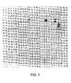
- FIG. 1 is a photograph showing the results of an electrophoresis analysis performed in accordance with Example 2, described below.
- FIG. 2 is a photograph showing the results of an electrophoresis analysis performed in accordance with Examples 3 and 4, described below.
- FIGS. 3A and 3B are photographs showing the results of an electrophoresis analysis performed in accordance with Example 5, described below.
- FIG. 4 is a photograph showing the results of an electrophoresis analysis performed in accordance with Example 6, described below.
- FIG. 5 is a photograph showing the results of an electrophoresis analysis performed in accordance with Example 7, described below.
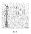
- FIG. 6 is a photograph showing the results of an electrophoresis analysis performed in accordance with Example 8, described below.
- FIGS. 7A and 7B are photographs showing the results of an electrophoresis analysis performed in accordance with Example 9, described below.
- FIG. 8 is a photograph showing the results of an electrophoresis analysis performed in accordance with Example 10, described below.
- FIGS. 9A and 9B are photographs showing the results of an electrophoresis analysis performed in accordance with Example 11, described below.
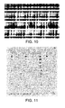
- FIG. 10 is a photograph showing the results of a blood card prepared in accordance with Example 12, described below.
- FIG. 11 is a photograph showing the results of an electrophoresis analysis performed in accordance with Example 12, described below.
- nucleic acids capable of being purified using the present invention include, but are not limited to DNA (single-stranded, double-stranded, covalently closed, and relaxed circular forms), RNA (single-stranded and double-stranded), PNA, and hybrids of the foregoing.
- the term “medium” encompasses any biological material, either naturally occurring or scientifically engineered, that contains at least one nucleic acid in addition to other non-nucleic acid material, such as biomolecules (e.g., proteins, polysaccharides, lipids, low molecular weight enzyme inhibitors, oligonucleotides, primers, templates), polyacrylamide, trace metals, organic solvents, etc.
- biomolecules e.g., proteins, polysaccharides, lipids, low molecular weight enzyme inhibitors, oligonucleotides, primers, templates
- polyacrylamide e.g., polyacrylamide, trace metals, organic solvents, etc.
- trace metals e.g., organic solvents, etc.
- organic solvents e.g., etc.
- naturally-occurring mediums include, but are not limited to, whole blood, plasma, and other body fluids, as well as tissue cell cultures obtained from humans, plants, or animals.
- scientifically-engineered mediums include, but are not limited to, lysates, nucleic acid samples eluted from agarose and/or polyacrylamide gels, solutions containing multiple species of DNA molecules resulting either from a polymerase chain reaction (PCR) amplification or from DNA size selection procedures, and solutions resulting from post-sequencing reactions.
- PCR polymerase chain reaction
- binding matrix encompasses any form capable of binding a nucleic acid. Those skilled in the art will be able to select an appropriate binding matrix for the nucleic acid(s) of interest.
- binding matrices examples include MAGAZORBTM paramagnetic particles (available from Promega, Madison, WI, catalog item MB1001), GENFINDTM particles (available from BECKMAN-COULTERTM, Fullerton, CA), MAGNESILTM Blue paramagnetic silica particles (available from Promega, catalog item A2201), zeolite particles (see Bitner et al., U.S. Published Patent Appln. No. 2007/0172855 , the entirety of which is incorporated by reference herein), and paramagnetic apple or citrus pectin particles (see Example 1 below).
- MAGAZORBTM paramagnetic particles available from Promega, Madison, WI, catalog item MB1001
- GENFINDTM particles available from BECKMAN-COULTERTM, Fullerton, CA
- MAGNESILTM Blue paramagnetic silica particles available from Promega, catalog item A2201
- zeolite particles see Bitner et al., U.S. Published Patent App
- binding matrices include, without limitation, paramagnetic silica particles, cellulose membranes, silica membranes, or columns such as cellulose acetate columns, nylon membrane columns, PVDF membrane columns, polypropylene columns, pure spin columns, and clearing columns for use in DNA IQTM spin baskets (available from Promega, catalog item V1221).
- suitable columns include DNA-IQTM columns, SV columns, Coming spin columns (available from Coming of Coming, NY, catalog item 8160), nylon membrane Coming spin columns (available from Coming, catalog item 8169), PVDF membrane in a polypropylene spin columns (available from Millipore Ultrafree-MC of Beverly, MA, catalog item VFC30GVNB), polypropylene membrane (available from Coming, catalog item AN0604700) in a DNA -IQTM column, HIGH PURETM Spin Filter tubes (available from Roche of Indianapolis, IN, catalog item1828665), and clearing columns (available from Promega, catalog item Z568A). Schleicher & Schuell cellulose cards (available from Keene of NH, catalog item GB003) also provide a useful binding matrix, particularly when storing and/or transporting nucleic acids for later use in downstream procedures.
- the ratio of binding matrix to medium is preferably from 0.005:1 to 0.5:1, more preferably from 0.2:1 to 0.4:1, by volume.
- One skilled in the art will be able to select optimal proportions, within or outside of these preferred ranges, depending on the nucleic acid(s) of interest and the type of binding matrix, among other factors.
- the GTC-A formulation acts as a lysis and/or binding solution that separates the nucleic acid of interest from its in vivo cellular environment and, if a binding matrix is present, facilitates the binding of the nucleic acid to the binding matrix.
- the formulation contains an amount of GTC and an amount of acetamide and/or one or more acetamide derivatives.
- the GTC and acetamide and/or one or more acetamide derivatives may be added together, or sequentially to the sample medium.
- the GTC may be added to the sample medium in a first solution, for example, to lyse cells, and the acetamide and/or one or more acetamide derivatives may be added in a second solution, for example, as a binding solution to promote binding of the DNA or RNA to the binding matrix.
- the respective amounts of GTC and acetamide and/or acetamide derivative(s) present in the GTC-A formulation can be adjusted to various concentrations.
- the concentration of GTC in the formulation is preferably from approximately 1.7M to approximately 4.3M, more preferably from approximately 4.0M to approximately 4.3M.
- the concentration of acetamide and/or acetamide derivative(s) in the formulation is preferably from approximately 5.0M to approximately 7.5M, more preferably from approximately 5.0M to approximately 7.1M.
- the proportion of formulation to medium depends on a number of variables, including, without limitation, the concentration of the formulation, the nucleic acid(s) of interest, whether a binding matrix is used, and, if so, what type.
- a preferred range of ratios of formulation to medium is from 1:1 to 30:1, by volume.
- a more preferred range is from 1.5:1 to 8:1, by volume.
- GTC can be purchased from Promega, catalog item V2791.
- Acetamide can be purchased from SIGMA-ALDRICHTM, catalog item A0500-500G.
- GTC can also be used with derivatives of acetamide.
- Preferred acetamide derivatives include methylacetamide (available from ACROS of Fair Lawn, NJ, catalog item 126141000) and dimethylacetamide (available from SIGMA-ALDRICHTM, catalog item D5511).
- the use of GTC and N,N-dimethylacetamide is preferred over the use of GTC and acetamide or GTC and N-methylacetamide.
- the GTC-A formulation can further contain one or more additional ingredients such as, for example, proteinase K, beta-mercaptoethanol, tris(2-carboxyethyl)phosphine, dithiothreitol, 1-TG, digitonin, lysis solutions, CHAPS, TERGITOLTM type NP-9, and TRITONTM X-100.
- additional ingredients such as, for example, proteinase K, beta-mercaptoethanol, tris(2-carboxyethyl)phosphine, dithiothreitol, 1-TG, digitonin, lysis solutions, CHAPS, TERGITOLTM type NP-9, and TRITONTM X-100.
- the GTC-A formulation can also be used as a wash solution for removing impurities, as described in the methods below.
- the GTC-A formulation can be combined with one or more binding matrices in a kit that can be used in the purification of nucleic acids.
- the ratio of binding matrix to formulation in the kit is within the range of from 1:400 to 1:1, by volume.
- the ratio of binding matrix to formulation can be varied within or outside of this preferred range, depending on the specific contents of the kit and the application for which the kit is intended.
- the kit may comprise GTC in a first container, and acetamide and/or one or more acetamide derivatives in a second container, combined with one or more binding matrices in a kit that can be used in the purification of nucleic acids.
- the GTC and acetamide and/or one or more acetamide derivatives may be combined in a single container.
- the one or more binding matrices may be combined within one of the above containers, or provided as a separate item in a kit that can be used in the purification of nucleic acids.
- the kit can include more than one type of binding matrix, each compatible with a different type of nucleic acid.
- the kit can be used in the selective purification of different types of nucleic acids.
- the invention relates to a method of purifying at least one nucleic acid contained in a medium.
- the medium containing the at least one nucleic acid is combined with at least one binding matrix and a GTC-A formulation in order to cause the at least one nucleic acid to separate from its in vivo cellular environment and bind to at least one binding matrix.
- the medium, binding matrices, and GTC-A formulation can be combined in any order or simultaneously.
- the binding matrices with at least one nucleic acid bound thereto then is separated from substantially the rest of the combined medium and formulation, for example, by using a magnetic rack, by centrifuging, or by filtration.
- bound binding matrix and the nucleic acid combinations can be washed using any suitable wash solution, including the GTC-A formulation, in order to remove any impurities.
- the at least one nucleic acid is eluted from the binding matrix, thereby obtaining the at least one nucleic acid in a substantially purified form.
- This elution step can immediately follow the aforementioned steps, or it can be performed at a later time.
- the nucleic acids can be stored for downstream activities.
- the elution step uses an elution buffer to separate the nucleic acid from the binding matrix, after which the substantially purified nucleic acid is contained in the elution buffer.
- Suitable elution buffers include, but are not limited to, nuclease-free water or aqueous solutions such as, for example, TRISTM-HCI, Tris-acetate, sucrose, and formamide solutions.
- a preferred elution buffer is a TRISTM buffer with ethylenediaminetetraacetic acid (EDTA). More preferably, the elution buffer is about 10mM TRISTM (pH 8.0) and about 1mM EDTA HEPESTM(pH 7.5). Elution of the nucleic acid from the binding matrix occurs quickly (e.g., in thirty seconds or less) when a suitable low ionic strength elution buffer is used.
- nucleic acids can be used in any of a number of known scientific procedures, including, without limitation, the isolation of genetic material, polymerase chain reactions, electrophoresis, sequencing, cloning, and the like.
- This method can also be used to purify at least two different nucleic acids contained in a single medium. This involves combining a medium containing at least two different nucleic acids with a first binding matrix compatible with binding a first nucleic acid contained in the medium, and a second binding matrix compatible with binding a second nucleic acid contained in the medium, and a GTC-A formulation as described above. This causes the first and second nucleic acids to separate from their in vivo cellular environments and to bind to the first and second binding matrices, respectively. Each of the first binding matrix with the first nucleic acid bound thereto and the second binding matrix with the second nucleic acid bound thereto then is separated from substantially the rest of the combined medium and formulation.
- this method involves combining a medium containing at least two different nucleic acids with a first binding matrix compatible with binding a first nucleic acid contained in the medium (e.g DNA-IQTM particles which are compatible with binding DNA), followed by a transfer of the remaining solution depleted of the first nucleic acid (e.g., the RNA remaining after DNA binding to DNA-IQTM particles) to a second binding matrix (e.g., paramagnetic zeolite particles) compatible with binding of the remaining nucleic acids, thereby separately obtaining each of the first and second nucleic acids in a substantially purified form.
- a medium containing at least two different nucleic acids with a first binding matrix compatible with binding a first nucleic acid contained in the medium (e.g DNA-IQTM particles which are compatible with binding DNA), followed by a transfer of the remaining solution depleted of the first nucleic acid (e.g., the RNA remaining after DNA binding to DNA-IQTM particles) to a second binding matrix (e.g., paramagne
- the invention relates to a method as described above, except that this method does not necessarily require eluting the nucleic acid from the binding matrix. Rather, the binding matrix with the nucleic acid(s) bound thereto can be stored and/or transported for later use in scientific procedures, which may or may not involve eluting the nucleic acid(s) from the binding matrix.
- This method is particularly useful when using the Schleicher & Schuell cellulose card as the binding matrix.
- a hole can be punched from the cellulose card and the nucleic acid bound thereto can be eluted for use in a scientific procedure.
- paramagnetic apple or citrus pectin particles suitable for use in certain embodiments of the present invention as a binding matrix were prepared as follows:
- Adjust the pH of the mixture first by adding 3.0ml of 56% KOH. After mixing for 5 minutes at about 21°C, lower the pH by adding 3.3ml of 3.0M HCL, with periodic testing of the mixture's pH using pH paper. The resulting pH should be about 3. Next, add 3.5ml of 1.32M KOAc having a pH of 4.8 so that the resulting pH of the mixture is between 4 and 5.
- any of the three sets of paramagnetic pectin particles -- "large,” “main,” and “first cut” -- can be used as a binding matrix; however, in the examples that follow, only the "main” particles are used.
- step 4 in order to wash the MAGAZORBTM paramagnetic particles a second time.
- FIG. 1 shows the results of the electrophoresis analysis of the samples prepared according to Example 2.
- the lanes are numbered 1-12, from left to right.
- Lanes 1-9 show the results for samples 1-9, respectively;
- lane 10 contains no sample;
- lane 11 shows the genomic DNA standard;
- lane 12 shows the 100bp DNA ladder of standard molecular weight.
- DNA from human whole blood samples was purified using a GTC-A formulation together with each of four different binding matrices using the following procedure:
- FIG. 2 shows the results of the electrophoresis analysis of the samples prepared according to Example 3.
- the lanes are numbered 1-12, from left to right.
- Lanes 1-8 show the results for samples 1-8, respectively. (Lanes 9-12 relate to Example 4, which is discussed below.)
- DNA from human whole blood samples was released using a GTC-A formulation together with one of two different binding matrices.
- the following procedure was used:
- binding matrices to samples 1-4 as follows: (a) to each of samples 1 and 2, add 100 ⁇ l (4.8mg) of MAGAZORBTM paramagnetic particles; and (b) to each of samples 3 and 4, add 100 ⁇ l (7mg) of "main" paramagnetic citrus pectin particles (from Example 1). Mix each sample by pipetting and allow the samples to sit for 10 minutes at about 21°C.
- step 8 For each sample, repeat the wash procedure of step 7 eight more times using 500 ⁇ l of D X each time.
- Lanes 9-12 of FIG. 2 show the results of the electrophoresis analysis of samples 1-4, respectively. (Lanes 1-8 of FIG. 2 relate to Example 3, discussed above.)
- RNA from human blood plasma samples was purified using different GTC-A formulations containing selected inhibitors of RNase activity. The following procedure was used:
- samples 1, 2, 13-15, and 23 by adding, to the other six tubes, the following: (a) for sample 1,1 ⁇ l luciferase control RNA and 200 ⁇ l of human plasma; (b) for sample 13, 1 ⁇ l luciferase control RNA; (c) for sample 14, 1 ⁇ l luciferase control RNA at a temperature of about -20°C; (d) for sample 23, 200 ⁇ l of human plasma; and (e) for each of samples 2 and 15, nothing.
- FIGS. 3A and 3B show the results of the electrophoresis analysis of the twenty-three samples prepared according to Example 5.
- the lanes are numbered 1-12, from left to right.
- the lanes are numbered 13-24, from left to right.
- Lanes 1-23 show the results for samples 1-23, respectively. Lane 24 is empty.
- the GTC-A formulations were effective in the purification of RNA notwithstanding the presence of certain inhibitors. Moreover, the use of certain inhibitors in addition to GTC-A enables the selective purification of RNA, while reducing the degradation of the RNA from RNase during the lysis process. In particular, GTC-A formulations containing BME alone (lanes 3-6) were effective in the purification of larger-sized RNA. Acting as an inhibitor of RNase, the combination of GTC-A and increasing BME concentrations reduced the degradation of the RNA.
- the GTC-A formulations containing TCEP (lanes 7-10) also inhibited RNA degradation and resulted in less RNA being bound to the MagaZorb® paramagnetic particles than that bound using the GTC-A formulations containing BME.
- the RNA bound to the MAGAZORBTM paramagnetic particles using the GTC-A formulations containing TCEP had a higher average molecular weight than that of the RNA bound using the GTC-A formulations containing BME.
- 1-TG added to the GTC-A formulation (lanes 16-18 and 20) also resulted in the purification of larger-sized RNA than that obtained using the GTC-A formulations containing BME.
- Luciferase control RNA added into human blood plasma was bound to the MAGAZORBTM paramagnetic particles using the GTC-A formulation.
- the absence of added human plasma (lane 1) demonstrated that the luciferase RNA was not visibly degraded.
- the addition of human plasma (lane 23) resulted in RNA degradation.
- DNA as small as about 25bp in size was purified from human whole blood samples using a GTC-A formulation. The following procedure was used:
- step 7 two more times for a total of three washes with 500 ⁇ l of W 1 for each sample.
- step 7 For each of samples 1 and 2, repeat step 7 an additional four times using W 1 for a total of seven washes with W 1 .
- step 7 For each of samples 3 and 4, repeat step 7 an additional four times using 500 ⁇ l of MAGAZORBTM DNA Binding Solution instead of W 1 .
- step 7 For each of samples 5 and 6, repeat step 7 an additional four times using 500 ⁇ l of Alcohol Wash, Blood (BW wash) (available from Promega, catalog item MB1001) solution instead of W 1 .
- BW wash Blood (BW wash) (available from Promega, catalog item MB1001) solution instead of W 1 .
- FIG. 4 shows the results of the electrophoresis analysis of the twelve samples prepared according to Example 6.
- the lanes are numbered 1-12, from left to right. Lanes 1-12 show the results for samples 1-12, respectively.
- the GTC-A formulation in conjunction with a binding matrix is suitable in purifying genomic DNA that is anywhere from 25 bases to 20 kilobase pairs or larger.
- lanes 2 and 10 show the purification of DNA which was about 25 bases in length and showed that oligo primers of 22 and 29 bases migrated to the bottom of the gel.
- BW wash (lanes 5 and 6) resulted in the purification of larger-sized DNA, about 75bp, than the 25bp obtained without using the BW wash.
- DNA bands in all three purification methods which can be seen, for example, between the 100bp band and 200bp band, as well as between the 200bp and 300bp bands, as a result of the added DNA bands being bound by the GTC-A formulation.
- nucleic acids from tissue culture cells were purified using a GTC-A formulation. The following procedure was used:
- sample 6 add 75 ⁇ l of CHO cells in GIBCOTM F-12 plus 10% FBS medium) (1 ⁇ 10 6 cells) and mix the sample by pipetting.
- step 8 two more times, except substituting a 500 ⁇ l mixture of 2.6M GTC and 7.1M acetamide for the 500 ⁇ l mixture of 4.0M GTC, 5.0M acetamide, and 20% (volume/volume) 1-TG used in step 8.
- step 8 For samples 2 and 5 only, repeat step 8 one more time, except substituting 500 ⁇ l of SV Total RNA Wash for the 500 ⁇ l mixture of 4.0M GTC, 5.0M acetamide, and 20% (volume/volume) 1-TG used in step 8.
- FIG. 5 shows the results of the electrophoresis analysis of the samples prepared according to Example 7.
- the lanes are numbered 1-12, from left to right.
- Lane 1 shows the results for sample 1;
- lane 2 shows the results for sample 2;
- lane 3 shows the results for sample 3;
- lane 4 shows the results for sample 7;
- lane 5 shows the results for sample 8;
- lane 6 shows the results for sample 9;
- lane 7 shows the results for sample 4;
- lane 8 shows the results for sample 5;
- lane 9 shows the results for sample 6;
- lane 10 shows the results for sample 10;
- lane 11 shows the results for sample 11; and
- lane 12 shows the results for sample 12.
- the use of the GTC-A formulation allowed the purification of genomic DNA and total RNA from each of the samples prepared according to Example 7.
- cytoplasmic RNA from tissue culture cell samples was purified using GTC-A formulations. The following procedure was used:
- HEK293 tissue culture cells in GIBCOTM DMEM medium. Allow 4ml of 5 ⁇ 10 6 HEK293 cells to settle at 1 ⁇ g, and then remove the growth medium by pipetting, so that about 2 ⁇ 10 7 cells are contained in 50 ⁇ l. Next, place the 50 ⁇ l of cells into a 1.5ml plastic tube and place on ice for 5 minutes.
- step 2 Add 6 ⁇ l of RNASINTM Plus (40 units per ⁇ l) (available from Promega, catalog item 1232) to the 50 ⁇ l of cells from step 1. Then add 100 ⁇ l of 120 ⁇ g/ml digitonin in 100mM EDTA 100mM HEPESTM (pH 7.5) (on ice) to the cells. Mix the contents by vortexing for 10 seconds, and then incubate on ice for 20 minutes. The incubation with 77 ⁇ g/ml digitonin promotes the lysis of the cytoplasmic membrane, without lysis of the cell nuclear membrane.
- RNASINTM Plus 40 units per ⁇ l
- step 6 Mix the four samples from step 5 by pipetting and incubating them at about 21°C for five minutes. Place each of samples 1-4 on a magnetic. After separation of the particles from the samples, discard the supernatants from the particles that are attracted to the side of the tubes.
- FIG. 6 shows the results of the electrophoresis analysis of the samples prepared according to Example 8.
- Lane 1 shows a 100bp DNA ladder for the purpose of comparing a DNA sequence
- lane 2 shows the results of 10 ⁇ l of initial cytoplasmic RNA supernatant to serve as a marker of non purified nucleic acid
- lane 3 shows the results for sample 3
- lane 4 shows the results for sample 4
- lane 5 shows the results for sample 1
- lane 6 shows the results for sample 2
- lane 7 shows the results for sample 5
- lane 8 shows the results for sample 6
- lane 9 shows 100bp DNA ladder for the purpose of comparing a DNA sequence
- lane 10 shows the results for sample 7
- lane 11 shows the results for sample 8
- lane 12 is empty.
- RNA is purified using the GTC-acetamide formulation and SV RNA Wash, using either MAGAZORBTM paramagnetic cellulose particles or MAGNESILTM paramagnetic silica particles.
- MAGAZORBTM paramagnetic cellulose particles or MAGNESILTM paramagnetic silica particles.
- ribosomal RNA and tRNA bands are visible in lanes 7 and 8, showing purification using GTC-A.
- the passage of the cytoplasmic RNA sample through a cellulose column (lanes 3 and 4), with a short sample incubation time of 20 seconds at a temperature of about 21°C, substantially removed nuclear DNA. This was accomplished without substantial removal of the cytoplasmic RNA, demonstrated by the subsequent purification using MAGNESILTM particles (lanes 10 and 11).
- DNA or RNA from tissue culture cell samples was purified using a GTC-A formulation with the aid of various binding matrices in order to determine additional binding matrices suitable for use with the present invention. The following procedure was used:
- step 7 three more times using 400 ⁇ l of SV RNA Wash Solution instead of the GTC-A formulation. After discarding the third RNA wash solution, air-dry the particle samples for 20 minutes at a temperature of about 21°C. Then elute the nucleic acids in 40 ⁇ l of nuclease-free water.
- FIGS. 7A and 7B show the results of the electrophoresis analysis of the samples prepared according to Example 9.
- the lanes are numbered 1-12, from left to right.
- the lanes are numbered 13-24, from left to right.
- Lanes 1-24 show the results for samples 1-24, respectively.
- genomic DNA was purified using cellulose acetate columns, PVDF columns, DNA-IQTM paramagnetic particles, and MAGNESILTM Blue particles.
- MAGAZORBTM paramagnetic particles purified a small amount of RNA as well as genomic DNA. Both RNA and DNA were purified using SV silica columns, nylon membrane columns, polypropylene membrane columns, HIGH PURETM Spin columns, clearing columns, and paramagnetic zeolite particles, which showed prominent tRNA bands.
- this example shows how to screen for the initial binding of nucleic acids to a binding matrix, followed by the retention of nucleic acids during sequential washes, and elution of the nucleic acids at the end of the procedure.
- previous examples have shown binding of both DNA and RNA to MAGAZORBTM paramagnetic particles, and in this example (with washes using SV RNA Wash) the RNA was less prominent in the elution. Therefore, in order to screen binding matrices for binding of nucleic acids, one could wash only with the initial GTC-A formulation to ensure retention of nucleic acids on the binding matrix. This example demonstrated a measure of the ability to retain the nucleic acids on the binding matrix during wash steps.
- this example also required that the nucleic acid was eluted from the binding matrix.
- the nucleic acid must also remain bound during washes with two different solutions, and also allows the nucleic acid to elute from the binding matrix at the end of the purification. Even with all three requirements, it is shown that all of the binding matrices show an ability to purify nucleic acid(s) using GTC-A formulations.
- DNA and RNA from the same tissue culture cell samples were purified using a GTC-A formulation. The following procedure was used:
- Samples 1 and 2 now consist of the DNA-IQTM particles minus the supernatants.
- FIG. 8 shows the results of an agarose gel electrophoresis analysis of the samples prepared according to Example 10.
- the lanes are numbered 1-12, from left to right.
- Lanes 1 and 2 show the results for samples 1 and 2, respectively; lane 3 is blank; lanes 4 and 5 show samples 1 and 2, respectively, digested with RNase ONE; lane 6 contains Promega 100BP DNA ladder; lanes 7 and 8 show results for samples 3 and 4, respectively; lane 9 is blank; lanes 10 and 11 show samples 3 and 4, respectively, digested with Promega RQ1 RNase-free DNase; lane 12 contains Promega genomic DNA standard.
- genomic DNA was purified using DNA-IQTM particles, and from the same medium, RNA was purified using paramagnetic zeolite particles.
- DNA and RNA can be purified from the same medium using a first binding matrix to bind DNA and a second binding matrix to bind RNA, without adding any additional solution or condition between the first binding matrix and the application of the second binding matrix.
- RNA from tissue culture cell mediums were purified using GTC-A formulations containing GTC and acetamide, N-methylacetamide or N,N-dimethylacetamide. The following procedure was used:
- FIGS. 9A and 9B show the results of the electrophoresis analysis of the samples prepared according to Example 11.
- the lanes are numbered 1-12, from left to right.
- the lanes are numbered 13-24, from left to right.
- Lanes 1-24 show the results for samples 1-24, respectively.
- DNA and RNA are bound to cellulose membranes in mini-columns, and purified using GTC and acetamide, GTC and N-methylacetamide, and GTC and N,N-dimethylacetamide, showing that derivatives of acetamide can also be used in conjunction with GTC to purify nucleic acids.
- DNA purified from human whole blood was bound to a blood card, which served as a binding matrix. Additionally, the blood sample can be washed on the blood card, and purified nucleic acids can be obtained by elution of blood card punches. The following procedure was used:
- RNA Marker available from Promega, catalog item G3191
- the sample 2 spot To the sample 2 spot, apply 150 ⁇ l of a mixture of 2.6M GTC and 7.1M acetamide in 10 ⁇ l increments, with successive additions being applied only after the previous addition has been absorbed by the cellulose card.
- the resulting blood spot should have a relatively white center, and a concentric red circle containing hemoglobin and other cellular debris.
- FIG. 10 shows the Schleicher & Schuell cellulose card prepared according to Example 12. The spot on the left corresponds to sample 1; the spot in the middle corresponds to sample 2; and the spot on the right corresponds to the blood plus RNA Marker.
- FIG. 11 shows the results of the electrophoresis analysis of the four elutions prepared according to Example 12.
- the lanes are numbered 1-12, from left to right.
- Lane 1 shows the results for the center punch of sample 1;
- lane 3 shows the results for the second punch of sample 1;
- lane 5 shows the results for the center punch of sample 2;
- lane 7 shows the results for the second punch of sample 2;
- lane 9 shows Promega 100bp DNA Ladder;
- lane 11 shows Promega genomic DNA standard; and the remaining lanes are empty.
Abstract
Description
- This application claims priority to
U.S. Patent Application No. 12/549806, filed August 28, 2009 - This invention relates to methods of purifying one or more nucleic acids, and also to a formulation and kit for use in performing such methods.
- The purification of nucleic acids plays an important role in scientific procedures. There are a number of known methods of purifying single- and double-stranded DNA contained in biological fluids such as human blood, serum, and cultured cells, as well as plants, animal and human tissues, and other specimens. However, such methods can result in very low yields and do not always work well when trying to extract small amounts of nucleic acids from large samples. Known methods are described in, for example,
Nargessi, U.S. Pat. No. 6,855,499 (2005 );Tereba et al., U.S. Pat. No. 6,673,631 (2004 );McKernan et al., U.S. Pat. No. 6,534,262 (2003 ); Taylor et al., J. Chromatography A, 890:159-166 (2000); Ahn et al., BioTechniques, 29:466-468 (2000); Scott Jr. et al., Lett. Appl. Microbiol., 31:95-99 (2000); Lin et al., BioTechniques, 29:460-466 (2000);Smith et al., U.S. Pat. No. 6,027,945 (2000 ); Mrazek et al., Acta Univ. Palacki. Olomuc., Fac. Med. 142:23-28 (1999);Hawkins, U.S. Pat. No. 5,898,071 (1999 ); andHawkins, U.S. Pat. No. 5,705,628 (1998 ). - The present invention represents an improvement over the known methods described in the aforementioned literature.
- In one aspect, the invention relates to a method of purifying at least one nucleic acid, such as deoxyribonucleic acid (DNA), ribonucleic acid (RNA), and/or peptide nucleic acid (PNA), which is contained in a medium, such as whole blood, plasma, or tissue cell cultures obtained from humans, plants, or animals. The method includes steps of (a) combining the medium containing the at least one nucleic acid with at least one binding matrix and a formulation in order to cause the at least one nucleic acid to separate from its in vivo cellular environment and bind to at least one binding matrix, (b) separating the binding matrix with at least one nucleic acid bound thereto from substantially the rest of the combined medium and formulation, and (c) eluting the at least one nucleic acid from the binding matrix, thereby obtaining the at least one nucleic acid in a substantially purified form.
- A nucleic acid is considered to be in a "substantially purified form" when the nucleic acid has been separated from its in vivo cellular environment and obtained in a form that is useful in one or more scientific procedures, such as the isolation of genetic material, polymerase chain reactions, electrophoresis, sequencing, and cloning, among others.
- The formulation used in the foregoing method contains an amount of guanidine thiocyanate and an amount of (i) acetamide, (ii) one or more acetamide derivatives, or (iii) a combination of acetamide and one or more acetamide derivatives. Preferred acetamide derivatives include methylacetamide and dimethylacetamide. Herein, guanidine thiocyanate is sometimes referred to as "GTC," and the combination of guanidine thiocyanate with acetamide and/or one or more acetamide derivatives is sometimes referred to as "GTC-A."
- In the above method, the respective amounts of GTC and acetamide and/or acetamide derivative(s) present in the formulation are sufficient to cause the at least one nucleic acid to separate from its in vivo cellular environment and bind to the binding matrix. Preferably, the concentration of GTC in the formulation is from approximately 1.7M to approximately 4.3M, more preferably from approximately 4.0M to approximately 4.3M. Preferably, the concentration of acetamide and/or acetamide derivative(s) in the formulation is from approximately 5.0M to approximately 7.5M, more preferably from approximately 5.0M to approximately 7.1M.
- Any of a number of known binding matrices can be used in the foregoing method, depending on the type of nucleic acids sought to be purified. Those skilled in the art will be able to select binding matrices that are compatible with the nucleic acid of interest. Examples of suitable binding matrices include, but are not limited to, paramagnetic cellulose particles, paramagnetic carboxy-cellulose particles, paramagnetic citrus pectin particles, paramagnetic apple pectin particles, paramagnetic zeolite particles, paramagnetic silica particles, cellulose membranes, silica membranes, cellulose acetate columns, nylon membrane columns, PVDF membrane columns, polypropylene columns, HIGH PURE™ spin columns (available from Roche-Diagnostics,
item 1 828 665), and clearing columns. - The ratio of formulation to medium used in the above method is preferably from 1:1 1 to 30:1, more preferably from 1.5:1 to 8:1, by volume. The ratio of binding matrix to medium is preferably from 0.005:1 to 0.5:1, more preferably from 0.2:1 to 0.4:1, by volume. One skilled in the art will be able to select proportions, within or outside of these preferred ranges, depending on the nucleic acid(s) of interest, the concentration of the formulation, and the type of binding matrix employed, among other variables. Therefore, the invention is not limited to these preferred ranges.
- Optionally, one or more additional ingredients can be combined with the medium, the binding matrix, and the formulation. For example, one or more enzymes which aid in the degradation and lysis of cellular structure can be used to facilitate the separation of nucleic acids from their mediums. Examples of suitable additional ingredients include, but are not limited to, proteinase K (available from Promega, catalog item V3021), beta-mercaptoethanol (BME), tris(carboxyethyl)phosphine (TCEP), dithiothreitol (DTT) (available from Sigma, catalog item 43815), 1-thioglycerol (1-TG) (available from SIGMA-ALDRICH™, catalog item M2172), digitonin, lysis solutions, CHAPS (3-[(3-cholamidopropyl)dimethylammonio]-1-propanesulfonate, available from SIGMA-ALDRICH™, catalog item C3023), TERGITOL™ type NP-9 (26-(4-nonylphenoxy)-3,6,9,12,15,18,21,24-.octaoxahcxacosan-1-ol available from SIGMA-ALDRICH™, catalog item np9), and TRITON™ X-100 (4-(1,1,3,3-tetramethylbutyl)phenyl-polyethylene glycol, available from Thermo Scientific, Waltham, MA, catalog item BP151). How these additional ingredients are employed is not critical. For example, they can be incorporated in the formulation or they can be added to the medium either before or after the medium is combined with the binding matrix and/or the formulation.
- This method can also be used to purify at least two different nucleic acids contained in a single medium. This involves combining a medium containing at least two different nucleic acids with a first binding matrix compatible with a first nucleic acid contained in the medium, a second binding matrix compatible with a second nucleic acid contained in the medium, and a GTC-A formulation as described above. This causes the first and second nucleic acids to separate from their in vivo cellular environments and to bind to the first and second binding matrices, respectively. Each of the first binding matrix with the first nucleic acid bound thereto and the second binding matrix with the second nucleic acid bound thereto then is separated from substantially the rest of the combined medium and formulation. Further, the first nucleic acid is eluted from the first binding matrix, and the second nucleic acid is eluted from the second binding matrix, thereby obtaining each of the first and second nucleic acids in a substantially purified form.
- Optionally, a medium containing the second nucleic acid and the formulation can be transferred after the first nucleic acid contained in the medium binds to the first binding matrix. This is followed by a transfer of the remaining medium to the second binding matrix compatible with the binding of the second nucleic acid contained in the medium, before separating each of the first and second nucleic acids from the first and second binding matrices, respectively.
- In another aspect, the invention relates to a method as described above, except that the final eluting step is not necessarily required, although such a step is not excluded. Thus, the binding matrix with the nucleic acid bound thereto can be stored and/or transported for later use in scientific procedures, which may or may not involve eluting the nucleic acid from the binding matrix.
- In another aspect, the invention relates to a kit for use in purifying nucleic acids and/or binding nucleic acids to a binding matrix. The kit includes a binding matrix and a GTC-A formulation as described above. Preferably, the ratio of binding matrix to formulation in the kit is within the range of from 1:400 to 1:1, by volume. The ratio of binding matrix to formulation can be varied within or outside of this preferred range depending on, among other things, the nucleic acid(s) of interest and the type of binding matrix employed.
-
FIG. 1 is a photograph showing the results of an electrophoresis analysis performed in accordance with Example 2, described below. -
FIG. 2 is a photograph showing the results of an electrophoresis analysis performed in accordance with Examples 3 and 4, described below. -
FIGS. 3A and 3B are photographs showing the results of an electrophoresis analysis performed in accordance with Example 5, described below. -
FIG. 4 is a photograph showing the results of an electrophoresis analysis performed in accordance with Example 6, described below. -
FIG. 5 is a photograph showing the results of an electrophoresis analysis performed in accordance with Example 7, described below. -
FIG. 6 is a photograph showing the results of an electrophoresis analysis performed in accordance with Example 8, described below. -
FIGS. 7A and 7B are photographs showing the results of an electrophoresis analysis performed in accordance with Example 9, described below. -
FIG. 8 is a photograph showing the results of an electrophoresis analysis performed in accordance with Example 10, described below. -
FIGS. 9A and 9B are photographs showing the results of an electrophoresis analysis performed in accordance with Example 11, described below. -
FIG. 10 is a photograph showing the results of a blood card prepared in accordance with Example 12, described below. -
FIG. 11 is a photograph showing the results of an electrophoresis analysis performed in accordance with Example 12, described below. - The following are non-limiting examples of preferred implementations of the present invention. Throughout, all volumes, pH levels, and concentrations are at room temperature unless stated otherwise.
- The nucleic acids capable of being purified using the present invention include, but are not limited to DNA (single-stranded, double-stranded, covalently closed, and relaxed circular forms), RNA (single-stranded and double-stranded), PNA, and hybrids of the foregoing.
- As used herein, the term "medium" encompasses any biological material, either naturally occurring or scientifically engineered, that contains at least one nucleic acid in addition to other non-nucleic acid material, such as biomolecules (e.g., proteins, polysaccharides, lipids, low molecular weight enzyme inhibitors, oligonucleotides, primers, templates), polyacrylamide, trace metals, organic solvents, etc. Examples of naturally-occurring mediums include, but are not limited to, whole blood, plasma, and other body fluids, as well as tissue cell cultures obtained from humans, plants, or animals. Examples of scientifically-engineered mediums include, but are not limited to, lysates, nucleic acid samples eluted from agarose and/or polyacrylamide gels, solutions containing multiple species of DNA molecules resulting either from a polymerase chain reaction (PCR) amplification or from DNA size selection procedures, and solutions resulting from post-sequencing reactions.
- Advantageously, one or more binding matrices can be used in the present invention. As used, herein, the term "binding matrix" encompasses any form capable of binding a nucleic acid. Those skilled in the art will be able to select an appropriate binding matrix for the nucleic acid(s) of interest.
- Examples of suitable binding matrices include MAGAZORB™ paramagnetic particles (available from Promega, Madison, WI, catalog item MB1001), GENFIND™ particles (available from BECKMAN-COULTER™, Fullerton, CA), MAGNESIL™ Blue paramagnetic silica particles (available from Promega, catalog item A2201), zeolite particles (see
Bitner et al., U.S. Published Patent Appln. No. 2007/0172855 , the entirety of which is incorporated by reference herein), and paramagnetic apple or citrus pectin particles (see Example 1 below). Other suitable binding matrices include, without limitation, paramagnetic silica particles, cellulose membranes, silica membranes, or columns such as cellulose acetate columns, nylon membrane columns, PVDF membrane columns, polypropylene columns, pure spin columns, and clearing columns for use in DNA IQ™ spin baskets (available from Promega, catalog item V1221). More specifically, suitable columns include DNA-IQ™ columns, SV columns, Coming spin columns (available from Coming of Coming, NY, catalog item 8160), nylon membrane Coming spin columns (available from Coming, catalog item 8169), PVDF membrane in a polypropylene spin columns (available from Millipore Ultrafree-MC of Beverly, MA, catalog item VFC30GVNB), polypropylene membrane (available from Coming, catalog item AN0604700) in a DNA -IQ™ column, HIGH PURE™ Spin Filter tubes (available from Roche of Indianapolis, IN, catalog item1828665), and clearing columns (available from Promega, catalog item Z568A). Schleicher & Schuell cellulose cards (available from Keene of NH, catalog item GB003) also provide a useful binding matrix, particularly when storing and/or transporting nucleic acids for later use in downstream procedures. - The ratio of binding matrix to medium is preferably from 0.005:1 to 0.5:1, more preferably from 0.2:1 to 0.4:1, by volume. One skilled in the art will be able to select optimal proportions, within or outside of these preferred ranges, depending on the nucleic acid(s) of interest and the type of binding matrix, among other factors.
- The GTC-A formulation acts as a lysis and/or binding solution that separates the nucleic acid of interest from its in vivo cellular environment and, if a binding matrix is present, facilitates the binding of the nucleic acid to the binding matrix. As mentioned above, the formulation contains an amount of GTC and an amount of acetamide and/or one or more acetamide derivatives. The GTC and acetamide and/or one or more acetamide derivatives may be added together, or sequentially to the sample medium. As an example of sequential addition, the GTC may be added to the sample medium in a first solution, for example, to lyse cells, and the acetamide and/or one or more acetamide derivatives may be added in a second solution, for example, as a binding solution to promote binding of the DNA or RNA to the binding matrix.
- As demonstrated by the examples below, other salts or salt and amide combinations, such as guanidine hydrochloride (available from Promega, catalog item H5381), guanidine hydrochloride and acetamide, GTC alone, or acetamide alone, are relatively ineffective in the purification of nucleic acids. Surprisingly, even though neither GTC alone nor acetamide alone is effective in purifying nucleic acids, the combination of GTC and acetamide and/or acetamide derivative(s) is highly effective.
- The respective amounts of GTC and acetamide and/or acetamide derivative(s) present in the GTC-A formulation can be adjusted to various concentrations. The concentration of GTC in the formulation is preferably from approximately 1.7M to approximately 4.3M, more preferably from approximately 4.0M to approximately 4.3M. The concentration of acetamide and/or acetamide derivative(s) in the formulation is preferably from approximately 5.0M to approximately 7.5M, more preferably from approximately 5.0M to approximately 7.1M.
- The proportion of formulation to medium depends on a number of variables, including, without limitation, the concentration of the formulation, the nucleic acid(s) of interest, whether a binding matrix is used, and, if so, what type. A preferred range of ratios of formulation to medium is from 1:1 to 30:1, by volume. A more preferred range is from 1.5:1 to 8:1, by volume.
- GTC can be purchased from Promega, catalog item V2791. Acetamide can be purchased from SIGMA-ALDRICH™, catalog item A0500-500G. As mentioned, GTC can also be used with derivatives of acetamide. Preferred acetamide derivatives include methylacetamide (available from ACROS of Fair Lawn, NJ, catalog item 126141000) and dimethylacetamide (available from SIGMA-ALDRICH™, catalog item D5511). Sometimes, such as when purifying RNA from HEK293 tissue cells, the use of GTC and N,N-dimethylacetamide is preferred over the use of GTC and acetamide or GTC and N-methylacetamide.
- Optionally, the GTC-A formulation can further contain one or more additional ingredients such as, for example, proteinase K, beta-mercaptoethanol, tris(2-carboxyethyl)phosphine, dithiothreitol, 1-TG, digitonin, lysis solutions, CHAPS, TERGITOL™ type NP-9, and TRITON™ X-100.
- In addition to being used as a lysis and/or binding solution, the GTC-A formulation can also be used as a wash solution for removing impurities, as described in the methods below.
- The GTC-A formulation can be combined with one or more binding matrices in a kit that can be used in the purification of nucleic acids. Preferably, the ratio of binding matrix to formulation in the kit is within the range of from 1:400 to 1:1, by volume. The ratio of binding matrix to formulation can be varied within or outside of this preferred range, depending on the specific contents of the kit and the application for which the kit is intended. The kit may comprise GTC in a first container, and acetamide and/or one or more acetamide derivatives in a second container, combined with one or more binding matrices in a kit that can be used in the purification of nucleic acids. Alternatively, the GTC and acetamide and/or one or more acetamide derivatives may be combined in a single container. The one or more binding matrices may be combined within one of the above containers, or provided as a separate item in a kit that can be used in the purification of nucleic acids.
- Advantageously, the kit can include more than one type of binding matrix, each compatible with a different type of nucleic acid. In that case, the kit can be used in the selective purification of different types of nucleic acids.
- In one preferred implementation, the invention relates to a method of purifying at least one nucleic acid contained in a medium. The medium containing the at least one nucleic acid is combined with at least one binding matrix and a GTC-A formulation in order to cause the at least one nucleic acid to separate from its in vivo cellular environment and bind to at least one binding matrix. The medium, binding matrices, and GTC-A formulation can be combined in any order or simultaneously. The binding matrices with at least one nucleic acid bound thereto then is separated from substantially the rest of the combined medium and formulation, for example, by using a magnetic rack, by centrifuging, or by filtration. Optionally, at this point, bound binding matrix and the nucleic acid combinations can be washed using any suitable wash solution, including the GTC-A formulation, in order to remove any impurities. Thereafter, the at least one nucleic acid is eluted from the binding matrix, thereby obtaining the at least one nucleic acid in a substantially purified form. This elution step can immediately follow the aforementioned steps, or it can be performed at a later time. By choosing to elute the nucleic acid from the binding matrix at a later time, the nucleic acids can be stored for downstream activities.
- In more detail, the elution step uses an elution buffer to separate the nucleic acid from the binding matrix, after which the substantially purified nucleic acid is contained in the elution buffer. Suitable elution buffers include, but are not limited to, nuclease-free water or aqueous solutions such as, for example, TRIS™-HCI, Tris-acetate, sucrose, and formamide solutions. A preferred elution buffer is a TRIS™ buffer with ethylenediaminetetraacetic acid (EDTA). More preferably, the elution buffer is about 10mM TRIS™ (pH 8.0) and about 1mM EDTA HEPES™(pH 7.5). Elution of the nucleic acid from the binding matrix occurs quickly (e.g., in thirty seconds or less) when a suitable low ionic strength elution buffer is used.
- Following purification, the nucleic acids can be used in any of a number of known scientific procedures, including, without limitation, the isolation of genetic material, polymerase chain reactions, electrophoresis, sequencing, cloning, and the like.
- This method can also be used to purify at least two different nucleic acids contained in a single medium. This involves combining a medium containing at least two different nucleic acids with a first binding matrix compatible with binding a first nucleic acid contained in the medium, and a second binding matrix compatible with binding a second nucleic acid contained in the medium, and a GTC-A formulation as described above. This causes the first and second nucleic acids to separate from their in vivo cellular environments and to bind to the first and second binding matrices, respectively. Each of the first binding matrix with the first nucleic acid bound thereto and the second binding matrix with the second nucleic acid bound thereto then is separated from substantially the rest of the combined medium and formulation. Then, the first nucleic acid is eluted from the first binding matrix, and the second nucleic acid is eluted from the second binding matrix, thereby obtaining each of the first and second nucleic acids in a substantially purified form. Alternatively, this method involves combining a medium containing at least two different nucleic acids with a first binding matrix compatible with binding a first nucleic acid contained in the medium (e.g DNA-IQ™ particles which are compatible with binding DNA), followed by a transfer of the remaining solution depleted of the first nucleic acid (e.g., the RNA remaining after DNA binding to DNA-IQ™ particles) to a second binding matrix (e.g., paramagnetic zeolite particles) compatible with binding of the remaining nucleic acids, thereby separately obtaining each of the first and second nucleic acids in a substantially purified form.
- In another implementation, the invention relates to a method as described above, except that this method does not necessarily require eluting the nucleic acid from the binding matrix. Rather, the binding matrix with the nucleic acid(s) bound thereto can be stored and/or transported for later use in scientific procedures, which may or may not involve eluting the nucleic acid(s) from the binding matrix.
- This method is particularly useful when using the Schleicher & Schuell cellulose card as the binding matrix. When there is a need for purified nucleic acids, a hole can be punched from the cellulose card and the nucleic acid bound thereto can be eluted for use in a scientific procedure.
- In this example, paramagnetic apple or citrus pectin particles suitable for use in certain embodiments of the present invention as a binding matrix were prepared as follows:
- 1. In a beaker, mix 0.5gm of Fe3O4 particles (magnetite, available from SIGMA-ALDRICH™ of St. Louis, MO, catalog item 31,006-9) with 1.0gm of apple pectin particles (available from SIGMA-ALDRICH™, catalog item P8471) or citrus pectin particles (available from SIGMA-ALDRICH™, catalog item P9135) in 8ml of water.
- 2. Adjust the pH of the mixture, first by adding 3.0ml of 56% KOH. After mixing for 5 minutes at about 21°C, lower the pH by adding 3.3ml of 3.0M HCL, with periodic testing of the mixture's pH using pH paper. The resulting pH should be about 3. Next, add 3.5ml of 1.32M KOAc having a pH of 4.8 so that the resulting pH of the mixture is between 4 and 5.
- 3. Allow the mixture to sit overnight at about 21°C. Some large (e.g., 1-2cm3) particles will form and settle to the bottom of the beaker. Pour off the rest of the mixture into a tube, and label the beaker containing the large particles as "large."
- 4. Place the tube containing the rest of the mixture on a magnetic rack and magnetize the mixture. After 2 minutes, pour off the portion of the mixture that has not been attracted to the side wall of the tube into another tube. Label the tube containing the particles that were attracted to the side wall as "main." There should be approximately 3ml of "main" particles, ranging anywhere from about 0.6µm to about 80µm in size.
- 5. Place the tube containing the rest of the mixture back on the magnetic rack and magnetize the mixture. After 10 minutes, pour off and discard the portion of the mixture that has not been attracted to the side wall of the tube. Label the tube containing the particles that were attracted to the side wall as "first cut." There should be approximately 1.5ml of "first cut" particles.
- 6. Wash the "large," "main," and "first cut" particles three times with nanopure water to remove any residual KOAc and/or residual pectin particles. For each wash, add 10ml of the nanopure water to the set of particles, allow the particles to settle, and then pour off the nanopure water.
- Any of the three sets of paramagnetic pectin particles -- "large," "main," and "first cut" -- can be used as a binding matrix; however, in the examples that follow, only the "main" particles are used.
- In this example, nine different lysis-binding formulations were used, some more successfully than others, in an attempt to purify DNA from human whole blood samples. The following procedure was used:
- 1 Prepare nine samples, 1-9, by adding 50µl of MAGAZORB™ paramagnetic particles, followed by 200µl of human whole blood (available from Bioreclamation, Inc., Hicksville, NY, catalog item HMPLEDTA3), to each of nine 1.5ml plastic tubes.
- 2. Add 800µl of one of the following nine formulations to a different one of samples 1-9 as follows: (a) to
sample 1, add 5.0M acetamide; (b) tosample 2, add 2.6M guanidine hydrochloride (GHCl); (c) to sample 3, add a mixture of 2.6M GHC1 and 5.0M acetamide; (d) to sample 4, add 2.6M GTC; (e) tosample 5, add a mixture of 2.6M GTC and 5.0M acetamide; (f) tosample 6, add a mixture of 4.3M GHC1 and 5.0M acetamide; (g) tosample 7, add 6.5M GHC1; (h) tosample 8, add 9.0M acetamide; and (i) tosample 9, add a mixture of 4.3M GTC and 5.0M acetamide. Mix each sample thoroughly by repeated pipetting. - 3. Allow each sample to sit for 10 minutes at about 21°C, and then place each sample on a magnetic rack in order to separate the MAGAZORB™ paramagnetic particles from the rest of the sample. Remove and discard the supernatants, leaving only the MAGAZORB™ paramagnetic particles.
- 4. Remove each of samples 1-9 from the magnetic rack and wash the MAGAZORB™ paramagnetic particles with 800µl of RNA Wash Solution (available from Promega, catalog item Z3091), mixing the particles into the solution by pipetting. Place each sample back on the magnetic rack in order to separate the MAGAZORB™ paramagnetic particles from the rest of the sample. Again, remove and discard the supernatants, leaving only the MAGAZORB™ paramagnetic particles.
- 5. Repeat step 4 in order to wash the MAGAZORB™ paramagnetic particles a second time.
- 6. Allow samples 1-9 to air dry on the magnetic rack for 20 minutes at a temperature of about 21°C.
- 7. Remove the samples from the magnetic rack and elute the DNA from the MAGAZORB™ paramagnetic particles by adding 200µl of nuclease-free water at a temperature of about 56°C to each sample. Allow the samples to elute for 20 minutes, occasionally vortexing each sample to release the DNA from the MAGAZORB™ paramagnetic particles into the nuclease-free water, and then place the samples back on the magnetic rack.
- 8. Using blue/orange 6X loading dye (available from Promega, catalog item G190), load 10µl of the DNA-containing, nuclease-free water from each of samples 1-9 into a respective one of nine agarose gel electrophoresis lanes containing a 15% TBE-urea gel (available from INVITROGEN™, Carlsbad, CA, catalog item EC68852BOX). To a tenth lane, add nothing. To an eleventh lane, add 10µl of genomic DNA standard (available from Promega, catalog item G3041) to serve as a control. To a twelfth lane, add 10µl of 100bp DNA ladder of standard molecular weight (available from Promega, catalog item G2101) to serve as a scale.
- 9. Perform electrophoresis using bromphenol blue dye at 120 volts for about 3 hours or until the bromphenol blue dye reaches the bottom slit of the gel. Stain each lane for 15 minutes with SYBR™ Gold (available from INVITROGEN™, catalog item S11494), and digitally image all of the lanes using an ALPHA INNOTECH FLUOROCHEM™ Imaging System, and, in particular, the Amersham TYPHOON™ platform with settings of: 1. ex488/em526.2. PMT 450.
-
FIG. 1 shows the results of the electrophoresis analysis of the samples prepared according to Example 2. InFIG. 1 , the lanes are numbered 1-12, from left to right. Lanes 1-9 show the results for samples 1-9, respectively;lane 10 contains no sample;lane 11 shows the genomic DNA standard; andlane 12 shows the 100bp DNA ladder of standard molecular weight. - As demonstrated in
FIG. 1 , in the case of human whole blood, the use of acetamide alone (lanes 1 and 8), GHC1 alone (lanes 2 and 7), GTC alone (lane 4), or the combination of acetamide and GHC1 (lanes 3 and 6) did not result in substantial DNA purification. Surprisingly, even though neither GTC alone nor acetamide alone was effective, using a combination of GTC and acetamide (lanes 5 and 9) resulted in substantial DNA purification. - In this example, DNA from human whole blood samples was purified using a GTC-A formulation together with each of four different binding matrices using the following procedure:
- 1. Prepare eight samples, 1-8, by adding 800µl of "L1," which consists of 2.7M GTC and 6.8M acetamide, to each of eight 1.5ml plastic tubes.
- 2. To each sample, add 20µl of 20mg/ml proteinase K and mix the sample by pipetting.
- 3. To each sample, add 100µl of human whole blood and mix six times by pipetting.
- 4. Incubate each sample for five minutes at a temperature of about 56°C, mix each sample by pipetting, and then incubate each sample for an additional five minutes at about 56°C.
- 5. Add one of the four aforementioned binding matrices to each of samples 1-8 as follows: (a) to each of
samples samples samples - 6. Magnetize the samples by placing them on a magnetic rack. Remove the excess fluid, so that only the magnetized particles remain for each sample.
- 7. Remove the samples from the magnetic rack and wash each sample by adding 500µl of L1 and mixing by pipetting. Next, magnetize the samples by placing them back on the magnetic rack. Remove the excess fluid, so that only the magnetized particles remain for each sample.
- 8. Repeat the wash a second and third time using 500µl of L1 by following the procedure of
step 7. - 9. Wash each sample one additional time using 500µl of Alcohol Wash, Blood (available from Promega, catalog item MD1411) by following the above wash procedure of
step 7, except substituting the Alcohol Wash, Blood for L1. - 10. Allow the samples to air dry on the magnetic rack for 10 minutes at a temperature of about 21°C.
- 11. Remove the samples from the magnetic rack and elute the DNA from the particles by adding 200µl of 10mM TRIS™ HC1 (pH 8.0) to each sample. Allow the samples to elute for 10 minutes, occasionally vortexing each sample to release the DNA from the particles into the TRIS™ HC1, and then place the samples on a magnetic rack.
- 12. Using blue/orange 6X loading dye, load 8µl of the DNA-containing TRIS™ HCl from each of samples 1-8 into a respective one of eight agarose gel electrophoresis lanes containing a 15% TBE-urea gel.
- 13. Perform electrophoresis using bromphenol blue dye at 120 volts for about 3 hours or until the bromphenol blue dye reaches the bottom slit of the gel. Stain each lane for 15 minutes with SYBR™ Gold, and digitally image all of the lanes using an ALPHA INNOTECH FLUOROCHEM™ Imaging System, and, in particular, the Amersham TYPHOON™ platform with settings of: 1. ex488/em526.2. PMT 450.
-
FIG. 2 shows the results of the electrophoresis analysis of the samples prepared according to Example 3. InFIG. 2 , the lanes are numbered 1-12, from left to right. Lanes 1-8 show the results for samples 1-8, respectively. (Lanes 9-12 relate to Example 4, which is discussed below.) - As shown in
FIG. 2 , in the case of human whole blood, all of the samples prepared in accordance with Example 3 achieved substantial purification of genomic DNA. The smaller amount of DNA obtained with the paramagnetic carboxy-cellulose GENFIND™ particles (lanes 3 and 4) is believed to be attributable to the small amount (3µl) of particles used per sample. - In this example, DNA from human whole blood samples was released using a GTC-A formulation together with one of two different binding matrices. The following procedure was used:
- 1. Prepare four samples, 1-4, by adding 800µl of "DX," which consists of 4.0M GTC, 5.0M acetamide, 20% (volume/volume) 1-TG, 0.64% (weight/volume) CHAPS, 0.64% (volume/volume) TERGITOL™ type NP-9 , and 0.16% (volume/volume) TRITON™ X-100, to each of four 1.5ml plastic tubes.
- 2. To each sample, add 20µl of 20mg/ml proteinase K and mix the sample by pipetting.
- 3. To each sample, add 500µl of human whole blood and mix ten times by pipetting.
- 4. Incubate each sample for 10 minutes at about 21°C, mix each sample by pipetting, and then incubate each sample for an additional 10 minutes at about 21°C.
- 5. Add one of the following binding matrices to samples 1-4 as follows: (a) to each of
samples - 6. Magnetize the samples by placing them on a magnetic rack. Remove the excess fluid, so that only the magnetized particles remain for each sample.
- 7. Remove the samples from the magnetic rack and wash each sample by adding 500µl of DX and mixing by pipetting. Next, magnetize the samples by placing them back on the magnetic rack. Remove the excess fluid, so that only the magnetized particles remain for each sample.
- 8. For each sample, repeat the wash procedure of
step 7 eight more times using 500µl of DX each time. - 9. Wash each sample twice with 500µl of Alcohol Wash, Blood using the wash procedure of
step 7, except substituting the Alcohol Wash, Blood for DX. - 10. Allow the samples to air dry on the magnetic rack for 20 minutes at about 21°C.
- 11. Remove the samples from the magnetic rack and elute the DNA from the particles by adding 200µl of 10mM TRIS™ HCl (pH 8.0) to each sample. Allow the samples to elute for 10 minutes, occasionally vortexing the samples to release the DNA from the particles into the TRIS™ HCl, and place the samples on a magnetic rack.
- 12. Using blue/orange 6X loading dye, load 8µl of the DNA-containing TRIS™ HCl from each of samples 1-4 into a respective one of four agarose gel electrophoresis lanes containing a 15% TBE-urea gel.
- 13. Perform electrophoresis using bromphenol blue dye at 120 volts for about 3 hours or until the bromphenol blue dye reaches the bottom slit of the gel. Stain each lane for 15 minutes with SYBR™ Gold, and digitally image all of the lanes using an ALPHA INNOTECH FLUOROCHEM™ Imaging System, and, in particular, the Amersham TYPHOON™ platform with settings of: 1. ex488/em526.2. PMT 450.
- Lanes 9-12 of
FIG. 2 show the results of the electrophoresis analysis of samples 1-4, respectively. (Lanes 1-8 ofFIG. 2 relate to Example 3, discussed above.) - As shown in
FIG. 2 , in the case of human whole blood, all of the samples prepared in accordance with Example 4 achieved substantial DNA purification. Purification using the MAGAZORB™ paramagnetic particles averaged 4.5µg of DNA (lanes 9 and 10) and purification using the "main" paramagnetic citrus pectin particles averaged 4.9µg of DNA (lanes 11 and 12). - In this example, RNA from human blood plasma samples was purified using different GTC-A formulations containing selected inhibitors of RNase activity. The following procedure was used:
- 1. In this example, twenty-three samples, 1-23, are prepared in twenty-three 1.5ml plastic tubes.
- 2. Prepare samples 3-12 and 16-22 by adding, to each of seventeen of the tubes, 800µl of a formulation consisting of 4.3M GTC, 6.0M acetamide, 0.8% CHAPS (weight/volume), 0.8% TERGITOL™ np-9 (volume/volume), 0.2% TRITON™ X-100 (volume/volume), and the indicated one of the following inhibitors to the final concentration indicated: (a) for sample 3, 0.008% BME (volume/volume); (b) for sample 4, 0.016% BME (volume/volume); (c) for sample 5, 0.032% BME (volume/volume); (d) for sample 6, 0.064% BME (volume/volume); (e) for sample 7, 12.5mg/ml TCEP; (f) for sample 8, 25mg/ml TCEP; (g) for sample 9, 50mg/ml TCEP; (h) for sample 10, 100mg/ml TCEP; (i) for sample 11, 125mM DTT; (j) for sample 12, 200mM DTT; (k) for sample 16, 12.5% 1-TG (volume/volume); (1) for sample 17, 20% 1-TG (volume/volume); (m) for sample 18, 33% 1-TG (volume/volume); (n) for sample 19, 330mM DTT; (o) for sample 20, a mixture of 12.5% 1-TG (volume/volume) and 0.016% BME (volume/volume); (p) for sample 21, a mixture of 20% 1-TG (volume/volume) and 0.032% BME (volume/volume); and (q) for sample 22, a mixture of 12.5% 1-TG (volume/volume) and 125mM DTT.
- 3. To each of samples 3-12 and 16-22, add 1.0µl of luciferase control RNA (available from Promega, catalog item L4561) and mix the sample by pipetting.
- 4. To each of samples 3-12 and 16-22, add 200µl of human plasma (available from Bioreclamation, Inc., Hicksville, NY, catalog item HMPLEDTA3) and mix the sample by pipetting.
- 5. Meanwhile, prepare
samples sample 1,1µl luciferase control RNA and 200µl of human plasma; (b) for sample 13, 1µl luciferase control RNA; (c) for sample 14, 1µl luciferase control RNA at a temperature of about -20°C; (d) for sample 23, 200µl of human plasma; and (e) for each ofsamples 2 and 15, nothing. - 6. Allow each of samples 1-23 to sit for five minutes at about 21°C. Then, add 2µl (1mg) of MAGAZORB™ paramagnetic particles to each sample and then mix each sample by pipetting.
- 7. Allow the samples to sit for three minutes at about 21°C, and then place the samples on a magnetic rack for two minutes. After the two minutes, remove the supernatants from the samples, leaving only the particles.
- 8. To each of samples 1-23, add 8µl of blue/orange loading dye (available from Promega, catalog item G1881) and resuspend the particles by mixing them by pipetting.
- 9.
Repeat step 8 five more times for each sample. - 10. Using blue/orange 6X loading dye, load 8µl of each of samples 1-23 into a respective one of twenty-three agarose gel electrophoresis lanes containing a 15% TBE-urea gel.
- 11. Perform electrophoresis using bromphenol blue dye at 120 volts for about 3 hours or until the bromphenol blue dye reaches the bottom slit of the gel. Stain each lane for 15 minutes with SYBR™ Gold, and digitally image all of the lanes using an ALPHA INNOTECH FLUOROCHEM™ Imaging System, and, in particular, the Amersham TYPHOON™ platform with settings of: 1. ex488/em526.2. PMT 450.
-
FIGS. 3A and 3B show the results of the electrophoresis analysis of the twenty-three samples prepared according to Example 5. InFIG. 3A , the lanes are numbered 1-12, from left to right. InFIG. 3B , the lanes are numbered 13-24, from left to right. Lanes 1-23 show the results for samples 1-23, respectively. Lane 24 is empty. - As shown in
FIGS. 3A and 3B , the GTC-A formulations were effective in the purification of RNA notwithstanding the presence of certain inhibitors. Moreover, the use of certain inhibitors in addition to GTC-A enables the selective purification of RNA, while reducing the degradation of the RNA from RNase during the lysis process. In particular, GTC-A formulations containing BME alone (lanes 3-6) were effective in the purification of larger-sized RNA. Acting as an inhibitor of RNase, the combination of GTC-A and increasing BME concentrations reduced the degradation of the RNA. The GTC-A formulations containing TCEP (lanes 7-10) also inhibited RNA degradation and resulted in less RNA being bound to the MagaZorb® paramagnetic particles than that bound using the GTC-A formulations containing BME. However, the RNA bound to the MAGAZORB™ paramagnetic particles using the GTC-A formulations containing TCEP had a higher average molecular weight than that of the RNA bound using the GTC-A formulations containing BME. 1-TG added to the GTC-A formulation (lanes 16-18 and 20) also resulted in the purification of larger-sized RNA than that obtained using the GTC-A formulations containing BME. 33% 1-TG (lane 18) demonstrated greater inhibition of RNase than 12.5% 1-TG (lane 16). The GTC-A formulations containing BME alone, TCEP alone, and 1-TG alone all demonstrated effective inhibition of RNase. Similarly, the inclusion of both 1-TG and BME in the GTC-A formulation (lane 8) also demonstrated effective RNase inhibition. - Luciferase control RNA added into human blood plasma (lanes 13 and 14) was bound to the MAGAZORB™ paramagnetic particles using the GTC-A formulation. In contrast, the absence of added human plasma (lane 1) demonstrated that the luciferase RNA was not visibly degraded. However, the addition of human plasma (lane 23) resulted in RNA degradation.
- In this example, DNA as small as about 25bp in size was purified from human whole blood samples using a GTC-A formulation. The following procedure was used:
- 1. Prepare twelve samples, 1-12, by adding 200µl of human whole blood and 800µl of "D1," the latter of which is made by adding 0.7gm of CHAPS, 400µl of TERGITOL™ type NP-9, and 400µl of TRITON™ X-100 to a 10ml mixture of 4.3M GTC / 5.9M acetamide, to each of twelve 1.5ml plastic tubes.
- 2. To each of samples 1-10, add 10µl of 100bp DNA ladder.
- 3. Mix each of samples 1-12 thoroughly by repeated pipetting.
- 4. To each of samples 1-12, add 30µl of MAGAZORB™ paramagnetic particles and again mix each sample by pipetting.
- 5. Incubate each sample for 20 minutes at about 21°C.
- 6. Into each of samples 1-12, insert a magnetic bar contained within a plastic sleeve, in order to collect the MAGAZORB™ paramagnetic particles.
- 7. Remove the sleeve/magnetic bar with the collected MAGAZORB™ paramagnetic particles.
- 8. Place the sleeve/magnetic bar and the collected sample 1-12 particles into fresh tubes, each containing a 500µl solution of "W1," which is a mixture of 2.6M GTC and 7.1M acetamide. Withdraw the magnets from the plastic sleeves, and then resuspend the sample 1-12 particles by mixing the sleeve with the W1 solution. Reinsert the magnetic bar into the plastic sleeves for a total of 30 seconds, and then remove the sleeve/magnetic bar with the collected MAGAZORB™ paramagnetic particles.
- 9.
Repeat step 7 two more times for a total of three washes with 500µl of W1 for each sample. - 10. For each of
samples step 7 an additional four times using W1 for a total of seven washes with W1. - 11. For each of samples 3 and 4, repeat
step 7 an additional four times using 500µl of MAGAZORB™ DNA Binding Solution instead of W1. - 12. For each of
samples step 7 an additional four times using 500µl of Alcohol Wash, Blood (BW wash) (available from Promega, catalog item MB1001) solution instead of W1. - 13. Place the sleeve/magnetic bar and the collected sample 1-12 particles into fresh tubes, respectively, each containing 40µl of WIZARD™ Genomic DNA Rehydration Solution (available from Promega, catalog item A7963). Withdraw the magnets from the plastic sleeves, and then resuspend the sample 1-12 particles by mixing the sleeve with the WIZARD™ Genomic DNA Rehydration Solution.
- 14. To each of
samples 2 and 4, add 0.3µl of 100bp DNA (to highlight the 100bp band in the sample). - 15. To each of
samples - 16. To each of
samples - 17. To each of
samples - 18. Using blue/orange 6X loading dye, load 8µl of each of samples 1-12 into a respective one of twelve agarose gel electrophoresis lanes containing a 15% TBE-urea gel.
- 19. Perform electrophoresis using bromphenol blue dye at 120 volts for about 3 hours or until the bromphenol blue dye reaches the bottom slit of the gel. Stain each lane for 15 minutes with SYBR™ Gold, and digitally image all of the lanes using an ALPHA INNOTECH FLUOROCHEM™ Imaging System, and, in particular, the Amersham TYPHOON™ platform with settings of: 1. ex488/em526.2. PMT 450.
-
FIG. 4 shows the results of the electrophoresis analysis of the twelve samples prepared according to Example 6. InFIG. 4 , the lanes are numbered 1-12, from left to right. Lanes 1-12 show the results for samples 1-12, respectively. - As shown in
FIG. 4 , the GTC-A formulation in conjunction with a binding matrix is suitable in purifying genomic DNA that is anywhere from 25 bases to 20 kilobase pairs or larger. Specifically,lanes lanes 5 and 6) resulted in the purification of larger-sized DNA, about 75bp, than the 25bp obtained without using the BW wash. Furthermore, there are DNA bands in all three purification methods which can be seen, for example, between the 100bp band and 200bp band, as well as between the 200bp and 300bp bands, as a result of the added DNA bands being bound by the GTC-A formulation. - In this example, nucleic acids from tissue culture cells were purified using a GTC-A formulation. The following procedure was used:
- 1. Prepare six samples, 1-6, by adding 600µl of a mixture of 4.0M GTC, 5.0M acetamide, 20% (volume/volume) 1-thio-glycerol, 0.64% (weight/volume) CHAPS, 0.64% (volume/volume) TERGITOL™ type NP-9, and 0.16% (volume/volume) TRITON™ X-100 to each of six 1.5ml plastic tubes.
- 2. To each of
samples - 3. To sample 3, add 100µl of HeLa cells in GIBCO™ DMEM medium (1 × 10⁶ cells) (available from INVITROGEN™, catalog item 11965092) and mix the sample by pipetting.
- 4. To each of
samples 4 and 5, add 150µl of Chinese Hamster Ovary (CHO) cells (2 × 10⁶ cells) and mix the sample by pipetting. - 5. To
sample 6, add 75µl of CHO cells in GIBCO™ F-12 plus 10% FBS medium) (1 × 10⁶ cells) and mix the sample by pipetting. - 6. Incubate each of samples 1-6 for 10 minutes at a temperature of about 21°C and then place the samples into a respective one of six DNA IQ™ spin basket tubes, each containing a cellulose membrane column.
- 7. After allowing samples 1-6 to sit for five minutes at about 21°C, centrifuge the samples for one minute at 12,000 × g (times gravity).
- 8. Remove the columns from each tube and then place the sample 1-6 columns into a respective clean 1.5ml tube. To each of samples 1-6, add a mixture of 500µl of 4.0M GTC, 5.0M acetamide, and 20% (volume/volume) 1-TG, and then centrifuge each sample for one minute at 12,000 × g.
- 9. Repeat step 8 a second time.
- 10.
Repeat step 8 two more times, except substituting a 500µl mixture of 2.6M GTC and 7.1M acetamide for the 500µl mixture of 4.0M GTC, 5.0M acetamide, and 20% (volume/volume) 1-TG used instep 8. - 11. For
samples repeat step 8 one more time, except substituting 500µl of SV Total RNA Wash for the 500µl mixture of 4.0M GTC, 5.0M acetamide, and 20% (volume/volume) 1-TG used instep 8. - 12. Place each of samples 1-6 into respective clean 1.5ml tubes and add 50µl of nuclease-free water to each tube.
- 13. Allow samples 1-6 to sit for two minutes at about 21°C, and then centrifuge the samples at 12,000 × g for one minute. Elute the samples in nuclease-free water in order to reduce the amount of GTC-A carried over from the previous washes.
- 14. To maximize the nucleic acid yield, prepare six additional samples, 7-12, by removing the columns from samples 1-6 and placing them into respective clean 1.5ml tubes.
- 15. Add 100µl of nuclease-free water to each of samples 7-12, and then incubate the samples at about 56°C for 15 minutes.
- 16. Centrifuge samples 7-12 at 12,000 × g for one minute. Elute the samples in nuclease-free water.
- 17. Add 2µl of 100bp DNA ladder to sample 1 (to provide molecular weight reference marker, thus serving as a control).
- 18. Using blue/orange 6X loading dye, load 5µl of each of samples 1-12 into a respective one of twelve agarose gel electrophoresis lanes containing a 10% TBE-urea gel (available from INVITROGEN™, Carlsbad, CA, catalog item EC68752BOX).
- 17. Perform electrophoresis using bromphenol blue dye at 120 volts for about 3 hours or until the bromphenol blue dye reaches the bottom slit of the gel. Stain each lane for 15 minutes with SYBR™ Gold, and digitally image all of the lanes using an ALPHA INNOTECH FLUOROCHEM™ Imaging System, and, in particular, the Amersham TYPHOON™ platform with settings of: 1. ex488/em526.2. PMT 450.
-
FIG. 5 shows the results of the electrophoresis analysis of the samples prepared according to Example 7. InFIG. 5 , the lanes are numbered 1-12, from left to right.Lane 1 shows the results forsample 1;lane 2 shows the results forsample 2; lane 3 shows the results for sample 3; lane 4 shows the results forsample 7;lane 5 shows the results forsample 8;lane 6 shows the results forsample 9;lane 7 shows the results for sample 4;lane 8 shows the results forsample 5;lane 9 shows the results forsample 6;lane 10 shows the results forsample 10;lane 11 shows the results forsample 11; andlane 12 shows the results forsample 12. - As shown in
FIG. 5 , in the case of tissue culture cells, the use of the GTC-A formulation allowed the purification of genomic DNA and total RNA from each of the samples prepared according to Example 7. - In this example, cytoplasmic RNA from tissue culture cell samples was purified using GTC-A formulations. The following procedure was used:
- 1. Grow Human Embryonic Kidney 293 (HEK293) tissue culture cells in GIBCO™ DMEM medium. Allow 4ml of 5 × 10⁶ HEK293 cells to settle at 1 × g, and then remove the growth medium by pipetting, so that about 2 × 10⁷ cells are contained in 50µl. Next, place the 50µl of cells into a 1.5ml plastic tube and place on ice for 5 minutes.
- 2. Add 6µl of RNASIN™ Plus (40 units per µl) (available from Promega, catalog item 1232) to the 50µl of cells from
step 1. Then add 100µl of 120µg/ml digitonin in 100mM EDTA 100mM HEPES™ (pH 7.5) (on ice) to the cells. Mix the contents by vortexing for 10 seconds, and then incubate on ice for 20 minutes. The incubation with 77µg/ml digitonin promotes the lysis of the cytoplasmic membrane, without lysis of the cell nuclear membrane. - 3. Centrifuge the suspension from
step 2 at 13,000 × g for five minutes at about 4°C to pellet the nuclei and cellular debris. - 4. Prepare four samples, 1-4, by dispensing 40µl of the supernatant of the suspension from step 3 into 1.5ml plastic tubes containing the following: (a) for each of
samples - 5. Mix each of samples 3 and 4 by pipetting, allow them to settle for 20 seconds at a temperature of about 21°C, and then spin samples 3 and 4 at 13,000 × g for one minute. Remove the column flowthrough of samples 3 and 4 and place them into fresh 1.5ml tubes. Then, add 10µl of MAGNESIL™ Blue to the column flowthrough of samples 3 and 4.
- 6. Mix the four samples from
step 5 by pipetting and incubating them at about 21°C for five minutes. Place each of samples 1-4 on a magnetic. After separation of the particles from the samples, discard the supernatants from the particles that are attracted to the side of the tubes. - 7. Wash the remaining particles of each of samples 1-4 with 400µl of a mixture of 4.0M GTC, 5.0M acetamide, and 20% (volume/volume) 1-TG.
- 8. Place the samples containing magnetic particles on magnetic stands, magnetize, and discard the supernatants from the particles that are attracted to the side of the tubes.
- 9. Transfer the DNA-IQ™ columns from
step 5 to clean 1.5ml tubes. Wash the columns using 400µl of SV RNA Wash Solution. Repeat this wash process an additional time for samples 2-4. - 10. Place all four samples containing paramagnetic particles on a magnetic stand, discard the supernatants, and allow the samples to air dry for 10 minutes.
- 11. Place the DNA-IQ™ columns into clean 1.5ml tubes and number these as
samples - 12. Elute each of samples 1-6 with 40µl of nuclease-free water per sample for 10 minutes at a temperature of about 21°C. Place the samples containing paramagnetic particles on a magnetic rack.
- 13. Using blue/orange 6X loading dye, load 10µl of sample from each tube into separate agarose gel electrophoresis lanes containing a 15% TBE-urea gel.
- 14. Perform electrophoresis using bromphenol blue dye at 120 volts for about 3 hours or until the bromphenol blue dye reaches the bottom slit of the gel. Stain each lane for 15 minutes with SYBR™ Gold, and digitally image all of the lanes using an ALPHA INNOTECH FLUOROCHEM™ Imaging System, and, in particular, the Amersham TYPHOON™ platform with settings of: 1. ex488/em526.2. PMT 450.
-
FIG. 6 shows the results of the electrophoresis analysis of the samples prepared according to Example 8.Lane 1 shows a 100bp DNA ladder for the purpose of comparing a DNA sequence;lane 2 shows the results of 10µl of initial cytoplasmic RNA supernatant to serve as a marker of non purified nucleic acid; lane 3 shows the results for sample 3; lane 4 shows the results for sample 4;lane 5 shows the results forsample 1;lane 6 shows the results forsample 2;lane 7 shows the results forsample 5;lane 8 shows the results forsample 6;lane 9 shows 100bp DNA ladder for the purpose of comparing a DNA sequence;lane 10 shows the results forsample 7;lane 11 shows the results forsample 8; andlane 12 is empty. - As demonstrated in
FIG. 6 , RNA is purified using the GTC-acetamide formulation and SV RNA Wash, using either MAGAZORB™ paramagnetic cellulose particles or MAGNESIL™ paramagnetic silica particles. In addition, ribosomal RNA and tRNA bands are visible inlanes lanes 10 and 11). - In this example, DNA or RNA from tissue culture cell samples was purified using a GTC-A formulation with the aid of various binding matrices in order to determine additional binding matrices suitable for use with the present invention. The following procedure was used:
- 1. Centrifuge 2.3 × 10⁷ HEK293 cells, grown in DMEM medium, at 1000 × g in a 50ml plastic tube for five minutes. Decant the supernatant and mix the pelleted cells by vortexing.
- 2. In order to lyse the HEK293 cells, add 8ml of a mixture of 4.0M GTC, 5.0M acetamide, 20% (volume/volume) 1-TG, 0.64% (weight/volume) CHAPS, 0.64% (volume/volume) TERGITOL™ type NP-9, and 0.16% (volume/volume) TRITON™ X-100. Mix the lysate by vortexing until a uniform mixture is obtained.
- 3. Prepare twenty-four samples, 1-24, by adding the indicated one of the following binding matrices to a 1.5ml plastic tube: (a) for
samples samples samples samples samples samples 19 and 20, 20µl (1mg) of MAGAZORB™ paramagnetic particles; (k) for samples 21 and 22, 10µl (1mg) of MAGNESIL™ Blue; and (1) for samples 23 and 24, 50µl (5mg) of DNA-IQ™ paramagnetic particles. - 4. Add 300µl of HEK293 lysate to each of samples 1-24.
- 5. Incubate all of samples 1-24 at about 21°C for 10 minutes. Then place samples 17-24, which contain particles, on a magnetic rack in order to separate the particles from the supernatants. Then discard the supernatants.
- 6. Centrifuge column samples 1-16 at 13,000 × g for five minutes. Note that the PVDF columns (
samples 9 and 10) allow only about half of the solution to flow through the membrane. Thus, the unprocessed fluid is to be discarded along with the flowthroughs. - 7. Resuspend each of particle samples 17-24 in 400µl of a mixture of 1.7M GTC and 7.5M acetamide by pipetting, and then place the particle samples back on the magnetic rack. Discard the supernatants.
- 8.
Repeat step 7 three more times using 400µl of SV RNA Wash Solution instead of the GTC-A formulation. After discarding the third RNA wash solution, air-dry the particle samples for 20 minutes at a temperature of about 21°C. Then elute the nucleic acids in 40µl of nuclease-free water. - 9. Wash each of column samples 1-16 with a 400µl mixture of 1.7M GTC and 7.5M acetamide, followed by centrifugation at 13,000 × g for five minutes. Discard the column flowthroughs.
- 10. Wash each of column samples 1-16 twice with 400µl of SV RNA Wash Solution, followed by centrifugation at 13,000 × g for five minutes, and then discard the column flowthroughs. Then, spin the column samples at 13,000 × g for one minute to ensure the columns are dry, and then elute each column sample with 40µl of nuclease-free water.
- 11. Store all of samples 1-24 at about -20°C for 16 hours.
- 13. After magnetic separation of samples 17-24, load 10µl of each sample into a respective one of twenty-four agarose gel electrophoresis lanes containing a 15% TBE-urea gel, using blue/orange 6X loading dye.
- 14. Perform electrophoresis using bromphenol blue dye at 120 volts for about 3 hours or until the bromphenol blue dye reaches the bottom slit of the gel. Stain each lane for 15 minutes with SYBR™ Gold, and digitally image all of the lanes using an ALPHA INNOTECH FLUOROCHEM™ Imaging System, and, in particular, the Amersham TYPHOON™ platform with settings of: 1. ex488/em526.2. PMT 450.
-
FIGS. 7A and 7B show the results of the electrophoresis analysis of the samples prepared according to Example 9. InFIG. 7A , the lanes are numbered 1-12, from left to right. InFIG. 7B , the lanes are numbered 13-24, from left to right. Lanes 1-24 show the results for samples 1-24, respectively. - As shown in
FIGS. 7A and 7B , genomic DNA was purified using cellulose acetate columns, PVDF columns, DNA-IQ™ paramagnetic particles, and MAGNESIL™ Blue particles. In addition, MAGAZORB™ paramagnetic particles purified a small amount of RNA as well as genomic DNA. Both RNA and DNA were purified using SV silica columns, nylon membrane columns, polypropylene membrane columns, HIGH PURE™ Spin columns, clearing columns, and paramagnetic zeolite particles, which showed prominent tRNA bands. - Furthermore, this example shows how to screen for the initial binding of nucleic acids to a binding matrix, followed by the retention of nucleic acids during sequential washes, and elution of the nucleic acids at the end of the procedure. For example previous examples have shown binding of both DNA and RNA to MAGAZORB™ paramagnetic particles, and in this example (with washes using SV RNA Wash) the RNA was less prominent in the elution. Therefore, in order to screen binding matrices for binding of nucleic acids, one could wash only with the initial GTC-A formulation to ensure retention of nucleic acids on the binding matrix. This example demonstrated a measure of the ability to retain the nucleic acids on the binding matrix during wash steps. Additionally, this example also required that the nucleic acid was eluted from the binding matrix. In addition to the requirement that the binding matrix has bound DNA or RNA, the nucleic acid must also remain bound during washes with two different solutions, and also allows the nucleic acid to elute from the binding matrix at the end of the purification. Even with all three requirements, it is shown that all of the binding matrices show an ability to purify nucleic acid(s) using GTC-A formulations.
- In this example, DNA and RNA from the same tissue culture cell samples were purified using a GTC-A formulation. The following procedure was used:
- 1. Centrifuge 1.6 × 10⁷ HEK293 cells grown in DMEM medium at 1000 × g in a 15ml plastic tube for five minutes.
- 2. Lyse the cells adding 6ml of DX. Then mix the lysate by repeated pipetting until a uniform mixture is obtained.
- 3. Prepare two samples, 1 and 2, by adding 400µl of the lysate from
step 2 to each of two 1.5ml plastic tubes, each containing 50µl (5mg) of DNA-IQ™ particles. Mix the samples, and then incubate for 10 minutes at about 21°C. - 4. Magnetize the particles in each of
samples - 5. Prepare two additional samples, 3 and 4, by transferring the supernatants from
samples Samples - 6. Mix and incubate each of samples 3 and 4 for 10 minutes at a temperature of about 21°C. Next, magnetize the particles in each of samples 3 and 4 using a magnetic rack and remove the supernatants from each of samples 3 and 4.
- 7. Wash each of samples 1-4 with 800µl of a mixture of 4.0M GTC, 5.0M acetamide, and 20% (volume/volume) 1-TG. Then wash each of the samples an additional three times with 800µl of SV RNA Wash Solution.
- 8. Magnetize the particles in samples 1-4, remove the supernatants, and then air dry the particles for 20 minutes at a temperature of about 21°C.
- 9. Elute the particles in each of samples 1-4 in 40µl of nuclease-free water for ten minutes at a temperature of about 21°C in order to allow the nucleic acids to elute from the particles.
- 10. Using blue/orange 6X loading dye, load 10µl of each of samples 1-4 into a respective one of four agarose gel electrophoresis lanes containing a 15% TBE-urea gel.
- 11. Perform electrophoresis using bromphenol blue dye at 120 volts for about 3 hours or until the bromphenol blue dye reaches the bottom slit of the gel. Stain each lane for 15 minutes with SYBR™ Gold, and digitally image all of the lanes using an ALPHA INNOTECH FLUOROCHEM™ Imaging System, and, in particular, the Amersham TYPHOON™ platform with settings of: 1. ex488/em526.2. PMT 450.
-
FIG. 8 shows the results of an agarose gel electrophoresis analysis of the samples prepared according to Example 10. InFIG. 8 , the lanes are numbered 1-12, from left to right.Lanes samples lanes 4 and 5show samples lane 6 contains Promega 100BP DNA ladder;lanes lane 9 is blank;lanes lane 12 contains Promega genomic DNA standard. - As shown in
FIG. 8 , genomic DNA was purified using DNA-IQ™ particles, and from the same medium, RNA was purified using paramagnetic zeolite particles. Thus, DNA and RNA can be purified from the same medium using a first binding matrix to bind DNA and a second binding matrix to bind RNA, without adding any additional solution or condition between the first binding matrix and the application of the second binding matrix. - In this example, RNA from tissue culture cell mediums were purified using GTC-A formulations containing GTC and acetamide, N-methylacetamide or N,N-dimethylacetamide. The following procedure was used:
- 1. Allow 25ml of 1.5 × 10⁶ HEK293 tissue culture cells, grown in GIBCO™ DMEM medium, to settle at 1 × g for sixty minutes at about 21°C, and then remove the growth medium supernatant by pipetting, so that about 3.7 × 10⁷ cells are contained in 1.5ml (about 2.5 × 10⁶ cells per 100µl).
- 2. Prepare twelve samples, 1-12, by adding the following to a respective one of twelve 1.5ml plastic tubes: (a) for samples 1-6, 100µl of HEK293 tissue culture cells; and (b) for samples 7-12, 100µl of human whole blood.
- 3. To each of
samples - 4. To each sample, add the indicated GTC-A formulation: (a) for each of
samples samples samples - 5. Mix each of samples 1-12 by vortexing, and then incubate the samples for five minutes at about 21°C.
- 6. Transfer each of samples 1-12 to one a respective 1.5ml plastic tube containing DNA-IQ™ cellulose mini-columns, and then centrifuge the samples at 11,000 × g for two minutes.
- 7. Transfer each of samplesl-12 DNA-IQ™ cellulose mini-columns to fresh 1.5ml tubes, then wash each mini-column with 500µl of the same GTC-A formulation used in step 4.
- 8. Elute each of samples 1-12 with 40µl of nuclease-free water and then remove the mini-columns.
- 9. Prepare twelve additional samples, 13-24, by placing the mini-columns removed from samples 1-12, respectively, into fresh 1.5ml tubes.
- 10. Elute each of samples 13-24 with 40µl of nuclease-free water and then discard the mini-columns.
- 11. Using blue/orange 6X loading dye, load 10µl of each of samples 1-24 into a respective one of twenty-four agarose gel electrophoresis lanes containing a 15% TBE-urea gel.
- 12. Perform electrophoresis using bromphenol blue dye at 120 volts for about 3 hours or until the bromphenol blue dye reaches the bottom slit of the gel. Stain each lane for 15 minutes with SYBR™ Gold, and digitally image all of the lanes using an ALPHA INNOTECH FLUOROCHEM™ Imaging System, and, in particular, the Amersham TYPHOON™ platform with settings of: 1. ex488/em526.2. PMT 450.
-
FIGS. 9A and 9B show the results of the electrophoresis analysis of the samples prepared according to Example 11. InFIG. 9A , the lanes are numbered 1-12, from left to right. InFIG. 9B , the lanes are numbered 13-24, from left to right. Lanes 1-24 show the results for samples 1-24, respectively. - As shown in
FIGS. 9A and 9B , DNA and RNA are bound to cellulose membranes in mini-columns, and purified using GTC and acetamide, GTC and N-methylacetamide, and GTC and N,N-dimethylacetamide, showing that derivatives of acetamide can also be used in conjunction with GTC to purify nucleic acids. - In this example, DNA purified from human whole blood was bound to a blood card, which served as a binding matrix. Additionally, the blood sample can be washed on the blood card, and purified nucleic acids can be obtained by elution of blood card punches. The following procedure was used:
- 1. Prepare two samples, 1 and 2, by adding 200µl of a mixture of 4.0M GTC, 5.0M acetamide, 20% (volume/volume) 1-TG, 0.64% (weight/volume) CHAPS, 0.64% (volume/volume) TERGITOL™ type NP-9, and 0.16% (volume/volume) TRITON™ X-100 to each of two 1.5ml plastic tubes.
- 2. Add 100µl of human whole blood to each of
samples - 3. To
sample 2, add 10µl of Promega 100bp ladder. - 4. Mix each of
samples - 5. In 10µl increments, spot each of
samples sample 1 and 310µl for sample 2) has been applied to the cellulose card. - 6. Spot 100µl of human whole blood plus 10µl of RNA Marker (available from Promega, catalog item G3191) onto the cellulose card in order to compare the nucleic acids.
- 7. To the
sample 2 spot, apply 150µl of a mixture of 2.6M GTC and 7.1M acetamide in 10µl increments, with successive additions being applied only after the previous addition has been absorbed by the cellulose card. The resulting blood spot should have a relatively white center, and a concentric red circle containing hemoglobin and other cellular debris. - 8. Take a central punch of about 2mm × 2mm from the center of each of the
sample - 9. Elute each the four punches in 20µl of nuclease-free water.
- 10. Using blue/orange 6X loading dye, load 10µl of the elution from each of the four punches into a respective one of four agarose gel electrophoresis lanes containing a 15% TBE-urea gel.
- 11. Perform electrophoresis using bromphenol blue dye at 120 volts for about 3 hours or until the bromphenol blue dye reaches the bottom slit of the gel. Stain each lane for 15 minutes with SYBR™ Gold, and digitally image all of the lanes using an ALPHA INNOTECH FLUOROCHEM™ Imaging System, and, in particular, the Amersham TYPHOON™ platform with settings of: 1. ex488/em526. 2. PMT 450.
-
FIG. 10 shows the Schleicher & Schuell cellulose card prepared according to Example 12. The spot on the left corresponds to sample 1; the spot in the middle corresponds to sample 2; and the spot on the right corresponds to the blood plus RNA Marker. -
FIG. 11 shows the results of the electrophoresis analysis of the four elutions prepared according to Example 12. InFIG. 11 , the lanes are numbered 1-12, from left to right.Lane 1 shows the results for the center punch ofsample 1; lane 3 shows the results for the second punch ofsample 1;lane 5 shows the results for the center punch ofsample 2;lane 7 shows the results for the second punch ofsample 2;lane 9 shows Promega 100bp DNA Ladder;lane 11 shows Promega genomic DNA standard; and the remaining lanes are empty. - As shown in
FIG. 11 , substantially more DNA was obtained from the center punches than from the second punches for each ofsamples - The invention is further defined by the following numbered embodiments:
- 1. A method of purifying a nucleic acid present in an in vivo cellular environment, the nucleic acid and in vivo cellular environment being contained in a medium, the method comprising: (a) combining a medium containing a nucleic acid with a binding matrix and a formulation, the formulation comprising: (b) guanidine thiocyanate; and (c) acetamide, or one or more acetamide derivatives, or a combination of acetamide and one or more acetamide derivatives, the amounts of the guanidine thiocyanate and the acetamide, the acetamide derivative(s), or the combination of acetamide and acetamide derivative(s) present in the formulation being sufficient to cause the nucleic acid to separate from its in vivo cellular environment and bind to the binding matrix; (d) separating the binding matrix with the nucleic acid bound thereto from substantially the rest of the combined medium and formulation; and (e) eluting the nucleic acid from the binding matrix, thereby obtaining the nucleic acid in a substantially purified form.
- 2. The method of
embodiment 1, wherein the acetamide derivative(s) are selected from the group consisting of N-methylacetamide and N,N-dimethylacetamide. - 3. The method of any one of the previous embodiments, wherein the concentration of the guanidine thiocyanate in the formulation is from approximately 1.7M to approximately 4.3M, and the concentration of the acetamide, the acetamide derivative(s), or the combination of acetamide and the acetamide derivative(s) in the formulation is from approximately 5.0M to approximately 7.5M.
- 4. The method of any one of the previous embodiments, wherein a second nucleic acid is present in the medium, the method further comprising (a) combining the rest of the combined medium remaining after step (b) of
embodiment 1 with a second binding matrix is capable of binding the second nucleic acid, wherein the second nucleic acid is bound to the second binding matrix, (b) separating the second binding matrix with the second nucleic acid bound thereto from substantially the rest of the combined medium and formulation; and (c) eluting the second nucleic acid from the second binding matrix, thereby obtaining the second nucleic acid in a substantially purified form. - 5. The method of any one of the previous embodiments, wherein the binding matrix comprises one or more materials selected from the group consisting of paramagnetic cellulose particles, paramagnetic carboxy-cellulose particles, paramagnetic citrus pectin particles, paramagnetic apple pectin particles, paramagnetic zeolite particles, paramagnetic silica particles, cellulose membranes, silica membranes, cellulose acetate columns, nylon membrane columns, PVDF membrane columns, polypropylene columns, and clearing columns.
- 6. The method of any one of the previous embodiments, wherein step (a) further comprises combining one or more additional ingredients with the medium, the binding matrix, and the formulation, the one or more additional ingredients being selected from the group consisting of proteinase K, beta-mercaptoethanol, tris(2-carboxyethyl)phosphine, dithiothreitol, 1-thioglycerol, digitonin, lysis solutions, 3-[(3-cholamidopropyl)dimethylammonio]-1-propanesulfonate, 26-(4-nonylphenoxy)-3,6,9,12,15,18,21,24-octaoxahexacosan-1-ol, and 4-(1,1,3,3-tetramethylbutyl)phenyl-polyethylene glycol.
- 7. A method of binding a nucleic acid present in an in vivo cellular environment, the nucleic acid and in vivo cellular environment being contained in a medium, to a binding matrix, the method comprising combining a medium containing the nucleic acid with the binding matrix and a formulation, the formulation comprising (a)guanidine thiocyanate; and (b) acetamide, or one or more acetamide derivatives, or a combination of acetamide and one or more acetamide derivative(s); the amounts of the guanidine thiocyanate, the acetamide, the acetamide derivative(s), or the combination of acetamide and acetamide derivative(s) present in the formulation being sufficient to cause the nucleic acid to separate from its in vivo cellular environment and bind to the binding matrix.
- 8. The method of
embodiment 7, wherein the acetamide derivative(s) are selected from the group consisting of N-methylacetamide and N,N-dimethylacetamide. - 9. The method of any one of
embodiments - 10. The method of
embodiment 9, wherein the concentration of the guanidine thiocyanate in the formulation is from approximately 4.0M to approximately 4.3M, and the concentration of the acetamide, the acetamide derivative(s), or the combination of acetamide and acetamide derivative(s) in the formulation is from approximately 5.0M to approximately 7.1M. - 11. The method in any one of
embodiments - 12. The method of
embodiment 11, wherein the ratio of the formulation to the medium is from 1.5:1 to 8:1, by volume, and the ratio of the binding matrix to the medium is from 0.2:1 to 0.4:1, by volume. - 13. The method in any one of
embodiments - 14. A kit comprising a binding matrix and a formulation, the formulation comprising an amount of guanidine thiocyanate and an amount of (i) acetamide, or (ii) one or more acetamide derivatives, or (iii) a combination of acetamide and one or more acetamide derivatives, the amounts of the guanidine thiocyanate and the acetamide, the acetamide derivative(s), or the combination of acetamide and acetamide derivative(s) present in the formulation being sufficient to cause a nucleic acid present in an in vivo cellular environment, the nucleic acid and in vivo cellular environment contained in a medium, to separate from its in vivo cellular environment and to bind to the binding matrix, when the medium containing the at least one nucleic acid is combined with the binding matrix and the formulation.
- 15. The kit of embodiment 14, wherein the acetamide derivative(s) are selected from the group consisting of N-methylacetamide and N,N-dimethylacetamide.
- 16. The kit of any one of embodiments 14 or 15, wherein the concentration of the guanidine thiocyanate in the formulation is from approximately 1.7M to approximately 4.3M, and the concentration of the acetamide, acetamide derivative(s), or the combination of acetamide and acetamide derivative(s) in the formulation is from approximately 5.0M to approximately 7.5M.
- 17. The kit of any one of embodiments 14, 15 or 16, wherein the concentration of the guanidine thiocyanate in the formulation is from approximately 4.0M to approximately 4.3M, and the concentration of the acetamide, the acetamide derivative(s), or the combination of acetamide and acetamide derivative(s) in the formulation is from approximately 5.0M to approximately 7.1M.
- 18. The kit of any one of embodiments 14, 15, 16 or 17 wherein the ratio of the binding matrix to the formulation in the kit is from 1:400 to 1:1, by volume.
- Although this invention has been described in certain specific exemplary embodiments, many additional modifications and variations would be apparent to those skilled in the art in light of this disclosure. It is, therefore, to be understood that this invention may be practiced otherwise than as specifically described. Thus, the exemplary embodiments of the invention should be considered in all respects to be illustrative and not restrictive, and the scope of the invention to be determined by any claims supported by this application, and the equivalents thereof, rather than by the foregoing description.
Claims (17)
- A method of purifying a nucleic acid present in an in vivo cellular environment, the nucleic acid and in vivo cellular environment being contained in a medium, the method comprising:(A) combining a medium containing a nucleic acid with a binding matrix and a formulation, the formulation comprising:(a) (i) guanidine thiocyanate plus (ii) one or more acetamide derivatives, or a combination of acetamide and one or more acetamide derivatives; or(b) (i) guanidine hydrochloride plus (ii) acetamide, one or more acetamide derivatives, or a combination of acetamide and one or more acetamide derivatives,
the amounts of the guanidine thiocyanate or guanidine hydrochloride and the acetamide, acetamide derivative(s), or the combination of acetamide and acetamide derivative(s) present in the formulation being sufficient to cause the nucleic acid to separate from its in vivo cellular environment and bind to the binding matrix;(B) separating the binding matrix with the nucleic acid bound thereto from substantially the rest of the combined medium and formulation; and(C) eluting the nucleic acid from the binding matrix, thereby obtaining the nucleic acid in a substantially purified form. - The method of claim 1, wherein the formulation comprises guanidine hydrochloride and the acetamide derivative(s) are selected from the group consisting of N-methylacetamide and N,N-dimethylacetamide.
- The method of any one of the previous claims, wherein a second nucleic acid is present in the medium, the method further comprising(a) combining the rest of the combined medium remaining after step (b) of claim 1 with a second binding matrix is capable of binding the second nucleic acid, wherein the second nucleic acid is bound to the second binding matrix.(b) separating the second binding matrix with the second nucleic acid bound thereto from substantially the rest of the combined medium and formulation; and(c) eluting the second nucleic acid from the second binding matrix, thereby obtaining the second nucleic acid in a substantially purified form.
- The method of any one of the previous claims, wherein the binding matrix comprises one or more materials selected from the group consisting of paramagnetic cellulose particles, paramagnetic carboxy-cellulose particles, paramagnetic citrus pectin particles, paramagnetic apple pectin particles, paramagnetic zeolite particles, paramagnetic silica particles, cellulose membranes, silica membranes, cellulose acetate columns, nylon membrane columns, PVDF membrane columns, polypropylene columns, and clearing columns.
- The method of any one of the previous claims, wherein step (a) further comprises combining one or more additional ingredients with the medium, the binding matrix, and the formulation, the one or more additional ingredients being selected from the group consisting of proteinase K, beta-mercaptoethanol, tris(2-carboxyethyl)phosphine, dithiothreitol, 1-thioglycerol, digitonin, lysis solutions, 3-[(3-cholamidopropyl)dimethylammonio]-1-propanesulfonate, 26-(4-nonylphenoxy)-3,6,9,12,15,18,21,24-octaoxahexacosan-1-ol, and 4-(1,1,3,3-tetramethylbutyl)phenyl-polyethylene glycol.
- A method of binding a nucleic acid present in an in vivo cellular environment, the nucleic acid and in vivo cellular environment being contained in a medium, to a binding matrix, the method comprising combining a medium containing the nucleic acid with the binding matrix and a formulation, the formulation comprising(a) (i) guanidine thiocyanate plus (ii) one or more acetamide derivatives, or a combination of acetamide and one or more acetamide derivatives; or(b) (i) guanidine hydrochloride plus (ii) acetamide, one or more acetamide derivatives, or a combination of acetamide and one or more acetamide derivatives;
the amounts of (a) or (b) present in the formulation being sufficient to cause the nucleic acid to separate from its in vivo cellular environment and bind to the binding matrix. - The method of claim 6, wherein the formulation comprises guanidine hydrochloride and the acetamide derivative(s) are selected from the group consisting of N-methylacetamide and N,N-dimethylacetamide.
- The method of claim 6, wherein the concentration of the guanidine thiocyanate in the formulation is from approximately 1.7M to approximately 4.3M, and the concentration of the acetamide, the acetamide derivative(s), or the combination of acetamide and acetamide derivative(s) in the formulation is from approximately 5.0M to approximately 7.5M.
- The method of claim 8, wherein the concentration of the guanidine thiocyanate in the formulation is from approximately 4.0M to approximately 4.3M, and the concentration of the acetamide, the acetamide derivative(s), or the combination of acetamide and acetamide derivative(s) in the formulation is from approximately 5.0M to approximately 7.1M.
- The method in any one of claims 6 to 9, wherein the ratio of the formulation to the medium is from 1:1 to 30:1, by volume, and the ratio of the binding matrix to the medium is from 0.005:1 to 0.5:1, by volume.
- The method of claim 10, wherein the ratio of the formulation to the medium is from 1.5:1 to 8:1, by volume, and the ratio of the binding matrix to the medium is from 0.2:1 to 0.4:1, by volume.
- The method in any one of claims 6 to 11, wherein the binding matrix comprises one or more materials selected from the group consisting of paramagnetic cellulose particles, paramagnetic carboxy-cellulose particles, paramagnetic citrus pectin particles, paramagnetic apple pectin particles, paramagnetic zeolite particles, paramagnetic silica particles, cellulose membranes, silica membranes, cellulose acetate columns, nylon membrane columns, PVDF membrane columns, and polypropylene columns.
- A kit comprising a binding matrix and a formulation, the formulation comprising(a) (i) guanidine thiocyanate plus (ii) one or more acetamide derivatives, or a combination of acetamide and one or more acetamide derivatives; or(b) (i) guanidine hydrochloride plus (ii) acetamide, one or more acetamide derivatives, or a combination of acetamide and one or more acetamide derivatives;
the amounts of (a) or (b) present in the formulation being sufficient to cause a nucleic acid present in an in vivo cellular environment, the nucleic acid and in vivo cellular environment contained in a medium, to separate from its in vivo cellular environment and to bind to the binding matrix, when the medium containing the at least one nucleic acid is combined with the binding matrix and the formulation. - The kit of claim 13, wherein the formulation comprises guanidine hydrochloride and the acetamide derivative(s) are selected from the group consisting of N-methylacetamide and N,N-dimethylacetamide.
- The kit of claim 13, wherein the concentration of the guanidine thiocyanate in the formulation is from approximately 1.7M to approximately 4.3M, and the concentration of the acetamide, acetamide derivative(s), or the combination of acetamide and acetamide derivative(s) in the formulation is from approximately 5.0M to approximately 7.5M.
- The kit of any one of claim 15, wherein the concentration of the guanidine thiocyanate in the formulation is from approximately 4.0M to approximately 4.3M, and the concentration of the acetamide, the acetamide derivative(s), or the combination of acetamide and acetamide derivative(s) in the formulation is from approximately 5.0M to approximately 7.1M.
- The kit of any one of claims 13 to 16, wherein the ratio of the binding matrix to the formulation in the kit is from 1:400 to 1:1, by volume.
Applications Claiming Priority (2)
| Application Number | Priority Date | Filing Date | Title |
|---|---|---|---|
| US12/549,806 US8039613B2 (en) | 2009-08-28 | 2009-08-28 | Methods of purifying a nucleic acid and formulation and kit for use in performing such methods |
| EP10752235.1A EP2470654B1 (en) | 2009-08-28 | 2010-08-30 | Methods of purifying a nucleic acid and formulation and kit for use in performing such methods |
Related Parent Applications (2)
| Application Number | Title | Priority Date | Filing Date |
|---|---|---|---|
| EP10752235.1A Division EP2470654B1 (en) | 2009-08-28 | 2010-08-30 | Methods of purifying a nucleic acid and formulation and kit for use in performing such methods |
| EP10752235.1 Division | 2010-08-30 |
Publications (2)
| Publication Number | Publication Date |
|---|---|
| EP2592143A1 true EP2592143A1 (en) | 2013-05-15 |
| EP2592143B1 EP2592143B1 (en) | 2015-10-07 |
Family
ID=43127683
Family Applications (2)
| Application Number | Title | Priority Date | Filing Date |
|---|---|---|---|
| EP13154260.7A Active EP2592143B1 (en) | 2009-08-28 | 2010-08-30 | Methods of purifying a nucleic acid and formulation and kit for use in performing such methods |
| EP10752235.1A Active EP2470654B1 (en) | 2009-08-28 | 2010-08-30 | Methods of purifying a nucleic acid and formulation and kit for use in performing such methods |
Family Applications After (1)
| Application Number | Title | Priority Date | Filing Date |
|---|---|---|---|
| EP10752235.1A Active EP2470654B1 (en) | 2009-08-28 | 2010-08-30 | Methods of purifying a nucleic acid and formulation and kit for use in performing such methods |
Country Status (4)
| Country | Link |
|---|---|
| US (2) | US8039613B2 (en) |
| EP (2) | EP2592143B1 (en) |
| JP (1) | JP5777221B2 (en) |
| WO (1) | WO2011026027A1 (en) |
Families Citing this family (13)
| Publication number | Priority date | Publication date | Assignee | Title |
|---|---|---|---|---|
| US8039613B2 (en) | 2009-08-28 | 2011-10-18 | Promega Corporation | Methods of purifying a nucleic acid and formulation and kit for use in performing such methods |
| US8222397B2 (en) * | 2009-08-28 | 2012-07-17 | Promega Corporation | Methods of optimal purification of nucleic acids and kit for use in performing such methods |
| KR101193765B1 (en) * | 2011-05-16 | 2012-10-24 | (주)나노헬릭스 | Rapid nucleic acid purification |
| US11021733B2 (en) | 2011-09-26 | 2021-06-01 | Qiagen Gmbh | Stabilization and isolation of extracellular nucleic acids |
| CA2849354C (en) | 2011-09-26 | 2021-11-09 | Preanalytix Gmbh | Stabilisation and isolation of extracellular nucleic acids |
| JP6608280B2 (en) | 2012-09-25 | 2019-11-20 | キアゲン ゲーエムベーハー | Biological sample stabilization |
| CN105164258B (en) * | 2013-03-18 | 2021-05-18 | 凯杰有限公司 | Stabilization and isolation of extracellular nucleic acids |
| CN105283550A (en) | 2013-03-18 | 2016-01-27 | 凯杰有限公司 | Stabilisation of biological samples |
| US10619152B2 (en) | 2014-02-10 | 2020-04-14 | Nancy Quan | Isolation of cell-free nucleic acids from bodily fluid samples using solid chaotropic agents |
| KR101701173B1 (en) * | 2014-07-28 | 2017-02-01 | (주) 알파젠 | Methods for isolation of nucleic acids from blood and isolation kits therefor |
| CN108291250B (en) | 2015-11-20 | 2022-05-27 | 凯杰有限公司 | Method for preparing sterilized composition for stabilizing extracellular nucleic acid |
| WO2020037262A1 (en) * | 2018-08-17 | 2020-02-20 | Cepheid | Nucleic acid isolation and related methods |
| US11819584B2 (en) | 2018-08-17 | 2023-11-21 | Cepheid | Nucleic acid decontamination methods |
Citations (7)
| Publication number | Priority date | Publication date | Assignee | Title |
|---|---|---|---|---|
| US3389133A (en) * | 1966-08-10 | 1968-06-18 | Schwarz Biores Inc | Process for isolating and purifying soluble ribonucleic acid |
| US5705628A (en) | 1994-09-20 | 1998-01-06 | Whitehead Institute For Biomedical Research | DNA purification and isolation using magnetic particles |
| WO1998031840A1 (en) * | 1997-01-21 | 1998-07-23 | Promega Corporation | Methods of isolating biological target materials using silica magnetic particles |
| US6534262B1 (en) | 1998-05-14 | 2003-03-18 | Whitehead Institute For Biomedical Research | Solid phase technique for selectively isolating nucleic acids |
| US6855499B1 (en) | 2001-02-16 | 2005-02-15 | Cortex Biochem, Inc. | Magnetic isolation and purification of nucleic acids |
| WO2007070381A2 (en) * | 2005-12-09 | 2007-06-21 | Promega Corporation | Nucleic acid purification with a binding matrix |
| US20090088560A1 (en) * | 2007-10-02 | 2009-04-02 | Hong Shen | Process for Nucleic Acid Purification |
Family Cites Families (141)
| Publication number | Priority date | Publication date | Assignee | Title |
|---|---|---|---|---|
| US3652761A (en) | 1969-09-04 | 1972-03-28 | Corning Glass Works | Immunochemical composites and antigen or antibody purification therewith |
| US3897309A (en) | 1974-02-15 | 1975-07-29 | Merck & Co Inc | Process for removing pyrogenic material from aqueous solutions |
| US4059512A (en) | 1974-12-27 | 1977-11-22 | Preventive Systems, Inc. | Process for removing endotoxin from biological fluids |
| GB1525022A (en) | 1975-05-21 | 1978-09-20 | Beecham Group Ltd | Cell culture method |
| US4743545A (en) | 1984-08-09 | 1988-05-10 | Torobin Leonard B | Hollow porous microspheres containing biocatalyst |
| US4233169A (en) | 1979-04-13 | 1980-11-11 | Corning Glass Works | Porous magnetic glass structure |
| US4395271A (en) | 1979-04-13 | 1983-07-26 | Corning Glass Works | Method for making porous magnetic glass and crystal-containing structures |
| US4297337A (en) | 1979-04-13 | 1981-10-27 | Corning Glass Works | Solid-phase immunoassays using magnetic glass |
| US4491660A (en) | 1980-01-10 | 1985-01-01 | Abbott Laboratories | Matrix polymers for binding endotoxins |
| US4298500A (en) | 1980-05-05 | 1981-11-03 | Varian Associates, Inc. | Mixed phase chromatographic compositions |
| DE3211309A1 (en) | 1982-03-26 | 1983-09-29 | Metin Dipl.-Ing. 6100 Darmstadt Colpan | CHROMATOGRAPHIC METHOD FOR INSULATING MACROMOLECULES |
| US5482834A (en) | 1982-05-17 | 1996-01-09 | Hahnemann University | Evaluation of nucleic acids in a biological sample hybridization in a solution of chaotrophic salt solubilized cells |
| US4866034A (en) | 1982-05-26 | 1989-09-12 | Ribi Immunochem Research Inc. | Refined detoxified endotoxin |
| US4659678A (en) | 1982-09-29 | 1987-04-21 | Serono Diagnostics Limited | Immunoassay of antigens |
| US4861705A (en) | 1983-01-31 | 1989-08-29 | Yeda Research And Development Company, Ltd. | Method for removing components of biological fluids |
| US4672040A (en) | 1983-05-12 | 1987-06-09 | Advanced Magnetics, Inc. | Magnetic particles for use in separations |
| US4554088A (en) | 1983-05-12 | 1985-11-19 | Advanced Magnetics Inc. | Magnetic particles for use in separations |
| US4695393A (en) | 1983-05-12 | 1987-09-22 | Advanced Magnetics Inc. | Magnetic particles for use in separations |
| JPH0738880B2 (en) | 1983-06-24 | 1995-05-01 | 東レ株式会社 | Endotoxemia treatment |
| US4523996A (en) | 1984-02-22 | 1985-06-18 | Westinghouse Electric Corp. | Method of separating cationic from anionic beads in mixed resin beds |
| US4966613A (en) | 1984-11-30 | 1990-10-30 | Ppg Industries, Inc. | Method of producing effective porous glass shapes |
| US4661407A (en) | 1985-01-07 | 1987-04-28 | Kms Fusion, Inc. | Glass-surface microcarrier for anchorage-dependent cell cultivation |
| US4683202A (en) | 1985-03-28 | 1987-07-28 | Cetus Corporation | Process for amplifying nucleic acid sequences |
| CH663728A5 (en) | 1985-06-10 | 1988-01-15 | Battelle Memorial Institute | PROCESS FOR THE PURIFICATION OF BIOACTIVE SUBSTANCES BY BIOSPECIFIC ADSORPTION. |
| US5076950A (en) | 1985-12-20 | 1991-12-31 | Syntex (U.S.A.) Inc. | Magnetic composition for particle separation |
| KR940005589B1 (en) | 1986-04-02 | 1994-06-21 | 다이닛뽄세이야꾸 가부시끼가이샤 | Method for removal of nucleic acids and/or endotoxin |
| US4927750A (en) | 1986-04-09 | 1990-05-22 | Jeanette Simpson | Cell separation process |
| US4927749A (en) | 1986-04-09 | 1990-05-22 | Jeanette Simpson | Reagent for cell separation |
| US6027750A (en) | 1986-09-04 | 2000-02-22 | Gautsch; James | Systems and methods for the rapid isolation of nucleic acids |
| DE3639949A1 (en) | 1986-11-22 | 1988-06-09 | Diagen Inst Molekularbio | METHOD FOR SEPARATING LONG CHAIN NUCLEIC ACIDS |
| US4935342A (en) | 1986-12-01 | 1990-06-19 | Syngene, Inc. | Method of isolating and purifying nucleic acids from biological samples |
| US4780423A (en) | 1986-12-09 | 1988-10-25 | Ciba Corning Diagnostics Corp. | Heterogeneous fluorescence assays using controlled pore glass particles |
| US4767670A (en) | 1987-01-21 | 1988-08-30 | E. I. Du Pont De Nemours And Company | Chromatographic supports for separation of oligonucleotides |
| DK255887D0 (en) | 1987-05-20 | 1987-05-20 | Claus Koch | IMMUNOASSAY |
| US4921805A (en) | 1987-07-29 | 1990-05-01 | Life Technologies, Inc. | Nucleic acid capture method |
| US4808314A (en) | 1987-09-18 | 1989-02-28 | Scripps Clinic And Research Foundation | Method for reducing bacterial endotoxin contamination in solutions of macromolecules |
| ATE117829T1 (en) | 1988-05-24 | 1995-02-15 | Anagen Uk Ltd | MAGNETICALLY ATTRACTABLE PARTICLES AND PRODUCTION METHOD. |
| US5512439A (en) | 1988-11-21 | 1996-04-30 | Dynal As | Oligonucleotide-linked magnetic particles and uses thereof |
| US5075430A (en) | 1988-12-12 | 1991-12-24 | Bio-Rad Laboratories, Inc. | Process for the purification of DNA on diatomaceous earth |
| US5234809A (en) | 1989-03-23 | 1993-08-10 | Akzo N.V. | Process for isolating nucleic acid |
| CA2011929A1 (en) | 1989-04-03 | 1990-10-03 | Rex M. Bitner | Metal oxide supports for nucleic acids |
| EP0411944B1 (en) | 1989-08-04 | 1998-06-10 | BEHRINGWERKE Aktiengesellschaft | Heterogeneous binding assays |
| US5169535A (en) | 1989-09-22 | 1992-12-08 | Kurita Water Industries Ltd. | Method of removing endotoxin |
| DE3935098C2 (en) | 1989-10-21 | 1995-05-24 | Macherey Nagel & Co Chem | Chromatographic support material and its use in a method for the chromatographic separation of nucleic acids |
| US5059527A (en) | 1989-12-08 | 1991-10-22 | White David C | Detection of endotoxins of gram negative bacteria |
| US5523231A (en) | 1990-02-13 | 1996-06-04 | Amersham International Plc | Method to isolate macromolecules using magnetically attractable beads which do not specifically bind the macromolecules |
| US5126239A (en) | 1990-03-14 | 1992-06-30 | E. I. Du Pont De Nemours And Company | Process for detecting polymorphisms on the basis of nucleotide differences |
| DE4018778A1 (en) | 1990-06-12 | 1991-12-19 | Braun Melsungen Ag | ADSORPTION MATERIAL FOR THE SELECTIVE REMOVAL OF LDL AND / AND VLDL |
| US5243037A (en) | 1990-09-21 | 1993-09-07 | E. I. Du Pont De Nemours And Company | Poly(fluoroalkyl) sugar reagents for surface modification of supports |
| AU654323B2 (en) | 1991-01-04 | 1994-11-03 | Perseptive Biosystems, Inc. | Sulfonamide bonded hydrophilic coating |
| US5371186A (en) | 1991-02-11 | 1994-12-06 | Biosynth S.R.L. | Synthetic peptides for detoxification of bacterial endotoxins and for the prevention and treatment of septic shock |
| US5155018A (en) | 1991-07-10 | 1992-10-13 | Hahnemann University | Process and kit for isolating and purifying RNA from biological sources |
| US5395498A (en) | 1991-11-06 | 1995-03-07 | Gombinsky; Moshe | Method for separating biological macromolecules and means therfor |
| US6051380A (en) | 1993-11-01 | 2000-04-18 | Nanogen, Inc. | Methods and procedures for molecular biological analysis and diagnostics |
| US5610274A (en) | 1991-11-20 | 1997-03-11 | Cpg, Inc. | Production and use of magnetic porous inorganic materials |
| US5734020A (en) | 1991-11-20 | 1998-03-31 | Cpg, Inc. | Production and use of magnetic porous inorganic materials |
| DE4139664A1 (en) | 1991-12-02 | 1993-06-03 | Diagen Inst Molekularbio | DEVICE AND METHOD FOR ISOLATING AND CLEANING NUCLEIC ACIDS |
| US5346994A (en) | 1992-01-28 | 1994-09-13 | Piotr Chomczynski | Shelf-stable product and process for isolating RNA, DNA and proteins |
| EP0555798B1 (en) | 1992-02-13 | 1999-05-06 | Becton, Dickinson and Company | Hydrated celite and purification of DNA |
| KR950701979A (en) | 1992-06-08 | 1995-05-17 | 다니엘 엘. 캐시앙 | Preparation of Nucleic Acid from Mononuclear Cells |
| US5906734A (en) | 1992-06-19 | 1999-05-25 | Biosepra Inc. | Passivated porous polymer supports and methods for the preparation and use of same |
| US5520917A (en) | 1992-07-27 | 1996-05-28 | Suzuki Yushi Industries Co., Ltd. | Materials in the form of colored spherical fine particles |
| DE4331358A1 (en) | 1992-10-12 | 1994-04-14 | Braun Melsungen Ag | Process for the quantitative selective removal or preparation of tumor necrosis factor (TNF) and / or lipopolysaccharides (LPS) from aqueous liquids |
| US5316680A (en) | 1992-10-21 | 1994-05-31 | Cornell Research Foundation, Inc. | Multimodal chromatographic separation media and process for using same |
| AT398973B (en) | 1992-11-18 | 1995-02-27 | Bonn Guenther Dr | METHOD FOR SEPARATING NUCLEIC ACIDS |
| WO1994018156A1 (en) | 1993-02-01 | 1994-08-18 | University Of Iowa Research Foundation | Quartenary amine surfactants and methods of using same in isolation of rna |
| DE4307262A1 (en) | 1993-03-02 | 1994-09-08 | Christian Bergemann | Magnetic polymeric silicon dioxide |
| US6103127A (en) | 1993-06-08 | 2000-08-15 | Cortex Biochem, Inc. | Methods for removing hazardous organic molecules from liquid waste |
| US5564104A (en) | 1993-06-08 | 1996-10-08 | Cortex Biochem, Inc. | Methods of removing radioactively labled biological molecules from liquid radioactive waste |
| JP3451667B2 (en) | 1993-08-24 | 2003-09-29 | 東ソー株式会社 | Method for extracting nucleic acid and detecting specific nucleic acid sequence |
| DE69433425T2 (en) | 1993-08-30 | 2004-10-07 | Promega Corp | COMPOSITIONS AND METHOD FOR PURIFYING NUCLEIC ACIDS |
| US5503816A (en) | 1993-09-27 | 1996-04-02 | Becton Dickinson And Company | Silicate compounds for DNA purification |
| US5561064A (en) | 1994-02-01 | 1996-10-01 | Vical Incorporated | Production of pharmaceutical-grade plasmid DNA |
| JP3847779B2 (en) | 1994-02-07 | 2006-11-22 | クイアジェン・ゲゼルシャフト・ミット・ベシュレンクテル・ハフツング | Method for producing nucleic acid and / or oligonucleotide free of endotoxin or reduced in endotoxin for gene therapy |
| JP3379758B2 (en) | 1994-02-07 | 2003-02-24 | クイアジェン・ゲゼルシャフト・ミット・ベシュレンクテル・ハフツング | Enhancement of transfection efficiency of nucleic acid using aqueous solution containing isopropanol |
| US5990301A (en) | 1994-02-07 | 1999-11-23 | Qiagen Gmbh | Process for the separation and purification of nucleic acids from biological sources |
| JP3256238B2 (en) | 1994-02-11 | 2002-02-12 | クイアジェン・ゲゼルシャフト・ミット・ベシュレンクテル・ハフツング | Method for separating double-stranded / single-stranded nucleic acid structure |
| US5576185A (en) | 1994-04-15 | 1996-11-19 | Coulter Corporation | Method of positive or negative selection of a population or subpopulation of a sample utilizing particles and gravity sedimentation |
| US5563068A (en) | 1994-04-21 | 1996-10-08 | Genetic Therapy, Inc. | Bioreactor |
| DE4420732A1 (en) | 1994-06-15 | 1995-12-21 | Boehringer Mannheim Gmbh | Device for the treatment of nucleic acids from a sample |
| US5789148A (en) | 1994-08-31 | 1998-08-04 | Dendreon Corporation | Cell separation composition |
| US5582988A (en) | 1994-09-15 | 1996-12-10 | Johnson & Johnson Clinical Diagnostics, Inc. | Methods for capture and selective release of nucleic acids using weakly basic polymer and amplification of same |
| US5652348A (en) | 1994-09-23 | 1997-07-29 | Massey University | Chromatographic resins and methods for using same |
| DE69527540T2 (en) | 1994-11-16 | 2003-01-30 | Amgen Inc | USE OF STEM CELL FACTOR AND SOLUBLE INTERLEUKIN-6 RECEPTOR FOR THE PROPAGATION OF HEMATOPOIETIC PRECELLER CELLS |
| US5654141A (en) | 1994-11-18 | 1997-08-05 | Thomas Jefferson University | Amplification based detection of bacterial infection |
| US5660984A (en) | 1994-12-09 | 1997-08-26 | Davis; Thomas E. | DNA isolating apparatus comprising a non-porous DNA binding, anion exchange resin and methods of use thereof |
| DE19512368A1 (en) | 1995-04-01 | 1996-10-02 | Boehringer Mannheim Gmbh | Nucleic acid release and isolation system |
| US6919175B1 (en) | 1995-04-01 | 2005-07-19 | Roche Diagnostics Gmbh | System for releasing and isolating nucleic acids |
| US5683875A (en) | 1995-05-04 | 1997-11-04 | Hewlett-Packard Company | Method for detecting a target nucleic acid analyte in a sample |
| EP0741141A2 (en) | 1995-05-04 | 1996-11-06 | Hewlett-Packard Company | Method of purifying ologonucleotide from biological samples |
| DE19520398B4 (en) | 1995-06-08 | 2009-04-16 | Roche Diagnostics Gmbh | Magnetic pigment |
| JP2965131B2 (en) | 1995-07-07 | 1999-10-18 | 東洋紡績株式会社 | Magnetic carrier for nucleic acid binding and nucleic acid isolation method using the same |
| DE19530132C2 (en) | 1995-08-16 | 1998-07-16 | Max Planck Gesellschaft | Process for the purification, stabilization or isolation of nucleic acids from biological materials |
| US5804684A (en) | 1995-08-24 | 1998-09-08 | The Theobald Smith Research Institute, Inc. | Method for isolating nucleic acids |
| US5783686A (en) | 1995-09-15 | 1998-07-21 | Beckman Instruments, Inc. | Method for purifying nucleic acids from heterogenous mixtures |
| EP0863749A2 (en) | 1995-11-30 | 1998-09-16 | Vical Incorporated | Complex cationic lipids |
| US5904848A (en) | 1996-02-21 | 1999-05-18 | Cpg, Inc. | Controlled pore glass-synthetic resin membrane |
| EP0929694A4 (en) | 1996-03-15 | 2002-05-02 | Penn State Res Found | Detection of extracellular tumor-associated nucleic acid in blood plasma or serum using nucleic acid amplification assays |
| US5981235A (en) | 1996-07-29 | 1999-11-09 | Promega Corporation | Methods for isolating nucleic acids using alkaline protease |
| US6074827A (en) | 1996-07-30 | 2000-06-13 | Aclara Biosciences, Inc. | Microfluidic method for nucleic acid purification and processing |
| CA2293820A1 (en) | 1997-06-25 | 1998-12-30 | Promega Corporation | Method of isolating rna |
| US6914137B2 (en) | 1997-12-06 | 2005-07-05 | Dna Research Innovations Limited | Isolation of nucleic acids |
| US6506559B1 (en) | 1997-12-23 | 2003-01-14 | Carnegie Institute Of Washington | Genetic inhibition by double-stranded RNA |
| US7078224B1 (en) | 1999-05-14 | 2006-07-18 | Promega Corporation | Cell concentration and lysate clearance using paramagnetic particles |
| US6194562B1 (en) | 1998-04-22 | 2001-02-27 | Promega Corporation | Endotoxin reduction in nucleic acid purification |
| US6048695A (en) | 1998-05-04 | 2000-04-11 | Baylor College Of Medicine | Chemically modified nucleic acids and methods for coupling nucleic acids to solid support |
| EP0992583B1 (en) | 1998-09-14 | 2003-03-26 | QIAGEN GmbH | Novel method for purifying covalently closed circular DNA |
| US5973138A (en) | 1998-10-30 | 1999-10-26 | Becton Dickinson And Company | Method for purification and manipulation of nucleic acids using paramagnetic particles |
| DE19900638C2 (en) | 1999-01-11 | 2002-12-19 | Max Planck Gesellschaft | Method of isolating DNA from biological materials |
| WO2000050564A2 (en) | 1999-02-26 | 2000-08-31 | Myriad Genetics, Inc. | Method for extracting dna from dried specimens |
| US6270970B1 (en) | 1999-05-14 | 2001-08-07 | Promega Corporation | Mixed-bed solid phase and its use in the isolation of nucleic acids |
| US6310199B1 (en) | 1999-05-14 | 2001-10-30 | Promega Corporation | pH dependent ion exchange matrix and method of use in the isolation of nucleic acids |
| US6617108B1 (en) | 1999-07-12 | 2003-09-09 | Technology Licensing Co. Llc | Methods and compositions for biotechnical separations using selective precipitation by compaction agents |
| JP2001139593A (en) * | 1999-11-12 | 2001-05-22 | Toyobo Co Ltd | Method for improved extraction of nucleic acid by using particle carrier |
| US6658548B1 (en) | 2000-01-10 | 2003-12-02 | International Business Machines Corporation | System and method in a data processing system for extracting data from a protected region of memory |
| US7005266B2 (en) | 2000-02-04 | 2006-02-28 | Qiagen Gmbh | Nucleic acid isolation from stool samples and other inhibitor-rich biological materials |
| EP1160212A1 (en) | 2000-05-31 | 2001-12-05 | Asahi Glass Co., Ltd. | Hollow glass microspheres and method for producing the same |
| FR2817343B1 (en) | 2000-11-29 | 2003-05-09 | Commissariat Energie Atomique | METHOD AND DEVICES FOR TRANSPORTING AND CONCENTRATING AN ANALYTE PRESENT IN A SAMPLE |
| US6818398B2 (en) | 2000-12-29 | 2004-11-16 | The University Of Chicago | Column device for isolation and labeling of nucleic acids |
| US6656587B2 (en) | 2001-05-02 | 2003-12-02 | Phillips Plastics Corporation | Composite particles |
| US6804684B2 (en) | 2001-05-07 | 2004-10-12 | Eastman Kodak Company | Method for associating semantic information with multiple images in an image database environment |
| CN1309825C (en) | 2001-10-02 | 2007-04-11 | 诺和诺德医疗保健公司 | Method for production of recombinant proteins in eukaryote cells |
| US20040137449A1 (en) | 2001-10-05 | 2004-07-15 | Nargessi R D | Magnetic isolation and purification of nucleic acids |
| AU2002333673B2 (en) | 2001-11-06 | 2008-08-14 | Promega Corporation | Isolation and purification of nucleic acids |
| JP4106026B2 (en) | 2001-11-28 | 2008-06-25 | アプレラ コーポレイション | Selective nucleic acid isolation methods and compositions |
| US20040016702A1 (en) | 2002-07-26 | 2004-01-29 | Applera Corporation | Device and method for purification of nucleic acids |
| US20040258570A1 (en) | 2002-11-04 | 2004-12-23 | Beebe David J. | Biochemical signal transfer using liposomes in a channel of a microfluidic device |
| US7601491B2 (en) | 2003-02-06 | 2009-10-13 | Becton, Dickinson And Company | Pretreatment method for extraction of nucleic acid from biological samples and kits therefor |
| US20040180445A1 (en) | 2003-03-12 | 2004-09-16 | Domanico Michael J. | Methods and compositions for purification of nucleic acid from a host cell |
| JP4241183B2 (en) | 2003-05-19 | 2009-03-18 | 株式会社日立ハイテクノロジーズ | Nucleic acid recovery method and nucleic acid recovery kit |
| US20050059024A1 (en) | 2003-07-25 | 2005-03-17 | Ambion, Inc. | Methods and compositions for isolating small RNA molecules |
| CA2482097C (en) | 2003-10-13 | 2012-02-21 | F. Hoffmann-La Roche Ag | Methods for isolating nucleic acids |
| DE10355409A1 (en) | 2003-11-25 | 2005-06-30 | Magnamedics Gmbh | Spherical, magnetic silica gel carriers with increased surface area for the purification of nucleic acids |
| AU2005200670B2 (en) | 2004-02-20 | 2007-05-03 | F. Hoffmann-La Roche Ag | Adsorption of nucleic acids to a solid phase |
| WO2005084272A2 (en) | 2004-02-28 | 2005-09-15 | Wang Chang-Ning J | Nucleic acid complexes |
| ES2529044T3 (en) | 2004-05-21 | 2015-02-16 | Mo Bio Laboratories, Inc. | Kits and procedures to remove nucleic acid contaminants in environmental and biological samples |
| JP2009500019A (en) | 2005-07-01 | 2009-01-08 | プロメガ・コーポレーション | Suspended particle network for the purification of biomolecules, and the use of buoyant particles or buoyant particle networks for the purification of biomolecules |
| US7135848B1 (en) | 2005-12-12 | 2006-11-14 | Xytrans, Inc. | Highly integrated radiometer sensor cell |
| JP2009528845A (en) * | 2006-03-08 | 2009-08-13 | プロメガ・コーポレーション | Purification method of low molecular weight RNA |
| US8222397B2 (en) | 2009-08-28 | 2012-07-17 | Promega Corporation | Methods of optimal purification of nucleic acids and kit for use in performing such methods |
| US8039613B2 (en) | 2009-08-28 | 2011-10-18 | Promega Corporation | Methods of purifying a nucleic acid and formulation and kit for use in performing such methods |
-
2009
- 2009-08-28 US US12/549,806 patent/US8039613B2/en active Active
-
2010
- 2010-08-30 EP EP13154260.7A patent/EP2592143B1/en active Active
- 2010-08-30 WO PCT/US2010/047137 patent/WO2011026027A1/en active Application Filing
- 2010-08-30 EP EP10752235.1A patent/EP2470654B1/en active Active
- 2010-08-30 JP JP2012527066A patent/JP5777221B2/en active Active
-
2011
- 2011-09-14 US US13/232,181 patent/US8519119B2/en active Active
Patent Citations (11)
| Publication number | Priority date | Publication date | Assignee | Title |
|---|---|---|---|---|
| US3389133A (en) * | 1966-08-10 | 1968-06-18 | Schwarz Biores Inc | Process for isolating and purifying soluble ribonucleic acid |
| US5705628A (en) | 1994-09-20 | 1998-01-06 | Whitehead Institute For Biomedical Research | DNA purification and isolation using magnetic particles |
| US5898071A (en) | 1994-09-20 | 1999-04-27 | Whitehead Institute For Biomedical Research | DNA purification and isolation using magnetic particles |
| WO1998031840A1 (en) * | 1997-01-21 | 1998-07-23 | Promega Corporation | Methods of isolating biological target materials using silica magnetic particles |
| US6027945A (en) | 1997-01-21 | 2000-02-22 | Promega Corporation | Methods of isolating biological target materials using silica magnetic particles |
| US6673631B1 (en) | 1997-01-21 | 2004-01-06 | Promega Corporation | Simultaneous isolation and quantitation of DNA |
| US6534262B1 (en) | 1998-05-14 | 2003-03-18 | Whitehead Institute For Biomedical Research | Solid phase technique for selectively isolating nucleic acids |
| US6855499B1 (en) | 2001-02-16 | 2005-02-15 | Cortex Biochem, Inc. | Magnetic isolation and purification of nucleic acids |
| WO2007070381A2 (en) * | 2005-12-09 | 2007-06-21 | Promega Corporation | Nucleic acid purification with a binding matrix |
| US20070172855A1 (en) | 2005-12-09 | 2007-07-26 | Promega Corporation | Nucleic acid purification with a binding matrix |
| US20090088560A1 (en) * | 2007-10-02 | 2009-04-02 | Hong Shen | Process for Nucleic Acid Purification |
Non-Patent Citations (7)
| Title |
|---|
| AHN ET AL., BIOTECHNIQUES, vol. 29, 2000, pages 466 - 468 |
| CHAKRABARTI R ET AL: "The enhancement of PCR amplification by low molecular weight amides", NUCLEIC ACIDS RESEARCH, OXFORD UNIVERSITY PRESS, SURREY, GB, vol. 29, no. 11, 1 June 2001 (2001-06-01), pages 2377 - 2381, XP002227234, ISSN: 0305-1048, DOI: 10.1093/NAR/29.11.2377 * |
| LIN ET AL., BIOTECHNIQUES, vol. 29, 2000, pages 460 - 466 |
| MCCORMICK ET AL: "A solid-phase extraction procedure for DNA purification", ANALYTICAL BIOCHEMISTRY, ACADEMIC PRESS INC, NEW YORK, vol. 181, no. 1, 15 August 1989 (1989-08-15), pages 66 - 74, XP024828321, ISSN: 0003-2697, [retrieved on 19890815], DOI: 10.1016/0003-2697(89)90394-1 * |
| MRAZEK ET AL., ACTA UNIV. PALACKI. OLOMUC., FAC. MED., vol. 142, 1999, pages 23 - 28 |
| SCOTT JR. ET AL., LETT. APPL. MICROBIOL., vol. 31, 2000, pages 95 - 99 |
| TAYLOR ET AL., J. CHROMATOGRAPHY A, vol. 890, 2000, pages 159 - 166 |
Also Published As
| Publication number | Publication date |
|---|---|
| US8039613B2 (en) | 2011-10-18 |
| EP2470654A1 (en) | 2012-07-04 |
| US20110054161A1 (en) | 2011-03-03 |
| WO2011026027A1 (en) | 2011-03-03 |
| JP5777221B2 (en) | 2015-09-09 |
| US20120022247A1 (en) | 2012-01-26 |
| JP2013502934A (en) | 2013-01-31 |
| US8519119B2 (en) | 2013-08-27 |
| EP2470654B1 (en) | 2013-07-31 |
| EP2592143B1 (en) | 2015-10-07 |
Similar Documents
| Publication | Publication Date | Title |
|---|---|---|
| EP2470654B1 (en) | Methods of purifying a nucleic acid and formulation and kit for use in performing such methods | |
| EP2470649B1 (en) | Methods of optimal purification of nucleic acids and kit for use in performing such methods | |
| CA2985652C (en) | Rapid methods for the extraction of nucleic acids from biological samples | |
| US20200239871A1 (en) | Method, lysis solution and kit for selectively depleting animal nucleic acids in a sample | |
| US20210163921A1 (en) | Nucleic acid purification | |
| US7977109B2 (en) | Method for enriching short-chain nucleic acids | |
| US8889393B2 (en) | Method and kit for sequential isolation of nucleotide species from a sample | |
| US20070249821A1 (en) | Small RNA purification | |
| WO2000034463A1 (en) | Formulations and methods for isolating nucleic acids from any complex starting material and subsequent complex genetic analysis | |
| US6852495B2 (en) | Process of extracting nucleic acid and process of simultaneously carrying out extraction and purification of nucleic acid | |
| DE102006019650A1 (en) | Formulations and methods for isolating nucleic acids from any complex starting materials and subsequent complex gene analysis | |
| AU2012205204B2 (en) | Method for enriching short-chain nucleic acids |
Legal Events
| Date | Code | Title | Description |
|---|---|---|---|
| PUAI | Public reference made under article 153(3) epc to a published international application that has entered the european phase |
Free format text: ORIGINAL CODE: 0009012 |
|
| AC | Divisional application: reference to earlier application |
Ref document number: 2470654 Country of ref document: EP Kind code of ref document: P |
|
| AK | Designated contracting states |
Kind code of ref document: A1 Designated state(s): AL AT BE BG CH CY CZ DE DK EE ES FI FR GB GR HR HU IE IS IT LI LT LU LV MC MK MT NL NO PL PT RO SE SI SK SM TR |
|
| 17P | Request for examination filed |
Effective date: 20131029 |
|
| RBV | Designated contracting states (corrected) |
Designated state(s): AL AT BE BG CH CY CZ DE DK EE ES FI FR GB GR HR HU IE IS IT LI LT LU LV MC MK MT NL NO PL PT RO SE SI SK SM TR |
|
| 17Q | First examination report despatched |
Effective date: 20140103 |
|
| GRAP | Despatch of communication of intention to grant a patent |
Free format text: ORIGINAL CODE: EPIDOSNIGR1 |
|
| INTG | Intention to grant announced |
Effective date: 20150213 |
|
| GRAP | Despatch of communication of intention to grant a patent |
Free format text: ORIGINAL CODE: EPIDOSNIGR1 |
|
| INTG | Intention to grant announced |
Effective date: 20150617 |
|
| GRAS | Grant fee paid |
Free format text: ORIGINAL CODE: EPIDOSNIGR3 |
|
| GRAA | (expected) grant |
Free format text: ORIGINAL CODE: 0009210 |
|
| AC | Divisional application: reference to earlier application |
Ref document number: 2470654 Country of ref document: EP Kind code of ref document: P |
|
| AK | Designated contracting states |
Kind code of ref document: B1 Designated state(s): AL AT BE BG CH CY CZ DE DK EE ES FI FR GB GR HR HU IE IS IT LI LT LU LV MC MK MT NL NO PL PT RO SE SI SK SM TR |
|
| REG | Reference to a national code |
Ref country code: GB Ref legal event code: FG4D |
|
| REG | Reference to a national code |
Ref country code: AT Ref legal event code: REF Ref document number: 753792 Country of ref document: AT Kind code of ref document: T Effective date: 20151015 Ref country code: CH Ref legal event code: EP |
|
| REG | Reference to a national code |
Ref country code: IE Ref legal event code: FG4D |
|
| REG | Reference to a national code |
Ref country code: DE Ref legal event code: R096 Ref document number: 602010028155 Country of ref document: DE |
|
| REG | Reference to a national code |
Ref country code: NL Ref legal event code: MP Effective date: 20151007 |
|
| REG | Reference to a national code |
Ref country code: AT Ref legal event code: MK05 Ref document number: 753792 Country of ref document: AT Kind code of ref document: T Effective date: 20151007 |
|
| REG | Reference to a national code |
Ref country code: LT Ref legal event code: MG4D |
|
| PG25 | Lapsed in a contracting state [announced via postgrant information from national office to epo] |
Ref country code: NO Free format text: LAPSE BECAUSE OF FAILURE TO SUBMIT A TRANSLATION OF THE DESCRIPTION OR TO PAY THE FEE WITHIN THE PRESCRIBED TIME-LIMIT Effective date: 20160107 Ref country code: LT Free format text: LAPSE BECAUSE OF FAILURE TO SUBMIT A TRANSLATION OF THE DESCRIPTION OR TO PAY THE FEE WITHIN THE PRESCRIBED TIME-LIMIT Effective date: 20151007 Ref country code: HR Free format text: LAPSE BECAUSE OF FAILURE TO SUBMIT A TRANSLATION OF THE DESCRIPTION OR TO PAY THE FEE WITHIN THE PRESCRIBED TIME-LIMIT Effective date: 20151007 Ref country code: IS Free format text: LAPSE BECAUSE OF FAILURE TO SUBMIT A TRANSLATION OF THE DESCRIPTION OR TO PAY THE FEE WITHIN THE PRESCRIBED TIME-LIMIT Effective date: 20160207 Ref country code: NL Free format text: LAPSE BECAUSE OF FAILURE TO SUBMIT A TRANSLATION OF THE DESCRIPTION OR TO PAY THE FEE WITHIN THE PRESCRIBED TIME-LIMIT Effective date: 20151007 Ref country code: ES Free format text: LAPSE BECAUSE OF FAILURE TO SUBMIT A TRANSLATION OF THE DESCRIPTION OR TO PAY THE FEE WITHIN THE PRESCRIBED TIME-LIMIT Effective date: 20151007 Ref country code: IT Free format text: LAPSE BECAUSE OF FAILURE TO SUBMIT A TRANSLATION OF THE DESCRIPTION OR TO PAY THE FEE WITHIN THE PRESCRIBED TIME-LIMIT Effective date: 20151007 |
|
| PG25 | Lapsed in a contracting state [announced via postgrant information from national office to epo] |
Ref country code: GR Free format text: LAPSE BECAUSE OF FAILURE TO SUBMIT A TRANSLATION OF THE DESCRIPTION OR TO PAY THE FEE WITHIN THE PRESCRIBED TIME-LIMIT Effective date: 20160108 Ref country code: AT Free format text: LAPSE BECAUSE OF FAILURE TO SUBMIT A TRANSLATION OF THE DESCRIPTION OR TO PAY THE FEE WITHIN THE PRESCRIBED TIME-LIMIT Effective date: 20151007 Ref country code: PT Free format text: LAPSE BECAUSE OF FAILURE TO SUBMIT A TRANSLATION OF THE DESCRIPTION OR TO PAY THE FEE WITHIN THE PRESCRIBED TIME-LIMIT Effective date: 20160208 Ref country code: PL Free format text: LAPSE BECAUSE OF FAILURE TO SUBMIT A TRANSLATION OF THE DESCRIPTION OR TO PAY THE FEE WITHIN THE PRESCRIBED TIME-LIMIT Effective date: 20151007 Ref country code: LV Free format text: LAPSE BECAUSE OF FAILURE TO SUBMIT A TRANSLATION OF THE DESCRIPTION OR TO PAY THE FEE WITHIN THE PRESCRIBED TIME-LIMIT Effective date: 20151007 Ref country code: SE Free format text: LAPSE BECAUSE OF FAILURE TO SUBMIT A TRANSLATION OF THE DESCRIPTION OR TO PAY THE FEE WITHIN THE PRESCRIBED TIME-LIMIT Effective date: 20151007 Ref country code: FI Free format text: LAPSE BECAUSE OF FAILURE TO SUBMIT A TRANSLATION OF THE DESCRIPTION OR TO PAY THE FEE WITHIN THE PRESCRIBED TIME-LIMIT Effective date: 20151007 |
|
| REG | Reference to a national code |
Ref country code: DE Ref legal event code: R097 Ref document number: 602010028155 Country of ref document: DE |
|
| PG25 | Lapsed in a contracting state [announced via postgrant information from national office to epo] |
Ref country code: CZ Free format text: LAPSE BECAUSE OF FAILURE TO SUBMIT A TRANSLATION OF THE DESCRIPTION OR TO PAY THE FEE WITHIN THE PRESCRIBED TIME-LIMIT Effective date: 20151007 |
|
| PLBE | No opposition filed within time limit |
Free format text: ORIGINAL CODE: 0009261 |
|
| STAA | Information on the status of an ep patent application or granted ep patent |
Free format text: STATUS: NO OPPOSITION FILED WITHIN TIME LIMIT |
|
| REG | Reference to a national code |
Ref country code: FR Ref legal event code: PLFP Year of fee payment: 7 |
|
| PG25 | Lapsed in a contracting state [announced via postgrant information from national office to epo] |
Ref country code: DK Free format text: LAPSE BECAUSE OF FAILURE TO SUBMIT A TRANSLATION OF THE DESCRIPTION OR TO PAY THE FEE WITHIN THE PRESCRIBED TIME-LIMIT Effective date: 20151007 Ref country code: SM Free format text: LAPSE BECAUSE OF FAILURE TO SUBMIT A TRANSLATION OF THE DESCRIPTION OR TO PAY THE FEE WITHIN THE PRESCRIBED TIME-LIMIT Effective date: 20151007 Ref country code: RO Free format text: LAPSE BECAUSE OF FAILURE TO SUBMIT A TRANSLATION OF THE DESCRIPTION OR TO PAY THE FEE WITHIN THE PRESCRIBED TIME-LIMIT Effective date: 20151007 Ref country code: SK Free format text: LAPSE BECAUSE OF FAILURE TO SUBMIT A TRANSLATION OF THE DESCRIPTION OR TO PAY THE FEE WITHIN THE PRESCRIBED TIME-LIMIT Effective date: 20151007 Ref country code: EE Free format text: LAPSE BECAUSE OF FAILURE TO SUBMIT A TRANSLATION OF THE DESCRIPTION OR TO PAY THE FEE WITHIN THE PRESCRIBED TIME-LIMIT Effective date: 20151007 |
|
| 26N | No opposition filed |
Effective date: 20160708 |
|
| PG25 | Lapsed in a contracting state [announced via postgrant information from national office to epo] |
Ref country code: SI Free format text: LAPSE BECAUSE OF FAILURE TO SUBMIT A TRANSLATION OF THE DESCRIPTION OR TO PAY THE FEE WITHIN THE PRESCRIBED TIME-LIMIT Effective date: 20151007 |
|
| PG25 | Lapsed in a contracting state [announced via postgrant information from national office to epo] |
Ref country code: BE Free format text: LAPSE BECAUSE OF FAILURE TO SUBMIT A TRANSLATION OF THE DESCRIPTION OR TO PAY THE FEE WITHIN THE PRESCRIBED TIME-LIMIT Effective date: 20151007 |
|
| PG25 | Lapsed in a contracting state [announced via postgrant information from national office to epo] |
Ref country code: MC Free format text: LAPSE BECAUSE OF FAILURE TO SUBMIT A TRANSLATION OF THE DESCRIPTION OR TO PAY THE FEE WITHIN THE PRESCRIBED TIME-LIMIT Effective date: 20151007 |
|
| REG | Reference to a national code |
Ref country code: CH Ref legal event code: PL |
|
| PG25 | Lapsed in a contracting state [announced via postgrant information from national office to epo] |
Ref country code: CH Free format text: LAPSE BECAUSE OF NON-PAYMENT OF DUE FEES Effective date: 20160831 Ref country code: LI Free format text: LAPSE BECAUSE OF NON-PAYMENT OF DUE FEES Effective date: 20160831 |
|
| REG | Reference to a national code |
Ref country code: IE Ref legal event code: MM4A |
|
| PG25 | Lapsed in a contracting state [announced via postgrant information from national office to epo] |
Ref country code: IE Free format text: LAPSE BECAUSE OF NON-PAYMENT OF DUE FEES Effective date: 20160830 |
|
| REG | Reference to a national code |
Ref country code: FR Ref legal event code: PLFP Year of fee payment: 8 |
|
| PG25 | Lapsed in a contracting state [announced via postgrant information from national office to epo] |
Ref country code: LU Free format text: LAPSE BECAUSE OF NON-PAYMENT OF DUE FEES Effective date: 20160830 |
|
| PG25 | Lapsed in a contracting state [announced via postgrant information from national office to epo] |
Ref country code: CY Free format text: LAPSE BECAUSE OF FAILURE TO SUBMIT A TRANSLATION OF THE DESCRIPTION OR TO PAY THE FEE WITHIN THE PRESCRIBED TIME-LIMIT Effective date: 20151007 Ref country code: HU Free format text: LAPSE BECAUSE OF FAILURE TO SUBMIT A TRANSLATION OF THE DESCRIPTION OR TO PAY THE FEE WITHIN THE PRESCRIBED TIME-LIMIT; INVALID AB INITIO Effective date: 20100830 |
|
| PG25 | Lapsed in a contracting state [announced via postgrant information from national office to epo] |
Ref country code: TR Free format text: LAPSE BECAUSE OF FAILURE TO SUBMIT A TRANSLATION OF THE DESCRIPTION OR TO PAY THE FEE WITHIN THE PRESCRIBED TIME-LIMIT Effective date: 20151007 Ref country code: MK Free format text: LAPSE BECAUSE OF FAILURE TO SUBMIT A TRANSLATION OF THE DESCRIPTION OR TO PAY THE FEE WITHIN THE PRESCRIBED TIME-LIMIT Effective date: 20151007 Ref country code: MT Free format text: LAPSE BECAUSE OF NON-PAYMENT OF DUE FEES Effective date: 20160831 |
|
| PG25 | Lapsed in a contracting state [announced via postgrant information from national office to epo] |
Ref country code: BG Free format text: LAPSE BECAUSE OF FAILURE TO SUBMIT A TRANSLATION OF THE DESCRIPTION OR TO PAY THE FEE WITHIN THE PRESCRIBED TIME-LIMIT Effective date: 20151007 |
|
| REG | Reference to a national code |
Ref country code: FR Ref legal event code: PLFP Year of fee payment: 9 |
|
| PG25 | Lapsed in a contracting state [announced via postgrant information from national office to epo] |
Ref country code: AL Free format text: LAPSE BECAUSE OF FAILURE TO SUBMIT A TRANSLATION OF THE DESCRIPTION OR TO PAY THE FEE WITHIN THE PRESCRIBED TIME-LIMIT Effective date: 20151007 |
|
| PGFP | Annual fee paid to national office [announced via postgrant information from national office to epo] |
Ref country code: GB Payment date: 20230828 Year of fee payment: 14 |
|
| PGFP | Annual fee paid to national office [announced via postgrant information from national office to epo] |
Ref country code: FR Payment date: 20230825 Year of fee payment: 14 Ref country code: DE Payment date: 20230829 Year of fee payment: 14 |