US6004770A - Sample presentation apparatus for mass spectrometry - Google Patents
Sample presentation apparatus for mass spectrometry Download PDFInfo
- Publication number
- US6004770A US6004770A US08/488,300 US48830095A US6004770A US 6004770 A US6004770 A US 6004770A US 48830095 A US48830095 A US 48830095A US 6004770 A US6004770 A US 6004770A
- Authority
- US
- United States
- Prior art keywords
- sulfosuccinimidyl
- pyridyldithio
- biomolecule
- succinimidyl
- sample presentation
- Prior art date
- Legal status (The legal status is an assumption and is not a legal conclusion. Google has not performed a legal analysis and makes no representation as to the accuracy of the status listed.)
- Expired - Lifetime
Links
Images


Classifications
-
- H—ELECTRICITY
- H01—ELECTRIC ELEMENTS
- H01J—ELECTRIC DISCHARGE TUBES OR DISCHARGE LAMPS
- H01J49/00—Particle spectrometers or separator tubes
- H01J49/02—Details
- H01J49/04—Arrangements for introducing or extracting samples to be analysed, e.g. vacuum locks; Arrangements for external adjustment of electron- or ion-optical components
- H01J49/0409—Sample holders or containers
- H01J49/0418—Sample holders or containers for laser desorption, e.g. matrix-assisted laser desorption/ionisation [MALDI] plates or surface enhanced laser desorption/ionisation [SELDI] plates
-
- C—CHEMISTRY; METALLURGY
- C12—BIOCHEMISTRY; BEER; SPIRITS; WINE; VINEGAR; MICROBIOLOGY; ENZYMOLOGY; MUTATION OR GENETIC ENGINEERING
- C12N—MICROORGANISMS OR ENZYMES; COMPOSITIONS THEREOF; PROPAGATING, PRESERVING, OR MAINTAINING MICROORGANISMS; MUTATION OR GENETIC ENGINEERING; CULTURE MEDIA
- C12N11/00—Carrier-bound or immobilised enzymes; Carrier-bound or immobilised microbial cells; Preparation thereof
- C12N11/14—Enzymes or microbial cells immobilised on or in an inorganic carrier
-
- C—CHEMISTRY; METALLURGY
- C12—BIOCHEMISTRY; BEER; SPIRITS; WINE; VINEGAR; MICROBIOLOGY; ENZYMOLOGY; MUTATION OR GENETIC ENGINEERING
- C12Q—MEASURING OR TESTING PROCESSES INVOLVING ENZYMES, NUCLEIC ACIDS OR MICROORGANISMS; COMPOSITIONS OR TEST PAPERS THEREFOR; PROCESSES OF PREPARING SUCH COMPOSITIONS; CONDITION-RESPONSIVE CONTROL IN MICROBIOLOGICAL OR ENZYMOLOGICAL PROCESSES
- C12Q1/00—Measuring or testing processes involving enzymes, nucleic acids or microorganisms; Compositions therefor; Processes of preparing such compositions
- C12Q1/68—Measuring or testing processes involving enzymes, nucleic acids or microorganisms; Compositions therefor; Processes of preparing such compositions involving nucleic acids
- C12Q1/6869—Methods for sequencing
- C12Q1/6872—Methods for sequencing involving mass spectrometry
-
- G—PHYSICS
- G01—MEASURING; TESTING
- G01N—INVESTIGATING OR ANALYSING MATERIALS BY DETERMINING THEIR CHEMICAL OR PHYSICAL PROPERTIES
- G01N33/00—Investigating or analysing materials by specific methods not covered by groups G01N1/00 - G01N31/00
- G01N33/48—Biological material, e.g. blood, urine; Haemocytometers
- G01N33/50—Chemical analysis of biological material, e.g. blood, urine; Testing involving biospecific ligand binding methods; Immunological testing
- G01N33/53—Immunoassay; Biospecific binding assay; Materials therefor
- G01N33/543—Immunoassay; Biospecific binding assay; Materials therefor with an insoluble carrier for immobilising immunochemicals
- G01N33/54366—Apparatus specially adapted for solid-phase testing
- G01N33/54373—Apparatus specially adapted for solid-phase testing involving physiochemical end-point determination, e.g. wave-guides, FETS, gratings
-
- Y—GENERAL TAGGING OF NEW TECHNOLOGICAL DEVELOPMENTS; GENERAL TAGGING OF CROSS-SECTIONAL TECHNOLOGIES SPANNING OVER SEVERAL SECTIONS OF THE IPC; TECHNICAL SUBJECTS COVERED BY FORMER USPC CROSS-REFERENCE ART COLLECTIONS [XRACs] AND DIGESTS
- Y10—TECHNICAL SUBJECTS COVERED BY FORMER USPC
- Y10T—TECHNICAL SUBJECTS COVERED BY FORMER US CLASSIFICATION
- Y10T436/00—Chemistry: analytical and immunological testing
- Y10T436/14—Heterocyclic carbon compound [i.e., O, S, N, Se, Te, as only ring hetero atom]
- Y10T436/142222—Hetero-O [e.g., ascorbic acid, etc.]
- Y10T436/143333—Saccharide [e.g., DNA, etc.]
-
- Y—GENERAL TAGGING OF NEW TECHNOLOGICAL DEVELOPMENTS; GENERAL TAGGING OF CROSS-SECTIONAL TECHNOLOGIES SPANNING OVER SEVERAL SECTIONS OF THE IPC; TECHNICAL SUBJECTS COVERED BY FORMER USPC CROSS-REFERENCE ART COLLECTIONS [XRACs] AND DIGESTS
- Y10—TECHNICAL SUBJECTS COVERED BY FORMER USPC
- Y10T—TECHNICAL SUBJECTS COVERED BY FORMER US CLASSIFICATION
- Y10T436/00—Chemistry: analytical and immunological testing
- Y10T436/14—Heterocyclic carbon compound [i.e., O, S, N, Se, Te, as only ring hetero atom]
- Y10T436/145555—Hetero-N
- Y10T436/147777—Plural nitrogen in the same ring [e.g., barbituates, creatinine, etc.]
- Y10T436/148888—Uric acid
-
- Y—GENERAL TAGGING OF NEW TECHNOLOGICAL DEVELOPMENTS; GENERAL TAGGING OF CROSS-SECTIONAL TECHNOLOGIES SPANNING OVER SEVERAL SECTIONS OF THE IPC; TECHNICAL SUBJECTS COVERED BY FORMER USPC CROSS-REFERENCE ART COLLECTIONS [XRACs] AND DIGESTS
- Y10—TECHNICAL SUBJECTS COVERED BY FORMER USPC
- Y10T—TECHNICAL SUBJECTS COVERED BY FORMER US CLASSIFICATION
- Y10T436/00—Chemistry: analytical and immunological testing
- Y10T436/24—Nuclear magnetic resonance, electron spin resonance or other spin effects or mass spectrometry
-
- Y—GENERAL TAGGING OF NEW TECHNOLOGICAL DEVELOPMENTS; GENERAL TAGGING OF CROSS-SECTIONAL TECHNOLOGIES SPANNING OVER SEVERAL SECTIONS OF THE IPC; TECHNICAL SUBJECTS COVERED BY FORMER USPC CROSS-REFERENCE ART COLLECTIONS [XRACs] AND DIGESTS
- Y10—TECHNICAL SUBJECTS COVERED BY FORMER USPC
- Y10T—TECHNICAL SUBJECTS COVERED BY FORMER US CLASSIFICATION
- Y10T436/00—Chemistry: analytical and immunological testing
- Y10T436/25—Chemistry: analytical and immunological testing including sample preparation
Definitions
- the present invention relates generally to derivatized mass spectrometry sample presentation apparatuses, and more specifically, to mass spectrometry sample presentation apparatuses derivatized with complexes including at least one molecule which modifies a biomolecule.
- mass spectrometry is a technique used to characterize analytes by determining their molecular weight.
- mass spectrometry involves the steps of: coating a sample presentation apparatus with an analyte, introducing the sample presentation apparatus into the mass spectrometer, volatilizing and ionizing the analyte, accelerating the ionized analyte toward a detector by exposing the ions to an electric and/or a magnetic field, and analyzing the data to determine the mass to charge ratio of specific analyte ions.
- an analyte remains intact throughout this process, data will be obtained which corresponds to a molecular weight for the entire intact analyte ion.
- the analyte is either modified before it is coated on the sample presentation apparatus, or during the volatilization and ionization steps, which occur inside the mass spectrometer.
- biomolecules e.g. polypeptides, deoxyribonucleic acid (DNA), ribonucleic acid (RNA), or carbohydrates
- DNA deoxyribonucleic acid
- RNA ribonucleic acid
- carbohydrates can be selectively digested at specific locations by exposing them to immobilized complexes.
- fragments are generated in solution in a reaction between the analyte and a reagent, the kinetics of the reaction may be rather slow, thus adding further delay. It is also disadvantageous to generate fragments during the volatilization and ionization steps as such methods typically provide little control over the analyte cleavage site and may lead to excessive degradation of the analyte.
- MALDI Matrix-Assisted Laser Desorption/Ionization
- MALDI matrix systems are well studied, and known to effectively and gently volatilize biomolecules.
- MALDI matrices cannot be used with the surface-associated molecule method because the matrix covers the surface-bound biomolecules, and thus would completely prevent the acquisition of useful data.
- Another object of the invention is to provide an improved sample presentation apparatus including a complex capable of modifying biomolecules wherein it is unnecessary to separate any reagent from the modified biomolecule to prevent contaminating data obtained from the modified biomolecule.
- Another object of the invention is to provide an improved derivatized mass spectrometry sample presentation apparatus which minimizes sample loss, and thus works effectively with extremely small amounts of biomolecules.
- Yet another object of the invention is to provide a mass spectrometry sample presentation apparatus which presents a complex capable of chemically modifying a biomolecule in a high effective concentration, and thus increases the rate of biomolecule modification reactions.
- Another object of the invention is to provide a mass spectrometry sample presentation apparatus which digests and/or purifies biomolecules.
- a further object of the invention is to provide a derivatized mass spectrometry sample presentation apparatus which can utilize existing optimized MALDI matrix systems.
- Another object of the invention is to provide a mass spectrometry method wherein biomolecules are reacted with one reagent, and then a portion of the reacted biomolecules are reacted with a different reagent.
- a further object of the invention is to provide a method for making a mass spectrometry sample presentation apparatus which overcomes the disadvantages of the prior art.
- Yet another object of the invention is to provide a method of analyzing a biomolecule, wherein the biomolecule is exposed to the derivatized surface of the sample presentation apparatus and then incubated in a moist atmosphere. Subsequently, a MALDI matrix material is added, and finally the sample presentation apparatus is introduced into the mass spectrometer.
- Another object of the invention is to provide a method of analyzing a polypeptide, wherein the molecular weight data is used to determine a limited amino acid sequence of the peptide.
- the limited amino acid sequence obtained by this method can then be used to generate corresponding nucleic acid sequences.
- These nucleic acid sequences can then be used to search a computer database containing known nucleic acid sequences. A match may identify a known nucleic acid sequence which codes for the polypeptide.
- a further object of the invention is to provide an improved mass spectrometer which incorporates an improved mass spectrometry sample presentation apparatus.
- a mass spectrometry sample presentation apparatus comprising a mass spectrometry sample presentation surface, wherein a complex is bound to the surface which includes at least one molecule which chemically modifies a biomolecule.
- a method of making a sample presentation apparatus is further provided wherein a complex, which includes at least one molecule which chemically modifies a biomolecule, is bound to the sample presentation apparatus surface.
- the objects of the present invention are further accomplished through a method of analyzing a biomolecule comprising the steps of:
- the objects of the present invention are further obtained by a mass spectrometer.
- the mass spectrometer has a sample presentation surface with a surface-bound complex including at least one molecule which chemically modifies a biomolecule.
- the mass spectrometer also has a vacuum interlocking device for introducing the surface into the machine, an apparatus for volatilizing and ionizing the chemically modified biomolecule, an electric field generator, a detector and electronics for determining the molecular weight to charge ratio of the modified biomolecule ions.
- the sample presentation apparatus, method of making a sample presentation apparatus, method of analyzing biomolecules and mass spectrometer of the present invention provide mass spectrometry information for biomolecules which is both easier to obtain and of higher quality, than was previously possible.
- FIG. 1 is a MALDI mass spectrum of 10 picomoles of chicken egg lysozyme digested for 10 minutes with surface-bound trypsin.
- FIG. 2 is a summary illustrating the correlation between the calculated and observed masses for chicken egg lysozyme fragments.
- FIG. 3 is a MALDI mass spectrum of 1 picomole of chicken egg lysozyme digested for 10 minutes with surface-bound trypsin.
- FIG. 4 is a MALDI mass spectrum of 1 picomole of chicken egg lysozyme digested for 10 minutes with an equimolar amount of unbound trypsin.
- FIG. 5 is a MALDI mass spectrum of 2 ⁇ l of a 500 ⁇ M solution of chicken egg lysozyme digested with an equal volume of slurried agarose immobilized trypsin.
- FIG. 6 is a summary of the residues of 1 picomole of chicken egg lysozyme obtained following a 10 minute digest with free trypsin and agarose-immobilized trypsin.
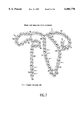
- FIG. 7 is a schematic illustration of the amino acid sequence of ⁇ -cobratoxin.
- FIG. 8 is a MALDI mass spectrum of undigested ⁇ -cobratoxin.
- FIG. 9 is a MALDI mass spectrum of approximately 10 picomoles of ⁇ -cobratoxin digested for 30 minutes with surface-bound trypsin sample presentation apparatus.
- FIG. 10 is a MALDI mass spectrum of approximately 10 picomoles of ⁇ -cobratoxin digested for 30 minutes with a surface-bound trypsin sample presentation apparatus in the presence of a reducing agent.
- FIG. 11 is an exploded view of the MALDI mass spectrum of FIG. 10 in the m/z range of 1,000-5,000 Pa.
- FIG. 12 is a positive ion MALDI mass spectrum of undigested human parathyroid hormone 1-34 (hPTH).
- FIG. 13 is a MALDI mass spectrum of hPTH digested for 10 minutes on a surface-bound ⁇ -chymotrypsin mass sample presentation apparatus.
- FIG. 14 is a MALDI mass spectrum of hPTH digested for 10 minutes on a surface-bound ⁇ -chymotrypsin sample presentation apparatus followed by pH adjustment to approximately 5 and addition of carboxypeptidase P (cpp).
- the inset illustrates the region of interest for final residue determination.
- FIG. 15 illustrates the partial peptide sequence of hPTH generated by analyzing the mass spectrometry data from ⁇ -chymotrypsin and ⁇ -chymotrypsin/carboxypeptidase P (cpp) fragments.
- FIG. 16 is a MALDI mass spectrum of hPTH digested for 5 minutes on a surface-bound trypsin sample presentation apparatus maintained at 45° C., pH 8.1.
- FIG. 17 is a protein ladder sequence generated by addition of cpp to the digested sample of FIG. 16.
- FIG. 18 is a summary of the partial peptide sequence generated by analyzing the mass spectrometry data from trypsin and trypsin/cpp fragments.
- FIG. 19 is an overlay of the mass spectra of trypsin and trypsin/cpp fragments illustrating the use of both the large-scale and ladder information to extend the sequence determination.
- FIG. 20 is a MALDI mass spectrum of hPTH digested for 8 minutes on a surface-bound pepsin sample presentation apparatus maintained at 45° C., pH 3.4.
- FIG. 21 is a MALDI mass spectrum of hPTH digested for 10 minutes on a surface-bound ⁇ -chymotrypsin mass sample presentation apparatus maintained at 45° C., pH 3.4.
- FIG. 22 is a MALDI mass spectrum of hPTH digested for 10 minutes on a surface-bound ⁇ -chymotrypsin mass sample presentation apparatus maintained at 45° C., pH 3.4, followed by a 10 minute digestion by surface-bound cpp maintained at 35° C., pH 5.
- FIG. 23(a) is a sample presentation apparatus having a mass spectrometry sample presentation surface contiguous with the sample presentation apparatus.
- FIG. 23(b) is a sample presentation apparatus having a mass spectrometry sample presentation surface separate from the sample presentation apparatus.
- FIGS. 23(a) and 23(b) depict a representative mass spectrometry sample presentation apparatus 100.
- the mass spectrometry sample presentation apparatus 100 has a mass spectrometry sample presentation surface 102 and a complex 104 immobilized on the surface.
- the present invention provides a conventional mass spectrometry sample presentation apparatus which has a surface derivatized with complexes which chemically modify biomolecules. This apparatus is typically exposed to a biomolecule, and incubated to allow the complex to modify the biomolecule. After incubation, the modified biomolecule is preferably surrounded with a MALDI matrix material and then introduced into a mass spectrometer.
- the invention also include further embodiments such as a mass spectrometer incorporating such a sample presentation apparatus. Further embodiments provide a method of performing mass spectrometry, and of making a sample presentation apparatus.
- a derivatized sample presentation apparatus in accordance with the invention may be composed of any suitable material.
- the material can be a solid or liquid. Suitable solid materials include, but are not limited to insulators such as quartz, semiconductors such as doped silicon and the like, and conductors including metals such as steel, gold and the like. Various insulating or conductive polymers may also be used.
- the surface of the sample presentation apparatus need not be made of the same material as the rest of the apparatus. It is preferable for the surface to be clean so that a complex may adhere to the surface.
- the complex attached to the presentation apparatus surface has two distinct functionalities.
- the term "complex” refers to a chemical which includes at least one molecule which chemically modifies a biomolecule.
- the complex has a "tethering" function, wherein it is capable of attaching itself at at least one end to the surface. Moreover, it also has a “reactive” function wherein it is capable of chemically modifying the analyte.
- the term “chemically modify” is meant to refer to purification or digestion of an analyte, but not binding an analyte.
- the complex may further comprise a tethering molecule which binds to the surface, and a reactive molecule which binds to the tethering molecule and modifies a biomolecule.
- the "tethering" function is associated with one group of atoms (i.e., the tethering molecule) while the “reactive” function is associated with a different group of atoms (i.e., the reactive molecule).
- the complex may comprise both a tethering molecule which binds to the sample presentation surface and a reactive molecule which binds to the tethering molecule and modifies a biomolecule.
- the tethering molecule is preferably selected from the class of molecules which has proven to be effective as an immobilized ligand. Many specific examples of such molecules are listed on pages T-156 to T-200 of the 1995 Pierce Corporation Catalog (Pierce Corp., P.O.
- Suitable tethering molecules include: dithiothreitol, dimethyladipimidate-2*HCL, dimethylpimelimidate*HCL, dimethylsuberimidate*2HCL, dimethyl 3,3'-dithiobispropionimidate*2HCL, disuccinimidyl glutarate, disuccinimidyl suberate, bis (sulfosuccinimidyl) suberate, dithiobis(succinimidylpropionate), dithiobis(sulfosuccinimidylpropionate), ethylene glycobis (succinimidylsuccinate), ethylene glycobis(sulfosuccinimidylsuccinate), disuccinimidyl tartarate, disulfosuccinimidyl tartarate, bis[2-(succinimidyloxycarbonyloxy)ethyl]sulfone, bis[2-(sulfone, bis[2-(sulfone,
- the complex may chemically modify a biomolecule analyte in several different ways.
- biomolecule is meant to refer to DNA, RNA, polypeptides, carbohydrates and combinations thereof.
- the complex may digest, or purify the biomolecule.
- digest is meant to refer to chemical or enzymatic cleavage of a biomolecule into fragments and the term “purify” means to remove undesirable materials from a solution containing the biomolecule.
- fragment is meant to refer to a part of a biomolecule which has been cleaved from the rest of the biomolecule using a chemical or enzymatic reagent.
- the complex will preferably comprise a chemical cleavage agent or an enzymatic protease.
- Enzymatic proteases are specific polypeptides which cleave polypeptides. Proteases may cleave themselves by a process known as autolysis. Several enzymatic proteases cleave polypeptides between specific amino acid residues. Examples of proteases which cleave nonspecifically include subtilisin, papain and thermolysin.
- proteases which cleave at least somewhat specifically include: aminopeptidase-M, carboxypeptidase-A, carboxypeptidase-P, carboxypeptidase-B, carboxypeptidase-Y, chymotrypsin, clostripain, trypsin, elastase, endoproteinase Arg-C, endoproteinase Glu-C, endoproteinase Lys-C, factor Xa, ficin, pepsin, plasmin, staphylococcus aureus V8 protease, proteinkinase K and thrombin.
- Chemical cleavage agents which cleave polypeptides between specific amino acid residues include cyanogen bromide, O-iodosobenzoate or O-iodosobenzoic acid, dilute hydrochloric acid, N-bromosuccinimide, sodium hydrazine, lithium aluminum hydride, hydroxylamine and 2-nitro-5-thiocyanobenzoate.
- Suitable chemical cleavage agents also include Sanger's Reagent, 2,4-dinitrofluorobenzene and immobilized derivatives thereof.
- coordinated transition metal complexes such as the tetradentate Co (III) complex, ⁇ -[Co(triethylenetetramine)-OH(H 2 O)], may also be used as effective cleavage agents. See Buckingham et al. 89 J.A.C.S. 1082 (1967).
- Disulfide reduction agents may be bound onto the sample presentation surface, or used in solution. Suitable disulfide reducing agents include ⁇ -mercaptoethanol, cysteine, and dithiothreitol (DTT). Carboxypeptidase-P, dithioerythritol (DTE) lipomide and N-acetylhomocysteine can be used as surface-bound disulfide reduction agents.
- restriction endonuclease or exonuclease can cleave DNA and RNA between specific nucleases, creating fragments of DNA or RNA.
- Specific restriction endonucleases and exonucleases are well known to those skilled in the art. Examples of enzymes which cleave both DNA and RNA include, but are not limited, to snake-venom phosphodiesterase and spleen phosphodiesterase. Enzymes which cleave only DNA include, but are not limited to, deoxy-ribonuclease I and II. Ribonuclease I (pancreas) and T. (mold) cleave RNA.
- fucase O- or N-glycanase
- mannase neuraminidase
- galactosidae glucosidase
- the complex attached to the sample presentation surface may also purify the biomolecule.
- the complex may comprise a reactive molecule which invisibly binds common impurities such as detergents, lipids, endotoxins or proteases.
- the present invention also provides a method for making the above-described derivatized mass spectrometry sample presentation apparatus.
- this method it is essential that a complex which modifies biomolecules be bound to the surface of the sample presentation apparatus.
- the entire complex is first synthesized or purified, and then reacted with the surface.
- the complex is formed by first reacting the sample presentation surface with a tethering molecule, and then later reacting the tethered molecule with a reactive molecule.
- Another embodiment of the present invention provides a method for analyzing a biomolecule. Specifically, the method involves: (1) providing a mass spectrometry sample presentation surface, (2) binding at least one complex on the surface wherein the complex includes at least one molecule which chemically modifies a biomolecule, (3) contacting the biomolecule with the surface, thereby chemically modifying the biomolecule (4) applying a MALDI matrix to the surface and (5) determining the molecular weight of the biomolecule in a mass spectrometer.
- the biomolecule may be contacted with the surface while in solution or in the vapor phase.
- the contacting step is preferably performed above room temperature, most preferably up to 55° C., to speed the reaction rate. However, it should not be performed at a temperature high enough to cause degradation of the analyte or the complex. For certain complexes, it may be advantageous to perform the contacting step below room temperature, most preferably above -10° C. This contacting step is allowed to proceed for a sufficient time, typically 5-30 minutes. The actual reaction time will depend on the particular complex and biomolecule. When proteases are used to digest polypeptide biomolecule, the contacting step is preferably performed in a moist atmosphere. Such an atmosphere may be created for example, by providing a water reservoir and enclosing it along with the sample presentation surface and biomolecule in a closed system.
- the MALDI matrix may be any material which solubilizes biomolecules, absorbs light energy at a frequency easily accessible by a laser, and is unreactive with respect to biomolecules.
- Suitable matrices include nicotinic acid, pyrozinoic acid, vanillic acid, succinic acid, caffeic acid, glycerol, urea or tris buffer (pH 7.3).
- Preferable matrices include ⁇ -cyano-4-hydroxycinnamic acid, ferulic acid, 2,5-dihydroxybenzoic acid, sinapic (or sinapinic) acid, 3,5-dimethoxy, 4-hydroxy-trans-cinnamic acid, other cinnamic acid derivatives, gentisic acid and combinations thereof.
- a further embodiment of the present invention provides a mass spectrometer which incorporates the above-described sample presentation apparatus.
- the mass spectrometer according to the present invention further comprises a vacuum interlocking device for introducing the sample presentation apparatus into a volatilization chamber, an apparatus for volatilizing and ionizing the analyte, an optional electric field generator which is oriented to cause biomolecule ions to travel toward a detector, and electronics for determining the molecular weight to charge ratio for a particular ion based on its trajectory.
- This mass spectrometer is compatible with all of the techniques normally used for volatilizing and ionizing analytes. Such techniques include, but are not limited to, electrospray ionization, Cf plasma desorption, field desorption, fast atom bombardment, liquid secondary ion MS, laser desorption, and thermospray ionization.
- the MALDI technique is used for volatilizing and analyzing biomolecules. The mechanical structures or equipment required in these techniques are well known to those skilled in the art.
- the spectrometer usually contains an electric field generator which has electrically charged metal structures between the area in the spectrometer where the ions are generated and the detector. These structures must be designed such that they attract ions, but the ions can travel past them to reach the detector. Preferably, the structures are charged metal grids with apertures that allow some of the ions to pass through and drift toward the detector. The structures must be located between the ion generation area and the detector. They must also have a potential difference which acts to accelerate ions toward the detector.
- Some mass spectrometers also use magnetic fields to deflect ions as they travel towards the detector. Such instruments determine the mass of an ion based on its deflection in the magnetic field. It is, however, preferable to use a time-of-fight mass spectrometer to determine the mass of biomolecules.
- the electronics for determining the molecular weight to charge ratio of an ion based on its trajectory typically includes an ion impact detector, data-acquisition electronics, and data analysis electronics, such as a computer.
- detectors are compatible with the present invention. Some specific examples of detectors include the Faraday cup, electron multipliers, electro-optical ion detectors and photographic emulsions.
- Circles of gold (Au) foil (99.9%), available from Johnson Matthey, 2.5 mm diameter and 0.01 mm thick were punched out and placed in a polyethylene microcentrifuge tube. Care was taken to avoid any contamination of the gold foil circles.
- the gold foil was activated according to the method of Katz. See, Katz, E. Y., J. Electroanal. Chem., Vol. 291, p. 257 (1990). The circles were treated with a crosslinking, or tethering molecule, solution of Dithiobis [succinimidylpropionate] (DSP) available from Pierce dissolved in isopropanol to approximately 0.1M. The DSP/isopropanol solution was essentially saturated at this concentration.
- DSP Dithiobis [succinimidylpropionate]
- the circles were treated with the DSP/isopropanol solution for fifteen minutes, with occasional mixing using a vortex mixer. This tethering molecule solution was poured off, and the gold circles were rinsed repetitively with pure isopropanol, followed by three rinses with pure ethanol. The gold circles were vacuum dried and transferred to new microcentrifuge tubes prior to treatment with a reactant molecule or biomolecule.
- a mass spectrometry sample presentation apparatus was prepared by binding trypsin to the surface of the apparatus as follows. Trypsin type XIII: tosyl-L-phenylalanine chloromethyl ketone (TPCK) treated, from bovine pancreas available from Sigma, was dissolved in 20 mM phosphate buffer, pH 7.8 to a concentration of 1 mg/mL for use in preparation of the derivatized sample presentation apparatus. To attach the enzyme to the crosslinker or tethering molecule on the gold foil circles prepared in Example 1, the circles were incubated with a 1 mg/mL solution of enzyme in 20 mM phosphate buffer, pH 7.8 overnight in a refrigerator with occasional agitation.
- TPCK tosyl-L-phenylalanine chloromethyl ketone
- the gold foil circles were rinsed vigorously with the phosphate buffer, and subsequently with a 0.1% aqueous solution of Triton-X100, using a vortex mixer for both rinses.
- the gold foil circles were then vacuum dried and stored in polyethylene microcentrifuge tubes. Physical attachment of the gold foil circle surfaces to the stainless steel sample presentation apparatuses conventionally used for routine MALDI analysis was accomplished by application of a small amount of spray adhesive to the conventional sample presentation surface or probe tip, which was then pressed onto one of the gold foil circles.
- the conventional stainless steel (304) mass spectrometer sample presentation apparatuses were electropolished and thoroughly rinsed in deionized water followed by ultrasonic cleaning in methanol. Even pressure was maintained on the probe tip and gold foil while the adhesive was allowed to bond. The sample presentation apparatus with bound trypsin was then used for protein digestion as described hereinbelow. In practice, multiple tips were made and stored in polyethylene tubes at room temperature until needed.
- Digestions were performed with the Au/trypsin active surfaces prepared in Example 2 by the application of aliquots (either 4 ⁇ L or 400 nL) of 250 nM lysozyme hen egg solution (dissolved in 20 mM phosphate; 10 mM dithiothreitol (DTT); pH 8.1 buffer) directly to the sample presentation apparatus surfaces.
- the surfaces were allowed to stand in a humid enclosure maintained at 40° C. The solution volume on the surface was monitored, and additional aliquots of 1-2 ⁇ L of phosphate buffer were added if the volume of liquid appeared to be less than 2 ⁇ L.
- a 1:1 mole ratio of trypsin to lysozyme was prepared by addition of 2 ⁇ L of 500 nM lysozyme with 2 ⁇ L of 500 nM trypsin (both prepared in a 20 mM phosphate; 10 mM DTT; pH 8.1 buffer). The combined volume was placed in a 600 ⁇ L microcentrifuge tube and incubated at 40° C. for 10 minutes. The reaction was halted by the addition of 2 ⁇ L of the MALDI matrix ACCA and the total volume of the digest/matrix mixture was placed on an inert mass spectrometer sample presentation apparatus and allowed to air dry.
- a 1:1 volume ratio of lysozyme to agarose/trypsin was prepared by addition of 2 ⁇ L of 500 nM lysozyme with 2 ⁇ L of slurried agarose/trypsin (both prepared in a 20 mM phosphate; 10 mM DTT; pH 8.1 buffer).
- the slurried agarose contained approximately 1 ⁇ L of beaded reagent.
- the combined volume was placed in a 600 ⁇ L microcentrifuge tube and incubated at 40° C. for 10 minutes.
- the reaction was halted by the addition of 2 ⁇ L of ACCA matrix and the total volume (with beaded reagent) of the digest/matrix mixture was placed on an inert mass spectrometer conventional sample presentation apparatus or probe tip and allowed to air dry.
- Mass spectrometry was performed using the sample presentation apparatus prepared in Example 3, Comparative Example 4, and Comparative Example 5 using a linear time-of-flight mass spectrometer. Desorption and ionization were accomplished using the third harmonic (355 nm) output of the Nd:YAG laser (Continuum Surelite I).
- An electrostatic particle guide was used to assist in ion transmission over the flight path.
- Detection was accomplished using a hybrid, microchannel-plate/discrete dynode, electron multiplier. Thresholds of ionization were determined empirically by increasing the intensity of the laser light at the sample surface while monitoring the ion signal real-time with a 50 Mhz oscilloscope (Tektronix TDS 310). Upon observation of stable ion signal, 50 laser shots were signal averaged using a 500 MS/s digital storage oscilloscope (Tektronix TDS 520A) and transferred to an IBM-compatible 486 computer. The data were analyzed using the PC compatible software LabCalc (Galactic Industries). All mass spectra were obtained in the positive-ion mode.
- FIG. 2 shows the correlation between the calculated masses of the enzymatically cleaved fragments and those observed in the mass spectrum.
- FIG. 3 shows the mass spectrum obtained from using the same procedure performed with only 1 pmol of lysozyme applied to the surface. Virtually the same fragmentation pattern is observed, with an expected sacrifice in the signal-to-noise ratio of the data.
- trypsin whole molecule signals also appear in the spectrum.
- the autolysis and parent signals contaminate the data because these fragments are incorporated in the matrix.
- the trypsin used in digestion is not bound to the surface, and therefore, there is nothing to prevent the trypsin molecular ion and autolysis products from incorporating into the matrix crystals.
- a decrease in the signal-to-noise ratio is also observed, relative to FIG. 3, and is attributed to loss of sample in transfer and handling.
- agarose is a porous medium, it is believed that digest fragments are lost through a combination of incorporation into the media, slow digestion due to poor localization of analyte around the immobilized enzyme, and inefficient elution using a small volume of matrix.
- ⁇ -cobratoxin was analyzed using an immobilized trypsin sample presentation apparatus as described in Example 2.
- the unfragmented ⁇ -cobratoxin was analyzed using the MALDI method. Signals were obtained which correspond the singly and doubly charged ions of the intact peptide. As can be seen from FIG. 7 and FIG. 8, singly and doubly charged ions corresponding to residue 1-71 are observed at 7,823 and 3,912 Da, respectfully. Signals less than m/z approximately 1,000 Da are due to the ACCA matrix.
- weights correspond to peptide sequences 1-68, 1-69 and 1-71, respectively, as shown in FIG. 7. These are the primary fragments expected from the tryptic treatment of the non-reduced peptide. Five other cleavage sites are possible, each producing different molecular weight fragments. However, these sites are stearically hindered and not available to trypsin. Therefore, these five other possible peaks were not observed.
- ⁇ -cobratoxin was further analyzed by repeating the above-described 10 picomole ⁇ -cobratoxin digestion by surface-bound trypsin in the presence of a reducing agent (10 pmol ⁇ -cobratoxin in 3 ⁇ L-10 mM phosphate; 10 mM DTT). As can be seen from FIG. 7, the reducing agent broke the disulfide bonds, thus enabling the generation of more fragments. The resulting spectrum shown in FIGS. 10 and 11 shows a set of peptide fragments indicative of reduced cobratoxin.
- a mass spectrometry sample presentation apparatus was prepared by binding ⁇ -chymotrypsin to the surface of the apparatus as follows. ⁇ -chymotrypsin or TPCK-treated ⁇ -chymotrypsin was dissolved in 20 mM phosphate buffer, pH 7.8 to a concentration of 1 mg/mL for use in preparation of a derivatized sample presentation apparatus as described in Example 2.
- Enzymatically active trypsin and ⁇ -chymotrypsin mass spectrometry sample presentation surfaces were prepared as described in Examples 2 and 8.
- the synthetic peptide human parathyroid hormone 1-34 (hPTH) was chosen as a test peptide for analyses.
- the sequence of the peptide is:
- Digestions were performed in the following manner. Three microliter aliquots of a 0.01 mg/mL solution of hPTH (in a buffer of 20 mM NaHPO 4 , pH 8.1) were applied to derivatized mass spectrometer sample presentation apparatus prepared in accordance with Example 2 or Example 8. The digestion was carried out in a high humidity environment at a temperature of between 40 and 50° C. The digestions were allowed to run for 5 to 10 minutes before 0.5-1.0 ⁇ L aliquots of the reaction mixtures were removed from the surfaces. These aliquots were MALDI analyzed for endopeptidase mapping.
- the pH was reduced to approximately 5 with the addition of an aliquot of 20 mM NH 4 COOCH 3 , the volume of which was equal to that of the remaining reaction mixture. 500 nL aliquots of 0.001 mg/mL carboxypeptidase P (CPP), in the acetate buffer, were then added to the reaction mixtures. Protein ladder digestions were allowed to run for between 1 to 3 minutes before the surfaces were removed from the heated high humidity environment and the reactions halted through the addition of an equal volume of MALDI matrix. The matrix/reaction mixture was allowed to air-dry and the enzymatically-active surfaces inserted directly into the mass spectrometer.
- CPP carboxypeptidase P
- FIG. 12 is an ACCA-MALDI mass spectrum of 2.5 picomoles of the hPTH, the starting point of reference for the following enzymatic analyses.
- the hPTH was further analyzed after a 10 minute digestion on an Au/ ⁇ -chymotrypsin surface maintained at 40° C., pH 8.1. The results of this digestion are shown in FIG. 13. Four major signals at 1,398.5, 1,706.2, 1850.0 and 2,738.7 Da are observed as shown in FIG. 13.
- FIG. 14 shows the MALDI mass spectrum obtained from this second sample.
- the first residue is a preferred cleavage site for ⁇ -chymotrypsin.
- FIG. 15 summarizes the masses of the signals observed in FIG. 14, the differences in mass between adjacent signals and the residues determined from the differences.
- the partial sequence read as shown in FIG. 15 from N to C-terminal is determined by sequential mass difference to give (pep)-MERVEW-COOH.
- FIG. 16 shows the results of the Au/trypsin digestion after 5 minutes at a temperature of 45° C. (pH 8.1).
- FIG. 18 summarizes the masses observed for the digestion.
- the trypsin digestion shown in FIG. 16 resulted in the production of several starting fragments, and indicated the presence of the short sequence (Arg/Lys)-Lys-Lys.
- FIG. 17 shows the resulting sequence ladder. From the observed mass differences summarized in FIG. 18 the sequence was determined to be (pep)-NSMERVEW(L/I)RK-COOH. The first residue cleaved by cpp is that of Arg. Arg is a preferred cleavage site for trypsin. This is obviously an extension of the partial sequence determined using Au/ ⁇ -chymotrypsin/cpp.
- a nominal mass difference of 128 Da was observed at several points in the spectrum resulting from the initial trypsin cleavage in the Au/trypsin/cpp analysis. Considering that trypsin will cleave only at lysine or arginine, the 128 Da mass difference is highly likely to result from a lysine C-terminal to either another lysine or an arginine. Likewise, two adjacent mass differences of 128 Da, as observed in the 3,000 to 3,300 Da region, indicates the presence of Lys-Lys in the sequence, with the third residue being either another lysine or an arginine.
- FIG. 19 illustrates a ladder sequence extended by an additional lysine determined from the (Arg/Lys)-Lys-Lys observed in the trypsin spectrum overlapping with the Arg-Lys of the ladder sequence.
- the 128 Da mass differences observed in this manner can be unambiguously assigned to Lys as opposed to Gln which has the same molecular weight.
- the result is that 12 residues of the sequence were determined to be (pep)-NSMERVEW(L/I)RKK-COOH.
- nucleic acid sequences which code for the peptide sequence. See, e.g., Biochemistry, Stryer, pp. 91-115 (1988). Since many different nucleic acid sequences code for a single peptide, several nucleic acid sequences can be generated from the peptide sequences. The nucleic acid sequences can be used to search a database of nucleic acid sequences, e.g., Genbank, to identify genes which code for a particular polypeptide.
- a mass spectrometry sample presentation apparatus was prepared by binding pepsin to the surface of the apparatus as follows.
- Gold foil was first activated with DSP according to the method given in Example 1. The DSP activated gold was then reacted with ethylene diamine (20% v/v in isopropanol) at room temperature for 30 minutes. After incubation, the gold foil was rinsed thoroughly with isopropanol and then reacted with 0.1 M[l-ethyl-3-(3-dimethylaminopropyl)carbodiimide] in 20 mM phosphate buffer at pH 4.5, for 30 minutes at room temperature. The foil was then rinsed thoroughly with the phosphate buffer. Next, a solution of 10 mg/ml pepsin in phosphate buffer was added to the surface and allowed to incubate for 4 hours at 4° C. After incubation the foil was rinsed repeatedly with phosphate buffer and finally vacuum dried.
- a digestion was performed with the Au/Pepsin active surfaces prepared in Example 10 by the application of approximately 5 pmole of hPTH 1-34 in 10 mM ammonium acetate buffer (pH 3.4).
- the hPTH was digested for 8 minutes at 45° C. before stopping the reaction by adding ACCA matrix.
- Time-of-Flight mass spectrometry was then performed.
- the resulting spectrum shown in FIG. 20 includes ion signals for the pepsin-generated peptides corresponding to amino acids 4-18, 9-24, 8-24, and 18-34.
- the combination of two different surface bound complexes was used to modify a biomolecule in sequence as follows. First hPTH was digested for 10 minutes at 35° C., pH 8.1 using an immobilized Au/ ⁇ -chymotrypsin sample presentation device as described in Example 2. A portion of the digest mixture was then transferred to a cpp activated apparatus prepared as described in Example 2 and the pH of the solution was adjusted to approximately 5. The cpp digestion was allowed to proceed for 10 minutes at 35° C. before termination by the addition of the ACCA matrix.
- the mass spectrum of the results of the initial Au/ ⁇ -chymotrypsin digestion is shown in FIG. 21. Ion signals resulting from the ⁇ -chymotrypsin cleavage of hPTH are observed.
- the mass spectrum of the results of the Au/cpp digestion of the ⁇ -chymotrypsin digestion mixture is shown in FIG. 22. New ion signals indicated by an asterisk "*" are observed. These new signals correspond to the C-terminal digestion of the original peptide species.
- the invention can be used to purify biomolecules as follows. For many biological studies, it is essential to remove undesirable proteases from biological solutions.
- Aprotinin is a single-chain basic 58-amino-acid polypeptide with a molecular mass of 6512 Daltons that has a polyvalent inhibitory action on proteases. Specifically, it inhibits a variety of enzymes belonging to the family of serine proteases by binding to the active site of the enzyme, forming tight complexes.
- a mass spectrometry sample presentation apparatus is prepared by tethering aprotinin to the surface in a manner similar to that described in Example 2. A solution of a polypeptide and a protease is added to the surface. The protease is removed from solution by the aprotinin. Mass spectrometry is performed and only the purified polypeptide should be observed.
- An ion exchange functionality such as a diethylaminoethyl derivative, a t-butyl amino derivative (for anion exchange), a sulfite derivative, a carboxylic acid derivative (for cation exchange), or a combination of the above is immobilized to a sample presentation surface prepared according to claim 1.
- a biomolecule-containing solution is added to a surface prepared according to Example 14.
- the ion exchange functionality bound to the sample presentation surface exchanges ions with detrimental ionic species present as impurities in the biomolecule-containing solution.
- the nuclease snake-venom phosphodiesterase
- This attachment may be accomplished using a tethering molecule in a manner similar to that described in Example 2.
- DNA is added to the surface. After the phosphodiesterase has produced DNA fragments, a MALDI matrix is added, and MALDI time-of-flight mass spectrometry is performed.
- An enzyme such as mannase which digests carbohydrates is attached to the sample presentation apparatus surface. This attachment may be accomplished using a tethering molecule in a manner similar to that described in Example 2. Next, a carbohydrate is added to the surface. After the mannase has produced carbohydrate fragments, a MALDI matrix is added, and MALDI time-of-flight mass spectrometry is performed.
Abstract
Description
______________________________________
102030
  SVSEIQLMHN LGKHLNSMER VEWLRKKLQD VHNF
______________________________________
Claims (12)
Priority Applications (8)
| Application Number | Priority Date | Filing Date | Title |
|---|---|---|---|
| US08/488,300 US6004770A (en) | 1995-06-07 | 1995-06-07 | Sample presentation apparatus for mass spectrometry |
| JP50142297A JP3879030B2 (en) | 1995-06-07 | 1996-06-06 | Sample presentation device for mass spectrometry |
| EP96921296A EP0832201B1 (en) | 1995-06-07 | 1996-06-06 | A sample presentation apparatus for mass spectrometry |
| AU62549/96A AU729513C (en) | 1995-06-07 | 1996-06-06 | A sample presentation apparatus for mass spectrometry |
| PCT/US1996/008994 WO1996040888A1 (en) | 1995-06-07 | 1996-06-06 | A sample presentation apparatus for mass spectrometry |
| CA002221250A CA2221250C (en) | 1995-06-07 | 1996-06-06 | A sample presentation apparatus for mass spectrometry |
| AT96921296T ATE305969T1 (en) | 1995-06-07 | 1996-06-06 | SAMPLER FOR MASS SPECTROMETRY |
| DE69635242T DE69635242T2 (en) | 1995-06-07 | 1996-06-06 | SAMPLE SPECTRUM FOR MASS SPECTROMETRY |
Applications Claiming Priority (1)
| Application Number | Priority Date | Filing Date | Title |
|---|---|---|---|
| US08/488,300 US6004770A (en) | 1995-06-07 | 1995-06-07 | Sample presentation apparatus for mass spectrometry |
Publications (1)
| Publication Number | Publication Date |
|---|---|
| US6004770A true US6004770A (en) | 1999-12-21 |
Family
ID=23939174
Family Applications (1)
| Application Number | Title | Priority Date | Filing Date |
|---|---|---|---|
| US08/488,300 Expired - Lifetime US6004770A (en) | 1995-06-07 | 1995-06-07 | Sample presentation apparatus for mass spectrometry |
Country Status (1)
| Country | Link |
|---|---|
| US (1) | US6004770A (en) |
Cited By (27)
| Publication number | Priority date | Publication date | Assignee | Title |
|---|---|---|---|---|
| US6379971B1 (en) * | 1998-02-24 | 2002-04-30 | Target Discovery, Inc. | Methods for sequencing proteins |
| US20020172961A1 (en) * | 2000-10-19 | 2002-11-21 | Target Discovery | Mass defect labeling for the determination of oligomer sequences |
| US20020177242A1 (en) * | 1997-06-20 | 2002-11-28 | Ciphergen Biosystems, Inc. | Retentate chromatography and protein chip arrays with applications in biology and medicine |
| US20020192676A1 (en) * | 2001-06-18 | 2002-12-19 | Madonna Angelo J. | Method for determining if a type of bacteria is present in a mixture |
| US6498039B2 (en) * | 1995-06-07 | 2002-12-24 | Arizona Board Of Regents | Sample presentation apparatus for mass spectrometry |
| US6569383B1 (en) * | 2000-03-11 | 2003-05-27 | Intrinsic Bioprobes, Inc. | Bioactive chip mass spectrometry |
| US20030116707A1 (en) * | 2001-08-17 | 2003-06-26 | Micromass Limited | Maldi sample plate |
| US6624409B1 (en) | 2002-07-30 | 2003-09-23 | Agilent Technologies, Inc. | Matrix assisted laser desorption substrates for biological and reactive samples |
| US20040038234A1 (en) * | 2000-06-30 | 2004-02-26 | Gut Ivo Glynne | Sample generation for genotyping by mass spectrometry |
| US20040050787A1 (en) * | 2002-09-13 | 2004-03-18 | Elena Chernokalskaya | Apparatus and method for sample preparation and direct spotting eluants onto a MALDI-TOF target |
| US6716636B1 (en) * | 1999-04-20 | 2004-04-06 | Target Discovery, Inc. | Methods for sequencing proteins |
| US20040185448A1 (en) * | 2003-03-20 | 2004-09-23 | Viorica Lopez-Avila | Methods and devices for performing matrix assisted laser desorption/lonization protocols |
| US20040219531A1 (en) * | 2003-04-30 | 2004-11-04 | Dicesare Joseph L. | Method of scanning a sample plate surface mask in an area adjacent to a conductive area using matrix-assisted laser desorption and ionization mass spectrometry |
| US6867002B2 (en) * | 1998-10-20 | 2005-03-15 | Matsushita Electric Industrial Co., Ltd. | Sample treating kit and sample treating method using the same for analysis with a biosensor |
| US6869572B1 (en) | 1999-09-13 | 2005-03-22 | Millipore Corporation | High density cast-in-place sample preparation card |
| US20050130222A1 (en) * | 2001-05-25 | 2005-06-16 | Lee Peter J.J. | Sample concentration maldi plates for maldi mass spectrometry |
| US20050153346A1 (en) * | 1999-04-20 | 2005-07-14 | Target Discovery, Inc. | Methods for conducting metabolic analyses |
| US20060016984A1 (en) * | 2003-02-10 | 2006-01-26 | Waters Investments Limited | Sample preparation plate for mass spectrometry |
| US7053366B2 (en) | 2001-05-25 | 2006-05-30 | Waters Investments Limited | Desalting plate for MALDI mass spectrometry |
| US20070059725A1 (en) * | 2005-03-31 | 2007-03-15 | Voorhees Kent J | Apparatus and method for detecting microscopic organisms using bacteriophage |
| AU2004216642B2 (en) * | 1999-04-20 | 2007-05-10 | Target Discovery, Inc. | Polypeptide fingerprinting methods, metabolic profiling, and bioinformatics database |
| US20070148638A1 (en) * | 2002-04-12 | 2007-06-28 | Colorado School Of Mines | Method for Detecting Low Concentrations of a Target Bacterium That Uses Phages to Infect Target Bacterial Cells |
| US20090246753A1 (en) * | 2008-01-11 | 2009-10-01 | Colorado School Of Mines | Detection of Phage Amplification by SERS Nanoparticles |
| US20090258341A1 (en) * | 2008-04-03 | 2009-10-15 | Colorado School Of Mines | Compositions and Methods for Detecting Bacteria |
| US20090325222A1 (en) * | 2006-05-26 | 2009-12-31 | Brandeis University | Tissue Sample Preparation and MALDI MS Imaging Thereof |
| US20110097702A1 (en) * | 2005-03-31 | 2011-04-28 | Voorhees Kent J | Methods and compositions for in situ detection of microorganisms on a surface |
| US7943294B2 (en) | 2004-07-30 | 2011-05-17 | Hologic, Inc. | Methods for detecting oncofetal fibronectin |
Citations (4)
| Publication number | Priority date | Publication date | Assignee | Title |
|---|---|---|---|---|
| US3878049A (en) * | 1973-04-03 | 1975-04-15 | Massachusetts Inst Technology | Biochemical temperature-sensitive probe and method for measuring reactant concentrations thereof |
| JPS52139781A (en) * | 1976-05-15 | 1977-11-21 | Nippi Inc | Production of fixed biologically active substance |
| WO1994028418A1 (en) * | 1993-05-28 | 1994-12-08 | Baylor College Of Medicine | Method and apparatus for desorption and ionization of analytes |
| US5516698A (en) * | 1990-04-11 | 1996-05-14 | Ludwig Institute For Cancer Research | Methods and apparatus allowing sequential chemical reactions |
-
1995
- 1995-06-07 US US08/488,300 patent/US6004770A/en not_active Expired - Lifetime
Patent Citations (4)
| Publication number | Priority date | Publication date | Assignee | Title |
|---|---|---|---|---|
| US3878049A (en) * | 1973-04-03 | 1975-04-15 | Massachusetts Inst Technology | Biochemical temperature-sensitive probe and method for measuring reactant concentrations thereof |
| JPS52139781A (en) * | 1976-05-15 | 1977-11-21 | Nippi Inc | Production of fixed biologically active substance |
| US5516698A (en) * | 1990-04-11 | 1996-05-14 | Ludwig Institute For Cancer Research | Methods and apparatus allowing sequential chemical reactions |
| WO1994028418A1 (en) * | 1993-05-28 | 1994-12-08 | Baylor College Of Medicine | Method and apparatus for desorption and ionization of analytes |
Non-Patent Citations (77)
| Title |
|---|
| A. S. Woods et al. Anal. Biochem. 1995, 226, 15 25. * |
| A. S. Woods et al. Anal. Biochem. 1995, 226, 15-25. |
| B. Johnsson et al. Anal. Biochem. 1991, 198, 268 277. * |
| B. Johnsson et al. Anal. Biochem. 1991, 198, 268-277. |
| B. M. C. Hoang et al. C. R. Acad. Sci 1994, 317, 5 10. * |
| B. M. C. Hoang et al. C. R. Acad. Sci 1994, 317, 5-10. |
| B. T. Chait et al. Sciences 1993, 262, 89 92. * |
| B. T. Chait et al. Sciences 1993, 262, 89-92. |
| B. Thiede et al. FEBS Lett. 1995, 357, 65 69. * |
| B. Thiede et al. FEBS Lett. 1995, 357, 65-69. |
| C. J. Aldrich et al. Cell, 1994, 79, 649 658. * |
| C. J. Aldrich et al. Cell, 1994, 79, 649-658. |
| C. W. Sutton et al. "Techniques in Protein Chemistry IV" R. H. Angelette, Ed., Academic Press, Inc.: San Diego, California, 1993, pp. 109-116. |
| C. W. Sutton et al. Techniques in Protein Chemistry IV R. H. Angelette, Ed., Academic Press, Inc.: San Diego, California, 1993, pp. 109 116. * |
| D. Bharadwaj et al. Indian J. Biochem. Biophys. 1992, 29, 375 377. * |
| D. Bharadwaj et al. Indian J. Biochem. Biophys. 1992, 29, 375-377. |
| D. Dogruel et al. Anal. Chem. 1995, 67, 4343 4348. * |
| D. Dogruel et al. Anal. Chem. 1995, 67, 4343-4348. |
| D. I. Papac et al. Protein Sci. 1994, 3, 1485 1492. * |
| D. I. Papac et al. Protein Sci. 1994, 3, 1485-1492. |
| D. L. Crimmins et al. Anal. Biochem. 1995, 226, 355 361s. * |
| D. L. Crimmins et al. Anal. Biochem. 1995, 226, 355-361s. |
| D. Pilosof et al. Biomed. Mass. Spec. 1984, 11, 403 407. * |
| D. Pilosof et al. Biomed. Mass. Spec. 1984, 11, 403-407. |
| D. Suckau et al. Proc. Natl. Acad. Sci USA 1990, 87, 9848 9852. * |
| D. Suckau et al. Proc. Natl. Acad. Sci USA 1990, 87, 9848-9852. |
| E. J. Zaluzec et al. J. Am. Soc. Mass Spectrom. 1994, 5, 230 237. * |
| E. J. Zaluzec et al. J. Am. Soc. Mass Spectrom. 1994, 5, 230-237. |
| E. Van Leemp u tten et al. Biotechnol. Bioeng. 1974, 16, 997 1003. * |
| E. Van Leemputten et al. Biotechnol. Bioeng. 1974, 16, 997-1003. |
| F. C. Church et al. J. Appl. Biochem. 1984, 6, 205 211. * |
| F. C. Church et al. J. Appl. Biochem. 1984, 6, 205-211. |
| G. Allmaier et al. Anal. Chim. Acta 1990, 241, 321 327. * |
| G. Allmaier et al. Anal. Chim. Acta 1990, 241, 321-327. |
| H. Ueki et al. J. Biochem 1974, 76, 191 199. * |
| H. Ueki et al. J. Biochem 1974, 76, 191-199. |
| H. Y. Kim et al. J. Am. Chem. Soc. 1984, 106, 7304 7309. * |
| H. Y. Kim et al. J. Am. Chem. Soc. 1984, 106, 7304-7309. |
| J. Bai et al. Anal. Chem. 1994, 66, 3423 3430. * |
| J. Bai et al. Anal. Chem. 1994, 66, 3423-3430. |
| J. Bai et al. Anal. Chem. 1995, 67, 1705 1710. * |
| J. Bai et al. Anal. Chem. 1995, 67, 1705-1710. |
| J. L. Garnett et al. Chem. Abstr. 1986, 104, 203089z. * |
| J. L. Garnett et al. Radiat Phys. Chem. 1986, 27, 301 309. * |
| J. L. Garnett et al. Radiat Phys. Chem. 1986, 27, 301-309. |
| K. K. Mock et al. Rapid Commun. Mass Spectrom. 1992, 6, 233 238. * |
| K. K. Mock et al. Rapid Commun. Mass Spectrom. 1992, 6, 233-238. |
| L. A. Amankuva et al. Anal. Chem. 1992, 64, 1610 1613. * |
| L. A. Amankuva et al. Anal. Chem. 1992, 64, 1610-1613. |
| M. Karas et al. Anal. Chim. Acta 1990, 241, 175 185. * |
| M. Karas et al. Anal. Chim. Acta 1990, 241, 175-185. |
| M. M. Vestling et al. Anal. Chem. 1994, 66, 471 477. * |
| M. M. Vestling et al. Anal. Chem. 1994, 66, 471-477. |
| Nelson et al., Mass Spectrometric Immunoassay, Analytical Chemistry, 34:1153 1158, (Apr. 1, 1995). * |
| Nelson et al., Mass Spectrometric Immunoassay, Analytical Chemistry, 34:1153-1158, (Apr. 1, 1995). |
| P. C. Andrews et al. "Techniques in Protein Chemistry III" R. H. Angeletti, Ed., Academic Press, Inc.: San Diego, Califfornia, 1992, pp. 515-523. |
| P. C. Andrews et al. Techniques in Protein Chemistry III R. H. Angeletti, Ed., Academic Press, Inc.: San Diego, Califfornia, 1992, pp. 515 523. * |
| R. Kaufmann J. Biotechnology. 1995, 41, 155 175. * |
| R. Kaufmann J. Biotechnology. 1995, 41, 155-175. |
| R. M. Wagner et al. Biomed. Environ. Mass Spectrom. 1987, 14, 235 239. * |
| R. M. Wagner et al. Biomed. Environ. Mass Spectrom. 1987, 14, 235-239. |
| R. S. Youngquist et al. Rapid Commun. Mass Spec. 1994, 8, 77 81. * |
| R. S. Youngquist et al. Rapid Commun. Mass Spec. 1994, 8, 77-81. |
| R. W. Nelson et al. Anal. Chem. 1994, 66, 1408 1415. * |
| R. W. Nelson et al. Anal. Chem. 1994, 66, 1408-1415. |
| R. W. Nelson et al. Rapid Commun. Mass Spectrom. 1992, 6, 4 8. * |
| R. W. Nelson et al. Rapid Commun. Mass Spectrom. 1992, 6, 4-8. |
| S. L. Cohen et al. Protein Sci. 1995, 4, 1088 1099. * |
| S. L. Cohen et al. Protein Sci. 1995, 4, 1088-1099. |
| T. L o tzbeyer et al. J. Electroanal. Chem. 1994, 377, 291 294. * |
| T. Lotzbeyer et al. J. Electroanal. Chem. 1994, 377, 291-294. |
| T. W. Hutchens et al. FEBS Lett. 1992, 296, 99 102. * |
| T. W. Hutchens et al. FEBS Lett. 1992, 296, 99-102. |
| T. W. Hutchens et al. Rapid Commun. Mass Spectrom. 1993, 7, 576 580. * |
| T. W. Hutchens et al. Rapid Commun. Mass Spectrom. 1993, 7, 576-580. |
| W. H. Fischer et al. Rapid Commun. Mass. Spectrom. 1993, 7, 225 228. * |
| W. H. Fischer et al. Rapid Commun. Mass. Spectrom. 1993, 7, 225-228. |
Cited By (58)
| Publication number | Priority date | Publication date | Assignee | Title |
|---|---|---|---|---|
| US6498039B2 (en) * | 1995-06-07 | 2002-12-24 | Arizona Board Of Regents | Sample presentation apparatus for mass spectrometry |
| US7112453B2 (en) * | 1997-06-20 | 2006-09-26 | Ciphergen Biosystems, Inc. | Retentate chromatography and protein chip arrays with applications in biology and medicine |
| US20020177242A1 (en) * | 1997-06-20 | 2002-11-28 | Ciphergen Biosystems, Inc. | Retentate chromatography and protein chip arrays with applications in biology and medicine |
| US7105339B2 (en) * | 1997-06-20 | 2006-09-12 | Ciphergen Biosystems, Inc. | Retentate chromatography and protein chip arrays with applications in biology and medicine |
| US20040142493A1 (en) * | 1997-06-20 | 2004-07-22 | Ciphergen Biosystems, Inc. | Retentate chromatography and protein chip arrays with applications in biology and medicine |
| US6706529B2 (en) * | 1998-02-24 | 2004-03-16 | Target Discovery, Inc. | Methods for sequencing proteins |
| US6379971B1 (en) * | 1998-02-24 | 2002-04-30 | Target Discovery, Inc. | Methods for sequencing proteins |
| US6867002B2 (en) * | 1998-10-20 | 2005-03-15 | Matsushita Electric Industrial Co., Ltd. | Sample treating kit and sample treating method using the same for analysis with a biosensor |
| US8003076B2 (en) | 1999-04-20 | 2011-08-23 | Target Discovery, Inc. | Methods for conducting metabolic analyses |
| US6716636B1 (en) * | 1999-04-20 | 2004-04-06 | Target Discovery, Inc. | Methods for sequencing proteins |
| US20090010915A1 (en) * | 1999-04-20 | 2009-01-08 | Target Discovery, Inc. | Methods for Conducting Metabolic Analyses |
| US7435406B2 (en) | 1999-04-20 | 2008-10-14 | Target Discovery, Inc. | Methods for conducting metabolic analyses |
| AU2004216642B2 (en) * | 1999-04-20 | 2007-05-10 | Target Discovery, Inc. | Polypeptide fingerprinting methods, metabolic profiling, and bioinformatics database |
| US20050153346A1 (en) * | 1999-04-20 | 2005-07-14 | Target Discovery, Inc. | Methods for conducting metabolic analyses |
| US20090090859A1 (en) * | 1999-09-13 | 2009-04-09 | William Kopaciewicz | High density cast-in-place sample preparation card |
| US6869572B1 (en) | 1999-09-13 | 2005-03-22 | Millipore Corporation | High density cast-in-place sample preparation card |
| US7951611B2 (en) | 1999-09-13 | 2011-05-31 | Millipore Corporation | High density cast-in-place sample preparation card |
| US20040126894A1 (en) * | 2000-03-11 | 2004-07-01 | Nelson Randall W. | Bioactive chip mass spectrometry |
| US6569383B1 (en) * | 2000-03-11 | 2003-05-27 | Intrinsic Bioprobes, Inc. | Bioactive chip mass spectrometry |
| US6887713B2 (en) | 2000-03-11 | 2005-05-03 | Intrinsic Bioprobes, Inc. | Bioactive chip mass spectrometry |
| US20040038234A1 (en) * | 2000-06-30 | 2004-02-26 | Gut Ivo Glynne | Sample generation for genotyping by mass spectrometry |
| US20060073485A1 (en) * | 2000-10-19 | 2006-04-06 | Taget Discovery, Inc. | Mass defect labeling for the determination of oligomer sequences |
| US20020172961A1 (en) * | 2000-10-19 | 2002-11-21 | Target Discovery | Mass defect labeling for the determination of oligomer sequences |
| US7316931B2 (en) | 2000-10-19 | 2008-01-08 | Target Discovery, Inc. | Mass defect labeling for the determination of oligomer sequences |
| US20070154900A1 (en) * | 2000-10-19 | 2007-07-05 | Target Discovery, Inc | Methods for determining protein and peptide terminal sequences |
| US8352193B2 (en) | 2000-10-19 | 2013-01-08 | Target Discovery, Inc. | Methods for deriving a cumulative ranking |
| US6962818B2 (en) | 2000-10-19 | 2005-11-08 | Target Discovery | Mass defect labeling for the determination of oligomer sequences |
| US20050130222A1 (en) * | 2001-05-25 | 2005-06-16 | Lee Peter J.J. | Sample concentration maldi plates for maldi mass spectrometry |
| US7053366B2 (en) | 2001-05-25 | 2006-05-30 | Waters Investments Limited | Desalting plate for MALDI mass spectrometry |
| US20020192676A1 (en) * | 2001-06-18 | 2002-12-19 | Madonna Angelo J. | Method for determining if a type of bacteria is present in a mixture |
| US7294831B2 (en) | 2001-08-17 | 2007-11-13 | Micromass Uk Limited | MALDI sample plate |
| US6952011B2 (en) | 2001-08-17 | 2005-10-04 | Micromass Uk Limited | MALDI sample plate |
| US20050274885A1 (en) * | 2001-08-17 | 2005-12-15 | Micromass Uk Limited | Maldi sample plate |
| US20030116707A1 (en) * | 2001-08-17 | 2003-06-26 | Micromass Limited | Maldi sample plate |
| US20070148638A1 (en) * | 2002-04-12 | 2007-06-28 | Colorado School Of Mines | Method for Detecting Low Concentrations of a Target Bacterium That Uses Phages to Infect Target Bacterial Cells |
| US20070275370A1 (en) * | 2002-04-12 | 2007-11-29 | Madonna Angelo J | Method for detecting concentrations of a target bacterium that uses phages to infect target bacterial cells |
| US7972773B2 (en) | 2002-04-12 | 2011-07-05 | Colorado School Of Mines | Method for detecting concentrations of a target bacterium that uses phages to infect target bacterial cells |
| US6624409B1 (en) | 2002-07-30 | 2003-09-23 | Agilent Technologies, Inc. | Matrix assisted laser desorption substrates for biological and reactive samples |
| US20060263259A1 (en) * | 2002-09-13 | 2006-11-23 | Elena Chernokalskaya | Apparatus and method for sample preparation and direct spotting eluants onto a MALDI-TOF target |
| US20040050787A1 (en) * | 2002-09-13 | 2004-03-18 | Elena Chernokalskaya | Apparatus and method for sample preparation and direct spotting eluants onto a MALDI-TOF target |
| US20060016984A1 (en) * | 2003-02-10 | 2006-01-26 | Waters Investments Limited | Sample preparation plate for mass spectrometry |
| US20040185448A1 (en) * | 2003-03-20 | 2004-09-23 | Viorica Lopez-Avila | Methods and devices for performing matrix assisted laser desorption/lonization protocols |
| US20040219531A1 (en) * | 2003-04-30 | 2004-11-04 | Dicesare Joseph L. | Method of scanning a sample plate surface mask in an area adjacent to a conductive area using matrix-assisted laser desorption and ionization mass spectrometry |
| US7858387B2 (en) * | 2003-04-30 | 2010-12-28 | Perkinelmer Health Sciences, Inc. | Method of scanning a sample plate surface mask in an area adjacent to a conductive area using matrix-assisted laser desorption and ionization mass spectrometry |
| US20110056311A1 (en) * | 2003-04-30 | 2011-03-10 | Dicesare Joseph L | Method of Scanning a Sample Plate Surface Mask in an Area Adjacent to a Conductive Area Using Matrix-Assisted Laser Desorption and Ionization Mass Spectrometry |
| US9513298B2 (en) | 2004-07-30 | 2016-12-06 | Hologic, Inc. | Methods for detecting oncofetal fibronectin |
| US7943294B2 (en) | 2004-07-30 | 2011-05-17 | Hologic, Inc. | Methods for detecting oncofetal fibronectin |
| US8372581B2 (en) | 2004-07-30 | 2013-02-12 | Cytyc Corporation | Methods for detecting oncofetal fibronectin |
| US8092990B2 (en) | 2005-03-31 | 2012-01-10 | Colorado School Of Mines | Apparatus and method for detecting microscopic organisms using bacteriophage |
| US20070059725A1 (en) * | 2005-03-31 | 2007-03-15 | Voorhees Kent J | Apparatus and method for detecting microscopic organisms using bacteriophage |
| US20110097702A1 (en) * | 2005-03-31 | 2011-04-28 | Voorhees Kent J | Methods and compositions for in situ detection of microorganisms on a surface |
| US20090325222A1 (en) * | 2006-05-26 | 2009-12-31 | Brandeis University | Tissue Sample Preparation and MALDI MS Imaging Thereof |
| US8945941B2 (en) * | 2006-05-26 | 2015-02-03 | Brandeis University | Tissue sample preparation and MALDI MS imaging thereof |
| US9625359B2 (en) | 2006-05-26 | 2017-04-18 | Brandeis University | Tissue sample preparation, and MALDI MS imaging thereof |
| US20090246753A1 (en) * | 2008-01-11 | 2009-10-01 | Colorado School Of Mines | Detection of Phage Amplification by SERS Nanoparticles |
| US8697434B2 (en) | 2008-01-11 | 2014-04-15 | Colorado School Of Mines | Detection of phage amplification by SERS nanoparticles |
| US20090258341A1 (en) * | 2008-04-03 | 2009-10-15 | Colorado School Of Mines | Compositions and Methods for Detecting Bacteria |
| US9441204B2 (en) | 2008-04-03 | 2016-09-13 | Colorado School Of Mines | Compositions and methods for detecting Yersinia pestis bacteria |
Similar Documents
| Publication | Publication Date | Title |
|---|---|---|
| US6004770A (en) | Sample presentation apparatus for mass spectrometry | |
| US6316266B1 (en) | Sample presentation apparatus for mass spectrometry | |
| US6093541A (en) | Mass spectrometer having a derivatized sample presentation apparatus | |
| Kaufmann | Matrix-assisted laser desorption ionization (MALDI) mass spectrometry: a novel analytical tool in molecular biology and biotechnology | |
| US6558902B1 (en) | Infrared matrix-assisted laser desorption/ionization mass spectrometric analysis of macromolecules | |
| Kinter et al. | Protein sequencing and identification using tandem mass spectrometry | |
| Yates III | Mass spectrometry and the age of the proteome | |
| AU676582B2 (en) | Method and apparatus for desorption and ionization of analytes | |
| US6582916B1 (en) | Metal ion-binding mass markers for nucleic acids | |
| US7491549B2 (en) | Apparatus for desorption and ionization of analytes | |
| DE19882657C2 (en) | Mass spectrometric detection of polypeptides | |
| Steen et al. | Analysis of protein–nucleic acid interactions by photochemical cross‐linking and mass spectrometry | |
| Gross et al. | Matrix-assisted laser desorption/ionisation–mass spectrometry applied to biological macromolecules | |
| Solouki et al. | High-resolution multistage MS, MS2, and MS3 matrix-assisted laser desorption/ionization FT-ICR mass spectra of peptides from a single laser shot | |
| EP1540698A1 (en) | System and method for the preparation of arrays of biological or other molecules | |
| AU729513C (en) | A sample presentation apparatus for mass spectrometry | |
| US20040185448A1 (en) | Methods and devices for performing matrix assisted laser desorption/lonization protocols | |
| AU756020B2 (en) | A sample presentation apparatus for mass spectrometry | |
| JP2007309673A (en) | Sample plate for maldi mass spectrometer enabling identification of trace amount of protein | |
| Devreese et al. | The important contribution of high precision mass spectrometric methods in the study of the structure–function relationship of proteins and enzymes | |
| Hofstadler et al. | ON-LINE MICROCOLUMN SEPARATIONS AND MANIPULATIONS WITH ELECTROSPRAY IONIZATION MASS SPECTROMETRY FOR BIOMOLECULAR IDENTIFICATION AND CHARACTERIZATION | |
| Jespersen et al. | Picoliter Vials: A New Sample Handling Technique for Matrix-Assisted Laser Desorption/Ionization Mass Spectrometry |
Legal Events
| Date | Code | Title | Description |
|---|---|---|---|
| AS | Assignment |
Owner name: ARIZONA STATE UNIVERSITY BOARD OF REGENTS, ARIZONA Free format text: ASSIGNMENT OF ASSIGNORS INTEREST;ASSIGNOR:NELSON, RANDALL W.;REEL/FRAME:007970/0012 Effective date: 19960529 |
|
| STCF | Information on status: patent grant |
Free format text: PATENTED CASE |
|
| FEPP | Fee payment procedure |
Free format text: PAYOR NUMBER ASSIGNED (ORIGINAL EVENT CODE: ASPN); ENTITY STATUS OF PATENT OWNER: SMALL ENTITY |
|
| FEPP | Fee payment procedure |
Free format text: PAT HOLDER CLAIMS SMALL ENTITY STATUS, ENTITY STATUS SET TO SMALL (ORIGINAL EVENT CODE: LTOS); ENTITY STATUS OF PATENT OWNER: SMALL ENTITY |
|
| REFU | Refund |
Free format text: REFUND - PAYMENT OF MAINTENANCE FEE, 4TH YEAR, LARGE ENTITY (ORIGINAL EVENT CODE: R1551); ENTITY STATUS OF PATENT OWNER: SMALL ENTITY |
|
| FPAY | Fee payment |
Year of fee payment: 4 |
|
| FPAY | Fee payment |
Year of fee payment: 8 |
|
| FPAY | Fee payment |
Year of fee payment: 12 |
|
| AS | Assignment |
Owner name: UNITED STATES DEPARTMENT OF ENERGY, DISTRICT OF COLUMBIA Free format text: CONFIRMATORY LICENSE;ASSIGNOR:ARIZONA STATE UNIVERSITY-TEMPE CAMPUS;REEL/FRAME:064290/0823 Effective date: 20230530 |