US6187572B1 - Method of inactivation of viral and bacterial blood contaminants - Google Patents

Method of inactivation of viral and bacterial blood contaminants Download PDF

Info

Publication number
US6187572B1
US6187572B1 US08/047,749 US4774993A US6187572B1 US 6187572 B1 US6187572 B1 US 6187572B1 US 4774993 A US4774993 A US 4774993A US 6187572 B1 US6187572 B1 US 6187572B1
Authority
US
United States
Prior art keywords
process according
sensitizer
viral
blood
cells
Prior art date
Legal status (The legal status is an assumption and is not a legal conclusion. Google has not performed a legal analysis and makes no representation as to the accuracy of the status listed.)
Expired - Fee Related
Application number
US08/047,749
Inventor
Matthew S. Platz
Raymond P. Goodrich, Jr.
Nagender Yerram
Current Assignee (The listed assignees may be inaccurate. Google has not performed a legal analysis and makes no representation or warranty as to the accuracy of the list.)
Baxter International Inc
Original Assignee
Baxter International Inc
Priority date (The priority date is an assumption and is not a legal conclusion. Google has not performed a legal analysis and makes no representation as to the accuracy of the date listed.)
Filing date
Publication date
Application filed by Baxter International Inc filed Critical Baxter International Inc
Priority to US08/047,749 priority Critical patent/US6187572B1/en
Priority to US08/091,674 priority patent/US5418130A/en
Priority to US08/165,305 priority patent/US5587490A/en
Priority to US08/311,125 priority patent/US5516629A/en
Priority to US08/343,680 priority patent/US6251644B1/en
Priority to US08/427,080 priority patent/US5789601A/en
Priority to US08/461,626 priority patent/US5869701A/en
Priority to US08/480,271 priority patent/US5955256A/en
Priority to US08/474,459 priority patent/US5798238A/en
Assigned to CREDIT MANAGERS ASSOCIATION OF CALIFORNIA reassignment CREDIT MANAGERS ASSOCIATION OF CALIFORNIA ASSIGNMENT OF ASSIGNORS INTEREST (SEE DOCUMENT FOR DETAILS). Assignors: CRYOPHARM CORPORATION
Assigned to BAXTER INTERNATIONAL INC. reassignment BAXTER INTERNATIONAL INC. ASSIGNMENT OF ASSIGNORS INTEREST (SEE DOCUMENT FOR DETAILS). Assignors: CREDIT MANAGERS ASSOCIATION OF CALIFORNIA
Priority to US09/148,238 priority patent/US6169109B1/en
Application granted granted Critical
Publication of US6187572B1 publication Critical patent/US6187572B1/en
Anticipated expiration legal-status Critical
Expired - Fee Related legal-status Critical Current

Links

Images

Classifications

    • AHUMAN NECESSITIES
    • A61MEDICAL OR VETERINARY SCIENCE; HYGIENE
    • A61LMETHODS OR APPARATUS FOR STERILISING MATERIALS OR OBJECTS IN GENERAL; DISINFECTION, STERILISATION OR DEODORISATION OF AIR; CHEMICAL ASPECTS OF BANDAGES, DRESSINGS, ABSORBENT PADS OR SURGICAL ARTICLES; MATERIALS FOR BANDAGES, DRESSINGS, ABSORBENT PADS OR SURGICAL ARTICLES
    • A61L2/00Methods or apparatus for disinfecting or sterilising materials or objects other than foodstuffs or contact lenses; Accessories therefor
    • A61L2/0005Methods or apparatus for disinfecting or sterilising materials or objects other than foodstuffs or contact lenses; Accessories therefor for pharmaceuticals, biologicals or living parts
    • A61L2/0011Methods or apparatus for disinfecting or sterilising materials or objects other than foodstuffs or contact lenses; Accessories therefor for pharmaceuticals, biologicals or living parts using physical methods
    • A61L2/0029Radiation
    • A61L2/0076Radiation using a photocatalyst or photosensitiser
    • AHUMAN NECESSITIES
    • A61MEDICAL OR VETERINARY SCIENCE; HYGIENE
    • A61KPREPARATIONS FOR MEDICAL, DENTAL OR TOILETRY PURPOSES
    • A61K35/00Medicinal preparations containing materials or reaction products thereof with undetermined constitution
    • A61K35/12Materials from mammals; Compositions comprising non-specified tissues or cells; Compositions comprising non-embryonic stem cells; Genetically modified cells
    • A61K35/14Blood; Artificial blood
    • AHUMAN NECESSITIES
    • A61MEDICAL OR VETERINARY SCIENCE; HYGIENE
    • A61KPREPARATIONS FOR MEDICAL, DENTAL OR TOILETRY PURPOSES
    • A61K41/00Medicinal preparations obtained by treating materials with wave energy or particle radiation ; Therapies using these preparations
    • A61K41/0038Radiosensitizing, i.e. administration of pharmaceutical agents that enhance the effect of radiotherapy
    • AHUMAN NECESSITIES
    • A61MEDICAL OR VETERINARY SCIENCE; HYGIENE
    • A61KPREPARATIONS FOR MEDICAL, DENTAL OR TOILETRY PURPOSES
    • A61K41/00Medicinal preparations obtained by treating materials with wave energy or particle radiation ; Therapies using these preparations
    • A61K41/10Inactivation or decontamination of a medicinal preparation prior to administration to an animal or a person
    • A61K41/17Inactivation or decontamination of a medicinal preparation prior to administration to an animal or a person by ultraviolet [UV] or infrared [IR] light, X-rays or gamma rays
    • AHUMAN NECESSITIES
    • A61MEDICAL OR VETERINARY SCIENCE; HYGIENE
    • A61LMETHODS OR APPARATUS FOR STERILISING MATERIALS OR OBJECTS IN GENERAL; DISINFECTION, STERILISATION OR DEODORISATION OF AIR; CHEMICAL ASPECTS OF BANDAGES, DRESSINGS, ABSORBENT PADS OR SURGICAL ARTICLES; MATERIALS FOR BANDAGES, DRESSINGS, ABSORBENT PADS OR SURGICAL ARTICLES
    • A61L2/00Methods or apparatus for disinfecting or sterilising materials or objects other than foodstuffs or contact lenses; Accessories therefor
    • A61L2/0005Methods or apparatus for disinfecting or sterilising materials or objects other than foodstuffs or contact lenses; Accessories therefor for pharmaceuticals, biologicals or living parts
    • A61L2/0011Methods or apparatus for disinfecting or sterilising materials or objects other than foodstuffs or contact lenses; Accessories therefor for pharmaceuticals, biologicals or living parts using physical methods
    • A61L2/0029Radiation
    • A61L2/0035Gamma radiation
    • AHUMAN NECESSITIES
    • A61MEDICAL OR VETERINARY SCIENCE; HYGIENE
    • A61LMETHODS OR APPARATUS FOR STERILISING MATERIALS OR OBJECTS IN GENERAL; DISINFECTION, STERILISATION OR DEODORISATION OF AIR; CHEMICAL ASPECTS OF BANDAGES, DRESSINGS, ABSORBENT PADS OR SURGICAL ARTICLES; MATERIALS FOR BANDAGES, DRESSINGS, ABSORBENT PADS OR SURGICAL ARTICLES
    • A61L2/00Methods or apparatus for disinfecting or sterilising materials or objects other than foodstuffs or contact lenses; Accessories therefor
    • A61L2/0005Methods or apparatus for disinfecting or sterilising materials or objects other than foodstuffs or contact lenses; Accessories therefor for pharmaceuticals, biologicals or living parts
    • A61L2/0082Methods or apparatus for disinfecting or sterilising materials or objects other than foodstuffs or contact lenses; Accessories therefor for pharmaceuticals, biologicals or living parts using chemical substances
    • CCHEMISTRY; METALLURGY
    • C07ORGANIC CHEMISTRY
    • C07KPEPTIDES
    • C07K14/00Peptides having more than 20 amino acids; Gastrins; Somatostatins; Melanotropins; Derivatives thereof
    • C07K14/435Peptides having more than 20 amino acids; Gastrins; Somatostatins; Melanotropins; Derivatives thereof from animals; from humans
    • C07K14/745Blood coagulation or fibrinolysis factors
    • C07K14/755Factors VIII, e.g. factor VIII C (AHF), factor VIII Ag (VWF)
    • CCHEMISTRY; METALLURGY
    • C12BIOCHEMISTRY; BEER; SPIRITS; WINE; VINEGAR; MICROBIOLOGY; ENZYMOLOGY; MUTATION OR GENETIC ENGINEERING
    • C12NMICROORGANISMS OR ENZYMES; COMPOSITIONS THEREOF; PROPAGATING, PRESERVING, OR MAINTAINING MICROORGANISMS; MUTATION OR GENETIC ENGINEERING; CULTURE MEDIA
    • C12N7/00Viruses; Bacteriophages; Compositions thereof; Preparation or purification thereof
    • CCHEMISTRY; METALLURGY
    • C12BIOCHEMISTRY; BEER; SPIRITS; WINE; VINEGAR; MICROBIOLOGY; ENZYMOLOGY; MUTATION OR GENETIC ENGINEERING
    • C12NMICROORGANISMS OR ENZYMES; COMPOSITIONS THEREOF; PROPAGATING, PRESERVING, OR MAINTAINING MICROORGANISMS; MUTATION OR GENETIC ENGINEERING; CULTURE MEDIA
    • C12N9/00Enzymes; Proenzymes; Compositions thereof; Processes for preparing, activating, inhibiting, separating or purifying enzymes
    • C12N9/14Hydrolases (3)
    • C12N9/48Hydrolases (3) acting on peptide bonds (3.4)
    • C12N9/50Proteinases, e.g. Endopeptidases (3.4.21-3.4.25)
    • C12N9/64Proteinases, e.g. Endopeptidases (3.4.21-3.4.25) derived from animal tissue
    • C12N9/6421Proteinases, e.g. Endopeptidases (3.4.21-3.4.25) derived from animal tissue from mammals
    • C12N9/6424Serine endopeptidases (3.4.21)
    • C12N9/644Coagulation factor IXa (3.4.21.22)
    • CCHEMISTRY; METALLURGY
    • C12BIOCHEMISTRY; BEER; SPIRITS; WINE; VINEGAR; MICROBIOLOGY; ENZYMOLOGY; MUTATION OR GENETIC ENGINEERING
    • C12YENZYMES
    • C12Y304/00Hydrolases acting on peptide bonds, i.e. peptidases (3.4)
    • C12Y304/21Serine endopeptidases (3.4.21)
    • C12Y304/21022Coagulation factor IXa (3.4.21.22)
    • AHUMAN NECESSITIES
    • A61MEDICAL OR VETERINARY SCIENCE; HYGIENE
    • A61KPREPARATIONS FOR MEDICAL, DENTAL OR TOILETRY PURPOSES
    • A61K38/00Medicinal preparations containing peptides
    • AHUMAN NECESSITIES
    • A61MEDICAL OR VETERINARY SCIENCE; HYGIENE
    • A61LMETHODS OR APPARATUS FOR STERILISING MATERIALS OR OBJECTS IN GENERAL; DISINFECTION, STERILISATION OR DEODORISATION OF AIR; CHEMICAL ASPECTS OF BANDAGES, DRESSINGS, ABSORBENT PADS OR SURGICAL ARTICLES; MATERIALS FOR BANDAGES, DRESSINGS, ABSORBENT PADS OR SURGICAL ARTICLES
    • A61L2202/00Aspects relating to methods or apparatus for disinfecting or sterilising materials or objects
    • A61L2202/20Targets to be treated
    • A61L2202/22Blood or products thereof
    • CCHEMISTRY; METALLURGY
    • C12BIOCHEMISTRY; BEER; SPIRITS; WINE; VINEGAR; MICROBIOLOGY; ENZYMOLOGY; MUTATION OR GENETIC ENGINEERING
    • C12NMICROORGANISMS OR ENZYMES; COMPOSITIONS THEREOF; PROPAGATING, PRESERVING, OR MAINTAINING MICROORGANISMS; MUTATION OR GENETIC ENGINEERING; CULTURE MEDIA
    • C12N2795/00Bacteriophages
    • C12N2795/00011Details
    • C12N2795/10011Details dsDNA Bacteriophages
    • C12N2795/10311Siphoviridae
    • C12N2795/10361Methods of inactivation or attenuation
    • C12N2795/10363Methods of inactivation or attenuation by chemical treatment

Definitions

  • This invention relates to the general field of biochemistry and medical sciences, and particularly to inactivation of viral contamination of compositions comprising peripheral blood cells (red blood cells, platelets, etc.), plasma protein fractions (albumin, clotting factors, etc.) from collected whole blood, the blood of virally infected persons, ex vivo, media used in the preparation of anti-viral vaccines, and cell culture media such as fetal bovine serum, bovine serum or products derived from these sources.
  • the invention is also applicable to a method for the selective inactivation of cancerous cells.
  • the blood-borne viruses that cause hepatitis and acquired immune deficiency syndrome (AIDS).
  • AIDS acquired immune deficiency syndrome
  • cell washing protocols may reduce the viral contamination load for samples of blood cells, by physical elution of the much smaller virus particles, such washing alone is insufficient to reduce viral contamination to safe levels.
  • some viruses are believed to be cell-associated, and unlikely to be removed by extensive washing and centrifugal pelleting of the cells.
  • Current theory suggests that safe levels will ultimately require at least a 6 log (6 orders of magnitude) demonstrated reduction in infectious viral titre for cellular blood components. This 6 log threshold may be greater for plasma protein components, especially the clotting factors (Factor VIII, Factor IX) that are administered throughout the life of some hemophilia patients.
  • Viral inactivation by stringent sterilization is not acceptable since this could also destroy the functional components of the blood, particularly the erythrocytes (red blood cells) and thrombocytes (platelets) and the labile plasma proteins.
  • Viable RBC's can be characterized by one or more of the following: capability of synthesizing ATP; cell morphology; P 50 values; oxyhemoglobin, methemoglobin and hemichrome values; MCV, MCH, and MCHC values; cell enzyme activity; and in vivo survival.
  • Methods that are currently employed with purified plasma protein fractions, often followed by lyophilization of the protein preparation include treatment with organic solvents and heat or extraction with detergents to disrupt the lipid coat of membrane enveloped viruses. Lyophilization (freeze-drying) alone has not proven sufficient to inactivate viruses, or to render blood proteins sufficiently stable to the effects of heat sterilization.
  • the organic solvent or detergent method employed with purified blood proteins cannot be used with blood cells as these chemicals destroy the lipid membrane that surrounds the cells.
  • beta-propiolactone Another viral inactivation approach for plasma proteins first demonstrated in 1958 has involved the use of a chemical compound, beta-propiolactone, with ultraviolet (U.V.) irradiation. This method has not found acceptance in the United States due to concern over the toxicity of beta-propiolactone in the amounts used to achieve some demonstrable viral inactivation and also due to unacceptable levels of damage to the proteins caused by the chemical agents. Concern has also been raised over the explosive potential for beta-propriolactone as well.
  • U.V. ultraviolet
  • Contamination problems also exist for blood plasma protein fractions, such as plasma fractions containing immune globulins and clotting factors.
  • plasma protein fractions containing immune globulins and clotting factors For example, new cases of non A, non B hepatitis have occurred in hemophilia patients receiving protein fractions containing Factor VIII which have been treated for viral inactivation according to approved methods. Therefore, there is a need for improved viral inactivation treatment of blood protein fractions.
  • the present invention thus provides a method for the inactivation of viral and bacterial contaminants present in blood and blood protein fractions.
  • the present invention also provides a method for viral or bacterial decontamination of frozen or lyophilized cells, recombinant protein preparations, blood components including red cells, platelets and leukocytes, stem cells, protein solutions or other frozen or lyophilized compositions intended for subsequent in vivo use such as plasma derived factors.
  • the present invention involves utilization of sensitizers which bind selectively to a viral nucleic acid, coat protein or membrane envelope.
  • the sensitizer is also a moiety which can be activated upon exposure to radiation, which may be in the form of ultra-violet radiation or ionizing radiation, such as X-rays, which can penetrate the sample containing the contamination. Damage to cells in lyophilized preparations by hydroxy radicals is minimal due to the absence of water molecules.
  • the present invention is also applicable to inactivation of blood-borne bacterial contaminants, and to blood-borne parasitic contaminants, since such infectious organisms rely on nucleic acids for their growth and propagation. Since purified blood plasma protein fractions are substantially free of human nucleic acids, and mature human peripheral blood cells, particularly red blood cells and platelets lack their own genomic DNA/RNA, the use of nucleic acid-binding sensitizers is especially useful for the problem of treating blood contaminants.
  • the present invention provides a method for viral/bacterial inactivation of dried (lyophilized or evaporatively dried), frozen, liquid or reconstituted cells (erythrocytes, platelets, hemosomes and other cellular or cell-like components) or blood protein fractions, which allows for the cells or protein fractions to be useful in a transfusable state, while still maintaining relatively high cell viability, ATP synthesis and oxygen transport, in the case of cellular components, and therapeutic efficacy, in the case of protein fractions.
  • the lyophilization and reconstitution media according to the present invention may be utilized to lyophilize and reconstitute proteins, particularly, blood plasma protein fractions.
  • the protein fraction may be virally/bacterially deactivated by mixing with a chemical sensitizer, lyophilized (freeze-dried) or frozen, then irradiated. If the lyophilization media of the invention is used, it is contemplated that the constituents of the media also serve to provide some degree of protection of the dry proteins during irradiation.
  • the present invention utilizes a class of compounds based on 3-carboethoxy psoralens, psoralens, angelicins, khellins and coumarins which contain a halogen substituent and a water solubilization moiety, such as, quaternary ammonium ion or phosphonium ion.
  • These materials comprise a relatively low toxicity class of compounds, which can selectively bind to the nucleic acid (single-stranded DNA, double-stranded DNA, or RNA) that comprise the genetic material of viruses.
  • the bound compound can be activated by exposure to radiation, such as ultraviolet radiation (U.V.
  • Activation of the selectively bound chemical sensitizer focuses the photochemistry and radiation chemistry to the viral nucleic acid or viral membranes and limits exposure to nearby cellular components or plasma proteins.
  • a radiation sensitizing chemical compound is added to a liquid suspension of infectious viruses, and the mixture is exposed to U.V. light or ionizing radiation.
  • Assays of viral infectivity demonstrate the effectiveness of the compounds in inactivating the viruses, compared to radiation treatment alone.
  • the present invention is also applicable to frozen or lyophilized and rehydrated blood components, in particular red blood cells, platelets, and plasma protein fractions.
  • An effective radiation sensitizer must bind specifically to nucleic acids and must not accumulate in significant amounts in lipid bilayers, which are common to viruses, erythrocytes, and platelets. Although there is evidence that psoralens bind to nucleic acids by intercalation, neutral psoralens such as 8-MOP are uncharged and thus also have a high affinity for the interior of lipid bilayers and cell membranes. The present invention thus provides an additional advantage in that the sensitizers do not bind to membranes.
  • AMT (4′-aminomethyl-4,5′,8-trimethylpsoralen)
  • AMT contains an acidic hydrogen which can bind to the phospholipid head group by hydrogen bonding, shown below.
  • AMT is believed to be an unacceptable sensitizer because it will indiscriminately sensitize damage to viral membranes and to the membranes of erythrocytes and platelets.
  • quaternary ammonium or phosphonium substituted halo-psoralens described herein do not accumulate in the interior of lipid bilayers (membranes) because of the presence of the charge, nor will they bind to the phospholipid head groups of the membrane because they lack acidic hydrogen for hydrogen bonding.
  • the psoralens (such as 8-MOP and AMT) must often be used in combination with a quencher (e.g. mannitol, vitamin E, etc.) to protect, repair or otherwise offset the deleterious effects of the sensitizer and light on cell membranes.
  • a quencher e.g. mannitol, vitamin E, etc.
  • the psoralen sensitizers herein do not accumulate in viral membranes and as a consequence do not require the presence of a quencher additive to the blood product.
  • a feature of the invention is that radicals and radical ions are generated preferably by X-ray activation for advantageous selective viral destruction.
  • the exposure of water and organic molecules to ionizing radiation produces hydroxyl radicals, hydrogen atoms, solvated electrons and organic radicals and radical ions. These are short lived species which react at nearly diffusion controlled rates with proteins and cell membranes. Ultimately these reactions result in cell death or protein denaturation, but according to the invention these chemical reactions are harnessed for viral inactivation.
  • X-rays of energy less than about 50 Kev interact with molecules primarily by the photoelectric effect.
  • a water molecule is ionized by an X-ray photon to produce a radical cation and a photoelectron of high energy.
  • the primary photoelectron has enough excess energy to ionize nearby water molecules to produce additional water radical cations and secondary electrons.
  • the secondary photoelectrons have less energy than the primary photoelectrons, but still contain enough energy to ionize nearby water molecules and produce tertiary photoelectrons.
  • the cross section (Mass Attenuation Coefficient) ratios of platinum to carbon, bromine to carbon, and iodine to carbon as a function of the X-ray source atom are shown in FIG. 1 .
  • These values suggest that Mo, Rb, Pd, and Ag X-ray target tubes could produce suitable wavelength radiation to provide a dramatic selectivity in X-ray cross section for bromine or platinum compared to carbon.
  • the selective absorption of X-ray radiation for bromine and platinum peaks with a molybdenum source as opposed to the widely used tungsten source available in most industrial or medical X-ray units.
  • the preferred source of radiation for viral inactivation is the combination of the soft X-rays produced from a molybdenum X-ray tube with a polybrominated radiation sensitizer.
  • the bromine atom K-shell absorption edge matches the energy of the characteristic X-ray emission spikes of the molybdenum tube thereby allowing selective deposition of the X-ray energy into the sensitizer.
  • the sensitizer is bound to the virus or in close proximity to it, this will lead to selective viral inactivation.
  • Exemplary molecules which meet these criteria include brominated psoralen derivatives as described in Appendix I and positively charged porphyrin compounds, Appendix II.
  • fluorescein derivatives such as fluorescein derivatives, merocyanine 540, hematoporphyrin, benzoporphyrin, and phthalocyanines may be utilized for this binding capacity upon derivatization to contain halogens or platinum or both (Appendix III). These may be activated by UV, as well as visible light or X-rays. This feature will permit their use in the solid state where the side reactions (such as generation of singlet oxygen) which hampers their use in the liquid state can be avoided or suppressed.
  • a preferred class of sensitizers is selected from the group consisting of compounds of the formula (A):
  • u is an integer from 1 to 6;
  • X is an anionic counterion;
  • Z is N or P;
  • R 1 , R 2 , R 3 , R 4 , R 5 , and R 6 are independently halo; H; linear or branched alkyl of 1-10 carbon atoms; linear or branched alkoxy of 1-10 carbon atoms; ⁇ CH 2 ⁇ m O ⁇ CH 2 ⁇ p Z ⁇ R′R′′R′′′ or —O ⁇ CH 2 ⁇ n Z ⁇ R′R′′R′′′ wherein n, m and p are independently integers from 1 to 10 and R′, R′′ and R′′′ are independently H or linear or branched alkyl of 1 to 10 carbon atoms with the proviso that on each Z atom, not more than one of R′, R′′ or R′′′ may be H; and at least one of R 1 , R 2 , R 3 , R 4 , R 5 , or R 6 is ⁇ CH 2
  • R 6 , R 5 , R 2 and R 1 are hydrogen and R 3 is H or halo, preferably bromo.
  • the above compounds may be made by halogenating psoralens and isolating the appropriately substituted isomers.
  • the ring substituent is a quaternary ammonium alkoxy or phosphonphonium alkoxy group
  • that group may be made from the corresponding hydroxy-substituted psoralen, as exemplified by the following scheme.
  • AMT 4′-aminomethyl-4,5′,8-trimethylpsoralen
  • sensitizing compounds for viral inactivation preferably do not contain substituents which possess free hydrogen groups capable of exhibiting hydrogen bonding to the cell membrane.
  • the present invention is applicable to lyophilized cells.
  • lyophilization is broadly defined as freezing a substance and then reducing the concentration of the solvent, namely water, by sublimation and desorption, to levels which will no longer support biological or chemical reactions.
  • the drying step is accomplished in a high vacuum.
  • the extent of drying (the amount of residual moisture) is of critical importance in the ability of cells to withstand long-term storage at room temperature.
  • cells may be lyophilized to a residual water content of less than 10 weight %, preferably less than 3%, and still be reconstituted to transfusable, therapeutically useful cells.
  • Cells with about 3 weight % water content using this procedure may be stored for up to two weeks at room temperature, and at 4° C. for longer than eight months, without decomposition. This far exceeds the current A.A.B.B. standard for refrigerated storage of red blood cells of six weeks at 4° C. or less than one day at room temperature without decomposition. These dried cells may be deactivated using a chemical sensitizer described herein.
  • the washed, packed cells are mixed with a chemical sensitizer, then washed to remove excess sensitizer not bound to viral or bacterial nucleic acid, and the treated cells are then dried or lyophilized.
  • the dry cell and sensitizer mixture will then be irradiated, typically with gamma radiation, at an intensity of about 3K-50K rads, for a period of time sufficient to destroy viruses (in particular, the single-stranded or double-stranded RNA/DNA viruses), without any substantial adverse effect on the recovery and usefulness of the cells.
  • Other wavelengths of electromagnetic radiation such as visible light or X-rays, may be used.
  • the chemical sensitizers may be added to liquid protein preparations, then lyophilized and irradiated.
  • blood protein preparations including but not limited to, plasma proteins, blood protein extracts, clotting factors (such as Factors VIII and IX), immune globulins and serum albumin.
  • Dry or lyophilized cells or protein fractions may be directly mixed with the chemical sensitizer, then irradiated.
  • the invention can be used to selectively bind a chemical sensitizer to blood-transmitted viruses, bacteria, or parasites.
  • monoclonal or polyclonal antibodies directed against specific viral antigens may be covalently coupled with a sensitizer compound.
  • cell compositions also comprise a variety of proteins
  • the method of decontamination of cells described herein is also applicable to protein fractions, particularly blood plasma protein fractions, including, but not limited to, fractions containing clotting factors (such as Factor VIII and Factor IX), serum albumin and/or immune globulins.
  • clotting factors such as Factor VIII and Factor IX
  • serum albumin serum albumin and/or immune globulins.
  • the viral and bacterial inactivation may be accomplished by treating a protein fraction with a sensitizer as described herein.
  • FIG. 1 is a plot of X-ray cross section for various sensitizers described in Example 3.
  • FIG. 2 is a plot of efficiency of sensitizers with Mo and W targets.
  • FIG. 3 is a plot of Phi 6 with inactivation according to the procedure of Example 5.
  • FIG. 4 is a plot of viral reduction in plasma according to the procedure of Example 6.
  • FIG. 5 is a plot of viral reduction in samples described in Example 7.
  • FIG. 6 is a plot of viral reduction in samples described in Example 8.
  • FIG. 7 is a plot of residual Factor VII in X-ray irradiated lyophilized plasma.
  • FIG. 8 is a plot of viral reduction of samples described in Example 9.
  • FIG. 9 is a plot of residual Factor VIII activity in UV irradiated plasma.
  • FIGS. 10, 11 and 12 are plots of viral reduction in samples described in Example 10.
  • FIGS. 13, 14 , 15 and 16 are plots of viral reduction in samples described in Example 11.
  • FIG. 17 is a plot of lambda virus reduction with psoralen sensitizer (with and without bromine) in hydrated plasma in sensitizer concentrations of 0.05, 0.1 and 0.2 mg/ml.
  • the sensitizers are psoralen (S#9) and Br-psoralen (S#8), activated with UV.
  • FIGS. 18 through 21 are graphs of platelet characteristics as a function of irradiation time according to the procedure of Example 12.
  • FIGS. 22 through 25 are graphs of platelet characteristics of platelet concentrates seeded with phi 6, irradiated in presence of senstizers 8B and 8C, according to Example 12.
  • FIG. 26 shows the viral reduction results of the test described in Example 13.
  • FIG. 27 shows the results of bacterial reduction described in Example 19.
  • FIG. 28 shows the attenuation coefficients described in Example 3.
  • FIG. 29 shows results of the test described in Example 21.
  • the present invention is directed to methods for reducing viral, bacterial and other parasitic contamination in blood, blood components, cell cultures or cell culture components by irradiation in the presence of a chemical sensitizer.
  • Sensitizers are disclosed which are particularly useful to decontaminate liquid compositions, such as blood, blood components, reconstituted lyophilized cells, and the like, using UV irradiation.
  • Other sensitizers are disclosed which are particularly useful to decontaminate solid compositions, such as frozen, dried or lyophilized cells or proteins.
  • One embodiment involves lyophilized cells (or proteins).
  • the cells are preferably prepared by immersing a plurality of erythrocytes, platelets and/or hemosomes, etc. in a physiologic buffered aqueous solution containing a carbohydrate, and one or more biologically compatible polymers, preferably having amphipathic properties.
  • amphipathic it is meant there are hydrophobic and hydrophilic portions on a single molecule.
  • This immersion is followed by freezing the solution, and drying the frozen solution to yield novel freeze-dried erythrocytes containing less than 10%, and preferably about 3% or less by weight of moisture, which, when reconstituted, produce a significant percentage of viable, transfusably useful red blood cells, platelets or hemosomes.
  • Preferred methods of reconstitution of the lyophilized composition are described below. Although described in connection with red blood cells, it will be understood that the methods are generally also useful to lyophilize platelets, hemosomes, and blood protein fractions.
  • the carbohydrate utilized to prepare erythrocyte, platelet and/or hemosome compositions according to the invention is biologically compatible with the erythrocytes, platelets or hemosomes, that is, non-disruptive to the cells or hemosome membrane, and one which permeates, or is capable of permeating, the membrane of the erythrocytes, platelets or hemosomes. It is also advantageous to stabilize proteins, especially labile blood proteins, with the carbohydrates during lyophilization and irradiation according to the invention.
  • the carbohydrate may be selected from the group consisting of monosaccharides, since disaccharides do not appear to permeate the membrane to any significant extent.
  • Monosaccharide pentoses and hexoses are preferred as is a final concentration of from about 7.0 to 37.5 weight % in phosphate buffered saline (PBS) or a phosphate buffered solution, preferably about 26%.
  • PBS phosphate buffered saline
  • Xylose, glucose, ribose, mannose and fructose are employed to particular advantage.
  • the cells may be lyophilized using other protocols and irradiated as described below. Although viral inactivation will be attained, the advantage of retaining a significant percentage of viable useful red blood cells is lost if the described lyophilization procedure is not followed.
  • erythrocytes erythrocytes
  • platelets hemosomes or other blood cell types or biological cells
  • protein fractions particularly plasma protein fractions.
  • the erythrocytes will preferably be prepared from whole blood centrifugation, removal of the plasma supernatant and resuspending the cells in PBS or a phosphate buffered solution or a commercial dextrose-saline solution. This wash cycle may be repeated 2-3 times preferably using a commercial dextrose-saline solution, then the packed cells are diluted with the lyophilization buffer described above so that the final diluted concentration of carbohydrate and polymer are maintained in the necessary ranges.
  • packed blood cells typically are prepared in CPDA (commercial solution containing citrate, phosphate, dextrose and adenine).
  • the cells will have been previously lyophilized using a lyophilization solution buffered in the range of pH of 7.0 to 7.4 preferably by a phosphate-buffered solution.
  • a typical phosphate-buffered lyophilization solution will comprise mono- and di-basic potassium and sodium phosphate (usually in the range of 1-10 mM each) and 5-10 mM adenine. This solution maintains the pH at around 7.2.
  • a preferred phosphate-buffered solution to be used as the lyophilization buffer will comprise nicotinic acid, reduced glutathione, glutamine, inosine, adenine, monopotassium phosphate, magnesium chloride disodium phosphate all of which will serve as a basic salt buffer at a pH of about 7.2.
  • this lyophilization buffer will contain a final concentration of about 26% weight by volume of a monosaccharide, preferably 1.7 M glucose, and a final concentration of about 3.0% weight by volume of polyvinylpyrrolidone (average molecular weight of 360K), and a final concentration of about 15% weight by volume of hydroxyethyl starch (average molecular weight of 500K).
  • the lyophilized cells may be maintained under vacuum in vacuum-tight containers, or under nitrogen or other inert gas, at room temperatures for extended periods of time in absence of or without significant degradation of their desirable properties when reconstituted for use as transfusable cells.
  • a particular advantage of the present invention is that the lyophilized cells may be stored at room temperature for extended periods of time, thus obviating the need for low temperature refrigeration which is required for storing liquid CPDA preserved red blood cells prepared by methods of the prior art.
  • the present invention also obviates the need for very low temperature ( ⁇ 80° C.) frozen storage of red blood cells in glycerol.
  • the lyophilized red blood cells may be reconstituted at normal temperatures, i.e. greater than about 4° C. up to about 37° C., which corresponds to normal human body temperature, and preferably at room temperature (about 22° C.).
  • the reconstitution medium is preferably a solution comprising a polymer or mixture of polymers having a molecular weight of from about 2.5K to 360 K, preferably 5K to about 360K, present in a concentration in the range of about 12 to 30% weight by volume. This polymer may be the same polymer utilized to lyophilize the red blood cells as described above.
  • polymers polyvinylpyrrolidone, hydroxyethyl starch, and dextran are particularly preferred and most preferred is polyvinylpyrrolidone (preferably molecular weight about 10K) present in a concentration of about 19% weight by volume in the reconstitution solution.
  • the reconstitution solution will be buffered again typically by phosphate-buffered solution comprising monopotassium phosphate and disodium phosphate as described hereinabove to maintain a pH within the range of about 7.0 to 7.4.
  • the most particularly preferred polymer is polyvinylpyrrolidone of an average molecular weight of about 10K.
  • the most preferred reconstitution buffer will also contain adenosine triphosphate (ATP) in a final concentration of about 5 nM.
  • ATP adenosine triphosphate
  • the polymers may be present in the various solutions from a final concentration of about 3.6K weight % up to saturation, and have a molecular weight in the range of from about 2.5K to about 360K.
  • the polymers Preferably, the polymers have molecular weights in the range of from about 2.5K to about 500K, most preferably from about 2.5K to 50K, and are present in a concentration of from about 3.6 weight % up to the limit of solubility of the polymer in the solution.
  • Polymers selected from the group consisting of polyvinylpyrrolidone (PVP) and polyvinylpyrrolidone derivatives, and dextran and dextran derivatives provide significant advantages.
  • polyvinylpyrrolidone an amphipathic polymer of average molecular weight in the range of 2.5-360K in an amount in the range of 3-20% weight by volume in the solution prior to lyophilization.
  • Amino acid based polymers i.e., proteins
  • dextrans or hydroxyethyl starch
  • M-HES hydroxyethyl starch
  • Other amphipathic polymers may be used, such as poloxamers in any of their various forms.
  • the use of the carbohydrate-polymer solution in the lyophilization of red blood cells allows for the recovery of intact cells, a significant percentage of which contain biologically-active hemoglobin.
  • the most preferred reconstitution buffer will be a solution comprising monopotassium phosphate, disodium phosphate and ATP, all of which form a basic salt buffer at a pH of about 7.2, which also contains about 19% weight by volume of polyvinylpyrrolidone (average molecular weight about 10K).
  • the reconstitution solution may also optionally contain a monosaccharide, preferably present in the concentration range of about 7.0 to 37.5% weight by volume.
  • the preferred monosaccharides are xylose, glucose, ribose, mannose and fructose.
  • the lyophilized erythrocytes can be reconstituted by mixing with an equal volume of the reconstitution buffer at a temperature of about 37° C. and mixed.
  • the solution is preferably diluted 1:1 with 1-4 additional volumes of the reconstitution buffer at a temperature of about 37° C. with added mixing until fully hydrated.
  • the rehydrated cells be washed according to the following procedure. It is realized, however, that once the cells are reconstituted with reconstitution buffer they are in a hydrated and useful form, but the combination of washings described hereinafter are preferred, specifically for clinical purposes.
  • the resulting packed cells are preferably resuspended at room temperature in (approximately the volume used in the initial reconstitution) a wash buffer comprising nicotinic acid, inosine, adenine, glutamine, and magnesium chloride, all present at about 0.4-10 mM further comprising sodium chloride and potassium chloride each at about 30 mM, buffered by 10 nM disodium phosphate to pH 7.2.
  • a wash buffer comprising nicotinic acid, inosine, adenine, glutamine, and magnesium chloride, all present at about 0.4-10 mM further comprising sodium chloride and potassium chloride each at about 30 mM, buffered by 10 nM disodium phosphate to pH 7.2.
  • This wash buffer further comprises a monosaccharide, preferably glucose at a concentration of about 20 mM, and a polymer, preferably polyvinylpyrrolidone, of a molecular weight 40K and present at a concentration of about 16% weight by volume. Separation by centrifugation completes the first post-rehydration step, a washing step.
  • a monosaccharide preferably glucose at a concentration of about 20 mM
  • a polymer preferably polyvinylpyrrolidone
  • the rehydrated cells may be suspended in a dextrose-saline transfusion buffer, if transfusion is the intended use of the cells, at room temperature which preferably contains polyvinylpyrrolidone at a 10% weight by volume final concentration, with an average 2.5K molecular weight.
  • the cells can be used as is or be returned to autologous plasma. Additional wash steps in a phosphate-buffered diluent buffer can further remove viruses, but this step is optional for preparation of rehydrated, transfusible cells.
  • the reconstituted cells have characteristics which render them transfusable and useful for therapeutic purposes in that their properties are similar to that of fresh (i.e. not previously lyophilized) red blood cells.
  • reconstituted red blood cells according to the present invention have an oxyhemoglobin content greater than about 90% of that in normal red blood cells.
  • Hemoglobin recovery prior to any washing step is typically in the range of 80 to 85%.
  • the overall cellular hemoglobin recovery including the post-hydration washing steps is about 20 to 30%.
  • the morphology of the reconstituted cells according to the present invention (by scanning electron microscope) typically shows no holes or gaps, and primarily discocytic with some stomatocytic morphology.
  • the oxygen carrying capacity of fresh red blood cells (as measured by P 50 , the oxygen partial pressure at which 50% of the oxygen molecules are bound) was measured to be in the range of about 26 to 28 (average 26.7); with an average Hill coefficient (a measure of the cooperative binding of oxygen molecules to native hemoglobin) of 1.95.
  • the typical P 50 for erythrocytes lyophilized and reconstituted according to the present invention is about 27.5 (average) with an average Hill coefficient of 2.08.
  • Assays of ATP in the reconstituted cells indicate ATP levels suggesting normal ATP to ADP metabolism. Normal hemagglutination by readily available blood typing antisera of red blood cells made according to the present invention is also typically found.
  • This lyophilization and reconstitution procedure advantageously and significantly diminishes viral/bacterial contamination in cell-like material (such as hemosomes), and protein fractions.
  • the contamination can be further reduced by the radiation sensitizing and treatment, particularly while the cells or protein fractions are in the dry state.
  • the starting packed red blood cells or proteins are mixed with a sufficient amount (based on total wet weight of cells) of a chemical sensitizer.
  • a chemical sensitizer Preferably, in a composition of packed red blood cells (about 10% hematocrit) about 0.1 to 1 mg of the chemical sensitizer will be used per ml of packed cells.
  • the mixture will be irradiated with gamma radiation in the range of 3K-50K rads, typically about 3K rads.
  • Preferred exposure is from 1-10 minutes, if using gamma radiation.
  • UV light 320 nm
  • Preferred exposure is from 1-10 minutes, preferably 3 minutes, if using UV radiation.
  • the present invention provides a selective method of generating free radicals derived from chemical sensitizers only in the vicinity of viral RNA or DNA.
  • Indiscriminate radiolysis of blood containing virus in a hydrated state produces hydroxyl radical.
  • the hydroxyl radical will damage both the red blood cells and associated proteins as well as the viral target.
  • viral inactivation would be achieved at the sacrifice of red cell viability. Therefore, sensitizers which bind to DNA, RNA, viral coat proteins, and/or viral membranes and which can be selected to generate radicals upon irradiation, are required. Since the radiolysis can be performed in the dry state (preferably less than 10% residual moisture), generation of hydroxyl radicals from water is greatly reduced. In this manner indiscriminate radical damage is further prevented.
  • Exemplary compounds include:
  • the radiation-sensitizing compound (which may also be modified to bear a metal atom substituent) may also be selected from the class consisting of DNA-binding drugs, including, but not limited to, netropsin, BD peptide (a consensus peptide from HMG-1), S2 peptide, and the like.
  • DNA-binding drugs including, but not limited to, netropsin, BD peptide (a consensus peptide from HMG-1), S2 peptide, and the like.
  • the radiation sensitizing compound (which may also bear a metal atom) can also comprise a class of DNA-binding proteins and/or polypeptides and/or peptides.
  • This class of DNA-binding proteins and/or polypeptides and/or peptides are disclosed in Churchill, M. E. A. and Travers, A. A. (1991) Trends in Biochemical Sciences 16, 92-97.
  • Specific examples of DNA-binding peptides include the SE peptide and BD peptide disclosed in the reference herein.
  • sensitizers comprises the positively charged porphorins and pthalocyanates, which bind DNA and RNA. These sensitizers can be activated by irradiation with visible light (500-700 nm).
  • the DNA-binding specificity can be achieved by covalently coupling the radiation sensitizing compound and/or metal atom to either a DNA-binding drug or to a DNA-binding protein or polypeptide or peptide.
  • sensitizers include specially designed molecules which form triplex DNA, such as those disclosed by Youngquist and Dervan PNAS 82 2565 (1985); Van Dyke and Dervan, Science 225 1122 (1984); Van Dyke and Dervan, Nuc. Acids Res. 11 5555 (1983); Barton and Raphael, PNAS 82 6460 (1985); Barton et al., JACS 106 2172 (1984); and Barton, PNAS 81 1961 (1984). These molecules bind to DNA and RNA, site specifically, if desired, and carry reactive moieties which can generate free radicals in the proximity of the DNA or RNA.
  • Electron capture by R ⁇ I leads to dissociation of RX with the formation of a radical.
  • the radical so generated will abstract a C—H hydrogen atom from a sugar moiety of a nearby nucleic acid which in turn will lead to DNA or RNA cleavage and viral inactivation.
  • the radical cation of the sensitizer (R ⁇ X +• ) will eventually abstract an electron from that component of the sample with the most favorable oxidation potential. This is most likely guanine.
  • the electron transfer reaction forms a guanine radical cation. This substance will react with O 2 upon reconstitution with aerated H 2 O. This process also leads to DNA cleavage and viral inactivation. Unreacted material and reaction by-products will be removed during the washing steps involved in the reconstitution of the lyophilized cells. This process will also further remove any virus not inactivated by the treatment described above.
  • Compounds (1) and (2) bind tightly to DNA and RNA by either intercalation and/or by electrostatic interactions between positively charged ammonium ion groups and the negatively charged phosphate groups of the nucleic acid target. Red blood cells do not contain nucleic acids and accordingly will not bind to such compounds by intercalation.
  • Dried or lyophilized solid formulations are preferably exposed to 10,000 rads of x-ray or gamma radiation. It is known that the red cells will survive these doses of radiation in the absence of a sensitizer. Lyophilized blood can withstand higher dosage levels of radiation than hydrated blood.
  • the gamma radiation or x-ray will be absorbed primarily by the heavy atom of the sensitizer, which will be bound to viral DNA or RNA.
  • the radiation will ionize the sensitizer as follows:
  • sensitizers may diffuse into the red blood cells prior to lyophilization.
  • Antioxidants such as glutathione (an excellent hydrogen atom donor) may be added to the preparation to augment the red cell defenses against free radical initiated damage. It will be understood that incorporation of the sensitizer into cells will also allow inactivation of intracellular viruses, especially viruses thought to reside inside white blood cells (most packed red blood cell units contain residual white cells), or intracellular blood parasites, such as malaria parasite which infects red blood cells.
  • the sensitizers are removed from the reconstituted blood serum or protein fraction by the washing protocol described above for lyophilized cells.
  • gamma or X-ray radiolysis take place in a dried lyophilized blood (or protein), virus, and sensitizer formulation rather than in a wet, fluid material for several reasons.
  • the dry material is less sensitive to radiation and can be exposed to larger doses of ⁇ -rays or other penetrating radiation without damage to red blood cells (Table 1). This increases the extent of radiolysis of the sensitizer.
  • sensitizer radicals bound to DNA or RNA in the dry state can not dissociate from the virus due to the lack of diffusion in the solid material. This will force the sensitizer radical to react with viral RNA or DNA.
  • the solid state conditions will enhance hydrogen atom transfer reactions of the sensitizer radical with the viral nucleic acid, perhaps by quantum mechanical tunneling.
  • the reconstitution and washing protocol used with lyophilized blood or protein fraction serves as a means to remove unreacted material or reaction by-products, and further removes any virus not affected by the treatment (Table 2).
  • ionizing radiation in general, such as X-ray radiation.
  • a metal and/or halogen atom may be a substituent on a chemical radiation sensitizer molecule which binds to nucleic acids, thereby targeting the embodiments such as bacteria, parasites and viruses.
  • Metal and halogen atom substituents of chemical sensitizers for this purpose include Pt, Br, I, Zn, Cl, Ca and F.
  • the X-ray source is preferably a tunable source, so that the radiation may be confined to a narrow wavelength and energy band, if so desired. The tunable feature allows for optimization of energy absorption by the metal atoms, thereby directing the absorbed penetrating radiation energy to the production of radicals by a chemical sensitizer bound to nucleic acid.
  • a preferred metal to be used as the radiation sensitizer is platinum.
  • Another preferred group comprises the halogens, bromine, iodine, chlorine and fluorine. Based on their increasing ability to interact with impinging X-rays, the order of enhancement of radiation sensitizing is expected to be platinum, which is much greater than bromine, which is much greater than iodine, chlorine and fluorine, all of which are much greater than hydrogen.
  • the sensitizer be UV-activated.
  • the most preferred sensitizers for this purpose are halogenated psoralens.
  • Psoralens are naturally occurring compounds which have been used therapeutically for millennia in Asia and Africa.
  • the action of psoralens and light has been used to treat vitiligo and psoriasis (PUVA therapy; Psoralen Ultra Violet A) and more recently various forms of lymphoma.
  • PUVA therapy Psoralen Ultra Violet A
  • Psoralen will bind to nucleic acid double helices by intercalation between base pairs; adenine, guanine, cytosine and thymine (DNA) or uracil (RNA). Upon absorption of a UVA photon the psoralen excited state will react with a thymine or uracil double bond and covalently attach to one nucleic acid helix.
  • the reaction is specific for a thymine (DNA) or uracil (RNA) base and will proceed only if the psoralen is intercalated in a site containing thymine or uracil.
  • the initial photoadduct can absorb a second UVA photon and react with a second thymine or uracil on the opposing strand of the double helix to crosslink the two strands of the double helix.
  • Lethal damage to a cell or virus occurs when a psoralen intercalated into a nucleic acid duplex in sites containing two thymines (or uracils) on opposing strands sequentially absorb 2 UVA photons. This is an inefficient process because two low probability events are required, the localization of the psoralen into sites with two thymines (or uracils) present and its sequential absorption of 2 UVA photons.
  • halogenated psoralens according to the present invention are improved and more efficient sensitizers because they require only a single UVA photon for activation and the presence of a single guanine base is the only requirement for the site of intercalation.
  • absorption of a UVA photon by a bromopsoralen in the presence of guanine leads to electron transfer and the formation of free radicals and ultimately nucleic acid cleavage and viral or cell death.
  • the psoralen radical 2 can inflict damage on the nucleic acid double helix to which it is bonded by abstraction of a ribose (RNA) or deoxyribose (DNA) sugar carbon hydrogen bond. This can lead to DNA cleavage by known mechanisms.
  • RNA ribose
  • DNA deoxyribose
  • the guanine radical cation is also known to react with molecular oxygen, initiating a series of reactions which cleave DNA.
  • the byproduct of the photochemistry is debrominated psoralen 4, which can sensitize DNA cross links as described above.
  • a disadvantage of PUVA therapy is that it increases the chances of DNA damage (cross linking of the double helix) to normal cells, if normal DNA-containing cells are present in the treated composition.
  • the angelicins, khellins, and coumarins can not cross link DNA because they lack a second double bond or this bond, is located in a position unsuitable for crosslinking.
  • halogenated angelicins, khellins and coumarins bearing a quaternary ammonium or phosphonium side chain will reduce the effects of this problem if the maintenance of integrity of the DNA in the cells is desired.
  • the best mode for using the invention on liquid samples is to add a UV-sensitive sensitizer to potentially contaminated blood suspensions, and to expose to UV radiation. Fluid solutions of blood are preferably exposed to 3000 rads.
  • the sensitizers will be utilized in conjunction with solvent detergent systems.
  • solvent detergent systems Such detergents are known to decrease the viral titre of plasma or separated plasma fractions, presumably by dissolution of the viruses.
  • Such detergents include, TWEEN®, sodium cholate, sodium deoxycholate, TRITON® and common organic solvents such as ether. Reduction of viral titre by use of these solvent detergents is described for example by Horowitz, et al., Transfusion, 25, (6), 516-522 (1985), and 25, (6), 523-527 (1985); and U.S. Pat. No. 4,946,648.
  • the level of reduction by such solvent detergents may vary as reported in literature to a reduction of one log to greater than five logs of viral titre for such viruses as VSV, Sindbis, and Sendai.
  • the present invention may enhance the reduction of viral titre by these solvent detergents when used in conjunction with the sensitizers and exposure to radiation as set forth herein. While not intending to be bound by a theory, it is believed that the solvent detergents act on the viral proteins or lipid membranes to denature or alter them in a manner which makes them more susceptible to the actions of the sensitizers through the changes induced by the detergents.
  • a particularly preferred class of sensitizers comprise DNA intercalators, such as hydroxyl, amino methyl, or methyl substituted psoralens, which may be added to plasma or plasma fractions followed by UV radiation to reduce the viral contamination therein.
  • the substituted psoralens are described in U.S. Pat. No. 4,727,027 wherein a reduction of about 4 to 7 logs of viral contamination was obtained with extended exposure to ultraviolet radiation.
  • the proposed mechanism of action is to form a photoadduct between the sensitizer and the DNA or RNA of viral origin, which results in loss of infectivity of the virus.
  • the reduction of viral contamination can be unexpectedly reduced by utilizing brominated psoralens or other halogenated psoralens.
  • the bromopsoralens are about 200,000 times more effective in reducing viral activity when compared to use of their non-brominated counterparts. While not intending to be bound by any theory, it is believed that the mechanism of action of the brominated psoralens may be a free radical generation in the proximity of the DNA or RNA resulting in damage of vital nucleic acids of viruses.
  • the brominated psoralens are in an improvement over the known psoralens and other substituted psoralens when used as sensitizers because only one photon of light is required to activate the brominated sensitizer whereas two photons are required to activate a non-brominated sensitizer.
  • a brominated psoralen is effective in virtually every intercalative site, whereas a non-brominated sensitizer is effective only in intercalation sites containing a uracil or thymine on different strands of the DNA or RNA.
  • the brominated psoralens may be activated by X-rays as well as UV light.
  • brominated or halogenated psoralens is particularly useful in activation in hydrated systems such as plasma, immune sera, tissue culture media containing animal serum or serum components (such as fetal calf serum), or recombinant products isolated from tissue culture media.
  • intercalators may be utilized besides the psoralens and substituted psoralens such as those listed below. These intercalators may be used to target viruses or other blood contaminants, or cancer cells. Thus, halogenated or metal atom-substituted derivatives of the following compounds may be utilized as sensitizers:
  • the present invention may be applied to treatment of liquid blood in ex vivo irradiation, such as by methods and apparatus described in U.S. Pat. Nos. 4,889,129 and 4,878,891.
  • lipophilic dyes which include dyes such as merocyanine 540 and phthalocyanine derivatives.
  • Merocyanine 540 has been disclosed as useful for the treatment of cancer and viral inactivation of blood cells and plasma proteins (Sieber, et al., Photo Chem. and Photo Biology, 46 707-711 (1987)).
  • Phthalocyanine derivatives and other lipophilic dyes are known to bind to the membranes of cancer cells or enveloped viruses. When these compounds are activated with suitable wavelength of electromagnetic radiation, they produce singlet molecular oxygen (Kalyanaraman, et al., PNAS, 84 2999-3003 (1987)), which damages the membrane resulting in the killing of the cancer cells or in viral inactivation.
  • radiation sensitizer atoms metal atoms or halogens
  • use of these compounds in combination with suitable radiation produces free radicals in the proximity of the DNA/RNA/viral membranes, which then results in destruction of the viral membranes or nucleic acid to inactivate the virus.
  • Halogenated or metal atom substituted fatty acids also may be utilized according to the present invention as radiation sensitizers.
  • Fatty acids per se have been used in viral inactivation by Horowitz, et al. as disclosed in U.S. Pat. No. 4,841,023.
  • these fatty acids may be utilized with sensitizer atoms to target viral membranes in plasma protein solutions, for example, and by subsequent activation with suitable radiation the free radicals are produced to inactivate the viral membrane.
  • sensitizers which contain or which may be modified to contain metal substituents or halogen substituents in accordance with the present invention:
  • sensitizers include compounds such as doxorubicin and daunomycin, which may be modified with halogens or metal atoms in accordance with the present invention, which are known in chemotherapy to attack dividing cells.
  • the sensitizers also may be utilized in vivo and delivered in liposomes (artificial cells) or drug-loaded natural cells. After introduction of the liposome or drug-loaded cell, the patient may be treated by radiation to activate the sensitizer.
  • the present invention is applicable to contaminants which comprise single or double-stranded nucleic acid chains, including RNA and DNA, and viruses, bacteria or other parasites comprising RNA and/or DNA.
  • red blood cells were lyophilized as described above, irradiated, and tested for erythrocyte characteristics measured. The results are shown in Table 1. The same procedure was then used, except that the bacteriophage T4 (in dextrose saline) was mixed with the cells and then washed successively with four different wash buffers. The results are shown in Table 2.
  • Packed human red blood cells purified from donated whole blood are washed free of the anticoagulant storage solution (commercially available CPDA, containing citrate/phosphate/dextrose/adenine), and suspended in dextrose-saline at a 10% hematocrit.
  • CPDA citrate/phosphate/dextrose/adenine
  • dextrose-saline at a 10% hematocrit.
  • Approximately 10 ml of washed packed red cells are placed in a quartz chamber and exposed to U.V. light, preferably at 320 nm, for 2 minute time intervals, up to a 10 minute total exposure. At each 2 minute interval the suspension is mixed and a small sample of red cells (10 microliters) is removed and diluted into 2 ml of water for spectrophotometric assay of hemoglobin.
  • Untreated red cells contain a high proportion of functional oxyhemoglobin (oxyHb), usually in the range of 96% or higher. Oxidation damage can form a semi-stable methemoglobin species (metHb), which can normally be reduced back to oxyhemoglobin by a cellular repair enzyme. Hemichrome represents a more severely damaged form, and can be irreversible. Normal red cells can tolerate a moderate level of methemoglobin.
  • oxyHb functional oxyhemoglobin
  • metalHb semi-stable methemoglobin species
  • Hemichrome degradation can produce free heme, the iron-porphyrin component of native hemoglobin, which is damaging to cell membranes. Thus it is desirable to minimize hemichrome levels.
  • Each hemoglobin species can be detected at a specific wavelength, using a standard spectrophotometer.
  • Each suspension of bacteriophage with a radiation sensitizing compound is then exposed to U.V. radiation of the preferred wavelength (320 nm) in a quartz chamber for the preferred time (3 minutes).
  • a control sample of each bacteriophage suspension, containing a sensitizer, is not exposed to U.V. light.
  • Serial dilutions are performed to quantitate the level of infectious titre, and aliquots of the various bacteriophage samples are then mixed with host bacteria and spread on nutrient agar. Following a normal growth period, the plates are assayed for plaques. Other bacteriophage suspensions are separately irradiated as above, but without added sensitizer, to demonstrate the effect of this dose of U.V. alone.
  • the X-ray cross-section ratios of bromine to carbon, iodine to carbon and platinum to carbon were calculated for their cross-sections in CM 2 /G for various X-ray target tubes.
  • the data (FIG. 28) suggest that Mo, Rh, Pd and Ag X-ray target tubes will produce suitable wave length radiation and selectivity in X-ray cross-sections for sensitizer atoms over the carbon element.
  • FIG. 2 shows the calculated mass attenuation coefficients (X-ray cross-section) of molybdenum and tungsten X-ray targets for cross-sections for iodine, bromine and platinum sensitizer compounds.
  • the graph indicates that platinum and bromine sensitizer atoms absorb 100 to 175 times more radiation energy than carbon for low atomic number elements using a molybdenum target. However, the selectivity is reduced with a tungsten target. This suggests that the combination of platinum and bromine sensitizer atoms and a molybdenum target tube will allow activation of these sensitizers in the presence of a large excess of cellular and protein material.
  • the viral inactivation obtained with molybdenum X-ray radiation and sensitizers with different heavy atoms are illustrated in FIG. 3 .
  • a phage containing irradiated sample was mixed with suitable phage host bacteria and 3 ml of melted soft agar. The mixture was poured over hard nutrient agar plate. After one day of incubation the lysed area stood out as plaque against the dense background. The plaques were counted with a colony counter. All samples were treated with X-ray radiation unless otherwise stated.
  • the samples were prepared as described above and transferred to 50 ml round bottom flasks, lyophilized on a bench freeze dryer for 16 hours. The dry powder was placed in Petri dishes and X-ray irradiated to 353 kr. The residual viral titre was determined in reconstituted plasma by the plaque method as described above. The results are consistent with the mass attenuation data shown in FIG. 2 .
  • the initial titre of ⁇ 6 virus 1.2 ⁇ 10 7 PFU/ml in compound 20 (structure given above) and 0.5 mg/ml were used in hydrated plasma.
  • the sample preparation and irradiation conditions were as described Example 5.
  • the resulting date are shown in FIG. 4 .
  • For R-17 virus the starting viral titre of 4.2 ⁇ 10 8 PFU/ml in compound No. 20 at 0.5 mg/ml were used.
  • the results indicate that enhanced viral reduction is obtained with the use of sensitizers in combination with the X-ray radiation treatment.
  • FIG. 5 shows the inactivation of enveloped and non-enveloped viruses obtained with molybdenum X-ray radiation treatment in lyophilized plasma preparations.
  • the residual moisture content of Sample 1 was about 7.7% and of Sample 2 was about 2.4% as determined by a Karl/Fisher titrater.
  • the starting concentrations of the viruses in the samples were ⁇ 6 (2.0 ⁇ 10 7 PFU/ml and R-17, 4.4 ⁇ 10 8 PFU/ml).
  • Sample 1 and Sample 2 were lyophilized in a Petri dish and round bottom flask respectively. All samples were treated with 353 kr dose and the final virus concentration was determined by the plaque method. The results indicate that the amount of the residual moisture in lyophilized samples significantly influences the degree of inactivation obtained with X-ray radiation treatment in the presence of and absence of a sensitizer.
  • Samples have starting ⁇ 6 virus titre of 8.1 ⁇ 10 8 PFU/ml and compound No. 20 (structure given above) at 0.5 mg/ml were used in lyophilized plasma.
  • the radiation dose employed was 420 kr.
  • For the R-17 virus the conditions were: initial titre 3.7 ⁇ 10 8 PFU/ml and compound No. 7 (structure given above) at 0.2 mg/ml, 353 kr radiation dose.
  • Samples not irradiated with X-ray radiation showed very small or no change in starting viral titre value. The results are shown in FIG. 6 where there is shown a 7 log reduction in viral titre of ⁇ 6 and a 4.6 log reduction in viral titre of R-17 using a sensitizer.
  • FIG. 6 where there is shown a 7 log reduction in viral titre of ⁇ 6 and a 4.6 log reduction in viral titre of R-17 using a sensitizer.
  • FIG. 7 shows the residual factor H activity X-ray irradiated lyophilized plasma (AHF concentrate) from the same samples.
  • the damage factor to Factor VIII activity in lyophilized plasma during viral inactivation treatment is negligible. Greater than 95% recovery of Factor VIII recovery is shown using the sensitizer.
  • a stock solution of bacterial phage ⁇ is added to plasma to obtain final titre of 1.2 ⁇ 10 8 PFU/ml and compound No. 3 (structure given above) at 0.1 mg/ml concentration.
  • the mixture was transferred to Pyrex glass photolysis cells and exposed to UV (300-360 nm blue lamp) for 5 minutes. After UV irradiation treatment the final viral titre and Factor VIII activity were measured as described earlier.
  • FIG. 8 shows viral reductions obtained. About 3-6 logs of increased viral inactivation was obtained with protein coated viruses using a sensitizer. The recovery of Factor VIII in these samples after inactivation treatments is shown in FIG. 9 . Only 10% of Factor VIII activity was recovered after viral inactivation treatment. However, addition of antioxidants, such as vitamin E, increase Factor VIII recovery under UV irradiation.
  • the starting titre of ⁇ 6 virus was 2.5 ⁇ 10 8 PFU/ml in these samples.
  • the plasma mixture was mixed with either 1% TWEEN 80 detergent or 1% tri-(n-butyl)phosphate (TNBP) for 2 hours at room temperature.
  • the samples were treated with 353 kr radiation dose in hydrated and lyophilized states as described above.
  • the results are shown in FIGS. 10 and 11.
  • the data indicate that the viral inactivation obtained with molybdenum X-ray radiation in hydrated and lyophilized plasma is substantially enhanced by addition of either organic solvent (TNBP) or detergent (TWEEN 80) individually or in combination.
  • Fresh platelets (24 hours old) were spun to remove residual red cells. The platelets were diluted with plasma to obtain approximately 500 ⁇ 10 6 cells/ml. The starting titre of ⁇ 6 virus in liquid platelet preparation was 2.0 ⁇ 10 5 PFU/ml. After addition of compound 20, 0.3 mg/ml of the sample was transferred to a polyolefin bag (2 inches by 2 inches dimensions) and irradiated to deliver approximately a radiation dose of 195.4 kr. Subsequently the sample was analyzed for morphological evaluation under microscope, cell recovery calculations were made, and aggregation response to collagen (200 ⁇ g/ml) was determined by an aggregometer. The final virus titre was determined by the plaque method. The liquid state results were shown in FIGS. 13 and 14.
  • fresh platelets 500 ⁇ 106 cells/ml were diluted with 10% DMSO in PBS buffer (1:1, v/v).
  • the starting ⁇ 6 virus titre was 2.0 ⁇ 10 6 PFU/ml.
  • the compound No. 20 was added at a concentration of 0.3 mg/ml and the sample (2 ml) was transferred to a polyolefin bag and frozen.
  • the frozen bag was treated with 26.3 kr radiation dose.
  • the sample bag temperature was maintained at ⁇ 80° C. using liquid nitrogen during the irradiation treatment. After irradiation the sample was thawed at 37° C. and diluted with PBS slowly to 10 ml.
  • the washed platelets were resuspended in plasma and analyzed for morphological evaluation, cell recovery and aggregation response to collagen (200 mg/ml).
  • the final virus titre after irradiation was determined by thawed samples by the plaque method. Results were shown in FIGS. 15 and 16. These results suggest that the addition of sensitizer increases the viral inactivation by 1 to 2 logs.
  • the irradiation damage to platelets is not significant (70% to 90% control of viability assays) as determined by the morphological score, cell recovery and aggregation response to collagen agonist.
  • Human platelet concentrates are mixed with ⁇ 6 bacteriophage, with or without sensitizer, then exposed to U.V. light.
  • Cell quality assays after treatment include morphology score conducted by visual inspection using a light microscope (using a Kunicki scoring system), hypotonic shock response (HSR), aggregation response induced by addition of ADP measured in an aggregometer, and cell number count.
  • HSR hypotonic shock response
  • PLT #137, 139, 140/S #8, 8-QA (UV): Sensitizer conc.: 0.050 mg/ml or 50 mg/ml Sensitizer stock: 2.00 mg/ml in PBS Irradiation times: 5′, 7.5′, 10′, 12.5′ Controls: 0′, 12.5′ no Sensitizer 0′ Sensitizer 8, 8-QA.
  • FIGS. 18-25 are graphs of the experimental data on platelet quality as a function of U.V. irradiation time.
  • platelet preparations (2 ml, PRP) were spiked with ⁇ 6 phage and brominated psoralen (8B) and non-brominated psoralen(8C) were added to obtain final sensitizer concentration of 0.1 mg/ml.
  • the samples were exposed to UVA light in a home made reactor consisting of 4 UVA lamps for the indicated time periods.
  • the petri dishes containing samples were mixed on an orbital shaker during UVA exposure.
  • the UVA light intensity at the sample position was measured at 3.3 mW/cm 2 .
  • the residual viral titre after each time point was measured by plaque method.
  • the viral reduction values are illustrated in FIG. 26 .
  • the viral inactivation values and in vitro properties of platelets subjected to viral inactivation treatment using various derivatives of psoralen are summarized in Table 1.
  • the psoralen derivative with acidic hydrogens (AMT) produced substantial reduction in hypotonic shock response (HSR) and ADP aggregation response relative to the control sample.
  • Single unit platelet concentrates were obtained 24 hours after collection from certified blood banks.
  • the platelets were subjected to soft spin to remove residual RBC and about 6 logs of ⁇ 6 phage and sensitizer 8B were added in PBS solution to obtain 0.03 mg/ml final sensitizer concentration.
  • the samples were transferred to Stericonpolyolefin bag (which transmits 60% of UVA light) and irradiated from top (3.6 mW/cm 2 )and bottom (3.4 mW/cm 2 ) in a home made reactor for 10 min.
  • the bags were continuously mixed on extended glass platform attached to orbital shaker.
  • the viral inactivation values and in vitro properties of platelet concentrates were monitored for 2 days after the treatment. The results are summarized in Table 2. The data indicates that >5.3 logs of viral reduction was obtained with excellent retention of platelet in vitro properties.
  • the viral inactivation kinetics in red cells is dependent on the intensity of the UVA light source.
  • the data in Table 4 indicate that the time required to achieve 6 logs of viral reduction varies with the intensity of the UVA light source used.
  • the time required to achieve 6 logs viral reduction in red cells is substantially reduced to 90 min. using a very high intensity UVA light source (40 mW/cm 2 (top) and 40 mW/cm 2 from bottom).
  • Viral inactivation in FBS was carried out in 100 ml serum samples in 225 cm 2 tissue culture flask in the presence of sensitizer 8B (0.03 mg/ml). The sample was exposed to UVA light from top (3.6 mW/cm 2 ) and bottom (3.4 mW/cm 2 ) while continuously mixing on an orbital shaker for 30 min. (8.4 J/cm 2 ). Human plasma was irradiated in a Pyrex glass photolysis cell in the presence of sensitizer 8 (0.1 mg/ml) for 5 min. Inactivation of various bacteriophages were carried out using brominated psoralens in combination with UVA light. The results in Table 5 clearly indicate that 5-7 logs of viral reduction was obtained with various phage (enveloped and non-enveloped) in human plasma fractions and fetal bovine serum products.
  • FBS (5 ml) sample was spiked with E.coli bacteria and sensitizer 8 (0.03 mg/ml) and irradiated for 15 min. (3.3 mW/cm 2 ).
  • the residual bacterial titre was determined by colony forming assay using agar plates. The results (FIG. 27) indicate that brominated psoralens effectively inactivate bacteria as well as viral contaminants.
  • the samples were prepared as described above and transferred to 50 ml round bottom flasks, lyophilized on a bench freeze dryer for 16 hours.
  • the dry powder was placed in petri dishes and X-ray irradiated to 353 Kr.
  • the residual viral titre was determined in reconstituted plasma by the plaque method.
  • the results in FIG. 2 clearly demonstrate that sensitizer compound containing bromine substitutients (EO) produced significantly higher viral reduction in combination with a Mo X-ray source. Higher viral reduction was obtained in the lyophilized state where the interference of water in selective activation of brominated sensitizer was reduced to minimal.
  • EO bromine substitutients
  • the samples spiked with ⁇ 6 phage had starting titres of 8.1 ⁇ 10 8 PFU/ml and compound 20 at 0.5 mg/ml was subjected to 420 Kr radiation treatment in lyophilized state.
  • the initial titre, 3.7 ⁇ 10 8 PFU, compound 8B 5 at 0.2 mg/ml and 353 Kr radiation dose were employed.
  • the samples not irradiated with X-ray radiation showed very small or no changes in starting viral titre value.
  • Table 7 shows about 3.6 log viral reduction with ⁇ 6 phage and 4.6 log reduction in viral titre of R-17 using the pentabromo psoralen derivative.
  • the damage to Factor VIII activity in viral inactivation treated samples is negligible. Greater than 95% recovery of the Factor VIII activity was retained after the treatment as measured by APTT times.
  • Packed red cells (2.5 ml) were spiked with ⁇ 6 phage to obtain final titre of 5 ⁇ 10 6 PFU/ml and suspended in 7.5 ml of lyophilization solution containing radiation sensitizer (eosin, 0.6 mg/ml).
  • the samples were frozen ( ⁇ 25° C.) or lyophilized in T-75 tissue culture flasks. Except liquid samples frozen and lyophilized samples were x-ray irradiated while maintaining the sample temperature at ⁇ 40° C. using dry ice/ethanol bath. Liquid samples were irradiated in the presence of radiation sensitizer (eosin, 0.6 mg/ml) in saline solution. The residual viral titre in each sample was measured after processing the sample with appropriate reconstitution buffers.
  • radiation sensitizer eosin, 0.6 mg/ml
  • the viral reduction values and in vitro properties of treated red cells are summarized in Table 8. Respectable levels of viral reductions were obtained in frozen and lyophilized state without significant alterations in red cell properties. The liquid samples produced lower viral reductions and abnormal red cell properties were observed due to the treatment.
  • 8-methoxypsoralen (8-MOP, Aldrich, Milwaukee, Wis., 0.75 g, 3.5 mmol) was dissolved in tetrahydrofuran (THF, 75 ml), Bromine (1.5 ml, 3.5 mmol) in THF (5 mL) was added dropwise. The resulting mixture was stirred at room temperature for overnight. 10% Sodium thiosulfate (5 ml) was added and the solution was basified using conc. aqueous NH 4 OH, and then extracted twice with chloroform. The organic layer was recrystallized from chloroform to give the product 5-bromo-8-methoxypsoralen (0.75 g, 73.5% yield).
  • 5-bromo-8-methoxypsoralen (0.75 g, 2.4 mmol) was dissolved in dry methylene chloride (50 ml) and added to a three-neck round bottom flask fitted with a reflux condenser under nitrogen. Boron tribromide in hexane (5.1 ml, 5 mmol) was then added in one portion to the flask via syringe. The resulting reaction mixture was stirred at room temperature for 7 hours after which some yellow solid precipitated. Water (125 ml) was added slowly and cautiously. The resulting heterogeneous mixture was stirred at room temperature for overnight. The crude product was collected by vacuum filtration and air dried. The crude product was recrystallized from acetonitrile (45 ml) to give the product 5-bromo-8-hydroxypsoralen (0.40 g, 58% yield).
  • 5-bromo-8-hydroxypsoralen (0.35 g, 1.22 mmol) was dissolved in anhydrous acetone (100 ml). 1,3-dibromopropane (1.5 ml) and anhydrous K 2 CO 3 (3.0 g) were added. The resulting mixture was refluxed for 48 hours. After cooling, the acetone solution was filtered and the solid residue was washed twice with acetone (950 ml). The combined acetone solutions were concentrated to yield an oil which was dissolved in benzene (150 ml). This was chromatographed through a silica plate column containing water (5%). The product eluted with benzene and was concentrated to give the product 5-bromo-8-( ⁇ -bromopropyloxy) psoralen (0.3 g, 60% yield).
  • 5-bromo-8-( ⁇ -bromopropyloxy)psoralen (0.3 g, 0.73 mmol) was dissolved in anhydrous ethanol (10 ml). Triethylamine was added and heated at 60° C. for 5 hours. After cooling, the brown solution was poured into water (100 ml) and extracted twice with chloroform (200 ml). The organic layer was dried with MgSO 4 and concentrated to give an oil. The hydrochloride salt of the product was precipitated by adding anhydrous ethanol (2 ml), conc. aqueous HCl (0.5 ml) and ether (2 ml).
  • the bags were irradiated from top (3.6 mW/cm 2 ) and bottom (3.4 mW/cm 2 ) in a home made reactor for 6 hours (151.2 J/cm 2 ).
  • the sample was mixed continuously during UVA exposure on a orbital shaker. Data was collected from 3 separate experiments and the values are mean ⁇ S.D with unpaired controls.

Abstract

A method is provided for inactivating viral and/or bacterial contamination in blood cellular matter, such as erythrocytes and platelets, or protein fractions. The cells or protein fractions are mixed with chemical sensitizers, frozen or freeze-dried, and irradiated with, for example, UV, visible, gamma or X-ray radiation while in the solid state.

Description

This is a continuation-in-part of Ser. No. 07/825,691, filed Jan. 27, 1992, now abandoned, which is a continuation-in-part of Ser. No. 07/685,931, filed Apr. 16, 1991, now abandoned, which is a continuation-in-part of Ser. No. 07/656,254, filed Feb. 15, 1991, now abandoned, and a continuation-in-part of Ser. No. 07/632,277, filed Dec. 20, 1990, now abandoned, and a continuation-in-part of Ser. No. 07/510,234, filed Apr. 16, 1990, now abandoned.
FIELD OF THE INVENTION
This invention relates to the general field of biochemistry and medical sciences, and particularly to inactivation of viral contamination of compositions comprising peripheral blood cells (red blood cells, platelets, etc.), plasma protein fractions (albumin, clotting factors, etc.) from collected whole blood, the blood of virally infected persons, ex vivo, media used in the preparation of anti-viral vaccines, and cell culture media such as fetal bovine serum, bovine serum or products derived from these sources. The invention is also applicable to a method for the selective inactivation of cancerous cells.
BACKGROUND OF THE INVENTION
A major concern in the transfusion of donated, stored whole human blood or the various blood cells or protein fractions isolated from whole blood is the possibility of viral contamination. Of particular concern are the blood-borne viruses that cause hepatitis and acquired immune deficiency syndrome (AIDS). While any number of cell washing protocols may reduce the viral contamination load for samples of blood cells, by physical elution of the much smaller virus particles, such washing alone is insufficient to reduce viral contamination to safe levels. In fact, some viruses are believed to be cell-associated, and unlikely to be removed by extensive washing and centrifugal pelleting of the cells. Current theory suggests that safe levels will ultimately require at least a 6 log (6 orders of magnitude) demonstrated reduction in infectious viral titre for cellular blood components. This 6 log threshold may be greater for plasma protein components, especially the clotting factors (Factor VIII, Factor IX) that are administered throughout the life of some hemophilia patients.
Viral inactivation by stringent sterilization is not acceptable since this could also destroy the functional components of the blood, particularly the erythrocytes (red blood cells) and thrombocytes (platelets) and the labile plasma proteins. Viable RBC's can be characterized by one or more of the following: capability of synthesizing ATP; cell morphology; P50 values; oxyhemoglobin, methemoglobin and hemichrome values; MCV, MCH, and MCHC values; cell enzyme activity; and in vivo survival. Thus, if lyophilized then reconstituted and virally inactivated cells are damaged to the extent that the cells are not capable of metabolizing or synthesizing ATP, or the cell circulation is compromised, then their utility in transfusion medicine is compromised.
Viral inactivation by stringent steam sterilization is not acceptable since this also destroys the functional components of the blood, particularly the blood cells and plasma proteins. Dry heat sterilization, like wet steam, is harmful to blood cells and blood proteins at the levels needed to reduce viral infectivity. Use of stabilizing agents such as carbohydrates does not provide sufficient protection to the delicate blood cells and proteins from the general effects of exposure to high temperature and pressure.
Methods that are currently employed with purified plasma protein fractions, often followed by lyophilization of the protein preparation, include treatment with organic solvents and heat or extraction with detergents to disrupt the lipid coat of membrane enveloped viruses. Lyophilization (freeze-drying) alone has not proven sufficient to inactivate viruses, or to render blood proteins sufficiently stable to the effects of heat sterilization. The organic solvent or detergent method employed with purified blood proteins cannot be used with blood cells as these chemicals destroy the lipid membrane that surrounds the cells.
Another viral inactivation approach for plasma proteins first demonstrated in 1958 has involved the use of a chemical compound, beta-propiolactone, with ultraviolet (U.V.) irradiation. This method has not found acceptance in the United States due to concern over the toxicity of beta-propiolactone in the amounts used to achieve some demonstrable viral inactivation and also due to unacceptable levels of damage to the proteins caused by the chemical agents. Concern has also been raised over the explosive potential for beta-propriolactone as well.
It is therefore a desideratum to devise an effective viral inactivation treatment for human blood components, which will not damage the valuable blood cells or proteins. The treatment must be nontoxic and selective for viruses, while allowing the intermingled blood cells or proteins to survive unharmed.
There is an immediate need to develop protocols for the deactivation of viruses that can be present in the human red blood cell supply. For example, only recently has a test been developed for Non A, Non B hepatitis, but such screening methods, while reducing the incidence of viral transmission, do not make the blood supply completely safe or virus free. Current statistics indicate that the transfusion risk per unit of transfused blood is as high as 1:100 for Non A, Non B hepatitis, and ranges from 1:40,000 to 1:1,000,000 for HIV, depending on geographic location. Clearly, it is desirable to develop a method which inactivates or removes virus indiscriminately from the blood.
Contamination problems also exist for blood plasma protein fractions, such as plasma fractions containing immune globulins and clotting factors. For example, new cases of non A, non B hepatitis have occurred in hemophilia patients receiving protein fractions containing Factor VIII which have been treated for viral inactivation according to approved methods. Therefore, there is a need for improved viral inactivation treatment of blood protein fractions.
The present invention thus provides a method for the inactivation of viral and bacterial contaminants present in blood and blood protein fractions.
The present invention also provides a method for viral or bacterial decontamination of frozen or lyophilized cells, recombinant protein preparations, blood components including red cells, platelets and leukocytes, stem cells, protein solutions or other frozen or lyophilized compositions intended for subsequent in vivo use such as plasma derived factors. The present invention involves utilization of sensitizers which bind selectively to a viral nucleic acid, coat protein or membrane envelope. The sensitizer is also a moiety which can be activated upon exposure to radiation, which may be in the form of ultra-violet radiation or ionizing radiation, such as X-rays, which can penetrate the sample containing the contamination. Damage to cells in lyophilized preparations by hydroxy radicals is minimal due to the absence of water molecules. However, while not intending to be limited to a particular theory, in frozen cell or protein containing compositions, some of the water is present in the form of ice but there is also unfrozen water trapped in a highly viscous glassy state. Water with molecules which are present in these glassy states have low mobility and may possibly form hydroxy radicals which can randomly damage cells. However, due to the low mobility in the glassy state, damage to cells from these hydroxy radicals is reduced. Therefore, by irradiating a frozen suspension of cells containing the sensitizers, random damage of the cells due to the hydroxy radicals may be avoided due to the inability of the sensitizer to migrate in the frozen suspension and the inability of the hydroxy radicals to form and migrate through the frozen suspension. In this manner, damage is localized on the targeted viral or bacterial particle.
The present invention is also applicable to inactivation of blood-borne bacterial contaminants, and to blood-borne parasitic contaminants, since such infectious organisms rely on nucleic acids for their growth and propagation. Since purified blood plasma protein fractions are substantially free of human nucleic acids, and mature human peripheral blood cells, particularly red blood cells and platelets lack their own genomic DNA/RNA, the use of nucleic acid-binding sensitizers is especially useful for the problem of treating blood contaminants.
SUMMARY OF THE INVENTION
The present invention provides a method for viral/bacterial inactivation of dried (lyophilized or evaporatively dried), frozen, liquid or reconstituted cells (erythrocytes, platelets, hemosomes and other cellular or cell-like components) or blood protein fractions, which allows for the cells or protein fractions to be useful in a transfusable state, while still maintaining relatively high cell viability, ATP synthesis and oxygen transport, in the case of cellular components, and therapeutic efficacy, in the case of protein fractions.
The lyophilization and reconstitution media according to the present invention may be utilized to lyophilize and reconstitute proteins, particularly, blood plasma protein fractions. The protein fraction may be virally/bacterially deactivated by mixing with a chemical sensitizer, lyophilized (freeze-dried) or frozen, then irradiated. If the lyophilization media of the invention is used, it is contemplated that the constituents of the media also serve to provide some degree of protection of the dry proteins during irradiation.
The present invention utilizes a class of compounds based on 3-carboethoxy psoralens, psoralens, angelicins, khellins and coumarins which contain a halogen substituent and a water solubilization moiety, such as, quaternary ammonium ion or phosphonium ion. These materials comprise a relatively low toxicity class of compounds, which can selectively bind to the nucleic acid (single-stranded DNA, double-stranded DNA, or RNA) that comprise the genetic material of viruses. The bound compound can be activated by exposure to radiation, such as ultraviolet radiation (U.V. light of a defined wavelength), or ionizing radiation such as x-rays, after which the activated compound damages the bound viral nucleic acid or viral membranes rendering the virus sterile and non-infectious. Activation of the selectively bound chemical sensitizer focuses the photochemistry and radiation chemistry to the viral nucleic acid or viral membranes and limits exposure to nearby cellular components or plasma proteins.
According to the present invention, a radiation sensitizing chemical compound is added to a liquid suspension of infectious viruses, and the mixture is exposed to U.V. light or ionizing radiation. Assays of viral infectivity demonstrate the effectiveness of the compounds in inactivating the viruses, compared to radiation treatment alone. The present invention is also applicable to frozen or lyophilized and rehydrated blood components, in particular red blood cells, platelets, and plasma protein fractions.
An effective radiation sensitizer must bind specifically to nucleic acids and must not accumulate in significant amounts in lipid bilayers, which are common to viruses, erythrocytes, and platelets. Although there is evidence that psoralens bind to nucleic acids by intercalation, neutral psoralens such as 8-MOP are uncharged and thus also have a high affinity for the interior of lipid bilayers and cell membranes. The present invention thus provides an additional advantage in that the sensitizers do not bind to membranes.
Figure US06187572-20010213-C00001
The binding of 8-MOP (8-methoxypsoralen) to cell membranes, shown above, would be acceptable if the psoralen bound to lipid was photochemically inert. However, Midden (W. R. Midden, Psoralen DNA photobiology, Vol II (ed. F. P. Gaspalloco) CRC press, pp. 1. (1988)) has presented evidence that psoralens photoreact with unsaturated lipids and photoreact with molecular oxygen to produce active oxygen species such as superoxide and singlet oxygen that cause lethal damage to membranes. Thus, we believe that 8-MOP is an unacceptable sensitizer because it sensitizes indiscriminate damage to both cells and viruses.
Positively charged psoralens such as AMT (4′-aminomethyl-4,5′,8-trimethylpsoralen) will not bind to the interior of phospholipid bilayers (membranes) because of the presence of the charge. However, AMT contains an acidic hydrogen which can bind to the phospholipid head group by hydrogen bonding, shown below.
Figure US06187572-20010213-C00002
Thus AMT is believed to be an unacceptable sensitizer because it will indiscriminately sensitize damage to viral membranes and to the membranes of erythrocytes and platelets.
The quaternary ammonium or phosphonium substituted halo-psoralens described herein do not accumulate in the interior of lipid bilayers (membranes) because of the presence of the charge, nor will they bind to the phospholipid head groups of the membrane because they lack acidic hydrogen for hydrogen bonding.
The psoralens (such as 8-MOP and AMT) must often be used in combination with a quencher (e.g. mannitol, vitamin E, etc.) to protect, repair or otherwise offset the deleterious effects of the sensitizer and light on cell membranes. The psoralen sensitizers herein do not accumulate in viral membranes and as a consequence do not require the presence of a quencher additive to the blood product.
In solid samples (frozen or lyophilized), a feature of the invention is that radicals and radical ions are generated preferably by X-ray activation for advantageous selective viral destruction. The exposure of water and organic molecules to ionizing radiation produces hydroxyl radicals, hydrogen atoms, solvated electrons and organic radicals and radical ions. These are short lived species which react at nearly diffusion controlled rates with proteins and cell membranes. Ultimately these reactions result in cell death or protein denaturation, but according to the invention these chemical reactions are harnessed for viral inactivation.
H2O+X-ray→H2O+•+e**   (1)
e**+H2O→H2O+•+2e*   (2)
e*+H2O→H2O*+2e   (3)
e*+H2O→e(H2O)   (4)
H2O+•+H2O→H3O++HO•  (5)
e(H2O)+H3O+→H•+H2O   (6)
e (H2O)+HO•→HO  (7)
When a composition containing red cells, water, and virus is exposed to X-ray radiation, water absorbs essentially all of the incident X-rays due to its presence in large molar excess. X-rays of energy less than about 50 Kev interact with molecules primarily by the photoelectric effect. A water molecule is ionized by an X-ray photon to produce a radical cation and a photoelectron of high energy. The primary photoelectron has enough excess energy to ionize nearby water molecules to produce additional water radical cations and secondary electrons. The secondary photoelectrons have less energy than the primary photoelectrons, but still contain enough energy to ionize nearby water molecules and produce tertiary photoelectrons. The cycle of ionization continues until the energy of the photoelectrons is degraded below the ionization threshold of water. The thermalized photoelectrons are then solvated by water. Water radical cations react with water to form hydroxy radicals and hydronium ions (Equation 5). Solvated electrons react with hydronium ions to form hydrogen atoms (Equation 6). This leads to the creation of hydroxyl radicals (see equation above) and solvated electrons randomly dispersed throughout the sample. These reactive intermediates are then free to diffuse throughout the sample and react indiscriminately with cells and virus. This protocol produces non-selective damage to the virus and cells that are suspended in the solution.
Figure US06187572-20010213-C00003
There are several strategic advantages which result from exposure of lyophilized or frozen solid samples to X-rays relative to the exposure of hydrated liquid samples to X-rays. As water is present in molar excess in liquid samples it will absorb the incident X-rays. This results in a random and unselective distribution of reactive intermediates in the sample. The water content in lyophilized samples is greatly reduced and thus the X-ray radiation can now be absorbed by a molecule other than water, in particular, a radiation sensitizer, which is in proximity to the virus and efficiently absorbs X-ray radiation, then the reactive intermediates will be generated in a cluster close to the virus. As there is little diffusion in a lyophilized sample, the reactive intermediates cannot migrate and must undergo reactions in the immediate vicinity in which they are generated. The only reactions possible in the solid state involve the movement of small, light hydrogen atoms and electrons, most likely by quantum mechanical tunneling (QMT). This scheme works equally well in the frozen state. In this case, the water molecules are restricted in their mobility. The most likely pathway for reaction for them involves the recombination of the radicals generated to form the neutral parent molecule. In the frozen system as well, the diffusion of reactive species is limited.
All atoms have a cross section for the absorption of X-rays. In general the larger the atomic number (Z) of the atom, the larger the cross section of the atom to high energy radiation. Secondly, the higher the energy of the X-ray photon, the smaller the cross section of the atom to radiation. Thus, a bromine atom absorbs X-rays more strongly than carbon, nitrogen, or oxygen atoms. X-rays of very high energy (>100 Kev) penetrate samples deeply because they have a small probability of absorption by a particular type of atom. For this reason high energy, deeply penetrating X-rays, are less selectively absorbed by a heavy atom such as bromine relative to a light atom such as carbon, nitrogen, or oxygen. Soft X-rays (<35 Kev) penetrate samples less deeply than hard X-rays (>100 Kev) but are more selectively absorbed by heavy atoms relative to light atoms.
The cross section (Mass Attenuation Coefficient) ratios of platinum to carbon, bromine to carbon, and iodine to carbon as a function of the X-ray source atom are shown in FIG. 1. These values suggest that Mo, Rb, Pd, and Ag X-ray target tubes could produce suitable wavelength radiation to provide a dramatic selectivity in X-ray cross section for bromine or platinum compared to carbon. The selective absorption of X-ray radiation for bromine and platinum peaks with a molybdenum source as opposed to the widely used tungsten source available in most industrial or medical X-ray units.
The preferred source of radiation for viral inactivation is the combination of the soft X-rays produced from a molybdenum X-ray tube with a polybrominated radiation sensitizer. The bromine atom K-shell absorption edge matches the energy of the characteristic X-ray emission spikes of the molybdenum tube thereby allowing selective deposition of the X-ray energy into the sensitizer. When the sensitizer is bound to the virus or in close proximity to it, this will lead to selective viral inactivation.
Exemplary molecules which meet these criteria include brominated psoralen derivatives as described in Appendix I and positively charged porphyrin compounds, Appendix II.
Other compounds such as fluorescein derivatives, merocyanine 540, hematoporphyrin, benzoporphyrin, and phthalocyanines may be utilized for this binding capacity upon derivatization to contain halogens or platinum or both (Appendix III). These may be activated by UV, as well as visible light or X-rays. This feature will permit their use in the solid state where the side reactions (such as generation of singlet oxygen) which hampers their use in the liquid state can be avoided or suppressed.
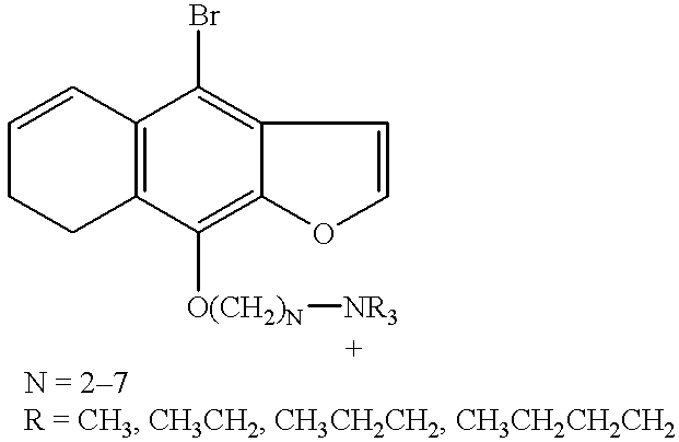
A preferred class of sensitizers is selected from the group consisting of compounds of the formula (A):
Figure US06187572-20010213-C00004
wherein u is an integer from 1 to 6; X is an anionic counterion; Z is N or P; R1, R2, R3, R4, R5, and R6 are independently halo; H; linear or branched alkyl of 1-10 carbon atoms; linear or branched alkoxy of 1-10 carbon atoms; CH2mOCH2pZR′R″R′″ or —OCH2nZR′R″R′″ wherein n, m and p are independently integers from 1 to 10 and R′, R″ and R′″ are independently H or linear or branched alkyl of 1 to 10 carbon atoms with the proviso that on each Z atom, not more than one of R′, R″ or R′″ may be H; and at least one of R1, R2, R3, R4, R5, or R6 is CH2mOCH2pNR′R″R′″ or —OCH2nNR′R″R′″. Particularly preferred are compounds wherein R4 is —OCH2nNR′R″R′″, especially wherein R′, R″ and R′″ are ethyl and n=3. Preferably, R6, R5, R2 and R1 are hydrogen and R3 is H or halo, preferably bromo.
In general, the above compounds may be made by halogenating psoralens and isolating the appropriately substituted isomers. For compounds wherein the ring substituent is a quaternary ammonium alkoxy or phosphonphonium alkoxy group, that group may be made from the corresponding hydroxy-substituted psoralen, as exemplified by the following scheme.
Figure US06187572-20010213-C00005
In general, derivatives possessing more than one free hydrogen atom on the preferred ammonium group show viral inactivation but unacceptable levels of cell damage. This is particularly the case for 4′-aminomethyl-4,5′,8-trimethylpsoralen (AMT), which is an experimentally utilized psoralen derivative.
Therefore, according to the invention, sensitizing compounds for viral inactivation preferably do not contain substituents which possess free hydrogen groups capable of exhibiting hydrogen bonding to the cell membrane.
In one embodiment, the present invention is applicable to lyophilized cells. The term lyophilization is broadly defined as freezing a substance and then reducing the concentration of the solvent, namely water, by sublimation and desorption, to levels which will no longer support biological or chemical reactions. Usually, the drying step is accomplished in a high vacuum. However, with respect to the storage of cells and particularly erythrocytes, the extent of drying (the amount of residual moisture) is of critical importance in the ability of cells to withstand long-term storage at room temperature. Using the procedure described herein, cells may be lyophilized to a residual water content of less than 10 weight %, preferably less than 3%, and still be reconstituted to transfusable, therapeutically useful cells. Cells with about 3 weight % water content using this procedure may be stored for up to two weeks at room temperature, and at 4° C. for longer than eight months, without decomposition. This far exceeds the current A.A.B.B. standard for refrigerated storage of red blood cells of six weeks at 4° C. or less than one day at room temperature without decomposition. These dried cells may be deactivated using a chemical sensitizer described herein.
According to the embodiment of the present invention for treating dried or lyophilized cells, the washed, packed cells are mixed with a chemical sensitizer, then washed to remove excess sensitizer not bound to viral or bacterial nucleic acid, and the treated cells are then dried or lyophilized. The dry cell and sensitizer mixture will then be irradiated, typically with gamma radiation, at an intensity of about 3K-50K rads, for a period of time sufficient to destroy viruses (in particular, the single-stranded or double-stranded RNA/DNA viruses), without any substantial adverse effect on the recovery and usefulness of the cells. Other wavelengths of electromagnetic radiation such as visible light or X-rays, may be used.
In another embodiment, the chemical sensitizers may be added to liquid protein preparations, then lyophilized and irradiated. Particularly preferred are blood protein preparations, including but not limited to, plasma proteins, blood protein extracts, clotting factors (such as Factors VIII and IX), immune globulins and serum albumin.
Dry or lyophilized cells or protein fractions may be directly mixed with the chemical sensitizer, then irradiated.
From the foregoing description, it will be realized that the invention can be used to selectively bind a chemical sensitizer to blood-transmitted viruses, bacteria, or parasites. Also monoclonal or polyclonal antibodies directed against specific viral antigens (either coat proteins or envelope proteins) may be covalently coupled with a sensitizer compound.
Since cell compositions also comprise a variety of proteins, the method of decontamination of cells described herein is also applicable to protein fractions, particularly blood plasma protein fractions, including, but not limited to, fractions containing clotting factors (such as Factor VIII and Factor IX), serum albumin and/or immune globulins.
The viral and bacterial inactivation may be accomplished by treating a protein fraction with a sensitizer as described herein.
Although described in connection with viruses, it will be understood that the methods of the present invention are generally also useful to inactivate any biological contaminant found in stored blood or blood products, including bacteria and blood-transmitted parasites.
BRIEF DESCRIPTION OF THE DRAWINGS
FIG. 1 is a plot of X-ray cross section for various sensitizers described in Example 3.
FIG. 2 is a plot of efficiency of sensitizers with Mo and W targets.
FIG. 3 is a plot of Phi 6 with inactivation according to the procedure of Example 5.
FIG. 4 is a plot of viral reduction in plasma according to the procedure of Example 6.
FIG. 5 is a plot of viral reduction in samples described in Example 7.
FIG. 6 is a plot of viral reduction in samples described in Example 8.
FIG. 7 is a plot of residual Factor VII in X-ray irradiated lyophilized plasma.
FIG. 8 is a plot of viral reduction of samples described in Example 9.
FIG. 9 is a plot of residual Factor VIII activity in UV irradiated plasma.
FIGS. 10, 11 and 12 are plots of viral reduction in samples described in Example 10.
FIGS. 13, 14, 15 and 16 are plots of viral reduction in samples described in Example 11.
FIG. 17 is a plot of lambda virus reduction with psoralen sensitizer (with and without bromine) in hydrated plasma in sensitizer concentrations of 0.05, 0.1 and 0.2 mg/ml. The sensitizers are psoralen (S#9) and Br-psoralen (S#8), activated with UV.
FIGS. 18 through 21 are graphs of platelet characteristics as a function of irradiation time according to the procedure of Example 12.
FIGS. 22 through 25 are graphs of platelet characteristics of platelet concentrates seeded with phi 6, irradiated in presence of senstizers 8B and 8C, according to Example 12.
FIG. 26 shows the viral reduction results of the test described in Example 13.
FIG. 27 shows the results of bacterial reduction described in Example 19.
FIG. 28 shows the attenuation coefficients described in Example 3.
FIG. 29 shows results of the test described in Example 21.
DETAILED DESCRIPTION OF THE INVENTION
The present invention is directed to methods for reducing viral, bacterial and other parasitic contamination in blood, blood components, cell cultures or cell culture components by irradiation in the presence of a chemical sensitizer. Sensitizers are disclosed which are particularly useful to decontaminate liquid compositions, such as blood, blood components, reconstituted lyophilized cells, and the like, using UV irradiation. Other sensitizers are disclosed which are particularly useful to decontaminate solid compositions, such as frozen, dried or lyophilized cells or proteins.
Lyophilized or Dried Cells or Proteins
One embodiment involves lyophilized cells (or proteins).
The cells are preferably prepared by immersing a plurality of erythrocytes, platelets and/or hemosomes, etc. in a physiologic buffered aqueous solution containing a carbohydrate, and one or more biologically compatible polymers, preferably having amphipathic properties. By the term amphipathic it is meant there are hydrophobic and hydrophilic portions on a single molecule. This immersion is followed by freezing the solution, and drying the frozen solution to yield novel freeze-dried erythrocytes containing less than 10%, and preferably about 3% or less by weight of moisture, which, when reconstituted, produce a significant percentage of viable, transfusably useful red blood cells, platelets or hemosomes. Preferred methods of reconstitution of the lyophilized composition are described below. Although described in connection with red blood cells, it will be understood that the methods are generally also useful to lyophilize platelets, hemosomes, and blood protein fractions.
The carbohydrate utilized to prepare erythrocyte, platelet and/or hemosome compositions according to the invention is biologically compatible with the erythrocytes, platelets or hemosomes, that is, non-disruptive to the cells or hemosome membrane, and one which permeates, or is capable of permeating, the membrane of the erythrocytes, platelets or hemosomes. It is also advantageous to stabilize proteins, especially labile blood proteins, with the carbohydrates during lyophilization and irradiation according to the invention. The carbohydrate may be selected from the group consisting of monosaccharides, since disaccharides do not appear to permeate the membrane to any significant extent. Monosaccharide pentoses and hexoses are preferred as is a final concentration of from about 7.0 to 37.5 weight % in phosphate buffered saline (PBS) or a phosphate buffered solution, preferably about 26%. Xylose, glucose, ribose, mannose and fructose are employed to particular advantage.
It will be understood that the cells may be lyophilized using other protocols and irradiated as described below. Although viral inactivation will be attained, the advantage of retaining a significant percentage of viable useful red blood cells is lost if the described lyophilization procedure is not followed.
The following description is written in connection with erythrocytes (RBC's) but it will be understood it is also applicable to platelets, hemosomes or other blood cell types or biological cells, as well as protein fractions, particularly plasma protein fractions.
The erythrocytes will preferably be prepared from whole blood centrifugation, removal of the plasma supernatant and resuspending the cells in PBS or a phosphate buffered solution or a commercial dextrose-saline solution. This wash cycle may be repeated 2-3 times preferably using a commercial dextrose-saline solution, then the packed cells are diluted with the lyophilization buffer described above so that the final diluted concentration of carbohydrate and polymer are maintained in the necessary ranges.
Alternatively, commercially available packed blood cells may be used, which typically are prepared in CPDA (commercial solution containing citrate, phosphate, dextrose and adenine).
Preferably, but not necessarily, the cells will have been previously lyophilized using a lyophilization solution buffered in the range of pH of 7.0 to 7.4 preferably by a phosphate-buffered solution. A typical phosphate-buffered lyophilization solution will comprise mono- and di-basic potassium and sodium phosphate (usually in the range of 1-10 mM each) and 5-10 mM adenine. This solution maintains the pH at around 7.2.
A preferred phosphate-buffered solution to be used as the lyophilization buffer will comprise nicotinic acid, reduced glutathione, glutamine, inosine, adenine, monopotassium phosphate, magnesium chloride disodium phosphate all of which will serve as a basic salt buffer at a pH of about 7.2. In addition this lyophilization buffer will contain a final concentration of about 26% weight by volume of a monosaccharide, preferably 1.7 M glucose, and a final concentration of about 3.0% weight by volume of polyvinylpyrrolidone (average molecular weight of 360K), and a final concentration of about 15% weight by volume of hydroxyethyl starch (average molecular weight of 500K).
Upon lyophilization to a moisture content of less than 10%, and preferably less than 3%, the lyophilized cells may be maintained under vacuum in vacuum-tight containers, or under nitrogen or other inert gas, at room temperatures for extended periods of time in absence of or without significant degradation of their desirable properties when reconstituted for use as transfusable cells. In using the preferred lyophilization method disclosed herein, a particular advantage of the present invention is that the lyophilized cells may be stored at room temperature for extended periods of time, thus obviating the need for low temperature refrigeration which is required for storing liquid CPDA preserved red blood cells prepared by methods of the prior art. The present invention also obviates the need for very low temperature (−80° C.) frozen storage of red blood cells in glycerol.
By using the preferred reconstitution method disclosed herein it is a further advantage that the lyophilized red blood cells may be reconstituted at normal temperatures, i.e. greater than about 4° C. up to about 37° C., which corresponds to normal human body temperature, and preferably at room temperature (about 22° C.). The reconstitution medium is preferably a solution comprising a polymer or mixture of polymers having a molecular weight of from about 2.5K to 360 K, preferably 5K to about 360K, present in a concentration in the range of about 12 to 30% weight by volume. This polymer may be the same polymer utilized to lyophilize the red blood cells as described above. Hence the polymers polyvinylpyrrolidone, hydroxyethyl starch, and dextran are particularly preferred and most preferred is polyvinylpyrrolidone (preferably molecular weight about 10K) present in a concentration of about 19% weight by volume in the reconstitution solution. The reconstitution solution will be buffered again typically by phosphate-buffered solution comprising monopotassium phosphate and disodium phosphate as described hereinabove to maintain a pH within the range of about 7.0 to 7.4. The most particularly preferred polymer is polyvinylpyrrolidone of an average molecular weight of about 10K. The most preferred reconstitution buffer will also contain adenosine triphosphate (ATP) in a final concentration of about 5 nM.
The polymers may be present in the various solutions from a final concentration of about 3.6K weight % up to saturation, and have a molecular weight in the range of from about 2.5K to about 360K. Preferably, the polymers have molecular weights in the range of from about 2.5K to about 500K, most preferably from about 2.5K to 50K, and are present in a concentration of from about 3.6 weight % up to the limit of solubility of the polymer in the solution. Polymers selected from the group consisting of polyvinylpyrrolidone (PVP) and polyvinylpyrrolidone derivatives, and dextran and dextran derivatives provide significant advantages. Most preferred is the use of polyvinylpyrrolidone (an amphipathic polymer) of average molecular weight in the range of 2.5-360K in an amount in the range of 3-20% weight by volume in the solution prior to lyophilization. Amino acid based polymers (i.e., proteins), dextrans or hydroxyethyl starch may also be employed. In the lyophilization buffer hydroxyethyl starch (M-HES) with an average molecular weight of about 500K is employed in a 15% weight by volume final concentration. Other amphipathic polymers may be used, such as poloxamers in any of their various forms. The use of the carbohydrate-polymer solution in the lyophilization of red blood cells allows for the recovery of intact cells, a significant percentage of which contain biologically-active hemoglobin.
The most preferred reconstitution buffer will be a solution comprising monopotassium phosphate, disodium phosphate and ATP, all of which form a basic salt buffer at a pH of about 7.2, which also contains about 19% weight by volume of polyvinylpyrrolidone (average molecular weight about 10K).
The reconstitution solution may also optionally contain a monosaccharide, preferably present in the concentration range of about 7.0 to 37.5% weight by volume. The preferred monosaccharides are xylose, glucose, ribose, mannose and fructose.
In the most preferred embodiment, the lyophilized erythrocytes can be reconstituted by mixing with an equal volume of the reconstitution buffer at a temperature of about 37° C. and mixed. By “equal” it is meant that the volume is the same as the starting volume prior to lyophilization. After initial reconstitution, the solution is preferably diluted 1:1 with 1-4 additional volumes of the reconstitution buffer at a temperature of about 37° C. with added mixing until fully hydrated.
Then, it is preferred that the rehydrated cells be washed according to the following procedure. It is realized, however, that once the cells are reconstituted with reconstitution buffer they are in a hydrated and useful form, but the combination of washings described hereinafter are preferred, specifically for clinical purposes.
After separating the cells from the reconstitution buffer by centrifugation, the resulting packed cells are preferably resuspended at room temperature in (approximately the volume used in the initial reconstitution) a wash buffer comprising nicotinic acid, inosine, adenine, glutamine, and magnesium chloride, all present at about 0.4-10 mM further comprising sodium chloride and potassium chloride each at about 30 mM, buffered by 10 nM disodium phosphate to pH 7.2. This wash buffer further comprises a monosaccharide, preferably glucose at a concentration of about 20 mM, and a polymer, preferably polyvinylpyrrolidone, of a molecular weight 40K and present at a concentration of about 16% weight by volume. Separation by centrifugation completes the first post-rehydration step, a washing step.
After the washing step the rehydrated cells may be suspended in a dextrose-saline transfusion buffer, if transfusion is the intended use of the cells, at room temperature which preferably contains polyvinylpyrrolidone at a 10% weight by volume final concentration, with an average 2.5K molecular weight. The cells can be used as is or be returned to autologous plasma. Additional wash steps in a phosphate-buffered diluent buffer can further remove viruses, but this step is optional for preparation of rehydrated, transfusible cells.
The reconstitution and washings described above will in most instances achieve about 4 log reduction of any viral and bacterial contamination, where 1 log reduction is achieved by drying and 3 log reduction is achieved by washing. Of course, different viruses may respond differently, potentially resulting in more than 4 log reduction of contamination.
The reconstituted cells have characteristics which render them transfusable and useful for therapeutic purposes in that their properties are similar to that of fresh (i.e. not previously lyophilized) red blood cells. Typically reconstituted red blood cells according to the present invention have an oxyhemoglobin content greater than about 90% of that in normal red blood cells. Hemoglobin recovery prior to any washing step is typically in the range of 80 to 85%. The overall cellular hemoglobin recovery including the post-hydration washing steps is about 20 to 30%. The morphology of the reconstituted cells according to the present invention (by scanning electron microscope) typically shows no holes or gaps, and primarily discocytic with some stomatocytic morphology. The oxygen carrying capacity of fresh red blood cells (as measured by P50, the oxygen partial pressure at which 50% of the oxygen molecules are bound) was measured to be in the range of about 26 to 28 (average 26.7); with an average Hill coefficient (a measure of the cooperative binding of oxygen molecules to native hemoglobin) of 1.95. The typical P50 for erythrocytes lyophilized and reconstituted according to the present invention is about 27.5 (average) with an average Hill coefficient of 2.08. Assays of ATP in the reconstituted cells indicate ATP levels suggesting normal ATP to ADP metabolism. Normal hemagglutination by readily available blood typing antisera of red blood cells made according to the present invention is also typically found.
This lyophilization and reconstitution procedure advantageously and significantly diminishes viral/bacterial contamination in cell-like material (such as hemosomes), and protein fractions. The contamination can be further reduced by the radiation sensitizing and treatment, particularly while the cells or protein fractions are in the dry state.
The starting packed red blood cells or proteins (which may initially be in a liquid or lyophilized state) are mixed with a sufficient amount (based on total wet weight of cells) of a chemical sensitizer. Preferably, in a composition of packed red blood cells (about 10% hematocrit) about 0.1 to 1 mg of the chemical sensitizer will be used per ml of packed cells. Preferably, the mixture will be irradiated with gamma radiation in the range of 3K-50K rads, typically about 3K rads. Preferred exposure is from 1-10 minutes, if using gamma radiation. Alternatively, UV light (320 nm) may be used, particularly for protein fractions. Preferred exposure is from 1-10 minutes, preferably 3 minutes, if using UV radiation. By this irradiation in the presence of a sensitizer, there will be about a 6 log reduction of viral and bacterial contamination, based on contamination present prior to washing and irradiation.
The present invention provides a selective method of generating free radicals derived from chemical sensitizers only in the vicinity of viral RNA or DNA. Indiscriminate radiolysis of blood containing virus in a hydrated state produces hydroxyl radical. However, the hydroxyl radical will damage both the red blood cells and associated proteins as well as the viral target. Thus, viral inactivation would be achieved at the sacrifice of red cell viability. Therefore, sensitizers which bind to DNA, RNA, viral coat proteins, and/or viral membranes and which can be selected to generate radicals upon irradiation, are required. Since the radiolysis can be performed in the dry state (preferably less than 10% residual moisture), generation of hydroxyl radicals from water is greatly reduced. In this manner indiscriminate radical damage is further prevented. Exemplary compounds include:
Figure US06187572-20010213-C00006
The preparations of these compounds are known. See Martin, R. F. and Kelly, D. P., Aust. J. Chem., 32, 2637-46 (1979); Firth, W., and Yielding, L. W., J. Org. Chem., 47, 3002 (1982). Other radical-generating reagents which generate radicals upon irradiation are disclosed by Platz et al., Proc. SPIE-Int. Soc. Opt. Eng. 847, 57-60 (1988) and Kanakarajan et al., JACS 110 6536-41 (1988).
The radiation-sensitizing compound (which may also be modified to bear a metal atom substituent) may also be selected from the class consisting of DNA-binding drugs, including, but not limited to, netropsin, BD peptide (a consensus peptide from HMG-1), S2 peptide, and the like. These and other DNA-binding drugs are disclosed in Pjura, P. E., Grzeskowiak, K. and Dickerson, R. E. (1987), J. Mol. Biol. 197, 267-271; and Tengi, M., Usman, N., Frederick, C. A. and Wang, A. H. J. (1988), Nucleic Acids Res. 16, 2671-2690.
The radiation sensitizing compound (which may also bear a metal atom) can also comprise a class of DNA-binding proteins and/or polypeptides and/or peptides. Examples of this class of DNA-binding proteins and/or polypeptides and/or peptides are disclosed in Churchill, M. E. A. and Travers, A. A. (1991) Trends in Biochemical Sciences 16, 92-97. Specific examples of DNA-binding peptides include the SE peptide and BD peptide disclosed in the reference herein.
Another class of sensitizers comprises the positively charged porphorins and pthalocyanates, which bind DNA and RNA. These sensitizers can be activated by irradiation with visible light (500-700 nm).
The DNA-binding specificity can be achieved by covalently coupling the radiation sensitizing compound and/or metal atom to either a DNA-binding drug or to a DNA-binding protein or polypeptide or peptide.
Figure US06187572-20010213-C00007
Other sensitizers include specially designed molecules which form triplex DNA, such as those disclosed by Youngquist and Dervan PNAS 82 2565 (1985); Van Dyke and Dervan, Science 225 1122 (1984); Van Dyke and Dervan, Nuc. Acids Res. 11 5555 (1983); Barton and Raphael, PNAS 82 6460 (1985); Barton et al., JACS 106 2172 (1984); and Barton, PNAS 81 1961 (1984). These molecules bind to DNA and RNA, site specifically, if desired, and carry reactive moieties which can generate free radicals in the proximity of the DNA or RNA.
R−I+e→R•I
R−I+*+Guanine→R−I+Guanine+*
While not intending to be bound by a theory, it is believed that the ejected electron will be captured by that site with the most favorable electron affinity, which is most likely a second molecule of sensitizer elsewhere in the sample. Electron capture by R−I (or R−Br) leads to dissociation of RX with the formation of a radical. The radical so generated will abstract a C—H hydrogen atom from a sugar moiety of a nearby nucleic acid which in turn will lead to DNA or RNA cleavage and viral inactivation.
The radical cation of the sensitizer (R−X+•) will eventually abstract an electron from that component of the sample with the most favorable oxidation potential. This is most likely guanine. The electron transfer reaction forms a guanine radical cation. This substance will react with O2 upon reconstitution with aerated H2O. This process also leads to DNA cleavage and viral inactivation. Unreacted material and reaction by-products will be removed during the washing steps involved in the reconstitution of the lyophilized cells. This process will also further remove any virus not inactivated by the treatment described above.
Compounds (1) and (2) bind tightly to DNA and RNA by either intercalation and/or by electrostatic interactions between positively charged ammonium ion groups and the negatively charged phosphate groups of the nucleic acid target. Red blood cells do not contain nucleic acids and accordingly will not bind to such compounds by intercalation.
Dried or lyophilized solid formulations are preferably exposed to 10,000 rads of x-ray or gamma radiation. It is known that the red cells will survive these doses of radiation in the absence of a sensitizer. Lyophilized blood can withstand higher dosage levels of radiation than hydrated blood.
The gamma radiation or x-ray will be absorbed primarily by the heavy atom of the sensitizer, which will be bound to viral DNA or RNA. The radiation will ionize the sensitizer as follows:
R−I+γ-Ray→R−I+•+e(X-Ray)
In some instances, particularly if the sensitizer and red blood cells are allowed to stand together for more than several minutes, sensitizers may diffuse into the red blood cells prior to lyophilization. Antioxidants such as glutathione (an excellent hydrogen atom donor) may be added to the preparation to augment the red cell defenses against free radical initiated damage. It will be understood that incorporation of the sensitizer into cells will also allow inactivation of intracellular viruses, especially viruses thought to reside inside white blood cells (most packed red blood cell units contain residual white cells), or intracellular blood parasites, such as malaria parasite which infects red blood cells.
The sensitizers are removed from the reconstituted blood serum or protein fraction by the washing protocol described above for lyophilized cells.
It is preferred that gamma or X-ray radiolysis take place in a dried lyophilized blood (or protein), virus, and sensitizer formulation rather than in a wet, fluid material for several reasons. Firstly, the dry material is less sensitive to radiation and can be exposed to larger doses of γ-rays or other penetrating radiation without damage to red blood cells (Table 1). This increases the extent of radiolysis of the sensitizer. Secondly, sensitizer radicals bound to DNA or RNA in the dry state can not dissociate from the virus due to the lack of diffusion in the solid material. This will force the sensitizer radical to react with viral RNA or DNA. Thirdly, the solid state conditions will enhance hydrogen atom transfer reactions of the sensitizer radical with the viral nucleic acid, perhaps by quantum mechanical tunneling. Fourthly, the reconstitution and washing protocol used with lyophilized blood or protein fraction serves as a means to remove unreacted material or reaction by-products, and further removes any virus not affected by the treatment (Table 2).
Other types of radiation may be used including ionizing radiation in general, such as X-ray radiation. In one embodiment a metal and/or halogen atom may be a substituent on a chemical radiation sensitizer molecule which binds to nucleic acids, thereby targeting the embodiments such as bacteria, parasites and viruses. Metal and halogen atom substituents of chemical sensitizers for this purpose include Pt, Br, I, Zn, Cl, Ca and F. The X-ray source is preferably a tunable source, so that the radiation may be confined to a narrow wavelength and energy band, if so desired. The tunable feature allows for optimization of energy absorption by the metal atoms, thereby directing the absorbed penetrating radiation energy to the production of radicals by a chemical sensitizer bound to nucleic acid.
A preferred metal to be used as the radiation sensitizer is platinum. Another preferred group comprises the halogens, bromine, iodine, chlorine and fluorine. Based on their increasing ability to interact with impinging X-rays, the order of enhancement of radiation sensitizing is expected to be platinum, which is much greater than bromine, which is much greater than iodine, chlorine and fluorine, all of which are much greater than hydrogen.
Compounds containing these atoms, when exposed to X-rays or other forms of ionizing radiation, are capable of forming a reactive species which can interact with the viral nucleic acid, coat protein or lipid envelope, thus destroying it and rendering it non-infectious. This process may be most effective in a dry state where quenching and side reactions due to the presence of water are avoided.
Treatment of Liquid Compositions Containing Cells or Proteins
If the composition suspected of being contaminated is to be treated in liquid form, i.e., as a suspension of cells or protein solution, it is preferred that the sensitizer be UV-activated. The most preferred sensitizers for this purpose are halogenated psoralens.
Psoralens are naturally occurring compounds which have been used therapeutically for millennia in Asia and Africa. The action of psoralens and light has been used to treat vitiligo and psoriasis (PUVA therapy; Psoralen Ultra Violet A) and more recently various forms of lymphoma.
Psoralen will bind to nucleic acid double helices by intercalation between base pairs; adenine, guanine, cytosine and thymine (DNA) or uracil (RNA). Upon absorption of a UVA photon the psoralen excited state will react with a thymine or uracil double bond and covalently attach to one nucleic acid helix.
Figure US06187572-20010213-C00008
The reaction is specific for a thymine (DNA) or uracil (RNA) base and will proceed only if the psoralen is intercalated in a site containing thymine or uracil. The initial photoadduct can absorb a second UVA photon and react with a second thymine or uracil on the opposing strand of the double helix to crosslink the two strands of the double helix.
Lethal damage to a cell or virus occurs when a psoralen intercalated into a nucleic acid duplex in sites containing two thymines (or uracils) on opposing strands sequentially absorb 2 UVA photons. This is an inefficient process because two low probability events are required, the localization of the psoralen into sites with two thymines (or uracils) present and its sequential absorption of 2 UVA photons.
The halogenated psoralens according to the present invention are improved and more efficient sensitizers because they require only a single UVA photon for activation and the presence of a single guanine base is the only requirement for the site of intercalation. As shown in Scheme 1 absorption of a UVA photon by a bromopsoralen in the presence of guanine leads to electron transfer and the formation of free radicals and ultimately nucleic acid cleavage and viral or cell death.
Figure US06187572-20010213-C00009
The psoralen radical 2 can inflict damage on the nucleic acid double helix to which it is bonded by abstraction of a ribose (RNA) or deoxyribose (DNA) sugar carbon hydrogen bond. This can lead to DNA cleavage by known mechanisms. The guanine radical cation is also known to react with molecular oxygen, initiating a series of reactions which cleave DNA. The byproduct of the photochemistry is debrominated psoralen 4, which can sensitize DNA cross links as described above.
A disadvantage of PUVA therapy is that it increases the chances of DNA damage (cross linking of the double helix) to normal cells, if normal DNA-containing cells are present in the treated composition. The angelicins, khellins, and coumarins can not cross link DNA because they lack a second double bond or this bond, is located in a position unsuitable for crosslinking. Thus halogenated angelicins, khellins and coumarins bearing a quaternary ammonium or phosphonium side chain will reduce the effects of this problem if the maintenance of integrity of the DNA in the cells is desired.
The structures of exemplary chemical sensitizers utilized in accordance with the present invention are listed in Appendix I. The syntheses of the parent heterocycles to these sensitizers are well known.
The best mode for using the invention on liquid samples is to add a UV-sensitive sensitizer to potentially contaminated blood suspensions, and to expose to UV radiation. Fluid solutions of blood are preferably exposed to 3000 rads.
In another embodiment of the present invention the sensitizers will be utilized in conjunction with solvent detergent systems. Such detergents are known to decrease the viral titre of plasma or separated plasma fractions, presumably by dissolution of the viruses. Such detergents include, TWEEN®, sodium cholate, sodium deoxycholate, TRITON® and common organic solvents such as ether. Reduction of viral titre by use of these solvent detergents is described for example by Horowitz, et al., Transfusion, 25, (6), 516-522 (1985), and 25, (6), 523-527 (1985); and U.S. Pat. No. 4,946,648. The level of reduction by such solvent detergents may vary as reported in literature to a reduction of one log to greater than five logs of viral titre for such viruses as VSV, Sindbis, and Sendai. The present invention may enhance the reduction of viral titre by these solvent detergents when used in conjunction with the sensitizers and exposure to radiation as set forth herein. While not intending to be bound by a theory, it is believed that the solvent detergents act on the viral proteins or lipid membranes to denature or alter them in a manner which makes them more susceptible to the actions of the sensitizers through the changes induced by the detergents.
A particularly preferred class of sensitizers comprise DNA intercalators, such as hydroxyl, amino methyl, or methyl substituted psoralens, which may be added to plasma or plasma fractions followed by UV radiation to reduce the viral contamination therein. The substituted psoralens are described in U.S. Pat. No. 4,727,027 wherein a reduction of about 4 to 7 logs of viral contamination was obtained with extended exposure to ultraviolet radiation. The proposed mechanism of action is to form a photoadduct between the sensitizer and the DNA or RNA of viral origin, which results in loss of infectivity of the virus. According to the present invention, the reduction of viral contamination can be unexpectedly reduced by utilizing brominated psoralens or other halogenated psoralens. For example, it was observed that the bromopsoralens are about 200,000 times more effective in reducing viral activity when compared to use of their non-brominated counterparts. While not intending to be bound by any theory, it is believed that the mechanism of action of the brominated psoralens may be a free radical generation in the proximity of the DNA or RNA resulting in damage of vital nucleic acids of viruses.
The brominated psoralens are in an improvement over the known psoralens and other substituted psoralens when used as sensitizers because only one photon of light is required to activate the brominated sensitizer whereas two photons are required to activate a non-brominated sensitizer. Secondly, a brominated psoralen is effective in virtually every intercalative site, whereas a non-brominated sensitizer is effective only in intercalation sites containing a uracil or thymine on different strands of the DNA or RNA. Thirdly, the brominated psoralens may be activated by X-rays as well as UV light.
The use of the brominated or halogenated psoralens is particularly useful in activation in hydrated systems such as plasma, immune sera, tissue culture media containing animal serum or serum components (such as fetal calf serum), or recombinant products isolated from tissue culture media.
Other types of intercalators may be utilized besides the psoralens and substituted psoralens such as those listed below. These intercalators may be used to target viruses or other blood contaminants, or cancer cells. Thus, halogenated or metal atom-substituted derivatives of the following compounds may be utilized as sensitizers:
dihematoporphyrin esters
hematoporphyrin derivatives
benzoporphyrin derivatives
hydrodibenzoporphyrin dimaleimade
hydrodibenzoporhyrin
dicyano disulfone
tetracarbethoxy hydrodibenzoporhyrin
tetracarbethoxy hydrodibenzoporhyrin dipropionamide
The above compounds in their non-halogenated or non-metal atom substituted forms are disclosed in U.S. Pat. Nos. 4,649,151, 4,866,168, 4,883,790, 5,053,423 and 5,059,619, incorporated by reference herein. When modified with halogen atoms or metal atoms, the above-identified classes of compounds may be sensitized with electromagnetic radiation, including visible light.
The present invention may be applied to treatment of liquid blood in ex vivo irradiation, such as by methods and apparatus described in U.S. Pat. Nos. 4,889,129 and 4,878,891.
The above compounds are included in a class named lipophilic dyes which include dyes such as merocyanine 540 and phthalocyanine derivatives. Merocyanine 540 has been disclosed as useful for the treatment of cancer and viral inactivation of blood cells and plasma proteins (Sieber, et al., Photo Chem. and Photo Biology, 46 707-711 (1987)). Phthalocyanine derivatives and other lipophilic dyes are known to bind to the membranes of cancer cells or enveloped viruses. When these compounds are activated with suitable wavelength of electromagnetic radiation, they produce singlet molecular oxygen (Kalyanaraman, et al., PNAS, 84 2999-3003 (1987)), which damages the membrane resulting in the killing of the cancer cells or in viral inactivation. With the addition of radiation sensitizer atoms (metal atoms or halogens) according to the present invention, use of these compounds in combination with suitable radiation produces free radicals in the proximity of the DNA/RNA/viral membranes, which then results in destruction of the viral membranes or nucleic acid to inactivate the virus.
Halogenated or metal atom substituted fatty acids also may be utilized according to the present invention as radiation sensitizers. Fatty acids per se have been used in viral inactivation by Horowitz, et al. as disclosed in U.S. Pat. No. 4,841,023. According to the present invention, these fatty acids may be utilized with sensitizer atoms to target viral membranes in plasma protein solutions, for example, and by subsequent activation with suitable radiation the free radicals are produced to inactivate the viral membrane.
Treatment of biological compositions with detergents is disclosed in U.S. Pat. Nos. 4,820,805 and 4,764,369. The clinical application of psoralens in conjunction with photodynamic treatment is discussed by Adelson, Scientific American 50-57 (August 1988).
The following compounds are illustrative of sensitizers which contain or which may be modified to contain metal substituents or halogen substituents in accordance with the present invention:
1. DNA or RNA Target Sensitizers
Psoralen Sensitizers:
Figure US06187572-20010213-C00010
Compound #1
Figure US06187572-20010213-C00011
Compound #2
Figure US06187572-20010213-C00012
Compound #3
Figure US06187572-20010213-C00013
Compound #4
Figure US06187572-20010213-C00014
Compound #5
Figure US06187572-20010213-C00015
Compound #6
X = I Compound 6b
Figure US06187572-20010213-C00016
Compound #
7
Figure US06187572-20010213-C00017
Compound #8
Figure US06187572-20010213-C00018
Compound #9
Figure US06187572-20010213-C00019
Compound #10
X = I, Compound #10b
Figure US06187572-20010213-C00020
Compound #10C
X = I, Compound #10d
2. Membrane Target Sensitizers
Phthalocyanine Sensitizers:
Figure US06187572-20010213-C00021
X = Br PHTH-Br Compound #11
X = I PHTH-I Compound #12
Figure US06187572-20010213-C00022
X = H PT-PHTH-H Compound #13
X = I PT-PHTH-I Compound #14
X = Br PT-PHTH-Br Compound #15
Merocyanine Sensitizers:
Figure US06187572-20010213-C00023
Compound #16
Fatty acid Sensitizers:
Figure US06187572-20010213-C00024
Compound #17
Figure US06187572-20010213-C00025
Compound #18
Biological Dyes:
Figure US06187572-20010213-C00026
Compound #19
Figure US06187572-20010213-C00027
Compound #20
Other sensitizers include compounds such as doxorubicin and daunomycin, which may be modified with halogens or metal atoms in accordance with the present invention, which are known in chemotherapy to attack dividing cells.
The sensitizers also may be utilized in vivo and delivered in liposomes (artificial cells) or drug-loaded natural cells. After introduction of the liposome or drug-loaded cell, the patient may be treated by radiation to activate the sensitizer.
The present invention is applicable to contaminants which comprise single or double-stranded nucleic acid chains, including RNA and DNA, and viruses, bacteria or other parasites comprising RNA and/or DNA.
To illustrate the invention in lyophilized cells, red blood cells were lyophilized as described above, irradiated, and tested for erythrocyte characteristics measured. The results are shown in Table 1. The same procedure was then used, except that the bacteriophage T4 (in dextrose saline) was mixed with the cells and then washed successively with four different wash buffers. The results are shown in Table 2.
TABLE 1
Influence of irradiation on lyophilized
reconstituted red blood cells. Doses as high as
20,000-50,000 rads do not affect cells in the dry
state according to the parameters assayed after
reconstitution and listed below.
Exposure of Lyophilized Cells to Gamma Irradiation
*Percentage of Control
Dosage Level 20,000 rads 50,000 rads
Hb Recovery
100  99
Oxy Hb No Change from No Change from
starting value starting value
Cell Indices
MCV 99 98
MCH 100  100 
MCHC 100  100 
Metabolism
ATP 79 79
(μmol/g Hb)
Lactate 86 79
(μmol/g
Hb/Hr)
*Control cells were non-irradiated, lyophilized reconstituted cells.
TABLE 2
Reduction in viral titre as a function of
washing of the red cells. The procedure used in
reconstituting the lyophilized cells involves several
washing steps which also reduce the viral titre. The
extent of reduction with each wash decreases until a
practical limit is attained. This represents an
approximate 4 log reduction in viral titre.
Washing Protocol Reduction of Viral Load in Blood
Total Amount
Buffer Wash Step of Virus Log Reduction
Experiment 1
(non-lyophilized
cells)
Reconstitution 7.30 × 10⁷ 0
Wash 4.80 × 10⁴ 3.2
Diluent 2.08 × 10⁴ 3.5
Transfusion 3.50 × 10⁴ 3.3
Experiment 2
(lyophilized
cells)
Lyophilization 3.68 × 10⁸ 0
Reconstitution 2.11 × 10⁷   1.2**
Wash 2.38 × 10⁴ 4.2
Diluent 2.00 × 10⁴ 4.3
Transfusion 4.06 × 10⁴ 4.0
In Experiment 1, the effects of lyophilization on viral reduction are not included.
In Experiment 2, these effects are included.
The marker virus used in these cases was bacteriophage T4. The extent of reduction was determined using the plaque assay.
**This shows an additional about 1 log reduction of contamination due to the drying step.
EXAMPLE 1
Packed human red blood cells purified from donated whole blood are washed free of the anticoagulant storage solution (commercially available CPDA, containing citrate/phosphate/dextrose/adenine), and suspended in dextrose-saline at a 10% hematocrit. Approximately 10 ml of washed packed red cells are placed in a quartz chamber and exposed to U.V. light, preferably at 320 nm, for 2 minute time intervals, up to a 10 minute total exposure. At each 2 minute interval the suspension is mixed and a small sample of red cells (10 microliters) is removed and diluted into 2 ml of water for spectrophotometric assay of hemoglobin. At each step the temperature of the irradiated red cell suspension is measured, to ensure that the suspension did not overheat. At no point does the suspension exceed 26 degrees C. (normal body temperature is 37 degrees C.). Untreated red cells contain a high proportion of functional oxyhemoglobin (oxyHb), usually in the range of 96% or higher. Oxidation damage can form a semi-stable methemoglobin species (metHb), which can normally be reduced back to oxyhemoglobin by a cellular repair enzyme. Hemichrome represents a more severely damaged form, and can be irreversible. Normal red cells can tolerate a moderate level of methemoglobin. Hemichrome degradation can produce free heme, the iron-porphyrin component of native hemoglobin, which is damaging to cell membranes. Thus it is desirable to minimize hemichrome levels. Each hemoglobin species can be detected at a specific wavelength, using a standard spectrophotometer.
The following data show the sensitivity of the hemoglobin to damage by the increased U.V. exposure.
An exposure of 3 minutes was judged to be usable for viral inactivation using a radiation sensitizer, without inflicting excessive damage to red blood cells.
EXPOSURE % % %
(Minutes) OXYHB METHB HEMI
0 96.6  3.4 0  
2 90.2  7.5 2.3
4 84.5 13.4 2.1
6 76.7 22.5 0.9
8 72.6 27.4 0  
10  66.4 33.6 0  
EXAMPLE 2
A suspension (0.1 ml) of bacteriophage lambda or bacteriophage phi-X174, of at least 10EV PFU/ml, is separately added to 4 ml of dextrose-saline containing 1 mg/ml of compounds I or II or III. Each suspension of bacteriophage with a radiation sensitizing compound is then exposed to U.V. radiation of the preferred wavelength (320 nm) in a quartz chamber for the preferred time (3 minutes). A control sample of each bacteriophage suspension, containing a sensitizer, is not exposed to U.V. light. Serial dilutions are performed to quantitate the level of infectious titre, and aliquots of the various bacteriophage samples are then mixed with host bacteria and spread on nutrient agar. Following a normal growth period, the plates are assayed for plaques. Other bacteriophage suspensions are separately irradiated as above, but without added sensitizer, to demonstrate the effect of this dose of U.V. alone.
Log10 Reduction of
Virus Titre
COMPOUND phi-X174 Lambda
I (X = N3) >6.0  >6.0
I (X = I) 4.0 >6.0
II 1.7 >6.0
No compound 2-3 2-3
From these data it can be seen that all three tested compounds significantly increase the sensitivity of double-stranded DNA virus (lambda) to U.V. of the preferred exposure. Compound I is also effective against a single-stranded DNA virus, phi-X174. Compound I is most preferred, showing a high (at least 6 log reduction) inactivation efficacy against both single-strand and double-strand DNA viruses.
EXAMPLE 3 Selection of X-Ray Target Source
Referring to FIG. 1, the X-ray cross-section ratios of bromine to carbon, iodine to carbon and platinum to carbon were calculated for their cross-sections in CM2/G for various X-ray target tubes. Reference: W. H. McMaster, et al. UCRL-50174 Section II, Section III, Section IV, and CRC Handbook of Chemistry and Physics, pp. E147, 1979. The data (FIG. 28) suggest that Mo, Rh, Pd and Ag X-ray target tubes will produce suitable wave length radiation and selectivity in X-ray cross-sections for sensitizer atoms over the carbon element.
EXAMPLE 4 Comparison of Calculated Ratios of X-Ray Cross-Sections
FIG. 2 shows the calculated mass attenuation coefficients (X-ray cross-section) of molybdenum and tungsten X-ray targets for cross-sections for iodine, bromine and platinum sensitizer compounds. The graph indicates that platinum and bromine sensitizer atoms absorb 100 to 175 times more radiation energy than carbon for low atomic number elements using a molybdenum target. However, the selectivity is reduced with a tungsten target. This suggests that the combination of platinum and bromine sensitizer atoms and a molybdenum target tube will allow activation of these sensitizers in the presence of a large excess of cellular and protein material. The viral inactivation obtained with molybdenum X-ray radiation and sensitizers with different heavy atoms are illustrated in FIG. 3.
EXAMPLE 5 Virus Inactivation in Dry/Wet Plasma
Stock solution of φ6 phage was added to plasma to obtain the final virus concentrate of 1.2×10⁷ PFU/ml. Compounds 19 and 20 (formulas given above) and fluorescein were added to the mixture to give the sensitizer concentration of 0.5 mg/ml. After addition of the sensitizer, the solution was mixed on a mechanical shaker for 1 hour at room temperature. The sample was transferred to a plastic Petri dish (35×10 mm) and irradiated in a Pantak HP 160 X-ray unit equipped with a Mo target tube operating at 28 ma and 40 kv settings. Approximate radiation dose delivered was 353 kr. After irradiation the residual viral titre was measured by the plaque method. A phage containing irradiated sample was mixed with suitable phage host bacteria and 3 ml of melted soft agar. The mixture was poured over hard nutrient agar plate. After one day of incubation the lysed area stood out as plaque against the dense background. The plaques were counted with a colony counter. All samples were treated with X-ray radiation unless otherwise stated. For dry lyophilized state irradiation, the samples were prepared as described above and transferred to 50 ml round bottom flasks, lyophilized on a bench freeze dryer for 16 hours. The dry powder was placed in Petri dishes and X-ray irradiated to 353 kr. The residual viral titre was determined in reconstituted plasma by the plaque method as described above. The results are consistent with the mass attenuation data shown in FIG. 2.
EXAMPLE 6 Viral Inactivation in Hydrated Plasma
The initial titre of φ6 virus 1.2×10⁷ PFU/ml in compound 20 (structure given above) and 0.5 mg/ml were used in hydrated plasma. The sample preparation and irradiation conditions were as described Example 5. The resulting date are shown in FIG. 4. For R-17 virus the starting viral titre of 4.2×10⁸ PFU/ml in compound No. 20 at 0.5 mg/ml were used. The results indicate that enhanced viral reduction is obtained with the use of sensitizers in combination with the X-ray radiation treatment.
EXAMPLE 7 Viral Inactivation with Molybdenum X-Ray Radiation in Lyophilized Plasma With Various Residual Moisture Contents
FIG. 5 shows the inactivation of enveloped and non-enveloped viruses obtained with molybdenum X-ray radiation treatment in lyophilized plasma preparations. The residual moisture content of Sample 1 was about 7.7% and of Sample 2 was about 2.4% as determined by a Karl/Fisher titrater. The starting concentrations of the viruses in the samples were φ6 (2.0×10⁷ PFU/ml and R-17, 4.4×10⁸ PFU/ml). Sample 1 and Sample 2 were lyophilized in a Petri dish and round bottom flask respectively. All samples were treated with 353 kr dose and the final virus concentration was determined by the plaque method. The results indicate that the amount of the residual moisture in lyophilized samples significantly influences the degree of inactivation obtained with X-ray radiation treatment in the presence of and absence of a sensitizer.
EXAMPLE 8 Viral Inactivation in Lyophilized Plasma in the Presence and Absence of a Sensitizer with Molybdenum X-Ray Radiation
Samples have starting φ6 virus titre of 8.1×10⁸ PFU/ml and compound No. 20 (structure given above) at 0.5 mg/ml were used in lyophilized plasma. The radiation dose employed was 420 kr. For the R-17 virus the conditions were: initial titre 3.7×10⁸ PFU/ml and compound No. 7 (structure given above) at 0.2 mg/ml, 353 kr radiation dose. Samples not irradiated with X-ray radiation showed very small or no change in starting viral titre value. The results are shown in FIG. 6 where there is shown a 7 log reduction in viral titre of φ6 and a 4.6 log reduction in viral titre of R-17 using a sensitizer. FIG. 7 shows the residual factor H activity X-ray irradiated lyophilized plasma (AHF concentrate) from the same samples. The damage factor to Factor VIII activity in lyophilized plasma during viral inactivation treatment is negligible. Greater than 95% recovery of Factor VIII recovery is shown using the sensitizer.
EXAMPLE 9 Viral Inactivation in Hydrated Plasma Using UV Irradiation Treatment
A stock solution of bacterial phage λ is added to plasma to obtain final titre of 1.2×10⁸ PFU/ml and compound No. 3 (structure given above) at 0.1 mg/ml concentration. The mixture was transferred to Pyrex glass photolysis cells and exposed to UV (300-360 nm blue lamp) for 5 minutes. After UV irradiation treatment the final viral titre and Factor VIII activity were measured as described earlier. FIG. 8 shows viral reductions obtained. About 3-6 logs of increased viral inactivation was obtained with protein coated viruses using a sensitizer. The recovery of Factor VIII in these samples after inactivation treatments is shown in FIG. 9. Only 10% of Factor VIII activity was recovered after viral inactivation treatment. However, addition of antioxidants, such as vitamin E, increase Factor VIII recovery under UV irradiation.
EXAMPLE 10 Viral Inactivation in Hydrated and Lyophilized Plasma in Presence and Absence of Organic Solvents
The starting titre of φ6 virus was 2.5×10⁸ PFU/ml in these samples. The plasma mixture was mixed with either 1% TWEEN 80 detergent or 1% tri-(n-butyl)phosphate (TNBP) for 2 hours at room temperature. The samples were treated with 353 kr radiation dose in hydrated and lyophilized states as described above. The results are shown in FIGS. 10 and 11. The data indicate that the viral inactivation obtained with molybdenum X-ray radiation in hydrated and lyophilized plasma is substantially enhanced by addition of either organic solvent (TNBP) or detergent (TWEEN 80) individually or in combination. FIG. 12 shows the results using a stock solution of R-17 virus added to plasma to give a starting titre of 6.2×10⁸ PFU/ml and 1% TWEEN 80 with 1% TNBP for two hours at room temperature. The hydrated and lyophilized samples were treated with 353 kr radiation dose and the final viral titre was determined as described above.
EXAMPLE 11 Viral Inactivation in Platelets (Frozen/Liquid)
Fresh platelets (24 hours old) were spun to remove residual red cells. The platelets were diluted with plasma to obtain approximately 500×106 cells/ml. The starting titre of φ6 virus in liquid platelet preparation was 2.0×10⁵ PFU/ml. After addition of compound 20, 0.3 mg/ml of the sample was transferred to a polyolefin bag (2 inches by 2 inches dimensions) and irradiated to deliver approximately a radiation dose of 195.4 kr. Subsequently the sample was analyzed for morphological evaluation under microscope, cell recovery calculations were made, and aggregation response to collagen (200 μg/ml) was determined by an aggregometer. The final virus titre was determined by the plaque method. The liquid state results were shown in FIGS. 13 and 14. For the frozen samples, fresh platelets 500×106 cells/ml were diluted with 10% DMSO in PBS buffer (1:1, v/v). The starting φ6 virus titre was 2.0×10⁶ PFU/ml. The compound No. 20 was added at a concentration of 0.3 mg/ml and the sample (2 ml) was transferred to a polyolefin bag and frozen. The frozen bag was treated with 26.3 kr radiation dose. The sample bag temperature was maintained at −80° C. using liquid nitrogen during the irradiation treatment. After irradiation the sample was thawed at 37° C. and diluted with PBS slowly to 10 ml. The washed platelets were resuspended in plasma and analyzed for morphological evaluation, cell recovery and aggregation response to collagen (200 mg/ml). The final virus titre after irradiation was determined by thawed samples by the plaque method. Results were shown in FIGS. 15 and 16. These results suggest that the addition of sensitizer increases the viral inactivation by 1 to 2 logs. The irradiation damage to platelets is not significant (70% to 90% control of viability assays) as determined by the morphological score, cell recovery and aggregation response to collagen agonist.
EXAMPLE 12
Human platelet concentrates are mixed with φ6 bacteriophage, with or without sensitizer, then exposed to U.V. light. Cell quality assays after treatment include morphology score conducted by visual inspection using a light microscope (using a Kunicki scoring system), hypotonic shock response (HSR), aggregation response induced by addition of ADP measured in an aggregometer, and cell number count.
Procedure for Irradiation of Platelets
1. Soft spin platelet unit at 1000 rpm for 5 min to remove RBCs.
2. Dilute concentrated platelets with spun autologous plasma to obtain a conc. of 500×106 cells/ml.
3. Add 5×10⁶ of φ6 to the platelet suspension and mix gently.
4. Add sensitizer to the platelet suspension and incubate for 15 min while protecting from light.
Control samples have PBS added.
5. Pipet 2 ml aliquots into small plastic petri dishes and irradiate with UV while rotating samples at 100 rpm on a shaker. Irradiate samples without cover.
6. Transfer irradiated samples to 15 ml plastic centrifuge tubes.
7. Determine morphology, HSR, ADP, Viral inactivation, cell count.
PLT #137, 139, 140/S #8, 8-QA (UV):
Sensitizer conc.: 0.050 mg/ml or 50 mg/ml
Sensitizer stock: 2.00 mg/ml in PBS
Irradiation times: 5′, 7.5′, 10′, 12.5′
Controls: 0′, 12.5′ no Sensitizer
0′ Sensitizer 8, 8-QA.
TABLE OF TESTED COMPOUNDS OF FORMULA (A)
(R6 = H in all cases)
No. R1 R2 R3 R4 R5
AMT —Me —CH2NH3 —H —Me —Me
8 —H —H —Br —O(CH2)3NH(Et)2 —H
8QA —H —H —Br —O(CH2)3NMe3 —H
8A —H —H —H —O(CH2)3NMe3 —H
8B —H —H —Br —O(CH2)3NEt3 H
8C —H —H —H —O(CH2)3NEt3 —H
8D —H —H —Br —O(CH2)6NMe3 —H
COMPARISON OF PSORALEN SENSITIZER
WITH DIFFERENT SIDE CHAINS
Note: Concentration of all sensitizers is 50 μg/ml
n = 3
Untreated
n = 3 n = 3 Platelets
Sensitizer #
8 Sensitizer #8QA (1 day old)
Morphology
0 min. 113 ± 9 113 ± 6 124 ± 8.7
5 min. *143 ± 4 111 ± 9
7.5 min. *125 ± 17 104 ± 3
10 min. *125 ± 7 103 ± 3
12.5 min. **0 ± 0 103 ± 8
ADP Response
0 min. 39.7 ± 17.2 60.2 ± 5.8 62 ± 6.6
5 min. Not Done 62.5 ± 2.6
7.5 min. 19.5 ± 4.4 40.1 ± 10.5
10 min. 18.6 ± 1.5 43.5 ± 20.3
12.5 min. 14.2 ± 4.6 33.9 ± 13.9
HSR Response
0 min. 77.3 ± 10.9 71.3 ± 13.9 64 ± 9.9
5 min. 9.3 ± 13.2 66.3 ± 8.2
7.5 min. 0 ± 0 53.3 ± 6.6
10 min. 0 ± 0 47.3 ± 1.9
12.5 min. 0 ± 0 37.9 ± 8.4
Viral Reduction
0 min. 0.06 ± 0.06 0.09 ± 0.08 N/A
5 min. 1.0 ± 0.7 1.6 ± 0.6
7.5 min. 2.2 ± 0.7 2.4 ± 0.6
10 min. 2.3 ± 0.5 2.9 ± 0.7
12.5 min. 2.2 ± 0.6 3.2 ± 0.7
*Although morphology score values are high, the cells appeared to be swollen under the microscope into a spherical form. This cell form is consistent with cells that have been found to be “fixed” and inactive in responses. This is also consistent with values observed in other assays.
**These samples were completely aggregated after treatment.
FIGS. 18-25 are graphs of the experimental data on platelet quality as a function of U.V. irradiation time.
EXAMPLE 13 The Comparison of Viral Inactivation Efficiencies of Brominated and Non-Brominated Sensitizers
To test the viral inactivation efficiency of brominated sensitizers, platelet preparations (2 ml, PRP) were spiked with φ6 phage and brominated psoralen (8B) and non-brominated psoralen(8C) were added to obtain final sensitizer concentration of 0.1 mg/ml. The samples were exposed to UVA light in a home made reactor consisting of 4 UVA lamps for the indicated time periods. The petri dishes containing samples were mixed on an orbital shaker during UVA exposure. The UVA light intensity at the sample position was measured at 3.3 mW/cm2. The residual viral titre after each time point was measured by plaque method. The viral reduction values are illustrated in FIG. 26. These results clearly indicate the enhancement in the level of viral inactivation observed with brominated compound when compared with non-brominated derivative under identical conditions. These results demonstrate that as much as a 400 fold improvement in the level of viral inactivation observed when the psoralen molecule is brominated.
EXAMPLE 14 The Influence of Sensitizer Side Chain on Platelet In Vitro Properties
The viral inactivation values and in vitro properties of platelets subjected to viral inactivation treatment using various derivatives of psoralen are summarized in Table 1. The psoralen derivative with acidic hydrogens (AMT) produced substantial reduction in hypotonic shock response (HSR) and ADP aggregation response relative to the control sample. These results indicate that the presence of acidic hydrogens on the side chain nitrogen is detrimental to blood cells because of the hydrogen bonding with cell membrane phospholipid head groups, whereas quaternary ammonium derivatives produced significantly lower damage to platelets due to a charge screening effect.
EXAMPLE 15 Viral Inactivation in Single Unit Platelet Concentrates
Single unit platelet concentrates were obtained 24 hours after collection from certified blood banks. The platelets were subjected to soft spin to remove residual RBC and about 6 logs of φ6 phage and sensitizer 8B were added in PBS solution to obtain 0.03 mg/ml final sensitizer concentration. The samples were transferred to Stericonpolyolefin bag (which transmits 60% of UVA light) and irradiated from top (3.6 mW/cm2)and bottom (3.4 mW/cm2) in a home made reactor for 10 min. The bags were continuously mixed on extended glass platform attached to orbital shaker. The viral inactivation values and in vitro properties of platelet concentrates were monitored for 2 days after the treatment. The results are summarized in Table 2. The data indicates that >5.3 logs of viral reduction was obtained with excellent retention of platelet in vitro properties.
EXAMPLE 16 Viral Inactivation in Red Blood Cells
Units of packed red cells were transferred to Stericon red cell freezing bags and the hematocrits were adjusted to 60% with PBS. The samples were spiked with 5×10⁶ PFU/ml φ6 phage and 0.3 mg/mL sensitizer 8B. The contents of the bag were exposed to UVA light from top (3.6 mW/cm2) and bottom (3.4 mW/cm2) for 6 hours (151.2 J/cm2). The samples were mixed continuously during UVA exposure on an orbital shaker. The final viral titre in the sample was determined by the plaque method. In vitro biochemical and biophysical properties of treated red cells were monitored for 7 days following the treatment. The results are summarized in Table 3. The data indicate that >6.6 logs (complete inactivation of added virus) of viral reduction was obtained without any significant alterations in in vitro biochemical and biophysical properties.
EXAMPLE 17 Effect of UVA Light Intensity on Viral Inactivation in Red Cells
The viral inactivation kinetics in red cells is dependent on the intensity of the UVA light source. The data in Table 4 indicate that the time required to achieve 6 logs of viral reduction varies with the intensity of the UVA light source used. The time required to achieve 6 logs viral reduction in red cells is substantially reduced to 90 min. using a very high intensity UVA light source (40 mW/cm2 (top) and 40 mW/cm2 from bottom).
EXAMPLE 18 Inactivation of Viruses in Human Plasma and Bovine Serum with UV Activation
Viral inactivation in FBS was carried out in 100 ml serum samples in 225 cm2tissue culture flask in the presence of sensitizer 8B (0.03 mg/ml). The sample was exposed to UVA light from top (3.6 mW/cm2) and bottom (3.4 mW/cm2) while continuously mixing on an orbital shaker for 30 min. (8.4 J/cm2). Human plasma was irradiated in a Pyrex glass photolysis cell in the presence of sensitizer 8 (0.1 mg/ml) for 5 min. Inactivation of various bacteriophages were carried out using brominated psoralens in combination with UVA light. The results in Table 5 clearly indicate that 5-7 logs of viral reduction was obtained with various phage (enveloped and non-enveloped) in human plasma fractions and fetal bovine serum products.
EXAMPLE 19 Inactivation of Bacteria (E coli) in Fetal Bovine Serum (FBS)
FBS (5 ml) sample was spiked with E.coli bacteria and sensitizer 8 (0.03 mg/ml) and irradiated for 15 min. (3.3 mW/cm2). The residual bacterial titre was determined by colony forming assay using agar plates. The results (FIG. 27) indicate that brominated psoralens effectively inactivate bacteria as well as viral contaminants.
EXAMPLE 20 Determination of Ames Mutagenicity (In Vitro) of Brominated Psoralens
The standard Ames mutagenicity test using salmonella mutants was performed on brominated sensitizer 8B at 0.6 mg/ml. The results are summarized in Table 6. The data indicate that the compound at 0.6 mg/ml concentration is non-mutagenic.
EXAMPLE 21 Virus Inactivation in Dry/Wet Plasma
Stock solution of φ6 phage was added to plasma to obtain the final virus concentration of 1.2×10⁶ PFU/ml. Compounds RB (rose bengal), EO (eosin Y) FL (fluoroscein) were added to the mixture to give the sensitizer concentration of 0.5 mg/ml. After addition of the sensitizer, the solution was mixed on a mechanical shaker for 1 hour at room temperature. The sample was transferred to a plastic petri dish (35×10 mm) and irradiated in a Pantak HP 160 X-ray unit equipped with a Mo target tube operating at 28 MA and 40 KV settings. Approximate radiation dose delivered was 353 Kr. After irradiation the residual viral titre was measured (FIG. 29) by the standard double agar plaque method.
For lyophilized state irradiation, the samples were prepared as described above and transferred to 50 ml round bottom flasks, lyophilized on a bench freeze dryer for 16 hours. The dry powder was placed in petri dishes and X-ray irradiated to 353 Kr. The residual viral titre was determined in reconstituted plasma by the plaque method. The results in FIG. 2 clearly demonstrate that sensitizer compound containing bromine substitutients (EO) produced significantly higher viral reduction in combination with a Mo X-ray source. Higher viral reduction was obtained in the lyophilized state where the interference of water in selective activation of brominated sensitizer was reduced to minimal.
EXAMPLE 22 Viral Inactivation in Lyophilized Plasma in the Presence of a Sensitizer with Molybdenum X-Ray Radiation
The samples spiked with φ6 phage had starting titres of 8.1×10⁸ PFU/ml and compound 20 at 0.5 mg/ml was subjected to 420 Kr radiation treatment in lyophilized state. For R17 phage samples the initial titre, 3.7×10⁸ PFU, compound 8B5 at 0.2 mg/ml and 353 Kr radiation dose were employed. The samples not irradiated with X-ray radiation showed very small or no changes in starting viral titre value. Table 7 shows about 3.6 log viral reduction with φ6 phage and 4.6 log reduction in viral titre of R-17 using the pentabromo psoralen derivative. The damage to Factor VIII activity in viral inactivation treated samples is negligible. Greater than 95% recovery of the Factor VIII activity was retained after the treatment as measured by APTT times.
EXAMPLE 23 Viral Inactivation in Red Cells Using Molybdenum X-Ray Radiation
Packed red cells (2.5 ml) were spiked with φ6 phage to obtain final titre of 5×10⁶ PFU/ml and suspended in 7.5 ml of lyophilization solution containing radiation sensitizer (eosin, 0.6 mg/ml). The samples were frozen (−25° C.) or lyophilized in T-75 tissue culture flasks. Except liquid samples frozen and lyophilized samples were x-ray irradiated while maintaining the sample temperature at −40° C. using dry ice/ethanol bath. Liquid samples were irradiated in the presence of radiation sensitizer (eosin, 0.6 mg/ml) in saline solution. The residual viral titre in each sample was measured after processing the sample with appropriate reconstitution buffers. The viral reduction values and in vitro properties of treated red cells are summarized in Table 8. Respectable levels of viral reductions were obtained in frozen and lyophilized state without significant alterations in red cell properties. The liquid samples produced lower viral reductions and abnormal red cell properties were observed due to the treatment.
EXAMPLE 24 Synthesis of 5-Bromo-8 (γ-Triethylaminopropyloxy) Psoralen Hydrochloride (#8B)
5-bromo-8-methoxypsoralen:
8-methoxypsoralen (8-MOP, Aldrich, Milwaukee, Wis., 0.75 g, 3.5 mmol) was dissolved in tetrahydrofuran (THF, 75 ml), Bromine (1.5 ml, 3.5 mmol) in THF (5 mL) was added dropwise. The resulting mixture was stirred at room temperature for overnight. 10% Sodium thiosulfate (5 ml) was added and the solution was basified using conc. aqueous NH4OH, and then extracted twice with chloroform. The organic layer was recrystallized from chloroform to give the product 5-bromo-8-methoxypsoralen (0.75 g, 73.5% yield).
5-bromo-8-hydroxypsoralen:
5-bromo-8-methoxypsoralen (0.75 g, 2.4 mmol) was dissolved in dry methylene chloride (50 ml) and added to a three-neck round bottom flask fitted with a reflux condenser under nitrogen. Boron tribromide in hexane (5.1 ml, 5 mmol) was then added in one portion to the flask via syringe. The resulting reaction mixture was stirred at room temperature for 7 hours after which some yellow solid precipitated. Water (125 ml) was added slowly and cautiously. The resulting heterogeneous mixture was stirred at room temperature for overnight. The crude product was collected by vacuum filtration and air dried. The crude product was recrystallized from acetonitrile (45 ml) to give the product 5-bromo-8-hydroxypsoralen (0.40 g, 58% yield).
5-bromo-8-(γ-bromopropyloxy) psoralen:
5-bromo-8-hydroxypsoralen (0.35 g, 1.22 mmol) was dissolved in anhydrous acetone (100 ml). 1,3-dibromopropane (1.5 ml) and anhydrous K2CO3 (3.0 g) were added. The resulting mixture was refluxed for 48 hours. After cooling, the acetone solution was filtered and the solid residue was washed twice with acetone (950 ml). The combined acetone solutions were concentrated to yield an oil which was dissolved in benzene (150 ml). This was chromatographed through a silica plate column containing water (5%). The product eluted with benzene and was concentrated to give the product 5-bromo-8-(γ-bromopropyloxy) psoralen (0.3 g, 60% yield).
5-bromo-8(γ-triethylaminopropyloxy)psoralen hydrochloride:
5-bromo-8-(γ-bromopropyloxy)psoralen (0.3 g, 0.73 mmol) was dissolved in anhydrous ethanol (10 ml). Triethylamine was added and heated at 60° C. for 5 hours. After cooling, the brown solution was poured into water (100 ml) and extracted twice with chloroform (200 ml). The organic layer was dried with MgSO4 and concentrated to give an oil. The hydrochloride salt of the product was precipitated by adding anhydrous ethanol (2 ml), conc. aqueous HCl (0.5 ml) and ether (2 ml). The resulting mixture was collected in the freezer overnight and the precipitate was vacuum filtered to give a white solid which was recrystallized from anhydrous ethanol: hexane (1:5, v/v) to give the product 5-bromo-8(γ-triethylaminopropyloxy)psoralen hydrochloride (0.15 g, 48% yield). Melting Point 196-198° C. The product was characterized by 1H NMR, 13C NMR and FAB mass spectrometry.
EXAMPLE 25
Figure US06187572-20010213-C00028
8-Hydroxyl-2,3,9-tribromopsoralen The mixture of aluminum chloride (13.4 g, 100 mmol) and 40 ml of 1,2-dichloroethane was purged with nitrogen, cooled with ice-salt bath, and stirred as the solution of 4.32 g (20.0 mmol) of 8-methoxypsoralen in 60 ml of 1,2-dichloroethane was added dropwise over 30 min. Then followed was the addition of bromine (9.6 g, 60 mmol) over 1 h. After being stirred at 0° C. for 2 h, the reaction was then allowed to stand still overnight at room temperature. The reaction mixture was poured into the mixture of ice (200 g) and concentrated hydrochloric acid (30 ml), and stirred over 3 h. The solid was collected by vacuum filtration, washed with acetone, and then transferred into 500 ml of acetone, which consequently was refluxed over 2 h. After cooling, the gray solid was collected, weighed 8.74 g (100%), mp 305-10° C., 1HNMR(DMSO-d6, ppm): 11.40 (Br, 1H), 8.12 (d, J=9.2 Hz, 1 H), 6.55 (d, J=9.2 Hz, 1 H). 13CNMR: the spectrum was taken due to the lack of suitable solvent, HRMS mass called for C11H3 79Br3O4 436.1441, found 435.7561, major fragments m/e (relative intensity %): 442(0.93, M++6), 440(2.98, M++4), 438(3.14, M++2), 436(0.95, M+), 396(20.76), 394(27.69), 317(11.70), 315(45.52), 313(34.56), 271(23.45), 269(35.28), 82(98.56), 80(100), 79(41.99).
8-(3-Bromopropan)oxyl-2,3,9-tribromopsoralen To the mixture of 4.39 g (10.0 mmol) of 8-Hydroxyl-2,3,9-tribromopsoralen and 10.1 g (50 mmol) of 1,3-dibromopropane in 8 mL of DMSO, was added 7.0 g of K2CO3. After stirred at 60° C. for 2 days, the reaction mixture then was poured into 250 ml of ethyl acetate, which was followed by 3 times of washing with 150 ml of water, dried over Na2SO4 and concentrated by rotatory evaporation to about 50 ml. About the same amount of petroleum ether was added to the residue to get precipitate, which was collected by filtration to give out 3.70 g (66%) of yellow solid. mp 116-8° C. 1HNMR(DMSO-d6, ppm): 8.13 (d.d., J1=10 Hz, J2=2 Hz, 1H), 6.59 (d, J=10 Hz, 1H), 4.43 (t., J=5.9 Hz, 2 H), 3.74 (t., J=6.5 Hz, 2 H), 2.27 (m. 2H). 13CNMR: HRMS mass called for C14H8 79Br4O4 556.2197, found 555.7178, major fragments m/e (relative intensity %): 564 (0.43, M++8), 562 (1.56, M++6), 560 (3.02, M++4), 558 (1.34, M++2), 556 (0.36, M+), 518 (14.90), 516 (23.75), 514 (18.28), 474 (11.00), 472 (34.13), 470 (36.94), 468 (13.86), 396 (44.12), 394 (60.20), 352 (47.04), 349 (100), 348 (60.91), 271 (33.81), 269 (54.70).
Figure US06187572-20010213-C00029
3-[8-(2,3,9-Tribromopsoralen)oxy]propyltriethyl ammonium bromide. 2.0 ml of triethylamine was added to the mixture of 0.561 g (1.00 mmol) of 8-(3-Bromopropan)oxyl-2,3,9-tribromopsoralen and 8 ml of dimethyl sulfone. After being stirred at 50-60° C. for 2 days, the reaction mixture was poured into about 30 ml of water, the produced precipitate was then collected by filtration, recrystallized from ethanol and ethyl acetate to give 0.435 g (66%) yellow solid. mp 224-6° C. (dec.). 1HNMR(DMSOd-6, ppm): 8.21 (d d, J1=10.0 Hz, J2=2.0 Hz, 1 H), 6.64 (d, J=10.0 Hz, 1 H), 4.46 (t, J=4.8 Hz, 2 H), 3.47-3.26 (m, 8H), 2.15 (m, 2 H), 1.23 (t, J=7.0 Hz, 2H).
Figure US06187572-20010213-C00030
3-[8-(2,3,9-Tribromopsoralen)oxy]propyldimethyl-2-dimethylamminoethyl ammonium bromide. 2.0 mL of N,N,N′,N′-tetramethylethylenediamine was added to the mixture of 1.12 g (2.00 mmol) of 8-(3-Bromopropan)oxyl-2,3,9-tribromopsoralen and 20 ml of dimethyl sulfone. After being stirred at 50-60° C. for 2 days, the reaction mixture was poured into about 100 ml of water, the produced precipitate was then collected by filtration, recrystallized from ethanol and ethyl acetate to give 0.700 g yellow solid. mp 189-92° C. (dec.) 1HNMR(CDCl3, ppm): 8.18, 8.12 (d d, J=10.0 Hz, 1 H), 6.45 (d, J=10.0 Hz, 1H), 4.49 (t, J=6.2 Hz, 2H), 4.12 (t, J=8.4 Hz, 2H), 3.88 (t, J=5.7 Hz, 2 H), 3.55 (s, 6 H), 2.89 (t, J=5.4 Hz, 2 H), 2.53-2.41 (M, J1=6.3 Hz, 2 H), 13CNMR(CDCl3, ppm): 159.7, 147.1, 146.9, 144.1, 142.5, 131.5, 127.8, 115.7, 115.6, 107.4, 105.6, 72.7, 72.6, 71.3, 70.6, 70.3, 61.6, 49.1, 46.9, 42.7, 27.7, 11.6. HRMS mass called for C24H33 35ClNO7 556.2197, found 555.7178, major fragments m/e (relative intensity %): 564(0.43, m++8), 562(1.56, M++6), 560(3.02, M++4), 558(1.34, M++2), 556(0.36, M+), 518(14.90), 516(23.75), 514(18.28), 474(11.00), 472(34.13), 470(36.94), 468(13.86), 396(44.12), 394(60.20), 352(47.04), 349(100), 348(60.91), 271(33.81), 269(54.70).
Figure US06187572-20010213-C00031
3-[8-(2,3,9-Tribromopsoralen)oxy]propyl-(2-hydroxyl)ethyldimethylammonium bromide. The mixture of 0.261 mg (0.500 mmol) of 8-(3-Bromopropan)oxyl-2,3,9-tribromopsoralen and 0.5 ml of the required amine in 5 ml of isopropanol was refluxed for 24 hrs. After being cooled, the mixture was poured in 10 ml of water. The yellow solid was collected by filtration and recrystallized from ethanol-ethyl acetate. Yield 0.221 g (67%). mp 225-8° C. (dec.) 1HNMR(DMSOd-6, ppm): 8.27 (d d, J1=10.0 Hz, J2=2.0 Hz, 1 H), 6.67 (d, J1=10.0 Hz, J2=1.3 Hz, 1 H), 4.44 (t, J=5.4 Hz, 2 H), 3.62-3.35 (m, 6 H), 3.10 (s, 6 H), 2.28-2.19 (m, 2 H), 1.92-1.83 (m, 2 H).
Figure US06187572-20010213-C00032
(3-Bromo)propyltriethylphosphonium bromide. The solution of 0.591 ml (0.473 g, 4.00 mmol) of triethylphosphine in 5 ml of acetonitrile was added to the refluxing solution of 4.04 g (20.0 mmol) of 1,3-dibromopropane in 15 ml of acetronitrile over 1 hr. under argon atmosphere. After the addition was completed, the reaction mixture was refluxed for another 6 hrs. After being cooled, acetonitrile was evaporated, 15 ml of ethyl acetate was then introduced into the residue. After being stored in refrigerator for a couple of hours, the highly hygroscopic white solid was collected by filtration. Yield 1.09 g (85%). 1HNMR(DMSOd-6, ppm): 3.64 (t, J=6.7 Hz, 2 H), 2.36-2.17 (m, 8 H) 2.08-2.02 (m, 2 H), 1.13 (d t, J1=18.2 Hz, J2=7.7 Hz, 1 H).
3-[8-(2,3,9-Tribromopsoralen)oxy]propyltriethylphosphonium bromide. The mixture of 0.439 g (1.00 mmol) of 8-Hydroxyl-2,3,9-tribromopsoralen, 0.351 g (1.10 mmol) of (3-Bromo)propyltriethylphosphonium bromide, and 0.4 g of potassium carbonate in 3 ml of acetone was refluxed under argon for 5 days. After acetone was evaporated, 30 ml of water was poured into the residue, and the water solution was then saturated with potassium bromide. The dark yellow crude product was collected by filtration and recrystallized with ethanol-ethyl acetate. Yield 0.439 g (65%). mp 225-8° C. (dec.). 1HNMR(CDCl3, ppm): 8.20 (d d, J1=10.0 Hz, J2=2.6 Hz, 1 H), 6.46 (d, J=10.0 Hz, 1 H), 4.52 (t, J=4.9 Hz, 2 H), 3.04-2.90 (m, 2 H), 2.60 (d q, J1=13.1 Hz, J2=7.7 Hz, 6 H), 2.33-2.27 (m, 2 H), 1.36 (d t, J1=18.1 Hz, J2=7.7 Hz, 9 H).
TABLE 1
Influence of Sensitizer Side Chain on The Viral Inactivation Treatment
ADP
Compounds HSR Aggregation Morphology
No Sensitizer
−light 64.2 ± 9.9 61.7 ± 6.6 285 ± 9
+light 44.2 ± 3.9 64.5 ± 8.9 248 ± 4
AMT 26.0 ± 2.9 0.4 ± 0.6 301 ± 9
8QA 37.0 ± 13.8 45.2 ± 13.2 239 ± 4
8C 44.9 ± 7.2 49.6 ± 14.3 251 ± 8
2 mL PRP samples (plastic disposable petri dishes) containing 0.1 mg/mL sensitizer were irradiated with UVA for 6 min. (1.19 J/cm2). The intensity of UVA radiation at sample position was measured at 3.3 mW/cm2. The data was obtained from three separate experiments using different platelet concentrates. Values are mean ± SD.
TABLE 2
Viral Inactivation in Single Unit Platelet Concentrates
Platelet In Vitro Properties
0 Hour 24 Hours 43 Hours
Assays Control* Treated Control Treated Control Treated
ADP Agg. 49 ± 19 43 ± 12 43 ± 21 34 ± 19 32 ± 22 26 ± 10
Collagen 70 ± 5 72 ± 3 63 ± 3 65 ± 3 64 ± 8 60 ± 4
HSR 74 ± 9 65 ± 8 75 ± 4 65 ± 6 73 ± 2 70 ± 6
pH 7.3 ± 0.1 7.2 ± 0.1 7.5 ± 0.1 7.1 ± 0.2 7.5 ± 0.1 6.8 ± 0.1
Morphology 286 ± 32 233 ± 17 263 ± 16 230 ± 19 229 ± 7 217 ± 11
Cell count 442 ± 31 474 ± 20 450 ± 60 499 ± 50 453 ± 42 506 ± 10
V.I (log10) 0 5.3 ± 0.2$
Single units of platelet concentrates in Cryocyte ™ bag containing 0.03 mg/mL #8B sensitizer (Appendix I) were irradiated from top (3.6 mW/cm2) and bottom (3.4 mW/cm2) using UVA radiation in a home made reactor for 10 min (4.2 J/cm2). After the treatment platelet concentrates were transferred into original platelet containers and stored in a platelet incubator for indicated period of time.
Platelet viability assays were carried out immediately after treatment and
following 24 & 48 hours storage in a platelet incubator. The viral reduction in treated samples was determined by the plaque assay. The values are mean ±S.D (n = 3)
Treated control exposed for 10 min. to UVA produced 0:4 logs of viral infection.
*Data represents n = 6
$Values are in log (n = 4)
Note:
Cyrocyte is a trademark of Baxter's Fenwal product line
TABLE 3
Properties of Red Cells Following Viral Inactivation Treatment (Full Units)
Red Cell In Vitro Properties
Day
1 Day 2 Day 7
Assays Control Treated Control Treated Control Treated
MCV 91.0 ± 4.5 88.8 ± 2.5 88.6 ± 1.3 89.8 ± 1.7 89.8 ± 1.7 89.8 ± 1.7
MCH 29.6 ± 0.9 30.9 ± 0.1 30.5 ± 0.3 30.3 ± 0.4 30.6 ± 0.6 30.1 ± 0.8
MCHC 33.7 ± 0.7 34.9 ± 0.4 34.4 ± 0.2 33.8 ± 0.9 34.0 ± 0.3 33.6 ± 1.6
% Oxy Hb. 92.3 ± 0.5 95.2 ± 0.1 99.4 ± 0.5 97.7 ± 1.7 99.1 ± 0.6 98.5 ± 1.0
% Met Hb. 0.5 ± 0.5 2.9 ± 1.5 0 ± 0 1.7 ± 1.7 0 ± 0 1.4 ± 1.2
% Hemichrome 0.3 ± 0.4 1.9 ± 1.5 0.6 ± 0.9 0.6 ± 0.9 0.9 ± 0.6 0.2 ± 0.2
DI Max % control 100 ± 0 97.4 ± 3.6 102 ± 3.3 99.6 ± 3.8 101 ± 5.9 99.0 ± 5.9
Viral Reduction (log) 0 6.6 ± 0.1
DI Max, Deformability index by ektacytometry
Viral inactivation treatment was carried out in packed red cells (full units, 60% Hct.) containing model virus (φ6 phage) and 8B sensitizer (0.3 mg/mL) in a Sericon red cells freezing bag (altered). The bags were irradiated from top (3.6 mW/cm2) and bottom (3.4 mW/cm2) in a home made reactor for 6 hours (151.2 J/cm2). The sample was mixed continuously during UVA exposure on a orbital shaker.
Data was collected from 3 separate experiments and the values are mean ±S.D with
unpaired controls.
TABLE 4
UVA Exposure Times for Red Cell Viral Inactivation
Time Required
UV Reactor Irradiation UVA Intensity for 6.0 logs
# (direction) (mW/cm2) V.I. Hours
1. Top 3.3 16.4 (n = 1)
2. Top + 3.6 7.0 ± 1.2 (n = 9)
Bottom 3.4
3. Top + 40 1.3 ± 0.4 (n = 3)
Bottom 40
UVA exposure time required to achieve 6.0 logs of viral reduction in full units of red cells (60% hematocrit) using 0.1 mg/mL #8B sensitizer.
TABLE 5
Viral Inactivation in Human Plasma And Fetal Bovine Serum
Viral Reduction
(log)
Sample φ6 λ R-17
Plasma 6.5 6.5 5.5
Fetal bovine serum 6.5 6.6 N/A
Experimental details are described in example 18.
TABLE 6
PLATE INCORPORATION ASSAY
Test Article: Evaluation of Sensitizer #8B for Mutagenicity (In
Vitro), Sensitizer #8B
Number of Revertan Colonies
(Average of Duplicate Plates)
Salmonella typhimurium TA15
Tester Strains TA98 TA100 TA1535 TA1537 38
DMSO (− control)  61  165  126  31  21
DMSO test artile solution  55  164  173  36  22
undiluted
DMSO w/S-9 (− control)  80  196  105  30  21
DMSO w/S-9 test article  83  245  197  26  25
solution (undiluted)
Dexon 1 mg/ml (+ 1963 1767 N/A 1021 N/A
control)
Dexon 1 mg/ml w/S-9 1649 1413 N/A  629 N/A
(+ control)
Sodium azide 0.1 mg/ml N/A N/A 4043 N/A N/A
(+ control)
Sodium azide N/A N/A 4475 N/A N/A
0.1 mg/ml w/S-9 (+
control)
2-nitroflurene 1 mg/ml N/A N/A N/A N/A 3965
(+ control)
2-nitroflurene w/S-9 (+ N/A N/A N/A N/A 1806
control)
2-aminoflurene 0.1 N/A  291 N/A N/A  81
mg/ml (+ control)
2-aminoflurene w/S-9 (+ N/A 3415 N/A N/A 4907
control)
N/A = Not Applicable
In no case was there a two-fold or greater increase in the reversion rate of the tester strains in the presence of the test article solution.
TABLE 7
Inactivation of Viruses in Plasma Using Molybdenum X-Ray Radiation
Residual
VIII Viral Reduction (log) Factor
Treatment φ6 R-17 λ Activity (%)
X-ray radiation 1.1 0.5 N.D. <7.0
(Hydrated state)
X-ray radiation 3.1 4.4 4.6 >95
(Lyophilized state)
Experimental details are described in example 22
Experimental details are described in example 22.
TABLE 8
Viral Inactivation in Red Cells Using X-Ray Radiation
Assay Without Viral Inactivation Treatment With Viral Inactivation Treatment
State Liquid Frozen Lyophilized Liquid Frozen Lyophilized
Viral
0 0 0 2.3 ± 0.05 4.9 ± 0.3 5.2 ± 1.3
Reduction
MCV 88.6 ± 0.1 94.3 ± 0.3 97.5 ± 0.5 100.4 ± 1.5 96.9 ± 0.2 100.3 ± 0.4
MCH 29.9 ± 0.3 32.4 ± 0.4 29.7 ± 0.5 46.3 ± 0.5** 32.0 ± 0.8 31.0 ± 2.4
MCHC 33.7 ± 0.5 34.3 ± 0.6 30.4 ± 1.5 46.1 ± 1.1** 33.0 ± 0.7 30.9 ± 2.2
Osmotic 88.9 ± 1.3 93.2 ± 1.4 92.2 ± 0.8 91.8 ± 2.8 95.9 ± 1.0 88.9 ± 6.6
stability
% Oxy 99.8 ± 0.3 99.3 ± 0.6 96.3 ± 0.9 96.3 ± 1.9 99.0 ± 0.8 93.0 ± 1.6
Hb.
% Met Hb 0.3 ± 0.5 0 0 3.2 ± 1.9 0 2.36 ± 2.33
% Hemi. 0 0.7 ± 0.6 3.72 ± 0.9 0.5 ± 0.9 1.0 ± 0.8 4.6 ± 1.1
D.I. Max 0.58 ± 0.01 0.58 ± .02 .052 ± 0.02 0.56 ± 0.02 0.57 ± 0.01 0.51*
D.I. Max 95.7 ± 2.3 97.3 ± 2.4 87.4 ± 2.4 92.6 ± 3.7 96.5 ± 1.8 86.5*
Control
The data represents average and standard deviation from 3 separate sets of samples.
Red cell indices were measured after reconstitution with saline solutions except liquid samples which were analyzed after centrifugation.
All preparations contained a sensitizer concentration of 0.6 mg/mL. The approximate delivered radiation dose was 161.3 rads as measured using liquid Frick dosimetry.
D.I., Deformability index as measured by ektacytometry, is the maximum deformability observed at 300 mOs.
*Data represents n = 2.
**Highly abnormal values for these samples are due to extensive aggregation following treatment. The aggregation inhibited the hematology analyzer cell count and produced inconsistent and highly suspect values. This occurs only for samples treated in the liquid state.
APPENDIX I
Nucleic Acid Target UV/X-Ray Radiation Sensitizers
Figure US06187572-20010213-C00033
Sensitizer
Designation X = R =
8 Br —OCH2CH2CH2N(C2H5)2.HCl
8QA Br
Figure US06187572-20010213-C00034
8A H
Figure US06187572-20010213-C00035
8B Br
Figure US06187572-20010213-C00036
8C H
Figure US06187572-20010213-C00037
8D Br
Figure US06187572-20010213-C00038
8E Br
Figure US06187572-20010213-C00039
8G Br
Figure US06187572-20010213-C00040
8I Br
Figure US06187572-20010213-C00041
8H Br
Figure US06187572-20010213-C00042
8K Br
Figure US06187572-20010213-C00043
8J Br
Figure US06187572-20010213-C00044
8B5
Figure US06187572-20010213-C00045
—OCH3
Figure US06187572-20010213-C00046
Figure US06187572-20010213-C00047
N = 2-7
R = CH3, CH3CH2, CH3CH2CH2, CH3CH2CH2CH2
Figure US06187572-20010213-C00048
Figure US06187572-20010213-C00049
Figure US06187572-20010213-C00050
and
Figure US06187572-20010213-C00051
Figure US06187572-20010213-C00052
Figure US06187572-20010213-C00053
X = H or Br
Figure US06187572-20010213-C00054
Figure US06187572-20010213-C00055
Figure US06187572-20010213-C00056
Figure US06187572-20010213-C00057
Figure US06187572-20010213-C00058
R = CH3, CH3CH2, CH3CH2CH2
Appendix II
(Nucleic Acid Target Visible/X-Ray Radiation Sensitizers)
(3) Positively Charged Porphyrins
Figure US06187572-20010213-C00059
M = H
M = Pt
Figure US06187572-20010213-C00060
Figure US06187572-20010213-C00061
Figure US06187572-20010213-C00062
Figure US06187572-20010213-C00063
Figure US06187572-20010213-C00064
Figure US06187572-20010213-C00065
Figure US06187572-20010213-C00066
Figure US06187572-20010213-C00067
Figure US06187572-20010213-C00068
Appendix III
(Membrane Target Visible/X-Ray Radiation Sensitizers)
Negatively Charged Phthalocyanine Negatively Charged Benzoporphyrins
Figure US06187572-20010213-C00069
Figure US06187572-20010213-C00070
Figure US06187572-20010213-C00071
Figure US06187572-20010213-C00072
Figure US06187572-20010213-C00073
MER-H X = H
MER-Br X = Br
Figure US06187572-20010213-C00074
Figure US06187572-20010213-C00075
Figure US06187572-20010213-C00076

Claims (58)

What is claimed is:
1. A process for reducing the concentration of viable viral, bacterial or parasitic contaminants in a composition comprising blood, a blood component, a cell culture, or a component of a cell culture, comprising the steps of:
(i) mixing said composition in a liquid state with a chemical radiation sensitizer which binds said viral, bacterial or parasitic contaminants wherein said chemical sensitizer inactivates said contaminants upon exposure to electromagnetic radiation and wherein said chemical sensitizer is selected from the group consisting of halo or metal derivatives of fatty acid based molecules; halo or metal derivatives of fluorescein, merocyanine 540, porphyrin, benzoporphyrin, phthalocyanine, netropsin, DNA binding proteins or peptides; doxorubicin; daunomycin; and a compound having the formula:
Figure US06187572-20010213-C00077
 wherein u is an integer from 1 to 6; X is an anionic counterion; R1, R2, R3, R4, R5, and R6 are independently halo; H; linear or branched alkyl of 1-10 carbon atoms; linear or branched alkoxy of 1-10 carbon atoms; (CH2)mO(CH2)pZR′R″R′″ or —O(CH2)nZR′R″R′″ wherein n,m, and p are independently integers from 1 to 10 and R′,R″ and R′″ are independently H or linear or branched alkyl of 1 to 10 carbon atoms with the proviso that on each Z atom, not more than one of R′, R″ or R′″ may be H; and at least one of R1, R2, R3, R4, R5, or R6 is (CH2)mO(CH2)pZR′R″R′″ or —O(CH2)nZR′R″R′″; Z is N or P;
(ii) freezing or freeze-drying said composition and sensitizer mixture; and
(iii) exposing said frozen or freeze-dried composition and sensitizer mixture to electromagnetic radiation selected from the group consisting of visible, UV, x-ray, and gamma radiation of sufficient wavelength and intensity for a period of time sufficient to activate said sensitizer whereby the activation of said sensitizer reduces said contamination in said composition.
2. The process according to claim 1 further comprising the step of subliming the irradiated solid state composition.
3. The method according to claim 2 further comprising the step of rehydrating the sublimed composition.
4. The process according to claim 1 further comprising the step of subsequently thawing said frozen mixture.
5. The process according to claim 1 wherein said composition comprises whole blood or a cellular fraction prepared from whole blood.
6. The process according to claim 5 wherein said blood cell fraction comprises red blood cells, platelets, white blood cells, or stem cells.
7. The process according to claim 1 wherein said composition comprises whole plasma or a blood plasma fraction.
8. The process according to claim 7 wherein said blood plasma fraction comprises plasma protein fractions.
9. The process according to claim 8 wherein said plasma protein fractions comprise serum albumin, immune globulins, or a clotting factor.
10. The process according to claim 9 wherein said clotting factor comprises Factor VIII.
11. The process according to claim 1 wherein said composition is a cell culture comprising growth media containing serum supplements.
12. The process according to claim 11 wherein said culture comprises recombinant mammalian cell lines expressing recombinant proteins.
13. The process according to claim 12 wherein said culture comprises recombinant serum albumin or recombinant clotting factor.
14. The process according to claim 13 wherein said recombinant clotting factor is recombinant Factor VIII.
15. The process according to claim 12 wherein said mammalian cell lines comprise hybridoma cell lines.
16. The process according to claim 15 wherein said hybridoma cell lines produce monoclonal antibodies.
17. The process according to claim 11 wherein said serum supplements comprise whole serum or fractions derived from whole serum.
18. The process according to claim 17 wherein said serum is bovine serum.
19. The process according to claim 18 wherein said bovine serum is fetal calf serum.
20. The process according to claim 1 wherein said composition comprises pharmaceutically useful proteins.
21. The process according to claim 20 wherein said proteins are hormones.
22. The process according to claim 1 wherein said electromagnetic radiation is ultraviolet light.
23. The process according to claim 22 wherein said ultraviolet light has a wavelength of 400 nanometers or shorter.
24. The process according to claim 1 wherein said electromagnetic radiation comprises ionizing radiation.
25. The process according to claim 24 wherein said ionizing radiation comprises X-rays or gamma rays.
26. The process according to claim 25 wherein said X-rays are produced by a metallic target source.
27. The process according to claim 26 wherein said target source is molybdenum.
28. The process according to claim 26 wherein said target source is palladium.
29. The process according to claim 26 wherein said target source is rhodium.
30. The process according to claim 26 wherein said target source is silver.
31. The process according to claim 26 wherein said target source is tungsten.
32. The process according to claim 26 wherein said target source comprises an element selected from the group consisting of titanium, chromium, manganese, iron, cobalt, nickel, copper, and zinc.
33. The process according to claim 1 wherein said electromagnetic radiation is visible light.
34. The process according to claim 1 wherein said chemical sensitizer is a halogenated molecule.
35. The process according to claim 34 wherein said halogenated molecules is selected from the group consisting of bromo-, chloro-, iodo-, and fluoro-derivatives.
36. The process according to claim 34 wherein said halogenated molecule has multiple halogens per molecule.
37. The process according to claim 1 wherein said chemical sensitizer molecules contain metal atoms.
38. The process according to claim 37 wherein said metal atom has an atomic number greater than atomic number=6.
39. The process according to claim 38 wherein said metal atom is platinum (atomic number=78).
40. The process according to claim 34, 36 or 37 wherein said chemical sensitizer molecules contain halogens or metal atoms which increase the overall mass attenuation coefficient of the sensitizer to radiation from a predetermined X-ray target source.
41. The process according to claim 1 wherein said sensitizer is porphyrin.
42. The process according to claim 1 further comprising the step of treating said composition with organic solvents or detergents.
43. The process according to claim 42 wherein said organic solvent comprises tri-N-butyl phosphate (TNBP).
44. The process according to claim 42 wherein said detergent is “Tween” 80.
45. The process according to claim 42 wherein said detergent is a nonionic detergent.
46. The process according to claim 1 wherein said freeze-dried solid has a residual moisture content of 10% or less.
47. The process according to claim 1 wherein R4 is —O(CH2)nNR′R″R′″.
48. The process according to claim 47 wherein R′, R″ and R′″ are ethyl.
49. The process according to claim 48 wherein R6, R5, R2 and R1 are hydrogen.
50. The process according to claim 49 wherein R3 is H or halo.
51. The process according to claim 50 wherein R3 is bromo.
52. The process according to claim 51 wherein n=3.
53. A process according to claim 1 wherein said composition containing said chemical sensitizer is irradiated in a blood radiation device.
54. A method according to claim 53 wherein said device comprises quartz chambers for ex vivo irradiation of blood or blood components using ultraviolet light.
55. A solid composition with a reduced concentration of viable viral, bacterial or parasitic contaminants prepared according to claim 1, 2, 3 or 4.
56. The composition according to claim 55 comprising human plasma or plasma protein fractions.
57. The composition according to claim 55 comprising frozen blood cells or frozen blood plasma and plasma proteins.
58. The composition according to claim 55 comprising frozen or freeze-dried cell culture or a component of a cell culture.
US08/047,749 1990-04-16 1993-04-14 Method of inactivation of viral and bacterial blood contaminants Expired - Fee Related US6187572B1 (en)

Priority Applications (10)

Application Number Priority Date Filing Date Title
US08/047,749 US6187572B1 (en) 1990-04-16 1993-04-14 Method of inactivation of viral and bacterial blood contaminants
US08/091,674 US5418130A (en) 1990-04-16 1993-07-13 Method of inactivation of viral and bacterial blood contaminants
US08/165,305 US5587490A (en) 1990-04-16 1993-12-10 Method of inactivation of viral and bacterial blood contaminants
US08/311,125 US5516629A (en) 1990-04-16 1994-09-22 Photoinactivation of viral and bacterial blood contaminants using halogenated coumarins
US08/343,680 US6251644B1 (en) 1990-04-16 1994-11-22 Method for inactivating non-enveloped viral contaminants with a photosensitizer by increasing viral permeability to the photosensitizer
US08/427,080 US5789601A (en) 1990-04-16 1995-04-21 Method of inactivation of viral and bacterial blood contaminants
US08/461,626 US5869701A (en) 1990-04-16 1995-06-05 Method of inactivation of viral and bacterial blood contaminants
US08/480,271 US5955256A (en) 1990-04-16 1995-06-07 Method of inactivation of viral and bacterial blood contaminants
US08/474,459 US5798238A (en) 1990-04-16 1995-06-07 Method of inactivation of viral and bacterial blood contaminants with quinolines as photosensitizer
US09/148,238 US6169109B1 (en) 1990-04-16 1998-09-04 Method of inactivation of viral and bacterial blood contaminants

Applications Claiming Priority (6)

Application Number Priority Date Filing Date Title
US51023490A 1990-04-16 1990-04-16
US63227790A 1990-12-20 1990-12-20
US65625491A 1991-02-15 1991-02-15
US68593191A 1991-04-16 1991-04-16
US82569192A 1992-01-27 1992-01-27
US08/047,749 US6187572B1 (en) 1990-04-16 1993-04-14 Method of inactivation of viral and bacterial blood contaminants

Related Parent Applications (2)

Application Number Title Priority Date Filing Date
US68593191A Continuation-In-Part 1990-04-16 1991-04-16
US82569192A Continuation-In-Part 1990-04-16 1992-01-27

Related Child Applications (2)

Application Number Title Priority Date Filing Date
US08/091,674 Continuation-In-Part US5418130A (en) 1990-04-16 1993-07-13 Method of inactivation of viral and bacterial blood contaminants
US08/165,305 Continuation-In-Part US5587490A (en) 1990-04-16 1993-12-10 Method of inactivation of viral and bacterial blood contaminants

Publications (1)

Publication Number Publication Date
US6187572B1 true US6187572B1 (en) 2001-02-13

Family

ID=27541808

Family Applications (1)

Application Number Title Priority Date Filing Date
US08/047,749 Expired - Fee Related US6187572B1 (en) 1990-04-16 1993-04-14 Method of inactivation of viral and bacterial blood contaminants

Country Status (1)

Country Link
US (1) US6187572B1 (en)

Cited By (49)

* Cited by examiner, † Cited by third party
Publication number Priority date Publication date Assignee Title
US20030031584A1 (en) * 2001-08-10 2003-02-13 Wilson Burgess Methods for sterilizing biological materials using dipeptide stabilizers
WO2003026703A1 (en) * 2001-09-24 2003-04-03 Clearant, Inc. Methods of sterilizing biological materials containing non-aqueous solvents
US20030162163A1 (en) * 2001-12-21 2003-08-28 Clearant, Inc. Method of sterilizing heart valves
US20030161753A1 (en) * 2001-03-23 2003-08-28 Macphee Martin Methods for sterilizing biological materials by multiple rates
US20030180181A1 (en) * 2002-02-01 2003-09-25 Teri Greib Methods for sterilizing tissue
US6635222B2 (en) 1993-07-22 2003-10-21 Clearant, Inc. Method of sterilizing products
US20030213920A1 (en) * 2001-08-31 2003-11-20 Miekka Shirley I. Methods for sterilizing preparations containing albumin
WO2004004691A1 (en) * 2002-07-02 2004-01-15 Federico Arcamone Phospholipid complexes of lexitropsins, their preparation and use in therapeutic formulations
US20040013562A1 (en) * 2002-07-18 2004-01-22 Wilson Burgess Methods for sterilizing milk.
US20040033160A1 (en) * 2002-07-18 2004-02-19 Macphee Martin Methods for sterilizing biological materials by irradiation over a temperature gradient
US6696060B2 (en) 2001-06-14 2004-02-24 Clearant, Inc. Methods for sterilizing preparations of monoclonal immunoglobulins
US20040067157A1 (en) * 1993-07-22 2004-04-08 Clearant, Inc. Methods for sterilizing biological materials
US20040086420A1 (en) * 2000-03-23 2004-05-06 Macphee Martin J. Methods for sterilizing serum or plasma
US6749851B2 (en) 2001-08-31 2004-06-15 Clearant, Inc. Methods for sterilizing preparations of digestive enzymes
US6783968B2 (en) 2001-09-24 2004-08-31 Clearant, Inc. Methods for sterilizing preparations of glycosidases
US20040211916A1 (en) * 2003-04-23 2004-10-28 Glenn Calvert Devices and methods for determining the amount of energy absorbed during irradiation
US6946098B2 (en) 2001-08-10 2005-09-20 Clearant, Inc. Methods for sterilizing biological materials
US20050250817A1 (en) * 2004-03-22 2005-11-10 Solvay Pharmaceuticals Gmbh Pharmaceutical compositions of lipase-containing products, in particular of pancreation
US7094378B1 (en) * 2000-06-15 2006-08-22 Gambro, Inc. Method and apparatus for inactivation of biological contaminants using photosensitizers
US20070148151A1 (en) * 2005-07-29 2007-06-28 Martin Frink Processes for the manufacture and use of pancreatin
US20070148152A1 (en) * 2005-08-15 2007-06-28 George Shlieout Process for the manufacture and use of pancreatin micropellet cores
US20070148153A1 (en) * 2005-08-15 2007-06-28 George Shlieout Controlled release pharmaceutical compositions for acid-labile drugs
US20070281915A1 (en) * 2002-12-23 2007-12-06 Destiny Pharma Limited Porphyrin Derivatives and Their Use in Photodynamic Therapy
US20080019959A1 (en) * 2006-05-22 2008-01-24 Dietmar Becher Process for separating and determining the viral load in a pancreatin sample
US20080080998A1 (en) * 2001-09-24 2008-04-03 Clearant, Inc. Methods for sterilizing tissue
US20080101982A1 (en) * 2006-10-31 2008-05-01 Ethicon, Inc. Sterilization of polymeric materials
US20080135782A1 (en) * 2006-12-11 2008-06-12 Ethicon, Inc. Apparatus and method for the irradiation of radiation sensitive materials
US20090130063A1 (en) * 2007-11-15 2009-05-21 Solvay Pharmaceuticals Gmbh Process for separating and determining the viral load in a pancreatin sample
EP2295126A1 (en) 2002-12-31 2011-03-16 CSL Behring LLC Method for purification of alpha-1-antitrypsin
US20110091353A1 (en) * 2001-09-24 2011-04-21 Wilson Burgess Methods for Sterilizing Tissue
CN102093376A (en) * 2009-12-09 2011-06-15 中国科学院上海药物研究所 Furocoumarin compound and application thereof
US7977474B2 (en) 2004-06-23 2011-07-12 Destiny Pharma Ltd. Uses of porphyrin compounds
EP2520583A1 (en) 2007-08-17 2012-11-07 CSL Behring GmbH Methods for purification of alpha-1-antitrypsin and apolipoprotein a-i
US20130209508A1 (en) * 2010-04-29 2013-08-15 Michael J. Daly Compositions Containing Purine and Pyrimidine Nucleotsides, Peptides, and Manganese and Their Uses
US8535421B2 (en) 2009-10-12 2013-09-17 New Health Sciences, Inc. Blood storage bag system and depletion devices with oxygen and carbon dioxide depletion capabilities
US8569052B2 (en) 2009-10-12 2013-10-29 New Health Sciences, Inc. Oxygen depletion devices and methods for removing oxygen from red blood cells
US8828226B2 (en) 2003-03-01 2014-09-09 The Trustees Of Boston University System for assessing the efficacy of stored red blood cells using microvascular networks
US9005343B2 (en) 2010-05-05 2015-04-14 New Health Sciences, Inc. Integrated leukocyte, oxygen and/or CO2 depletion, and plasma separation filter device
US9067004B2 (en) 2011-03-28 2015-06-30 New Health Sciences, Inc. Method and system for removing oxygen and carbon dioxide during red cell blood processing using an inert carrier gas and manifold assembly
US9199016B2 (en) 2009-10-12 2015-12-01 New Health Sciences, Inc. System for extended storage of red blood cells and methods of use
WO2016069922A1 (en) * 2014-10-29 2016-05-06 Biotium, Inc. Nucleic acid modifying agents and uses thereof
US9339025B2 (en) 2010-08-25 2016-05-17 New Health Sciences, Inc. Method for enhancing red blood cell quality and survival during storage
US9801784B2 (en) 2015-04-23 2017-10-31 New Health Sciences, Inc. Anaerobic blood storage containers
US9877476B2 (en) 2013-02-28 2018-01-30 New Health Sciences, Inc. Gas depletion and gas addition devices for blood treatment
US10058091B2 (en) 2015-03-10 2018-08-28 New Health Sciences, Inc. Oxygen reduction disposable kits, devices and methods of use thereof
US10136635B2 (en) 2010-05-05 2018-11-27 New Health Sciences, Inc. Irradiation of red blood cells and anaerobic storage
US10583192B2 (en) 2016-05-27 2020-03-10 New Health Sciences, Inc. Anaerobic blood storage and pathogen inactivation method
US11013771B2 (en) 2015-05-18 2021-05-25 Hemanext Inc. Methods for the storage of whole blood, and compositions thereof
US11284616B2 (en) 2010-05-05 2022-03-29 Hemanext Inc. Irradiation of red blood cells and anaerobic storage

Citations (99)

* Cited by examiner, † Cited by third party
Publication number Priority date Publication date Assignee Title
US2720485A (en) 1950-02-16 1955-10-11 Allied Lab Inc Preparation of concentrated, purified ultraviolet inactivated hog cholera vaccine
US3019168A (en) 1956-02-20 1962-01-30 Parke Davis & Co Heat and ultra-violet light attenuation of polio virus
US3333583A (en) 1963-09-25 1967-08-01 Bruce R Bodell Artificial gill
US3339264A (en) 1964-11-06 1967-09-05 Halliburton Co Prestressing pump fluid ends
US3686395A (en) 1966-04-06 1972-08-22 Biotest Serum Institut Gmbh Process for preparation of storage stable hepatitis-free serum
US3743480A (en) 1971-04-09 1973-07-03 Allied Chem Sterilization of biological fluids
US3779706A (en) 1971-10-04 1973-12-18 Energy Sciences Inc Process for bulk sterilization, minimizing chemical and physical damage
US3940249A (en) 1973-05-29 1976-02-24 Streck Laboratories, Inc. Laboratory testing procedure
US3962252A (en) 1973-02-28 1976-06-08 Mead Johnson & Company 10-imidoylacridans
US4008136A (en) 1974-08-09 1977-02-15 Temple University Process for the treatment of waste water by heterogeneous photosensitized oxidation
US4021551A (en) 1972-06-07 1977-05-03 Boehringer Ingelheim Gmbh 3,6-Bis-(heterocyclic aminoacyl-amino)-acridines and salts thereof
US4039413A (en) 1972-12-23 1977-08-02 Rohm Gmbh Method of bonding a polypeptide to a macromolecular polymeric carrier by irradiation with light
US4071412A (en) 1974-05-17 1978-01-31 The State Of Israel And Isorad Isotope And Radiation Enterprise, Ltd. Ready made sterilized culture media and process of preparation
BE859912A (en) 1976-10-20 1978-04-19 Univ California NEW DERIVATIVES OF PSORALENE
US4150134A (en) 1976-04-16 1979-04-17 Sterling Drug Inc. Aminoalkoxy substituted 9(aryl or aralkyl)-acridines
US4160644A (en) 1977-06-13 1979-07-10 Streck Laboratories, Inc. Platelet reference control and method of preparation
US4197088A (en) 1977-09-23 1980-04-08 Akro-Medic Engineering, Inc. Method for qualitative and quantitative determination of immunological reactions
US4250139A (en) * 1979-02-01 1981-02-10 Collagen Corporation Microwave sterilization of dry protein
US4251437A (en) 1978-11-01 1981-02-17 Nordisk Insulinlaboratorium Process for producing an antihemophilic factor preparation from human blood plasma by irradiating with electromagnetic waves
US4268279A (en) 1978-06-15 1981-05-19 Mitsubishi Rayon Co., Ltd. Gas transfer process with hollow fiber membrane
US4291034A (en) 1980-03-20 1981-09-22 Warner Lambert Company 7-Chloro-3-substituted aryl-3,4-dihydro-1,9(2H,10H) and 10 hydroxy acridinedioneimines having antimalarial activity
US4308249A (en) 1979-12-13 1981-12-29 G. D. Searle & Co. Radiopharmaceutical complexes of N-(tri-substituted alkyl)-iminodiacetic acids
US4314061A (en) 1979-10-31 1982-02-02 Murdock Keith C Certain 3,6-bis-(heteroaminoalkoxy) acridines
US4321919A (en) 1979-12-11 1982-03-30 Leukocyte Research, Inc. Method and system for externally treating human blood
US4325715A (en) 1976-03-24 1982-04-20 Bowman Donald B Apparatus for degassing hemodialysis liquid
US4370264A (en) 1980-09-10 1983-01-25 Biotest-Serum-Institut Gmbh Method for the cold sterilization of preparations containing blood coagulation factor VIII
CH635436A5 (en) 1978-11-22 1983-03-31 Erba Strumentazione ROOM IN WHICH AT LEAST ONE TOGRAFIC GAS-CHROME COLUMN.
US4398031A (en) 1980-06-11 1983-08-09 The Regents Of The University Of California Coumarin derivatives and method for synthesizing 5'-methyl psoralens therefrom
US4401647A (en) 1980-03-03 1983-08-30 The Regents Of The University Of Ca Radiolabeled neoglycopeptides
US4402318A (en) 1975-11-28 1983-09-06 Swartz Mitchell R Method for inactivating viruses, bacteria, etc. in vitro and production of vaccines
US4409105A (en) 1980-12-18 1983-10-11 Asahi Kasei Kogyo Kabushiki Kaisha Dried, sterilized, gamma-globulin-fixed column and a process for preparing the same
US4411518A (en) 1977-09-23 1983-10-25 Gamma Biologicals, Inc. Apparatus for use in qualitative determination of immunological reactions
US4436821A (en) 1982-04-12 1984-03-13 Streck Laboratories, Inc. Simulated human platelets from red blood cells
US4472509A (en) 1982-06-07 1984-09-18 Gansow Otto A Metal chelate conjugated monoclonal antibodies
EP0124363A2 (en) 1983-05-02 1984-11-07 Diamond Scientific Co. Photochemical decontamination treatment of whole blood or blood components
US4503039A (en) 1982-12-21 1985-03-05 Biotest-Serum-Institut Gmbh Coagulant plasma-protein solution
WO1985002628A1 (en) 1983-12-12 1985-06-20 Hri Research, Inc. Nucleic acid hybridization assay
US4540573A (en) 1983-07-14 1985-09-10 New York Blood Center, Inc. Undenatured virus-free biologically active protein derivatives
US4545987A (en) * 1983-12-20 1985-10-08 Advanced Genetics Research Institute Psoralen inactivated double-stranded RNA viral vaccines
US4613501A (en) 1984-12-21 1986-09-23 New York Blood Center, Inc. Inactivation of viruses in labile blood derivatives
EP0196515A1 (en) 1985-03-14 1986-10-08 Baxter Travenol Laboratories, Inc. Photodynamic inactivation of virus in therapeutic protein compositions
US4620908A (en) 1983-10-03 1986-11-04 Biocell Laboratories, Inc. Method for destroying microbial contamination in protein materials
US4683120A (en) 1983-10-28 1987-07-28 Gamma Biologicals, Inc. Biological fluid assay system
US4684521A (en) 1982-12-08 1987-08-04 Frederic A. Bourke, Jr. Method and system for externally treating the blood
US4693981A (en) 1983-12-20 1987-09-15 Advanced Genetics Research Institute Preparation of inactivated viral vaccines
US4727027A (en) 1983-05-02 1988-02-23 Diamond Scientific Co. Photochemical decontamination treatment of whole blood or blood components
US4729959A (en) 1986-02-24 1988-03-08 Streck Laboratories, Inc. Glucose reference control for glucose test strips
US4748120A (en) 1983-05-02 1988-05-31 Diamond Scientific Co. Photochemical decontamination treatment of whole blood or blood components
US4755684A (en) 1985-09-16 1988-07-05 Avl Ag Method for tumor diagnosis and arrangement for implementing this method
US4764369A (en) 1983-07-14 1988-08-16 New York Blood Center Inc. Undenatured virus-free biologically active protein derivatives
US4788139A (en) 1987-08-06 1988-11-29 Streck Laboratories, Inc. Platelet aggregation reagent, reagent container and method of determining platelet aggregation in EDTA-anticoagulated blood
WO1989001630A1 (en) 1987-08-12 1989-02-23 Nemeth Peter Procedure for relieving cell mixtures and tissues of unwanted populations
US4820805A (en) 1983-07-14 1989-04-11 New York Blood Center, Inc. Undenatured virus-free trialkyl phosphate treated biologically active protein derivatives
US4837160A (en) 1983-10-28 1989-06-06 Gamma Biologicals, Inc. Biological fluid assay system and method
US4841023A (en) 1986-06-25 1989-06-20 New York Blood Center, Inc. Inactivation of viruses in labile protein-containing compositions using fatty acids
US4866282A (en) 1986-08-26 1989-09-12 Baxter International Inc. Irradiation of blood products
US4866073A (en) 1984-11-20 1989-09-12 Burroughs Wellcome Co. Acridinylmethylamino derivatives and pharmaceutical compositions containing them
US4870018A (en) 1984-08-03 1989-09-26 Gesellschaft Fur Biotechnologische Forschung Mbh (Gbf) Process and apparatus for gassing liquids
EP0334679A2 (en) 1988-03-25 1989-09-27 National Research Development Corporation Improvements in or relating to the inactivation of infectious agents
US4874690A (en) 1988-08-26 1989-10-17 Cryopharm Corporation Lyophilization of red blood cells
US4877866A (en) 1986-11-27 1989-10-31 Biotest Pharma Gmbh Method of producing a virus safe, storage-stable, and intravenously tolerable immunoglobulin-G preparation
US4878891A (en) 1987-06-25 1989-11-07 Baylor Research Foundation Method for eradicating infectious biological contaminants in body tissues
US4891319A (en) 1985-07-09 1990-01-02 Quadrant Bioresources Limited Protection of proteins and the like
US4898891A (en) 1987-08-07 1990-02-06 Yeda Research And Development Company Ltd. Antiviral compositions
WO1990001563A1 (en) 1988-08-01 1990-02-22 Cimino George D Identification of allele specific nucleic acid sequences by hybridization with crosslinkable oligonucleotide probes
US4909940A (en) 1987-12-30 1990-03-20 New York Blood Center, Inc. Extraction of process chemicals from labile biological mixtures with organic alcohols or with halogenated hydrocarbons
WO1990003187A1 (en) 1988-09-30 1990-04-05 Baylor Research Foundation Pre-activated therapeutic agents derived from photoactive compounds
US4927502A (en) 1989-01-31 1990-05-22 Board Of Regents, The University Of Texas Methods and apparatus using galvanic immunoelectrodes
US4946648A (en) 1987-09-11 1990-08-07 Biotest Pharma Gmbh Method of sterilizing plasma or plasma fractions
US4950665A (en) 1988-10-28 1990-08-21 Oklahoma Medical Research Foundation Phototherapy using methylene blue
US4952812A (en) 1986-08-26 1990-08-28 Baxter International Inc. Irradiation of blood products
WO1990012581A1 (en) 1989-04-14 1990-11-01 Prp, Inc. Platelet membrane microparticles
US4971760A (en) 1986-03-10 1990-11-20 University Of Southern California Novel method for disinfecting red blood cells, blood platelets, blood plasma, and optical corneas and sclerae
US4973327A (en) 1989-11-01 1990-11-27 Cryopharm Blood bag for lyophilization
EP0402637A2 (en) 1989-06-14 1990-12-19 Quadra Logic Technologies Inc. Stable freeze dried polyhematoporphyrin ether/ester composition
US4992272A (en) 1987-03-09 1991-02-12 Diamond Scientific Canine distemper virus vaccine
US5000951A (en) 1987-03-09 1991-03-19 Diamond Scientific Company Multivalent canine distemper virus vaccine
US5004355A (en) 1989-10-04 1991-04-02 Streck Laboratories, Inc. Temperature measuring apparatus
WO1991003933A1 (en) 1989-09-13 1991-04-04 Blutspendedienst Der Landesverbände Des Deutschen Roten Kreuzes Niedersachsen, Oldenburg Und Bremen G.G.M.B.H. Process for inactivating viruses in blood and blood products
US5008201A (en) 1990-05-24 1991-04-16 Streck Laboratories, Inc. Simulated human platelets from red blood cells
US5011695A (en) 1988-02-22 1991-04-30 Biotest Pharma Gmbh Sterilization of blood and its derivatives with vitamins
WO1991006843A1 (en) 1989-10-26 1991-05-16 David Paul Hearst Device and method for photoactivation
WO1991006665A1 (en) 1989-10-26 1991-05-16 Isaacs Stephen T Activation compounds and methods for nucleic acid sterilization
US5026566A (en) 1987-06-29 1991-06-25 Quadrant Bioresources, Limited Dried food containing trehalose and method for preparing same
US5049589A (en) 1986-08-08 1991-09-17 Yeda Research And Development Company, Ltd. Antiviral aerosol compositions and pressurized containers containing same
US5053121A (en) 1986-08-11 1991-10-01 Baxter International Inc. Blood cell washing systems and methods
US5053423A (en) 1990-03-22 1991-10-01 Quadra Logic Technologies Inc. Compositions for photodynamic therapy
WO1992007957A1 (en) 1990-10-31 1992-05-14 Cimino George D Methods and compositions for minimizing inhibition of nucleic acid amplification
WO1992008807A1 (en) 1990-11-14 1992-05-29 Hri Research, Inc. Preparation of nucleic acid samples
US5120412A (en) 1989-11-15 1992-06-09 Yeda Research And Development Co. Ltd. Preparation of hypericin
US5120649A (en) 1990-05-15 1992-06-09 New York Blood Center, Inc. Photodynamic inactivation of viruses in blood cell-containing compositions
US5149653A (en) 1988-01-21 1992-09-22 Quadrant Bioresources Limited Preservation of viruses
US5176921A (en) 1983-05-02 1993-01-05 Diamond Scientific Co. Method of blood component decontamination by glucose addition
US5182111A (en) 1987-11-17 1993-01-26 Boston University Research Foundation In vivo delivery of active factors by co-cultured cell implants
US5185371A (en) 1986-03-10 1993-02-09 University Of Southern California Method for disinfecting red blood cells
US5186945A (en) 1988-11-23 1993-02-16 Edward Shanbrom Blood plasma antiviral process and composition
US5213814A (en) * 1989-04-10 1993-05-25 Cryopharm Corporation Lyophilized and reconstituted blood platelet compositions
US5216176A (en) * 1989-01-23 1993-06-01 Lehigh University 7-alkoxycoumarins, dihydropsoralens, and benzodipyranones as photo-activated therapeutic agents and inhibitors of epidermal growth factor
US5263925A (en) 1991-07-22 1993-11-23 Gilmore Jr Thomas F Photopheresis blood treatment

Patent Citations (103)

* Cited by examiner, † Cited by third party
Publication number Priority date Publication date Assignee Title
US2720485A (en) 1950-02-16 1955-10-11 Allied Lab Inc Preparation of concentrated, purified ultraviolet inactivated hog cholera vaccine
US3019168A (en) 1956-02-20 1962-01-30 Parke Davis & Co Heat and ultra-violet light attenuation of polio virus
US3333583A (en) 1963-09-25 1967-08-01 Bruce R Bodell Artificial gill
US3339264A (en) 1964-11-06 1967-09-05 Halliburton Co Prestressing pump fluid ends
US3686395A (en) 1966-04-06 1972-08-22 Biotest Serum Institut Gmbh Process for preparation of storage stable hepatitis-free serum
US3743480A (en) 1971-04-09 1973-07-03 Allied Chem Sterilization of biological fluids
US3779706A (en) 1971-10-04 1973-12-18 Energy Sciences Inc Process for bulk sterilization, minimizing chemical and physical damage
US4021551A (en) 1972-06-07 1977-05-03 Boehringer Ingelheim Gmbh 3,6-Bis-(heterocyclic aminoacyl-amino)-acridines and salts thereof
US4039413A (en) 1972-12-23 1977-08-02 Rohm Gmbh Method of bonding a polypeptide to a macromolecular polymeric carrier by irradiation with light
US3962252A (en) 1973-02-28 1976-06-08 Mead Johnson & Company 10-imidoylacridans
US3940249A (en) 1973-05-29 1976-02-24 Streck Laboratories, Inc. Laboratory testing procedure
US4071412A (en) 1974-05-17 1978-01-31 The State Of Israel And Isorad Isotope And Radiation Enterprise, Ltd. Ready made sterilized culture media and process of preparation
US4008136A (en) 1974-08-09 1977-02-15 Temple University Process for the treatment of waste water by heterogeneous photosensitized oxidation
US4402318A (en) 1975-11-28 1983-09-06 Swartz Mitchell R Method for inactivating viruses, bacteria, etc. in vitro and production of vaccines
US4325715A (en) 1976-03-24 1982-04-20 Bowman Donald B Apparatus for degassing hemodialysis liquid
US4150134A (en) 1976-04-16 1979-04-17 Sterling Drug Inc. Aminoalkoxy substituted 9(aryl or aralkyl)-acridines
BE859912A (en) 1976-10-20 1978-04-19 Univ California NEW DERIVATIVES OF PSORALENE
US4124598A (en) 1976-10-20 1978-11-07 Hoffmann-La Roche Inc. Psoralens
US4160644A (en) 1977-06-13 1979-07-10 Streck Laboratories, Inc. Platelet reference control and method of preparation
US4197088A (en) 1977-09-23 1980-04-08 Akro-Medic Engineering, Inc. Method for qualitative and quantitative determination of immunological reactions
US4411518A (en) 1977-09-23 1983-10-25 Gamma Biologicals, Inc. Apparatus for use in qualitative determination of immunological reactions
US4268279A (en) 1978-06-15 1981-05-19 Mitsubishi Rayon Co., Ltd. Gas transfer process with hollow fiber membrane
US4251437A (en) 1978-11-01 1981-02-17 Nordisk Insulinlaboratorium Process for producing an antihemophilic factor preparation from human blood plasma by irradiating with electromagnetic waves
CH635436A5 (en) 1978-11-22 1983-03-31 Erba Strumentazione ROOM IN WHICH AT LEAST ONE TOGRAFIC GAS-CHROME COLUMN.
US4250139A (en) * 1979-02-01 1981-02-10 Collagen Corporation Microwave sterilization of dry protein
US4314061A (en) 1979-10-31 1982-02-02 Murdock Keith C Certain 3,6-bis-(heteroaminoalkoxy) acridines
US4321919A (en) 1979-12-11 1982-03-30 Leukocyte Research, Inc. Method and system for externally treating human blood
US4308249A (en) 1979-12-13 1981-12-29 G. D. Searle & Co. Radiopharmaceutical complexes of N-(tri-substituted alkyl)-iminodiacetic acids
US4401647A (en) 1980-03-03 1983-08-30 The Regents Of The University Of Ca Radiolabeled neoglycopeptides
US4291034A (en) 1980-03-20 1981-09-22 Warner Lambert Company 7-Chloro-3-substituted aryl-3,4-dihydro-1,9(2H,10H) and 10 hydroxy acridinedioneimines having antimalarial activity
US4398031A (en) 1980-06-11 1983-08-09 The Regents Of The University Of California Coumarin derivatives and method for synthesizing 5'-methyl psoralens therefrom
US4370264A (en) 1980-09-10 1983-01-25 Biotest-Serum-Institut Gmbh Method for the cold sterilization of preparations containing blood coagulation factor VIII
US4409105A (en) 1980-12-18 1983-10-11 Asahi Kasei Kogyo Kabushiki Kaisha Dried, sterilized, gamma-globulin-fixed column and a process for preparing the same
US4436821A (en) 1982-04-12 1984-03-13 Streck Laboratories, Inc. Simulated human platelets from red blood cells
US4472509A (en) 1982-06-07 1984-09-18 Gansow Otto A Metal chelate conjugated monoclonal antibodies
US4684521A (en) 1982-12-08 1987-08-04 Frederic A. Bourke, Jr. Method and system for externally treating the blood
US4503039A (en) 1982-12-21 1985-03-05 Biotest-Serum-Institut Gmbh Coagulant plasma-protein solution
EP0124363A2 (en) 1983-05-02 1984-11-07 Diamond Scientific Co. Photochemical decontamination treatment of whole blood or blood components
US5176921A (en) 1983-05-02 1993-01-05 Diamond Scientific Co. Method of blood component decontamination by glucose addition
US4748120A (en) 1983-05-02 1988-05-31 Diamond Scientific Co. Photochemical decontamination treatment of whole blood or blood components
US4727027A (en) 1983-05-02 1988-02-23 Diamond Scientific Co. Photochemical decontamination treatment of whole blood or blood components
US4764369A (en) 1983-07-14 1988-08-16 New York Blood Center Inc. Undenatured virus-free biologically active protein derivatives
US4820805A (en) 1983-07-14 1989-04-11 New York Blood Center, Inc. Undenatured virus-free trialkyl phosphate treated biologically active protein derivatives
US4540573A (en) 1983-07-14 1985-09-10 New York Blood Center, Inc. Undenatured virus-free biologically active protein derivatives
US4620908A (en) 1983-10-03 1986-11-04 Biocell Laboratories, Inc. Method for destroying microbial contamination in protein materials
US4683120A (en) 1983-10-28 1987-07-28 Gamma Biologicals, Inc. Biological fluid assay system
US4837160A (en) 1983-10-28 1989-06-06 Gamma Biologicals, Inc. Biological fluid assay system and method
WO1985002628A1 (en) 1983-12-12 1985-06-20 Hri Research, Inc. Nucleic acid hybridization assay
US4693981A (en) 1983-12-20 1987-09-15 Advanced Genetics Research Institute Preparation of inactivated viral vaccines
US4545987A (en) * 1983-12-20 1985-10-08 Advanced Genetics Research Institute Psoralen inactivated double-stranded RNA viral vaccines
US4870018A (en) 1984-08-03 1989-09-26 Gesellschaft Fur Biotechnologische Forschung Mbh (Gbf) Process and apparatus for gassing liquids
US4866073A (en) 1984-11-20 1989-09-12 Burroughs Wellcome Co. Acridinylmethylamino derivatives and pharmaceutical compositions containing them
US4613501A (en) 1984-12-21 1986-09-23 New York Blood Center, Inc. Inactivation of viruses in labile blood derivatives
EP0196515A1 (en) 1985-03-14 1986-10-08 Baxter Travenol Laboratories, Inc. Photodynamic inactivation of virus in therapeutic protein compositions
JPS61275228A (en) 1985-03-14 1986-12-05 バクスタ−、トラベノ−ル、ラボラトリ−ズ、インコ−ポレイテツド Photodynamic inactivity of virus in therapeutical protein composition
US4891319A (en) 1985-07-09 1990-01-02 Quadrant Bioresources Limited Protection of proteins and the like
US4755684A (en) 1985-09-16 1988-07-05 Avl Ag Method for tumor diagnosis and arrangement for implementing this method
US4729959A (en) 1986-02-24 1988-03-08 Streck Laboratories, Inc. Glucose reference control for glucose test strips
US5185371A (en) 1986-03-10 1993-02-09 University Of Southern California Method for disinfecting red blood cells
US4971760A (en) 1986-03-10 1990-11-20 University Of Southern California Novel method for disinfecting red blood cells, blood platelets, blood plasma, and optical corneas and sclerae
US4841023A (en) 1986-06-25 1989-06-20 New York Blood Center, Inc. Inactivation of viruses in labile protein-containing compositions using fatty acids
US5049589A (en) 1986-08-08 1991-09-17 Yeda Research And Development Company, Ltd. Antiviral aerosol compositions and pressurized containers containing same
US5053121A (en) 1986-08-11 1991-10-01 Baxter International Inc. Blood cell washing systems and methods
US4866282A (en) 1986-08-26 1989-09-12 Baxter International Inc. Irradiation of blood products
US4952812A (en) 1986-08-26 1990-08-28 Baxter International Inc. Irradiation of blood products
US4877866A (en) 1986-11-27 1989-10-31 Biotest Pharma Gmbh Method of producing a virus safe, storage-stable, and intravenously tolerable immunoglobulin-G preparation
US4992272A (en) 1987-03-09 1991-02-12 Diamond Scientific Canine distemper virus vaccine
US5000951A (en) 1987-03-09 1991-03-19 Diamond Scientific Company Multivalent canine distemper virus vaccine
US5030200A (en) 1987-06-25 1991-07-09 Baylor Research Foundation Method for eradicating infectious biological contaminants in body tissues
US4878891A (en) 1987-06-25 1989-11-07 Baylor Research Foundation Method for eradicating infectious biological contaminants in body tissues
US5026566A (en) 1987-06-29 1991-06-25 Quadrant Bioresources, Limited Dried food containing trehalose and method for preparing same
US4788139A (en) 1987-08-06 1988-11-29 Streck Laboratories, Inc. Platelet aggregation reagent, reagent container and method of determining platelet aggregation in EDTA-anticoagulated blood
US4898891A (en) 1987-08-07 1990-02-06 Yeda Research And Development Company Ltd. Antiviral compositions
WO1989001630A1 (en) 1987-08-12 1989-02-23 Nemeth Peter Procedure for relieving cell mixtures and tissues of unwanted populations
US4946648A (en) 1987-09-11 1990-08-07 Biotest Pharma Gmbh Method of sterilizing plasma or plasma fractions
US5182111A (en) 1987-11-17 1993-01-26 Boston University Research Foundation In vivo delivery of active factors by co-cultured cell implants
US4909940A (en) 1987-12-30 1990-03-20 New York Blood Center, Inc. Extraction of process chemicals from labile biological mixtures with organic alcohols or with halogenated hydrocarbons
US5149653A (en) 1988-01-21 1992-09-22 Quadrant Bioresources Limited Preservation of viruses
US5011695A (en) 1988-02-22 1991-04-30 Biotest Pharma Gmbh Sterilization of blood and its derivatives with vitamins
EP0334679A2 (en) 1988-03-25 1989-09-27 National Research Development Corporation Improvements in or relating to the inactivation of infectious agents
WO1990001563A1 (en) 1988-08-01 1990-02-22 Cimino George D Identification of allele specific nucleic acid sequences by hybridization with crosslinkable oligonucleotide probes
US4874690A (en) 1988-08-26 1989-10-17 Cryopharm Corporation Lyophilization of red blood cells
WO1990003187A1 (en) 1988-09-30 1990-04-05 Baylor Research Foundation Pre-activated therapeutic agents derived from photoactive compounds
US4950665A (en) 1988-10-28 1990-08-21 Oklahoma Medical Research Foundation Phototherapy using methylene blue
US5186945A (en) 1988-11-23 1993-02-16 Edward Shanbrom Blood plasma antiviral process and composition
US5216176A (en) * 1989-01-23 1993-06-01 Lehigh University 7-alkoxycoumarins, dihydropsoralens, and benzodipyranones as photo-activated therapeutic agents and inhibitors of epidermal growth factor
US4927502A (en) 1989-01-31 1990-05-22 Board Of Regents, The University Of Texas Methods and apparatus using galvanic immunoelectrodes
US5213814A (en) * 1989-04-10 1993-05-25 Cryopharm Corporation Lyophilized and reconstituted blood platelet compositions
WO1990012581A1 (en) 1989-04-14 1990-11-01 Prp, Inc. Platelet membrane microparticles
US5059619A (en) 1989-06-14 1991-10-22 Quadra Logic Technologies, Inc. Stable freeze-dried polyhematoporphyrin ether/ester
EP0402637A2 (en) 1989-06-14 1990-12-19 Quadra Logic Technologies Inc. Stable freeze dried polyhematoporphyrin ether/ester composition
WO1991003933A1 (en) 1989-09-13 1991-04-04 Blutspendedienst Der Landesverbände Des Deutschen Roten Kreuzes Niedersachsen, Oldenburg Und Bremen G.G.M.B.H. Process for inactivating viruses in blood and blood products
US5004355A (en) 1989-10-04 1991-04-02 Streck Laboratories, Inc. Temperature measuring apparatus
WO1991006843A1 (en) 1989-10-26 1991-05-16 David Paul Hearst Device and method for photoactivation
WO1991006665A1 (en) 1989-10-26 1991-05-16 Isaacs Stephen T Activation compounds and methods for nucleic acid sterilization
US4973327A (en) 1989-11-01 1990-11-27 Cryopharm Blood bag for lyophilization
US5120412A (en) 1989-11-15 1992-06-09 Yeda Research And Development Co. Ltd. Preparation of hypericin
US5053423A (en) 1990-03-22 1991-10-01 Quadra Logic Technologies Inc. Compositions for photodynamic therapy
US5120649A (en) 1990-05-15 1992-06-09 New York Blood Center, Inc. Photodynamic inactivation of viruses in blood cell-containing compositions
US5008201A (en) 1990-05-24 1991-04-16 Streck Laboratories, Inc. Simulated human platelets from red blood cells
WO1992007957A1 (en) 1990-10-31 1992-05-14 Cimino George D Methods and compositions for minimizing inhibition of nucleic acid amplification
WO1992008807A1 (en) 1990-11-14 1992-05-29 Hri Research, Inc. Preparation of nucleic acid samples
US5263925A (en) 1991-07-22 1993-11-23 Gilmore Jr Thomas F Photopheresis blood treatment

Non-Patent Citations (33)

* Cited by examiner, † Cited by third party
Title
"Anthracyclines", DeVita, Newer Methods of Cancer Treatment, pp. 311-312.
Acheson, R., et al., "Acridines", published 1956 by Interscience Publishers, Inc. (N. Y.), pp. 339-361.
Bigbee, P., J. Forensic Sci., 34:1303-10 (1989).
Brasch, R. "Contrast-Enhances NMR Imaging: Animal Studies Using Gaddinium-DPTA Complex" AJR 142:625-630, Mar. 1984.
Brown, et al., BBRC, 86:1139-1145 (86).
Debart, F., et al., "Synthesis, DNA Binding . . . " J. Med. Chem., 1989, vol. 32, pp. 1074-1083.
Dougherty, T., et al., "Photodynamic Sensitizers", Newer Methods of Cancer Treatment, De Vita, Section 4, pp. 2272-2279.
Espersen, G., et al., "Irradiated Blood Platelet Concentrates Stored for Five Days" Vox Sang., vol. 55, issued 1988, pp. 218-221.
Firth, W., et al., "Azido Analogs of Acridine", Mutation Research, vol. 81, issued 1981, pp. 295-309.
Gamshirt, K., et al., "A five-bag System for Washing Fresh and Frozen Erythrocytes and their preservation", Vox Sang., vol. 26, Issued 1974, pp. 66-73.
Gibbs Jr., C., et al., "Unusual resistance to ionizing radiation of the viruses of Kuru, Creutzfeldt-Jakob disease, and scrapie", Proc. Nat'l. Acad. Sci., USA 75 (1978).
Grant and Hackh's Chemical Dictionary, pp. 320, 1989.
Kalas, D., "Photodynamic Inactivation of some Bacillus Subtilis Bacteriophages".
Khan, N. C., et al., "Photodynamic Treatment of Herpes Simplex Virus During it's replicative cycle".
Kitchen, A. D., et al., "Effect of Gamma Irradiation on the Human Immunodeficiency Virus . . . " Vox Sang, 1989: 56: 223-229.
Lantz, C. H., "Plasmodium berghei" Experimental Parasitology, vol. 31, pp. 255-261, (1972).
Lin, L., et al., Blood, 74:517-525, (1989).
Lown, et al., "Novel Linked Antiviral and Antitumor Agents Related to Netropsin and Distamycin" J. Med. Chem., 1989, No. 32, pp. 2368-2375.
Mahnel, H., Zentralbl Bakteriol, [B], 170 57-70 (1990), Abstract.
Martin and Kelly, Australian J. Chem. (1979) 32, 2637-46.
Melnick, J. L. et al., "Photodynamic Inactivation of Herpes Simplex Virus: A Status Report".
Myhrvoid, V. "Cryopreservation of Sheep Red Blood Cells" Acta Vet Scand, vol. 20, issued 1979, pp. 531-536.
Phillips, T. L., "Radiation Sensitizers and Protectors", Newer Methods of Cancer Treatment, DeVita, Section 3, pp. 2256-2271.
Piszkiewicz, D., et al., "Virus Inactivation by Heat Treatment of Lyphilized Coagulation Factor Concentrates", Curr. Stud Hematol. Blood Transfus. No. 56, pp. 44-54.
Pjura, P., et al., "Binding of Hoechst 33258 to the Minor Groove of B-DNA", Mol. Biol., vol. 197, pp. 257-271, (1987).
Polak, J., et al. "The Bacterial and Fungicidal Activity of some Quinolium Compounds" Pharm. Delt., Epistom Ekoosis 1(2), 27-33 (1970).
Pribor, D., "Studies with Dextran 40 in Cryopreservation of Blood" Cryobiology, vol. 10, issued 1973, pp. 93-103.
Rowley, S.D., "Hematopoietic Stem Cell Cryopreservation: A review of current techniques" J. of Hematotherapy, vol. 1, No. 3, Fall 1992, p. 223.
Scott, J., et al., "Metalloporphyrin Phototoxicity", J. of Photochemistry and Photobiology, B: Biology, 7, (1990), 149-157.
Screening Donated Blood, Plasma, Organs, Tissues and Semen for Hepatitis B and Hepatitis C Viruses, CCBC Newsletter, Dec. 29, 1990.
Sigma Chemical Company Catalog, p. 868.
Sun, I., et al., J. Clin. Microbiology, 8: 604-611 (1978).
Verwoerd, O. W., et al., Biochemical transformation of Mouse Cell by Herpes Simplex Virus Type Enhancement by Means of Low-Level Phtdymc. Im.

Cited By (104)

* Cited by examiner, † Cited by third party
Publication number Priority date Publication date Assignee Title
US20080176306A1 (en) * 1993-07-22 2008-07-24 Macphee Martin J Methods for Sterilizing Biological Materials
US6635222B2 (en) 1993-07-22 2003-10-21 Clearant, Inc. Method of sterilizing products
US20040067157A1 (en) * 1993-07-22 2004-04-08 Clearant, Inc. Methods for sterilizing biological materials
US20040101436A1 (en) * 1993-07-22 2004-05-27 Clearant, Inc. Methods for sterilizing biological materials
US20040086420A1 (en) * 2000-03-23 2004-05-06 Macphee Martin J. Methods for sterilizing serum or plasma
US7094378B1 (en) * 2000-06-15 2006-08-22 Gambro, Inc. Method and apparatus for inactivation of biological contaminants using photosensitizers
US6682695B2 (en) 2001-03-23 2004-01-27 Clearant, Inc. Methods for sterilizing biological materials by multiple rates
US20040091388A1 (en) * 2001-03-23 2004-05-13 Clearant, Inc. Methods for sterilizing biological materials by multiple rates
US20030161753A1 (en) * 2001-03-23 2003-08-28 Macphee Martin Methods for sterilizing biological materials by multiple rates
US20040249135A1 (en) * 2001-06-14 2004-12-09 Teri Grieb Methods for sterilizing preparations of monoclonal immunoglobulins
US6696060B2 (en) 2001-06-14 2004-02-24 Clearant, Inc. Methods for sterilizing preparations of monoclonal immunoglobulins
US20030031584A1 (en) * 2001-08-10 2003-02-13 Wilson Burgess Methods for sterilizing biological materials using dipeptide stabilizers
US6946098B2 (en) 2001-08-10 2005-09-20 Clearant, Inc. Methods for sterilizing biological materials
US6749851B2 (en) 2001-08-31 2004-06-15 Clearant, Inc. Methods for sterilizing preparations of digestive enzymes
US20030213920A1 (en) * 2001-08-31 2003-11-20 Miekka Shirley I. Methods for sterilizing preparations containing albumin
US7252799B2 (en) 2001-08-31 2007-08-07 Clearant, Inc. Methods for sterilizing preparations containing albumin
EP1438077A4 (en) * 2001-09-24 2005-08-31 Clearant Inc Methods of sterilizing biological materials containing non-aqueous solvents
US7848487B2 (en) 2001-09-24 2010-12-07 Clearant, Inc. Methods for sterilizing biological materials containing non-aqueous solvents
EP1438077A1 (en) * 2001-09-24 2004-07-21 Clearant, Inc. Methods of sterilizing biological materials containing non-aqueous solvents
US6783968B2 (en) 2001-09-24 2004-08-31 Clearant, Inc. Methods for sterilizing preparations of glycosidases
WO2003026703A1 (en) * 2001-09-24 2003-04-03 Clearant, Inc. Methods of sterilizing biological materials containing non-aqueous solvents
AU2002326816B2 (en) * 2001-09-24 2008-02-21 Clearant, Inc. Methods of sterilizing biological materials containing non-aqueous solvents
US20080080998A1 (en) * 2001-09-24 2008-04-03 Clearant, Inc. Methods for sterilizing tissue
US20030095890A1 (en) * 2001-09-24 2003-05-22 Shirley Miekka Methods for sterilizing biological materials containing non-aqueous solvents
US20090202039A1 (en) * 2001-09-24 2009-08-13 Shirley Miekka Methods for Sterilizing Biological Materials Containing Non-Aqueous Solvents
US20110091353A1 (en) * 2001-09-24 2011-04-21 Wilson Burgess Methods for Sterilizing Tissue
US20030162163A1 (en) * 2001-12-21 2003-08-28 Clearant, Inc. Method of sterilizing heart valves
US20030180181A1 (en) * 2002-02-01 2003-09-25 Teri Greib Methods for sterilizing tissue
US20060067997A1 (en) * 2002-07-02 2006-03-30 Federico Arcamone Phospholipid complexes of lexitropsins, their preparation and use in therapeutic formulations
WO2004004691A1 (en) * 2002-07-02 2004-01-15 Federico Arcamone Phospholipid complexes of lexitropsins, their preparation and use in therapeutic formulations
US20040033160A1 (en) * 2002-07-18 2004-02-19 Macphee Martin Methods for sterilizing biological materials by irradiation over a temperature gradient
US6908591B2 (en) 2002-07-18 2005-06-21 Clearant, Inc. Methods for sterilizing biological materials by irradiation over a temperature gradient
US20040013562A1 (en) * 2002-07-18 2004-01-22 Wilson Burgess Methods for sterilizing milk.
US20070281915A1 (en) * 2002-12-23 2007-12-06 Destiny Pharma Limited Porphyrin Derivatives and Their Use in Photodynamic Therapy
US8084602B2 (en) 2002-12-23 2011-12-27 Destiny Pharma Limited Porphyrin derivatives and their use in photodynamic therapy
US11224643B2 (en) 2002-12-31 2022-01-18 Csl Behring L.L.C. Methods of treatment using alpha-1-antitrypsin compositions
EP2295126A1 (en) 2002-12-31 2011-03-16 CSL Behring LLC Method for purification of alpha-1-antitrypsin
EP3006462A1 (en) 2002-12-31 2016-04-13 CSL Behring LLC Method for purification of alpha-1-antitrypsin
US9320783B2 (en) 2002-12-31 2016-04-26 Csl Behring L.L.C. Methods of treatment using alpha-1-antitrypsin compositions
US8722624B2 (en) 2002-12-31 2014-05-13 Csl Behring Llc Alpha-1-antitrypsin compositions and methods of use
US9950046B2 (en) 2002-12-31 2018-04-24 Csl Behring L.L.C. Methods of treatment using alpha-1-antitrypsin compositions
US10335467B2 (en) 2002-12-31 2019-07-02 Csl Behring L.L.C. Methods of treatment using alpha-1-antitrypsin compositions
EP3543254A1 (en) 2002-12-31 2019-09-25 CSL Behring LLC Method for purification of alpha-1-antitrypsin
US8828226B2 (en) 2003-03-01 2014-09-09 The Trustees Of Boston University System for assessing the efficacy of stored red blood cells using microvascular networks
WO2004095062A2 (en) * 2003-04-23 2004-11-04 Clearant, Inc. Devices and methods for determining the amount of energy absorbed during irradiation
US20040211916A1 (en) * 2003-04-23 2004-10-28 Glenn Calvert Devices and methods for determining the amount of energy absorbed during irradiation
WO2004095062A3 (en) * 2003-04-23 2006-10-12 Clearant Inc Devices and methods for determining the amount of energy absorbed during irradiation
US6979829B2 (en) * 2003-04-23 2005-12-27 Clearant Inc. Devices and methods for determining the amount of energy absorbed during irradiation
US8802087B2 (en) 2004-03-22 2014-08-12 Abbott Products Gmbh Pharmaceutical compositions of lipase-containing products, in particular of pancreation
US20050250817A1 (en) * 2004-03-22 2005-11-10 Solvay Pharmaceuticals Gmbh Pharmaceutical compositions of lipase-containing products, in particular of pancreation
US7977474B2 (en) 2004-06-23 2011-07-12 Destiny Pharma Ltd. Uses of porphyrin compounds
US20070148151A1 (en) * 2005-07-29 2007-06-28 Martin Frink Processes for the manufacture and use of pancreatin
US10704037B2 (en) 2005-07-29 2020-07-07 Abbott Products Gmbh Processes for the manufacture and use of pancreatin
US20070148152A1 (en) * 2005-08-15 2007-06-28 George Shlieout Process for the manufacture and use of pancreatin micropellet cores
US9198871B2 (en) 2005-08-15 2015-12-01 Abbott Products Gmbh Delayed release pancreatin compositions
US11266607B2 (en) 2005-08-15 2022-03-08 AbbVie Pharmaceuticals GmbH Process for the manufacture and use of pancreatin micropellet cores
US20070148153A1 (en) * 2005-08-15 2007-06-28 George Shlieout Controlled release pharmaceutical compositions for acid-labile drugs
US20080019959A1 (en) * 2006-05-22 2008-01-24 Dietmar Becher Process for separating and determining the viral load in a pancreatin sample
US10072256B2 (en) 2006-05-22 2018-09-11 Abbott Products Gmbh Process for separating and determining the viral load in a pancreatin sample
US8580192B2 (en) 2006-10-31 2013-11-12 Ethicon, Inc. Sterilization of polymeric materials
US20080101982A1 (en) * 2006-10-31 2008-05-01 Ethicon, Inc. Sterilization of polymeric materials
US8585965B2 (en) 2006-10-31 2013-11-19 Ethicon, Inc. Sterilization of polymeric materials
US7776260B2 (en) 2006-12-11 2010-08-17 Ethicon, Inc. Apparatus and method for the irradiation of radiation sensitive materials
US20080135782A1 (en) * 2006-12-11 2008-06-12 Ethicon, Inc. Apparatus and method for the irradiation of radiation sensitive materials
EP2522674A1 (en) 2007-08-17 2012-11-14 CSL Behring GmbH Methods for purification of alpha-1-antitrypsin and apolipoprotein a-i
EP2520583A1 (en) 2007-08-17 2012-11-07 CSL Behring GmbH Methods for purification of alpha-1-antitrypsin and apolipoprotein a-i
US20090130063A1 (en) * 2007-11-15 2009-05-21 Solvay Pharmaceuticals Gmbh Process for separating and determining the viral load in a pancreatin sample
US8569052B2 (en) 2009-10-12 2013-10-29 New Health Sciences, Inc. Oxygen depletion devices and methods for removing oxygen from red blood cells
US9199016B2 (en) 2009-10-12 2015-12-01 New Health Sciences, Inc. System for extended storage of red blood cells and methods of use
US11433164B2 (en) 2009-10-12 2022-09-06 Hemanext Inc. System for extended storage of red blood cells and methods of use
US9296990B2 (en) 2009-10-12 2016-03-29 New Health Sciences, Inc. Oxygen depletion devices and methods for removing oxygen from red blood cells
US10603417B2 (en) 2009-10-12 2020-03-31 Hemanext Inc. System for extended storage of red blood cells and methods of use
US9095662B2 (en) 2009-10-12 2015-08-04 New Health Sciences, Inc. Blood storage bag system and depletion devices with oxygen and carbon dioxide depletion capabilities
US8535421B2 (en) 2009-10-12 2013-09-17 New Health Sciences, Inc. Blood storage bag system and depletion devices with oxygen and carbon dioxide depletion capabilities
US9844615B2 (en) 2009-10-12 2017-12-19 New Health Sciences, Inc. System for extended storage of red blood cells and methods of use
CN102093376A (en) * 2009-12-09 2011-06-15 中国科学院上海药物研究所 Furocoumarin compound and application thereof
CN102093376B (en) * 2009-12-09 2013-04-10 中国科学院上海药物研究所 Furocoumarin compound and application thereof
US9758760B2 (en) 2010-04-29 2017-09-12 The Henry M. Jackson Foundation For The Advancement Of Military Medicine, Inc. Compositions containing amino acids, phosphate and manganese and their uses
US20130209508A1 (en) * 2010-04-29 2013-08-15 Michael J. Daly Compositions Containing Purine and Pyrimidine Nucleotsides, Peptides, and Manganese and Their Uses
US9234168B2 (en) * 2010-04-29 2016-01-12 The Henry M. Jackson Foundation For The Advancement Of Military Medicine, Inc. Compositions containing amino acids, phosphate and manganese and their uses
US11284616B2 (en) 2010-05-05 2022-03-29 Hemanext Inc. Irradiation of red blood cells and anaerobic storage
US10065134B2 (en) 2010-05-05 2018-09-04 New Health Sciences, Inc. Integrated leukocyte, oxygen and/or CO2 depletion, and plasma separation filter device
US9005343B2 (en) 2010-05-05 2015-04-14 New Health Sciences, Inc. Integrated leukocyte, oxygen and/or CO2 depletion, and plasma separation filter device
US10136635B2 (en) 2010-05-05 2018-11-27 New Health Sciences, Inc. Irradiation of red blood cells and anaerobic storage
US9539375B2 (en) 2010-05-05 2017-01-10 New Health Sciences, Inc. Integrated leukocyte, oxygen and/or CO2 depletion, and plasma separation filter device
US10251387B2 (en) 2010-08-25 2019-04-09 New Health Sciences, Inc. Method for enhancing red blood cell quality and survival during storage
US9339025B2 (en) 2010-08-25 2016-05-17 New Health Sciences, Inc. Method for enhancing red blood cell quality and survival during storage
EP3539381A1 (en) 2010-11-05 2019-09-18 New Health Sciences, Inc. Irradiation of red blood cells and anaerobic storage
US9067004B2 (en) 2011-03-28 2015-06-30 New Health Sciences, Inc. Method and system for removing oxygen and carbon dioxide during red cell blood processing using an inert carrier gas and manifold assembly
US9968718B2 (en) 2011-03-28 2018-05-15 New Health Sciences, Inc. Method and system for removing oxygen and carbon dioxide during red cell blood processing using an inert carrier gas and manifold assembly
US9877476B2 (en) 2013-02-28 2018-01-30 New Health Sciences, Inc. Gas depletion and gas addition devices for blood treatment
US10687526B2 (en) 2013-02-28 2020-06-23 Hemanext Inc. Gas depletion and gas addition devices for blood treatment
US10570463B2 (en) 2014-10-29 2020-02-25 Biotium, Inc. Nucleic acid modifying agents and uses thereof
WO2016069922A1 (en) * 2014-10-29 2016-05-06 Biotium, Inc. Nucleic acid modifying agents and uses thereof
US10058091B2 (en) 2015-03-10 2018-08-28 New Health Sciences, Inc. Oxygen reduction disposable kits, devices and methods of use thereof
US11350626B2 (en) 2015-03-10 2022-06-07 Hemanext Inc. Oxygen reduction disposable kits, devices and methods of use thereof (ORDKit)
US11375709B2 (en) 2015-03-10 2022-07-05 Hemanext Inc. Oxygen reduction disposable kits, devices and methods of use thereof
US11638421B2 (en) 2015-03-10 2023-05-02 Hemanext Inc. Oxygen reduction disposable kits, devices and methods of use thereof
US9801784B2 (en) 2015-04-23 2017-10-31 New Health Sciences, Inc. Anaerobic blood storage containers
US10849824B2 (en) 2015-04-23 2020-12-01 Hemanext Inc. Anaerobic blood storage containers
US11013771B2 (en) 2015-05-18 2021-05-25 Hemanext Inc. Methods for the storage of whole blood, and compositions thereof
US11147876B2 (en) 2016-05-27 2021-10-19 Hemanext Inc. Anaerobic blood storage and pathogen inactivation method
US10583192B2 (en) 2016-05-27 2020-03-10 New Health Sciences, Inc. Anaerobic blood storage and pathogen inactivation method
US11911471B2 (en) 2016-05-27 2024-02-27 Hemanext Inc. Anaerobic blood storage and pathogen inactivation method

Similar Documents

Publication Publication Date Title
US6187572B1 (en) Method of inactivation of viral and bacterial blood contaminants
US5587490A (en) Method of inactivation of viral and bacterial blood contaminants
US5418130A (en) Method of inactivation of viral and bacterial blood contaminants
US6235508B1 (en) Method of inactivation of viral and bacterial blood contaminants
US6169109B1 (en) Method of inactivation of viral and bacterial blood contaminants
US5798238A (en) Method of inactivation of viral and bacterial blood contaminants with quinolines as photosensitizer
EP0633786B1 (en) Method of inactivation of viral, parasitic and bacterial blood contaminants
EP0483304B1 (en) Method of inactivation of viral and bacterial blood contaminants
US5955256A (en) Method of inactivation of viral and bacterial blood contaminants
EP0707476B1 (en) Compounds for the photodecontamination of pathogens in blood
US5593823A (en) Method for inactivating pathogens in blood using photoactivation of 4&#39;-primary amino-substituted psoralens
US6194139B1 (en) Methods for photodecontamination of pathogens in blood
US6566046B2 (en) Synthetic media for blood components
US5342752A (en) Method of inactivation of viral blood contaminants using acridine deriatives
US5871900A (en) Method of inactivating pathogens in biological fluids using photoactivated 5-primaryamino psoralens
AU691672B2 (en) Photodynamic inactivation of viral and bacterial blood contaminants with halogenated coumarin and furocoumarin sensitizers
JP3373201B2 (en) Inactivation method of blood contaminants of viruses and bacteria
Goodrich et al. zSttS SC| ENCE
US6251644B1 (en) Method for inactivating non-enveloped viral contaminants with a photosensitizer by increasing viral permeability to the photosensitizer
AU703108B2 (en) Method of inactivation of viral and bacterial blood contaminants
JP2000500137A (en) Virus inactivation of red blood cells using phthalocyanine and red light
Sieber et al. Antiviral effects of photosensitizing merocyanine dyes: implications for transfusion and bone marrow transplantation
AuBuchon et al. Inactivation of microbial contaminants of blood components
JPH10506391A (en) Photodynamic inactivation of viral and bacterial blood contaminants using halogenated coumarins and furocoumarin sensitizers
Kagan Synthesis of photoactive psoralen derivatives and evaluation of their potential as antiviral agents

Legal Events

Date Code Title Description
AS Assignment

Owner name: CREDIT MANAGERS ASSOCIATION OF CALIFORNIA, CALIFOR

Free format text: ASSIGNMENT OF ASSIGNORS INTEREST;ASSIGNOR:CRYOPHARM CORPORATION;REEL/FRAME:007968/0858

Effective date: 19960529

AS Assignment

Owner name: BAXTER INTERNATIONAL INC., ILLINOIS

Free format text: ASSIGNMENT OF ASSIGNORS INTEREST;ASSIGNOR:CREDIT MANAGERS ASSOCIATION OF CALIFORNIA;REEL/FRAME:008212/0095

Effective date: 19960815

REMI Maintenance fee reminder mailed
LAPS Lapse for failure to pay maintenance fees
STCH Information on status: patent discontinuation

Free format text: PATENT EXPIRED DUE TO NONPAYMENT OF MAINTENANCE FEES UNDER 37 CFR 1.362

FP Lapsed due to failure to pay maintenance fee

Effective date: 20050213