US6989140B2 - Methods for cancer imaging - Google Patents
Methods for cancer imaging Download PDFInfo
- Publication number
- US6989140B2 US6989140B2 US10/327,226 US32722602A US6989140B2 US 6989140 B2 US6989140 B2 US 6989140B2 US 32722602 A US32722602 A US 32722602A US 6989140 B2 US6989140 B2 US 6989140B2
- Authority
- US
- United States
- Prior art keywords
- conjugate
- cancer
- deoxyglucose
- cells
- glucose
- Prior art date
- Legal status (The legal status is an assumption and is not a legal conclusion. Google has not performed a legal analysis and makes no representation as to the accuracy of the status listed.)
- Expired - Fee Related, expires
Links
- 206010028980 Neoplasm Diseases 0.000 title claims abstract description 133
- 238000000034 method Methods 0.000 title claims abstract description 115
- 201000011510 cancer Diseases 0.000 title claims abstract description 88
- 238000003384 imaging method Methods 0.000 title claims description 25
- WQZGKKKJIJFFOK-GASJEMHNSA-N Glucose Natural products OC[C@H]1OC(O)[C@H](O)[C@@H](O)[C@@H]1O WQZGKKKJIJFFOK-GASJEMHNSA-N 0.000 claims abstract description 87
- VRYALKFFQXWPIH-PBXRRBTRSA-N (3r,4s,5r)-3,4,5,6-tetrahydroxyhexanal Chemical compound OC[C@@H](O)[C@@H](O)[C@H](O)CC=O VRYALKFFQXWPIH-PBXRRBTRSA-N 0.000 claims abstract description 78
- 239000008103 glucose Substances 0.000 claims abstract description 75
- WQZGKKKJIJFFOK-VFUOTHLCSA-N beta-D-glucose Chemical compound OC[C@H]1O[C@@H](O)[C@H](O)[C@@H](O)[C@@H]1O WQZGKKKJIJFFOK-VFUOTHLCSA-N 0.000 claims abstract description 72
- 238000001514 detection method Methods 0.000 claims abstract description 43
- 125000005647 linker group Chemical group 0.000 claims description 47
- 238000001356 surgical procedure Methods 0.000 claims description 25
- MSWZFWKMSRAUBD-UHFFFAOYSA-N beta-D-galactosamine Natural products NC1C(O)OC(CO)C(O)C1O MSWZFWKMSRAUBD-UHFFFAOYSA-N 0.000 claims description 22
- LYCAIKOWRPUZTN-UHFFFAOYSA-N Ethylene glycol Chemical compound OCCO LYCAIKOWRPUZTN-UHFFFAOYSA-N 0.000 claims description 19
- 238000002271 resection Methods 0.000 claims description 16
- MSWZFWKMSRAUBD-IVMDWMLBSA-N 2-amino-2-deoxy-D-glucopyranose Chemical compound N[C@H]1C(O)O[C@H](CO)[C@@H](O)[C@@H]1O MSWZFWKMSRAUBD-IVMDWMLBSA-N 0.000 claims description 14
- 239000012636 effector Substances 0.000 claims description 10
- 238000000638 solvent extraction Methods 0.000 claims description 10
- WGCNASOHLSPBMP-UHFFFAOYSA-N hydroxyacetaldehyde Natural products OCC=O WGCNASOHLSPBMP-UHFFFAOYSA-N 0.000 claims description 8
- 229910052760 oxygen Inorganic materials 0.000 claims description 8
- 201000001441 melanoma Diseases 0.000 claims description 7
- 229910052799 carbon Inorganic materials 0.000 claims description 5
- 229910052739 hydrogen Inorganic materials 0.000 claims description 5
- 230000004807 localization Effects 0.000 claims description 5
- 238000002360 preparation method Methods 0.000 claims description 5
- 125000004400 (C1-C12) alkyl group Chemical group 0.000 claims description 4
- OKTJSMMVPCPJKN-UHFFFAOYSA-N Carbon Chemical group [C] OKTJSMMVPCPJKN-UHFFFAOYSA-N 0.000 claims description 4
- OVRNDRQMDRJTHS-UHFFFAOYSA-N N-acelyl-D-glucosamine Natural products CC(=O)NC1C(O)OC(CO)C(O)C1O OVRNDRQMDRJTHS-UHFFFAOYSA-N 0.000 claims description 4
- OVRNDRQMDRJTHS-FMDGEEDCSA-N N-acetyl-beta-D-glucosamine Chemical compound CC(=O)N[C@H]1[C@H](O)O[C@H](CO)[C@@H](O)[C@@H]1O OVRNDRQMDRJTHS-FMDGEEDCSA-N 0.000 claims description 4
- 125000002252 acyl group Chemical group 0.000 claims description 4
- 239000003937 drug carrier Substances 0.000 claims description 4
- 238000002647 laser therapy Methods 0.000 claims description 4
- 208000020816 lung neoplasm Diseases 0.000 claims description 4
- 229950006780 n-acetylglucosamine Drugs 0.000 claims description 4
- 229910052757 nitrogen Inorganic materials 0.000 claims description 4
- 229910052710 silicon Inorganic materials 0.000 claims description 4
- 208000023514 Barrett esophagus Diseases 0.000 claims description 3
- 208000023665 Barrett oesophagus Diseases 0.000 claims description 3
- 206010005003 Bladder cancer Diseases 0.000 claims description 3
- 206010006187 Breast cancer Diseases 0.000 claims description 3
- 208000026310 Breast neoplasm Diseases 0.000 claims description 3
- 206010008342 Cervix carcinoma Diseases 0.000 claims description 3
- 206010009944 Colon cancer Diseases 0.000 claims description 3
- 208000000461 Esophageal Neoplasms Diseases 0.000 claims description 3
- 206010058467 Lung neoplasm malignant Diseases 0.000 claims description 3
- 206010030155 Oesophageal carcinoma Diseases 0.000 claims description 3
- 206010060862 Prostate cancer Diseases 0.000 claims description 3
- 208000000236 Prostatic Neoplasms Diseases 0.000 claims description 3
- 208000007097 Urinary Bladder Neoplasms Diseases 0.000 claims description 3
- 208000006105 Uterine Cervical Neoplasms Diseases 0.000 claims description 3
- 201000010881 cervical cancer Diseases 0.000 claims description 3
- 208000029742 colonic neoplasm Diseases 0.000 claims description 3
- 201000004101 esophageal cancer Diseases 0.000 claims description 3
- 201000010536 head and neck cancer Diseases 0.000 claims description 3
- 208000014829 head and neck neoplasm Diseases 0.000 claims description 3
- 201000005202 lung cancer Diseases 0.000 claims description 3
- 229910052698 phosphorus Inorganic materials 0.000 claims description 3
- 230000005855 radiation Effects 0.000 claims description 3
- 229910052717 sulfur Inorganic materials 0.000 claims description 3
- 201000005112 urinary bladder cancer Diseases 0.000 claims description 3
- 208000022559 Inflammatory bowel disease Diseases 0.000 claims description 2
- 230000037406 food intake Effects 0.000 claims description 2
- 238000001990 intravenous administration Methods 0.000 claims description 2
- ZOXJGFHDIHLPTG-UHFFFAOYSA-N Boron Chemical compound [B] ZOXJGFHDIHLPTG-UHFFFAOYSA-N 0.000 claims 11
- 229910052796 boron Inorganic materials 0.000 claims 11
- OVTCUIZCVUGJHS-UHFFFAOYSA-N dipyrrin Chemical compound C=1C=CNC=1C=C1C=CC=N1 OVTCUIZCVUGJHS-UHFFFAOYSA-N 0.000 claims 11
- 208000035984 Colonic Polyps Diseases 0.000 claims 1
- 230000000699 topical effect Effects 0.000 claims 1
- 210000004027 cell Anatomy 0.000 description 99
- 210000001519 tissue Anatomy 0.000 description 72
- 230000003902 lesion Effects 0.000 description 27
- WEVYAHXRMPXWCK-UHFFFAOYSA-N Acetonitrile Chemical compound CC#N WEVYAHXRMPXWCK-UHFFFAOYSA-N 0.000 description 24
- 239000000975 dye Substances 0.000 description 22
- OKKJLVBELUTLKV-UHFFFAOYSA-N Methanol Chemical compound OC OKKJLVBELUTLKV-UHFFFAOYSA-N 0.000 description 21
- XLYOFNOQVPJJNP-UHFFFAOYSA-N water Substances O XLYOFNOQVPJJNP-UHFFFAOYSA-N 0.000 description 17
- 230000005284 excitation Effects 0.000 description 15
- 150000001875 compounds Chemical class 0.000 description 13
- 125000000524 functional group Chemical group 0.000 description 11
- 229960002442 glucosamine Drugs 0.000 description 11
- 239000002953 phosphate buffered saline Substances 0.000 description 11
- 239000000126 substance Substances 0.000 description 11
- -1 Hydroxy Tryptamine Chemical compound 0.000 description 10
- ZMANZCXQSJIPKH-UHFFFAOYSA-N Triethylamine Chemical compound CCN(CC)CC ZMANZCXQSJIPKH-UHFFFAOYSA-N 0.000 description 10
- 239000000047 product Substances 0.000 description 10
- 238000001574 biopsy Methods 0.000 description 9
- 230000003211 malignant effect Effects 0.000 description 9
- 238000012216 screening Methods 0.000 description 9
- 0 C*C1C(C)OC(CO)[C@@H](O)[C@@H]1O.C*[C@@H]1C(O)C(O)OC(CO)[C@H]1O Chemical compound C*C1C(C)OC(CO)[C@@H](O)[C@@H]1O.C*[C@@H]1C(O)C(O)OC(CO)[C@H]1O 0.000 description 8
- 206010025323 Lymphomas Diseases 0.000 description 8
- VYPSYNLAJGMNEJ-UHFFFAOYSA-N Silicium dioxide Chemical compound O=[Si]=O VYPSYNLAJGMNEJ-UHFFFAOYSA-N 0.000 description 8
- 238000001839 endoscopy Methods 0.000 description 8
- 239000005090 green fluorescent protein Substances 0.000 description 8
- QUTFFEUUGHUPQC-ILWYWAAHSA-N (2r,3r,4s,5r)-3,4,5,6-tetrahydroxy-2-[(4-nitro-2,1,3-benzoxadiazol-7-yl)amino]hexanal Chemical compound OC[C@@H](O)[C@@H](O)[C@H](O)[C@H](C=O)NC1=CC=C([N+]([O-])=O)C2=NON=C12 QUTFFEUUGHUPQC-ILWYWAAHSA-N 0.000 description 7
- 238000006243 chemical reaction Methods 0.000 description 7
- 238000003745 diagnosis Methods 0.000 description 7
- 150000002148 esters Chemical class 0.000 description 7
- 150000002303 glucose derivatives Chemical class 0.000 description 7
- 239000003068 molecular probe Substances 0.000 description 7
- 239000000243 solution Substances 0.000 description 7
- GVEBOQDOPLERMC-BGCUHRRXSA-N (3r,4r,5s,6r)-6-(hydroxymethyl)-3-[(4-nitro-2,1,3-benzoxadiazol-7-yl)amino]oxane-2,4,5-triol Chemical compound O[C@H]1[C@H](O)[C@@H](CO)OC(O)[C@@H]1NC1=CC=C([N+]([O-])=O)C2=NON=C12 GVEBOQDOPLERMC-BGCUHRRXSA-N 0.000 description 6
- DNIAPMSPPWPWGF-UHFFFAOYSA-N Propylene glycol Chemical compound CC(O)CO DNIAPMSPPWPWGF-UHFFFAOYSA-N 0.000 description 6
- 230000015572 biosynthetic process Effects 0.000 description 6
- ZYGHJZDHTFUPRJ-UHFFFAOYSA-N coumarin Chemical compound C1=CC=C2OC(=O)C=CC2=C1 ZYGHJZDHTFUPRJ-UHFFFAOYSA-N 0.000 description 6
- 201000010099 disease Diseases 0.000 description 6
- 208000037265 diseases, disorders, signs and symptoms Diseases 0.000 description 6
- 239000000203 mixture Substances 0.000 description 6
- 210000003819 peripheral blood mononuclear cell Anatomy 0.000 description 6
- 238000003786 synthesis reaction Methods 0.000 description 6
- YMZMTOFQCVHHFB-UHFFFAOYSA-N 5-carboxytetramethylrhodamine Chemical compound C=12C=CC(N(C)C)=CC2=[O+]C2=CC(N(C)C)=CC=C2C=1C1=CC=C(C(O)=O)C=C1C([O-])=O YMZMTOFQCVHHFB-UHFFFAOYSA-N 0.000 description 5
- 125000000217 alkyl group Chemical group 0.000 description 5
- 150000001413 amino acids Chemical class 0.000 description 5
- 125000004429 atom Chemical group 0.000 description 5
- 210000004369 blood Anatomy 0.000 description 5
- 239000008280 blood Substances 0.000 description 5
- 150000003141 primary amines Chemical class 0.000 description 5
- 125000002924 primary amino group Chemical group [H]N([H])* 0.000 description 5
- 239000000523 sample Substances 0.000 description 5
- WQZGKKKJIJFFOK-SVZMEOIVSA-N (+)-Galactose Chemical compound OC[C@H]1OC(O)[C@H](O)[C@@H](O)[C@H]1O WQZGKKKJIJFFOK-SVZMEOIVSA-N 0.000 description 4
- AUUIARVPJHGTSA-UHFFFAOYSA-N 3-(aminomethyl)chromen-2-one Chemical compound C1=CC=C2OC(=O)C(CN)=CC2=C1 AUUIARVPJHGTSA-UHFFFAOYSA-N 0.000 description 4
- HSHNITRMYYLLCV-UHFFFAOYSA-N 4-methylumbelliferone Chemical compound C1=C(O)C=CC2=C1OC(=O)C=C2C HSHNITRMYYLLCV-UHFFFAOYSA-N 0.000 description 4
- NJYVEMPWNAYQQN-UHFFFAOYSA-N 5-carboxyfluorescein Chemical compound C12=CC=C(O)C=C2OC2=CC(O)=CC=C2C21OC(=O)C1=CC(C(=O)O)=CC=C21 NJYVEMPWNAYQQN-UHFFFAOYSA-N 0.000 description 4
- 108010004469 allophycocyanin Proteins 0.000 description 4
- 150000001408 amides Chemical class 0.000 description 4
- 125000003277 amino group Chemical group 0.000 description 4
- 125000003118 aryl group Chemical group 0.000 description 4
- 230000002496 gastric effect Effects 0.000 description 4
- 125000004435 hydrogen atom Chemical group [H]* 0.000 description 4
- 206010020718 hyperplasia Diseases 0.000 description 4
- 230000002390 hyperplastic effect Effects 0.000 description 4
- 210000000056 organ Anatomy 0.000 description 4
- 239000008188 pellet Substances 0.000 description 4
- 239000006187 pill Substances 0.000 description 4
- 238000012636 positron electron tomography Methods 0.000 description 4
- QZAYGJVTTNCVMB-UHFFFAOYSA-N serotonin Chemical compound C1=C(O)C=C2C(CCN)=CNC2=C1 QZAYGJVTTNCVMB-UHFFFAOYSA-N 0.000 description 4
- 230000000007 visual effect Effects 0.000 description 4
- 239000001018 xanthene dye Substances 0.000 description 4
- QKPLRMLTKYXDST-NSEZLWDYSA-N (3r,4r,5s,6r)-3-amino-6-(hydroxymethyl)oxane-2,4,5-triol;hydrochloride Chemical compound Cl.N[C@H]1C(O)O[C@H](CO)[C@@H](O)[C@@H]1O QKPLRMLTKYXDST-NSEZLWDYSA-N 0.000 description 3
- BGWLYQZDNFIFRX-UHFFFAOYSA-N 5-[3-[2-[3-(3,8-diamino-6-phenylphenanthridin-5-ium-5-yl)propylamino]ethylamino]propyl]-6-phenylphenanthridin-5-ium-3,8-diamine;dichloride Chemical compound [Cl-].[Cl-].C=1C(N)=CC=C(C2=CC=C(N)C=C2[N+]=2CCCNCCNCCC[N+]=3C4=CC(N)=CC=C4C4=CC=C(N)C=C4C=3C=3C=CC=CC=3)C=1C=2C1=CC=CC=C1 BGWLYQZDNFIFRX-UHFFFAOYSA-N 0.000 description 3
- IHHSSHCBRVYGJX-UHFFFAOYSA-N 6-chloro-2-methoxyacridin-9-amine Chemical compound C1=C(Cl)C=CC2=C(N)C3=CC(OC)=CC=C3N=C21 IHHSSHCBRVYGJX-UHFFFAOYSA-N 0.000 description 3
- LFQSCWFLJHTTHZ-UHFFFAOYSA-N Ethanol Chemical compound CCO LFQSCWFLJHTTHZ-UHFFFAOYSA-N 0.000 description 3
- GUBGYTABKSRVRQ-QKKXKWKRSA-N Lactose Natural products OC[C@H]1O[C@@H](O[C@H]2[C@H](O)[C@@H](O)C(O)O[C@@H]2CO)[C@H](O)[C@@H](O)[C@H]1O GUBGYTABKSRVRQ-QKKXKWKRSA-N 0.000 description 3
- 206010027476 Metastases Diseases 0.000 description 3
- 208000015914 Non-Hodgkin lymphomas Diseases 0.000 description 3
- 208000007660 Residual Neoplasm Diseases 0.000 description 3
- AUNGANRZJHBGPY-SCRDCRAPSA-N Riboflavin Chemical compound OC[C@@H](O)[C@@H](O)[C@@H](O)CN1C=2C=C(C)C(C)=CC=2N=C2C1=NC(=O)NC2=O AUNGANRZJHBGPY-SCRDCRAPSA-N 0.000 description 3
- XSQUKJJJFZCRTK-UHFFFAOYSA-N Urea Chemical compound NC(N)=O XSQUKJJJFZCRTK-UHFFFAOYSA-N 0.000 description 3
- 230000002159 abnormal effect Effects 0.000 description 3
- 125000002015 acyclic group Chemical group 0.000 description 3
- 150000001414 amino alcohols Chemical class 0.000 description 3
- 238000013459 approach Methods 0.000 description 3
- 239000007864 aqueous solution Substances 0.000 description 3
- 150000001732 carboxylic acid derivatives Chemical class 0.000 description 3
- 230000001413 cellular effect Effects 0.000 description 3
- 239000003153 chemical reaction reagent Substances 0.000 description 3
- KRKNYBCHXYNGOX-UHFFFAOYSA-N citric acid Chemical compound OC(=O)CC(O)(C(O)=O)CC(O)=O KRKNYBCHXYNGOX-UHFFFAOYSA-N 0.000 description 3
- 210000001072 colon Anatomy 0.000 description 3
- 229960000956 coumarin Drugs 0.000 description 3
- 235000001671 coumarin Nutrition 0.000 description 3
- 125000004122 cyclic group Chemical group 0.000 description 3
- 230000001419 dependent effect Effects 0.000 description 3
- 238000011156 evaluation Methods 0.000 description 3
- GNBHRKFJIUUOQI-UHFFFAOYSA-N fluorescein Chemical class O1C(=O)C2=CC=CC=C2C21C1=CC=C(O)C=C1OC1=CC(O)=CC=C21 GNBHRKFJIUUOQI-UHFFFAOYSA-N 0.000 description 3
- MHMNJMPURVTYEJ-UHFFFAOYSA-N fluorescein-5-isothiocyanate Chemical compound O1C(=O)C2=CC(N=C=S)=CC=C2C21C1=CC=C(O)C=C1OC1=CC(O)=CC=C21 MHMNJMPURVTYEJ-UHFFFAOYSA-N 0.000 description 3
- 238000002875 fluorescence polarization Methods 0.000 description 3
- 239000007850 fluorescent dye Substances 0.000 description 3
- 125000002791 glucosyl group Chemical group C1([C@H](O)[C@@H](O)[C@H](O)[C@H](O1)CO)* 0.000 description 3
- 238000010253 intravenous injection Methods 0.000 description 3
- 239000000463 material Substances 0.000 description 3
- 150000002772 monosaccharides Chemical class 0.000 description 3
- 229940100688 oral solution Drugs 0.000 description 3
- 239000000546 pharmaceutical excipient Substances 0.000 description 3
- 230000010287 polarization Effects 0.000 description 3
- 238000012746 preparative thin layer chromatography Methods 0.000 description 3
- 238000001959 radiotherapy Methods 0.000 description 3
- PYWVYCXTNDRMGF-UHFFFAOYSA-N rhodamine B Chemical class [Cl-].C=12C=CC(=[N+](CC)CC)C=C2OC2=CC(N(CC)CC)=CC=C2C=1C1=CC=CC=C1C(O)=O PYWVYCXTNDRMGF-UHFFFAOYSA-N 0.000 description 3
- 230000035945 sensitivity Effects 0.000 description 3
- 239000000377 silicon dioxide Substances 0.000 description 3
- 239000000725 suspension Substances 0.000 description 3
- QQWYQAQQADNEIC-RVDMUPIBSA-N tert-butyl [(z)-[cyano(phenyl)methylidene]amino] carbonate Chemical compound CC(C)(C)OC(=O)O\N=C(/C#N)C1=CC=CC=C1 QQWYQAQQADNEIC-RVDMUPIBSA-N 0.000 description 3
- MPLHNVLQVRSVEE-UHFFFAOYSA-N texas red Chemical compound [O-]S(=O)(=O)C1=CC(S(Cl)(=O)=O)=CC=C1C(C1=CC=2CCCN3CCCC(C=23)=C1O1)=C2C1=C(CCC1)C3=[N+]1CCCC3=C2 MPLHNVLQVRSVEE-UHFFFAOYSA-N 0.000 description 3
- 238000002560 therapeutic procedure Methods 0.000 description 3
- 229940086542 triethylamine Drugs 0.000 description 3
- 108091005957 yellow fluorescent proteins Proteins 0.000 description 3
- DEPMSUUWSGUYKQ-IWXIMVSXSA-N (2r,3s,4r,5r)-2,3,4,5-tetrahydroxy-6-[(4-nitro-2,1,3-benzoxadiazol-7-yl)amino]hexanal Chemical compound O=C[C@H](O)[C@@H](O)[C@H](O)[C@H](O)CNC1=CC=C([N+]([O-])=O)C2=NON=C12 DEPMSUUWSGUYKQ-IWXIMVSXSA-N 0.000 description 2
- QGKMIGUHVLGJBR-UHFFFAOYSA-M (4z)-1-(3-methylbutyl)-4-[[1-(3-methylbutyl)quinolin-1-ium-4-yl]methylidene]quinoline;iodide Chemical compound [I-].C12=CC=CC=C2N(CCC(C)C)C=CC1=CC1=CC=[N+](CCC(C)C)C2=CC=CC=C12 QGKMIGUHVLGJBR-UHFFFAOYSA-M 0.000 description 2
- YCMJMIVIVJRZPY-UHFFFAOYSA-N 10-oxa-3-azapentacyclo[12.8.0.02,11.04,9.015,20]docosa-1(14),2(11),4,6,8,12,15,17,19,21-decaene Chemical compound C1=CC=C2C3=CC=C4OC5=CC=CC=C5NC4=C3C=CC2=C1 YCMJMIVIVJRZPY-UHFFFAOYSA-N 0.000 description 2
- VWCLQNINSPFHFV-UHFFFAOYSA-N 10-oxapentacyclo[12.8.0.02,11.04,9.015,20]docosa-1(14),2(11),4,6,8,12,15,17,19,21-decaene Chemical compound C1=CC=C2C3=CC=C4OC5=CC=CC=C5CC4=C3C=CC2=C1 VWCLQNINSPFHFV-UHFFFAOYSA-N 0.000 description 2
- TZMSYXZUNZXBOL-UHFFFAOYSA-N 10H-phenoxazine Chemical compound C1=CC=C2NC3=CC=CC=C3OC2=C1 TZMSYXZUNZXBOL-UHFFFAOYSA-N 0.000 description 2
- XDFNWJDGWJVGGN-UHFFFAOYSA-N 2-(2,7-dichloro-3,6-dihydroxy-9h-xanthen-9-yl)benzoic acid Chemical compound OC(=O)C1=CC=CC=C1C1C2=CC(Cl)=C(O)C=C2OC2=CC(O)=C(Cl)C=C21 XDFNWJDGWJVGGN-UHFFFAOYSA-N 0.000 description 2
- JVDYWBQJOUXQBB-UHFFFAOYSA-N 2-nitro-1,3-benzoxazole Chemical compound C1=CC=C2OC([N+](=O)[O-])=NC2=C1 JVDYWBQJOUXQBB-UHFFFAOYSA-N 0.000 description 2
- ZVDGOJFPFMINBM-UHFFFAOYSA-N 3-(6-methoxyquinolin-1-ium-1-yl)propane-1-sulfonate Chemical compound [O-]S(=O)(=O)CCC[N+]1=CC=CC2=CC(OC)=CC=C21 ZVDGOJFPFMINBM-UHFFFAOYSA-N 0.000 description 2
- MJKVTPMWOKAVMS-UHFFFAOYSA-N 3-hydroxy-1-benzopyran-2-one Chemical compound C1=CC=C2OC(=O)C(O)=CC2=C1 MJKVTPMWOKAVMS-UHFFFAOYSA-N 0.000 description 2
- VIIIJFZJKFXOGG-UHFFFAOYSA-N 3-methylchromen-2-one Chemical compound C1=CC=C2OC(=O)C(C)=CC2=C1 VIIIJFZJKFXOGG-UHFFFAOYSA-N 0.000 description 2
- UNGMOMJDNDFGJG-UHFFFAOYSA-N 5-carboxy-X-rhodamine Chemical compound [O-]C(=O)C1=CC(C(=O)O)=CC=C1C1=C(C=C2C3=C4CCCN3CCC2)C4=[O+]C2=C1C=C1CCCN3CCCC2=C13 UNGMOMJDNDFGJG-UHFFFAOYSA-N 0.000 description 2
- VWOLRKMFAJUZGM-UHFFFAOYSA-N 6-carboxyrhodamine 6G Chemical compound [Cl-].C=12C=C(C)C(NCC)=CC2=[O+]C=2C=C(NCC)C(C)=CC=2C=1C1=CC(C(O)=O)=CC=C1C(=O)OCC VWOLRKMFAJUZGM-UHFFFAOYSA-N 0.000 description 2
- YXHLJMWYDTXDHS-IRFLANFNSA-N 7-aminoactinomycin D Chemical compound C[C@H]1OC(=O)[C@H](C(C)C)N(C)C(=O)CN(C)C(=O)[C@@H]2CCCN2C(=O)[C@@H](C(C)C)NC(=O)[C@H]1NC(=O)C1=C(N)C(=O)C(C)=C2OC(C(C)=C(N)C=C3C(=O)N[C@@H]4C(=O)N[C@@H](C(N5CCC[C@H]5C(=O)N(C)CC(=O)N(C)[C@@H](C(C)C)C(=O)O[C@@H]4C)=O)C(C)C)=C3N=C21 YXHLJMWYDTXDHS-IRFLANFNSA-N 0.000 description 2
- 108700012813 7-aminoactinomycin D Proteins 0.000 description 2
- OYPRJOBELJOOCE-UHFFFAOYSA-N Calcium Chemical compound [Ca] OYPRJOBELJOOCE-UHFFFAOYSA-N 0.000 description 2
- WWZKQHOCKIZLMA-UHFFFAOYSA-N Caprylic acid Natural products CCCCCCCC(O)=O WWZKQHOCKIZLMA-UHFFFAOYSA-N 0.000 description 2
- RFSUNEUAIZKAJO-VRPWFDPXSA-N D-Fructose Natural products OC[C@H]1OC(O)(CO)[C@@H](O)[C@@H]1O RFSUNEUAIZKAJO-VRPWFDPXSA-N 0.000 description 2
- 108091005941 EBFP Proteins 0.000 description 2
- WSFSSNUMVMOOMR-UHFFFAOYSA-N Formaldehyde Chemical compound O=C WSFSSNUMVMOOMR-UHFFFAOYSA-N 0.000 description 2
- RFSUNEUAIZKAJO-ARQDHWQXSA-N Fructose Chemical compound OC[C@H]1O[C@](O)(CO)[C@@H](O)[C@@H]1O RFSUNEUAIZKAJO-ARQDHWQXSA-N 0.000 description 2
- FZHXIRIBWMQPQF-UHFFFAOYSA-N Glc-NH2 Natural products O=CC(N)C(O)C(O)C(O)CO FZHXIRIBWMQPQF-UHFFFAOYSA-N 0.000 description 2
- PEDCQBHIVMGVHV-UHFFFAOYSA-N Glycerine Chemical compound OCC(O)CO PEDCQBHIVMGVHV-UHFFFAOYSA-N 0.000 description 2
- CSNNHWWHGAXBCP-UHFFFAOYSA-L Magnesium sulfate Chemical compound [Mg+2].[O-][S+2]([O-])([O-])[O-] CSNNHWWHGAXBCP-UHFFFAOYSA-L 0.000 description 2
- 241001465754 Metazoa Species 0.000 description 2
- YHIPILPTUVMWQT-UHFFFAOYSA-N Oplophorus luciferin Chemical compound C1=CC(O)=CC=C1CC(C(N1C=C(N2)C=3C=CC(O)=CC=3)=O)=NC1=C2CC1=CC=CC=C1 YHIPILPTUVMWQT-UHFFFAOYSA-N 0.000 description 2
- KPKZJLCSROULON-QKGLWVMZSA-N Phalloidin Chemical compound N1C(=O)[C@@H]([C@@H](O)C)NC(=O)[C@H](C)NC(=O)[C@H](C[C@@](C)(O)CO)NC(=O)[C@H](C2)NC(=O)[C@H](C)NC(=O)[C@@H]3C[C@H](O)CN3C(=O)[C@@H]1CSC1=C2C2=CC=CC=C2N1 KPKZJLCSROULON-QKGLWVMZSA-N 0.000 description 2
- XYFCBTPGUUZFHI-UHFFFAOYSA-N Phosphine Chemical compound P XYFCBTPGUUZFHI-UHFFFAOYSA-N 0.000 description 2
- 208000037062 Polyps Diseases 0.000 description 2
- 208000000453 Skin Neoplasms Diseases 0.000 description 2
- PJANXHGTPQOBST-VAWYXSNFSA-N Stilbene Natural products C=1C=CC=CC=1/C=C/C1=CC=CC=C1 PJANXHGTPQOBST-VAWYXSNFSA-N 0.000 description 2
- 238000009825 accumulation Methods 0.000 description 2
- 239000002253 acid Substances 0.000 description 2
- PEJLNXHANOHNSU-UHFFFAOYSA-N acridine-3,6-diamine;10-methylacridin-10-ium-3,6-diamine;chloride Chemical compound [Cl-].C1=CC(N)=CC2=NC3=CC(N)=CC=C3C=C21.C1=C(N)C=C2[N+](C)=C(C=C(N)C=C3)C3=CC2=C1 PEJLNXHANOHNSU-UHFFFAOYSA-N 0.000 description 2
- 150000001298 alcohols Chemical class 0.000 description 2
- PMMURAAUARKVCB-UHFFFAOYSA-N alpha-D-ara-dHexp Natural products OCC1OC(O)CC(O)C1O PMMURAAUARKVCB-UHFFFAOYSA-N 0.000 description 2
- 125000003368 amide group Chemical group 0.000 description 2
- 238000004458 analytical method Methods 0.000 description 2
- 230000008901 benefit Effects 0.000 description 2
- GONOPSZTUGRENK-UHFFFAOYSA-N benzyl(trichloro)silane Chemical compound Cl[Si](Cl)(Cl)CC1=CC=CC=C1 GONOPSZTUGRENK-UHFFFAOYSA-N 0.000 description 2
- MSWZFWKMSRAUBD-QZABAPFNSA-N beta-D-glucosamine Chemical compound N[C@H]1[C@H](O)O[C@H](CO)[C@@H](O)[C@@H]1O MSWZFWKMSRAUBD-QZABAPFNSA-N 0.000 description 2
- 239000011230 binding agent Substances 0.000 description 2
- DEGAKNSWVGKMLS-UHFFFAOYSA-N calcein Chemical compound O1C(=O)C2=CC=CC=C2C21C1=CC(CN(CC(O)=O)CC(O)=O)=C(O)C=C1OC1=C2C=C(CN(CC(O)=O)CC(=O)O)C(O)=C1 DEGAKNSWVGKMLS-UHFFFAOYSA-N 0.000 description 2
- 229910052791 calcium Inorganic materials 0.000 description 2
- 239000011575 calcium Substances 0.000 description 2
- 235000021074 carbohydrate intake Nutrition 0.000 description 2
- 150000001721 carbon Polymers 0.000 description 2
- 125000003178 carboxy group Chemical group [H]OC(*)=O 0.000 description 2
- TUESWZZJYCLFNL-DAFODLJHSA-N chembl1301 Chemical compound C1=CC(C(=N)N)=CC=C1\C=C\C1=CC=C(C(N)=N)C=C1O TUESWZZJYCLFNL-DAFODLJHSA-N 0.000 description 2
- 230000001684 chronic effect Effects 0.000 description 2
- 238000002052 colonoscopy Methods 0.000 description 2
- 230000000536 complexating effect Effects 0.000 description 2
- 230000021615 conjugation Effects 0.000 description 2
- GLNDAGDHSLMOKX-UHFFFAOYSA-N coumarin 120 Chemical compound C1=C(N)C=CC2=C1OC(=O)C=C2C GLNDAGDHSLMOKX-UHFFFAOYSA-N 0.000 description 2
- 230000006378 damage Effects 0.000 description 2
- 125000001295 dansyl group Chemical group [H]C1=C([H])C(N(C([H])([H])[H])C([H])([H])[H])=C2C([H])=C([H])C([H])=C(C2=C1[H])S(*)(=O)=O 0.000 description 2
- 230000007423 decrease Effects 0.000 description 2
- 238000010511 deprotection reaction Methods 0.000 description 2
- 238000011161 development Methods 0.000 description 2
- WQZGKKKJIJFFOK-UKLRSMCWSA-N dextrose-2-13c Chemical class OC[C@H]1OC(O)[13C@H](O)[C@@H](O)[C@@H]1O WQZGKKKJIJFFOK-UKLRSMCWSA-N 0.000 description 2
- GFZPJHFJZGRWMQ-UHFFFAOYSA-M diOC18(3) dye Chemical compound [O-]Cl(=O)(=O)=O.O1C2=CC=CC=C2[N+](CCCCCCCCCCCCCCCCCC)=C1C=CC=C1N(CCCCCCCCCCCCCCCCCC)C2=CC=CC=C2O1 GFZPJHFJZGRWMQ-UHFFFAOYSA-M 0.000 description 2
- 238000007435 diagnostic evaluation Methods 0.000 description 2
- 238000002405 diagnostic procedure Methods 0.000 description 2
- JVXZRNYCRFIEGV-UHFFFAOYSA-M dilC18(3) dye Chemical compound [O-]Cl(=O)(=O)=O.CC1(C)C2=CC=CC=C2N(CCCCCCCCCCCCCCCCCC)C1=CC=CC1=[N+](CCCCCCCCCCCCCCCCCC)C2=CC=CC=C2C1(C)C JVXZRNYCRFIEGV-UHFFFAOYSA-M 0.000 description 2
- LOKCTEFSRHRXRJ-UHFFFAOYSA-I dipotassium trisodium dihydrogen phosphate hydrogen phosphate dichloride Chemical compound P(=O)(O)(O)[O-].[K+].P(=O)(O)([O-])[O-].[Na+].[Na+].[Cl-].[K+].[Cl-].[Na+] LOKCTEFSRHRXRJ-UHFFFAOYSA-I 0.000 description 2
- OOYIOIOOWUGAHD-UHFFFAOYSA-L disodium;2',4',5',7'-tetrabromo-4,5,6,7-tetrachloro-3-oxospiro[2-benzofuran-1,9'-xanthene]-3',6'-diolate Chemical compound [Na+].[Na+].O1C(=O)C(C(=C(Cl)C(Cl)=C2Cl)Cl)=C2C21C1=CC(Br)=C([O-])C(Br)=C1OC1=C(Br)C([O-])=C(Br)C=C21 OOYIOIOOWUGAHD-UHFFFAOYSA-L 0.000 description 2
- YJHDFAAFYNRKQE-YHPRVSEPSA-L disodium;5-[[4-anilino-6-[bis(2-hydroxyethyl)amino]-1,3,5-triazin-2-yl]amino]-2-[(e)-2-[4-[[4-anilino-6-[bis(2-hydroxyethyl)amino]-1,3,5-triazin-2-yl]amino]-2-sulfonatophenyl]ethenyl]benzenesulfonate Chemical compound [Na+].[Na+].N=1C(NC=2C=C(C(\C=C\C=3C(=CC(NC=4N=C(N=C(NC=5C=CC=CC=5)N=4)N(CCO)CCO)=CC=3)S([O-])(=O)=O)=CC=2)S([O-])(=O)=O)=NC(N(CCO)CCO)=NC=1NC1=CC=CC=C1 YJHDFAAFYNRKQE-YHPRVSEPSA-L 0.000 description 2
- VYFYYTLLBUKUHU-UHFFFAOYSA-N dopamine Chemical compound NCCC1=CC=C(O)C(O)=C1 VYFYYTLLBUKUHU-UHFFFAOYSA-N 0.000 description 2
- 239000002552 dosage form Substances 0.000 description 2
- 230000000694 effects Effects 0.000 description 2
- WTOSNONTQZJEBC-UHFFFAOYSA-N erythrosin Chemical compound OC(=O)C1=CC=CC=C1C(C1C(C(=C(O)C(I)=C1)I)O1)=C2C1=C(I)C(=O)C(I)=C2 WTOSNONTQZJEBC-UHFFFAOYSA-N 0.000 description 2
- 238000002474 experimental method Methods 0.000 description 2
- 238000011347 external beam therapy Methods 0.000 description 2
- 238000001917 fluorescence detection Methods 0.000 description 2
- 238000002866 fluorescence resonance energy transfer Methods 0.000 description 2
- DVGHHMFBFOTGLM-UHFFFAOYSA-L fluorogold Chemical compound F[Au][Au]F DVGHHMFBFOTGLM-UHFFFAOYSA-L 0.000 description 2
- 230000006377 glucose transport Effects 0.000 description 2
- 230000004190 glucose uptake Effects 0.000 description 2
- 239000001963 growth medium Substances 0.000 description 2
- 125000000717 hydrazino group Chemical group [H]N([*])N([H])[H] 0.000 description 2
- 125000004356 hydroxy functional group Chemical group O* 0.000 description 2
- 125000002887 hydroxy group Chemical group [H]O* 0.000 description 2
- 229950005911 hydroxystilbamidine Drugs 0.000 description 2
- 238000002347 injection Methods 0.000 description 2
- 239000007924 injection Substances 0.000 description 2
- 210000000936 intestine Anatomy 0.000 description 2
- 150000002540 isothiocyanates Chemical class 0.000 description 2
- 239000003446 ligand Substances 0.000 description 2
- 210000004072 lung Anatomy 0.000 description 2
- 210000004324 lymphatic system Anatomy 0.000 description 2
- HQKMJHAJHXVSDF-UHFFFAOYSA-L magnesium stearate Chemical compound [Mg+2].CCCCCCCCCCCCCCCCCC([O-])=O.CCCCCCCCCCCCCCCCCC([O-])=O HQKMJHAJHXVSDF-UHFFFAOYSA-L 0.000 description 2
- 238000005259 measurement Methods 0.000 description 2
- 230000009401 metastasis Effects 0.000 description 2
- HQCYVSPJIOJEGA-UHFFFAOYSA-N methoxycoumarin Chemical compound C1=CC=C2OC(=O)C(OC)=CC2=C1 HQCYVSPJIOJEGA-UHFFFAOYSA-N 0.000 description 2
- AHEWZZJEDQVLOP-UHFFFAOYSA-N monobromobimane Chemical compound BrCC1=C(C)C(=O)N2N1C(C)=C(C)C2=O AHEWZZJEDQVLOP-UHFFFAOYSA-N 0.000 description 2
- FUZZWVXGSFPDMH-UHFFFAOYSA-N n-hexanoic acid Natural products CCCCCC(O)=O FUZZWVXGSFPDMH-UHFFFAOYSA-N 0.000 description 2
- 229960002378 oftasceine Drugs 0.000 description 2
- AFAIELJLZYUNPW-UHFFFAOYSA-N pararosaniline free base Chemical compound C1=CC(N)=CC=C1C(C=1C=CC(N)=CC=1)=C1C=CC(=N)C=C1 AFAIELJLZYUNPW-UHFFFAOYSA-N 0.000 description 2
- 210000005259 peripheral blood Anatomy 0.000 description 2
- 239000011886 peripheral blood Substances 0.000 description 2
- 239000008194 pharmaceutical composition Substances 0.000 description 2
- 230000026731 phosphorylation Effects 0.000 description 2
- 238000006366 phosphorylation reaction Methods 0.000 description 2
- 229920001223 polyethylene glycol Polymers 0.000 description 2
- 238000012545 processing Methods 0.000 description 2
- BBEAQIROQSPTKN-UHFFFAOYSA-N pyrene Chemical compound C1=CC=C2C=CC3=CC=CC4=CC=C1C2=C43 BBEAQIROQSPTKN-UHFFFAOYSA-N 0.000 description 2
- INCIMLINXXICKS-UHFFFAOYSA-M pyronin Y Chemical compound [Cl-].C1=CC(=[N+](C)C)C=C2OC3=CC(N(C)C)=CC=C3C=C21 INCIMLINXXICKS-UHFFFAOYSA-M 0.000 description 2
- 239000000741 silica gel Substances 0.000 description 2
- 229910002027 silica gel Inorganic materials 0.000 description 2
- 201000000849 skin cancer Diseases 0.000 description 2
- 239000007787 solid Substances 0.000 description 2
- 239000002904 solvent Substances 0.000 description 2
- 230000003595 spectral effect Effects 0.000 description 2
- PJANXHGTPQOBST-UHFFFAOYSA-N stilbene Chemical compound C=1C=CC=CC=1C=CC1=CC=CC=C1 PJANXHGTPQOBST-UHFFFAOYSA-N 0.000 description 2
- 235000021286 stilbenes Nutrition 0.000 description 2
- 229940124530 sulfonamide Drugs 0.000 description 2
- 150000003456 sulfonamides Chemical class 0.000 description 2
- 239000000829 suppository Substances 0.000 description 2
- 239000003826 tablet Substances 0.000 description 2
- 125000005931 tert-butyloxycarbonyl group Chemical group [H]C([H])([H])C(OC(*)=O)(C([H])([H])[H])C([H])([H])[H] 0.000 description 2
- JGVWCANSWKRBCS-UHFFFAOYSA-N tetramethylrhodamine thiocyanate Chemical compound [Cl-].C=12C=CC(N(C)C)=CC2=[O+]C2=CC(N(C)C)=CC=C2C=1C1=CC=C(SC#N)C=C1C(O)=O JGVWCANSWKRBCS-UHFFFAOYSA-N 0.000 description 2
- UMGDCJDMYOKAJW-UHFFFAOYSA-N thiourea Chemical compound NC(N)=S UMGDCJDMYOKAJW-UHFFFAOYSA-N 0.000 description 2
- 238000011200 topical administration Methods 0.000 description 2
- SFLSHLFXELFNJZ-QMMMGPOBSA-N (-)-norepinephrine Chemical compound NC[C@H](O)C1=CC=C(O)C(O)=C1 SFLSHLFXELFNJZ-QMMMGPOBSA-N 0.000 description 1
- IJJLRUSZMLMXCN-SLPGGIOYSA-N (2r,3r,4s,5r)-2,3,4,6-tetrahydroxy-5-sulfanylhexanal Chemical compound OC[C@@H](S)[C@@H](O)[C@H](O)[C@@H](O)C=O IJJLRUSZMLMXCN-SLPGGIOYSA-N 0.000 description 1
- VQVUBYASAICPFU-UHFFFAOYSA-N (6'-acetyloxy-2',7'-dichloro-3-oxospiro[2-benzofuran-1,9'-xanthene]-3'-yl) acetate Chemical compound O1C(=O)C2=CC=CC=C2C21C1=CC(Cl)=C(OC(C)=O)C=C1OC1=C2C=C(Cl)C(OC(=O)C)=C1 VQVUBYASAICPFU-UHFFFAOYSA-N 0.000 description 1
- CHADEQDQBURGHL-UHFFFAOYSA-N (6'-acetyloxy-3-oxospiro[2-benzofuran-1,9'-xanthene]-3'-yl) acetate Chemical compound O1C(=O)C2=CC=CC=C2C21C1=CC=C(OC(C)=O)C=C1OC1=CC(OC(=O)C)=CC=C21 CHADEQDQBURGHL-UHFFFAOYSA-N 0.000 description 1
- CTTVWDKXMPBZMQ-UHFFFAOYSA-N 1-[6-(dimethylamino)naphthalen-2-yl]undecan-1-one Chemical compound CCCCCCCCCCC(=O)c1ccc2cc(ccc2c1)N(C)C CTTVWDKXMPBZMQ-UHFFFAOYSA-N 0.000 description 1
- PRDFBSVERLRRMY-UHFFFAOYSA-N 2'-(4-ethoxyphenyl)-5-(4-methylpiperazin-1-yl)-2,5'-bibenzimidazole Chemical compound C1=CC(OCC)=CC=C1C1=NC2=CC=C(C=3NC4=CC(=CC=C4N=3)N3CCN(C)CC3)C=C2N1 PRDFBSVERLRRMY-UHFFFAOYSA-N 0.000 description 1
- UFBJCMHMOXMLKC-UHFFFAOYSA-N 2,4-dinitrophenol Chemical compound OC1=CC=C([N+]([O-])=O)C=C1[N+]([O-])=O UFBJCMHMOXMLKC-UHFFFAOYSA-N 0.000 description 1
- NOFPXGWBWIPSHI-UHFFFAOYSA-N 2,7,9-trimethylacridine-3,6-diamine;hydrochloride Chemical compound Cl.CC1=C(N)C=C2N=C(C=C(C(C)=C3)N)C3=C(C)C2=C1 NOFPXGWBWIPSHI-UHFFFAOYSA-N 0.000 description 1
- HIXDQWDOVZUNNA-UHFFFAOYSA-N 2-(3,4-dimethoxyphenyl)-5-hydroxy-7-methoxychromen-4-one Chemical compound C=1C(OC)=CC(O)=C(C(C=2)=O)C=1OC=2C1=CC=C(OC)C(OC)=C1 HIXDQWDOVZUNNA-UHFFFAOYSA-N 0.000 description 1
- RUVJFMSQTCEAAB-UHFFFAOYSA-M 2-[3-[5,6-dichloro-1,3-bis[[4-(chloromethyl)phenyl]methyl]benzimidazol-2-ylidene]prop-1-enyl]-3-methyl-1,3-benzoxazol-3-ium;chloride Chemical compound [Cl-].O1C2=CC=CC=C2[N+](C)=C1C=CC=C(N(C1=CC(Cl)=C(Cl)C=C11)CC=2C=CC(CCl)=CC=2)N1CC1=CC=C(CCl)C=C1 RUVJFMSQTCEAAB-UHFFFAOYSA-M 0.000 description 1
- ALVZYHNBPIMLFM-UHFFFAOYSA-N 2-[4-[2-(4-carbamimidoylphenoxy)ethoxy]phenyl]-1h-indole-6-carboximidamide;dihydrochloride Chemical compound Cl.Cl.C1=CC(C(=N)N)=CC=C1OCCOC1=CC=C(C=2NC3=CC(=CC=C3C=2)C(N)=N)C=C1 ALVZYHNBPIMLFM-UHFFFAOYSA-N 0.000 description 1
- PDDJAJCJQXFQCW-UHFFFAOYSA-N 2-[4-[bis(carboxymethyl)amino]-3-(carboxymethoxy)phenyl]-1h-indole-6-carboxylic acid Chemical compound C1=C(N(CC(O)=O)CC(O)=O)C(OCC(=O)O)=CC(C=2NC3=CC(=CC=C3C=2)C(O)=O)=C1 PDDJAJCJQXFQCW-UHFFFAOYSA-N 0.000 description 1
- PDURUKZNVHEHGO-UHFFFAOYSA-N 2-[6-[bis(carboxymethyl)amino]-5-(carboxymethoxy)-1-benzofuran-2-yl]-1,3-oxazole-5-carboxylic acid Chemical compound O1C=2C=C(N(CC(O)=O)CC(O)=O)C(OCC(=O)O)=CC=2C=C1C1=NC=C(C(O)=O)O1 PDURUKZNVHEHGO-UHFFFAOYSA-N 0.000 description 1
- RJPSHDMGSVVHFA-UHFFFAOYSA-N 2-[carboxymethyl-[(7-hydroxy-4-methyl-2-oxochromen-8-yl)methyl]amino]acetic acid Chemical compound OC(=O)CN(CC(O)=O)CC1=C(O)C=CC2=C1OC(=O)C=C2C RJPSHDMGSVVHFA-UHFFFAOYSA-N 0.000 description 1
- KFKRXESVMDBTNQ-UHFFFAOYSA-N 3-[18-(2-carboxylatoethyl)-8,13-bis(1-hydroxyethyl)-3,7,12,17-tetramethyl-22,23-dihydroporphyrin-21,24-diium-2-yl]propanoate Chemical compound N1C2=C(C)C(C(C)O)=C1C=C(N1)C(C)=C(C(O)C)C1=CC(C(C)=C1CCC(O)=O)=NC1=CC(C(CCC(O)=O)=C1C)=NC1=C2 KFKRXESVMDBTNQ-UHFFFAOYSA-N 0.000 description 1
- HAPJROQJVSPKCJ-UHFFFAOYSA-N 3-[4-[2-[6-(dibutylamino)naphthalen-2-yl]ethenyl]pyridin-1-ium-1-yl]propane-1-sulfonate Chemical compound C1=CC2=CC(N(CCCC)CCCC)=CC=C2C=C1C=CC1=CC=[N+](CCCS([O-])(=O)=O)C=C1 HAPJROQJVSPKCJ-UHFFFAOYSA-N 0.000 description 1
- IXFSUSNUALIXLU-UHFFFAOYSA-N 3-[4-[2-[6-(dioctylamino)naphthalen-2-yl]ethenyl]pyridin-1-ium-1-yl]propane-1-sulfonate Chemical compound C1=CC2=CC(N(CCCCCCCC)CCCCCCCC)=CC=C2C=C1C=CC1=CC=[N+](CCCS([O-])(=O)=O)C=C1 IXFSUSNUALIXLU-UHFFFAOYSA-N 0.000 description 1
- NJIRSTSECXKPCO-UHFFFAOYSA-M 3-[n-methyl-4-[2-(1,3,3-trimethylindol-1-ium-2-yl)ethenyl]anilino]propanenitrile;chloride Chemical compound [Cl-].C1=CC(N(CCC#N)C)=CC=C1\C=C\C1=[N+](C)C2=CC=CC=C2C1(C)C NJIRSTSECXKPCO-UHFFFAOYSA-M 0.000 description 1
- QWZHDKGQKYEBKK-UHFFFAOYSA-N 3-aminochromen-2-one Chemical compound C1=CC=C2OC(=O)C(N)=CC2=C1 QWZHDKGQKYEBKK-UHFFFAOYSA-N 0.000 description 1
- PQJVKBUJXQTCGG-UHFFFAOYSA-N 3-n,6-n-dibenzylacridine-3,6-diamine;hydrochloride Chemical compound Cl.C=1C=CC=CC=1CNC(C=C1N=C2C=3)=CC=C1C=C2C=CC=3NCC1=CC=CC=C1 PQJVKBUJXQTCGG-UHFFFAOYSA-N 0.000 description 1
- FWBHETKCLVMNFS-UHFFFAOYSA-N 4',6-Diamino-2-phenylindol Chemical compound C1=CC(C(=N)N)=CC=C1C1=CC2=CC=C(C(N)=N)C=C2N1 FWBHETKCLVMNFS-UHFFFAOYSA-N 0.000 description 1
- YSCNMFDFYJUPEF-OWOJBTEDSA-N 4,4'-diisothiocyano-trans-stilbene-2,2'-disulfonic acid Chemical compound OS(=O)(=O)C1=CC(N=C=S)=CC=C1\C=C\C1=CC=C(N=C=S)C=C1S(O)(=O)=O YSCNMFDFYJUPEF-OWOJBTEDSA-N 0.000 description 1
- LHYQAEFVHIZFLR-UHFFFAOYSA-L 4-(4-diazonio-3-methoxyphenyl)-2-methoxybenzenediazonium;dichloride Chemical compound [Cl-].[Cl-].C1=C([N+]#N)C(OC)=CC(C=2C=C(OC)C([N+]#N)=CC=2)=C1 LHYQAEFVHIZFLR-UHFFFAOYSA-L 0.000 description 1
- YPGZWUVVEWKKDQ-UHFFFAOYSA-M 4-(4-dihexadecylaminostyryl)-N-methylpyridium iodide Chemical compound [I-].C1=CC(N(CCCCCCCCCCCCCCCC)CCCCCCCCCCCCCCCC)=CC=C1C=CC1=CC=[N+](C)C=C1 YPGZWUVVEWKKDQ-UHFFFAOYSA-M 0.000 description 1
- YOQMJMHTHWYNIO-UHFFFAOYSA-N 4-[6-[16-[2-(2,4-dicarboxyphenyl)-5-methoxy-1-benzofuran-6-yl]-1,4,10,13-tetraoxa-7,16-diazacyclooctadec-7-yl]-5-methoxy-1-benzofuran-2-yl]benzene-1,3-dicarboxylic acid Chemical compound COC1=CC=2C=C(C=3C(=CC(=CC=3)C(O)=O)C(O)=O)OC=2C=C1N(CCOCCOCC1)CCOCCOCCN1C(C(=CC=1C=2)OC)=CC=1OC=2C1=CC=C(C(O)=O)C=C1C(O)=O YOQMJMHTHWYNIO-UHFFFAOYSA-N 0.000 description 1
- NZVGXJAQIQJIOY-UHFFFAOYSA-N 4-[6-[6-(4-methylpiperazin-1-yl)-1h-benzimidazol-2-yl]-1h-benzimidazol-2-yl]benzenesulfonamide;trihydrochloride Chemical compound Cl.Cl.Cl.C1CN(C)CCN1C1=CC=C(N=C(N2)C=3C=C4NC(=NC4=CC=3)C=3C=CC(=CC=3)S(N)(=O)=O)C2=C1 NZVGXJAQIQJIOY-UHFFFAOYSA-N 0.000 description 1
- QCQCHGYLTSGIGX-GHXANHINSA-N 4-[[(3ar,5ar,5br,7ar,9s,11ar,11br,13as)-5a,5b,8,8,11a-pentamethyl-3a-[(5-methylpyridine-3-carbonyl)amino]-2-oxo-1-propan-2-yl-4,5,6,7,7a,9,10,11,11b,12,13,13a-dodecahydro-3h-cyclopenta[a]chrysen-9-yl]oxy]-2,2-dimethyl-4-oxobutanoic acid Chemical compound N([C@@]12CC[C@@]3(C)[C@]4(C)CC[C@H]5C(C)(C)[C@@H](OC(=O)CC(C)(C)C(O)=O)CC[C@]5(C)[C@H]4CC[C@@H]3C1=C(C(C2)=O)C(C)C)C(=O)C1=CN=CC(C)=C1 QCQCHGYLTSGIGX-GHXANHINSA-N 0.000 description 1
- WCKQPPQRFNHPRJ-UHFFFAOYSA-N 4-[[4-(dimethylamino)phenyl]diazenyl]benzoic acid Chemical compound C1=CC(N(C)C)=CC=C1N=NC1=CC=C(C(O)=O)C=C1 WCKQPPQRFNHPRJ-UHFFFAOYSA-N 0.000 description 1
- IPJDHSYCSQAODE-UHFFFAOYSA-N 5-chloromethylfluorescein diacetate Chemical compound O1C(=O)C2=CC(CCl)=CC=C2C21C1=CC=C(OC(C)=O)C=C1OC1=CC(OC(=O)C)=CC=C21 IPJDHSYCSQAODE-UHFFFAOYSA-N 0.000 description 1
- ZMERMCRYYFRELX-UHFFFAOYSA-N 5-{[2-(iodoacetamido)ethyl]amino}naphthalene-1-sulfonic acid Chemical compound C1=CC=C2C(S(=O)(=O)O)=CC=CC2=C1NCCNC(=O)CI ZMERMCRYYFRELX-UHFFFAOYSA-N 0.000 description 1
- VDBJCDWTNCKRTF-UHFFFAOYSA-N 6'-hydroxyspiro[2-benzofuran-3,9'-9ah-xanthene]-1,3'-dione Chemical compound O1C(=O)C2=CC=CC=C2C21C1C=CC(=O)C=C1OC1=CC(O)=CC=C21 VDBJCDWTNCKRTF-UHFFFAOYSA-N 0.000 description 1
- HWQQCFPHXPNXHC-UHFFFAOYSA-N 6-[(4,6-dichloro-1,3,5-triazin-2-yl)amino]-3',6'-dihydroxyspiro[2-benzofuran-3,9'-xanthene]-1-one Chemical compound C=1C(O)=CC=C2C=1OC1=CC(O)=CC=C1C2(C1=CC=2)OC(=O)C1=CC=2NC1=NC(Cl)=NC(Cl)=N1 HWQQCFPHXPNXHC-UHFFFAOYSA-N 0.000 description 1
- IDLISIVVYLGCKO-UHFFFAOYSA-N 6-carboxy-4',5'-dichloro-2',7'-dimethoxyfluorescein Chemical compound O1C(=O)C2=CC=C(C(O)=O)C=C2C21C1=CC(OC)=C(O)C(Cl)=C1OC1=C2C=C(OC)C(O)=C1Cl IDLISIVVYLGCKO-UHFFFAOYSA-N 0.000 description 1
- FWEOQOXTVHGIFQ-UHFFFAOYSA-N 8-anilinonaphthalene-1-sulfonic acid Chemical compound C=12C(S(=O)(=O)O)=CC=CC2=CC=CC=1NC1=CC=CC=C1 FWEOQOXTVHGIFQ-UHFFFAOYSA-N 0.000 description 1
- SGAOZXGJGQEBHA-UHFFFAOYSA-N 82344-98-7 Chemical compound C1CCN2CCCC(C=C3C4(OC(C5=CC(=CC=C54)N=C=S)=O)C4=C5)=C2C1=C3OC4=C1CCCN2CCCC5=C12 SGAOZXGJGQEBHA-UHFFFAOYSA-N 0.000 description 1
- 108010000239 Aequorin Proteins 0.000 description 1
- 239000012099 Alexa Fluor family Substances 0.000 description 1
- GUBGYTABKSRVRQ-XLOQQCSPSA-N Alpha-Lactose Chemical compound O[C@@H]1[C@@H](O)[C@@H](O)[C@@H](CO)O[C@H]1O[C@@H]1[C@@H](CO)O[C@H](O)[C@H](O)[C@H]1O GUBGYTABKSRVRQ-XLOQQCSPSA-N 0.000 description 1
- PAYRUJLWNCNPSJ-UHFFFAOYSA-N Aniline Chemical compound NC1=CC=CC=C1 PAYRUJLWNCNPSJ-UHFFFAOYSA-N 0.000 description 1
- 208000003950 B-cell lymphoma Diseases 0.000 description 1
- 208000011691 Burkitt lymphomas Diseases 0.000 description 1
- FHIWOKWGOWWNGN-UHFFFAOYSA-N C.CC(C)(C)OC(=O)NCCOCC(=O)O.CC(C)(C)OC(=O)NCCOCCC(=O)O.CC(C)(C)OC(=O)NCCOCCOCC(=O)O.CC(C)(C)OC(=O)NCCOCCOCC(=O)O.CC(C)(C)OC(=O)NCCOCCOCCC(=O)O.CC(C)(C)OC(=O)NCCOCCOCCC(=O)O Chemical compound C.CC(C)(C)OC(=O)NCCOCC(=O)O.CC(C)(C)OC(=O)NCCOCCC(=O)O.CC(C)(C)OC(=O)NCCOCCOCC(=O)O.CC(C)(C)OC(=O)NCCOCCOCC(=O)O.CC(C)(C)OC(=O)NCCOCCOCCC(=O)O.CC(C)(C)OC(=O)NCCOCCOCCC(=O)O FHIWOKWGOWWNGN-UHFFFAOYSA-N 0.000 description 1
- MWNLTKCQHFZFHN-UHFFFAOYSA-N CBQCA reagent Chemical compound C1=CC(C(=O)O)=CC=C1C(=O)C1=CC2=CC=CC=C2N=C1C=O MWNLTKCQHFZFHN-UHFFFAOYSA-N 0.000 description 1
- RCSUAFUHTLHDJK-UHFFFAOYSA-N CC(C)(C)OC(=O)NCCC(=O)O.CC(C)(C)OC(=O)NCCCO Chemical compound CC(C)(C)OC(=O)NCCC(=O)O.CC(C)(C)OC(=O)NCCCO RCSUAFUHTLHDJK-UHFFFAOYSA-N 0.000 description 1
- QQPVCBDFCLSGTL-UHFFFAOYSA-N CC(C)(C)OC(=O)NCCOCCO.CC(C)(C)OC(=O)NCCOCCOCCO Chemical compound CC(C)(C)OC(=O)NCCOCCO.CC(C)(C)OC(=O)NCCOCCOCCO QQPVCBDFCLSGTL-UHFFFAOYSA-N 0.000 description 1
- RHFFFPWYAGFQBD-UNODLCSSSA-N CC(C)(C)OC(=O)NCCOCCOCCC(=O)N[C@H]1C(O)O[C@H](CO)[C@@H](O)[C@@H]1O.CC(C)(C)OC(=O)NCCOCCOCCC(=O)O.CC(C)(C)OC(=O)NCCOCCOCCC(=O)ON1C(=O)CCC1=O.Cl.N[C@H]1C(O)O[C@H](CO)[C@@H](O)[C@@H]1O.O=C1C=CC2=C3OC4=C5C=CC(O)=CC5=CC=C4C(C4=CC(C(=O)NCCOCCOCCC(=O)N[C@H]5C(O)O[C@H](CO)[C@@H](O)[C@@H]5O)=CC=C4C(=O)O)=C3C=CC2=C1.O=C1C=CC2=C3OC4=C5C=CC(O)=CC5=CC=C4C(C4=CC(C(=O)NS)=CC=C4C(=O)O)=C3C=CC2=C1 Chemical compound CC(C)(C)OC(=O)NCCOCCOCCC(=O)N[C@H]1C(O)O[C@H](CO)[C@@H](O)[C@@H]1O.CC(C)(C)OC(=O)NCCOCCOCCC(=O)O.CC(C)(C)OC(=O)NCCOCCOCCC(=O)ON1C(=O)CCC1=O.Cl.N[C@H]1C(O)O[C@H](CO)[C@@H](O)[C@@H]1O.O=C1C=CC2=C3OC4=C5C=CC(O)=CC5=CC=C4C(C4=CC(C(=O)NCCOCCOCCC(=O)N[C@H]5C(O)O[C@H](CO)[C@@H](O)[C@@H]5O)=CC=C4C(=O)O)=C3C=CC2=C1.O=C1C=CC2=C3OC4=C5C=CC(O)=CC5=CC=C4C(C4=CC(C(=O)NS)=CC=C4C(=O)O)=C3C=CC2=C1 RHFFFPWYAGFQBD-UNODLCSSSA-N 0.000 description 1
- UCBLKXILBRFEMH-XCLVTHCPSA-N CC(C)(C)OC(=O)NCCOCCOCCN[C@H]1C(O[Ac])O[C@H](CO[Ac])[C@@H](O[Ac])[C@@H]1O[Ac].CC(C)(C)OC(=O)NCCOCCOCCO.N[C@H]1C(O[Ac])O[C@H](CO[Ac])[C@@H](O[Ac])[C@@H]1O[Ac].O=C1C=CC2=C3OC4=C5C=CC(O)=CC5=CC=C4C(C4=CC(C(=O)NCCOCCOCCN[C@H]5C(O)O[C@H](CO)[C@@H](O)[C@@H]5O)=CC=C4C(=O)O)=C3C=CC2=C1.O=C1C=CC2=C3OC4=C5C=CC(O)=CC5=CC=C4C(C4=CC(C(=O)NS)=CC=C4C(=O)O)=C3C=CC2=C1.[H]C(=O)CCOCCOCCNC(=O)OC(C)(C)C Chemical compound CC(C)(C)OC(=O)NCCOCCOCCN[C@H]1C(O[Ac])O[C@H](CO[Ac])[C@@H](O[Ac])[C@@H]1O[Ac].CC(C)(C)OC(=O)NCCOCCOCCO.N[C@H]1C(O[Ac])O[C@H](CO[Ac])[C@@H](O[Ac])[C@@H]1O[Ac].O=C1C=CC2=C3OC4=C5C=CC(O)=CC5=CC=C4C(C4=CC(C(=O)NCCOCCOCCN[C@H]5C(O)O[C@H](CO)[C@@H](O)[C@@H]5O)=CC=C4C(=O)O)=C3C=CC2=C1.O=C1C=CC2=C3OC4=C5C=CC(O)=CC5=CC=C4C(C4=CC(C(=O)NS)=CC=C4C(=O)O)=C3C=CC2=C1.[H]C(=O)CCOCCOCCNC(=O)OC(C)(C)C UCBLKXILBRFEMH-XCLVTHCPSA-N 0.000 description 1
- TXPOJEYKBLQLTC-UHFFFAOYSA-N CC1=CC(C)=C2C=C3=CC=C(CCC(=O)NCCCCCC(=O)NC4C(O)OC(CO)C(O)C4O)N3B(F)(F)N12 Chemical compound CC1=CC(C)=C2C=C3=CC=C(CCC(=O)NCCCCCC(=O)NC4C(O)OC(CO)C(O)C4O)N3B(F)(F)N12 TXPOJEYKBLQLTC-UHFFFAOYSA-N 0.000 description 1
- KJMRWDHBVCNLTQ-UHFFFAOYSA-N CN(c(cccc1)c1C(O1)=O)C1=O Chemical compound CN(c(cccc1)c1C(O1)=O)C1=O KJMRWDHBVCNLTQ-UHFFFAOYSA-N 0.000 description 1
- YRHQIOSCUPXIMO-GAHSJCLHSA-N CN1C(=O)OC(=O)C2=C1C=CC=C2.CNC1=C(C(=O)N[C@H]2C(O)O[C@H](CO)[C@@H](O)[C@@H]2O)C=CC=C1.OC[C@H]1OC(O)[C@H](NCl)[C@@H](O)[C@@H]1O Chemical compound CN1C(=O)OC(=O)C2=C1C=CC=C2.CNC1=C(C(=O)N[C@H]2C(O)O[C@H](CO)[C@@H](O)[C@@H]2O)C=CC=C1.OC[C@H]1OC(O)[C@H](NCl)[C@@H](O)[C@@H]1O YRHQIOSCUPXIMO-GAHSJCLHSA-N 0.000 description 1
- UXVMQQNJUSDDNG-UHFFFAOYSA-L Calcium chloride Chemical compound [Cl-].[Cl-].[Ca+2] UXVMQQNJUSDDNG-UHFFFAOYSA-L 0.000 description 1
- KXDHJXZQYSOELW-UHFFFAOYSA-M Carbamate Chemical compound NC([O-])=O KXDHJXZQYSOELW-UHFFFAOYSA-M 0.000 description 1
- IVOMOUWHDPKRLL-KQYNXXCUSA-N Cyclic adenosine monophosphate Chemical compound C([C@H]1O2)OP(O)(=O)O[C@H]1[C@@H](O)[C@@H]2N1C(N=CN=C2N)=C2N=C1 IVOMOUWHDPKRLL-KQYNXXCUSA-N 0.000 description 1
- BRDJPCFGLMKJRU-UHFFFAOYSA-N DDAO Chemical compound ClC1=C(O)C(Cl)=C2C(C)(C)C3=CC(=O)C=CC3=NC2=C1 BRDJPCFGLMKJRU-UHFFFAOYSA-N 0.000 description 1
- XPDXVDYUQZHFPV-UHFFFAOYSA-N Dansyl Chloride Chemical compound C1=CC=C2C(N(C)C)=CC=CC2=C1S(Cl)(=O)=O XPDXVDYUQZHFPV-UHFFFAOYSA-N 0.000 description 1
- 108090000204 Dipeptidase 1 Proteins 0.000 description 1
- 108090000790 Enzymes Proteins 0.000 description 1
- 102000004190 Enzymes Human genes 0.000 description 1
- 229910052693 Europium Inorganic materials 0.000 description 1
- 102000018711 Facilitative Glucose Transport Proteins Human genes 0.000 description 1
- 229920001917 Ficoll Polymers 0.000 description 1
- OZLGRUXZXMRXGP-UHFFFAOYSA-N Fluo-3 Chemical compound CC1=CC=C(N(CC(O)=O)CC(O)=O)C(OCCOC=2C(=CC=C(C=2)C2=C3C=C(Cl)C(=O)C=C3OC3=CC(O)=C(Cl)C=C32)N(CC(O)=O)CC(O)=O)=C1 OZLGRUXZXMRXGP-UHFFFAOYSA-N 0.000 description 1
- OUVXYXNWSVIOSJ-UHFFFAOYSA-N Fluo-4 Chemical compound CC1=CC=C(N(CC(O)=O)CC(O)=O)C(OCCOC=2C(=CC=C(C=2)C2=C3C=C(F)C(=O)C=C3OC3=CC(O)=C(F)C=C32)N(CC(O)=O)CC(O)=O)=C1 OUVXYXNWSVIOSJ-UHFFFAOYSA-N 0.000 description 1
- 208000036119 Frailty Diseases 0.000 description 1
- 102220566453 GDNF family receptor alpha-1_Y66F_mutation Human genes 0.000 description 1
- 102220566451 GDNF family receptor alpha-1_Y66H_mutation Human genes 0.000 description 1
- 108010010803 Gelatin Proteins 0.000 description 1
- 108091052347 Glucose transporter family Proteins 0.000 description 1
- 108010043121 Green Fluorescent Proteins Proteins 0.000 description 1
- 102000004144 Green Fluorescent Proteins Human genes 0.000 description 1
- 102000005548 Hexokinase Human genes 0.000 description 1
- 108700040460 Hexokinases Proteins 0.000 description 1
- 239000004705 High-molecular-weight polyethylene Substances 0.000 description 1
- 241000282412 Homo Species 0.000 description 1
- 240000007472 Leucaena leucocephala Species 0.000 description 1
- 235000010643 Leucaena leucocephala Nutrition 0.000 description 1
- 102000006830 Luminescent Proteins Human genes 0.000 description 1
- 108010047357 Luminescent Proteins Proteins 0.000 description 1
- 208000025205 Mantle-Cell Lymphoma Diseases 0.000 description 1
- SNIXRMIHFOIVBB-UHFFFAOYSA-N N-Hydroxyl-tryptamine Chemical compound C1=CC=C2C(CCNO)=CNC2=C1 SNIXRMIHFOIVBB-UHFFFAOYSA-N 0.000 description 1
- KIMWOULVHFLJIU-UHFFFAOYSA-N N-Methylanthranilamide Chemical compound CNC(=O)C1=CC=CC=C1N KIMWOULVHFLJIU-UHFFFAOYSA-N 0.000 description 1
- 206010061309 Neoplasm progression Diseases 0.000 description 1
- CTQNGGLPUBDAKN-UHFFFAOYSA-N O-Xylene Chemical compound CC1=CC=CC=C1C CTQNGGLPUBDAKN-UHFFFAOYSA-N 0.000 description 1
- BBUNCDXJCQCJKE-UHFFFAOYSA-N O=C(CCCCCNS(=O)(=O)C1=CC=C(C2=C3C=C4CCC[N+]5=C4C(=C3OC3=C4CCCN6CCCC(=C46)C=C32)CCC5)C(S(=O)(=O)[O-])=C1)NC1C(O)OC(CO)C(O)C1O Chemical compound O=C(CCCCCNS(=O)(=O)C1=CC=C(C2=C3C=C4CCC[N+]5=C4C(=C3OC3=C4CCCN6CCCC(=C46)C=C32)CCC5)C(S(=O)(=O)[O-])=C1)NC1C(O)OC(CO)C(O)C1O BBUNCDXJCQCJKE-UHFFFAOYSA-N 0.000 description 1
- DZLITIOWENBWRL-UHFFFAOYSA-N O=C1C=CC2=C3OC4=C5C=CC(O)=CC5=CC=C4C(C4=CC(C(=O)NC5C(O)OC(CO)C(O)C5O)=CC=C4C(=O)O)=C3C=CC2=C1 Chemical compound O=C1C=CC2=C3OC4=C5C=CC(O)=CC5=CC=C4C(C4=CC(C(=O)NC5C(O)OC(CO)C(O)C5O)=CC=C4C(=O)O)=C3C=CC2=C1 DZLITIOWENBWRL-UHFFFAOYSA-N 0.000 description 1
- RHERVUWPUKUOMV-SLPGGIOYSA-N OC[C@H]([C@H]([C@@H]1O)O)OC[C@@H]1NCl Chemical compound OC[C@H]([C@H]([C@@H]1O)O)OC[C@@H]1NCl RHERVUWPUKUOMV-SLPGGIOYSA-N 0.000 description 1
- 108010009711 Phalloidine Proteins 0.000 description 1
- 108010004729 Phycoerythrin Proteins 0.000 description 1
- QBKMWMZYHZILHF-UHFFFAOYSA-L Po-Pro-1 Chemical compound [I-].[I-].O1C2=CC=CC=C2[N+](C)=C1C=C1C=CN(CCC[N+](C)(C)C)C=C1 QBKMWMZYHZILHF-UHFFFAOYSA-L 0.000 description 1
- CZQJZBNARVNSLQ-UHFFFAOYSA-L Po-Pro-3 Chemical compound [I-].[I-].O1C2=CC=CC=C2[N+](C)=C1C=CC=C1C=CN(CCC[N+](C)(C)C)C=C1 CZQJZBNARVNSLQ-UHFFFAOYSA-L 0.000 description 1
- BOLJGYHEBJNGBV-UHFFFAOYSA-J PoPo-1 Chemical compound [I-].[I-].[I-].[I-].O1C2=CC=CC=C2[N+](C)=C1C=C1C=CN(CCC[N+](C)(C)CCC[N+](C)(C)CCCN2C=CC(=CC3=[N+](C4=CC=CC=C4O3)C)C=C2)C=C1 BOLJGYHEBJNGBV-UHFFFAOYSA-J 0.000 description 1
- GYPIAQJSRPTNTI-UHFFFAOYSA-J PoPo-3 Chemical compound [I-].[I-].[I-].[I-].O1C2=CC=CC=C2[N+](C)=C1C=CC=C1C=CN(CCC[N+](C)(C)CCC[N+](C)(C)CCCN2C=CC(=CC=CC3=[N+](C4=CC=CC=C4O3)C)C=C2)C=C1 GYPIAQJSRPTNTI-UHFFFAOYSA-J 0.000 description 1
- 208000006664 Precursor Cell Lymphoblastic Leukemia-Lymphoma Diseases 0.000 description 1
- 238000001069 Raman spectroscopy Methods 0.000 description 1
- XUIMIQQOPSSXEZ-UHFFFAOYSA-N Silicon Chemical compound [Si] XUIMIQQOPSSXEZ-UHFFFAOYSA-N 0.000 description 1
- DBMJMQXJHONAFJ-UHFFFAOYSA-M Sodium laurylsulphate Chemical compound [Na+].CCCCCCCCCCCCOS([O-])(=O)=O DBMJMQXJHONAFJ-UHFFFAOYSA-M 0.000 description 1
- 229920002472 Starch Polymers 0.000 description 1
- 229930006000 Sucrose Natural products 0.000 description 1
- CZMRCDWAGMRECN-UGDNZRGBSA-N Sucrose Chemical compound O[C@H]1[C@H](O)[C@@H](CO)O[C@@]1(CO)O[C@@H]1[C@H](O)[C@@H](O)[C@H](O)[C@@H](CO)O1 CZMRCDWAGMRECN-UGDNZRGBSA-N 0.000 description 1
- 206010042971 T-cell lymphoma Diseases 0.000 description 1
- 208000027585 T-cell non-Hodgkin lymphoma Diseases 0.000 description 1
- 239000004098 Tetracycline Substances 0.000 description 1
- GNVMUORYQLCPJZ-UHFFFAOYSA-M Thiocarbamate Chemical compound NC([S-])=O GNVMUORYQLCPJZ-UHFFFAOYSA-M 0.000 description 1
- 229920000398 Thiolyte Polymers 0.000 description 1
- ATJFFYVFTNAWJD-UHFFFAOYSA-N Tin Chemical compound [Sn] ATJFFYVFTNAWJD-UHFFFAOYSA-N 0.000 description 1
- DPXHITFUCHFTKR-UHFFFAOYSA-L To-Pro-1 Chemical compound [I-].[I-].S1C2=CC=CC=C2[N+](C)=C1C=C1C2=CC=CC=C2N(CCC[N+](C)(C)C)C=C1 DPXHITFUCHFTKR-UHFFFAOYSA-L 0.000 description 1
- QHNORJFCVHUPNH-UHFFFAOYSA-L To-Pro-3 Chemical compound [I-].[I-].S1C2=CC=CC=C2[N+](C)=C1C=CC=C1C2=CC=CC=C2N(CCC[N+](C)(C)C)C=C1 QHNORJFCVHUPNH-UHFFFAOYSA-L 0.000 description 1
- MZZINWWGSYUHGU-UHFFFAOYSA-J ToTo-1 Chemical compound [I-].[I-].[I-].[I-].C12=CC=CC=C2C(C=C2N(C3=CC=CC=C3S2)C)=CC=[N+]1CCC[N+](C)(C)CCC[N+](C)(C)CCC[N+](C1=CC=CC=C11)=CC=C1C=C1N(C)C2=CC=CC=C2S1 MZZINWWGSYUHGU-UHFFFAOYSA-J 0.000 description 1
- APJYDQYYACXCRM-UHFFFAOYSA-N Tryptamine Natural products C1=CC=C2C(CCN)=CNC2=C1 APJYDQYYACXCRM-UHFFFAOYSA-N 0.000 description 1
- IVOMOUWHDPKRLL-UHFFFAOYSA-N UNPD107823 Natural products O1C2COP(O)(=O)OC2C(O)C1N1C(N=CN=C2N)=C2N=C1 IVOMOUWHDPKRLL-UHFFFAOYSA-N 0.000 description 1
- ULHRKLSNHXXJLO-UHFFFAOYSA-L Yo-Pro-1 Chemical compound [I-].[I-].C1=CC=C2C(C=C3N(C4=CC=CC=C4O3)C)=CC=[N+](CCC[N+](C)(C)C)C2=C1 ULHRKLSNHXXJLO-UHFFFAOYSA-L 0.000 description 1
- ZVUUXEGAYWQURQ-UHFFFAOYSA-L Yo-Pro-3 Chemical compound [I-].[I-].O1C2=CC=CC=C2[N+](C)=C1C=CC=C1C2=CC=CC=C2N(CCC[N+](C)(C)C)C=C1 ZVUUXEGAYWQURQ-UHFFFAOYSA-L 0.000 description 1
- GRRMZXFOOGQMFA-UHFFFAOYSA-J YoYo-1 Chemical compound [I-].[I-].[I-].[I-].C12=CC=CC=C2C(C=C2N(C3=CC=CC=C3O2)C)=CC=[N+]1CCC[N+](C)(C)CCC[N+](C)(C)CCC[N+](C1=CC=CC=C11)=CC=C1C=C1N(C)C2=CC=CC=C2O1 GRRMZXFOOGQMFA-UHFFFAOYSA-J 0.000 description 1
- JSBNEYNPYQFYNM-UHFFFAOYSA-J YoYo-3 Chemical compound [I-].[I-].[I-].[I-].C12=CC=CC=C2C(C=CC=C2N(C3=CC=CC=C3O2)C)=CC=[N+]1CCC(=[N+](C)C)CCCC(=[N+](C)C)CC[N+](C1=CC=CC=C11)=CC=C1C=CC=C1N(C)C2=CC=CC=C2O1 JSBNEYNPYQFYNM-UHFFFAOYSA-J 0.000 description 1
- ZYVSOIYQKUDENJ-UHFFFAOYSA-N [6-[[6-[4-[4-(5-acetyloxy-4-hydroxy-4,6-dimethyloxan-2-yl)oxy-5-hydroxy-6-methyloxan-2-yl]oxy-5-hydroxy-6-methyloxan-2-yl]oxy-7-(3,4-dihydroxy-1-methoxy-2-oxopentyl)-4,10-dihydroxy-3-methyl-5-oxo-7,8-dihydro-6h-anthracen-2-yl]oxy]-4-(4-hydroxy-5-methoxy-6 Chemical compound CC=1C(O)=C2C(O)=C3C(=O)C(OC4OC(C)C(O)C(OC5OC(C)C(O)C(OC6OC(C)C(OC(C)=O)C(C)(O)C6)C5)C4)C(C(OC)C(=O)C(O)C(C)O)CC3=CC2=CC=1OC(OC(C)C1OC(C)=O)CC1OC1CC(O)C(OC)C(C)O1 ZYVSOIYQKUDENJ-UHFFFAOYSA-N 0.000 description 1
- RZUBARUFLYGOGC-MTHOTQAESA-L acid fuchsin Chemical compound [Na+].[Na+].[O-]S(=O)(=O)C1=C(N)C(C)=CC(C(=C\2C=C(C(=[NH2+])C=C/2)S([O-])(=O)=O)\C=2C=C(C(N)=CC=2)S([O-])(=O)=O)=C1 RZUBARUFLYGOGC-MTHOTQAESA-L 0.000 description 1
- DPKHZNPWBDQZCN-UHFFFAOYSA-N acridine orange free base Chemical compound C1=CC(N(C)C)=CC2=NC3=CC(N(C)C)=CC=C3C=C21 DPKHZNPWBDQZCN-UHFFFAOYSA-N 0.000 description 1
- IVHDZUFNZLETBM-IWSIBTJSSA-N acridine red 3B Chemical compound [Cl-].C1=C\C(=[NH+]/C)C=C2OC3=CC(NC)=CC=C3C=C21 IVHDZUFNZLETBM-IWSIBTJSSA-N 0.000 description 1
- BGLGAKMTYHWWKW-UHFFFAOYSA-N acridine yellow Chemical compound [H+].[Cl-].CC1=C(N)C=C2N=C(C=C(C(C)=C3)N)C3=CC2=C1 BGLGAKMTYHWWKW-UHFFFAOYSA-N 0.000 description 1
- 239000004480 active ingredient Substances 0.000 description 1
- 230000001154 acute effect Effects 0.000 description 1
- 238000009098 adjuvant therapy Methods 0.000 description 1
- 239000000783 alginic acid Substances 0.000 description 1
- 235000010443 alginic acid Nutrition 0.000 description 1
- 229920000615 alginic acid Polymers 0.000 description 1
- 229960001126 alginic acid Drugs 0.000 description 1
- 150000004781 alginic acids Chemical class 0.000 description 1
- RGCKGOZRHPZPFP-UHFFFAOYSA-N alizarin Chemical compound C1=CC=C2C(=O)C3=C(O)C(O)=CC=C3C(=O)C2=C1 RGCKGOZRHPZPFP-UHFFFAOYSA-N 0.000 description 1
- PWIGYBONXWGOQE-UHFFFAOYSA-N alizarin complexone Chemical compound O=C1C2=CC=CC=C2C(=O)C2=C1C=C(CN(CC(O)=O)CC(=O)O)C(O)=C2O PWIGYBONXWGOQE-UHFFFAOYSA-N 0.000 description 1
- 229910052782 aluminium Inorganic materials 0.000 description 1
- XAGFODPZIPBFFR-UHFFFAOYSA-N aluminium Chemical compound [Al] XAGFODPZIPBFFR-UHFFFAOYSA-N 0.000 description 1
- 210000000436 anus Anatomy 0.000 description 1
- 239000000010 aprotic solvent Substances 0.000 description 1
- 239000003125 aqueous solvent Substances 0.000 description 1
- 239000007900 aqueous suspension Substances 0.000 description 1
- 150000001491 aromatic compounds Chemical class 0.000 description 1
- 125000006615 aromatic heterocyclic group Chemical group 0.000 description 1
- 150000001492 aromatic hydrocarbon derivatives Chemical class 0.000 description 1
- 238000003556 assay Methods 0.000 description 1
- 206010003549 asthenia Diseases 0.000 description 1
- QVGXLLKOCUKJST-UHFFFAOYSA-N atomic oxygen Chemical compound [O] QVGXLLKOCUKJST-UHFFFAOYSA-N 0.000 description 1
- JPIYZTWMUGTEHX-UHFFFAOYSA-N auramine O free base Chemical compound C1=CC(N(C)C)=CC=C1C(=N)C1=CC=C(N(C)C)C=C1 JPIYZTWMUGTEHX-UHFFFAOYSA-N 0.000 description 1
- DZBUGLKDJFMEHC-UHFFFAOYSA-N benzoquinolinylidene Natural products C1=CC=CC2=CC3=CC=CC=C3N=C21 DZBUGLKDJFMEHC-UHFFFAOYSA-N 0.000 description 1
- OJVABJMSSDUECT-UHFFFAOYSA-L berberin sulfate Chemical compound [O-]S([O-])(=O)=O.C1=C2CC[N+]3=CC4=C(OC)C(OC)=CC=C4C=C3C2=CC2=C1OCO2.C1=C2CC[N+]3=CC4=C(OC)C(OC)=CC=C4C=C3C2=CC2=C1OCO2 OJVABJMSSDUECT-UHFFFAOYSA-L 0.000 description 1
- 102000006635 beta-lactamase Human genes 0.000 description 1
- 230000001588 bifunctional effect Effects 0.000 description 1
- 239000006177 biological buffer Substances 0.000 description 1
- 108091005948 blue fluorescent proteins Proteins 0.000 description 1
- 230000037396 body weight Effects 0.000 description 1
- 238000002725 brachytherapy Methods 0.000 description 1
- 210000000481 breast Anatomy 0.000 description 1
- 239000006227 byproduct Substances 0.000 description 1
- 239000001110 calcium chloride Substances 0.000 description 1
- 229910001628 calcium chloride Inorganic materials 0.000 description 1
- 239000002775 capsule Substances 0.000 description 1
- 239000004202 carbamide Substances 0.000 description 1
- 150000003943 catecholamines Chemical class 0.000 description 1
- 239000013522 chelant Substances 0.000 description 1
- NAXWWTPJXAIEJE-UHFFFAOYSA-N chembl1398678 Chemical compound C1=CC=CC2=C(O)C(N=NC3=CC=C(C=C3)C3=NC4=CC=C(C(=C4S3)S(O)(=O)=O)C)=CC(S(O)(=O)=O)=C21 NAXWWTPJXAIEJE-UHFFFAOYSA-N 0.000 description 1
- HQKOBNMULFASAN-UHFFFAOYSA-N chembl1991515 Chemical compound OC1=CC=C(Cl)C=C1N=NC1=C(O)C=CC2=CC=CC=C12 HQKOBNMULFASAN-UHFFFAOYSA-N 0.000 description 1
- GTRGJJDVSJFNTE-UHFFFAOYSA-N chembl2009633 Chemical compound OC1=CC=C2C=C(S(O)(=O)=O)C=CC2=C1N=NC1=CC=CC=C1 GTRGJJDVSJFNTE-UHFFFAOYSA-N 0.000 description 1
- 239000003795 chemical substances by application Substances 0.000 description 1
- 229930002875 chlorophyll Natural products 0.000 description 1
- 235000019804 chlorophyll Nutrition 0.000 description 1
- ATNHDLDRLWWWCB-AENOIHSZSA-M chlorophyll a Chemical compound C1([C@@H](C(=O)OC)C(=O)C2=C3C)=C2N2C3=CC(C(CC)=C3C)=[N+]4C3=CC3=C(C=C)C(C)=C5N3[Mg-2]42[N+]2=C1[C@@H](CCC(=O)OC\C=C(/C)CCC[C@H](C)CCC[C@H](C)CCCC(C)C)[C@H](C)C2=C5 ATNHDLDRLWWWCB-AENOIHSZSA-M 0.000 description 1
- 238000004587 chromatography analysis Methods 0.000 description 1
- 238000003776 cleavage reaction Methods 0.000 description 1
- 238000004040 coloring Methods 0.000 description 1
- 238000002591 computed tomography Methods 0.000 description 1
- 238000010276 construction Methods 0.000 description 1
- 239000012059 conventional drug carrier Substances 0.000 description 1
- 238000011443 conventional therapy Methods 0.000 description 1
- 239000002537 cosmetic Substances 0.000 description 1
- AFYCEAFSNDLKSX-UHFFFAOYSA-N coumarin 460 Chemical compound CC1=CC(=O)OC2=CC(N(CC)CC)=CC=C21 AFYCEAFSNDLKSX-UHFFFAOYSA-N 0.000 description 1
- 230000008878 coupling Effects 0.000 description 1
- 238000010168 coupling process Methods 0.000 description 1
- 238000005859 coupling reaction Methods 0.000 description 1
- 239000006071 cream Substances 0.000 description 1
- 108010082025 cyan fluorescent protein Proteins 0.000 description 1
- 229940095074 cyclic amp Drugs 0.000 description 1
- 230000009849 deactivation Effects 0.000 description 1
- 230000034994 death Effects 0.000 description 1
- 231100000517 death Toxicity 0.000 description 1
- 239000008121 dextrose Substances 0.000 description 1
- 238000009792 diffusion process Methods 0.000 description 1
- 239000003085 diluting agent Substances 0.000 description 1
- 239000007884 disintegrant Substances 0.000 description 1
- HRMOLDWRTCFZRP-UHFFFAOYSA-L disodium 5-acetamido-3-[(4-acetamidophenyl)diazenyl]-4-hydroxynaphthalene-2,7-disulfonate Chemical compound [Na+].OC1=C(C(=CC2=CC(=CC(=C12)NC(C)=O)S(=O)(=O)[O-])S(=O)(=O)[O-])N=NC1=CC=C(C=C1)NC(C)=O.[Na+] HRMOLDWRTCFZRP-UHFFFAOYSA-L 0.000 description 1
- BMAUDWDYKLUBPY-UHFFFAOYSA-L disodium;3-[[4-[(4,6-dichloro-1,3,5-triazin-2-yl)amino]-2-methylphenyl]diazenyl]naphthalene-1,5-disulfonate Chemical compound [Na+].[Na+].C=1C=C(N=NC=2C=C3C(=CC=CC3=C(C=2)S([O-])(=O)=O)S([O-])(=O)=O)C(C)=CC=1NC1=NC(Cl)=NC(Cl)=N1 BMAUDWDYKLUBPY-UHFFFAOYSA-L 0.000 description 1
- BDYOOAPDMVGPIQ-QDBORUFSSA-L disodium;5-[(4-anilino-6-methoxy-1,3,5-triazin-2-yl)amino]-2-[(e)-2-[4-[(4-anilino-6-methoxy-1,3,5-triazin-2-yl)amino]-2-sulfonatophenyl]ethenyl]benzenesulfonate Chemical compound [Na+].[Na+].N=1C(NC=2C=C(C(\C=C\C=3C(=CC(NC=4N=C(OC)N=C(NC=5C=CC=CC=5)N=4)=CC=3)S([O-])(=O)=O)=CC=2)S([O-])(=O)=O)=NC(OC)=NC=1NC1=CC=CC=C1 BDYOOAPDMVGPIQ-QDBORUFSSA-L 0.000 description 1
- 229960003638 dopamine Drugs 0.000 description 1
- 238000012377 drug delivery Methods 0.000 description 1
- 229910052876 emerald Inorganic materials 0.000 description 1
- 239000010976 emerald Substances 0.000 description 1
- 239000003995 emulsifying agent Substances 0.000 description 1
- 239000000839 emulsion Substances 0.000 description 1
- 238000011846 endoscopic investigation Methods 0.000 description 1
- 108010045262 enhanced cyan fluorescent protein Proteins 0.000 description 1
- 108010048367 enhanced green fluorescent protein Proteins 0.000 description 1
- 239000003623 enhancer Substances 0.000 description 1
- YQGOJNYOYNNSMM-UHFFFAOYSA-N eosin Chemical compound [Na+].OC(=O)C1=CC=CC=C1C1=C2C=C(Br)C(=O)C(Br)=C2OC2=C(Br)C(O)=C(Br)C=C21 YQGOJNYOYNNSMM-UHFFFAOYSA-N 0.000 description 1
- 210000003743 erythrocyte Anatomy 0.000 description 1
- 210000003238 esophagus Anatomy 0.000 description 1
- RTZKZFJDLAIYFH-UHFFFAOYSA-N ether Substances CCOCC RTZKZFJDLAIYFH-UHFFFAOYSA-N 0.000 description 1
- ZMMJGEGLRURXTF-UHFFFAOYSA-N ethidium bromide Chemical compound [Br-].C12=CC(N)=CC=C2C2=CC=C(N)C=C2[N+](CC)=C1C1=CC=CC=C1 ZMMJGEGLRURXTF-UHFFFAOYSA-N 0.000 description 1
- 229960005542 ethidium bromide Drugs 0.000 description 1
- OGPBJKLSAFTDLK-UHFFFAOYSA-N europium atom Chemical compound [Eu] OGPBJKLSAFTDLK-UHFFFAOYSA-N 0.000 description 1
- NNMXSTWQJRPBJZ-UHFFFAOYSA-K europium(iii) chloride Chemical compound Cl[Eu](Cl)Cl NNMXSTWQJRPBJZ-UHFFFAOYSA-K 0.000 description 1
- FWTLKTVVDHEQMM-UHFFFAOYSA-M exciton Chemical compound [O-]Cl(=O)(=O)=O.S1C2=CC=CC=C2[N+](CC)=C1C=CC=CC1=CC=C(N(C)C)C=C1 FWTLKTVVDHEQMM-UHFFFAOYSA-M 0.000 description 1
- 239000000945 filler Substances 0.000 description 1
- 239000000796 flavoring agent Substances 0.000 description 1
- 239000012530 fluid Substances 0.000 description 1
- GVEPBJHOBDJJJI-UHFFFAOYSA-N fluoranthrene Natural products C1=CC(C2=CC=CC=C22)=C3C2=CC=CC3=C1 GVEPBJHOBDJJJI-UHFFFAOYSA-N 0.000 description 1
- 238000002189 fluorescence spectrum Methods 0.000 description 1
- 238000001215 fluorescent labelling Methods 0.000 description 1
- 108010021843 fluorescent protein 583 Proteins 0.000 description 1
- 108091006047 fluorescent proteins Proteins 0.000 description 1
- 102000034287 fluorescent proteins Human genes 0.000 description 1
- 201000003444 follicular lymphoma Diseases 0.000 description 1
- 235000013355 food flavoring agent Nutrition 0.000 description 1
- 235000003599 food sweetener Nutrition 0.000 description 1
- 238000009472 formulation Methods 0.000 description 1
- 208000021302 gastroesophageal reflux disease Diseases 0.000 description 1
- 210000001035 gastrointestinal tract Anatomy 0.000 description 1
- 239000008273 gelatin Substances 0.000 description 1
- 229920000159 gelatin Polymers 0.000 description 1
- 239000007903 gelatin capsule Substances 0.000 description 1
- 235000019322 gelatine Nutrition 0.000 description 1
- 235000011852 gelatine desserts Nutrition 0.000 description 1
- 229910052732 germanium Inorganic materials 0.000 description 1
- GNPVGFCGXDBREM-UHFFFAOYSA-N germanium atom Chemical compound [Ge] GNPVGFCGXDBREM-UHFFFAOYSA-N 0.000 description 1
- 235000011187 glycerol Nutrition 0.000 description 1
- 238000010438 heat treatment Methods 0.000 description 1
- BHEPBYXIRTUNPN-UHFFFAOYSA-N hydridophosphorus(.) (triplet) Chemical compound [PH] BHEPBYXIRTUNPN-UHFFFAOYSA-N 0.000 description 1
- 230000007062 hydrolysis Effects 0.000 description 1
- 238000006460 hydrolysis reaction Methods 0.000 description 1
- 230000003301 hydrolyzing effect Effects 0.000 description 1
- 238000003018 immunoassay Methods 0.000 description 1
- 230000006872 improvement Effects 0.000 description 1
- 238000000338 in vitro Methods 0.000 description 1
- 238000001727 in vivo Methods 0.000 description 1
- PNDZEEPOYCVIIY-UHFFFAOYSA-N indo-1 Chemical compound CC1=CC=C(N(CC(O)=O)CC(O)=O)C(OCCOC=2C(=CC=C(C=2)C=2N=C3[CH]C(=CC=C3C=2)C(O)=O)N(CC(O)=O)CC(O)=O)=C1 PNDZEEPOYCVIIY-UHFFFAOYSA-N 0.000 description 1
- 239000003701 inert diluent Substances 0.000 description 1
- 230000010365 information processing Effects 0.000 description 1
- 238000001802 infusion Methods 0.000 description 1
- 239000004615 ingredient Substances 0.000 description 1
- 230000002401 inhibitory effect Effects 0.000 description 1
- 230000003993 interaction Effects 0.000 description 1
- 238000007918 intramuscular administration Methods 0.000 description 1
- 150000002500 ions Chemical class 0.000 description 1
- 239000012948 isocyanate Substances 0.000 description 1
- 150000002513 isocyanates Chemical class 0.000 description 1
- 238000002955 isolation Methods 0.000 description 1
- 239000008101 lactose Substances 0.000 description 1
- 231100000518 lethal Toxicity 0.000 description 1
- 230000001665 lethal effect Effects 0.000 description 1
- 208000032839 leukemia Diseases 0.000 description 1
- 210000000265 leukocyte Anatomy 0.000 description 1
- XJENLUNLXRJLEZ-UHFFFAOYSA-M lissamine rhodamine Chemical compound [Na+].C=12C=C(C)C(N(CC)CC)=CC2=[O+]C=2C=C(N(CC)CC)C(C)=CC=2C=1C1=CC=C(S([O-])(=O)=O)C=C1S([O-])(=O)=O XJENLUNLXRJLEZ-UHFFFAOYSA-M 0.000 description 1
- IOOMXAQUNPWDLL-UHFFFAOYSA-M lissamine rhodamine anion Chemical compound C=12C=CC(=[N+](CC)CC)C=C2OC2=CC(N(CC)CC)=CC=C2C=1C1=CC=C(S([O-])(=O)=O)C=C1S([O-])(=O)=O IOOMXAQUNPWDLL-UHFFFAOYSA-M 0.000 description 1
- 239000000314 lubricant Substances 0.000 description 1
- DLBFLQKQABVKGT-UHFFFAOYSA-L lucifer yellow dye Chemical compound [Li+].[Li+].[O-]S(=O)(=O)C1=CC(C(N(C(=O)NN)C2=O)=O)=C3C2=CC(S([O-])(=O)=O)=CC3=C1N DLBFLQKQABVKGT-UHFFFAOYSA-L 0.000 description 1
- 208000037841 lung tumor Diseases 0.000 description 1
- 201000011649 lymphoblastic lymphoma Diseases 0.000 description 1
- NGCVJRFIBJVSFI-UHFFFAOYSA-I magnesium green Chemical compound [K+].[K+].[K+].[K+].[K+].C1=C(N(CC([O-])=O)CC([O-])=O)C(OCC(=O)[O-])=CC(NC(=O)C=2C=C3C(C4(C5=CC(Cl)=C([O-])C=C5OC5=CC([O-])=C(Cl)C=C54)OC3=O)=CC=2)=C1 NGCVJRFIBJVSFI-UHFFFAOYSA-I 0.000 description 1
- 235000019359 magnesium stearate Nutrition 0.000 description 1
- 229910052943 magnesium sulfate Inorganic materials 0.000 description 1
- 238000002595 magnetic resonance imaging Methods 0.000 description 1
- 230000014759 maintenance of location Effects 0.000 description 1
- 229940107698 malachite green Drugs 0.000 description 1
- FDZZZRQASAIRJF-UHFFFAOYSA-M malachite green Chemical compound [Cl-].C1=CC(N(C)C)=CC=C1C(C=1C=CC=CC=1)=C1C=CC(=[N+](C)C)C=C1 FDZZZRQASAIRJF-UHFFFAOYSA-M 0.000 description 1
- 230000035800 maturation Effects 0.000 description 1
- 229960000901 mepacrine Drugs 0.000 description 1
- DWCZIOOZPIDHAB-UHFFFAOYSA-L methyl green Chemical compound [Cl-].[Cl-].C1=CC(N(C)C)=CC=C1C(C=1C=CC(=CC=1)[N+](C)(C)C)=C1C=CC(=[N+](C)C)C=C1 DWCZIOOZPIDHAB-UHFFFAOYSA-L 0.000 description 1
- VWKNUUOGGLNRNZ-UHFFFAOYSA-N methylbimane Chemical compound CC1=C(C)C(=O)N2N1C(C)=C(C)C2=O VWKNUUOGGLNRNZ-UHFFFAOYSA-N 0.000 description 1
- CFCUWKMKBJTWLW-BKHRDMLASA-N mithramycin Chemical compound O([C@@H]1C[C@@H](O[C@H](C)[C@H]1O)OC=1C=C2C=C3C[C@H]([C@@H](C(=O)C3=C(O)C2=C(O)C=1C)O[C@@H]1O[C@H](C)[C@@H](O)[C@H](O[C@@H]2O[C@H](C)[C@H](O)[C@H](O[C@@H]3O[C@H](C)[C@@H](O)[C@@](C)(O)C3)C2)C1)[C@H](OC)C(=O)[C@@H](O)[C@@H](C)O)[C@H]1C[C@@H](O)[C@H](O)[C@@H](C)O1 CFCUWKMKBJTWLW-BKHRDMLASA-N 0.000 description 1
- FZTMEYOUQQFBJR-UHFFFAOYSA-M mitoTracker Orange Chemical compound [Cl-].C=12C=CC(=[N+](C)C)C=C2OC2=CC(N(C)C)=CC=C2C=1C1=CC=C(CCl)C=C1 FZTMEYOUQQFBJR-UHFFFAOYSA-M 0.000 description 1
- IKEOZQLIVHGQLJ-UHFFFAOYSA-M mitoTracker Red Chemical compound [Cl-].C1=CC(CCl)=CC=C1C(C1=CC=2CCCN3CCCC(C=23)=C1O1)=C2C1=C(CCC1)C3=[N+]1CCCC3=C2 IKEOZQLIVHGQLJ-UHFFFAOYSA-M 0.000 description 1
- 230000004048 modification Effects 0.000 description 1
- 238000012986 modification Methods 0.000 description 1
- SUIPVTCEECPFIB-UHFFFAOYSA-N monochlorobimane Chemical compound ClCC1=C(C)C(=O)N2N1C(C)=C(C)C2=O SUIPVTCEECPFIB-UHFFFAOYSA-N 0.000 description 1
- 210000004877 mucosa Anatomy 0.000 description 1
- VMGAPWLDMVPYIA-HIDZBRGKSA-N n'-amino-n-iminomethanimidamide Chemical compound N\N=C\N=N VMGAPWLDMVPYIA-HIDZBRGKSA-N 0.000 description 1
- VMCOQLKKSNQANE-UHFFFAOYSA-N n,n-dimethyl-4-[6-[6-(4-methylpiperazin-1-yl)-1h-benzimidazol-2-yl]-1h-benzimidazol-2-yl]aniline Chemical compound C1=CC(N(C)C)=CC=C1C1=NC2=CC=C(C=3NC4=CC(=CC=C4N=3)N3CCN(C)CC3)C=C2N1 VMCOQLKKSNQANE-UHFFFAOYSA-N 0.000 description 1
- 210000000822 natural killer cell Anatomy 0.000 description 1
- VOFUROIFQGPCGE-UHFFFAOYSA-N nile red Chemical compound C1=CC=C2C3=NC4=CC=C(N(CC)CC)C=C4OC3=CC(=O)C2=C1 VOFUROIFQGPCGE-UHFFFAOYSA-N 0.000 description 1
- IJGRMHOSHXDMSA-UHFFFAOYSA-N nitrogen Substances N#N IJGRMHOSHXDMSA-UHFFFAOYSA-N 0.000 description 1
- QJGQUHMNIGDVPM-UHFFFAOYSA-N nitrogen group Chemical group [N] QJGQUHMNIGDVPM-UHFFFAOYSA-N 0.000 description 1
- 230000009871 nonspecific binding Effects 0.000 description 1
- 229960002748 norepinephrine Drugs 0.000 description 1
- SFLSHLFXELFNJZ-UHFFFAOYSA-N norepinephrine Natural products NCC(O)C1=CC=C(O)C(O)=C1 SFLSHLFXELFNJZ-UHFFFAOYSA-N 0.000 description 1
- 238000010606 normalization Methods 0.000 description 1
- QIQXTHQIDYTFRH-UHFFFAOYSA-N octadecanoic acid Chemical compound CCCCCCCCCCCCCCCCCC(O)=O QIQXTHQIDYTFRH-UHFFFAOYSA-N 0.000 description 1
- 239000002674 ointment Substances 0.000 description 1
- 239000003960 organic solvent Substances 0.000 description 1
- 239000001301 oxygen Substances 0.000 description 1
- VYNDHICBIRRPFP-UHFFFAOYSA-N pacific blue Chemical compound FC1=C(O)C(F)=C2OC(=O)C(C(=O)O)=CC2=C1 VYNDHICBIRRPFP-UHFFFAOYSA-N 0.000 description 1
- 238000007911 parenteral administration Methods 0.000 description 1
- 230000008807 pathological lesion Effects 0.000 description 1
- 238000012831 peritoneal equilibrium test Methods 0.000 description 1
- NTGBUUXKGAZMSE-UHFFFAOYSA-N phenyl n-[4-[4-(4-methoxyphenyl)piperazin-1-yl]phenyl]carbamate Chemical compound C1=CC(OC)=CC=C1N1CCN(C=2C=CC(NC(=O)OC=3C=CC=CC=3)=CC=2)CC1 NTGBUUXKGAZMSE-UHFFFAOYSA-N 0.000 description 1
- NMHMNPHRMNGLLB-UHFFFAOYSA-N phloretic acid Chemical compound OC(=O)CCC1=CC=C(O)C=C1 NMHMNPHRMNGLLB-UHFFFAOYSA-N 0.000 description 1
- 229910000073 phosphorus hydride Inorganic materials 0.000 description 1
- 229920002120 photoresistant polymer Polymers 0.000 description 1
- 239000003504 photosensitizing agent Substances 0.000 description 1
- INAAIJLSXJJHOZ-UHFFFAOYSA-N pibenzimol Chemical compound C1CN(C)CCN1C1=CC=C(N=C(N2)C=3C=C4NC(=NC4=CC=3)C=3C=CC(O)=CC=3)C2=C1 INAAIJLSXJJHOZ-UHFFFAOYSA-N 0.000 description 1
- 229960003171 plicamycin Drugs 0.000 description 1
- 229920000889 poly(m-phenylene isophthalamide) Polymers 0.000 description 1
- 150000004032 porphyrins Chemical class 0.000 description 1
- 238000002600 positron emission tomography Methods 0.000 description 1
- 238000012877 positron emission topography Methods 0.000 description 1
- 239000000843 powder Substances 0.000 description 1
- RSRNHSYYBLEMOI-UHFFFAOYSA-M primuline Chemical compound [Na+].S1C2=C(S([O-])(=O)=O)C(C)=CC=C2N=C1C(C=C1S2)=CC=C1N=C2C1=CC=C(N)C=C1 RSRNHSYYBLEMOI-UHFFFAOYSA-M 0.000 description 1
- ZDWVWKDAWBGPDN-UHFFFAOYSA-O propidium Chemical compound C12=CC(N)=CC=C2C2=CC=C(N)C=C2[N+](CCC[N+](C)(CC)CC)=C1C1=CC=CC=C1 ZDWVWKDAWBGPDN-UHFFFAOYSA-O 0.000 description 1
- 210000002307 prostate Anatomy 0.000 description 1
- 125000006239 protecting group Chemical group 0.000 description 1
- 102000004169 proteins and genes Human genes 0.000 description 1
- 108090000623 proteins and genes Proteins 0.000 description 1
- 230000002797 proteolythic effect Effects 0.000 description 1
- KXXXUIKPSVVSAW-UHFFFAOYSA-K pyranine Chemical compound [Na+].[Na+].[Na+].C1=C2C(O)=CC(S([O-])(=O)=O)=C(C=C3)C2=C2C3=C(S([O-])(=O)=O)C=C(S([O-])(=O)=O)C2=C1 KXXXUIKPSVVSAW-UHFFFAOYSA-K 0.000 description 1
- CXZRDVVUVDYSCQ-UHFFFAOYSA-M pyronin B Chemical compound [Cl-].C1=CC(=[N+](CC)CC)C=C2OC3=CC(N(CC)CC)=CC=C3C=C21 CXZRDVVUVDYSCQ-UHFFFAOYSA-M 0.000 description 1
- GPKJTRJOBQGKQK-UHFFFAOYSA-N quinacrine Chemical compound C1=C(OC)C=C2C(NC(C)CCCN(CC)CC)=C(C=CC(Cl)=C3)C3=NC2=C1 GPKJTRJOBQGKQK-UHFFFAOYSA-N 0.000 description 1
- UKOBAUFLOGFCMV-UHFFFAOYSA-N quinacrine mustard Chemical compound C1=C(Cl)C=CC2=C(NC(C)CCCN(CCCl)CCCl)C3=CC(OC)=CC=C3N=C21 UKOBAUFLOGFCMV-UHFFFAOYSA-N 0.000 description 1
- 150000003254 radicals Chemical class 0.000 description 1
- 230000002285 radioactive effect Effects 0.000 description 1
- 239000000700 radioactive tracer Substances 0.000 description 1
- 229910052761 rare earth metal Inorganic materials 0.000 description 1
- 150000002910 rare earth metals Chemical class 0.000 description 1
- 239000011541 reaction mixture Substances 0.000 description 1
- 210000000664 rectum Anatomy 0.000 description 1
- BOLDJAUMGUJJKM-LSDHHAIUSA-N renifolin D Natural products CC(=C)[C@@H]1Cc2c(O)c(O)ccc2[C@H]1CC(=O)c3ccc(O)cc3O BOLDJAUMGUJJKM-LSDHHAIUSA-N 0.000 description 1
- HSSLDCABUXLXKM-UHFFFAOYSA-N resorufin Chemical compound C1=CC(=O)C=C2OC3=CC(O)=CC=C3N=C21 HSSLDCABUXLXKM-UHFFFAOYSA-N 0.000 description 1
- 201000006845 reticulosarcoma Diseases 0.000 description 1
- 208000029922 reticulum cell sarcoma Diseases 0.000 description 1
- 239000010979 ruby Substances 0.000 description 1
- 229910001750 ruby Inorganic materials 0.000 description 1
- 238000005070 sampling Methods 0.000 description 1
- 229910052594 sapphire Inorganic materials 0.000 description 1
- 239000010980 sapphire Substances 0.000 description 1
- 230000007017 scission Effects 0.000 description 1
- DYPYMMHZGRPOCK-UHFFFAOYSA-N seminaphtharhodafluor Chemical compound O1C(=O)C2=CC=CC=C2C21C(C=CC=1C3=CC=C(O)C=1)=C3OC1=CC(N)=CC=C21 DYPYMMHZGRPOCK-UHFFFAOYSA-N 0.000 description 1
- 238000000926 separation method Methods 0.000 description 1
- 229940076279 serotonin Drugs 0.000 description 1
- 150000004760 silicates Chemical class 0.000 description 1
- 239000010703 silicon Substances 0.000 description 1
- 231100000444 skin lesion Toxicity 0.000 description 1
- 206010040882 skin lesion Diseases 0.000 description 1
- 230000036555 skin type Effects 0.000 description 1
- 239000002002 slurry Substances 0.000 description 1
- ZSOMPVKQDGLTOT-UHFFFAOYSA-J sodium green Chemical compound C[N+](C)(C)C.C[N+](C)(C)C.C[N+](C)(C)C.C[N+](C)(C)C.COC=1C=C(NC(=O)C=2C=C(C(=CC=2)C2=C3C=C(Cl)C(=O)C=C3OC3=CC([O-])=C(Cl)C=C32)C([O-])=O)C(OC)=CC=1N(CCOCC1)CCOCCOCCN1C(C(=C1)OC)=CC(OC)=C1NC(=O)C1=CC=C(C2=C3C=C(Cl)C(=O)C=C3OC3=CC([O-])=C(Cl)C=C32)C(C([O-])=O)=C1 ZSOMPVKQDGLTOT-UHFFFAOYSA-J 0.000 description 1
- 235000019333 sodium laurylsulphate Nutrition 0.000 description 1
- UGJCNRLBGKEGEH-UHFFFAOYSA-N sodium-binding benzofuran isophthalate Chemical compound COC1=CC=2C=C(C=3C(=CC(=CC=3)C(O)=O)C(O)=O)OC=2C=C1N(CCOCC1)CCOCCOCCN1C(C(=CC=1C=2)OC)=CC=1OC=2C1=CC=C(C(O)=O)C=C1C(O)=O UGJCNRLBGKEGEH-UHFFFAOYSA-N 0.000 description 1
- GFWRVVCDTLRWPK-KPKJPENVSA-N sofalcone Chemical compound C1=CC(OCC=C(C)C)=CC=C1\C=C\C(=O)C1=CC=C(OCC=C(C)C)C=C1OCC(O)=O GFWRVVCDTLRWPK-KPKJPENVSA-N 0.000 description 1
- 239000008247 solid mixture Substances 0.000 description 1
- 125000006850 spacer group Chemical group 0.000 description 1
- 238000001228 spectrum Methods 0.000 description 1
- 230000007480 spreading Effects 0.000 description 1
- 238000010561 standard procedure Methods 0.000 description 1
- 239000008107 starch Substances 0.000 description 1
- 235000019698 starch Nutrition 0.000 description 1
- 239000008174 sterile solution Substances 0.000 description 1
- 238000007920 subcutaneous administration Methods 0.000 description 1
- 239000005720 sucrose Substances 0.000 description 1
- 230000004083 survival effect Effects 0.000 description 1
- 239000000375 suspending agent Substances 0.000 description 1
- 238000013268 sustained release Methods 0.000 description 1
- 239000012730 sustained-release form Substances 0.000 description 1
- 239000003765 sweetening agent Substances 0.000 description 1
- 238000010189 synthetic method Methods 0.000 description 1
- 230000009885 systemic effect Effects 0.000 description 1
- 239000000454 talc Substances 0.000 description 1
- 229910052623 talc Inorganic materials 0.000 description 1
- 235000012222 talc Nutrition 0.000 description 1
- 230000008685 targeting Effects 0.000 description 1
- 238000012360 testing method Methods 0.000 description 1
- 229960002180 tetracycline Drugs 0.000 description 1
- 229930101283 tetracycline Natural products 0.000 description 1
- 235000019364 tetracycline Nutrition 0.000 description 1
- 150000003522 tetracyclines Chemical class 0.000 description 1
- WGTODYJZXSJIAG-UHFFFAOYSA-N tetramethylrhodamine chloride Chemical compound [Cl-].C=12C=CC(N(C)C)=CC2=[O+]C2=CC(N(C)C)=CC=C2C=1C1=CC=CC=C1C(O)=O WGTODYJZXSJIAG-UHFFFAOYSA-N 0.000 description 1
- ACOJCCLIDPZYJC-UHFFFAOYSA-M thiazole orange Chemical compound CC1=CC=C(S([O-])(=O)=O)C=C1.C1=CC=C2C(C=C3N(C4=CC=CC=C4S3)C)=CC=[N+](C)C2=C1 ACOJCCLIDPZYJC-UHFFFAOYSA-M 0.000 description 1
- 229910052718 tin Inorganic materials 0.000 description 1
- 231100000027 toxicology Toxicity 0.000 description 1
- 210000004881 tumor cell Anatomy 0.000 description 1
- 230000004614 tumor growth Effects 0.000 description 1
- 230000005751 tumor progression Effects 0.000 description 1
- 230000004222 uncontrolled growth Effects 0.000 description 1
- 210000002438 upper gastrointestinal tract Anatomy 0.000 description 1
- 210000003708 urethra Anatomy 0.000 description 1
- 210000003932 urinary bladder Anatomy 0.000 description 1
- 210000004291 uterus Anatomy 0.000 description 1
- 210000001215 vagina Anatomy 0.000 description 1
- 230000035899 viability Effects 0.000 description 1
- 238000012800 visualization Methods 0.000 description 1
- 238000005406 washing Methods 0.000 description 1
- 239000008096 xylene Substances 0.000 description 1
Images


Classifications
-
- A—HUMAN NECESSITIES
- A61—MEDICAL OR VETERINARY SCIENCE; HYGIENE
- A61K—PREPARATIONS FOR MEDICAL, DENTAL OR TOILETRY PURPOSES
- A61K49/00—Preparations for testing in vivo
- A61K49/001—Preparation for luminescence or biological staining
- A61K49/0013—Luminescence
- A61K49/0017—Fluorescence in vivo
- A61K49/005—Fluorescence in vivo characterised by the carrier molecule carrying the fluorescent agent
- A61K49/0052—Small organic molecules
-
- A—HUMAN NECESSITIES
- A61—MEDICAL OR VETERINARY SCIENCE; HYGIENE
- A61K—PREPARATIONS FOR MEDICAL, DENTAL OR TOILETRY PURPOSES
- A61K49/00—Preparations for testing in vivo
- A61K49/001—Preparation for luminescence or biological staining
- A61K49/0013—Luminescence
- A61K49/0017—Fluorescence in vivo
- A61K49/0019—Fluorescence in vivo characterised by the fluorescent group, e.g. oligomeric, polymeric or dendritic molecules
- A61K49/0021—Fluorescence in vivo characterised by the fluorescent group, e.g. oligomeric, polymeric or dendritic molecules the fluorescent group being a small organic molecule
- A61K49/0041—Xanthene dyes, used in vivo, e.g. administered to a mice, e.g. rhodamines, rose Bengal
Definitions
- cancer generally refers to one of a group of more than 100 diseases that are caused by the uncontrolled growth and spread of abnormal cells and can take the form of solid tumors, lymphomas and non-solid cancers such as leukemia. Unlike normal cells, which reproduce until maturation is attained and then only reproduce as necessary to replace wounded cells, cancer cells grow and divide endlessly without differentiating to mature, functional cells, crowding out nearby cells and eventually spreading to other parts of the body.
- the most common sites in which cancer develops include the skin, lungs, female breasts, prostate, colon, rectum, bladder, uterus, blood-forming tissues, and lymphatic system. Cancer cells that have developed at one of these sites will grow rapidly into a malignant tumor, invading and destroying nearby tissues. Malignant cancer tumors will eventually metastasize, or spread to other parts of the body, unless their progression is stopped.
- Cancers are easier to treat and cure if they are discovered and treated prior to metastasis.
- the survival of a patient with cancer is generally influenced by the stage at which the cancer is diagnosed.
- the stage generally categorized as 1–4, is determined by the extent of disease, with stage 1 cancers being those that are small and not invading the surrounding tissues, while stage 4 cancers have established tumors in tissues other than the organ in which the cancer arose.
- stage 1 cancers being those that are small and not invading the surrounding tissues
- stage 4 cancers have established tumors in tissues other than the organ in which the cancer arose.
- PET scan positron emission tomography
- FDG 18 F-labeled glucose derivative
- FDG 18 F-2-fluoro-2-deoxyglucose
- This glucose derivative lacking a hydroxy group at the 2-position, cannot be further metabolized by the cells and is simply accumulated in the cells.
- This method of detection reveals live, growing tumor cells and therefore has advantages over anatomic detection methods which show abnormal structures but are insensitive to the viability of these abnormal tissues.
- PET scanning is quite useful to image the entire patient for cancer, it requires expensive and cumbersome equipment for detection and construction of the positron used for the image. Accordingly, PET scanning cannot be used for the detection of cancer in many clinical settings, such as in the physician's office during the time of physical examination or in the operating room at the time of surgery.
- Detection of a skin cancer such as melanoma has typically been through physical examination of the skin followed by biopsy of selected lesions suspected to be cancerous.
- Drawbacks to this procedure reside in the experience of the examiner, and errors in diagnosis can be life threatening. In instances where cancers are missed and then spread beyond the original site of disease, mortality can increase. Conversely, some skin lesions are biopsied which are not cancerous, and the patients are thus subjected to unnecessary harm. In some cases, biopsy samples are taken from the face, leading to cosmetic debility.
- the physician generally determines if the cancer can be removed, or resected, by surgery. Patients who have cancer that has not spread beyond a local area, stage 1 or 2, frequently may be cured by completely resecting the tumor. Prior to the surgery, various images of the tumor are obtained such as X-rays, CT scans, MRI scans or PET images. These tests provide guidance for surgery, but at the time of surgery, these images cannot be generated in real time to guide the surgeon to the tumor. As a result, the surgeon must use the unaided senses of sight and feel to determine the location and extent of the tumor.
- the surgeon will obtain biopsy samples in the area of the resected tumor prior to completing the operation. These samples are examined under the microscope to determine if all of the cancer has been removed.
- this procedure is often not conducted, as it requires a highly trained pathologist to be present at the surgery and to rapidly analyze the tissue sample while the patient remains on the operating table. If this analysis is used and the cancer remains in the patient, the surgeon continues with unaided senses to try to resect any residual tumor.
- residual tumor will be left inside the patient about 15–25% of the time. Studies have shown that these patients are at greater risk of dying of the cancer than those that have the tumor completely resected. Because of this, these patients require further, often debilitating, costly therapy in an attempt to arrest and treat the cancer left in the patient at the time of surgery.
- the present invention provides compounds, compositions and methods for detecting and imaging cancerous and pre-cancerous cells and tissues.
- the present invention provides methods for detecting or imaging cancer in a subject, comprising:
- the fluorophore conjugate has the formula: Fl-L-Glc wherein Fl is a fluorophore; L is a bond or a linking group; and Glc is glucose, deoxyglucose, or a glucose or deoxyglucose derivative.
- Preferred glucose derivatives are D-(+)-deoxyglucose, D-(+)-glucosamine and N-acetyl D-glucosamine.
- this method finds broad applicability to the detection of a number of cancers, due in large part to its ability to detect and/or image cancer at the cellular level from such cancers as lung cancer, breast cancer, prostate cancer, colon cancer, cervical cancer, esophageal cancer, bladder cancer, head and neck cancer, and melanoma.
- additional steps can be employed, such as a preliminary step of reducing glucose ingestion in a subject prior to administering the fluorophore glucose or deoxyglucose conjugate. Typically, this can be accomplished by withholding carbohydrate consumption for a period of up to 18 hours prior to administration of the conjugate. In other embodiments, carbohydrate consumption is stopped for a period of 8 to 48 hours prior to administration of the conjugate.
- the present invention provides methods for detecting or imaging pre-cancerous cells in a subject, comprising:
- the present invention provides a method for cancer or pre-cancer detection during an operative or endoscopic procedure, the method comprising:
- the procedure is conducted within about 24 hours of administering the conjugate. More preferably, the method further comprises treating sites of conjugate accretion by external beam radiation, laser therapy or surgical removal.
- the present invention provides a fluorophore glucose or deoxyglucose conjugate having the formula: Fl-L-Glc wherein Fl is a fluorophore having an emission wavelength of from about 400 nm to about 1200 nm; L is a bond or a linking group; and Glc is glucose or deoxyglucose or a glucose or deoxyglucose derivative, with the proviso that the conjugate is other than 2-(N-(7-nitrobenz-2-oxa-1,3-diazol-4-yl)amino)-2-deoxyglucose (2-NBDG).
- Fl fluorophore glucose or deoxyglucose conjugate having the formula: Fl-L-Glc wherein Fl is a fluorophore having an emission wavelength of from about 400 nm to about 1200 nm; L is a bond or a linking group; and Glc is glucose or deoxyglucose or a glucose or deoxyglucose derivative, with the pro
- kits comprising a container with a sterile preparation for human use of a fluorophore glucose or deoxyglucose conjugate and a pharmaceutically acceptable carrier.
- FIG. 1 is a graph illustrating the results of a time-course uptake of 2-(N-(7-nitrobenz-2-oxa-1,3-diazol-4-yl)amino)-2-deoxyglucose in Raji lymphoma cells.
- FIG. 2 is a histogram providing a comparison of 2-(N-(7-nitrobenz-2-oxa-1,3-diazol-4-yl)amino)-2-deoxyglucose uptake in Raji lymphoma cells versus non-cancer PBMC cells ( FIG. 2A ) and where the signals are normalized for the presence of red blood cells in PBMC ( FIG. 2B ).
- FIG. 3 is a graph illustrating the results of a time-course uptake of FGC-002 relative to 2-(N-(7-nitrobenz-2-oxa-1,3-diazol-4-yl)amino)-2-deoxyglucose in Raji lymphoma cells.
- the present invention arises in part from the discovery that cancerous and pre-cancerous cells exhibit an enhanced rate of uptake of glucose fluorophore (fluorescent) conjugates.
- the present invention makes possible the imaging of tumor tissue during screening visits to a doctor and at the time of surgery by using fluorophore glucose or deoxyglucose conjugates.
- Some of these conjugates are derived by chemically modifying 2-deoxyglucose or an analog thereof, which is taken into and accumulated in cancerous cells and pre-cancerous cells (e.g., dysplastic cells) preferentially, compared to normal cells.
- pre-cancerous cells e.g., dysplastic cells
- an examination can be made of the cancer or of the patient with either a camera or other device to view or capture the fluorescence. This method allows cancerous tissue to be detected more specifically and with greater sensitivity than existing methods of detection.
- the method is used by a surgeon to visualize, in real time, tumor tissue during a surgical resection and to assess the margin of the tumor, allowing a more complete resection of the tumor tissue and a better outcome for the patient.
- administration of the fluorophore conjugate to a human prior to examination can be carried out to determine if the human subject has cancerous or pre-cancerous tissue in any observed organ.
- an individual who is at high risk for developing melanoma because of skin type and family history can be administered a fluorophore conjugate prior to examination.
- visualization of the tumor can be made with appropriate equipment.
- a diagnosis can be made by determining if any of the pigmented lesions on the skin are cancerous and must be removed. The present invention eliminates many errors of diagnosis that can lead to either the unneeded removal of benign tissue or the mistake of leaving a cancer on the skin, allowing it to spread throughout the body.
- the methods of the present invention are useful in the course of surgery, visual examinations, endoscopic examinations, and the like.
- the methods will involve contacting sites on a subject suspected of being cancerous sites with a suitable fluorophore conjugate, and visualizing the cells and tissues that have taken up the conjugate to determine in real time whether cancerous or pre-cancerous cells are present in the subject.
- the methods described herein do not require the processing of images but instead can be carried out by the clinician or surgeon with the use of, e.g., an intraoperative probe, an intravascular probe, or an endoscope to scan areas of suspected cancerous cells or tissue and to correlate the level of fluorescence observed with the presence or absence of cancerous cells and to discriminate cancerous cells and tissue from non-cancerous cells and tissue more precisely.
- an intraoperative probe e.g., an intraoperative probe, an intravascular probe, or an endoscope
- the surgeon or clinician can more precisely define tumor borders for surgical resection or diagnostic evaluation.
- drug delivery, laser therapy, radiation therapy, external beam therapy, and the like can be more specifically targeted to cancerous cells and tissues harboring them that have been identified by the methods described herein.
- the present invention provides, in one aspect, methods for detecting or imaging cancerous cells or tissue in a subject, comprising:
- the present methods find broad applicability in the detection of a number of cancers, due in large part to their utility in detecting cancer at the cellular level from all types of cancers, including but not limited to such cancers as lung cancer, breast cancer, prostate cancer, colon cancer, cervical cancer, esophageal cancer, bladder cancer, head and neck cancer, melanoma, low grade non-Hodgkin's Lymphoma, intermediate grade non-Hodgkin's Lymphoma, follicular lymphoma, large cell lymphoma, B-cell lymphoma, T-cell lymphoma, Mantle cell lymphoma, Burkitt's lymphoma, NK cell lymphoma, and acute lymphoblastic lymphoma.
- cancers as lung cancer, breast cancer, prostate cancer, colon cancer, cervical cancer, esophageal cancer, bladder cancer, head and neck cancer, melanoma, low grade non-Hodgkin's Lymphoma, intermediate grade non-Hodgkin'
- the fluorophore conjugate used in this aspect of the invention is preferably one having the formula: Fl-L-Glc (I) wherein Fl is a fluorophore; L is a bond or a linking group; and Glc is glucose or deoxyglucose or a glucose or deoxyglucose derivative.
- fluorophores are useful in this aspect of the invention, including certain commercially available fluorophores such as fluorescein derivatives (e.g., fluorescein isothiocyanate), rhodamine derivatives and derivatives of the chelates of rare earth metals such as europium.
- fluorescein derivatives e.g., fluorescein isothiocyanate
- rhodamine derivatives e.g., rhodamine derivatives
- derivatives of the chelates of rare earth metals such as europium.
- Selection of a suitable fluorophore generally involves consideration of certain physical and chemical properties such as fluorescence intensity, fluorescence lifetime, excitation and emission wavelength maxima, polarization, and non-specific binding behavior.
- Fluorescence intensity is the intensity of the fluorescence produced upon excitation of the fluorophore with light (such as from a laser source) and can be dependent on the nature of the solvent used for the fluorescence measurement.
- Use of an aqueous solvent system, such as a biological buffer, is convenient and preferred for certain applications, such as immunoassays.
- the excitation and emission wavelengths are, respectively, the wavelengths of light required to produce fluorescence and the wavelengths of light at which fluorescence emission occurs. Fluorescence emission occurs at a longer wavelength than the excitation wavelength.
- Ultraviolet, visible, and infrared light typically wavelengths in the range of about 200 nanometers to about 1000 nanometers are considered to be wavelengths that are potentially useful in exciting a fluorophore molecule and thereby producing detectable fluorescence.
- the patent reports that, when tissue is monochromatically excited at 300 nm, the resulting native fluorescence spectrum from 320 nm to 600 nm in cancerous tissue is substantially different from that of either benign or healthy tissue.
- the patent further disclosed that, by avoiding the use of fluorescent emissions between about 380 nm and 430 nm, the fluorescent effect from blood could be ignored.
- the patent reported that, at excitation wavelengths above 315 nm, the ratios of fluorescent-emission intensities are indistinguishable between cancerous and benign cells.
- the diverse fluorophore deoxyglucose conjugates of this invention allow the practitioner to select the conjugate best suited to detect cancer cells in the tissue or tissues of interest given such parameters.
- the lifetime of the fluorescence produced by the fluorophore may vary from less than one nanosecond to several milliseconds.
- Some organic dyes that exhibit fluorescence lifetimes in the range of 3 to 50 nanoseconds belong to the general class of aromatic compounds, exemplified by aromatic hydrocarbon derivatives such as perlene carboxylic acid and aromatic heterocyclic compounds such as phthalocyanines and naturally occurring porphyrins. These dyes have a characteristic fluorescence lifetime, that is, the time period following excitation during which they emit light and during which the fluorescence intensity decreases to about 37% (1/e) of its initial value in the absence of any deactivating factors.
- the measured fluorescence decay time is the time period during which the decrease to the 37% (1/e) level of fluorescence intensity is observed.
- the measured decay time of a particular compound may be solvent dependent. Under conditions which minimize deactivation, measured decay time may approach fluorescence lifetime.
- fluorophores When a fluorescent substance in solution is excited with polarized light, it emits partially polarized light as fluorescence.
- the degree of polarization of fluorescence can be measured and is related to the molecular volume of the fluorophore.
- generally preferred fluorophores are those which efficiently fluoresce upon excitation with light whose wavelength falls within the range of about 200 to about 1000 nanometers, preferably in the range of about 600 to 800 nanometers.
- Suitable fluorophores include those which absorb and/or emit at wavelengths which are distinguishable from the excitation and emission maxima of the other solution components (such as proteins present in a sample) to minimize background fluorescence.
- fluorophores having excitation and/or emission wavelengths of at least about 500 nanometers are preferred.
- the use of such fluorophores reduces interference from the ambient fluorescence of other biological components.
- Preferred fluorophores also exhibit a high degree of fluorescence polarization, preferably greater than about 10% of the theoretical maximum value for an observable polarization.
- Preferred fluorophore moieties include fluorescent dyes having (a) high fluorescence intensity; (b) sufficiently long excitation and emission wavelength maxima so that interference from natural fluorescence of either diseased or normal tissue is minimized; (c) sufficiently long measured fluorescence decay time to allow accurate measurement of emitted light over background fluorescence and scattering (at least about 2, preferably at least about 10 nanoseconds); and (d) high degree of fluorescence polarization.
- a fluorophore glucose, deoxyglucose or derivative conjugate that is transportable by the glucose transporter is provided and is preferred in some applications of the methods of the invention.
- the preferred fluorophore has the additional quality of small size.
- Preferred fluorophores include, for example, 5-(and 6) carboxynaphtofluorescein and Texas Red, as described in the examples below, especially for conjugation to the terminal primary amino group of a linker attached to glucosamine.
- other preferred fluorophores are: (i) BODIPY 630/650, which can be conveniently coupled to primary amines using an active ester derivative that is available commercially (Molecular Probes, Inc., Catalog number, B-10003); (ii) BODIPY 650/665, which can be conveniently coupled to primary amines using an active ester derivative that is available commercially (Molecular Probes, Inc., Catalog number, B-10005); (iii) Dansyl, which can be conveniently coupled to primary amines using dansyl chloride that is commercially available (Aldrich Chemical); (iv) Rhodamine, which can be conveniently coupled to primary amines using an active ester derivative that is available commercially (Molecular Probes, Inc., Catalog number, R-6107); (v
- preferred fluorophores include macrocyclic fluorescent dye compounds, especially compounds having aromatic ⁇ -electron systems. These dye compounds act as multidentate macrocyclic ligands to chelate a central complexing atom. Thus, these preferred fluorophore moieties may comprise a substantially planar multidentate macrocyclic ligand coordinated to a complexing central ion or atom.
- Preferred elements include aluminum, phosphorous, and the group IVB elements, e.g. silicon, germanium, and tin.
- fluorophores include coumarin dyes, nitrobenzoxazole dyes, cyanine dyes, dipyrrometheneboron dyes, xanthene dyes (including the benzo- and naphtho-xanthene dyes), phenoxazine dyes (as well as the benzo- and naphtho-phenoxazine dyes) and compounds from other classes of dyes well known to those of skill in the art.
- fluorophores include the fluorophores in the following non-exclusive list: 1,5 IAEDANS; 1,8-ANS; 4-Methylumbelliferone; 5-carboxy-2,7-dichlorofluorescein; 5-Carboxyfluorescein (5-FAM); 5-Carboxynapthofluorescein; 5-Carboxytetramethylrhodamine (5-TAMRA); 5-FAM (5-Carboxyfluorescein); 5-HAT (Hydroxy Tryptamine); 5-Hydroxy Tryptamine (HAT); 5-ROX (carboxy-X-rhodamine); 5-TAMRA (5-Carboxytetramethylrhodamine); 6-Carboxyrhodamine 6G; 6-CR 6G; 6-JOE; 7-Amino-4-methylcoumarin; 7-Aminoactinomycin D (7-AAD); 7-Hydroxy-4-methylcoumarin; 9-Amino-6-chloro-2-methoxya
- the fluorophores used herein can be attached to the glucose or deoxyglucose or glucose or deoxyglucose derivative using any linkage chemistry that is compatible with the fluorophore and the glucose or deoxyglucose or glucose or deoxyglucose derivative portions of the conjugate.
- the fluorophore can be attached directly to glucose or deoxyglucose or a glucose or deoxyglucose derivative using, for example, a fluorophore isothiocyanate (e.g., fluorescein isothiocyanate) and a suitably protected glucose or glucose derivative (e.g., tetra O-acetyl-2-deoxy-2-aminoglucose) to form a desired conjugate.
- a fluorophore isothiocyanate e.g., fluorescein isothiocyanate
- a suitably protected glucose or glucose derivative e.g., tetra O-acetyl-2-deoxy-2-aminoglucose
- the glucose or deoxyglucose moiety having “n” hydroxy or amino groups will have “n ⁇ 1” protecting groups, thereby leaving one available reactive functional group as an attachment site for either the fluorophore or a linking group. Additionally, the unprotected functional group will be at a known location on the glucose, deoxyglucose or derivative thereof to prepare conjugates of a desired structure.
- a linking group is used to attach the fluorophore to the glucose or deoxyglucose or glucose or deoxyglucose derivative covalently.
- any commercially available bifunctional linking groups preferably, heterobifunctional linking groups (see Pierce Catalog) can be used.
- one of skill in the art can construct a linking group having two or more reactive functional groups to attach the fluorophore and the glucose or deoxyglucose moiety (e.g., heterobifunctional or homobifunctional).
- either a plurality of glucose or deoxyglucose moieties or a plurality of fluorophores or both are attached to a multifunctional linking group to provide a conjugate of the invention.
- linker and “linking group” refer to a moiety that is used to connect various portions of the conjugate to one another.
- a linker or linking group has functional groups that are used to interact with and form covalent bonds with functional groups in the components (e.g., fluorophores and glucose or glucose derivatives) of the conjugates described and used herein.
- functional groups on the linking groups include —NH 2 , —NHNH 2 , —ONH 2 , —NHC ⁇ (O)NHNH 2 , —OH, —CO 2 H or —SH.
- each of these functional groups can form a covalent linkage to a suitable functional group on the glucose portion or the fluorophore portion of the conjugate during synthesis of the conjugate.
- amino, hydroxy and hydrazino groups can each form a covalent bond with a reactive carboxyl group (e.g., a carboxylic acid chloride or activated ester such as an N-hydroxysuccinimide ester (NHS)).
- a reactive carboxyl group e.g., a carboxylic acid chloride or activated ester such as an N-hydroxysuccinimide ester (NHS)
- Other suitable bond forming groups are well-known in the literature and can be used to prepare conjugates of the present invention.
- the linking group can include linear or acyclic portions, cyclic portions, aromatic rings or combinations thereof. More specifically, the linking group L will typically have from 3 to 100 main chain atoms other than hydrogen atoms, selected from C, N, O, S, P and Si, and will be cyclic, acyclic, aromatic or a combination thereof. Additionally, the linking groups will be sufficiently robust so that they are stable to reaction conditions used in conjugate assembly, e.g., the protection/deprotection chemistries used to prepare the conjugates of the invention. Illustrative linkers and synthetic chemistries are described in more detail below.
- U.S. Pat. Nos. 5,512,667; 5,451,463; and 5,141,813 describe other linking groups that can be used to prepare conjugates of the present invention.
- U.S. Pat. Nos. 5,696,251; 5,585,422; and 6,031,091 describe certain tetrafunctional linking groups that can be useful in preparing conjugates of the present invention, such as, for example, conjugates comprising two or more fluorophores.
- Functional groups or linkers useful in preparing conjugates of the present invention include primary and secondary nitrogen, primary and secondary OH, and —SH.
- a preferred deoxyglucose moiety for forming the conjugates of the present invention is D-glucosamine as the hydrochloride salt.
- This compound has an amino group that provides a chemical handle for conjugations. Fluorophores can be conjugated directly to this amino group via alkyl, amide, or sulfonamide linkages, for example. Alternatively, a linker or spacer can separate the fluorophore and the glucosamine. The glucosamine is then not sterically impacted by the fluorophore and is free to act like glucose in term of uptake via the glucose transport system and phosphorylation via the hexokinase enzymes.
- Optimal linkers are oligomers of ethylene glycol or straight alkyl chains. These linkers are attached to the glucosamine amine via either an alkyl or amide connection. The fluorophore is attached to the other end via an amide, sulfonamide, or ether connection. The optimum length of the linker is from 4 to 16 atoms. Illustrative synthetic schemes for forming such conjugates of the invention are shown below for several preferred linkers of the invention.
- the 5-(and 6) carboxynaphtofluorescein is shown as an example of a fluorophore (only one isomer shown in schemes), but any fluorophore as an N-hydroxysuccimide (NHS) ester can be used in these illustrative schemes to form a conjugate of the invention.
- the length of the linker arm can be varied.
- These t-boc protected amino acids can be derived from the corresponding amino acid and BOC-ON (19,337-2, Aldrich Chemical). The amino acids in turn can be obtained from published procedures in the chemical literature.
- the length of the linker arm can be varied.
- These t-boc protected amino alcohols can be derived, for example, from the corresponding amino alcohols and BOC-ON (19,337-2, Aldrich Chemical).
- the amino alcohols in turn can be obtained from published procedures in the chemical literature.
- the starting amino acids and alcohols for the conjugates can comprise all alkyl chains as shown below.
- the amino acids and alcohols are available commercially (Aldrich Chemical) and can be derivatized with BOC-ON (19,337-2, Aldrich Chemical) to produce the reagents shown below.
- Glucose and Deoxyglucose and their Derivatives are available commercially (Aldrich Chemical) and can be derivatized with BOC-ON (19,337-2, Aldrich Chemical) to produce the reagents shown below.
- the glucose, deoxyglucose or derivative components of the conjugates described herein are preferably D-(+)-deoxyglucose, D-(+)-glucosamine, and N-acetyl D-glucosamine.
- Other monosaccharides e.g., 5-thio-glucose, D-galactose and D-fructose
- any of the monosaccharides and their derivatives described in PCT patent publication WO 01/82926, incorporated herein by reference, can be used to prepare a conjugate of the invention.
- 6-[N-(7-nitrobenz-2-oxa-1,3-diazol-4-yl)amino]-6-deoxyglucose (6-NBDG) was used to study glucose uptake in certain cells. See, for example, Yoshioka et al., 1996 , Biochimica et Biophysica Acta 1289: 5–9; Yamada et al., 21 Jul. 2000 , J. Biol. Chem. 275(29): 22278–22283; Ball et al., 2002 , Can. J. Physiol. Pharmacol.
- conjugates that can be phosphorylated after uptake by a cell results in conjugates that can be phosphorylated after uptake by a cell, with the consequence that such conjugates are less likely to diffuse out of the cells.
- deoxyglucose derivatives are often preferred, as they are less likely to be hydrolyzed by esters and other enzymes in the cell.
- the fluorophore glucose, deoxyglucose or derivative conjugate of the present invention has a formula selected from: wherein Fl and L have the meanings provided above.
- fluorophore glucose, deoxyglucose or derivative conjugate of the invention has the formula: wherein Fl is a fluorophore; L is a linking group; each X is independently selected from the group consisting of O and NH; and R 1 , R 3 and R 4 are each members independently selected from the group consisting of H, (C 1 –C 12 )alkyl, (C 1 –C 12 )acyl and a solubility or partitioning effector component.
- the solubility or partitioning effector component is a component that increases the solubility of the resultant conjugate in aqueous solution relative to a conjugate having a hydrogen atom at the same position.
- Suitable solubility or partitioning effector components include oligoethylene glycol, oligopropylene glycol, polyhydroxylated carbon chains (typically two to thirty carbons in length) and the like.
- the fluorophore glucose, deoxyglucose or derivative conjugate has the formula Ic in which each X is O, and R 1 , R 3 and R 4 are each H.
- the fluorophore glucose, deoxyglucose or derivative conjugate has the formula Ic in which each X is O, and two of R 1 , R 3 and R 4 are H, with the remaining member of R 1 , R 3 and R 4 being a solubility or partitioning effector component.
- L is attached to a glucose or deoxyglucose derivative by means of an amino or amido linkage (e.g., the glucose derivative is a 2-amino-2-deoxyglucose derivative).
- Administration of a fluorophore glucose or deoxyglucose conjugate provided herein can be effected by any method that enables delivery of the conjugates to the site of the cancer or suspected cancer. In one embodiment, delivery is via circulation in the bloodstream.
- the methods of administration include oral, buccal intraduodenal, parenteral injection (including intravenous, subcutaneous, intramuscular, intravascular, or infusion), topical administration, and rectal.
- an effective dosage is typically in the range of about 0.001 to about 100 mg per kg body weight, preferably about 1 to about 35 mg/kg/day, in single or divided doses. For a 70 kg human, this dosage would amount to about 0.05 to about 7 g/day, preferably about 0.2 to about 2.5 g/day. In some instances, dosage levels below the lower limit of the aforesaid range may be more than adequate, while in other cases still larger doses may be employed without causing any harmful side effect, although such larger doses may be divided into several smaller doses for administration throughout the day.
- the imaging fluorophore conjugate composition may, for example, be in a form suitable for oral administration, such as a tablet, capsule, pill, powder, sustained release formulation, solution, or suspension; for parenteral injection, such as a sterile solution, suspension or emulsion; for topical administration, such as an ointment or cream; or for rectal administration, such as a suppository.
- the imaging fluorophore conjugate composition may be in unit dosage forms suitable for single administration of precise dosages and can include a conventional pharmaceutical carrier or excipient.
- Exemplary parenteral administration forms include solutions or suspensions of the imaging conjugate in sterile aqueous solutions, for example, aqueous propylene glycol or dextrose solutions. Such dosage forms can be suitably buffered, if desired.
- Suitable pharmaceutical carriers include inert diluents or fillers, water, and various organic solvents.
- the pharmaceutical compositions may, if desired, contain additional ingredients such as flavorings, binders, excipients, and the like.
- excipients such as citric acid
- various disintegrants such as starch, alginic acid, and certain complex silicates
- binding agents such as sucrose, gelatin, and acacia.
- lubricating agents such as magnesium stearate, sodium lauryl sulfate, and talc are often useful for tableting purposes.
- Solid compositions of a similar type may also be employed in soft and hard filled gelatin capsules.
- Preferred materials include lactose or milk sugar and high molecular weight polyethylene glycols.
- the imaging fluorophore conjugate therein may be combined with various sweetening or flavoring agents, coloring matters or dyes, and, if desired, emulsifying agents or suspending agents, together with diluents such as water, ethanol, propylene glycol, glycerin, or combinations thereof.
- the imaging fluorescent conjugates of the present invention are preferentially taken up by cancer cells, it is possible to obtain an image of or visually confirm the presence of cancer cells that have taken up the conjugate. Detection of the conjugates can be performed using essentially any fluorescence detection device to obtain an image of the cancerous tissues or cells.
- Detection and imaging of tissues or cells that take up the conjugates described herein can be accomplished using visual techniques or via two-dimensional image information processing by direct continuous observation with a fluorescence microscope. While spatial resolution can be difficult for certain visual methods (unaided by spectral enhancers or microscopes), a typical fluorescence microscope can provide sufficient resolution at a single cell level.
- 3-dimensional stereoscopic image information with a resolution of about 1 ⁇ m can be continuously obtained in real time from tissues in vivo.
- a variety of known methods can be adapted for use with the conjugates of the present invention.
- the conjugates of the invention can be used in the endoscopic technique described in U.S. Pat. No. 5,261,410, in which an infrared monochromatic light source is employed and the Raman shift in emission radiation is measured to assess the tissue.
- PCT patent publication No. WO 96/10363 discloses a method of normalization by dividing the intensity at each wavelength by the integrated area under the spectrum. Differences in the resulting curves are then used as the basis for diagnosis.
- fluorescence detection means either microscopic or macroscopic, can be employed that is capable of detecting the fluorophore glucose or deoxyglucose conjugate localized in a particular lesion, tissue, organ, or cell.
- the detection means can be in the form of an endoscope inserted into a body cavity through an orifice, such as the mouth, nose, ear, anus, urethra, vagina or an incision.
- endoscope is used here to refer to any scope introduced into a body cavity, e.g., an anally introduced endoscope, an orally introduced bronchoscope, a urethrally introduced cystoscope, an abdominally introduced laparoscope, and the like.
- the miniaturization of scope components has greatly enhanced the utility of an endoscope, making endoscopes particularly useful in the practice of the present invention.
- certain embodiments of the present invention relate to intraoperative, laparoscopic, intravascular, and endoscopic examination, biopsy and treatment of tissues and/or organs with a fluorophore conjugate detecting means capable of close approach to suspected sites of tumor recurrence, metastasis, or incomplete removal of cancer tissue.
- endoscopic procedures include laparoscopic procedures.
- Embodiments of the present invention also relate to the intravascular, intraoperative, laparoscopic, and endoscopic examination of lesions with a fluorophore conjugate detecting means capable of close approach to suspected sites of the lesions, especially non-malignant pathological lesions.
- Lesions include cancerous, hyperplasic, and pre-cancerous cells or tissues.
- a surgeon or clinician through the use of, e.g., an intraoperative, laparoscopic, intravascular probe or an endoscope, can quickly scan areas of suspected tumor growth and use the level of fluorescence to more precisely discriminate tumor tissue from non-tumor tissue and thereby more precisely define tumor borders for surgical resection or diagnostic evaluation, or for laser or radiation therapy, including brachytherapy and external beam therapy, or for improved biopsy procedures.
- the conjugate is useful for therapy of the detected tumor by emitting oxygen free radicals or other byproducts which damage the cells in which there has been accumulation of the conjugate.
- the emission of such damaging agents can be aided or induced by the energy which excites the fluorophore.
- the above detection methods can be carried out in combination with a surgical procedure, such as a cancer resection.
- the method of detecting can be carried out endoscopically, for example, or visually as part of a skin examination for melanoma screening.
- the present invention provides methods for detecting pre-cancerous cells in a subject, comprising:
- the fluorophore conjugate used in this aspect of the invention is preferably one having the formula provided above as (I); more preferably having the formula provided above as (Ia) or (Ib); and most preferably one having the formula (Ic).
- pre-cancerous cells e.g., dysplastic cells
- pre-cancerous cells e.g., dysplastic cells
- conjugates, methods of administration and detection are the same as have been described above with respect to the detection and imaging of cancer cells.
- the detection methods provided herein are broadly applicable and can be particularly effective when used in combination with surgical or endoscopic procedures.
- the present invention provides a method for cancer or pre-cancer detection during an operative or endoscopic procedure, the method comprising:
- a fluorescent conjugate is administered to a patient prior to or coincident with a surgical procedure for the removal of cancerous tissue.
- the conjugate can be administered by any method designed to bring the conjugate into close proximity with the cancerous tissue, tumor, or lesion to be removed. In this manner, uptake of the conjugate by cancer cells will provide a more accurate determination of the cancer margins.
- the conjugate will typically be one that exhibits a rate of uptake in cancerous or pre-cancerous cells that is at least two times greater than the rate of uptake in normal cells (e.g., PBMC cells). Evaluation of such uptake can be carried out as described in the Examples below.
- the rate of uptake in cancerous or pre-cancerous cells is at least 10 times the rate of uptake in normal cells, more preferably at least 20 times, and still more preferably at least 40 times.
- the fluorescent conjugate is administered prior to the operative or endoscopic procedure, it will preferably be administered from about 1 hour to about 48 hours prior to the procedure, more preferably from about 1 hour to about 16 hours prior to the procedure, and most preferably from about 1 hour to about 6 hours prior to the procedure.
- detection can be visual.
- the fluorescence of cells that have taken up the conjugate can be enhanced by excitation of the fluorophore with light of a suitable wavelength. Accordingly, once a portion of the tumor or lesion is removed, the remaining tissue can be subjected to a suitable light source to excite the fluorescent conjugates that remain and additional resection can be accomplished.
- detection can be accomplished using fluoroscopes and other detection devices known to those of skill in the art.
- the present invention provides fluorophore glucose, deoxyglucose and derivative conjugates having the formula: Fl-L-Glc wherein Fl is a fluorophore having an emission wavelength of from about 400 nm to about 1200 nm; L is a bond or a linking group; and Glc is glucose, deoxyglucose or derivative, including deoxyglucose and derivatives thereof, with the proviso that the conjugate is other than 2-(N-(7-nitrobenz-2-oxa-1,3-diazol-4-yl)amino)-2-deoxyglucose.
- this aspect of the invention is directed to a variety of glucose, deoxyglucose and derivative conjugates.
- the fluorophores used in this aspect of the invention will typically be selected from coumarin dyes, nitrobenzoxazole dyes, cyanine dyes, dipyrrometheneboron dyes, xanthene dyes (including the benzo- and naphtho-xanthene dyes), phenoxazine dyes (as well as the benzo- and naphtho-phenoxazine dyes) and other classes well known to those of skill in the art.
- Illustrative embodiments of the fluorophore glucose or deoxyglucose conjugates of the invention and methods for their preparation are provided in the Examples below.
- L is a bond such that the fluorophore Fl is directly attached to Glc. Typically, this attachment is accomplished via coupling of a functional group on Fl with a compatible (e.g., linkage-forming) functional group on Glc.
- Fl has an isocyanate, isothiocyanate or carboxylic acid functional group that is used to attach Fl to a hydroxy or amino group present on Glc to form a carbamate, thiocarbamate, urea or thiourea linkage between the components.
- L is a linking group which can be essentially any of the linking groups discussed above and generally known to those of skill in the art.
- the linking group in this aspect of the invention typically has from 3 to 100 main chain atoms other than hydrogen atoms, selected from C, N, O, S, P and Si, and can be cyclic, acyclic, aromatic or a combination thereof. Additionally, the linking groups are sufficiently robust so that they are stable not only to reaction conditions used in conjugate assembly, e.g., the protection/deprotection chemistries used to prepare the conjugates, but are also stable to conditions within a cancer cell.
- the linking group is one that is stable to hydrolytic and proteolytic conditions that are typically found in a cancer cell (e.g., exhibits a t 1/2 for hydrolysis or cleavage of the linkage of at least about 4 hours at ambient temperatures in a cancer cellular milieu).
- Preferred linking groups include both homo- and hetero-bifunctional linking groups, as are commercially available or readily prepared according to known methods.
- the glucose or glucose derivative components of the conjugates described herein are preferably D-(+)-deoxyglucose, D-(+)-glucosamine and N-acetyl D-glucosamine.
- Other monosaccharides e.g., D-galactose and D-fructose can also be used.
- the fluorophore glucose or deoxyglucose conjugate has a formula selected from: wherein Fl and L have the meanings provided above.
- Still other preferred embodiments are those fluorophore glucose, deoxyglucose or derivative conjugates wherein the glucose portion is a 2-deoxy glucose, the conjugate having the formula: wherein Fl is a fluorophore; L is a linking group; each X is independently selected from the group consisting of O and NH; R 1 , R 3 and R 4 are each members independently selected from the group consisting of H, (C 1 –C 12 )alkyl, (C 1 –C 12 )acyl and a solubility or partitioning effector component.
- the solubility or partitioning effector component can be essentially any component that increases the solubility of the resultant conjugate in aqueous solution relative to the conjugate having a hydrogen atom at the same position. Suitable solubility or partitioning effector components include oligoethylene glycol, oligopropylene glycol, polyhydroxylated carbon chains (typically two to thirty carbons in length) and the like.
- the fluorophore conjugate has the formula Ic in which each X is O, and R 1 , R 3 and R 4 are each H.
- the fluorophore conjugate has the formula Ic in which each X is O, and two of R 1 , R 3 and R 4 are H, with the remaining member of R 1 , R 3 and R 4 being a solubility or partitioning effector component.
- L is attached to the glucose framework by means of an amino or amido linkage (e.g., the deoxyglucose is a 2-amino-2-deoxyglucose).
- kits for use by a clinician having instructions for administration and cancer detection along with a sterile preparation of a fluorophore conjugate and a pharmaceutically acceptable carrier.
- the fluorophore conjugates of the invention while highly useful for the detection of cancer cells and tissues, can also be used in any application where a fluorescently labeled glucose, deoxyglucose or derivative molecule is useful.
- the compounds of the invention are used to measure glucose uptake.
- the compounds of the invention are used in an assay to determine whether a compound binds glucose or deoxyglucose.
- the methods and conjugates of the invention are especially valuable for the diagnosis and detection of cancer in humans, the methods and conjugates of the invention will also find application in the study and treatment of cancer in animals.
- This example illustrates the time course of uptake of 2-NBDG in Raji lymphoma cells.
- This example illustrates a comparison of 2-NBDG uptake in Raji lymphoma cells and peripheral blood white cells and illustrates the preferred uptake of a fluorophore deoxyglucose conjugate by a cancer cell as compared to a normal cell.
- PBMC peripheral blood mononuclear cells
- 2-NBDG 0.1 M in water.
- Culture medium with cells (either Raji or PBMC, 15 mL) were centrifuged for 10 min at 800 ⁇ g. The pellet of cells was resuspended in PBS and centrifuged for 10 min at 800 ⁇ g, and the resulting pellet was resuspended in 1.5 mL PBS. Cells were counted and adjusted to 1 ⁇ 10 6 per mL for both. Aliquots (100 uL) were placed into each of 2 tubes for each cell type, and the tubes were incubated at 37° C. for 15 min. The fluorescent deoxyglucose conjugate 2-NBDG was added to each tube to provide a concentration of 200 uM. At 40 min, the tubes were removed from the 37° C. bath and immediately placed on ice.
- FIG. 2 shows the results of the time course experiment.
- This example illustrates the preparation of the fluorophore deoxyglucose conjugate of the invention FGC-002.
- the fluorophore is BODIPY
- the glucose derivative is glucosamine.
- the FGC-002 conjugate is prepared by treating 6-((4,4-difluoro-5,7-dimethyl-4-bora-3a,4a-diaza-s-indacene-3-propionyl)amino)hexanoic acid, succinimidyl ester (BODIPY from Molecular Probes, D-2184, MW 502) with an excess of D-glucosamine (Sigma) in an aprotic solvent with gentle heating. Isolation of the product (FGC-002) can be accomplished via chromatography.
- FIG. 3 illustrates a time-course uptake of FGC-002 relative to 2-(N-(7-nitrobenz-2-oxa-1,3-diazol-4-yl)amino)-2-deoxyglucose in Raji lymphoma cells.
- the uptake study was conducted substantially in accordance with the protocol described in Example 1.
- This example illustrates the application of fluorophore conjugates of the present invention to procedures such as gastrointestinal colonoscopy.
- Endoscopic evaluation of the gastrointestinal tract is recommended for all individuals older than 50 years of age.
- This screening examination helps the physician identify polyps or other lesions which may be precancerous or cancerous but still localized in the intestine. Identification and removal of these lesions prevent spread of the disease and can cure most patients.
- This procedure suffers from some significant limitations, including difficulty in distinguishing between hyperplastic lesions and adenomatous lesions, the latter of which must be removed. With knowledge that a polyp is hyperplastic and not cancerous, the lesion can be ignored without removal and biopsy. In addition, some lesions are small and flat. These lesions are frequently overlooked and can be or become cancerous. Due to current limitations, some lesions are incompletely removed during the endoscopic procedure, which leads to an increased risk of recurrence.
- fluorophore conjugates are administered to the patient by intravenous injection from 1 to 48 hours, preferably 2 to 6 hours, prior to the endoscopic procedure.
- the conjugate can be administered as an oral solution or by pill or as a suppository.
- the fluorophore conjugate circulates in the blood and accumulates in cancerous or precancerous tissue relative to normal or hyperplastic tissue.
- the physician can view the colon under both white light and fluorescent capture. Specifically, while using white light, the physician sees a lesion that appears to be either hyperplastic or adenomatous. In particular, these lesions are viewed with fluorescent capture to determine if the lesion must be resected or can be ignored.
- the fluorophore conjugate can be used for patients who have conditions that put the patient at increased risk for the development of cancer. Patients who suffer this increased risk include patients with chronic inflammatory bowel disease and patients with Barrett's esophagus.
- This example illustrates the utility of fluorophore conjugates of the present invention to tumor resection procedures.
- fluorophore conjugates are administered to the patient by intravenous injection 1 to 48 hours, preferably 2 to 6 hours, prior to the tumor resection.
- the conjugate can alternatively be administered by oral solution or pill, circulates in the blood and accumulates in cancerous or precancerous tissue relative to normal tissue.
- the physician views the tissue under both white light and fluorescent capture. Specifically, the physician views an area that the physician has difficulty determining if it is normal or cancerous. In particular, the surgeon views the area of resected tissue prior to completing the operation to determine that all cancerous tissue has been removed. Areas remaining that emit fluorescence and therefore are clearly malignant or pre-malignant are removed.
- the fluorophore conjugate is used for patients who have cancers where such definitive removal is currently used. This includes removal of skin cancer by the Mohs' procedure that involves examination of successively collected sections of tissue to determine where the cancer remains and thus where to direct subsequent resection.
- the use of the fluorophore conjugate can improve the speed and ability of the surgeon to remove this type of cancer.
- This example illustrates the utility of fluorophore glucose or deoxyglucose conjugates of the present invention in upper gastrointestinal endoscopy.
- Endoscopic evaluation of the upper gastrointestinal tract is recommended for many patients who have chronic gastroesophageal reflux and all individuals diagnosed with Barrett's esophagus.
- This screening examination helps the physician identify lesions that may be dysplastic, precancerous, or cancerous but still localized in the intestine. Identification and removal of these lesions prevents spread of the disease and can cure most patients.
- This procedure suffers from some significant limitations including difficulty in distinguishing between normal mucosa and dysplastic or precancerous lesions that must be removed. If the physician knew that there were areas of precancerous tissue, the patient would undergo surgical removal of the same and the lethal development of a cancer would be avoided. Due to current limitations, areas of precancerous tissue are often overlooked during endoscopy. Because the physician has no aid to find the areas of precancerous tissue, the physician must obtain biopsies in a random fashion, hoping to discover by chance the offending tissue if it exists.
- a fluorescent conjugate of the invention is administered to the patient by intravenous injection 1 to 48 hours, preferably 2 to 6 hours, prior to the endoscopic procedure.
- This compound may alternatively be given by oral solution or pill.
- the conjugate circulates in the blood and accumulates in cancerous or precancerous tissue relative to normal tissue.
- the physician views the esophagus under both white light and fluorescent capture. Specifically, using fluorescent capture, the physician can identify areas of increased fluorescence, which are deemed to be precancerous and which can be biopsied or removed as the physician deems appropriate.
Abstract
Methods are provided for cancer and pre-cancer detection by increased uptake of fluorophore glucose or deoxyglucose conjugates in cancerous and pre-cancerous cells relative to normal cells.
Description
This application claims priority to provisional U.S. patent application Ser. No. 60/342,313, filed 21 Dec. 2001, which is incorporated herein by reference in its entirety.
The term “cancer” generally refers to one of a group of more than 100 diseases that are caused by the uncontrolled growth and spread of abnormal cells and can take the form of solid tumors, lymphomas and non-solid cancers such as leukemia. Unlike normal cells, which reproduce until maturation is attained and then only reproduce as necessary to replace wounded cells, cancer cells grow and divide endlessly without differentiating to mature, functional cells, crowding out nearby cells and eventually spreading to other parts of the body.
The most common sites in which cancer develops include the skin, lungs, female breasts, prostate, colon, rectum, bladder, uterus, blood-forming tissues, and lymphatic system. Cancer cells that have developed at one of these sites will grow rapidly into a malignant tumor, invading and destroying nearby tissues. Malignant cancer tumors will eventually metastasize, or spread to other parts of the body, unless their progression is stopped.
Cancers are easier to treat and cure if they are discovered and treated prior to metastasis. The survival of a patient with cancer is generally influenced by the stage at which the cancer is diagnosed. The stage, generally categorized as 1–4, is determined by the extent of disease, with stage 1 cancers being those that are small and not invading the surrounding tissues, while stage 4 cancers have established tumors in tissues other than the organ in which the cancer arose. Once cancer cells metastasize by leaving a tumor, they will travel through the bloodstream or lymphatic system to other parts of the body, where the cells begin multiplying and developing into new tumors. This sort of tumor progression makes cancer dangerously fatal. Although there have been great improvements in diagnosis, general patient care, surgical techniques, and local and systemic adjuvant therapies, most deaths from cancer are still due to metastases that are either resistant to conventional therapies or are undetected by current diagnostic methods.
The majority of diagnostic methods depend on microscopic observation of tissue biopsies. Many biopsy methods are invasive and require surgical removal of tissue for analysis.
Other useful methods for detecting cancer tissue in an animal include the use of positron emission tomography (PET scan). This method takes advantage of the increased uptake and retention by malignant cells of glucose and uses an 18F-labeled glucose derivative (FDG, 18F-2-fluoro-2-deoxyglucose). This glucose derivative, lacking a hydroxy group at the 2-position, cannot be further metabolized by the cells and is simply accumulated in the cells. This method of detection reveals live, growing tumor cells and therefore has advantages over anatomic detection methods which show abnormal structures but are insensitive to the viability of these abnormal tissues. While PET scanning is quite useful to image the entire patient for cancer, it requires expensive and cumbersome equipment for detection and construction of the positron used for the image. Accordingly, PET scanning cannot be used for the detection of cancer in many clinical settings, such as in the physician's office during the time of physical examination or in the operating room at the time of surgery.
Detection of a skin cancer such as melanoma has typically been through physical examination of the skin followed by biopsy of selected lesions suspected to be cancerous. Drawbacks to this procedure reside in the experience of the examiner, and errors in diagnosis can be life threatening. In instances where cancers are missed and then spread beyond the original site of disease, mortality can increase. Conversely, some skin lesions are biopsied which are not cancerous, and the patients are thus subjected to unnecessary harm. In some cases, biopsy samples are taken from the face, leading to cosmetic debility.
For those instances in which a patient has been diagnosed with cancer, the physician generally determines if the cancer can be removed, or resected, by surgery. Patients who have cancer that has not spread beyond a local area, stage 1 or 2, frequently may be cured by completely resecting the tumor. Prior to the surgery, various images of the tumor are obtained such as X-rays, CT scans, MRI scans or PET images. These tests provide guidance for surgery, but at the time of surgery, these images cannot be generated in real time to guide the surgeon to the tumor. As a result, the surgeon must use the unaided senses of sight and feel to determine the location and extent of the tumor.
In some instances, the surgeon will obtain biopsy samples in the area of the resected tumor prior to completing the operation. These samples are examined under the microscope to determine if all of the cancer has been removed. However, this procedure is often not conducted, as it requires a highly trained pathologist to be present at the surgery and to rapidly analyze the tissue sample while the patient remains on the operating table. If this analysis is used and the cancer remains in the patient, the surgeon continues with unaided senses to try to resect any residual tumor. Unfortunately, despite such efforts, residual tumor will be left inside the patient about 15–25% of the time. Studies have shown that these patients are at greater risk of dying of the cancer than those that have the tumor completely resected. Because of this, these patients require further, often debilitating, costly therapy in an attempt to arrest and treat the cancer left in the patient at the time of surgery.
Screening for tumors in the colon or lung is currently carried out by endoscopy using white light and a video capture screen. Due to the native fluorescence of lung tumor tissue, a special adapter for the endoscope is used to detect this autofluorescence, thereby enabling the observer to detect smaller tumors at an earlier time. However, even with this enhanced method of screening, smaller tumors and cancer cells can go undetected.
To image cancer tissue at the time of screening or at the time of surgery, attempts have been made to use radioactive isotopes or photosensitizers linked to targeting entities such as monoclonal antibodies. These detection methods are limited in that they cannot be used in the general physician's practice for screening large numbers of patients nor can they be used at the time of surgery to locate the residual tumor. Other detection methods are described in U.S. Pat. Nos. 6,256,530, 6,091,985, 6,083,487; EP patent publication No. 0588994 A1; and PCT patent publication Nos. WO 96/10363 and WO 93/13403.
What is needed are methods and reagents for detecting cancer tissue, including tumors, non-solid cancers, and cancer cells, that are convenient, without the drawbacks of the methods and reagents noted above, and that are widely applicable in a variety of clinical settings, including surgery. The present invention provides such methods, as well as compounds and compositions useful in the methods.
The present invention provides compounds, compositions and methods for detecting and imaging cancerous and pre-cancerous cells and tissues.
In one aspect, the present invention provides methods for detecting or imaging cancer in a subject, comprising:
(a) administering to the subject an effective amount of a fluorophore glucose or deoxyglucose conjugate; and
(b) detecting or imaging cells that take up the fluorophore conjugate or derivative to determine cancerous or pre-cancerous cells or tissue are present in the subject.
In one embodiment, the fluorophore conjugate has the formula:
Fl-L-Glc
wherein Fl is a fluorophore; L is a bond or a linking group; and Glc is glucose, deoxyglucose, or a glucose or deoxyglucose derivative. Preferred glucose derivatives are D-(+)-deoxyglucose, D-(+)-glucosamine and N-acetyl D-glucosamine. The above method can be carried out in combination with a surgical procedure, such as a cancer resection. The method of detecting can be carried out endoscopically for example, or visually, for example as part of a skin examination for melanoma screening.
Additionally, this method finds broad applicability to the detection of a number of cancers, due in large part to its ability to detect and/or image cancer at the cellular level from such cancers as lung cancer, breast cancer, prostate cancer, colon cancer, cervical cancer, esophageal cancer, bladder cancer, head and neck cancer, and melanoma.
In some embodiments, additional steps can be employed, such as a preliminary step of reducing glucose ingestion in a subject prior to administering the fluorophore glucose or deoxyglucose conjugate. Typically, this can be accomplished by withholding carbohydrate consumption for a period of up to 18 hours prior to administration of the conjugate. In other embodiments, carbohydrate consumption is stopped for a period of 8 to 48 hours prior to administration of the conjugate.
In a related aspect, the present invention provides methods for detecting or imaging pre-cancerous cells in a subject, comprising:
(a) administering to the subject an effective amount of a fluorophore glucose or deoxyglucose conjugate; and
(b) detecting or imaging pre-cancerous cells that take up the fluorophore glucose or deoxyglucose conjugate to determine if pre-cancerous cells are present in the subject.
In another related aspect, the present invention provides a method for cancer or pre-cancer detection during an operative or endoscopic procedure, the method comprising:
(a) administering to a patient subject to such a procedure, an effective amount of a fluorophore glucose or deoxyglucose conjugate, the conjugate having a rate of uptake in cancerous or pre-cancerous cells that is at least two times greater than the rate of uptake in normal cells;
(b) conducting the procedure within 48 hours of administering the conjugate; and
(c) scanning the patient with a detection means for detecting the localization of the fluorophore conjugate in the cancerous or pre-cancerous cells.
In preferred embodiments of this aspect, the procedure is conducted within about 24 hours of administering the conjugate. More preferably, the method further comprises treating sites of conjugate accretion by external beam radiation, laser therapy or surgical removal.
In yet another aspect, the present invention provides a fluorophore glucose or deoxyglucose conjugate having the formula:
Fl-L-Glc
wherein Fl is a fluorophore having an emission wavelength of from about 400 nm to about 1200 nm; L is a bond or a linking group; and Glc is glucose or deoxyglucose or a glucose or deoxyglucose derivative, with the proviso that the conjugate is other than 2-(N-(7-nitrobenz-2-oxa-1,3-diazol-4-yl)amino)-2-deoxyglucose (2-NBDG).
The present invention further provides kits comprising a container with a sterile preparation for human use of a fluorophore glucose or deoxyglucose conjugate and a pharmaceutically acceptable carrier.
The present invention arises in part from the discovery that cancerous and pre-cancerous cells exhibit an enhanced rate of uptake of glucose fluorophore (fluorescent) conjugates.
The present invention makes possible the imaging of tumor tissue during screening visits to a doctor and at the time of surgery by using fluorophore glucose or deoxyglucose conjugates. Some of these conjugates are derived by chemically modifying 2-deoxyglucose or an analog thereof, which is taken into and accumulated in cancerous cells and pre-cancerous cells (e.g., dysplastic cells) preferentially, compared to normal cells. After allowing the fluorophore glucose conjugate to accumulate in the cancerous cells, an examination can be made of the cancer or of the patient with either a camera or other device to view or capture the fluorescence. This method allows cancerous tissue to be detected more specifically and with greater sensitivity than existing methods of detection.
In one embodiment of the present invention, the method is used by a surgeon to visualize, in real time, tumor tissue during a surgical resection and to assess the margin of the tumor, allowing a more complete resection of the tumor tissue and a better outcome for the patient.
Additionally, administration of the fluorophore conjugate to a human prior to examination can be carried out to determine if the human subject has cancerous or pre-cancerous tissue in any observed organ. For example, an individual who is at high risk for developing melanoma because of skin type and family history can be administered a fluorophore conjugate prior to examination. After accumulation of the conjugate in any tumor tissue, visualization of the tumor can be made with appropriate equipment. In addition to standard methods of examination, a diagnosis can be made by determining if any of the pigmented lesions on the skin are cancerous and must be removed. The present invention eliminates many errors of diagnosis that can lead to either the unneeded removal of benign tissue or the mistake of leaving a cancer on the skin, allowing it to spread throughout the body.
The methods of the present invention are useful in the course of surgery, visual examinations, endoscopic examinations, and the like. Typically, the methods will involve contacting sites on a subject suspected of being cancerous sites with a suitable fluorophore conjugate, and visualizing the cells and tissues that have taken up the conjugate to determine in real time whether cancerous or pre-cancerous cells are present in the subject.
The methods described herein do not require the processing of images but instead can be carried out by the clinician or surgeon with the use of, e.g., an intraoperative probe, an intravascular probe, or an endoscope to scan areas of suspected cancerous cells or tissue and to correlate the level of fluorescence observed with the presence or absence of cancerous cells and to discriminate cancerous cells and tissue from non-cancerous cells and tissue more precisely. Using this method, the surgeon or clinician can more precisely define tumor borders for surgical resection or diagnostic evaluation. Additionally, drug delivery, laser therapy, radiation therapy, external beam therapy, and the like can be more specifically targeted to cancerous cells and tissues harboring them that have been identified by the methods described herein.
Methods of Detecting or Imaging Cancer
In view of the above, the present invention provides, in one aspect, methods for detecting or imaging cancerous cells or tissue in a subject, comprising:
(a) administering to the subject an effective amount of a fluorophore glucose or deoxyglucose conjugate; and
(b) detecting or imaging cells that take up the fluorophore conjugate to determine if cancer is present in the subject.
The present methods find broad applicability in the detection of a number of cancers, due in large part to their utility in detecting cancer at the cellular level from all types of cancers, including but not limited to such cancers as lung cancer, breast cancer, prostate cancer, colon cancer, cervical cancer, esophageal cancer, bladder cancer, head and neck cancer, melanoma, low grade non-Hodgkin's Lymphoma, intermediate grade non-Hodgkin's Lymphoma, follicular lymphoma, large cell lymphoma, B-cell lymphoma, T-cell lymphoma, Mantle cell lymphoma, Burkitt's lymphoma, NK cell lymphoma, and acute lymphoblastic lymphoma.
The fluorophore conjugate used in this aspect of the invention is preferably one having the formula:
Fl-L-Glc (I)
wherein Fl is a fluorophore; L is a bond or a linking group; and Glc is glucose or deoxyglucose or a glucose or deoxyglucose derivative.
Fluorophores
Fl-L-Glc (I)
wherein Fl is a fluorophore; L is a bond or a linking group; and Glc is glucose or deoxyglucose or a glucose or deoxyglucose derivative.
Fluorophores
A variety of fluorophores are useful in this aspect of the invention, including certain commercially available fluorophores such as fluorescein derivatives (e.g., fluorescein isothiocyanate), rhodamine derivatives and derivatives of the chelates of rare earth metals such as europium.
Selection of a suitable fluorophore generally involves consideration of certain physical and chemical properties such as fluorescence intensity, fluorescence lifetime, excitation and emission wavelength maxima, polarization, and non-specific binding behavior.
(a) Fluorescence Intensity
Fluorescence intensity is the intensity of the fluorescence produced upon excitation of the fluorophore with light (such as from a laser source) and can be dependent on the nature of the solvent used for the fluorescence measurement. Use of an aqueous solvent system, such as a biological buffer, is convenient and preferred for certain applications, such as immunoassays.
(b) Excitation and Emission Wavelengths
The excitation and emission wavelengths are, respectively, the wavelengths of light required to produce fluorescence and the wavelengths of light at which fluorescence emission occurs. Fluorescence emission occurs at a longer wavelength than the excitation wavelength. Ultraviolet, visible, and infrared light (typically wavelengths in the range of about 200 nanometers to about 1000 nanometers) are considered to be wavelengths that are potentially useful in exciting a fluorophore molecule and thereby producing detectable fluorescence. Due to the abundance of naturally occurring substances which fluoresce upon excitation at relatively short wavelengths (in the range of about 200 nm to about 500 nm), improved sensitivity of detection can be achieved by using a conjugate having a fluorophore which, in the conjugate, fluoresces upon excitation by light of wavelength greater than about 500 nm, preferably in the spectral range of about 500 nm to about 900 nm.
Those of skill in the art will be able to select a conjugate of the invention having the desired properties for the particular application of interest to the practitioner. There are a variety of fluorophores that can be selected to avoid or minimize the background signal from autofluorescence of the surrounding tissue or fluids or to enhance fluorescence from a fluorophore present in cancerous as opposed to surrounding benign tissue. Thus, as but one example, U.S. Pat. No. 5,131,398, to Alfano et al., describes diagnosis of cancerous cells by using a substantially monochromatic excitation light and two detection bands at ˜340 and 440 nm. The patent reports that, when tissue is monochromatically excited at 300 nm, the resulting native fluorescence spectrum from 320 nm to 600 nm in cancerous tissue is substantially different from that of either benign or healthy tissue. The patent further disclosed that, by avoiding the use of fluorescent emissions between about 380 nm and 430 nm, the fluorescent effect from blood could be ignored. In addition, the patent reported that, at excitation wavelengths above 315 nm, the ratios of fluorescent-emission intensities are indistinguishable between cancerous and benign cells. The diverse fluorophore deoxyglucose conjugates of this invention allow the practitioner to select the conjugate best suited to detect cancer cells in the tissue or tissues of interest given such parameters.
(c) Fluorescence Lifetime and Fluorescence Decay Time
The lifetime of the fluorescence produced by the fluorophore may vary from less than one nanosecond to several milliseconds. Some organic dyes that exhibit fluorescence lifetimes in the range of 3 to 50 nanoseconds belong to the general class of aromatic compounds, exemplified by aromatic hydrocarbon derivatives such as perlene carboxylic acid and aromatic heterocyclic compounds such as phthalocyanines and naturally occurring porphyrins. These dyes have a characteristic fluorescence lifetime, that is, the time period following excitation during which they emit light and during which the fluorescence intensity decreases to about 37% (1/e) of its initial value in the absence of any deactivating factors. The measured fluorescence decay time is the time period during which the decrease to the 37% (1/e) level of fluorescence intensity is observed. The measured decay time of a particular compound may be solvent dependent. Under conditions which minimize deactivation, measured decay time may approach fluorescence lifetime.
(d) Fluorescence Polarization
When a fluorescent substance in solution is excited with polarized light, it emits partially polarized light as fluorescence. The degree of polarization of fluorescence can be measured and is related to the molecular volume of the fluorophore. Accordingly, generally preferred fluorophores are those which efficiently fluoresce upon excitation with light whose wavelength falls within the range of about 200 to about 1000 nanometers, preferably in the range of about 600 to 800 nanometers. Suitable fluorophores include those which absorb and/or emit at wavelengths which are distinguishable from the excitation and emission maxima of the other solution components (such as proteins present in a sample) to minimize background fluorescence. For those methods involving detection or imaging during surgery, fluorophores having excitation and/or emission wavelengths of at least about 500 nanometers are preferred. The use of such fluorophores reduces interference from the ambient fluorescence of other biological components. Preferred fluorophores also exhibit a high degree of fluorescence polarization, preferably greater than about 10% of the theoretical maximum value for an observable polarization.
Preferred fluorophore moieties include fluorescent dyes having (a) high fluorescence intensity; (b) sufficiently long excitation and emission wavelength maxima so that interference from natural fluorescence of either diseased or normal tissue is minimized; (c) sufficiently long measured fluorescence decay time to allow accurate measurement of emitted light over background fluorescence and scattering (at least about 2, preferably at least about 10 nanoseconds); and (d) high degree of fluorescence polarization. Additionally, in one embodiment of the invention, a fluorophore glucose, deoxyglucose or derivative conjugate that is transportable by the glucose transporter is provided and is preferred in some applications of the methods of the invention. In this embodiment, the preferred fluorophore has the additional quality of small size.
Preferred fluorophores include, for example, 5-(and 6) carboxynaphtofluorescein and Texas Red, as described in the examples below, especially for conjugation to the terminal primary amino group of a linker attached to glucosamine. Additionally, other preferred fluorophores are: (i) BODIPY 630/650, which can be conveniently coupled to primary amines using an active ester derivative that is available commercially (Molecular Probes, Inc., Catalog number, B-10003); (ii) BODIPY 650/665, which can be conveniently coupled to primary amines using an active ester derivative that is available commercially (Molecular Probes, Inc., Catalog number, B-10005); (iii) Dansyl, which can be conveniently coupled to primary amines using dansyl chloride that is commercially available (Aldrich Chemical); (iv) Rhodamine, which can be conveniently coupled to primary amines using an active ester derivative that is available commercially (Molecular Probes, Inc., Catalog number, R-6107); (v) 5-TAMRA, which can be conveniently coupled to primary amines using an active ester derivative that is available commercially (Molecular Probes, Inc., Catalog number, C-2211).
As noted, in one aspect of the present invention, preferred fluorophores include macrocyclic fluorescent dye compounds, especially compounds having aromatic π-electron systems. These dye compounds act as multidentate macrocyclic ligands to chelate a central complexing atom. Thus, these preferred fluorophore moieties may comprise a substantially planar multidentate macrocyclic ligand coordinated to a complexing central ion or atom. Preferred elements include aluminum, phosphorous, and the group IVB elements, e.g. silicon, germanium, and tin.
Other suitable fluorophores include coumarin dyes, nitrobenzoxazole dyes, cyanine dyes, dipyrrometheneboron dyes, xanthene dyes (including the benzo- and naphtho-xanthene dyes), phenoxazine dyes (as well as the benzo- and naphtho-phenoxazine dyes) and compounds from other classes of dyes well known to those of skill in the art. Other suitable fluorophores include the fluorophores in the following non-exclusive list: 1,5 IAEDANS; 1,8-ANS; 4-Methylumbelliferone; 5-carboxy-2,7-dichlorofluorescein; 5-Carboxyfluorescein (5-FAM); 5-Carboxynapthofluorescein; 5-Carboxytetramethylrhodamine (5-TAMRA); 5-FAM (5-Carboxyfluorescein); 5-HAT (Hydroxy Tryptamine); 5-Hydroxy Tryptamine (HAT); 5-ROX (carboxy-X-rhodamine); 5-TAMRA (5-Carboxytetramethylrhodamine); 6-Carboxyrhodamine 6G; 6-CR 6G; 6-JOE; 7-Amino-4-methylcoumarin; 7-Aminoactinomycin D (7-AAD); 7-Hydroxy-4-methylcoumarin; 9-Amino-6-chloro-2-methoxyacridine; ABQ; Acid Fuchsin; ACMA (9-Amino-6-chloro-2-methoxyacridine); Acridine Orange; Acridine Red; Acridine Yellow; Acriflavin; Acriflavin Feulgen SITSA; Aequorin (Photoprotein); AutoFluorescent Protein-(Quantum Biotechnologies) see sgGFP, sgBFP; Alexa Fluor (various)™; Alizarin Complexon; Alizarin Red; Allophycocyanin (APC); AMC, AMCA-S; AMCA Aminomethylcoumarin); AMCA-X; Aminoactinomycin D; Aminocoumarin; Aminomethylcoumarin (AMCA); Anilin Blue 600; Anthrocyl stearate; APC (Allophycocyanin); APC-Cy7; APTRA-BTC=Ratio Dye, Zn2+; APTS; Astrazon Brilliant Red 4G, Orange R, Red 6B, and Yellow 7 GLL; Atabrine; ATTO-TAG™ CBQCA and FQ; Auramine; Aurophosphine; BAO 9 (Bisaminophenyloxadiazole); BCECF (high or low pH); Berberine Sulphate; Beta Lactamase; BFP blue shifted GFP; Blue Fluorescent Protein; blue shifted GFP (Y66H); BFP/GFP FRET; Bimane; Bisbenzamide; bis-BTC=Ratio Dye, Zn2+; Blancophor FFG and SV; BOBO™-1 and -3; Bodipy (various); BO-PRO™-1 and -3; Brilliant Sulphoflavin; BTC—Ratio Dye Ca2+; BTC-5N; Calcein; Calcein Blue; Calcium Crimson™; Calcium Green (various) and Orange; Calcofluor White; Carboxy-X-rhodamine (5-ROX); Cascade Blue™; Cascade Yellow; Catecholamine; CCF2 (GeneBlazer); CFDA; CFP—Cyan Fluorescent Protein; Cyan Fluorescent Protein; CFP/YFP FRET; Chlorophyll; Chromomycin A; CL-NERF (Ratio Dye, pH); CMFDA; Coelenterazine Dye (various); Coumarin Phalloidin; C-phycocyanine; Methylcoumarin; Methylcoumarin CTC; CTC Formazan; Cy2™ and various others; Cyan GFP; cyclic AMP Fluorosensor (FiCRhR); Dabcyl (various); Dansyl (various); DAPI; Dapoxyl (various); DCFDA; DCFH (Dichlorodihydrofluorescein Diacetate); DDAO; DHR (Dihydorhodamine 123); Di-4-ANEPPS; Di-8-ANEPPS; DiA (4-Di-16-ASP); Dichlorodihydrofluorescein Diacetate (DCFH); DiD—Lipophilic Tracer; DiD (DiIC18(5)); DIDS; Dihydorhodamine 123 (DHR); DiI (DiIC18(3)); Dinitrophenol; DiO (DiOC18(3)); DiR (various); DM-NERF (high pH); DNP; Dopamine; DsRed; DTAF; DY-630-NHS; DY-635-NHS; EBFP; Enhanced Blue Fluorescent Protein; Enhanced Cyan Fluorescent Protein; Enhanced Green Fluorescent Protein; ELF; Eosin; Erythrosin; Erythrosin; Ethidium Bromide; Ethidium homodimer-1 (EthD-1); Euchrysin; EukoLight; Europium (III) chloride; EYFP; Enhanced Yellow Fluorescent Protein; Fast Blue; FDA; Feulgen (Pararosaniline); FIF (Formaldehyd Induced Fluorescence); Flazo Orange; Fluo-3; Fluo-4; Fluorescein (FITC); Fluorescein Diacetate; Fluoro-Emerald; Fluoro-Gold (Hydroxystilbamidine); Fluor-Ruby; FluorX; FM 1-43™; FM 4-46; Fura Red™ (various); Genacryl Brilliant Red B, Brilliant Yellow 10GF, Pink 3G, and Yellow 5GF; GeneBlazer (CCF2); GFP (various); Gloxalic Acid; Granular Blue; Haematoporphyrin; Hoechst 33258, 33342, and 34580; HPTS; Hydroxycoumarin; Hydroxystilbamidine (FluoroGold); Hydroxytryptamine; Indo-1 (various); Indodicarbocyanine (DiD); Indotricarbocyanine (DiR); Intrawhite Cf; JC-1; JO-JO-1; JO-PRO-1; LaserPro; Laurodan; LDS 751; Leucophor PAF, SF, and WS; Lissamine Rhodamine (various); Calcein/Ethidium homodimer; LOLO-1; LO-PRO-1; Lucifer Yellow; Lyso Tracker (various); LysoSensor (various); Mag Green; Magdala Red (Phloxin B); Mag-Fura Red; Mag-Fura-2; Mag-Fura-5; Mag-Indo-1; Magnesium Green and Orange; Malachite Green; Marina Blue; Maxilon Brilliant Flavin; Maxilon Brilliant Flavin; Merocyanin; Methoxycoumarin; Mitotracker Green, Orange, and Red; Mitramycin; Monobromobimane; Monobromobimane (mBBr-GSH); Monochlorobimane; MPS (Methyl Green Pyronine Stilbene); NBD; NBD Amine; Nile Red; Nitrobenzoxadidole; Noradrenaline; Nuclear Fast Red; Nuclear Yellow; Nylosan Brilliant lavin; Oregon Green™ (various); Pacific Blue; Pararosaniline (Feulgen); PBFI; PE-Cy5 and Cy7; PerCP; PE-TexasRed [Red 613]; Phloxin B (Magdala Red); Phorwite (various); Phosphine; PhotoResist; Phycoerythrin B and R; PKH26 and 67; PMIA; Pontochrome Blue Black; POPO-1 and -3; PO-PRO-1 and -3; Primuline; Procion Yellow; Propidium lodid (PI); PYMPO; Pyrene; Pyronine; Pyronine B; Pyrozal Brilliant Flavin; QSY; Quinacrine Mustard; Red 613 [PE-TexasRed]; Resorufin; Rhodamine (various); Rose Bengal; R-phycocyanine; R-phycoerythrin (PE); red shifted GFP (various); Sapphire GFP; SBFI; Serotonin; Sevron Brilliant Red, Orange, and Yellow L; SuperGlo™GFP (various); SITS (various); SNAFL (various); SNARF; Sodium Green; SpectrumAqua, Green, Orange, and Red; SPQ (6-methoxy-N-(3-sulfopropyl)quinolinium); Stilbene; Sulphorhodamine B and G; SYTO (various); SYTOX Blue, Green, and Orange; Tetracycline; Tetramethylrhodamine (TRITC); Texas Red™; Texas Red-X™ conjugate; Thiadicarbocyanine (DiSC3); Thiazine Red R; Thiazole Orange; Thioflavin 5, S, and TCN; Thiolyte; Thiozole Orange; Tinopol CBS (Calcofluor White); TMR; TO-PRO-1, -3 and -5; TOTO-1 and -3; TriColor (PE-Cy5); TRITC; TetramethylRodamineIsoThioCyanate; True Blue; TruRed; Ultralite; Uranine B; Uvitex SFC; X-Rhodamine; XRITC; Xylene Orange; Y66F, H, and W; Yellow shifted Green Fluorescent Protein; Yellow Fluorescent Protein; YO-PRO-1 and -3; and; YOYO-1 and -3.
Linking Groups
The fluorophores used herein can be attached to the glucose or deoxyglucose or glucose or deoxyglucose derivative using any linkage chemistry that is compatible with the fluorophore and the glucose or deoxyglucose or glucose or deoxyglucose derivative portions of the conjugate.
In some embodiments, the fluorophore can be attached directly to glucose or deoxyglucose or a glucose or deoxyglucose derivative using, for example, a fluorophore isothiocyanate (e.g., fluorescein isothiocyanate) and a suitably protected glucose or glucose derivative (e.g., tetra O-acetyl-2-deoxy-2-aminoglucose) to form a desired conjugate. A number of protected glucose or deoxyglucose compounds or derivatives are commercially available or can be prepared according to well-established methods. In many instances, the glucose or deoxyglucose moiety having “n” hydroxy or amino groups, will have “n−1” protecting groups, thereby leaving one available reactive functional group as an attachment site for either the fluorophore or a linking group. Additionally, the unprotected functional group will be at a known location on the glucose, deoxyglucose or derivative thereof to prepare conjugates of a desired structure.
In certain embodiments, a linking group is used to attach the fluorophore to the glucose or deoxyglucose or glucose or deoxyglucose derivative covalently. In these embodiments, any commercially available bifunctional linking groups, preferably, heterobifunctional linking groups (see Pierce Catalog), can be used. Alternatively, one of skill in the art can construct a linking group having two or more reactive functional groups to attach the fluorophore and the glucose or deoxyglucose moiety (e.g., heterobifunctional or homobifunctional). In some embodiments, either a plurality of glucose or deoxyglucose moieties or a plurality of fluorophores or both are attached to a multifunctional linking group to provide a conjugate of the invention.
The terms “linker” and “linking group” refer to a moiety that is used to connect various portions of the conjugate to one another. Typically a linker or linking group has functional groups that are used to interact with and form covalent bonds with functional groups in the components (e.g., fluorophores and glucose or glucose derivatives) of the conjugates described and used herein. Examples of functional groups on the linking groups (prior to interaction with other components) include —NH2, —NHNH2, —ONH2, —NHC═(O)NHNH2, —OH, —CO2H or —SH. One of skill in the art will understand that each of these functional groups can form a covalent linkage to a suitable functional group on the glucose portion or the fluorophore portion of the conjugate during synthesis of the conjugate. For example, amino, hydroxy and hydrazino groups can each form a covalent bond with a reactive carboxyl group (e.g., a carboxylic acid chloride or activated ester such as an N-hydroxysuccinimide ester (NHS)). Other suitable bond forming groups are well-known in the literature and can be used to prepare conjugates of the present invention.
The linking group can include linear or acyclic portions, cyclic portions, aromatic rings or combinations thereof. More specifically, the linking group L will typically have from 3 to 100 main chain atoms other than hydrogen atoms, selected from C, N, O, S, P and Si, and will be cyclic, acyclic, aromatic or a combination thereof. Additionally, the linking groups will be sufficiently robust so that they are stable to reaction conditions used in conjugate assembly, e.g., the protection/deprotection chemistries used to prepare the conjugates of the invention. Illustrative linkers and synthetic chemistries are described in more detail below.
In addition to commercially available linking groups, U.S. Pat. Nos. 5,512,667; 5,451,463; and 5,141,813 describe other linking groups that can be used to prepare conjugates of the present invention. U.S. Pat. Nos. 5,696,251; 5,585,422; and 6,031,091 describe certain tetrafunctional linking groups that can be useful in preparing conjugates of the present invention, such as, for example, conjugates comprising two or more fluorophores. Functional groups or linkers useful in preparing conjugates of the present invention include primary and secondary nitrogen, primary and secondary OH, and —SH.
As discussed in the following section, a preferred deoxyglucose moiety for forming the conjugates of the present invention is D-glucosamine as the hydrochloride salt. This compound has an amino group that provides a chemical handle for conjugations. Fluorophores can be conjugated directly to this amino group via alkyl, amide, or sulfonamide linkages, for example. Alternatively, a linker or spacer can separate the fluorophore and the glucosamine. The glucosamine is then not sterically impacted by the fluorophore and is free to act like glucose in term of uptake via the glucose transport system and phosphorylation via the hexokinase enzymes. Optimal linkers are oligomers of ethylene glycol or straight alkyl chains. These linkers are attached to the glucosamine amine via either an alkyl or amide connection. The fluorophore is attached to the other end via an amide, sulfonamide, or ether connection. The optimum length of the linker is from 4 to 16 atoms. Illustrative synthetic schemes for forming such conjugates of the invention are shown below for several preferred linkers of the invention. The 5-(and 6) carboxynaphtofluorescein is shown as an example of a fluorophore (only one isomer shown in schemes), but any fluorophore as an N-hydroxysuccimide (NHS) ester can be used in these illustrative schemes to form a conjugate of the invention.
Synthesis of Carboxynaphtofluorescin-oligo-PEG Amido-glucosamine Conjugates 
Synthesis of Carboxynaphtofluorescin-oligo-PEG Amido-glucosamine Conjugates
The length of the linker arm can be varied. For example, in the case of the amide connections, one can use the derivatives shown below in place of N-t-boc-amido-PEG4-acid, product number 10220, Quantabiodesign Inc. 
These t-boc protected amino acids can be derived from the corresponding amino acid and BOC-ON (19,337-2, Aldrich Chemical). The amino acids in turn can be obtained from published procedures in the chemical literature.
These t-boc protected amino acids can be derived from the corresponding amino acid and BOC-ON (19,337-2, Aldrich Chemical). The amino acids in turn can be obtained from published procedures in the chemical literature.
Synthesis of Carboxynaphtofluorescin-oligo-PEG-alkyl-glucosamine Conjugate 
The length of the linker arm can be varied. For example, in the case of the alkyl connection, one can use the derivatives shown below in place of N-t-boc-amido-PEG4-alcohol, (product number 10250, Quantabiodesign Inc.). 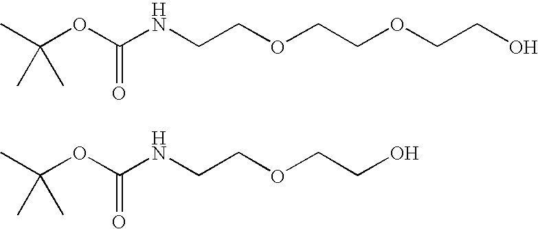
These t-boc protected amino alcohols can be derived, for example, from the corresponding amino alcohols and BOC-ON (19,337-2, Aldrich Chemical). The amino alcohols in turn can be obtained from published procedures in the chemical literature.
These t-boc protected amino alcohols can be derived, for example, from the corresponding amino alcohols and BOC-ON (19,337-2, Aldrich Chemical). The amino alcohols in turn can be obtained from published procedures in the chemical literature.
Alternatively, the starting amino acids and alcohols for the conjugates can comprise all alkyl chains as shown below. The amino acids and alcohols are available commercially (Aldrich Chemical) and can be derivatized with BOC-ON (19,337-2, Aldrich Chemical) to produce the reagents shown below. 
Glucose and Deoxyglucose and their Derivatives
Glucose and Deoxyglucose and their Derivatives
The glucose, deoxyglucose or derivative components of the conjugates described herein are preferably D-(+)-deoxyglucose, D-(+)-glucosamine, and N-acetyl D-glucosamine. Other monosaccharides (e.g., 5-thio-glucose, D-galactose and D-fructose) and their derivatives can also be used. In addition, any of the monosaccharides and their derivatives described in PCT patent publication WO 01/82926, incorporated herein by reference, can be used to prepare a conjugate of the invention.
Some compounds are preferred for the conjugates used in the present methods. In other work, 6-[N-(7-nitrobenz-2-oxa-1,3-diazol-4-yl)amino]-6-deoxyglucose (6-NBDG) was used to study glucose uptake in certain cells. See, for example, Yoshioka et al., 1996, Biochimica et Biophysica Acta 1289: 5–9; Yamada et al., 21 Jul. 2000, J. Biol. Chem. 275(29): 22278–22283; Ball et al., 2002, Can. J. Physiol. Pharmacol. 80: 205–209; and Leira et al., 2002, Toxicology in Vitro 16: 267–273, each of which is incorporated herein by reference. This particular deoxyglucose conjugate is labeled with NBD at the 6-carbon position. The hydroxy group at the 6-carbon position is typically phosphorylated upon entry into a cell, inhibiting its diffusion from the cell. With conjugates carrying a fluorophore at this position, phosphorylation is less likely to occur, and the conjugate will thus be more likely to diffuse out of the cells. Conversely, fluorescent labeling of glucose or deoxyglucose at the 2-position or 3-position results in conjugates that can be phosphorylated after uptake by a cell, with the consequence that such conjugates are less likely to diffuse out of the cells. In addition, deoxyglucose derivatives are often preferred, as they are less likely to be hydrolyzed by esters and other enzymes in the cell. By accumulating the conjugate in the cells, sensitivity of the present method is increased; thus, conjugates of the present invention that have the fluorophore linked to the 2- or 3-position of the glucose, deoxyglucose or derivative are preferred.
Accordingly, in one embodiment, the fluorophore glucose, deoxyglucose or derivative conjugate of the present invention has a formula selected from: 
wherein Fl and L have the meanings provided above.
wherein Fl and L have the meanings provided above.
Another preferred embodiment of the fluorophore glucose, deoxyglucose or derivative conjugate of the invention has the formula: 
wherein Fl is a fluorophore; L is a linking group; each X is independently selected from the group consisting of O and NH; and R1, R3 and R4 are each members independently selected from the group consisting of H, (C1–C12)alkyl, (C1–C12)acyl and a solubility or partitioning effector component. The solubility or partitioning effector component is a component that increases the solubility of the resultant conjugate in aqueous solution relative to a conjugate having a hydrogen atom at the same position. Suitable solubility or partitioning effector components include oligoethylene glycol, oligopropylene glycol, polyhydroxylated carbon chains (typically two to thirty carbons in length) and the like.
wherein Fl is a fluorophore; L is a linking group; each X is independently selected from the group consisting of O and NH; and R1, R3 and R4 are each members independently selected from the group consisting of H, (C1–C12)alkyl, (C1–C12)acyl and a solubility or partitioning effector component. The solubility or partitioning effector component is a component that increases the solubility of the resultant conjugate in aqueous solution relative to a conjugate having a hydrogen atom at the same position. Suitable solubility or partitioning effector components include oligoethylene glycol, oligopropylene glycol, polyhydroxylated carbon chains (typically two to thirty carbons in length) and the like.
In a preferred embodiment, the fluorophore glucose, deoxyglucose or derivative conjugate has the formula Ic in which each X is O, and R1, R3 and R4 are each H.
In another preferred embodiment, the fluorophore glucose, deoxyglucose or derivative conjugate has the formula Ic in which each X is O, and two of R1, R3 and R4 are H, with the remaining member of R1, R3 and R4 being a solubility or partitioning effector component.
In a most preferred embodiment, L is attached to a glucose or deoxyglucose derivative by means of an amino or amido linkage (e.g., the glucose derivative is a 2-amino-2-deoxyglucose derivative).
Administering the Fluorophore Glucose or Deoxyglucose Conjugate
Administration of a fluorophore glucose or deoxyglucose conjugate provided herein can be effected by any method that enables delivery of the conjugates to the site of the cancer or suspected cancer. In one embodiment, delivery is via circulation in the bloodstream. To place the conjugates in contact with cancerous tissues or cells, the methods of administration include oral, buccal intraduodenal, parenteral injection (including intravenous, subcutaneous, intramuscular, intravascular, or infusion), topical administration, and rectal.
The amount of the conjugate administered will be dependent upon the subject being treated, the severity of the cancer, localization of the cancer, the rate of administration, the disposition of the conjugate (e.g., solubility and fluorescence intensity) and the discretion of the administrator. However, an effective dosage is typically in the range of about 0.001 to about 100 mg per kg body weight, preferably about 1 to about 35 mg/kg/day, in single or divided doses. For a 70 kg human, this dosage would amount to about 0.05 to about 7 g/day, preferably about 0.2 to about 2.5 g/day. In some instances, dosage levels below the lower limit of the aforesaid range may be more than adequate, while in other cases still larger doses may be employed without causing any harmful side effect, although such larger doses may be divided into several smaller doses for administration throughout the day.
The imaging fluorophore conjugate composition may, for example, be in a form suitable for oral administration, such as a tablet, capsule, pill, powder, sustained release formulation, solution, or suspension; for parenteral injection, such as a sterile solution, suspension or emulsion; for topical administration, such as an ointment or cream; or for rectal administration, such as a suppository. The imaging fluorophore conjugate composition may be in unit dosage forms suitable for single administration of precise dosages and can include a conventional pharmaceutical carrier or excipient.
Exemplary parenteral administration forms include solutions or suspensions of the imaging conjugate in sterile aqueous solutions, for example, aqueous propylene glycol or dextrose solutions. Such dosage forms can be suitably buffered, if desired.
Suitable pharmaceutical carriers include inert diluents or fillers, water, and various organic solvents. The pharmaceutical compositions may, if desired, contain additional ingredients such as flavorings, binders, excipients, and the like. Thus for oral administration, tablets containing various excipients, such as citric acid, may be employed together with various disintegrants such as starch, alginic acid, and certain complex silicates, and with binding agents such as sucrose, gelatin, and acacia. Additionally, lubricating agents such as magnesium stearate, sodium lauryl sulfate, and talc are often useful for tableting purposes. Solid compositions of a similar type may also be employed in soft and hard filled gelatin capsules. Preferred materials, therefore, include lactose or milk sugar and high molecular weight polyethylene glycols. When aqueous suspensions or elixirs are desired for oral administration the imaging fluorophore conjugate therein may be combined with various sweetening or flavoring agents, coloring matters or dyes, and, if desired, emulsifying agents or suspending agents, together with diluents such as water, ethanol, propylene glycol, glycerin, or combinations thereof.
Methods of preparing various pharmaceutical compositions with a specific amount of an active ingredient that are suitable for use with the active imaging fluorophore conjugates of the present invention are known, or will be apparent upon consideration of the disclosure herein, to those skilled in this art. For examples, see Remington's Pharmaceutical Sciences, Mack Publishing Company, Easter, Pa., 15th Edition (1975).
Because the imaging fluorescent conjugates of the present invention are preferentially taken up by cancer cells, it is possible to obtain an image of or visually confirm the presence of cancer cells that have taken up the conjugate. Detection of the conjugates can be performed using essentially any fluorescence detection device to obtain an image of the cancerous tissues or cells.
Detection of Cancer
Detection and imaging of tissues or cells that take up the conjugates described herein can be accomplished using visual techniques or via two-dimensional image information processing by direct continuous observation with a fluorescence microscope. While spatial resolution can be difficult for certain visual methods (unaided by spectral enhancers or microscopes), a typical fluorescence microscope can provide sufficient resolution at a single cell level.
For example, with a confocal laser scanning fluorescent microscope, 3-dimensional stereoscopic image information with a resolution of about 1 μm can be continuously obtained in real time from tissues in vivo. A variety of known methods can be adapted for use with the conjugates of the present invention. For example, the conjugates of the invention can be used in the endoscopic technique described in U.S. Pat. No. 5,261,410, in which an infrared monochromatic light source is employed and the Raman shift in emission radiation is measured to assess the tissue. Likewise, PCT patent publication No. WO 96/10363 discloses a method of normalization by dividing the intensity at each wavelength by the integrated area under the spectrum. Differences in the resulting curves are then used as the basis for diagnosis.
One of skill in the art will appreciate that essentially any fluorescence detection means, either microscopic or macroscopic, can be employed that is capable of detecting the fluorophore glucose or deoxyglucose conjugate localized in a particular lesion, tissue, organ, or cell.
In some embodiments, the detection means can be in the form of an endoscope inserted into a body cavity through an orifice, such as the mouth, nose, ear, anus, urethra, vagina or an incision. The term “endoscope” is used here to refer to any scope introduced into a body cavity, e.g., an anally introduced endoscope, an orally introduced bronchoscope, a urethrally introduced cystoscope, an abdominally introduced laparoscope, and the like. The miniaturization of scope components has greatly enhanced the utility of an endoscope, making endoscopes particularly useful in the practice of the present invention.
In addition to methods of detecting cancer as generally described above, certain embodiments of the present invention relate to intraoperative, laparoscopic, intravascular, and endoscopic examination, biopsy and treatment of tissues and/or organs with a fluorophore conjugate detecting means capable of close approach to suspected sites of tumor recurrence, metastasis, or incomplete removal of cancer tissue. As used herein, endoscopic procedures include laparoscopic procedures.
Embodiments of the present invention also relate to the intravascular, intraoperative, laparoscopic, and endoscopic examination of lesions with a fluorophore conjugate detecting means capable of close approach to suspected sites of the lesions, especially non-malignant pathological lesions. Lesions include cancerous, hyperplasic, and pre-cancerous cells or tissues.
As noted above, the methods of the present invention do not require processing of images. Rather, in one embodiment, a surgeon or clinician, through the use of, e.g., an intraoperative, laparoscopic, intravascular probe or an endoscope, can quickly scan areas of suspected tumor growth and use the level of fluorescence to more precisely discriminate tumor tissue from non-tumor tissue and thereby more precisely define tumor borders for surgical resection or diagnostic evaluation, or for laser or radiation therapy, including brachytherapy and external beam therapy, or for improved biopsy procedures.
Other embodiments enable the intravascular, intraoperative, laparoscopic, or endoscopic detection means to be similarly used to define and treat lesions. In another embodiment, the conjugate is useful for therapy of the detected tumor by emitting oxygen free radicals or other byproducts which damage the cells in which there has been accumulation of the conjugate. The emission of such damaging agents can be aided or induced by the energy which excites the fluorophore.
The above detection methods can be carried out in combination with a surgical procedure, such as a cancer resection. The method of detecting can be carried out endoscopically, for example, or visually as part of a skin examination for melanoma screening.
Methods of Detecting or Imaging Pre-Cancer
In a related aspect, the present invention provides methods for detecting pre-cancerous cells in a subject, comprising:
(a) administering to the subject an effective amount of a fluorophore glucose, deoxyglucose or derivative conjugate; and
(b) detecting cells that take up the fluorophore glucose or deoxyglucose conjugate to determine if pre-cancerous cells are present in the subject.
The fluorophore conjugate used in this aspect of the invention is preferably one having the formula provided above as (I); more preferably having the formula provided above as (Ia) or (Ib); and most preferably one having the formula (Ic).
The application of the present method to detection and imaging of pre-cancerous cells (e.g., dysplastic cells) resides in the upregulated glucose transport that is exhibited by pre-cancerous cells. Generally, the conjugates, methods of administration and detection are the same as have been described above with respect to the detection and imaging of cancer cells.
Methods for Cancer or Pre-cancer Detection During an Operative or Endoscopic Procedure
As noted above, the detection methods provided herein are broadly applicable and can be particularly effective when used in combination with surgical or endoscopic procedures.
Accordingly, in another related aspect, the present invention provides a method for cancer or pre-cancer detection during an operative or endoscopic procedure, the method comprising:
(a) administering to a patient subject to said procedure an effective amount of a fluorescent glucose, deoxyglucose or derivative conjugate, the conjugate having a rate of uptake in cancerous or pre-cancerous cells that is at least two times greater than the rate of uptake in normal cells;
(b) conducting the procedure within 48 hours of administering the conjugate; and
(c) scanning the patient with a detection means for detecting the localization of the fluorescent conjugate in cancerous or pre-cancerous cells.
In this aspect, a fluorescent conjugate is administered to a patient prior to or coincident with a surgical procedure for the removal of cancerous tissue. The conjugate can be administered by any method designed to bring the conjugate into close proximity with the cancerous tissue, tumor, or lesion to be removed. In this manner, uptake of the conjugate by cancer cells will provide a more accurate determination of the cancer margins.
To facilitate detection during the operative or endoscopic procedure, the conjugate will typically be one that exhibits a rate of uptake in cancerous or pre-cancerous cells that is at least two times greater than the rate of uptake in normal cells (e.g., PBMC cells). Evaluation of such uptake can be carried out as described in the Examples below. Preferably, the rate of uptake in cancerous or pre-cancerous cells is at least 10 times the rate of uptake in normal cells, more preferably at least 20 times, and still more preferably at least 40 times.
For those embodiments in which the fluorescent conjugate is administered prior to the operative or endoscopic procedure, it will preferably be administered from about 1 hour to about 48 hours prior to the procedure, more preferably from about 1 hour to about 16 hours prior to the procedure, and most preferably from about 1 hour to about 6 hours prior to the procedure.
During the procedure, and depending upon the conjugate used, detection can be visual. In some embodiments, the fluorescence of cells that have taken up the conjugate can be enhanced by excitation of the fluorophore with light of a suitable wavelength. Accordingly, once a portion of the tumor or lesion is removed, the remaining tissue can be subjected to a suitable light source to excite the fluorescent conjugates that remain and additional resection can be accomplished.
In other embodiments, detection can be accomplished using fluoroscopes and other detection devices known to those of skill in the art.
Fluorophore Glucose, Deoxyglucose and Derivative Conjugates
In yet another aspect, the present invention provides fluorophore glucose, deoxyglucose and derivative conjugates having the formula:
Fl-L-Glc
wherein Fl is a fluorophore having an emission wavelength of from about 400 nm to about 1200 nm; L is a bond or a linking group; and Glc is glucose, deoxyglucose or derivative, including deoxyglucose and derivatives thereof, with the proviso that the conjugate is other than 2-(N-(7-nitrobenz-2-oxa-1,3-diazol-4-yl)amino)-2-deoxyglucose.
As with the methods above, this aspect of the invention is directed to a variety of glucose, deoxyglucose and derivative conjugates. In particular, the fluorophores used in this aspect of the invention will typically be selected from coumarin dyes, nitrobenzoxazole dyes, cyanine dyes, dipyrrometheneboron dyes, xanthene dyes (including the benzo- and naphtho-xanthene dyes), phenoxazine dyes (as well as the benzo- and naphtho-phenoxazine dyes) and other classes well known to those of skill in the art. Illustrative embodiments of the fluorophore glucose or deoxyglucose conjugates of the invention and methods for their preparation are provided in the Examples below.
In one group of embodiments, L is a bond such that the fluorophore Fl is directly attached to Glc. Typically, this attachment is accomplished via coupling of a functional group on Fl with a compatible (e.g., linkage-forming) functional group on Glc. In certain preferred embodiments, Fl has an isocyanate, isothiocyanate or carboxylic acid functional group that is used to attach Fl to a hydroxy or amino group present on Glc to form a carbamate, thiocarbamate, urea or thiourea linkage between the components.
In another group of embodiments, L is a linking group which can be essentially any of the linking groups discussed above and generally known to those of skill in the art. The linking group in this aspect of the invention typically has from 3 to 100 main chain atoms other than hydrogen atoms, selected from C, N, O, S, P and Si, and can be cyclic, acyclic, aromatic or a combination thereof. Additionally, the linking groups are sufficiently robust so that they are stable not only to reaction conditions used in conjugate assembly, e.g., the protection/deprotection chemistries used to prepare the conjugates, but are also stable to conditions within a cancer cell. More particularly, the linking group is one that is stable to hydrolytic and proteolytic conditions that are typically found in a cancer cell (e.g., exhibits a t1/2 for hydrolysis or cleavage of the linkage of at least about 4 hours at ambient temperatures in a cancer cellular milieu). Preferred linking groups include both homo- and hetero-bifunctional linking groups, as are commercially available or readily prepared according to known methods.
The glucose or glucose derivative components of the conjugates described herein are preferably D-(+)-deoxyglucose, D-(+)-glucosamine and N-acetyl D-glucosamine. Other monosaccharides (e.g., D-galactose and D-fructose) can also be used.
In one group of preferred embodiments, the fluorophore glucose or deoxyglucose conjugate has a formula selected from: 
wherein Fl and L have the meanings provided above.
wherein Fl and L have the meanings provided above.
Still other preferred embodiments are those fluorophore glucose, deoxyglucose or derivative conjugates wherein the glucose portion is a 2-deoxy glucose, the conjugate having the formula: 
wherein Fl is a fluorophore; L is a linking group; each X is independently selected from the group consisting of O and NH; R1, R3 and R4 are each members independently selected from the group consisting of H, (C1–C12)alkyl, (C1–C12)acyl and a solubility or partitioning effector component. The solubility or partitioning effector component can be essentially any component that increases the solubility of the resultant conjugate in aqueous solution relative to the conjugate having a hydrogen atom at the same position. Suitable solubility or partitioning effector components include oligoethylene glycol, oligopropylene glycol, polyhydroxylated carbon chains (typically two to thirty carbons in length) and the like.
wherein Fl is a fluorophore; L is a linking group; each X is independently selected from the group consisting of O and NH; R1, R3 and R4 are each members independently selected from the group consisting of H, (C1–C12)alkyl, (C1–C12)acyl and a solubility or partitioning effector component. The solubility or partitioning effector component can be essentially any component that increases the solubility of the resultant conjugate in aqueous solution relative to the conjugate having a hydrogen atom at the same position. Suitable solubility or partitioning effector components include oligoethylene glycol, oligopropylene glycol, polyhydroxylated carbon chains (typically two to thirty carbons in length) and the like.
In a most preferred embodiment, the fluorophore conjugate has the formula Ic in which each X is O, and R1, R3 and R4 are each H.
In another most preferred embodiment, the fluorophore conjugate has the formula Ic in which each X is O, and two of R1, R3 and R4 are H, with the remaining member of R1, R3 and R4 being a solubility or partitioning effector component.
In the most preferred embodiment, L is attached to the glucose framework by means of an amino or amido linkage (e.g., the deoxyglucose is a 2-amino-2-deoxyglucose).
Kits for Cancer Detection
In still another aspect, the present invention provides kits for use by a clinician, the kits having instructions for administration and cancer detection along with a sterile preparation of a fluorophore conjugate and a pharmaceutically acceptable carrier.
Other Applications
The fluorophore conjugates of the invention, while highly useful for the detection of cancer cells and tissues, can also be used in any application where a fluorescently labeled glucose, deoxyglucose or derivative molecule is useful. In one embodiment, the compounds of the invention are used to measure glucose uptake. In another embodiment, the compounds of the invention are used in an assay to determine whether a compound binds glucose or deoxyglucose. Likewise, while the methods and conjugates of the invention are especially valuable for the diagnosis and detection of cancer in humans, the methods and conjugates of the invention will also find application in the study and treatment of cancer in animals.
The following examples are offered by way of illustration and not by way of limitation.
This example illustrates the time course of uptake of 2-NBDG in Raji lymphoma cells.
Materials:
Raji cells from culture of one t-75 flask. Phosphate-buffered saline plus 2.5 mM CaCl2, 1.2 mM MgSO4, 4 mM KCl and 5 mM glucose (PBS+). 2-NBDG can be prepared in accordance with the method reported in Yoshioka et al., 1996, Biochimica et Biophysica Acta 1289: 5–9, incorporated herein by reference.
Uptake Reaction:
Culture medium with cells (15 mL) was centrifuged for 10 min at 800×g. The pellet of cells was resuspended in PBS+ and centrifuged for 10 min at 800×g, and the resulting pellet was resuspended in 1.5 mL PBS+. Aliquots (100 uL) were placed into each of 8 tubes and incubated at 37° C. for 15 min. The fluorophore deoxyglucose conjugate 2-NBDG was added to each tube to provide a concentration of 200 uM. A tube was removed from the 37° C. bath at each of 0, 1, 3, 5, 10, 20, 30 and 40 min and immediately placed on ice. The cells were washed twice with PBS and resuspended in 100 uL of PBS. A 20 uL aliquot was placed in each of 5 wells of an LJL 96-well plate and fluorescence was counted by a LJL plate reader. FIG. 1 shows the results of the time course experiment.
This example illustrates a comparison of 2-NBDG uptake in Raji lymphoma cells and peripheral blood white cells and illustrates the preferred uptake of a fluorophore deoxyglucose conjugate by a cancer cell as compared to a normal cell.
Materials:
Raji cells from culture one t-75 flask. Peripheral blood white cells from Stanford Blood Bank. Leukocytes were further separated by Ficoll separation (twice). These cells are referred to as PBMC (peripheral blood mononuclear cells). Phosphate-buffered saline. 2-NBDG, 0.1 M in water.
Uptake Reaction:
Culture medium with cells (either Raji or PBMC, 15 mL) were centrifuged for 10 min at 800×g. The pellet of cells was resuspended in PBS and centrifuged for 10 min at 800×g, and the resulting pellet was resuspended in 1.5 mL PBS. Cells were counted and adjusted to 1×10⁶ per mL for both. Aliquots (100 uL) were placed into each of 2 tubes for each cell type, and the tubes were incubated at 37° C. for 15 min. The fluorescent deoxyglucose conjugate 2-NBDG was added to each tube to provide a concentration of 200 uM. At 40 min, the tubes were removed from the 37° C. bath and immediately placed on ice. The cells were washed twice with PBS and resuspended in 100 uL of PBS. A 20 uL aliquot was placed in each of 5 wells of an LJL 96-well plate, and fluorescence was counted by a LJL plate reader. FIG. 2 shows the results of the time course experiment.
This example illustrates the preparation of the fluorophore deoxyglucose conjugate of the invention FGC-002. In the conjugate, the fluorophore is BODIPY, and the glucose derivative is glucosamine. 
Generally, the FGC-002 conjugate is prepared by treating 6-((4,4-difluoro-5,7-dimethyl-4-bora-3a,4a-diaza-s-indacene-3-propionyl)amino)hexanoic acid, succinimidyl ester (BODIPY from Molecular Probes, D-2184, MW 502) with an excess of D-glucosamine (Sigma) in an aprotic solvent with gentle heating. Isolation of the product (FGC-002) can be accomplished via chromatography.
More particularly, 2.2 mg (4 micromoles) of 6-((4,4-difluoro-5,7-dimethyl-4-bora-3a,4a-diaza-s-indacene-3-propionyl)amino)hexanoic acid, succinimidyl ester was dissolved in 0.3 ml of DMF followed by the addition of 6 mg (28 micromoles) of glucosamine HCL dissolved in 0.3 ml of water and 3.9 microliters (28 micromoles) of triethyl amine. The reaction was stirred for 24 hrs at room temperature, evaporated under reduced pressure, and dissolved in a minimum of methanol. This solution was spread on a preparative TLC plate (silica), dried, and developed with 2.5% water in acetonitrile. The plate was then dried and the product visualized with a UV light. The only fluorescent band was scraped off the plate and then eluted with 10% water in acetonitrile and evaporated under reduced pressure, yielding 1 mg of product, which was soluble in methanol and sparingly soluble in water.
This example provides synthetic methods for other illustrative fluorophore deoxyglucose conjugates of the invention.
A. Synthesis of Texas Red Glucosamine Conjugate 
A. Synthesis of Texas Red Glucosamine Conjugate
About 4 mg (4.8 micromoles) of Texas Red®-X, succinimidyl ester *single isomer* (816.94 MW, Molecular Probes, T-20175) were dissolved in 1.2 ml of methanol and combined with 4 mg (18 micromoles) of glucosamine HCl (Sigma) in 600 microliters of water. About 2.5 microliters (18 micromoles) of triethylamine were then added. The reaction was stirred overnight at 20° C. and then evaporated under reduced pressure. The gum was dissolved in methanol and spread on a preparative TLC plate (silica), dried, and developed with 10% water in acetonitrile. The plate was then dried and the product visualized with a UV light. The only red fluorescent band was scraped off the plate and then eluted with 20% water in acetonitrile and evaporated under reduced pressure, yielding 2 mgs of product which was soluble in methanol and sparingly soluble in water.
B. Synthesis of 5-(and 6) Carboxynaphtofluorescein Glucosamine Conjugate 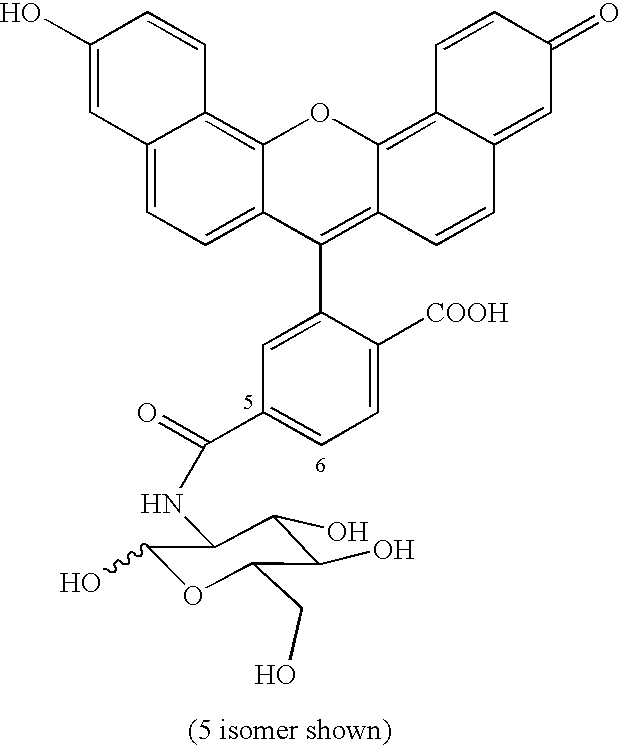
B. Synthesis of 5-(and 6) Carboxynaphtofluorescein Glucosamine Conjugate
About 8.3 mg (14 micromoles) of 5-(and-6)-carboxynaphthofluorescein, succinimidyl ester *mixed isomers* (MW 573.51, Molecular Probes, C-653) were dissolved in 2 ml of DMF, and 10 mg (46 micromoles) of glucosamine HCl (Sigma) in 1 ml of water was added. About 5.8 microliters (46 micromoles) of triethylamine were then added. The reaction was stirred overnight at 20° C. and then evaporated under reduced pressure. The gum was dissolved in 25% water in methanol and spread on a preparative TLC plate (silica), dried, and developed with 10% water in acetonitrile. The plate was then dried and the product visualized with a UV light. The only fluorescent band was scraped off the plate and then eluted with 20% water in acetonitrile and evaporated under reduced pressure, yielding 2 mgs of product, which was soluble in methanol and sparingly soluble in water.
C. Synthesis of Glucosamine N-Methylanthranilamide Conjugate 
C. Synthesis of Glucosamine N-Methylanthranilamide Conjugate
To a slurry of glucosamine (0.25 g, 1.2 mmol) and NEt3 (1.0 mL, 7.0 mmol) in DMF (1 mL) was added a solution of N-methylitacoicanhydride (0.1 g, 0.56 mmol), and the resulting mixture was stirred at 80° C. for 18 h. The reaction mixture was filtered, adsorbed upon silica gel and separated on a silica gel column by washing with acetonitrile and eluting with 10% water in acetonitrile to yield 5 mg of the desired product.
This example illustrates the application of fluorophore conjugates of the present invention to procedures such as gastrointestinal colonoscopy.
Gastrointestinal Colonoscopy:
Endoscopic evaluation of the gastrointestinal tract is recommended for all individuals older than 50 years of age. This screening examination helps the physician identify polyps or other lesions which may be precancerous or cancerous but still localized in the intestine. Identification and removal of these lesions prevent spread of the disease and can cure most patients. This procedure suffers from some significant limitations, including difficulty in distinguishing between hyperplastic lesions and adenomatous lesions, the latter of which must be removed. With knowledge that a polyp is hyperplastic and not cancerous, the lesion can be ignored without removal and biopsy. In addition, some lesions are small and flat. These lesions are frequently overlooked and can be or become cancerous. Due to current limitations, some lesions are incompletely removed during the endoscopic procedure, which leads to an increased risk of recurrence.
To eliminate or ameliorate these limitations of endoscopy, fluorophore conjugates are administered to the patient by intravenous injection from 1 to 48 hours, preferably 2 to 6 hours, prior to the endoscopic procedure. The conjugate can be administered as an oral solution or by pill or as a suppository. The fluorophore conjugate circulates in the blood and accumulates in cancerous or precancerous tissue relative to normal or hyperplastic tissue. During endoscopy, the physician can view the colon under both white light and fluorescent capture. Specifically, while using white light, the physician sees a lesion that appears to be either hyperplastic or adenomatous. In particular, these lesions are viewed with fluorescent capture to determine if the lesion must be resected or can be ignored. Lesions that are highly fluorescent are clearly malignant or premalignant and are removed. Prior to completing this removal, the physician views the surrounding area by fluorescent capture to ensure that all of the malignant tissue is removed. In particular, the fluorophore conjugate can be used for patients who have conditions that put the patient at increased risk for the development of cancer. Patients who suffer this increased risk include patients with chronic inflammatory bowel disease and patients with Barrett's esophagus.
This example illustrates the utility of fluorophore conjugates of the present invention to tumor resection procedures.
Tumor Resection:
Patients diagnosed with a cancerous lesion can be cured if the lesion can be completely resected. Frequently, the surgeon takes tissue samples around the area of the resected tumor to examine the margins at the time of surgery. If the margins appear to contain cancer cells, the surgeon continues to remove tissue to complete the resection. This tissue examination helps the physician identify areas that may be precancerous or cancerous but not yet removed from the patient. Identification and removal of these lesions prevents spread of the disease and can cure the patient. This procedure suffers from some significant limitations, as it is time consuming, and samples from only a small area can be examined. Accordingly, many surgeons do not undertake this tissue sampling during the resection of the tumor but instead examine the tissue only after the surgery is completed. If malignant tissue has been left in the patient, further therapy, such as radiation therapy, is given to the patient. If the physician knew that malignant tissue remained in the patient at the time of surgery, a more complete resection could be effected, and an improved outcome for the patient would result.
To eliminate these limitations of surgical resection of cancer, fluorophore conjugates are administered to the patient by intravenous injection 1 to 48 hours, preferably 2 to 6 hours, prior to the tumor resection. The conjugate can alternatively be administered by oral solution or pill, circulates in the blood and accumulates in cancerous or precancerous tissue relative to normal tissue. During the surgery, the physician views the tissue under both white light and fluorescent capture. Specifically, the physician views an area that the physician has difficulty determining if it is normal or cancerous. In particular, the surgeon views the area of resected tissue prior to completing the operation to determine that all cancerous tissue has been removed. Areas remaining that emit fluorescence and therefore are clearly malignant or pre-malignant are removed. In particular, the fluorophore conjugate is used for patients who have cancers where such definitive removal is currently used. This includes removal of skin cancer by the Mohs' procedure that involves examination of successively collected sections of tissue to determine where the cancer remains and thus where to direct subsequent resection. The use of the fluorophore conjugate can improve the speed and ability of the surgeon to remove this type of cancer.
This example illustrates the utility of fluorophore glucose or deoxyglucose conjugates of the present invention in upper gastrointestinal endoscopy.
Upper Gastrointestinal Endoscopy:
Endoscopic evaluation of the upper gastrointestinal tract is recommended for many patients who have chronic gastroesophageal reflux and all individuals diagnosed with Barrett's esophagus. This screening examination helps the physician identify lesions that may be dysplastic, precancerous, or cancerous but still localized in the intestine. Identification and removal of these lesions prevents spread of the disease and can cure most patients. This procedure suffers from some significant limitations including difficulty in distinguishing between normal mucosa and dysplastic or precancerous lesions that must be removed. If the physician knew that there were areas of precancerous tissue, the patient would undergo surgical removal of the same and the lethal development of a cancer would be avoided. Due to current limitations, areas of precancerous tissue are often overlooked during endoscopy. Because the physician has no aid to find the areas of precancerous tissue, the physician must obtain biopsies in a random fashion, hoping to discover by chance the offending tissue if it exists.
To eliminate these limitations of endoscopy, a fluorescent conjugate of the invention is administered to the patient by intravenous injection 1 to 48 hours, preferably 2 to 6 hours, prior to the endoscopic procedure. This compound may alternatively be given by oral solution or pill. The conjugate circulates in the blood and accumulates in cancerous or precancerous tissue relative to normal tissue. During endoscopy, the physician views the esophagus under both white light and fluorescent capture. Specifically, using fluorescent capture, the physician can identify areas of increased fluorescence, which are deemed to be precancerous and which can be biopsied or removed as the physician deems appropriate.
All publications and patent applications cited in this specification are herein incorporated by reference as if each individual publication or patent application were specifically and individually incorporated by reference. Although the foregoing invention has been described in some detail by way of illustration and example for purposes of clarity of understanding, it will be readily apparent to those of ordinary skill in the art in light of the teachings of this invention that certain changes and modifications may be made thereto without departing from the spirit or scope of the appended claims.
Claims (17)
1. A method for detecting or imaging cancer in a subject, said method comprising:
(a) administering to said subject an effective amount of a dipyrromethene boron dye glucose or deoxyglucose conjugate; and
(b) detecting or imaging cells that take up said conjugate to determine if cancer is present in said subject.
2. A method in accordance with claim 1 , wherein said conjugate has the formula:
Fl-L-Glc
Fl-L-Glc
wherein
Fl is a dipyrromethene boron dye;
L is a bond or a linking group; and
Glc is glucose or deoxyglucose.
3. A method in accordance with claim 2 , wherein said Glc is deoxyglucose or a deoxyglucose derivative selected from the group consisting of D-(+)-deoxyglucose, D-(+)-glucosamine and N-acetyl D-glucosamine, and said Fl is a dipyrromethene boron dye having an emission wavelength of from about 400 nm to about 1200 nm.
4. A method in accordance with claim 2 , wherein L is a linking group having from 2 to 30 chain atoms selected from the group consisting of C, N, O, P, S and Si.
5. A method in accordance with claim 2 , wherein said conjugate has a formula selected from the group consisting of: 
wherein
Fl is a dipyrromethene boron dye;
L is a linking group;
each X is independently selected from the group consisting of O and NH; and
R1, R3 and R4 are each members independently selected from the group consisting of H, (C1–C12)alkyl, (C1–C12)acyl and a solubility or partitioning effector component selected from the group consisting of oligoethylene glycol, oligopropylene glycol, and a polyhydroxylated (C2–C30)carbon chain.
6. A method in accordance with claim 1 , wherein said detecting or imaging is carried out in combination with a cancer resection.
7. A method in accordance with claim 1 , wherein said cancer cells are selected from the group consisting of lung cancer cells, breast cancer cells, prostate cancer cells, colon cancer cells, cervical cancer cells, esophageal cancer cells, bladder cancer cells, head and neck cancer cells and melanoma cells.
8. A method in accordance with claim 1 , further comprising a preliminary step of reducing glucose ingestion in said subject.
9. A method of detecting pre-cancerous cells in a subject, said method comprising:
(a) administering to said subject an effective amount of a dipyrromethene boron dye glucose or deoxyglucose conjugate; and
(b) detecting or imaging pre-cancer cells that take up said conjugate to determine if pre-cancer cells are present in said subject.
10. A method in accordance with claim 9 , wherein said conjugate has the formula:
Fl-L-Glc
Fl-L-Glc
wherein
Fl is a dipyrromethene boron dye;
L is a bond or a linking group; and
Glc is glucose or deoxyglucose.
11. A method in accordance with claim 9 , wherein said administering is intravenous, oral or topical.
12. A method in accordance with claim 11 , wherein said pre-cancerous cells are associated with Barrett Esophagus, colonic polyps and inflammatory bowel disease.
13. A fluorophore conjugate having the formula:
Fl-L-Glc
Fl-L-Glc
wherein
Fl is a dipyrromethene boron dye having an emission wavelength of from about 400 nm to about 1200 nm;
L is a bond or a linking group; and
Glc is glucose or deoxyglucose.
14. A fluorophore conjugate of claim 13 , having a formula selected from the group consisting of: 
wherein
Fl is a dipyrromethene boron dye;
L is a linking group;
each X is independently selected from the group consisting of O and NH; and
R1, R3 and R4 are each members independently selected from the group consisting of H, (C1–C12)alkyl, (C1–C12)acyl and a solubility or partitioning effector component selected from the group consisting of oligoethylene glycol, oligopropylene glycol, and a polyhydroxylated (C2–C30)carbon chain.
15. A method for cancer or pre-cancer detection during an operative or endoscopic procedure, said method comprising:
(a) administering to a patient subject to said procedure, an effective amount of a dipyrromethene boron dye glucose or deoxyglucose conjugate, said conjugate having a rate of uptake in cancerous or pre-cancerous cells that is at least two times greater than the rate of uptake in normal cells;
(b) conducting said procedure within 48 hours of said administering; and
(c) scanning said patient with a detection means for detecting the localization of said conjugate in said cancer or pre-cancer cells, said detection means comprising a fluorescence microscope or a confocal laser scanning fluorescent microscope.
16. A method for cancer or pre-cancer detection in a patient undergoing an operative procedure or an endoscopic procedure, said method comprising:
(a) administering to said patient an effective amount of a dipyrromethene boron dye glucose or deoxyglucose conjugate, said conjugate having a rate of uptake in cancerous or pre-cancerous cells that is at least two times greater than the rate of uptake in normal cells;
(b) conducting said procedure within 24 hours after said administering step;
(c) scanning said patient with a detection means for detecting localization of said conjugate in said cancer or pre-cancer cells, said detection means comprising a fluorescence microscope or a confocal laser scanning fluorescent microscope; and
(d) treating sites of conjugate accretion by external beam radiation, laser therapy, or surgical removal.
17. A kit comprising a vial containing a sterile preparation for human use of a dipyrromethene boron dye glucose or deoxyglucose conjugate and a pharmaceutically acceptable carrier.
Priority Applications (2)
| Application Number | Priority Date | Filing Date | Title |
|---|---|---|---|
| US10/327,226 US6989140B2 (en) | 2001-12-21 | 2002-12-20 | Methods for cancer imaging |
| US11/291,531 US20060165597A1 (en) | 2001-12-21 | 2005-11-30 | Methods for cancer imaging |
Applications Claiming Priority (2)
| Application Number | Priority Date | Filing Date | Title |
|---|---|---|---|
| US34231301P | 2001-12-21 | 2001-12-21 | |
| US10/327,226 US6989140B2 (en) | 2001-12-21 | 2002-12-20 | Methods for cancer imaging |
Related Child Applications (1)
| Application Number | Title | Priority Date | Filing Date |
|---|---|---|---|
| US11/291,531 Continuation US20060165597A1 (en) | 2001-12-21 | 2005-11-30 | Methods for cancer imaging |
Publications (2)
| Publication Number | Publication Date |
|---|---|
| US20030152518A1 US20030152518A1 (en) | 2003-08-14 |
| US6989140B2 true US6989140B2 (en) | 2006-01-24 |
Family
ID=23341275
Family Applications (2)
| Application Number | Title | Priority Date | Filing Date |
|---|---|---|---|
| US10/327,226 Expired - Fee Related US6989140B2 (en) | 2001-12-21 | 2002-12-20 | Methods for cancer imaging |
| US11/291,531 Abandoned US20060165597A1 (en) | 2001-12-21 | 2005-11-30 | Methods for cancer imaging |
Family Applications After (1)
| Application Number | Title | Priority Date | Filing Date |
|---|---|---|---|
| US11/291,531 Abandoned US20060165597A1 (en) | 2001-12-21 | 2005-11-30 | Methods for cancer imaging |
Country Status (3)
| Country | Link |
|---|---|
| US (2) | US6989140B2 (en) |
| AU (1) | AU2002360766A1 (en) |
| WO (1) | WO2003059149A2 (en) |
Cited By (24)
| Publication number | Priority date | Publication date | Assignee | Title |
|---|---|---|---|---|
| US20050014207A1 (en) * | 2002-06-14 | 2005-01-20 | Immunomedics, Inc. | Monoclonal antibody hPAM4 |
| US20050214221A1 (en) * | 2002-03-11 | 2005-09-29 | Visen Medical, Inc. | Optical imaging probes |
| US20060171893A1 (en) * | 2003-06-09 | 2006-08-03 | The Trustees Of The University Of Pennsylvania | Antineoplastic agents targeted via glut transporters |
| US20060280688A1 (en) * | 2000-09-19 | 2006-12-14 | Li-Cor, Inc. | Optical fluorescent imaging |
| WO2008117918A1 (en) * | 2007-03-26 | 2008-10-02 | Snu R&Db Foundation | Fluorescent dye-labeled glucose bioprobe, synthesis method and usage thereof |
| WO2009012109A2 (en) * | 2007-07-13 | 2009-01-22 | Emory University | Cyanine-containing compounds for cancer imaging and treatment |
| US20090214439A1 (en) * | 2006-03-15 | 2009-08-27 | Trustees Of Tufts College | Fluorinated Carbohydrates and Their Use in Tumor Visualization, Tissue Engineering, and Cancer Chemotherapy |
| US20090304580A1 (en) * | 2002-06-14 | 2009-12-10 | Immunomedics, Inc. | Anti-Pancreatic Cancer Antibodies |
| US20100134605A1 (en) * | 2005-11-30 | 2010-06-03 | Demos Stavros G | In vivo spectral micro-imaging of tissue |
| US20100234762A1 (en) * | 2009-02-27 | 2010-09-16 | Gary Pond | Compositions and methods for detecting oral neoplasm |
| US20110070156A1 (en) * | 2002-06-14 | 2011-03-24 | Immunomedics, Inc. | Combining Radioimmunotherapy and Antibody-Drug Conjugates for Improved Cancer Therapy |
| US20110085974A1 (en) * | 2008-06-13 | 2011-04-14 | Cedars-Sinai Medical Center | Small molecule ligand-drug conjugates for targeted cancer therapy |
| US20110165072A1 (en) * | 2002-06-14 | 2011-07-07 | Immunomedics, Inc. | Detection of Early-Stage Pancreatic Adenocarcinoma |
| US20110189708A1 (en) * | 2008-08-08 | 2011-08-04 | Hirosaki University | Method for evaluating specific incorporation of d-glucose into cells |
| KR101150967B1 (en) | 2010-03-18 | 2012-07-11 | 서울대학교산학협력단 | Fluorescent glucose analogue and usage thereof |
| WO2012133688A1 (en) | 2011-03-31 | 2012-10-04 | 国立大学法人弘前大学 | Method for detecting cancer cell using fluorescently labeled l-glucose derivative, and cancer cell-imaging agent comprising fluorescently labeled l-glucose derivative |
| US8574854B2 (en) | 2002-06-14 | 2013-11-05 | Immunomedics, Inc. | Anti-mucin antibodies for early detection and treatment of pancreatic cancer |
| WO2014054601A1 (en) | 2012-10-03 | 2014-04-10 | 国立大学法人弘前大学 | Method for imaging cell using fluorescence-labeled sugar derivative having coumarin derivative bound thereto, and imaging agent |
| US8821868B2 (en) | 2002-06-14 | 2014-09-02 | Immunomedics, Inc. | Anti-pancreatic cancer antibodies |
| US9005613B2 (en) | 2003-06-16 | 2015-04-14 | Immunomedics, Inc. | Anti-mucin antibodies for early detection and treatment of pancreatic cancer |
| US9382329B2 (en) | 2012-08-14 | 2016-07-05 | Ibc Pharmaceuticals, Inc. | Disease therapy by inducing immune response to Trop-2 expressing cells |
| US9452228B2 (en) | 2013-04-01 | 2016-09-27 | Immunomedics, Inc. | Antibodies reactive with an epitope located in the N-terminal region of MUC5AC comprising cysteine-rich subdomain 2 (Cys2) |
| US9599619B2 (en) | 2002-06-14 | 2017-03-21 | Immunomedics, Inc. | Anti-pancreatic cancer antibodies |
| US10001487B2 (en) | 2014-04-08 | 2018-06-19 | Hirosaki University | Glucose derivative, and cell imaging method and imaging agent using said derivative |
Families Citing this family (17)
| Publication number | Priority date | Publication date | Assignee | Title |
|---|---|---|---|---|
| US8188116B2 (en) * | 2002-09-04 | 2012-05-29 | Vanderbilt University | Agents for therapy efficacy monitoring and deep tissue imaging |
| US7754884B2 (en) * | 2005-01-03 | 2010-07-13 | Vanderbilt University | Targeted, NIR imaging agents for therapy efficacy monitoring, deep tissue disease demarcation and deep tissue imaging |
| US20050148534A1 (en) * | 2003-09-22 | 2005-07-07 | Castellino Angelo J. | Small molecule compositions and methods for increasing drug efficiency using compositions thereof |
| WO2005054854A1 (en) * | 2003-11-05 | 2005-06-16 | The United States Of America As Represented By The Secretary Of The Navy Naval Medical Research Center | Fluorescence polarization instruments and methods for detection of exposure to biological materials by fluorescence polarization immunoassay of saliva, oral or bodily fluids |
| US7408640B2 (en) * | 2003-11-05 | 2008-08-05 | The United States Of America As Represented By The Secretary Of The Navy | Fluorescence polarization instruments and methods for detection of exposure to biological materials by fluorescence polarization immunoassay of saliva, oral or bodily fluids |
| WO2006017342A2 (en) * | 2004-07-13 | 2006-02-16 | Y's Therapeutics, Inc. | Assay methods for enzymes associated with sulfation |
| US20060067467A1 (en) * | 2004-09-28 | 2006-03-30 | Minnesota Medical Physics Llc | Apparatus and method for conformal radiation brachytherapy for breast and other tumors |
| US7208611B2 (en) | 2005-02-23 | 2007-04-24 | Xenoport, Inc. | Platinum-containing compounds exhibiting cytostatic activity, synthesis and methods of use |
| WO2006102167A2 (en) * | 2005-03-18 | 2006-09-28 | Stanford University | Compounds for tissue imaging and methods of use thereof |
| EP2173166A4 (en) * | 2007-07-03 | 2010-08-11 | Childrens Hosp & Res Ct Oak | Inhibitors of polysialic acid de-n-acetylase and methods for using the same |
| US8236284B1 (en) * | 2008-04-02 | 2012-08-07 | University Of Central Florida Research Foundation, Inc. | Multimodal, multifunctional polymer coated nanoparticles |
| US20100240878A1 (en) * | 2009-03-20 | 2010-09-23 | Barbeau Donald L | Fluorescent saccharide conjugates |
| US20130066199A1 (en) * | 2010-04-15 | 2013-03-14 | Cedars-Sinai Medical Center | Tumor margin detection method based on nuclear morphometry and tissue topology |
| US20140126787A1 (en) * | 2012-11-08 | 2014-05-08 | Lasarow Healthcare Technologies Limited | Device and method for generating melanoma risk assessments |
| US20170112947A1 (en) * | 2014-06-05 | 2017-04-27 | The Trustees Of Columbia University In The City Of New York | Composition for use in imaging |
| IT201900025840A1 (en) * | 2019-12-31 | 2021-07-01 | Univ Bologna Alma Mater Studiorum | COMPOUNDS ISOTHIOCIANATES AND ISOSELENCIANATES |
| CN113687068A (en) * | 2021-08-24 | 2021-11-23 | 江苏耀研达科技发展有限公司 | Tumor single cell double-fluorescence labeling detection kit, preparation method and application thereof |
Citations (20)
| Publication number | Priority date | Publication date | Assignee | Title |
|---|---|---|---|---|
| US4716225A (en) | 1984-12-27 | 1987-12-29 | Georgetown University | Radioopaque sugar derivatives and a method of metabolic mapping using the same |
| EP0413145A1 (en) | 1989-07-13 | 1991-02-20 | Nihon Medi-Physics Co., Ltd. | Radioactive diagnostic agent |
| US5057301A (en) | 1988-04-06 | 1991-10-15 | Neorx Corporation | Modified cellular substrates used as linkers for increased cell retention of diagnostic and therapeutic agents |
| US5131398A (en) | 1990-01-22 | 1992-07-21 | Mediscience Technology Corp. | Method and apparatus for distinguishing cancerous tissue from benign tumor tissue, benign tissue or normal tissue using native fluorescence |
| WO1993013403A1 (en) | 1991-12-21 | 1993-07-08 | Sune Svanberg | FLUORESCENCE DIAGNOSTICS OF CANCER USING δ-AMINO LEVULINIC ACID |
| US5261410A (en) | 1991-02-07 | 1993-11-16 | Alfano Robert R | Method for determining if a tissue is a malignant tumor tissue, a benign tumor tissue, or a normal or benign tissue using Raman spectroscopy |
| US5451463A (en) | 1989-08-28 | 1995-09-19 | Clontech Laboratories, Inc. | Non-nucleoside 1,3-diol reagents for labeling synthetic oligonucleotides |
| WO1996010363A1 (en) | 1994-09-30 | 1996-04-11 | Lockheed Martin Energy Systems, Inc. | Laser-induced differential normalized fluorescence method for cancer diagnosis |
| US5512667A (en) | 1990-08-28 | 1996-04-30 | Reed; Michael W. | Trifunctional intermediates for preparing 3'-tailed oligonucleotides |
| US5585422A (en) | 1995-09-20 | 1996-12-17 | Ciba-Geigy Corporation | Hybrid s-triazine light stabilizers substituted by benzotriazole or benzophenone moieties and compositions stabilized therewith |
| WO1999020316A1 (en) | 1997-10-20 | 1999-04-29 | Board Of Regents, The University Of Texas System | Single photon-emitting radiotraces and methods for their use |
| US6031091A (en) | 1987-09-21 | 2000-02-29 | Gen-Probe Incorporated | Non-nucleotide linking reagents for nucleotide probes |
| US6083487A (en) | 1997-08-25 | 2000-07-04 | Advanced Photodynamic Technologies, Inc. | Methylene blue and toluidene blue mediated fluorescence diagnosis of cancer |
| US6091985A (en) | 1998-01-23 | 2000-07-18 | Research Foundation Of City College Of New York | Detection of cancer and precancerous conditions in tissues and/or cells using native fluorescence excitation spectroscopy |
| US6099822A (en) | 1997-09-05 | 2000-08-08 | Ozker; Suleyman Kutlan | Imaging methods and compositions |
| US6207136B1 (en) | 1997-03-11 | 2001-03-27 | Tokyo University Of Agriculture And Technology | Fluorescent imaging method of saccharide uptake activity of living tissue |
| US6226352B1 (en) | 1998-09-08 | 2001-05-01 | Veritas Pharmaceuticals, Inc. | System and method for radiographic imaging of tissue |
| US6256530B1 (en) | 1998-09-15 | 2001-07-03 | Denvu, L.L.C. | Optical instrument and technique for cancer diagnosis using in-vivo fluorescence emission of test tissue |
| US6284223B1 (en) | 1998-10-15 | 2001-09-04 | Fluoroprobe, Inc. | Method for viewing tumor tissue located within a body cavity |
| US6489302B1 (en) | 1995-07-05 | 2002-12-03 | Deutches Krebsforschungszentrum Stiftung Des Offentlichen Rechts | Saccharide conjugates |
Family Cites Families (3)
| Publication number | Priority date | Publication date | Assignee | Title |
|---|---|---|---|---|
| US5512567A (en) * | 1988-08-24 | 1996-04-30 | Sankyo Company, Limited | Analgesic compounds, their preparation, and pharmaceutical compositions containing them |
| DE4003799C1 (en) * | 1990-02-08 | 1991-04-18 | Cramer Gmbh & Co Kg, 5750 Menden, De | |
| US5672332A (en) * | 1996-05-13 | 1997-09-30 | Mallinckrodt Medical, Inc. | Delta 1,2 bicyclo 4,4,0! functional dyes for contrast enhancement in optical imaging |
-
2002
- 2002-12-20 AU AU2002360766A patent/AU2002360766A1/en not_active Abandoned
- 2002-12-20 US US10/327,226 patent/US6989140B2/en not_active Expired - Fee Related
- 2002-12-20 WO PCT/US2002/041339 patent/WO2003059149A2/en not_active Application Discontinuation
-
2005
- 2005-11-30 US US11/291,531 patent/US20060165597A1/en not_active Abandoned
Patent Citations (20)
| Publication number | Priority date | Publication date | Assignee | Title |
|---|---|---|---|---|
| US4716225A (en) | 1984-12-27 | 1987-12-29 | Georgetown University | Radioopaque sugar derivatives and a method of metabolic mapping using the same |
| US6031091A (en) | 1987-09-21 | 2000-02-29 | Gen-Probe Incorporated | Non-nucleotide linking reagents for nucleotide probes |
| US5057301A (en) | 1988-04-06 | 1991-10-15 | Neorx Corporation | Modified cellular substrates used as linkers for increased cell retention of diagnostic and therapeutic agents |
| EP0413145A1 (en) | 1989-07-13 | 1991-02-20 | Nihon Medi-Physics Co., Ltd. | Radioactive diagnostic agent |
| US5451463A (en) | 1989-08-28 | 1995-09-19 | Clontech Laboratories, Inc. | Non-nucleoside 1,3-diol reagents for labeling synthetic oligonucleotides |
| US5131398A (en) | 1990-01-22 | 1992-07-21 | Mediscience Technology Corp. | Method and apparatus for distinguishing cancerous tissue from benign tumor tissue, benign tissue or normal tissue using native fluorescence |
| US5512667A (en) | 1990-08-28 | 1996-04-30 | Reed; Michael W. | Trifunctional intermediates for preparing 3'-tailed oligonucleotides |
| US5261410A (en) | 1991-02-07 | 1993-11-16 | Alfano Robert R | Method for determining if a tissue is a malignant tumor tissue, a benign tumor tissue, or a normal or benign tissue using Raman spectroscopy |
| WO1993013403A1 (en) | 1991-12-21 | 1993-07-08 | Sune Svanberg | FLUORESCENCE DIAGNOSTICS OF CANCER USING δ-AMINO LEVULINIC ACID |
| WO1996010363A1 (en) | 1994-09-30 | 1996-04-11 | Lockheed Martin Energy Systems, Inc. | Laser-induced differential normalized fluorescence method for cancer diagnosis |
| US6489302B1 (en) | 1995-07-05 | 2002-12-03 | Deutches Krebsforschungszentrum Stiftung Des Offentlichen Rechts | Saccharide conjugates |
| US5585422A (en) | 1995-09-20 | 1996-12-17 | Ciba-Geigy Corporation | Hybrid s-triazine light stabilizers substituted by benzotriazole or benzophenone moieties and compositions stabilized therewith |
| US6207136B1 (en) | 1997-03-11 | 2001-03-27 | Tokyo University Of Agriculture And Technology | Fluorescent imaging method of saccharide uptake activity of living tissue |
| US6083487A (en) | 1997-08-25 | 2000-07-04 | Advanced Photodynamic Technologies, Inc. | Methylene blue and toluidene blue mediated fluorescence diagnosis of cancer |
| US6099822A (en) | 1997-09-05 | 2000-08-08 | Ozker; Suleyman Kutlan | Imaging methods and compositions |
| WO1999020316A1 (en) | 1997-10-20 | 1999-04-29 | Board Of Regents, The University Of Texas System | Single photon-emitting radiotraces and methods for their use |
| US6091985A (en) | 1998-01-23 | 2000-07-18 | Research Foundation Of City College Of New York | Detection of cancer and precancerous conditions in tissues and/or cells using native fluorescence excitation spectroscopy |
| US6226352B1 (en) | 1998-09-08 | 2001-05-01 | Veritas Pharmaceuticals, Inc. | System and method for radiographic imaging of tissue |
| US6256530B1 (en) | 1998-09-15 | 2001-07-03 | Denvu, L.L.C. | Optical instrument and technique for cancer diagnosis using in-vivo fluorescence emission of test tissue |
| US6284223B1 (en) | 1998-10-15 | 2001-09-04 | Fluoroprobe, Inc. | Method for viewing tumor tissue located within a body cavity |
Non-Patent Citations (5)
| Title |
|---|
| Ball, S.W. et al. (2002). "A fluorescent compound for glucose uptake measurements in isolated rat cardiomyocytes," Can. J. Physiol. Pharmacol 80:205-209. |
| Leira, F. et al. (2002). "Fluorescent microplate cell assay to measure uptake and metabolism of glucose in normal human lung fibroblasts," Toxicology in Vitro 16:267-273. |
| Som, P. et al. (1980). "A Fluorinated Glucose Analog, 2-fluoro-2-deoxy-D-glucose (F-18): Nontoxic Tracer for Rapid Tumor Detection," J. Nucl. Med 21:670-675. |
| Yamada, K. et al. (2000). "Measurement of Glucose Uptake and Intracellular Calcium Concentration in Single, Living Pancreatic beta-Cells," The Journal of Biological Chemistry 275:22278-22283. |
| Yoshioka, K. et al. (1996). "A Novel fluorescent derivative of glucose applicable to the assessment of glucose uptake activity of Escherichia coli," Biochemica et Biophysica Acta 1289:5-9. |
Cited By (56)
| Publication number | Priority date | Publication date | Assignee | Title |
|---|---|---|---|---|
| US7597878B2 (en) | 2000-09-19 | 2009-10-06 | Li-Cor, Inc. | Optical fluorescent imaging |
| US20060280688A1 (en) * | 2000-09-19 | 2006-12-14 | Li-Cor, Inc. | Optical fluorescent imaging |
| US8303936B2 (en) | 2000-09-19 | 2012-11-06 | Li-Cor, Inc. | Optical fluorescent imaging |
| US20100080758A1 (en) * | 2000-09-19 | 2010-04-01 | Li-Cor,Inc. | Optical fluorescent imaging |
| US9272057B2 (en) | 2002-02-26 | 2016-03-01 | Immunomedics, Inc. | Combining radioimmunotherapy and antibody-drug conjugates for improved cancer therapy |
| US8992931B2 (en) | 2002-03-01 | 2015-03-31 | Immunomedics, Inc. | Combining radioimmunotherapy and antibody-drug conjugates for improved cancer therapy |
| US20050214221A1 (en) * | 2002-03-11 | 2005-09-29 | Visen Medical, Inc. | Optical imaging probes |
| US20110171136A1 (en) * | 2002-03-11 | 2011-07-14 | Poss Kirtland G | Optical imaging probes |
| US9238081B2 (en) | 2002-06-14 | 2016-01-19 | Immunomedics, Inc. | Detection of early-stage pancreatic adenocarcinoma |
| US20050014207A1 (en) * | 2002-06-14 | 2005-01-20 | Immunomedics, Inc. | Monoclonal antibody hPAM4 |
| US9599619B2 (en) | 2002-06-14 | 2017-03-21 | Immunomedics, Inc. | Anti-pancreatic cancer antibodies |
| US20090304580A1 (en) * | 2002-06-14 | 2009-12-10 | Immunomedics, Inc. | Anti-Pancreatic Cancer Antibodies |
| US9513293B2 (en) | 2002-06-14 | 2016-12-06 | Immunomedics, Inc. | Detection of early-stage pancreatic adenocarcinoma |
| US9089618B2 (en) | 2002-06-14 | 2015-07-28 | Immunomedics, Inc. | Anti-mucin antibodies for early detection and treatment of pancreatic cancer |
| US7282567B2 (en) * | 2002-06-14 | 2007-10-16 | Immunomedics, Inc. | Monoclonal antibody hPAM4 |
| US8974784B2 (en) | 2002-06-14 | 2015-03-10 | Immunomedics, Inc. | Anti-pancreatic cancer antibodies |
| US8821868B2 (en) | 2002-06-14 | 2014-09-02 | Immunomedics, Inc. | Anti-pancreatic cancer antibodies |
| US20110070156A1 (en) * | 2002-06-14 | 2011-03-24 | Immunomedics, Inc. | Combining Radioimmunotherapy and Antibody-Drug Conjugates for Improved Cancer Therapy |
| US8795662B2 (en) | 2002-06-14 | 2014-08-05 | Immunomedics, Inc. | Anti-mucin antibodies for early detection and treatment of pancreatic cancer |
| US8586050B2 (en) | 2002-06-14 | 2013-11-19 | Immunomedics, Inc. | Combining radioimmunotherapy and antibody-drug conjugates for improved cancer therapy |
| US20110165072A1 (en) * | 2002-06-14 | 2011-07-07 | Immunomedics, Inc. | Detection of Early-Stage Pancreatic Adenocarcinoma |
| US8574854B2 (en) | 2002-06-14 | 2013-11-05 | Immunomedics, Inc. | Anti-mucin antibodies for early detection and treatment of pancreatic cancer |
| US8491896B2 (en) | 2002-06-14 | 2013-07-23 | Immunomedics, Inc. | Anti-pancreatic cancer antibodies |
| US8435529B2 (en) | 2002-06-14 | 2013-05-07 | Immunomedics, Inc. | Combining radioimmunotherapy and antibody-drug conjugates for improved cancer therapy |
| US20060171893A1 (en) * | 2003-06-09 | 2006-08-03 | The Trustees Of The University Of Pennsylvania | Antineoplastic agents targeted via glut transporters |
| US20080269112A1 (en) * | 2003-06-09 | 2008-10-30 | The Trustees Of The University Of Pennsylvania | Antineoplastic Agents Targeted via Glut Transporters |
| US7943586B2 (en) * | 2003-06-09 | 2011-05-17 | The Trustees Of The University Of Pennsylvania | Antineoplastic agents targeted via glut transporters |
| US9238084B2 (en) | 2003-06-16 | 2016-01-19 | Immunomedics, Inc. | Anti-mucin antibodies for early detection and treatment of pancreatic cancer |
| US9005613B2 (en) | 2003-06-16 | 2015-04-14 | Immunomedics, Inc. | Anti-mucin antibodies for early detection and treatment of pancreatic cancer |
| US20100134605A1 (en) * | 2005-11-30 | 2010-06-03 | Demos Stavros G | In vivo spectral micro-imaging of tissue |
| US8320650B2 (en) * | 2005-11-30 | 2012-11-27 | Lawrence Livermore National Security, Llc | In vivo spectral micro-imaging of tissue |
| US20090214439A1 (en) * | 2006-03-15 | 2009-08-27 | Trustees Of Tufts College | Fluorinated Carbohydrates and Their Use in Tumor Visualization, Tissue Engineering, and Cancer Chemotherapy |
| KR100874761B1 (en) | 2007-03-26 | 2008-12-19 | 재단법인서울대학교산학협력재단 | Glucose analogs labeled with fluorescent dyes, synthetic methods thereof and uses thereof |
| WO2008117918A1 (en) * | 2007-03-26 | 2008-10-02 | Snu R&Db Foundation | Fluorescent dye-labeled glucose bioprobe, synthesis method and usage thereof |
| US20100105149A1 (en) * | 2007-03-26 | 2010-04-29 | Snu R&Db Foundation | Fluorescent dye-labeled glucose bioprobe, synthesis method and usage thereof |
| US8679854B2 (en) | 2007-03-26 | 2014-03-25 | Snu & Db Foundation | Fluorescent dye-labeled glucose bioprobe, synthesis method and usage thereof |
| WO2009012109A3 (en) * | 2007-07-13 | 2009-12-30 | Emory University | Cyanine-containing compounds for cancer imaging and treatment |
| US11738095B2 (en) | 2007-07-13 | 2023-08-29 | Emory University | Cyanine-containing compounds for cancer imaging and treatment |
| WO2009012109A2 (en) * | 2007-07-13 | 2009-01-22 | Emory University | Cyanine-containing compounds for cancer imaging and treatment |
| US20110085974A1 (en) * | 2008-06-13 | 2011-04-14 | Cedars-Sinai Medical Center | Small molecule ligand-drug conjugates for targeted cancer therapy |
| US8986656B2 (en) * | 2008-08-08 | 2015-03-24 | Hirosaki University | Method for evaluating specific incorporation of D-glucose into cells |
| US20110189708A1 (en) * | 2008-08-08 | 2011-08-04 | Hirosaki University | Method for evaluating specific incorporation of d-glucose into cells |
| US20150212094A1 (en) * | 2008-08-08 | 2015-07-30 | Hirosaki University | L-glucose derivatives having fluorescent chromophore |
| US9958450B2 (en) * | 2008-08-08 | 2018-05-01 | Hirosaki University | L-glucose derivatives having fluorescent chromophore |
| US20100234762A1 (en) * | 2009-02-27 | 2010-09-16 | Gary Pond | Compositions and methods for detecting oral neoplasm |
| KR101150967B1 (en) | 2010-03-18 | 2012-07-11 | 서울대학교산학협력단 | Fluorescent glucose analogue and usage thereof |
| WO2012133688A1 (en) | 2011-03-31 | 2012-10-04 | 国立大学法人弘前大学 | Method for detecting cancer cell using fluorescently labeled l-glucose derivative, and cancer cell-imaging agent comprising fluorescently labeled l-glucose derivative |
| US9670286B2 (en) | 2012-08-14 | 2017-06-06 | Ibc Pharmaceuticals, Inc. | Disease therapy by inducing immune response to Trop-2 expressing cells |
| US9879088B2 (en) | 2012-08-14 | 2018-01-30 | Ibc Pharmaceuticals, Inc. | Disease therapy by inducing immune response to Trop-2 expressing cells |
| US9382329B2 (en) | 2012-08-14 | 2016-07-05 | Ibc Pharmaceuticals, Inc. | Disease therapy by inducing immune response to Trop-2 expressing cells |
| US10662252B2 (en) | 2012-08-14 | 2020-05-26 | Ibc Pharmaceuticals, Inc. | Disease therapy by inducing immune response to Trop-2 expressing cells |
| US10288604B2 (en) | 2012-10-03 | 2019-05-14 | Hirosaki University | Method for imaging cell using fluorescence-labeled sugar derivative having coumarin derivative bound thereto, and imaging agent |
| WO2014054601A1 (en) | 2012-10-03 | 2014-04-10 | 国立大学法人弘前大学 | Method for imaging cell using fluorescence-labeled sugar derivative having coumarin derivative bound thereto, and imaging agent |
| US9452228B2 (en) | 2013-04-01 | 2016-09-27 | Immunomedics, Inc. | Antibodies reactive with an epitope located in the N-terminal region of MUC5AC comprising cysteine-rich subdomain 2 (Cys2) |
| US10001487B2 (en) | 2014-04-08 | 2018-06-19 | Hirosaki University | Glucose derivative, and cell imaging method and imaging agent using said derivative |
| US10509041B2 (en) | 2014-04-08 | 2019-12-17 | Hirosaki University | Glucose derivative, and cell imaging method and imaging agent using said derivative |
Also Published As
| Publication number | Publication date |
|---|---|
| US20060165597A1 (en) | 2006-07-27 |
| AU2002360766A8 (en) | 2003-07-30 |
| US20030152518A1 (en) | 2003-08-14 |
| WO2003059149A3 (en) | 2003-09-18 |
| WO2003059149A2 (en) | 2003-07-24 |
| AU2002360766A1 (en) | 2003-07-30 |
Similar Documents
| Publication | Publication Date | Title |
|---|---|---|
| US6989140B2 (en) | Methods for cancer imaging | |
| US10695445B2 (en) | Methods of using optical agents | |
| Weissleder et al. | In vivo imaging of tumors with protease-activated near-infrared fluorescent probes | |
| Rosenthal et al. | The status of contemporary image-guided modalities in oncologic surgery | |
| US6846311B2 (en) | Method and apparatus for in VIVO treatment of mammary ducts by light induced fluorescence | |
| AU2010239272B2 (en) | Light-emitting dye for intraoperative imaging or sentinel lymph node biopsy | |
| US20120114563A1 (en) | Optical imaging agents | |
| US20090280061A1 (en) | Esters of 5-aminolevulinic acid as photosensitizing agents in photochemotherapy | |
| US10918741B2 (en) | PSMA-targeting imaging agents | |
| AU2007249226A1 (en) | Imaging agents and methods | |
| CN104661994A (en) | Compounds | |
| de Oliveira Silva et al. | Enhancement of blood porphyrin emission intensity with aminolevulinic acid administration: A new concept for photodynamic diagnosis of early prostate cancer | |
| US20190076555A1 (en) | Tumor targeting nanoagent for imaging and fluorescent guided resection of tumors | |
| JPH0638967A (en) | Apparatus and method using biopsy needle based on fluorescence intensified by photointensifier | |
| WO2010032248A2 (en) | Multifunctional albumin conjugates | |
| US20070196278A1 (en) | Compositions and methods for locating an internal bleeding site | |
| Li et al. | Research progress of fluorescence imaging in intraoperative navigation based on VOSviewer bibliometric analysis | |
| Fu et al. | Improving oral squamous cell carcinoma diagnosis and treatment with fluorescence molecular imaging | |
| GB2343186A (en) | Tetrasulphamoyl- phthalocyanine & naphthalocyanine dye derivatives for use in tissue demarcation, imaging, & diagnosis of tumour cells & diseased lymph nodes | |
| US20080167544A1 (en) | Compositions And Methods For Locating An Internal Bleeding Site | |
| Chuchueva et al. | THE ASSESSMENT OF A CELLULAR PATTERN OF THE EPITHELIUM AS A DIAGNOSTIC TOOL FOR LARYNGEAL TUMORS DETECTION | |
| US20100047173A1 (en) | Methods for Using Optical Agents | |
| CN117042811A (en) | Composition for cancer imaging | |
| JP2023537627A (en) | Compounds and compositions for tumor detection and surgical guidance | |
| EP4041213A1 (en) | Psma-targeting imaging agents |
Legal Events
| Date | Code | Title | Description |
|---|---|---|---|
| AS | Assignment |
Owner name: THRESHOLD PHARMACEUTICAL, INC., CALIFORNIA Free format text: ASSIGNMENT OF ASSIGNORS INTEREST;ASSIGNORS:TIDMARSH, GEORGE;MATTEUCCI, MARK;REEL/FRAME:013622/0624 Effective date: 20021217 |
|
| FPAY | Fee payment |
Year of fee payment: 4 |
|
| REMI | Maintenance fee reminder mailed | ||
| LAPS | Lapse for failure to pay maintenance fees | ||
| LAPS | Lapse for failure to pay maintenance fees |
Free format text: PATENT EXPIRED FOR FAILURE TO PAY MAINTENANCE FEES (ORIGINAL EVENT CODE: EXP.) |
|
| FP | Lapsed due to failure to pay maintenance fee |
Effective date: 20140124 |
|
| STCH | Information on status: patent discontinuation |
Free format text: PATENT EXPIRED DUE TO NONPAYMENT OF MAINTENANCE FEES UNDER 37 CFR 1.362 |