US7247703B2 - Immune reactivity to HER-2/neu protein for diagnosis and treatment of malignancies in which the HER-2/neu oncogene is associated - Google Patents
Immune reactivity to HER-2/neu protein for diagnosis and treatment of malignancies in which the HER-2/neu oncogene is associated Download PDFInfo
- Publication number
- US7247703B2 US7247703B2 US10/647,005 US64700503A US7247703B2 US 7247703 B2 US7247703 B2 US 7247703B2 US 64700503 A US64700503 A US 64700503A US 7247703 B2 US7247703 B2 US 7247703B2
- Authority
- US
- United States
- Prior art keywords
- neu
- cells
- protein
- peptides
- seq
- Prior art date
- Legal status (The legal status is an assumption and is not a legal conclusion. Google has not performed a legal analysis and makes no representation as to the accuracy of the status listed.)
- Expired - Fee Related, expires
Links
- 101001012157 Homo sapiens Receptor tyrosine-protein kinase erbB-2 Proteins 0.000 title abstract description 181
- 108090000623 proteins and genes Proteins 0.000 title abstract description 179
- 102000004169 proteins and genes Human genes 0.000 title abstract description 172
- 102100030086 Receptor tyrosine-protein kinase erbB-2 Human genes 0.000 title abstract description 160
- 206010028980 Neoplasm Diseases 0.000 title abstract description 59
- 230000036210 malignancy Effects 0.000 title abstract description 35
- 108700020796 Oncogene Proteins 0.000 title abstract description 32
- 238000003745 diagnosis Methods 0.000 title abstract description 5
- 238000011282 treatment Methods 0.000 title abstract description 5
- 230000009257 reactivity Effects 0.000 title description 7
- 108090000765 processed proteins & peptides Proteins 0.000 claims abstract description 254
- 125000003275 alpha amino acid group Chemical group 0.000 claims description 27
- 102000004196 processed proteins & peptides Human genes 0.000 abstract description 167
- 210000001744 T-lymphocyte Anatomy 0.000 abstract description 121
- 230000004044 response Effects 0.000 abstract description 72
- 238000000034 method Methods 0.000 abstract description 47
- 201000011510 cancer Diseases 0.000 abstract description 26
- 238000000338 in vitro Methods 0.000 abstract description 25
- 239000000203 mixture Substances 0.000 abstract description 24
- 238000001514 detection method Methods 0.000 abstract description 20
- 210000001124 body fluid Anatomy 0.000 abstract description 16
- 239000010839 body fluid Substances 0.000 abstract description 16
- 230000035755 proliferation Effects 0.000 abstract description 13
- 238000012544 monitoring process Methods 0.000 abstract description 5
- 230000006044 T cell activation Effects 0.000 abstract description 4
- 210000004027 cell Anatomy 0.000 description 89
- 241000700159 Rattus Species 0.000 description 73
- 241001465754 Metazoa Species 0.000 description 51
- 239000000427 antigen Substances 0.000 description 47
- 108091007433 antigens Proteins 0.000 description 47
- 102000036639 antigens Human genes 0.000 description 47
- 230000027455 binding Effects 0.000 description 44
- 230000003053 immunization Effects 0.000 description 44
- 101100501691 Rattus norvegicus Erbb2 gene Proteins 0.000 description 35
- 238000002649 immunization Methods 0.000 description 34
- 230000005875 antibody response Effects 0.000 description 32
- 206010006187 Breast cancer Diseases 0.000 description 30
- 208000026310 Breast neoplasm Diseases 0.000 description 29
- 238000003556 assay Methods 0.000 description 29
- 108091003079 Bovine Serum Albumin Proteins 0.000 description 28
- 230000000638 stimulation Effects 0.000 description 27
- 230000005867 T cell response Effects 0.000 description 26
- 238000010790 dilution Methods 0.000 description 26
- 239000012895 dilution Substances 0.000 description 26
- 150000001413 amino acids Chemical class 0.000 description 24
- 101000856426 Mus musculus Cullin-7 Proteins 0.000 description 22
- LOKCTEFSRHRXRJ-UHFFFAOYSA-I dipotassium trisodium dihydrogen phosphate hydrogen phosphate dichloride Chemical compound P(=O)(O)(O)[O-].[K+].P(=O)(O)([O-])[O-].[Na+].[Na+].[Cl-].[K+].[Cl-].[Na+] LOKCTEFSRHRXRJ-UHFFFAOYSA-I 0.000 description 22
- 239000002953 phosphate buffered saline Substances 0.000 description 22
- 108700018351 Major Histocompatibility Complex Proteins 0.000 description 21
- 229940098773 bovine serum albumin Drugs 0.000 description 21
- 230000020382 suppression by virus of host antigen processing and presentation of peptide antigen via MHC class I Effects 0.000 description 21
- 238000004458 analytical method Methods 0.000 description 20
- 102000051957 human ERBB2 Human genes 0.000 description 18
- 102000008949 Histocompatibility Antigens Class I Human genes 0.000 description 17
- 108010088652 Histocompatibility Antigens Class I Proteins 0.000 description 17
- 210000001151 cytotoxic T lymphocyte Anatomy 0.000 description 16
- 238000002965 ELISA Methods 0.000 description 15
- 230000003834 intracellular effect Effects 0.000 description 15
- 102000000588 Interleukin-2 Human genes 0.000 description 14
- 108010002350 Interleukin-2 Proteins 0.000 description 14
- 108700020302 erbB-2 Genes Proteins 0.000 description 14
- 239000002671 adjuvant Substances 0.000 description 13
- 239000012133 immunoprecipitate Substances 0.000 description 13
- 230000014509 gene expression Effects 0.000 description 12
- 239000012528 membrane Substances 0.000 description 12
- 210000004379 membrane Anatomy 0.000 description 12
- 239000000047 product Substances 0.000 description 12
- 210000003719 b-lymphocyte Anatomy 0.000 description 11
- 210000004698 lymphocyte Anatomy 0.000 description 11
- 210000000952 spleen Anatomy 0.000 description 11
- 108010001336 Horseradish Peroxidase Proteins 0.000 description 10
- 238000002474 experimental method Methods 0.000 description 10
- 238000001727 in vivo Methods 0.000 description 10
- 210000001165 lymph node Anatomy 0.000 description 10
- 125000006853 reporter group Chemical group 0.000 description 10
- 210000002966 serum Anatomy 0.000 description 10
- 238000002560 therapeutic procedure Methods 0.000 description 10
- 238000001262 western blot Methods 0.000 description 10
- 102000001301 EGF receptor Human genes 0.000 description 9
- 108060006698 EGF receptor Proteins 0.000 description 9
- 102000025850 HLA-A2 Antigen Human genes 0.000 description 9
- 108010074032 HLA-A2 Antigen Proteins 0.000 description 9
- 230000006870 function Effects 0.000 description 9
- 210000005105 peripheral blood lymphocyte Anatomy 0.000 description 9
- 210000000612 antigen-presenting cell Anatomy 0.000 description 8
- 239000006166 lysate Substances 0.000 description 8
- 239000002243 precursor Substances 0.000 description 8
- 241000283973 Oryctolagus cuniculus Species 0.000 description 7
- 238000001516 cell proliferation assay Methods 0.000 description 7
- 239000012091 fetal bovine serum Substances 0.000 description 7
- 239000000499 gel Substances 0.000 description 7
- 238000001114 immunoprecipitation Methods 0.000 description 7
- 230000037452 priming Effects 0.000 description 7
- 230000009696 proliferative response Effects 0.000 description 7
- 229960000814 tetanus toxoid Drugs 0.000 description 7
- 239000000020 Nitrocellulose Substances 0.000 description 6
- 238000006243 chemical reaction Methods 0.000 description 6
- 230000028993 immune response Effects 0.000 description 6
- 230000036039 immunity Effects 0.000 description 6
- 238000011534 incubation Methods 0.000 description 6
- 230000000977 initiatory effect Effects 0.000 description 6
- 230000003211 malignant effect Effects 0.000 description 6
- 239000013642 negative control Substances 0.000 description 6
- 229920001220 nitrocellulos Polymers 0.000 description 6
- 230000002018 overexpression Effects 0.000 description 6
- 239000008188 pellet Substances 0.000 description 6
- 210000003819 peripheral blood mononuclear cell Anatomy 0.000 description 6
- 238000002360 preparation method Methods 0.000 description 6
- 230000007480 spreading Effects 0.000 description 6
- 238000003892 spreading Methods 0.000 description 6
- 102000008072 Lymphokines Human genes 0.000 description 5
- 108010074338 Lymphokines Proteins 0.000 description 5
- 241000699666 Mus <mouse, genus> Species 0.000 description 5
- 102000043276 Oncogene Human genes 0.000 description 5
- 230000006052 T cell proliferation Effects 0.000 description 5
- 238000013459 approach Methods 0.000 description 5
- 230000010261 cell growth Effects 0.000 description 5
- 239000003153 chemical reaction reagent Substances 0.000 description 5
- 238000003119 immunoblot Methods 0.000 description 5
- 230000002101 lytic effect Effects 0.000 description 5
- 230000003287 optical effect Effects 0.000 description 5
- 239000013641 positive control Substances 0.000 description 5
- 239000003656 tris buffered saline Substances 0.000 description 5
- 102000004190 Enzymes Human genes 0.000 description 4
- 108090000790 Enzymes Proteins 0.000 description 4
- 108060003951 Immunoglobulin Proteins 0.000 description 4
- 241001529936 Murinae Species 0.000 description 4
- 241001494479 Pecora Species 0.000 description 4
- PXIPVTKHYLBLMZ-UHFFFAOYSA-N Sodium azide Chemical compound [Na+].[N-]=[N+]=[N-] PXIPVTKHYLBLMZ-UHFFFAOYSA-N 0.000 description 4
- FAPWRFPIFSIZLT-UHFFFAOYSA-M Sodium chloride Chemical compound [Na+].[Cl-] FAPWRFPIFSIZLT-UHFFFAOYSA-M 0.000 description 4
- IQFYYKKMVGJFEH-XLPZGREQSA-N Thymidine Chemical compound O=C1NC(=O)C(C)=CN1[C@@H]1O[C@H](CO)[C@@H](O)C1 IQFYYKKMVGJFEH-XLPZGREQSA-N 0.000 description 4
- 230000004913 activation Effects 0.000 description 4
- 125000000539 amino acid group Chemical group 0.000 description 4
- 238000010367 cloning Methods 0.000 description 4
- 230000000875 corresponding effect Effects 0.000 description 4
- 230000009089 cytolysis Effects 0.000 description 4
- 230000003247 decreasing effect Effects 0.000 description 4
- 239000012636 effector Substances 0.000 description 4
- 229940088598 enzyme Drugs 0.000 description 4
- 238000003018 immunoassay Methods 0.000 description 4
- 102000018358 immunoglobulin Human genes 0.000 description 4
- YBYRMVIVWMBXKQ-UHFFFAOYSA-N phenylmethanesulfonyl fluoride Chemical compound FS(=O)(=O)CC1=CC=CC=C1 YBYRMVIVWMBXKQ-UHFFFAOYSA-N 0.000 description 4
- 230000002062 proliferating effect Effects 0.000 description 4
- 210000004989 spleen cell Anatomy 0.000 description 4
- UCSJYZPVAKXKNQ-HZYVHMACSA-N streptomycin Chemical compound CN[C@H]1[C@H](O)[C@@H](O)[C@H](CO)O[C@H]1O[C@@H]1[C@](C=O)(O)[C@H](C)O[C@H]1O[C@@H]1[C@@H](NC(N)=N)[C@H](O)[C@@H](NC(N)=N)[C@H](O)[C@H]1O UCSJYZPVAKXKNQ-HZYVHMACSA-N 0.000 description 4
- DGVVWUTYPXICAM-UHFFFAOYSA-N β‐Mercaptoethanol Chemical compound OCCS DGVVWUTYPXICAM-UHFFFAOYSA-N 0.000 description 4
- IQFYYKKMVGJFEH-OFKYTIFKSA-N 1-[(2r,4s,5r)-4-hydroxy-5-(tritiooxymethyl)oxolan-2-yl]-5-methylpyrimidine-2,4-dione Chemical compound C1[C@H](O)[C@@H](CO[3H])O[C@H]1N1C(=O)NC(=O)C(C)=C1 IQFYYKKMVGJFEH-OFKYTIFKSA-N 0.000 description 3
- 241000283707 Capra Species 0.000 description 3
- 230000006820 DNA synthesis Effects 0.000 description 3
- 108010052285 Membrane Proteins Proteins 0.000 description 3
- 206010061535 Ovarian neoplasm Diseases 0.000 description 3
- 210000000481 breast Anatomy 0.000 description 3
- 125000003178 carboxy group Chemical group [H]OC(*)=O 0.000 description 3
- 230000024245 cell differentiation Effects 0.000 description 3
- 210000001072 colon Anatomy 0.000 description 3
- 230000001461 cytolytic effect Effects 0.000 description 3
- 230000001086 cytosolic effect Effects 0.000 description 3
- 238000012217 deletion Methods 0.000 description 3
- 230000037430 deletion Effects 0.000 description 3
- 238000000432 density-gradient centrifugation Methods 0.000 description 3
- 229940042399 direct acting antivirals protease inhibitors Drugs 0.000 description 3
- 230000000694 effects Effects 0.000 description 3
- 238000011156 evaluation Methods 0.000 description 3
- 238000003125 immunofluorescent labeling Methods 0.000 description 3
- 230000002163 immunogen Effects 0.000 description 3
- 230000000670 limiting effect Effects 0.000 description 3
- 210000004072 lung Anatomy 0.000 description 3
- 230000004048 modification Effects 0.000 description 3
- 238000012986 modification Methods 0.000 description 3
- 210000000822 natural killer cell Anatomy 0.000 description 3
- 239000000137 peptide hydrolase inhibitor Substances 0.000 description 3
- 238000003127 radioimmunoassay Methods 0.000 description 3
- 238000012216 screening Methods 0.000 description 3
- 230000006641 stabilisation Effects 0.000 description 3
- 238000011105 stabilization Methods 0.000 description 3
- 239000000126 substance Substances 0.000 description 3
- 230000001225 therapeutic effect Effects 0.000 description 3
- 238000012546 transfer Methods 0.000 description 3
- 230000009466 transformation Effects 0.000 description 3
- 230000001131 transforming effect Effects 0.000 description 3
- 125000002987 valine group Chemical group [H]N([H])C([H])(C(*)=O)C([H])(C([H])([H])[H])C([H])([H])[H] 0.000 description 3
- 239000003981 vehicle Substances 0.000 description 3
- AHOKKYCUWBLDST-QYULHYBRSA-N (2s)-2-[[(2s)-2-[[(2s)-2-[[(2s)-2-[[(2s)-2-[[2-[[(2s)-2-[[(2s,3s)-2-[[(2s)-2,6-diaminohexanoyl]amino]-3-methylpentanoyl]amino]-3-phenylpropanoyl]amino]acetyl]amino]-3-hydroxypropanoyl]amino]-4-methylpentanoyl]amino]propanoyl]amino]-3-phenylpropanoyl]amino Chemical compound C([C@H](NC(=O)[C@@H](NC(=O)[C@@H](N)CCCCN)[C@@H](C)CC)C(=O)NCC(=O)N[C@@H](CO)C(=O)N[C@@H](CC(C)C)C(=O)N[C@@H](C)C(=O)N[C@@H](CC=1C=CC=CC=1)C(=O)N[C@@H](CC(C)C)C(O)=O)C1=CC=CC=C1 AHOKKYCUWBLDST-QYULHYBRSA-N 0.000 description 2
- QKNYBSVHEMOAJP-UHFFFAOYSA-N 2-amino-2-(hydroxymethyl)propane-1,3-diol;hydron;chloride Chemical compound Cl.OCC(N)(CO)CO QKNYBSVHEMOAJP-UHFFFAOYSA-N 0.000 description 2
- 229920000936 Agarose Polymers 0.000 description 2
- 108010039627 Aprotinin Proteins 0.000 description 2
- 108010017384 Blood Proteins Proteins 0.000 description 2
- 102000004506 Blood Proteins Human genes 0.000 description 2
- 206010011968 Decreased immune responsiveness Diseases 0.000 description 2
- KCXVZYZYPLLWCC-UHFFFAOYSA-N EDTA Chemical compound OC(=O)CN(CC(O)=O)CCN(CC(O)=O)CC(O)=O KCXVZYZYPLLWCC-UHFFFAOYSA-N 0.000 description 2
- 229920001917 Ficoll Polymers 0.000 description 2
- 102000008070 Interferon-gamma Human genes 0.000 description 2
- 108010074328 Interferon-gamma Proteins 0.000 description 2
- ZDXPYRJPNDTMRX-VKHMYHEASA-N L-glutamine Chemical compound OC(=O)[C@@H](N)CCC(N)=O ZDXPYRJPNDTMRX-VKHMYHEASA-N 0.000 description 2
- 229930182816 L-glutamine Natural products 0.000 description 2
- ROHFNLRQFUQHCH-UHFFFAOYSA-N Leucine Natural products CC(C)CC(N)C(O)=O ROHFNLRQFUQHCH-UHFFFAOYSA-N 0.000 description 2
- 102000018697 Membrane Proteins Human genes 0.000 description 2
- 206010033128 Ovarian cancer Diseases 0.000 description 2
- 229930182555 Penicillin Natural products 0.000 description 2
- JGSARLDLIJGVTE-MBNYWOFBSA-N Penicillin G Chemical compound N([C@H]1[C@H]2SC([C@@H](N2C1=O)C(O)=O)(C)C)C(=O)CC1=CC=CC=C1 JGSARLDLIJGVTE-MBNYWOFBSA-N 0.000 description 2
- 108091000080 Phosphotransferase Proteins 0.000 description 2
- 208000000236 Prostatic Neoplasms Diseases 0.000 description 2
- 102000007056 Recombinant Fusion Proteins Human genes 0.000 description 2
- 108010008281 Recombinant Fusion Proteins Proteins 0.000 description 2
- 206010070834 Sensitisation Diseases 0.000 description 2
- 238000012300 Sequence Analysis Methods 0.000 description 2
- DBMJMQXJHONAFJ-UHFFFAOYSA-M Sodium laurylsulphate Chemical compound [Na+].CCCCCCCCCCCCOS([O-])(=O)=O DBMJMQXJHONAFJ-UHFFFAOYSA-M 0.000 description 2
- 108091008874 T cell receptors Proteins 0.000 description 2
- 102000016266 T-Cell Antigen Receptors Human genes 0.000 description 2
- KZSNJWFQEVHDMF-UHFFFAOYSA-N Valine Natural products CC(C)C(N)C(O)=O KZSNJWFQEVHDMF-UHFFFAOYSA-N 0.000 description 2
- 230000003321 amplification Effects 0.000 description 2
- 230000000890 antigenic effect Effects 0.000 description 2
- 229960004405 aprotinin Drugs 0.000 description 2
- PXXJHWLDUBFPOL-UHFFFAOYSA-N benzamidine Chemical compound NC(=N)C1=CC=CC=C1 PXXJHWLDUBFPOL-UHFFFAOYSA-N 0.000 description 2
- 239000000872 buffer Substances 0.000 description 2
- 239000008366 buffered solution Substances 0.000 description 2
- 238000004113 cell culture Methods 0.000 description 2
- 239000013592 cell lysate Substances 0.000 description 2
- 230000005859 cell recognition Effects 0.000 description 2
- 238000005119 centrifugation Methods 0.000 description 2
- 208000029742 colonic neoplasm Diseases 0.000 description 2
- 239000002299 complementary DNA Substances 0.000 description 2
- 210000004748 cultured cell Anatomy 0.000 description 2
- 230000034994 death Effects 0.000 description 2
- 230000001419 dependent effect Effects 0.000 description 2
- 238000011161 development Methods 0.000 description 2
- 230000018109 developmental process Effects 0.000 description 2
- 239000003085 diluting agent Substances 0.000 description 2
- 201000010099 disease Diseases 0.000 description 2
- 208000037265 diseases, disorders, signs and symptoms Diseases 0.000 description 2
- 239000000839 emulsion Substances 0.000 description 2
- 239000002158 endotoxin Substances 0.000 description 2
- 239000012634 fragment Substances 0.000 description 2
- 210000002443 helper t lymphocyte Anatomy 0.000 description 2
- 210000000987 immune system Anatomy 0.000 description 2
- 230000003308 immunostimulating effect Effects 0.000 description 2
- ZPNFWUPYTFPOJU-LPYSRVMUSA-N iniprol Chemical compound C([C@H]1C(=O)NCC(=O)NCC(=O)N[C@H]2CSSC[C@H]3C(=O)N[C@@H](CCCCN)C(=O)N[C@@H](C)C(=O)N[C@@H](CCCNC(N)=N)C(=O)N[C@H](C(N[C@H](C(=O)N[C@@H](CCCNC(N)=N)C(=O)N[C@@H](CC=4C=CC(O)=CC=4)C(=O)N[C@@H](CC=4C=CC=CC=4)C(=O)N[C@@H](CC=4C=CC(O)=CC=4)C(=O)N[C@@H](CC(N)=O)C(=O)N[C@@H](C)C(=O)N[C@@H](CCCCN)C(=O)N[C@@H](C)C(=O)NCC(=O)N[C@@H](CC(C)C)C(=O)N[C@@H](CSSC[C@H](NC(=O)[C@H](CC(O)=O)NC(=O)[C@H](CCC(O)=O)NC(=O)[C@H](C)NC(=O)[C@H](CO)NC(=O)[C@H](CCCCN)NC(=O)[C@H](CC=4C=CC=CC=4)NC(=O)[C@H](CC(N)=O)NC(=O)[C@H](CC(N)=O)NC(=O)[C@H](CCCNC(N)=N)NC(=O)[C@H](CCCCN)NC(=O)[C@H](C)NC(=O)[C@H](CCCNC(N)=N)NC2=O)C(=O)N[C@@H](CCSC)C(=O)N[C@@H](CCCNC(N)=N)C(=O)N[C@@H]([C@@H](C)O)C(=O)N[C@@H](CSSC[C@H](NC(=O)[C@H](CC=2C=CC=CC=2)NC(=O)[C@H](CC(O)=O)NC(=O)[C@H]2N(CCC2)C(=O)[C@@H](N)CCCNC(N)=N)C(=O)N[C@@H](CC(C)C)C(=O)N[C@@H](CCC(O)=O)C(=O)N2[C@@H](CCC2)C(=O)N2[C@@H](CCC2)C(=O)N[C@@H](CC=2C=CC(O)=CC=2)C(=O)N[C@@H]([C@@H](C)O)C(=O)NCC(=O)N2[C@@H](CCC2)C(=O)N3)C(=O)NCC(=O)NCC(=O)N[C@@H](C)C(O)=O)C(=O)N[C@@H](CCC(N)=O)C(=O)N[C@H](C(=O)N[C@@H](CC=2C=CC=CC=2)C(=O)N[C@H](C(=O)N1)C(C)C)[C@@H](C)O)[C@@H](C)CC)=O)[C@@H](C)CC)C1=CC=C(O)C=C1 ZPNFWUPYTFPOJU-LPYSRVMUSA-N 0.000 description 2
- 238000013383 initial experiment Methods 0.000 description 2
- 230000003993 interaction Effects 0.000 description 2
- 208000020816 lung neoplasm Diseases 0.000 description 2
- 125000003588 lysine group Chemical group [H]N([H])C([H])([H])C([H])([H])C([H])([H])C([H])([H])C([H])(N([H])[H])C(*)=O 0.000 description 2
- 239000012139 lysis buffer Substances 0.000 description 2
- 238000004519 manufacturing process Methods 0.000 description 2
- 239000002609 medium Substances 0.000 description 2
- MIKKOBKEXMRYFQ-WZTVWXICSA-N meglumine amidotrizoate Chemical compound C[NH2+]C[C@H](O)[C@@H](O)[C@H](O)[C@H](O)CO.CC(=O)NC1=C(I)C(NC(C)=O)=C(I)C(C([O-])=O)=C1I MIKKOBKEXMRYFQ-WZTVWXICSA-N 0.000 description 2
- 210000003071 memory t lymphocyte Anatomy 0.000 description 2
- 230000011278 mitosis Effects 0.000 description 2
- 238000003199 nucleic acid amplification method Methods 0.000 description 2
- 238000011275 oncology therapy Methods 0.000 description 2
- 230000002611 ovarian Effects 0.000 description 2
- 239000002245 particle Substances 0.000 description 2
- 230000037361 pathway Effects 0.000 description 2
- 229940049954 penicillin Drugs 0.000 description 2
- 230000000737 periodic effect Effects 0.000 description 2
- 210000005259 peripheral blood Anatomy 0.000 description 2
- 239000011886 peripheral blood Substances 0.000 description 2
- 102000020233 phosphotransferase Human genes 0.000 description 2
- 238000007747 plating Methods 0.000 description 2
- 229920002401 polyacrylamide Polymers 0.000 description 2
- 229920000136 polysorbate Polymers 0.000 description 2
- 125000002924 primary amino group Chemical group [H]N([H])* 0.000 description 2
- 230000008569 process Effects 0.000 description 2
- 108020001580 protein domains Proteins 0.000 description 2
- 102000016914 ras Proteins Human genes 0.000 description 2
- 108010014186 ras Proteins Proteins 0.000 description 2
- 230000008313 sensitization Effects 0.000 description 2
- 239000011780 sodium chloride Substances 0.000 description 2
- 238000000527 sonication Methods 0.000 description 2
- 230000009870 specific binding Effects 0.000 description 2
- 230000003393 splenic effect Effects 0.000 description 2
- 229960005322 streptomycin Drugs 0.000 description 2
- 239000006228 supernatant Substances 0.000 description 2
- 238000001356 surgical procedure Methods 0.000 description 2
- 238000012360 testing method Methods 0.000 description 2
- 102000035160 transmembrane proteins Human genes 0.000 description 2
- 108091005703 transmembrane proteins Proteins 0.000 description 2
- 229960005486 vaccine Drugs 0.000 description 2
- 239000004474 valine Substances 0.000 description 2
- 238000005406 washing Methods 0.000 description 2
- FTZIQBGFCYJWKA-UHFFFAOYSA-N 3-(4,5-dimethylthiazol-2-yl)-2,5-diphenyltetrazolium Chemical compound S1C(C)=C(C)N=C1[N+]1=NC(C=2C=CC=CC=2)=NN1C1=CC=CC=C1 FTZIQBGFCYJWKA-UHFFFAOYSA-N 0.000 description 1
- 102000002260 Alkaline Phosphatase Human genes 0.000 description 1
- 108020004774 Alkaline Phosphatase Proteins 0.000 description 1
- 206010003445 Ascites Diseases 0.000 description 1
- 238000011725 BALB/c mouse Methods 0.000 description 1
- DWRXFEITVBNRMK-UHFFFAOYSA-N Beta-D-1-Arabinofuranosylthymine Natural products O=C1NC(=O)C(C)=CN1C1C(O)C(O)C(CO)O1 DWRXFEITVBNRMK-UHFFFAOYSA-N 0.000 description 1
- 102100026189 Beta-galactosidase Human genes 0.000 description 1
- 241000212384 Bifora Species 0.000 description 1
- 101100444309 Bos taurus DHFR gene Proteins 0.000 description 1
- 241001227713 Chiron Species 0.000 description 1
- VYZAMTAEIAYCRO-UHFFFAOYSA-N Chromium Chemical compound [Cr] VYZAMTAEIAYCRO-UHFFFAOYSA-N 0.000 description 1
- 206010009944 Colon cancer Diseases 0.000 description 1
- 108010062580 Concanavalin A Proteins 0.000 description 1
- 102000004127 Cytokines Human genes 0.000 description 1
- 108090000695 Cytokines Proteins 0.000 description 1
- 230000004544 DNA amplification Effects 0.000 description 1
- 239000006144 Dulbecco’s modified Eagle's medium Substances 0.000 description 1
- 238000012286 ELISA Assay Methods 0.000 description 1
- 108050001049 Extracellular proteins Proteins 0.000 description 1
- 229920001503 Glucan Polymers 0.000 description 1
- WQZGKKKJIJFFOK-GASJEMHNSA-N Glucose Natural products OC[C@H]1OC(O)[C@H](O)[C@@H](O)[C@@H]1O WQZGKKKJIJFFOK-GASJEMHNSA-N 0.000 description 1
- 108010015776 Glucose oxidase Proteins 0.000 description 1
- 239000004366 Glucose oxidase Substances 0.000 description 1
- 108010017213 Granulocyte-Macrophage Colony-Stimulating Factor Proteins 0.000 description 1
- 102100039620 Granulocyte-macrophage colony-stimulating factor Human genes 0.000 description 1
- 102100028967 HLA class I histocompatibility antigen, alpha chain G Human genes 0.000 description 1
- 101710197836 HLA class I histocompatibility antigen, alpha chain G Proteins 0.000 description 1
- 241000282412 Homo Species 0.000 description 1
- 101000841411 Homo sapiens Protein ecdysoneless homolog Proteins 0.000 description 1
- 241000701044 Human gammaherpesvirus 4 Species 0.000 description 1
- 102000013462 Interleukin-12 Human genes 0.000 description 1
- 108010065805 Interleukin-12 Proteins 0.000 description 1
- 102000003812 Interleukin-15 Human genes 0.000 description 1
- 108090000172 Interleukin-15 Proteins 0.000 description 1
- ROHFNLRQFUQHCH-YFKPBYRVSA-N L-leucine Chemical group CC(C)C[C@H](N)C(O)=O ROHFNLRQFUQHCH-YFKPBYRVSA-N 0.000 description 1
- FBOZXECLQNJBKD-ZDUSSCGKSA-N L-methotrexate Chemical compound C=1N=C2N=C(N)N=C(N)C2=NC=1CN(C)C1=CC=C(C(=O)N[C@@H](CCC(O)=O)C(O)=O)C=C1 FBOZXECLQNJBKD-ZDUSSCGKSA-N 0.000 description 1
- KZSNJWFQEVHDMF-BYPYZUCNSA-N L-valine Chemical compound CC(C)[C@H](N)C(O)=O KZSNJWFQEVHDMF-BYPYZUCNSA-N 0.000 description 1
- 206010058467 Lung neoplasm malignant Diseases 0.000 description 1
- KDXKERNSBIXSRK-UHFFFAOYSA-N Lysine Natural products NCCCCC(N)C(O)=O KDXKERNSBIXSRK-UHFFFAOYSA-N 0.000 description 1
- 239000004472 Lysine Substances 0.000 description 1
- 206010064912 Malignant transformation Diseases 0.000 description 1
- 101150076359 Mhc gene Proteins 0.000 description 1
- 241000699670 Mus sp. Species 0.000 description 1
- 102000007079 Peptide Fragments Human genes 0.000 description 1
- 108010033276 Peptide Fragments Proteins 0.000 description 1
- 206010035226 Plasma cell myeloma Diseases 0.000 description 1
- 206010060862 Prostate cancer Diseases 0.000 description 1
- 102000004022 Protein-Tyrosine Kinases Human genes 0.000 description 1
- 108090000412 Protein-Tyrosine Kinases Proteins 0.000 description 1
- 108090000244 Rat Proteins Proteins 0.000 description 1
- 239000006146 Roswell Park Memorial Institute medium Substances 0.000 description 1
- BQCADISMDOOEFD-UHFFFAOYSA-N Silver Chemical compound [Ag] BQCADISMDOOEFD-UHFFFAOYSA-N 0.000 description 1
- 229940100514 Syk tyrosine kinase inhibitor Drugs 0.000 description 1
- 230000024932 T cell mediated immunity Effects 0.000 description 1
- 239000007983 Tris buffer Substances 0.000 description 1
- 230000001594 aberrant effect Effects 0.000 description 1
- 208000009956 adenocarcinoma Diseases 0.000 description 1
- 238000007818 agglutination assay Methods 0.000 description 1
- 238000011166 aliquoting Methods 0.000 description 1
- 230000004075 alteration Effects 0.000 description 1
- AZDRQVAHHNSJOQ-UHFFFAOYSA-N alumane Chemical class [AlH3] AZDRQVAHHNSJOQ-UHFFFAOYSA-N 0.000 description 1
- 238000010171 animal model Methods 0.000 description 1
- 230000001093 anti-cancer Effects 0.000 description 1
- 230000002788 anti-peptide Effects 0.000 description 1
- 239000002518 antifoaming agent Substances 0.000 description 1
- 239000002246 antineoplastic agent Substances 0.000 description 1
- 230000008901 benefit Effects 0.000 description 1
- 108010005774 beta-Galactosidase Proteins 0.000 description 1
- IQFYYKKMVGJFEH-UHFFFAOYSA-N beta-L-thymidine Natural products O=C1NC(=O)C(C)=CN1C1OC(CO)C(O)C1 IQFYYKKMVGJFEH-UHFFFAOYSA-N 0.000 description 1
- 230000004071 biological effect Effects 0.000 description 1
- 230000015572 biosynthetic process Effects 0.000 description 1
- 230000000903 blocking effect Effects 0.000 description 1
- 230000037396 body weight Effects 0.000 description 1
- 201000008274 breast adenocarcinoma Diseases 0.000 description 1
- 230000003185 calcium uptake Effects 0.000 description 1
- 239000000969 carrier Substances 0.000 description 1
- 230000015556 catabolic process Effects 0.000 description 1
- 230000032823 cell division Effects 0.000 description 1
- 210000000170 cell membrane Anatomy 0.000 description 1
- 239000006285 cell suspension Substances 0.000 description 1
- 230000001413 cellular effect Effects 0.000 description 1
- 230000008859 change Effects 0.000 description 1
- 239000003795 chemical substances by application Substances 0.000 description 1
- 238000002512 chemotherapy Methods 0.000 description 1
- 229910052804 chromium Inorganic materials 0.000 description 1
- 239000011651 chromium Substances 0.000 description 1
- 230000009668 clonal growth Effects 0.000 description 1
- 239000003636 conditioned culture medium Substances 0.000 description 1
- 230000002596 correlated effect Effects 0.000 description 1
- 230000016396 cytokine production Effects 0.000 description 1
- 210000000172 cytosol Anatomy 0.000 description 1
- 230000007423 decrease Effects 0.000 description 1
- 238000006731 degradation reaction Methods 0.000 description 1
- 229960003964 deoxycholic acid Drugs 0.000 description 1
- 230000002074 deregulated effect Effects 0.000 description 1
- 230000003292 diminished effect Effects 0.000 description 1
- 238000009826 distribution Methods 0.000 description 1
- 239000003937 drug carrier Substances 0.000 description 1
- 238000013399 early diagnosis Methods 0.000 description 1
- 210000003162 effector t lymphocyte Anatomy 0.000 description 1
- 230000012202 endocytosis Effects 0.000 description 1
- 239000012645 endogenous antigen Substances 0.000 description 1
- 239000013604 expression vector Substances 0.000 description 1
- 235000013861 fat-free Nutrition 0.000 description 1
- 239000000945 filler Substances 0.000 description 1
- MHMNJMPURVTYEJ-UHFFFAOYSA-N fluorescein-5-isothiocyanate Chemical compound O1C(=O)C2=CC(N=C=S)=CC=C2C21C1=CC=C(O)C=C1OC1=CC(O)=CC=C21 MHMNJMPURVTYEJ-UHFFFAOYSA-N 0.000 description 1
- 230000004927 fusion Effects 0.000 description 1
- 229940044627 gamma-interferon Drugs 0.000 description 1
- 239000008103 glucose Substances 0.000 description 1
- 229940116332 glucose oxidase Drugs 0.000 description 1
- 235000019420 glucose oxidase Nutrition 0.000 description 1
- 230000012010 growth Effects 0.000 description 1
- 239000003102 growth factor Substances 0.000 description 1
- 239000001963 growth medium Substances 0.000 description 1
- 102000047791 human ECD Human genes 0.000 description 1
- 230000004727 humoral immunity Effects 0.000 description 1
- 230000002209 hydrophobic effect Effects 0.000 description 1
- 210000004201 immune sera Anatomy 0.000 description 1
- 229940042743 immune sera Drugs 0.000 description 1
- 230000000984 immunochemical effect Effects 0.000 description 1
- 230000005847 immunogenicity Effects 0.000 description 1
- 238000003364 immunohistochemistry Methods 0.000 description 1
- 201000004933 in situ carcinoma Diseases 0.000 description 1
- 239000000411 inducer Substances 0.000 description 1
- 208000015181 infectious disease Diseases 0.000 description 1
- 229960003130 interferon gamma Drugs 0.000 description 1
- 238000001990 intravenous administration Methods 0.000 description 1
- 208000030776 invasive breast carcinoma Diseases 0.000 description 1
- 108010045069 keyhole-limpet hemocyanin Proteins 0.000 description 1
- 238000002372 labelling Methods 0.000 description 1
- 125000001909 leucine group Chemical group [H]N(*)C(C(*)=O)C([H])([H])C(C([H])([H])[H])C([H])([H])[H] 0.000 description 1
- 208000032839 leukemia Diseases 0.000 description 1
- 239000003446 ligand Substances 0.000 description 1
- 239000002502 liposome Substances 0.000 description 1
- 239000007788 liquid Substances 0.000 description 1
- 239000012160 loading buffer Substances 0.000 description 1
- 201000005202 lung cancer Diseases 0.000 description 1
- 230000002934 lysing effect Effects 0.000 description 1
- 230000014759 maintenance of location Effects 0.000 description 1
- 230000036212 malign transformation Effects 0.000 description 1
- 238000013507 mapping Methods 0.000 description 1
- 239000000463 material Substances 0.000 description 1
- 230000007246 mechanism Effects 0.000 description 1
- 229960000485 methotrexate Drugs 0.000 description 1
- 239000003094 microcapsule Substances 0.000 description 1
- 239000008267 milk Substances 0.000 description 1
- 210000004080 milk Anatomy 0.000 description 1
- 235000013336 milk Nutrition 0.000 description 1
- 230000007193 modulation by symbiont of host erythrocyte aggregation Effects 0.000 description 1
- 229940126619 mouse monoclonal antibody Drugs 0.000 description 1
- 201000000050 myeloid neoplasm Diseases 0.000 description 1
- 238000006386 neutralization reaction Methods 0.000 description 1
- 239000002777 nucleoside Substances 0.000 description 1
- 150000003833 nucleoside derivatives Chemical class 0.000 description 1
- 229920002113 octoxynol Polymers 0.000 description 1
- 231100000590 oncogenic Toxicity 0.000 description 1
- 230000002246 oncogenic effect Effects 0.000 description 1
- 210000001672 ovary Anatomy 0.000 description 1
- 244000052769 pathogen Species 0.000 description 1
- 230000001717 pathogenic effect Effects 0.000 description 1
- 229940023041 peptide vaccine Drugs 0.000 description 1
- 210000003200 peritoneal cavity Anatomy 0.000 description 1
- 235000020030 perry Nutrition 0.000 description 1
- 108091005981 phosphorylated proteins Proteins 0.000 description 1
- 230000035479 physiological effects, processes and functions Effects 0.000 description 1
- 239000002504 physiological saline solution Substances 0.000 description 1
- 210000003281 pleural cavity Anatomy 0.000 description 1
- 238000010837 poor prognosis Methods 0.000 description 1
- 238000009258 post-therapy Methods 0.000 description 1
- 239000003755 preservative agent Substances 0.000 description 1
- 230000002335 preservative effect Effects 0.000 description 1
- 238000012545 processing Methods 0.000 description 1
- 210000002307 prostate Anatomy 0.000 description 1
- 230000004853 protein function Effects 0.000 description 1
- 230000005855 radiation Effects 0.000 description 1
- 238000001959 radiotherapy Methods 0.000 description 1
- 239000011541 reaction mixture Substances 0.000 description 1
- 230000002829 reductive effect Effects 0.000 description 1
- 238000011160 research Methods 0.000 description 1
- 150000003839 salts Chemical class 0.000 description 1
- 238000005070 sampling Methods 0.000 description 1
- 238000007423 screening assay Methods 0.000 description 1
- 239000013049 sediment Substances 0.000 description 1
- 229910052709 silver Inorganic materials 0.000 description 1
- 239000004332 silver Substances 0.000 description 1
- FHHPUSMSKHSNKW-SMOYURAASA-M sodium deoxycholate Chemical compound [Na+].C([C@H]1CC2)[C@H](O)CC[C@]1(C)[C@@H]1[C@@H]2[C@@H]2CC[C@H]([C@@H](CCC([O-])=O)C)[C@@]2(C)[C@@H](O)C1 FHHPUSMSKHSNKW-SMOYURAASA-M 0.000 description 1
- 239000000243 solution Substances 0.000 description 1
- 210000001082 somatic cell Anatomy 0.000 description 1
- 241000894007 species Species 0.000 description 1
- 210000004988 splenocyte Anatomy 0.000 description 1
- 230000004936 stimulating effect Effects 0.000 description 1
- 239000011550 stock solution Substances 0.000 description 1
- 238000007920 subcutaneous administration Methods 0.000 description 1
- 239000000758 substrate Substances 0.000 description 1
- 239000000725 suspension Substances 0.000 description 1
- 238000003786 synthesis reaction Methods 0.000 description 1
- 230000002194 synthesizing effect Effects 0.000 description 1
- 229940104230 thymidine Drugs 0.000 description 1
- 210000001519 tissue Anatomy 0.000 description 1
- 238000004448 titration Methods 0.000 description 1
- 238000002054 transplantation Methods 0.000 description 1
- LENZDBCJOHFCAS-UHFFFAOYSA-N tris Chemical compound OCC(N)(CO)CO LENZDBCJOHFCAS-UHFFFAOYSA-N 0.000 description 1
- 210000004881 tumor cell Anatomy 0.000 description 1
- 238000012795 verification Methods 0.000 description 1
- XLYOFNOQVPJJNP-UHFFFAOYSA-N water Substances O XLYOFNOQVPJJNP-UHFFFAOYSA-N 0.000 description 1
Images


Classifications
-
- C—CHEMISTRY; METALLURGY
- C12—BIOCHEMISTRY; BEER; SPIRITS; WINE; VINEGAR; MICROBIOLOGY; ENZYMOLOGY; MUTATION OR GENETIC ENGINEERING
- C12N—MICROORGANISMS OR ENZYMES; COMPOSITIONS THEREOF; PROPAGATING, PRESERVING, OR MAINTAINING MICROORGANISMS; MUTATION OR GENETIC ENGINEERING; CULTURE MEDIA
- C12N15/00—Mutation or genetic engineering; DNA or RNA concerning genetic engineering, vectors, e.g. plasmids, or their isolation, preparation or purification; Use of hosts therefor
- C12N15/09—Recombinant DNA-technology
- C12N15/11—DNA or RNA fragments; Modified forms thereof; Non-coding nucleic acids having a biological activity
-
- A—HUMAN NECESSITIES
- A61—MEDICAL OR VETERINARY SCIENCE; HYGIENE
- A61K—PREPARATIONS FOR MEDICAL, DENTAL OR TOILETRY PURPOSES
- A61K39/00—Medicinal preparations containing antigens or antibodies
- A61K39/0005—Vertebrate antigens
- A61K39/0011—Cancer antigens
- A61K39/001102—Receptors, cell surface antigens or cell surface determinants
- A61K39/001103—Receptors for growth factors
- A61K39/001106—Her-2/neu/ErbB2, Her-3/ErbB3 or Her 4/ErbB4
-
- A—HUMAN NECESSITIES
- A61—MEDICAL OR VETERINARY SCIENCE; HYGIENE
- A61K—PREPARATIONS FOR MEDICAL, DENTAL OR TOILETRY PURPOSES
- A61K39/00—Medicinal preparations containing antigens or antibodies
- A61K39/46—Cellular immunotherapy
- A61K39/461—Cellular immunotherapy characterised by the cell type used
- A61K39/4611—T-cells, e.g. tumor infiltrating lymphocytes [TIL], lymphokine-activated killer cells [LAK] or regulatory T cells [Treg]
-
- A—HUMAN NECESSITIES
- A61—MEDICAL OR VETERINARY SCIENCE; HYGIENE
- A61K—PREPARATIONS FOR MEDICAL, DENTAL OR TOILETRY PURPOSES
- A61K39/00—Medicinal preparations containing antigens or antibodies
- A61K39/46—Cellular immunotherapy
- A61K39/464—Cellular immunotherapy characterised by the antigen targeted or presented
- A61K39/4643—Vertebrate antigens
- A61K39/4644—Cancer antigens
- A61K39/464402—Receptors, cell surface antigens or cell surface determinants
- A61K39/464403—Receptors for growth factors
- A61K39/464406—Her-2/neu/ErbB2, Her-3/ErbB3 or Her 4/ ErbB4
-
- A—HUMAN NECESSITIES
- A61—MEDICAL OR VETERINARY SCIENCE; HYGIENE
- A61P—SPECIFIC THERAPEUTIC ACTIVITY OF CHEMICAL COMPOUNDS OR MEDICINAL PREPARATIONS
- A61P35/00—Antineoplastic agents
-
- A—HUMAN NECESSITIES
- A61—MEDICAL OR VETERINARY SCIENCE; HYGIENE
- A61P—SPECIFIC THERAPEUTIC ACTIVITY OF CHEMICAL COMPOUNDS OR MEDICINAL PREPARATIONS
- A61P37/00—Drugs for immunological or allergic disorders
-
- A—HUMAN NECESSITIES
- A61—MEDICAL OR VETERINARY SCIENCE; HYGIENE
- A61P—SPECIFIC THERAPEUTIC ACTIVITY OF CHEMICAL COMPOUNDS OR MEDICINAL PREPARATIONS
- A61P43/00—Drugs for specific purposes, not provided for in groups A61P1/00-A61P41/00
-
- C—CHEMISTRY; METALLURGY
- C07—ORGANIC CHEMISTRY
- C07K—PEPTIDES
- C07K14/00—Peptides having more than 20 amino acids; Gastrins; Somatostatins; Melanotropins; Derivatives thereof
- C07K14/435—Peptides having more than 20 amino acids; Gastrins; Somatostatins; Melanotropins; Derivatives thereof from animals; from humans
- C07K14/46—Peptides having more than 20 amino acids; Gastrins; Somatostatins; Melanotropins; Derivatives thereof from animals; from humans from vertebrates
- C07K14/47—Peptides having more than 20 amino acids; Gastrins; Somatostatins; Melanotropins; Derivatives thereof from animals; from humans from vertebrates from mammals
- C07K14/4701—Peptides having more than 20 amino acids; Gastrins; Somatostatins; Melanotropins; Derivatives thereof from animals; from humans from vertebrates from mammals not used
- C07K14/4702—Regulators; Modulating activity
- C07K14/4705—Regulators; Modulating activity stimulating, promoting or activating activity
-
- C—CHEMISTRY; METALLURGY
- C07—ORGANIC CHEMISTRY
- C07K—PEPTIDES
- C07K14/00—Peptides having more than 20 amino acids; Gastrins; Somatostatins; Melanotropins; Derivatives thereof
- C07K14/435—Peptides having more than 20 amino acids; Gastrins; Somatostatins; Melanotropins; Derivatives thereof from animals; from humans
- C07K14/705—Receptors; Cell surface antigens; Cell surface determinants
- C07K14/71—Receptors; Cell surface antigens; Cell surface determinants for growth factors; for growth regulators
-
- C—CHEMISTRY; METALLURGY
- C07—ORGANIC CHEMISTRY
- C07K—PEPTIDES
- C07K7/00—Peptides having 5 to 20 amino acids in a fully defined sequence; Derivatives thereof
- C07K7/04—Linear peptides containing only normal peptide links
- C07K7/06—Linear peptides containing only normal peptide links having 5 to 11 amino acids
-
- C—CHEMISTRY; METALLURGY
- C12—BIOCHEMISTRY; BEER; SPIRITS; WINE; VINEGAR; MICROBIOLOGY; ENZYMOLOGY; MUTATION OR GENETIC ENGINEERING
- C12N—MICROORGANISMS OR ENZYMES; COMPOSITIONS THEREOF; PROPAGATING, PRESERVING, OR MAINTAINING MICROORGANISMS; MUTATION OR GENETIC ENGINEERING; CULTURE MEDIA
- C12N15/00—Mutation or genetic engineering; DNA or RNA concerning genetic engineering, vectors, e.g. plasmids, or their isolation, preparation or purification; Use of hosts therefor
- C12N15/09—Recombinant DNA-technology
- C12N15/63—Introduction of foreign genetic material using vectors; Vectors; Use of hosts therefor; Regulation of expression
- C12N15/79—Vectors or expression systems specially adapted for eukaryotic hosts
- C12N15/85—Vectors or expression systems specially adapted for eukaryotic hosts for animal cells
- C12N15/86—Viral vectors
-
- G—PHYSICS
- G01—MEASURING; TESTING
- G01N—INVESTIGATING OR ANALYSING MATERIALS BY DETERMINING THEIR CHEMICAL OR PHYSICAL PROPERTIES
- G01N33/00—Investigating or analysing materials by specific methods not covered by groups G01N1/00 - G01N31/00
- G01N33/48—Biological material, e.g. blood, urine; Haemocytometers
- G01N33/50—Chemical analysis of biological material, e.g. blood, urine; Testing involving biospecific ligand binding methods; Immunological testing
- G01N33/53—Immunoassay; Biospecific binding assay; Materials therefor
- G01N33/574—Immunoassay; Biospecific binding assay; Materials therefor for cancer
- G01N33/57407—Specifically defined cancers
-
- G—PHYSICS
- G01—MEASURING; TESTING
- G01N—INVESTIGATING OR ANALYSING MATERIALS BY DETERMINING THEIR CHEMICAL OR PHYSICAL PROPERTIES
- G01N33/00—Investigating or analysing materials by specific methods not covered by groups G01N1/00 - G01N31/00
- G01N33/48—Biological material, e.g. blood, urine; Haemocytometers
- G01N33/50—Chemical analysis of biological material, e.g. blood, urine; Testing involving biospecific ligand binding methods; Immunological testing
- G01N33/53—Immunoassay; Biospecific binding assay; Materials therefor
- G01N33/574—Immunoassay; Biospecific binding assay; Materials therefor for cancer
- G01N33/57484—Immunoassay; Biospecific binding assay; Materials therefor for cancer involving compounds serving as markers for tumor, cancer, neoplasia, e.g. cellular determinants, receptors, heat shock/stress proteins, A-protein, oligosaccharides, metabolites
- G01N33/57488—Immunoassay; Biospecific binding assay; Materials therefor for cancer involving compounds serving as markers for tumor, cancer, neoplasia, e.g. cellular determinants, receptors, heat shock/stress proteins, A-protein, oligosaccharides, metabolites involving compounds identifable in body fluids
-
- A—HUMAN NECESSITIES
- A61—MEDICAL OR VETERINARY SCIENCE; HYGIENE
- A61K—PREPARATIONS FOR MEDICAL, DENTAL OR TOILETRY PURPOSES
- A61K38/00—Medicinal preparations containing peptides
-
- A—HUMAN NECESSITIES
- A61—MEDICAL OR VETERINARY SCIENCE; HYGIENE
- A61K—PREPARATIONS FOR MEDICAL, DENTAL OR TOILETRY PURPOSES
- A61K39/00—Medicinal preparations containing antigens or antibodies
-
- G—PHYSICS
- G01—MEASURING; TESTING
- G01N—INVESTIGATING OR ANALYSING MATERIALS BY DETERMINING THEIR CHEMICAL OR PHYSICAL PROPERTIES
- G01N2333/00—Assays involving biological materials from specific organisms or of a specific nature
- G01N2333/435—Assays involving biological materials from specific organisms or of a specific nature from animals; from humans
- G01N2333/705—Assays involving receptors, cell surface antigens or cell surface determinants
- G01N2333/71—Assays involving receptors, cell surface antigens or cell surface determinants for growth factors; for growth regulators
-
- G—PHYSICS
- G01—MEASURING; TESTING
- G01N—INVESTIGATING OR ANALYSING MATERIALS BY DETERMINING THEIR CHEMICAL OR PHYSICAL PROPERTIES
- G01N2333/00—Assays involving biological materials from specific organisms or of a specific nature
- G01N2333/82—Translation products from oncogenes
-
- Y—GENERAL TAGGING OF NEW TECHNOLOGICAL DEVELOPMENTS; GENERAL TAGGING OF CROSS-SECTIONAL TECHNOLOGIES SPANNING OVER SEVERAL SECTIONS OF THE IPC; TECHNICAL SUBJECTS COVERED BY FORMER USPC CROSS-REFERENCE ART COLLECTIONS [XRACs] AND DIGESTS
- Y10—TECHNICAL SUBJECTS COVERED BY FORMER USPC
- Y10S—TECHNICAL SUBJECTS COVERED BY FORMER USPC CROSS-REFERENCE ART COLLECTIONS [XRACs] AND DIGESTS
- Y10S530/00—Chemistry: natural resins or derivatives; peptides or proteins; lignins or reaction products thereof
- Y10S530/806—Antigenic peptides or proteins
-
- Y—GENERAL TAGGING OF NEW TECHNOLOGICAL DEVELOPMENTS; GENERAL TAGGING OF CROSS-SECTIONAL TECHNOLOGIES SPANNING OVER SEVERAL SECTIONS OF THE IPC; TECHNICAL SUBJECTS COVERED BY FORMER USPC CROSS-REFERENCE ART COLLECTIONS [XRACs] AND DIGESTS
- Y10—TECHNICAL SUBJECTS COVERED BY FORMER USPC
- Y10S—TECHNICAL SUBJECTS COVERED BY FORMER USPC CROSS-REFERENCE ART COLLECTIONS [XRACs] AND DIGESTS
- Y10S530/00—Chemistry: natural resins or derivatives; peptides or proteins; lignins or reaction products thereof
- Y10S530/827—Proteins from mammals or birds
- Y10S530/828—Cancer
Definitions
- the present invention is generally directed toward the detection, monitoring, and treatment of malignancies, in which the HER-2/neu oncogene is associated, through the use of a cancer patient's own immune reactivity to the HER-2/neu protein expressed by the HER-2/neu gene.
- cancer is the leading cause of death in women between the ages of 35 and 74.
- Breast cancer is the most common malignancy in women and the incidence for developing breast cancer is on the rise.
- Standard approaches to cure breast cancer have centered around a combination of surgery, radiation and chemotherapy. These approaches have resulted in some dramatic successes in certain malignancies.
- breast cancer is most often incurable, when diagnosed beyond a certain stage.
- Alternative approaches to early diagnosis and therapy are necessary.
- a common characteristic of malignancies is uncontrolled cell growth. Cancer cells appear to have undergone a process of transformation from the normal phenotype to a malignant phenotype capable of autonomous growth. Amplification and overexpression of somatic cell genes is considered to be a common primary event that results in the transformation of normal cells to malignant cells. The malignant phenotypic characteristics encoded by the oncogenic genes are passed on during cell division to the progeny of the transformed cells.
- Ongoing research involving oncogenes has identified at least forty oncogenes operative in malignant cells and responsible for, or associated with, transformation. Oncogenes have been classified into different groups based on the putative function or location of their gene products (such as the protein expressed by the oncogene).
- Oncogenes are believed to be essential for certain aspects of normal cellular physiology.
- the HER-2/neu oncogene is a member of the tyrosine protein kinase family of oncogenes and shares a high degree of homology with the epidermal growth factor receptor.
- HER-2/neu presumably plays a role in cell growth and/or differentiation.
- HER-2/neu appears to induce malignancies through quantitative mechanisms that result from increased or deregulated expression of an essentially normal gene product.
- HER-2/neu (p185) is the protein product of the HER-2/neu oncogene.
- the HER-2/neu gene is amplified and the HER-2/neu protein is overexpressed in a variety of cancers including breast, ovarian, colon, lung and prostate cancer.
- HER-2/neu is related to malignant transformation. It is found in 50%-60% of ductal in situ carcinoma and 20%-40% of all breast cancers, as well as a substantial fraction of adenocarcinomas arising in the ovaries, prostate, colon and lung.
- HER-2/neu is intimately associated not only with the malignant phenotype, but also with the aggressiveness of the malignancy, being found in one-fourth of all invasive breast cancers.
- HER-2/neu overexpression is correlated with a poor prognosis in both breast and ovarian cancer.
- HER-2/neu is a transmembrane protein with a relative molecular mass of 185 kd that is approximately 1255 amino acids (aa) in length. It has an extracellular binding domain (ECD) of approximately 645 aa, with 40% homology to epidermal growth factor receptor (EGFR), a highly hydrophobic transmembrane anchor domain (TMD), and a carboxyterminal cytoplasmic domain (CD) of approximately 580 aa with 80% homology to EGFR.
- ECD extracellular binding domain
- the present invention provides a variety of methods for the detection of a malignancy in a warm-blooded animal, wherein a HER-2/neu oncogene is associated with the malignancy.
- the methods may be used on a one time basis when a malignancy is suspected or on a periodic basis, e.g., to monitor an individual with an elevated risk of acquiring or reacquiring a malignancy.
- the method comprises the steps of: (a) isolating CD4 + T cells from a warm-blooded animal; (b) incubating the T cells with HER-2/neu protein; and (c) detecting the presence or absence of specific activation of the T cells, thereby determining the presence or absence of the malignancy.
- the method comprises the steps of: (a) isolating CD8 + T cells from a warm-blooded animal; (b) incubating the T cells with HER-2/neu protein; and (c) detecting the presence or absence of specific activation of the T cells, thereby determining the presence or absence of the malignancy.
- the method comprises the steps of: (a) contacting a body fluid, suspected of containing antibodies specific for HER-2/neu protein, with HER-2/neu protein; (b) incubating the body fluid under conditions and for a time sufficient to allow immunocomplexes to form; and (c) detecting the presence or absence of immunocomplexes formed between the HER-2/neu protein and antibodies in the body fluid specific for the HER-2/neu protein, thereby determining the presence or absence of the malignancy.
- the present invention provides methods for monitoring the effectiveness of cancer therapy in a warm-blooded animal with a malignancy, wherein a HER-2/neu oncogene is associated with the malignancy.
- Uses of such methods include the early detection of relapse.
- the method comprises the steps of: (a) contacting a first body fluid sample, taken from the warm-blooded animal prior to initiation of therapy, with HER-2/neu protein; (b) incubating the body fluid under conditions and for a time sufficient to allow immunocomplexes to form; (c) detecting immunocomplexes formed between the HER-2/neu protein and antibodies in the body fluid specific for the HER-2/neu protein; (d) repeating steps (a), (b), and (c) on a second body fluid sample taken from the animal subsequent to the initiation of therapy; and (e) comparing the number of immunocomplexes detected in the first and second body fluid samples, thereby monitoring the effectiveness of the therapy in the animal.
- the present invention is also directed toward methods for treating a malignancy in a warm-blooded animal, wherein a HER-2/neu oncogene is associated with the malignancy.
- the method comprises the steps of: (a) isolating CD4 + T cells from a warm-blooded animal; (b) incubating the T cells in the presence of HER-2/neu protein, such that the T cells proliferate; and (c) administering to the warm-blooded animal an effective amount of the proliferated T cells.
- the method comprises the steps of: (a) isolating CD8 + T cells from a warm-blooded animal; (b) incubating the T cells in the presence of HER-2/neu protein, such that the T cells proliferate; and (c) administering to the warm-blooded animal an effective amount of the proliferated T cells.
- the method comprises the steps of: (a) isolating CD4 + T cells from a warm-blooded animal; (b) incubating the T cells in the presence of HER-2/neu protein, such that the T cells proliferate; (c) cloning one or more cells that proliferated in the presence of HER-2/neu protein; and (d) administering to the warm-blooded animal an effective amount of the cloned T cells.
- the method comprises the steps of: (a) isolating CD8 + T cells from a warm-blooded animal; (b) incubating the T cells in the presence of HER-2/neu protein, such that the T cells proliferate; (c) cloning one or more cells that proliferated in the presence of HER-2/neu protein; and (d) administering to the warm-blooded animal an effective amount of the cloned T cells.
- the method comprises immunizing the animal with a HER-2/neu peptide recognized by T cells, the peptide not being the extracellular domain of the protein expression product of a HER-2/neu oncogene.
- the present invention provides anti-cancer therapeutic compositions comprising T cells proliferated in the presence of HER-2/neu protein, in combination with a pharmaceutically acceptable carrier or diluent.
- a variety of peptides designated for CD8 + T cell responses are provided which include peptides consisting essentially of:
- peptides designated for CD4+T cell responses include peptides consisting essentially of:
- Additional peptides include a peptide consisting essentially of the amino acid sequence of FIG. 1 from lysine, amino acid 676, to valine, amino acid 1255.
- FIG. 1 shows that p185 HER-2/neu protein (SEQ ID NO: 68) contains multiple segments with amino acid sequences appropriate for binding to class II MHC molecules.
- Each outlined amino acid represents the center point of an 11-mer peptide with alpha-helical periodicity and amphipathicity.
- Each underlined amino acid segment represents an epitope corresponding to Rothbard and Taylor motifs.
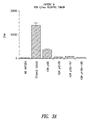
- FIG. 2 graphically illustrates the results of a low frequency event screening from a normal individual indicating that a CD4 + T cell response can be detected against p185 HER-2/neu and peptides derived from its amino acid sequence.
- the graph represents the data from one normal individual analyzed with the low frequency screening assayed described further below. Positive responses to the intact protein and two peptides were detected.
- FIG. 3 graphically illustrates that CD4 + T cells reactive to p185 HER-2/neu protein and peptides can be detected in high frequency from patients with HER-2/neu positive breast cancer and can also be detected in some patients with tumors that test negatively for expression of p185 HER-2/neu protein.
- Patient A had a primary tumor that tested negatively for overexpression of p185 HER-2/neu .
- the other three patients had HER-2/neu positive tumors.
- a proliferation assay was performed using purified peripheral blood mononuclear cells (PBMC) as described below, with each experimental group done in 24 well replicate. Two ⁇ 10 5 PBMC/well were incubated with no antigen, tetanus toxoid (5 ⁇ g/ml), p185 HER-2/neu (50 ⁇ g/ml), or HER-2/neu derived peptides (50 ⁇ g/ml) as described further below. After 4 days, wells were pulsed with 1 ⁇ Ci of tritiated thymidine ( 3 H-TdR) for 6-8 hours and then counted. The data represents the mean of 24 determinations of the c.p.m. with standard error bars expressed.
- PBMC peripheral blood mononuclear cells
- FIG. 4 panels A and B graphically illustrates that CD8 + CTL specific for HER-2/neu peptides 48-56 and 789-797 can be generated by in vitro immunization.
- Three ⁇ 10 7 PBMC from a homozygous HLA-A2 normal donor were incubated with p48-56 or p789-797 HER-2/neu peptides at concentrations of 10 ⁇ g/ml.
- the lymphocytes were tested for lytic activity after 10 in vitro sensitizations (IVS). Data is depicted after the tenth IVS with p48-56 (panel A) or with p789-797 (panel B).
- Target cells consisted of 51 Cr-labeled autologous EBV transformed B lymphocytes which had been incubated with p48-56 or p789-797 HER-2/neu or an irrelevant peptide for 2 hours prior to use.
- a four hour chromium release assay (CRA) was performed. The results represent the percent specific lysis at the indicated effector:target (E:T) ratio.
- Target controls of 51 Cr-labeled K562 and Daudi cells were also included to evaluate NK and LAK activity. The execution of the CRA is as described. The results represent the percent specific lysis at the indicated effector:target (E:T) ratio.
- FIG. 5 pictorially illustrates that antibodies are detectable against p185 HER-2/neu in the sera of a breast cancer patient.
- Lane 1 represents the immunoblot of p185 using a HER-2/neu positive breast cancer patient's sera (1:1000 dilution) as primary antibody. The blot was analyzed as described further below.
- Lane 2 represents the control strip from that experiment developed with c-neu antibody.
- FIG. 6 pictorially shows that antibodies in the sera of a breast cancer patient identify the same p185 band as does a known HER-2/neu-specific antibody (“control antibody”).
- a membrane preparation from NIH3T3 cells (a murine cell line) that had been transfected with HER-2/neu cDNA (“NIH3T3+H2N”) was tested against control antibody (lane A) or patient sera (lane D).
- a membrane preparation from untransfected cells (“NIH3T3”) was tested against control antibody (lane B) or patient sera (lane C).
- FIG. 7 pictorially illustrates that some breast cancer patients have antibodies directed to both the extracellular and intracellular domain of the HER-2/neu protein. Sera of breast cancer patients is tested against the extracellular domain (“ECD protein”) or the intracellular domain (“ICD protein”), in lanes A and B, respectively.
- ECD protein extracellular domain
- ICD protein intracellular domain
- FIG. 8 graphically illustrates that rats immunized with peptides derived from the intracellular domain (ICD) portion of rat neu protein develop antibody responses to neu protein.
- ICD intracellular domain
- FIG. 9 graphically illustrates that rats immunized with peptides derived from the extracellular domain (ECD) portion of rat neu protein develop antibody responses to neu protein.
- ECD extracellular domain
- FIG. 10 graphically shows that epitope analysis of ICD antibody responses demonstrates dominant B cell epitopes as well as “determinant spreading” between domains.
- ELISA analysis for peptide epitopes was performed. Each animal's sera was evaluated at dilutions of 1:25, 1:50, 1:100, and 1:200 for each peptide analyzed. Antibody responses titered with decreasing serum concentrations. All data shown is at a rat sera concentration of 1:50. Control sera analyzed was pooled sera from 5 non-immunized animals. Results were reproducible in 3 separately run assays.
- FIG. 11 graphically shows that epitope analysis of ECD antibody responses demonstrates dominant B cell epitopes.
- ELISA analysis for peptide epitopes was performed. Each animal's sera was evaluated at dilutions of 1:25, 1:50, 1:100, and 1:200 for each peptide analyzed. Antibody responses titered with decreasing serum concentrations. All data shown is at a rat sera concentration of 1:50. Control sera analyzed was pooled sera from 5 non-immunized animals. Results were reproducible in 3 separately run assays.
- FIG. 12 pictorially illustrates that antibodies elicited by immunization to either ICD or ECD peptides are specific for and can immunoprecipitate both rat neu protein and human HER-2/neu protein.
- Panel A shows the results of an immunoprecipitation experiment with immunized rat sera and lysates of DHFRG-8. Each sera was able to immunoprecipitate rat neu from the cell lysates. The immunoprecipitates were resolved on a 7.5% SDS-acrylamide gel and transferred to nitrocellulose. The blots were probed with primary antibody, c-neu-Ab-3, at a 1:1000 dilution. Control sera of an animal immunized with the adjuvant alone showed no evidence of reactivity to rat neu.
- Panel B depicts the results of an immunoprecipitation experiment with immunized rat sera and lysates of SKBR3, a source of human neu. Immunoblotting was performed in an identical manner and all experimental animal sera were able to immunoprecipitate human neu. The control sera, again, showed no evidence of reactivity.
- FIG. 13 pictorially illustrates that B cell epitopes that are cross reactive between human and rat neu are present in both domains of the protein. Shown here are the results of Western blot analysis of protein domain epitope mapping from representative animals in each immunized group.
- Animal 1.2 was immunized with the ECD group of peptides, animal 2.2 with the ICD group of peptides, and the control animal was immunized with adjuvant alone. Proteins were electrophoresed. After transfer to nitrocellulose the blots were incubated for 18 hours in rat sera at a 1:500 dilution. Antibody responses were detected with a second step goat anti-rat Ig HRP antibody at a dilution of 1:5000.
- FIG. 14 graphically illustrates that immunization of rats with ICD peptides elicits neu peptide-specific T cell responses.
- 2 ⁇ 10 5 immunized spleen cells were incubated with 25 ⁇ g/ml of the various peptides.
- the “Mix” group consisted of 25 ⁇ g/ml each of the immunizing peptides.
- a proliferation assay was performed. Each experimental group was done in 6 well replicates. The data is expressed in terms of a stimulation index (SI) which is the mean of the experimental wells divided by the mean of the control (no antigen) wells. Stimulation indices greater than 2 are considered to be indicative of a primed response. Animals immunized with adjuvant alone showed no stimulation index greater than 0.9 to any of the tested peptides (data not shown).
- SI stimulation index
- FIG. 15 graphically illustrates that immunization of rats with ICD peptides elicits neu protein-specific T cell responses.
- 1 ⁇ 10 5 cultured T cells derived from immunized spleen were incubated with 1 ⁇ 10 5 syngeneic spleen as APC (antigen presenting cells) and 1 ⁇ g/ml of purified rat neu protein.
- APC antigen presenting cells
- Each experimental group was done in 6 well replicates.
- the data is expressed in terms of a stimulation index which is the mean of the experimental wells divided by the mean of the control (no antigen) wells. Stimulation indices greater than 2 are considered to be indicative of a primed response.
- Wild type ras protein was the irrelevant protein used in the assay.
- FIG. 16 graphically shows that immunization of rats with ECD peptides elicits only weak peptide-specific T cell responses.
- 2 ⁇ 10 5 immunized spleen cells were incubated with 25 ⁇ g/ml of the various peptides.
- the “Mix” group consisted of 25 ⁇ g/ml each of the immunizing peptides.
- Each experimental group was done in 6 well replicates.
- the data is expressed in terms of a stimulation index which is the mean of the experimental wells divided by the mean of the control (no antigen) wells. Stimulation indices greater than 2 are considered to be indicative of a primed response.
- Animals immunized with adjuvant alone showed no stimulation index greater than 1.0 to any of the tested peptides (data not shown).
- FIG. 17 graphically shows that immunization of rats with ECD peptides elicits weak, but positive, responses to neu protein.
- 1 ⁇ 10 5 cultured T cells derived from immunized spleen or lymph nodes were incubated with 1 ⁇ 10 5 syngeneic spleen as APC and 1 ⁇ g/ml of purified rat neu protein.
- Each experimental group was done in 6 well replicates.
- the data is expressed in terms of a stimulation index which is the mean of the experimental wells divided by the mean of the control (no antigen) wells. Stimulation indices greater than 2 are considered to be indicative of a primed response.
- Wild type ras protein was the irrelevant protein used in the assay.
- HER-2/neu Protein refers to the p185 protein (also known as c-erbB2) and peptides thereof which are recognized by helper T cells or cytotoxic T cells; and may be naturally derived, synthetically produced, genetically engineered, or a functional equivalent thereof, e.g., where one or more amino acids are replaced by other amino acid(s) or non-amino acid(s) which do not substantially affect function.
- Proliferation of T cells includes the multiplication of T cells as well as the stimulation of T cells leading to multiplication, i.e., the initiation of events leading to mitosis and mitosis itself. Methods for detecting proliferation of T cells are discussed below.
- the present invention is directed toward methods and compositions for the diagnosis, monitoring and treatment of malignancies in a warm-blooded animal, wherein an amplified HER-2/neu gene is associated with the malignancies.
- Association of an amplified HER-2/neu gene with a malignancy does not require that the protein expression product of the gene be present on the tumor.
- overexpression of the protein expression product may be involved with initiation of a tumor, but the protein expression may subsequently be lost.
- An effective autochthonous immune response may convert a HER-2/neu positive tumor to HER-2/neu negative, but existent immunity will be present and allow diagnosis.
- the disclosure of the present invention shows that the protein expression product of the HER-2/neu gene can be recognized by thymus-dependent lymphocytes (hereinafter “T cells”) and, therefore, the autochthonous immune T cell response can be utilized to diagnose, monitor and treat malignancies in which such a protein is or has been overexpressed.
- T cells thymus-dependent lymphocytes
- the disclosure of the present invention also shows, in another aspect, that sera of patients with a malignancy, in which an amplified HER-2/neu oncogene is associated, contain antibodies to HER-2/neu protein.
- the autochthonous antibody response can be used to diagnose, monitor and treat malignancies in which such a protein is overexpressed.
- the two major arms of the immune system are: (1) cell-mediated immunity with immune T cells and (2) humoral immunity with antibodies. Further, the immune system normally functions to recognize and destroy any foreign or aberrant cells in the body. Since the HER-2/neu protein is expressed by some normal cells, tolerance and/or anergy (i.e., diminished reactivity to a specific antigen) is expected. Thus, it is surprising that, as disclosed within the present invention, both T cell and antibody responses to HER-2/neu are detected.
- CD4 + T cell populations are considered to function as helpers/inducers through the release of lymphokines when stimulated by a specific antigen; however, a subset of CD4 + cells can act as cytotoxic T lymphocytes (CTL).
- CD8 + T cells are considered to function by directly lysing antigenic targets; however, under a variety of circumstances they can secrete lymphokines to provide helper or DTH function.
- the phenotypic CD4 and CD8 markers are linked to the recognition of peptides bound to class II or class I MHC antigens.
- CD4 + and CD8 + T cells respond to different antigens or the same antigen presented under different circumstances.
- the binding of immunogenic peptides to class II MHC antigens most commonly occurs for antigens ingested by antigen presenting cells. Therefore, CD4 + T cells generally recognize antigens that have been external to the tumor cells.
- binding of peptides to class I MHC occurs only for proteins present in the cytosol and synthesized by the target itself, proteins in the external environment are excluded.
- An exception to this is the binding of exogenous peptides with a precise class I binding motif which are present outside the cell in high concentration.
- CD4 + and CD8 + T cells have broadly different functions and tend to recognize different antigens as a reflection of where the antigens normally reside.
- the protein product expressed by the HER-2/neu oncogene is recognized by T cells.
- T cells Such a protein expression product “turns over” within cells, i.e., undergoes a cycle wherein a synthesized protein functions and then eventually is degraded and replaced by a newly synthesized molecule.
- peptide fragments from the protein bind to major histocompatibility complex (MHC) antigens.
- MHC major histocompatibility complex
- T cells expressing a T cell receptor with high affinity binding of the peptide-MHC complex will bind to the peptide-MHC complex and thereby become activated and induced to proliferate.
- small numbers of immune T cells will secrete lymphokines, proliferate and differentiate into effector and memory T cells.
- the primary immune response will occur in vivo but has been difficult to detect in vitro. Subsequent encounter with the same antigen by the memory T cell will lead to a faster and more intense immune response.
- the secondary response will occur either in vivo or in vitro.
- the in vitro response is easily gauged by measuring the degree of proliferation, the degree of cytokine production, or the generation of cytolytic activity of the T cell population re-exposed in the antigen.
- Substantial proliferation of the T cell population in response to a particular antigen is considered to be indicative of prior exposure or priming to the antigen.
- a malignancy in which a HER-2/neu oncogene is associated may be detected.
- malignancies include breast, ovarian, colon, lung and prostate cancers.
- An immune response to the HER-2/neu protein, once generated, can be long-lived and can be detected long after immunization, regardless of whether the protein is present or absent in the body at the time of testing.
- prior exposure of a warm-blooded animal, such as humans, to the HER-2/neu protein can be detected by examining for the presence or absence of specific activation of CD4 + or CD8 + T cells.
- T cells isolated from an individual by routine techniques are incubated with HER-2/neu protein.
- T cells may be incubated in vitro for 2-9 days (typically 4 days) at 37° C. with HER-2/neu protein (typically, 5 ⁇ g/ml of whole protein or 25 ⁇ g/ml of an appropriate peptide or graded numbers of cells synthesizing HER-2/neu protein). It may be desirable to incubate another aliquot of a T cell sample in the absence of HER-2/neu protein to serve as a control.
- Specific activation of CD4 + or CD8 + T cells may be detected in a variety of ways.
- Methods for detecting specific T cell activation include detecting the proliferation of T cells, the production of cytokines (e.g., lymphokines), or the generation of cytolytic activity (i.e., generation of cytotoxic T cells specific for HER-2/neu protein).
- cytokines e.g., lymphokines
- cytolytic activity i.e., generation of cytotoxic T cells specific for HER-2/neu protein.
- CD4 + T cells a preferred method for detecting specific T cell activation is the detection of the proliferation of T cells.
- CD8 + T cells a preferred method for detecting specific T cell activation is the detection of the generation of cytolytic activity.
- T cell proliferation can be detected by measuring the rate of DNA synthesis.
- T cells which have been stimulated to proliferate exhibit an increased rate of DNA synthesis.
- a typical way to measure the rate of DNA synthesis is, for example, by pulse-labeling cultures of T cells with tritiated thymidine, a nucleoside precursor which is incorporated into newly synthesized DNA. The amount of tritiated thymidine incorporated can be determined using a liquid scintillation spectrophotometer.
- T cell proliferation examples include measuring increases in interleukin-2 (IL-2) production, Ca 2+ flux, or dye uptake, such as 3-(4,5-dimethylthiazol-2-yl)-2,5-diphenyl-tetrazolium.
- IL-2 interleukin-2
- dye uptake such as 3-(4,5-dimethylthiazol-2-yl)-2,5-diphenyl-tetrazolium.
- lymphokines such as interferon-gamma
- the relative number of T cells that can respond to intact p185 HER-2/neu protein or peptide may be quantified.
- Intact p185 HER-2/neu protein or peptides thereof which are recognized by cytotoxic T cells may be used within the present invention.
- the peptides may be naturally derived or produced based upon an identified sequence.
- the peptides for CD8 + T cell responses are generally 8-10 amino acids in length and the peptides for CD4 + T cell responses are longer, e.g., 15-18 amino acids in length.
- Peptides for CD8 + T cell responses vary according to each individual's class I MHC molecules.
- peptides appropriate for CD8 + T cell responses are peptides which are 8-10 amino acids in length and contain a leucine at position 2 and/or a leucine or valine at position 9.
- peptides designated by one letter abbreviations for amino acids and followed in parentheses by which residues of p185 they correspond
- suitable within the present invention for CD8 + T cell responses in individuals that are HLA-A2.1 include peptides consisting essentially of: HLYQGCQVV (p48-56) (Seq. ID No. 1); OLFEDNYAL (p106-114) (Seq. ID No.
- KIFGSLAFL p369-377) (Seq. ID No. 27); PLQPEQLQV (p391-399) (Seq. ID No. 2); PLTSIISAV (p650-658) (Seq. ID No. 3); ILLVVVLGV (p661-669) (Seq. ID No. 4); LLVVVLGW (p662-670) (Seq. ID No. 5); RLLQETELV (p689-697) (Seq. ID No. 6); ILDEAYVMAGV (p767-777) (Seq. ID No. 28); VMAGVGSPYV (p773-782) (Seq. ID No.
- CLTSTVQLV (p789-797) (Seq. ID No. 7); DLAARNVLV (p845-853) (Seq. ID No. 8); VLVKSPNHV (p851-859) (Seq. ID No. 9); TLSPGKNGV (p1172-1180) (Seq. ID No. 10); VLGVVFGIL (p666-674) (Seq. ID No. 11); LIKRRQQKI (p674-682) (Seq. ID No. 12); KIPVAIKVL (p747-755) (Seq. ID No. 13); ILDEAYVMA (p767-775) (Seq. ID No.
- QLMPYGCLL (p799-807) (Seq. ID No. 15); QIAKGMSYL (p829-836) (Seq. ID No. 16); LLNWCMQIA (p822-830) (Seq. ID No. 17); RLVHRDLAA (p840-848) (Seq. ID No. 18); DIDETEYHA (p871-879) (Seq. ID No. 19); DLLEKGERL (p933-941) (Seq. ID No. 20); TIDVYMLMV (p948-956) (Seq. ID No. 21); MIMVKCWMI (p953-961) (Seq. ID No.
- DLVDAEEYL (p1016-1024) (Seq. ID No. 23); GLEPSEEEA (p1062-1070) (Seq. ID No. 24); or YLTPQGGAA (p1196-1204) (Seq. ID No. 25).
- Peptides for CD4 + T cell responses vary according to each individual's class II MHC molecules.
- Examples of peptides suitable within the present invention for CD4 + T cell responses include peptides consisting essentially of: HLDMLRHLYQGCQVV (p42-56) (Seq. ID No. 30); PLQRLRIVRGTQLFE (p95-109) (Seq. ID No. 31); RLRIVRGTQLFEDNYAL (p98-114) (Seq. ID No. 61); LRSLTEILKGGVLIQ (p142-156) (Seq. ID No. 32); VTYNTDTFESMPNPE (p272-286) (Seq. ID No.
- NQEVTAEDGTQRCEK (p319-333) (Seq. ID No. 56); TQRCEKCSKPCARVCYGL (p328-345) (Seq. ID No. 60); HLREVRAVTSANIQE (p349-363) (Seq. ID No. 34); VRAVTSANIQEFAGC (p353-367) (Seq. ID No. 35); NIQEFAGCKKIFGSL (p360-374) (Seq. ID No. 36); KIFGSLAFLPESFDGD (p369-384) (Seq. ID No. 62); LQVFETLEEITGYLY (p397-411) (Seq. ID No.
- QVFETLEEITGYLYI (p398-412) (Seq. ID No. 37); QVIRGRILHNGAYSL (p429-443) (Seq. ID No. 57); QECVEECRVLQGLPR (p538-552) (Seq. ID No. 38); ASPLTSIISAVVGIL (p648-662) (Seq. ID No. 59); VVVLGVVFGILIKRR (p664-678) (Seq. ID No. 39); KYTMRRLLQETELVE (p684-698) (Seq. ID No. 40); RRLLQETELVEPLTPS (p688-703) (Seq. ID No.
- GAMPNQAQMRILKET (p704-718) (Seq. ID No. 41); VKVLGSGAFGTVYKG (p723-737) (Seq. ID No. 42); SPKANKEILDEAYVM (p760-774) (Seq. ID No. 43); GVGSPYVSRLLGICL (p776-790) (Seq. ID No. 44); SRLLGICLTSTVQLV (p783-797) (Seq. ID No. 45); GSQDLLNWCMQIAKG (p818-832) (Seq. ID No. 46); VKITDFGLARLLDID (p859-873) (Seq. ID No.
- peptides may be produced for use within the present invention, both for the HLA-A2.1 class I MHC molecule as well as for the other class I and class II molecules.
- a variety of techniques are well known for isolating or constructing peptides. Suitable peptides are readily identified based upon the disclosure provided herein. Additional suitable peptides include those which are longer in length.
- another peptide has an amino acid sequence corresponding to that disclosed in FIG. 1 beginning at about the lysine residue at amino acid position 676 and extending to about the valine residue at amino acid position 1255.
- Such a peptide may be extended (e.g., by the addition of one or more amino acid residues selected, for example, from position 675 to about position 646 of FIG. 1 ) and/or truncated (e.g., by the deletion of one or more amino acid residues from the carboxyl terminus which is position 1255 of FIG. 1 ).
- suitable peptides may be variations on other preferred peptides disclosed herein.
- variations on the peptide designated herein as p650-658 include the extension and/or truncation by the addition or deletion, respectively, of one or more amino acid residues beginning at either position 650 or position 658 or both positions.
- amino acids are removed from the amino terminus of p650-658 and four amino acids, such as the four adjacent to position 658, are added to its carboxyl terminus.
- this particular peptide variation results in a peptide with the same number of total amino acids (nine), a peptide variation on a preferred peptide need not be identical in length. Variations in amino acid sequence that yield peptides having substantially the same desired biological activity are within the scope of the present invention.
- CD4 + or CD8 + T cells that proliferate in the presence of HER-2/neu protein can be expanded in number either in vitro or in vivo. Proliferation of such T cells in vitro may be accomplished in a variety of ways. For example, the T cells can be re-exposed to HER-2/neu protein. It may be desirable to repeat the exposure of T cells to the HER-2/neu protein to induce proliferation. It may be further desirable to include T cell growth factors, such as interleukin-2, and/or stimulator cells which synthesize HER-2/neu protein. The addition of stimulator cells is preferred where generating CD8 + T cell responses.
- HER-2/neu protein-specific T cells can be grown to large numbers in vitro with retention of specificity in response to intermittent restimulation with the immunizing HER-2/neu protein.
- IVS primary in vitro stimulation
- large numbers of lymphocytes e.g., greater than 4 ⁇ 10 7
- HER-2/neu protein e.g., peptide at 10 ⁇ g/ml
- the flasks are incubated at 37° C. for 7 days.
- T cells are harvested and placed in new flasks with 2-3 ⁇ 10 7 irradiated peripheral blood mononuclear cells.
- HER-2/neu protein e.g., peptide at 10 ⁇ g/ml is added directly.
- the flasks are incubated at 37° C. for 7 days.
- 2-5 units of interleukin-2 (IL-2) is added.
- the T cells are placed in wells (e.g., 24 well plates).
- the T cells are stimulated with the individual's own EBV transformed B cells coated with the peptide.
- IL-2 is added on days 2 and 4 of each cycle. As soon as the cells are shown to be specific cytotoxic T cells, they are changed to a 10 day stimulation cycle with higher IL-2 (20 units) on days 2, 4 and 6 to expand them.
- one or more T cells that proliferate in the presence of HER-2/neu protein can be expanded in number by cloning.
- Methods for cloning cells are well known in the art.
- T cell lines may be established in vitro and cloned by limiting dilution.
- Responder T cells are purified from the peripheral blood of sensitized patients by density gradient centrifugation and sheep red cell rosetting and established in culture by stimulating with the nominal antigen in the presence of irradiated autologous filler cells.
- HER-2/neu protein is used as the antigenic stimulus and autologous peripheral blood lymphocytes (PBL) or lymphoblastoid cell lines (LCL) immortalized by infection with Epstein Barr virus are used as antigen presenting cells.
- PBL peripheral blood lymphocytes
- LCL lymphoblastoid cell lines
- autologous antigen-presenting cells transfected with an expression vector which produces relevant HER-2/neu protein may be used as stimulator cells.
- Established T cell lines are cloned 2-4 days following antigen stimulation by plating stimulated T cells at a frequency of 0.5 cells per well in 96-well flat-bottom plates with 1 ⁇ 10 6 irradiated PBL or LCL cells and recombinant interleukin-2 (rIL2) (50 U/ml).
- Wells with established clonal growth are identified at approximately 2-3 weeks after initial plating and restimulated with appropriate antigen in the presence of autologous antigen-presenting cells, then subsequently expanded by the addition of low doses of rIL2(10 U/ml) 2-3 days following antigen stimulation. T cell clones are maintained in 24-well plates by periodic restimulation with antigen and rIL2 approximately every two weeks.
- the T cells may be administered to the individual as an anti-cancer composition in an amount effective for therapeutic attack against a tumor.
- a patient's own T cells autochthonous T cells
- T cells/M 2 can be used as reagents to mediate specific tumor therapy.
- about 1 ⁇ 10 9 to 1 ⁇ 10 11 T cells/M 2 will be administered intravenously or intracavitary, e.g., in pleural or peritoneal cavities, or in the bed of a resected tumor. It will be evident to those skilled in the art that the number and frequency of administration will be dependent upon the response of the patient.
- Pharmaceutically suitable carriers or diluents for T cells include physiological saline or sera. It will be recognized by one skilled in the art that the composition should be prepared in sterile form.
- T cells may also be proliferated in vivo.
- immunization of an individual with a HER-2/neu peptide i.e., as a vaccine
- about 0.01 ⁇ g/kg to about 100 mg/kg body weight will be administered by the intradermal, subcutaneous or intravenous route.
- a preferred dosage is about 1 ⁇ g/kg to about 1 mg/kg, with about 5 ⁇ g/kg to about 200 ⁇ g/kg particularly preferred. It will be evident to those skilled in the art that the number and frequency of administration will be dependent upon the response of the patient.
- HER-2/neu peptide may be administered repetitively. It will be evident to those skilled in this art that more than one HER-2/neu peptide may be administered, either simultaneously or sequentially. For example, a combination of about 8-15 peptides may be used for immunization.
- Preferred peptides for immunization are those that include all or a portion of the amino acid sequence shown in FIG. 1 beginning at about the lysine residue at amino acid position 676 and extending to about the valine residue at amino acid position 1255. One or more peptides from other portions of the amino acid sequence shown in FIG. 1 may be added to one or more of the preferred peptides.
- p185 HER-2/neu protein nor a peptide having the amino acid sequence of its entire extracellular domain (i.e., a peptide having an amino acid sequence of the entire amino acid sequence shown in FIG. 1 up to amino acid position 650, plus or minus about one to five positions, and with or without the first 21 amino acid positions) are used alone for immunization.
- HER-2/neu peptide which functions as an antigen
- other components in the vaccine such as a vehicle for antigen delivery and immunostimulatory substances designed to enhance the protein's immunogenicity.
- vehicles for antigen delivery include aluminum salts, water-in-oil emulsions, biodegradable oil vehicles, oil-in-water emulsions, biodegradable microcapsules, and liposomes.
- immunostimulatory substances examples include N-acetylmuramyl-L-alanine-D-isoglutamine (MDP), lipopoly-saccharides (LPS), glucan, IL-12, GM-CSF, gamma interferon and IL-15.
- MDP N-acetylmuramyl-L-alanine-D-isoglutamine
- LPS lipopoly-saccharides
- glucan examples include N-acetylmuramyl-L-alanine-D-isoglutamine (MDP), lipopoly-saccharides (LPS), glucan, IL-12, GM-CSF, gamma interferon and IL-15.
- LPS lipopoly-saccharides
- glucan IL-12
- GM-CSF gamma interferon and IL-15.
- a carrier substance such as keyhole limpet hemocyanin.
- the present invention also discloses that HER-2/neu protein, in addition to being immunogenic to T cells, appears to stimulate B-cells to produce antibodies capable of recognizing HER-2/neu protein. Detection of such antibodies provides another way to diagnose a malignancy in which a HER-2/neu oncogene is associated with the malignancy.
- Antibodies specific i.e., which exhibit a binding affinity of about 10 7 liters/mole or better
- HER-2/neu protein may be found in a variety of body fluids including sera and ascites. Briefly, a body fluid sample is isolated from a warm-blooded animal, such as a human, for whom it is desired to determine whether antibodies specific for HER-2/neu are present.
- the body fluid is incubated with HER-2/neu protein under conditions and for a time sufficient to permit immunocomplexes to form between the protein and antibodies specific for the protein.
- a body fluid and HER-2/neu protein may be incubated at 4° C. for 24-48 hours.
- the reaction mixture is tested for the presence of immunocomplexes. Detection of one or more immunocomplexes formed between HER-2/neu protein and antibodies specific for HER-2/neu protein may be accomplished by a variety of known techniques, such as radioimmunoassays (RIA) and enzyme linked immunosorbent assays (ELISA).
- RIA radioimmunoassays
- ELISA enzyme linked immunosorbent assays
- Suitable immunoassays include the double monoclonal antibody sandwich immunoassay technique of David et al. (U.S. Pat. No. 4,376,110); monoclonal-polyclonal antibody sandwich assays (Wide et al., in Kirkham and Hunter, eds., Radioimmunoassay Methods , E. and S. Livingstone, Edinburgh, 1970); the “western blot” method of Gordon et al. (U.S. Pat. No. 4,452,901); immunoprecipitation of labeled ligand (Brown et al., J. Biol. Chem .
- HER-2/neu protein (“antigen”) may either be labeled or unlabeled.
- the antigen find use in agglutination assays.
- unlabeled antigen can be used in combination with labeled molecules that are reactive with immunocomplexes, or in combination with labeled antibodies (second antibodies) that are reactive with the antibody directed against HER-2/neu protein, such as antibodies specific for immunoglobulin.
- the antigen can be directly labeled.
- the reporter group can include radioisotopes, fluorophores, enzymes, luminescers, or dye particles.
- antigen is adsorbed to the surface of a microtiter well. Residual protein-binding sites on the surface are then blocked with an appropriate agent, such as bovine serum albumin (BSA), heat-inactivated normal goat serum (NGS), or BLOTTO (buffered solution of nonfat dry milk which also contains a preservative, salts, and an antifoaming agent).
- BSA bovine serum albumin
- NGS heat-inactivated normal goat serum
- BLOTTO buffered solution of nonfat dry milk which also contains a preservative, salts, and an antifoaming agent.
- the well is then incubated with a sample suspected of containing specific antibody.
- the sample can be applied neat, or, more often, it can be diluted, usually in a buffered solution which contains a small amount (0.1%-5.0% by weight) of protein, such as BSA, NGS, or BLOTTO.
- the well After incubating for a sufficient length of time to allow specific binding to occur, the well is washed to remove unbound protein and then incubated with an anti-species specific immunoglobulin antibody labeled with a reporter group.
- the reporter group can be chosen from a variety of enzymes, including horseradish peroxidase, beta-galactosidase, alkaline phosphatase, and glucose oxidase. Sufficient time is allowed for specific binding to occur, then the well is again washed to remove unbound conjugate, and the substrate for the enzyme is added. Color is allowed to develop and the optical density of the contents of the well is determined visually or instrumentally.
- a reporter group is bound to HER-2/neu protein.
- the step of detecting immunocomplexes involves removing substantially any unbound HER-2/neu protein and then detecting the presence or absence of the reporter group.
- a reporter group is bound to a second antibody capable of binding to the antibodies specific for HER-2/neu protein.
- the step of detecting immunocomplexes involves (a) removing substantially any unbound antibody, (b) adding the second antibody, (c) removing substantially any unbound second antibody and then (d) detecting the presence or absence of the reporter group.
- the second antibody is an anti-human antibody.
- a reporter group is bound to a molecule capable of binding to the immunocomplexes.
- the step of detecting involves (a) adding the molecule, (b) removing substantially any unbound molecule, and then (c) detecting the presence or absence of the reporter group.
- An example of a molecule capable of binding to the immunocomplexes is protein A.
- Reporter groups suitable for use in any of the methods include radioisotopes, fluorophores, enzymes, luminescers, and dye particles.
- detection of immunocomplexes formed between HER-2/neu protein and antibodies in body fluid which are specific for HER-2/neu protein may be used to monitor the effectiveness of cancer therapy for a malignancy in which the HER-2/neu oncogene is associated.
- Samples of body fluid taken from an individual prior to and subsequent to initiation of therapy may be analyzed for the immunocomplexes by the methodologies described above. Briefly, the number of immunocomplexes detected in both samples are compared. A substantial change in the number of immunocomplexes in the second sample (post-therapy initiation) relative to the first sample (pre-therapy) reflects successful therapy.
- CD4+T Cells Responsive to p185(HER-2/neu) Protein and Peptides can be Detected in Higher Frequency in Patients with Breast Cancer than Normal Individuals
- Soluble proteins are classically processed in the class II MHC pathway.
- p185 HER-2/neu protein is a transmembrane protein present at the cell surface. When overexpressed, it has been found to be soluble and in the extracellular environment both in vitro and in vivo.
- In vitro studies of human breast cancer cell lines found the extracellular domain of p185 HER-2/neu in culture media of rapidly growing cells (Alper et al., Cell Growth and Differentiation 1:591-599, 1990; Zabrecky et al., J. Biol. Chem . 266:1716-1720, 1991).
- In vivo studies identified circulating portions of the protein in the sera of patients with breast cancer (Leitzel et al., J. Clin. Oncol . 10:1436-1443, 1992; Mori et al., Jpn. J. Cancer Res . 81:489-494, 1990).
- T Sites A protein sequence analysis package, T Sites, that incorporates several computer algorithms designed to distinguish potential sites for T cell recognition was used (Feller and de la Cruz, Nature 349:720-721, 1991). Two searching algorithms were used: (1) the AMPHI algorithm described by Margalit (Feller and de la Cruz, Nature 349:720-721, 1991; Margalit et al., J. Immunol .
- Peptides each 15 amino acids in length, that encompass both the AMPHI and Rothbard motifs were constructed.
- the optimal peptide length for class II MHC binding depends upon the particular MHC molecule and may be shorter than 15 amino acids. However, class II MHC responses to exogenous peptides allow for endocytosis and intracellular processing of longer peptides.
- One of the synthetic peptides (p42-56), HLDMLRHLYQGCQVV (Seq. ID No. 30), is located in the extracellular domain and has 33% homology to epidermal growth factor receptor (EGFR).
- Two other synthetic peptides, SRLLGICLTSTVQLV (p783-797) (Seq. ID No.
- TLERPKTLSPGKNGV p1166-1180
- p1166-1180 TLERPKTLSPGKNGV
- p185(HER-2/neu) Protein can be Obtained and Purified from the Human Breast Adenocarcinoma Cell Line SKBR3
- SKBR3 has been reported on extensively in the literature as a commonly used standard cell line with increased HER-2/neu gene copy number and HER-2/neu protein overexpression. In one study, SKBR3 cells were found to contain a mean HER-2/neu oncogene copy number of 43 copies/cell compared with 2.5 copies/cell for MCF-7, a breast cancer cell line considered to be a standard cell line without HER-2/neu gene amplification (Kallionieme et al., Proc. Nat. Acad. Sci. USA 89:5321-5325, 1992).
- SKBR3 is reported to be one of the highest known expressors of p185 HER-2/neu protein by immunohistochemistry, 4+ compared to 1+ in MCF-7 (Kerns et al., J. Histochem . & Cytochem . 38:1823-1830, 1990).
- the same HER-2/neu bands as described in the literature were validated in the present experiments by Western analysis. Bands detected included p185, p105 (extracellular domain), and several smaller bands that presumably represent fragments of phosphorylated protein (Alper et al., Cell Growth and Differentiation 1:591-599, 1990; Zabrecky et al., J. Biol. Chem . 266:1716-1720, 1991; Stern et al., Mol. Cell. Biol . 8:3969-3973, 1988).
- the antibodies used for detecting the HER-2/neu protein immunoblotting were commercially prepared by Oncogene Science (Manhasset, N.Y.).
- the antibody most commonly used in the present experiments was c-neu Ab-3; derived by immunization of BALB/c mice with a peptide sequence, TAENPEYLGLDVPV (Seq. ID No. 65), from the carboxyl domain of human c-neu gene product, and fusion of mouse splenocytes with SP2/0 myeloma cells.
- a second antibody, c-neu AB-1 gave very faint bands when compared with c-neu Ab-3.
- This antibody was a polyclonal rabbit affinity purified antibody against the peptide sequence, LARLLDIDETEYAD (Seq. ID No. 66), from the kinase domain of the human c-neu gene product.
- Transmembrane p185 HER-2/neu protein was purified from the cell membrane fraction of SKBR3 by modifications of described methods for other membrane-associated proteins (Dhut et al, Leukemia 4:745-750, 1990; Montgomeryzner et al., J. Exp. Med . 165:1041-1057, 1987).
- Three ⁇ 10 6 SKBR3 cells were harvested and suspended in phosphate buffered saline (PBS) with the following protease inhibitors; 1 mM PMSF, 1 mM benzamidine, 5 ⁇ g/ml aprotinin. All procedures were done on ice or at 4° C.
- PBS phosphate buffered saline
- the cells were then disrupted by sonication at 75W for a total of 1 minute using a high intensity sonifier equipped with a microtip (Branson Instruments, Inc., Stamford, Conn.). The resulting suspension was then centrifuged for 1 hour at 35,000 rpm to sediment membranes from cytosolic fraction. The membrane pellet was washed in ice cold PBS with protease inhibitors and the cycle of sonication/centrifugation was repeated twice. All cytosolic (supernatant) and membranous fractions were tested for the presence of p185 HER-2/neu by Western analysis. The protein was noted to be strongly concentrated in the membrane fraction.
- Protein concentration of one of these enriched membrane pellets was determined to be 2625 ⁇ g/ml (Protein BioRad assay).
- p185 HER-2/neu is an estimated 8% of membrane protein in SKBR3 (Leitzel et al., J. Clin. Oncol . 10:1436-1443, 1992); therefore, an estimated 210 ⁇ g of p185 HER-2/neu were present in the membrane pellet from 3 ⁇ 10 6 SKBR3 cells.
- the membrane preparation may be further enriched for p185 HER-2/neu , e.g., by immunoprecipitation. Briefly, 1 ⁇ g of c-neu 3 antibody and 15 ⁇ l protein A agarose were added to the sonicated membrane pellet. The mixture was incubated at 4° C. on a rocker for 24 hours. The immunoprecipitate was collected by centrifugation in a micro-centrifuge at 2500 rpm for 15 minutes at 4° C., and the resulting pellet was washed several times with PBS, 1% Trition X-100, 0.5% sodium deoxycholate, and 0.1% sodium dodecyl sulfate. Silver stain and Western analysis showed increased concentration of p185 HER-2/neu protein and decreases in extraneous membrane proteins when compared to membrane enriched pellets alone.
- CD4+T Cells Reactive to p185(HER-2/neu) Protein can be Elicited from PBL of Normal Individuals by Using an Assay Designed for Detecting Low Frequency Lymphocyte Precursors
- a standard proliferation assay a screening method for low frequency events
- LDA limiting dilution assay
- Conventional proliferative assays are capable of readily detecting primed responses.
- the proliferative response stimulation index provides a rough correlation with precursor frequency of antigen-reactive T cells. Any specific proliferative response detected from PBL is considered to be a primed response.
- the assay system developed for detecting low lymphocyte precursor frequency responses (described below) is used. This assay is simple and cost-effective. In circumstances in which more precision is needed, the precursor frequency is validated by limiting dilution assays (Bishop and Orosz, Transplantation 47:671-677, 1989).
- primed CD4 + T cell responses can be detected in conventional proliferative assays, whereas unprimed responses are not detectable in the same assays. Detection of small numbers of unprimed T cells is limited by confounding background thymidine uptake including the autologous mixed lymphocyte response (AMLR) to self MHC antigen plus responses to processed self serum proteins and exogenously added serum proteins.
- AMLR autologous mixed lymphocyte response
- an assay system for low frequency responses based on Poisson sampling statistics was used (In: Pinnacles , Chiron Corporation, 1:1-2, 1991). This type of analysis applies specifically to low frequency events in that, if the precursor frequency is less than the number of cells in one replicate culture, many replicates are required to detect a statistically significant number of positives. Theoretically, the analysis will correct for autologous responses by setting up a known positive control (such as PHA or tetanus toxoid) and known negative control (no antigen) and evaluating all data points from lowest to highest irrespective of the experimental group to which they belong.
- a known positive control such as PHA or tetanus toxoid
- known negative control no antigen
- M arithmetic mean
- SD standard deviation.
- CD4+T Cells Reactive to p185(HER-2/neu) and Peptide can be Detected in the Peripheral Blood of Patients with HER-2/neu Positive Breast Cancer in Levels Consistent with a Primed Response
- CD8 + CTL Specific for HER-2/neu Peptides can be Generated from PBL of Normal Individuals by Primary in Vitro Immunization to Synthetic Peptides Derived from the Normal Amino Acid Sequence of p185(HER-2/neu) Protein
- CD8 + T cells recognize peptide bound to class I MHC molecules.
- peptide determinants are derived from endogenously synthesized proteins. The rules which determine the ability of a protein to be processed and complexed with class I MHC molecules are not completely understood. Recently, however, it has been determined that peptides binding to particular MHC molecules share discernible sequence motifs (Falk et al., Nature 351:290-296, 1991).
- a peptide motif for binding in the groove of HLA-A2.1 has been defined by Edman degradation of peptides stripped from HLA-A2.1 molecules of a cultured cell line (Table 2, from Falk et al., supra).
- the method identified the typical or average HLA-A2.1 binding peptide as being 9 amino acids in length with dominant anchor residues occurring at positions 2 (L) and 9 (V). Commonly occurring strong binding residues have been identified at positions 2 (M), 4 (E,K), 6 (V), and 8 (K). The identified motif represents the average of many binding peptides.
- the derived peptide motif as currently defined is not particularly stringent. Some HLA-A2.1 binding peptides do not contain both dominant anchor residues and the amino acids flanking the dominant anchor residues play major roles in allowing or disallowing binding. Not every peptide with the current described binding motif will bind, and some peptides without the motif will bind. However, the current motif is valid enough to allow identification of some peptides capable of binding.
- the p185 HER-2/neu protein contains a substantial number of peptides with amino acid sequences possibly appropriate for binding to the class I MHC antigen HLA-A2.1.
- Evaluation of the 1255 aa structure of p185 HER-2/neu revealed at least 19 peptide segments of 9 aa in length that contained at least one of the dominant anchor residues.
- the current HLA-A2.1 motif places 6 amino acids between the dominant anchor amino acids at residues 2 and 9.
- Recent studies show that alterations in secondary structure of peptides can sometimes allow for additional intervening residues, and thus longer binding peptides. In the present experiment, 9-mer peptides were evaluated. The 10 peptides with both dominant residues were considered.
- T2 is a human T-B cell hybrid that has a large homozygous deletion within the MHC gene region (Riberdy and Cresswell, J. Immunol . 148:2586-2590, 1992; Trousdale et al., Nature 348:741-744, 1990; Spies et al., Nature 348:744-747, 1990).
- T2 determine HLA-A2.1 binding peptides has been well defined. T2 does not appropriately process endogenous antigen for presentation with class I MHC molecules. Consequently, cell surface expression of class I MHC molecules is markedly reduced. However, provision of exogenous peptides which bind to and stabilize class I MHC in the presence of B2 microglobulin results in increased levels of class I at cell surface which can be easily detected by immunofluorescent staining. T2 without exogenous peptide has low expression of HLA-A2 (30%-50%). When incubated with peptides able to bind A2, the level of class I MHC stabilizes on the cell surface and can be measured by immunofluorescent staining. Thus, the T2 line fails to present internal proteins in the class I pathway, but can bind exogenous peptides, providing that the exogenous peptides have the appropriate HLA-A2.1 binding motif.
- CD8 + CTL Specific for HER-2/neu p48-56 and p789-797 can be Generated by Primary in Vitro Immunization
- Conditions for detecting immunity to standard recall antigens were used and no peptide-specific CTL could be detected.
- a set of conditions were derived which have allowed priming to 4 of 4 of the binding peptides tested to date. The conditions were derived by empiric experimentation but are consistent with the current paradigm. Conditions include: (1) large numbers of T cells; (2) a concurrent stimulated primed CD4 + T cell response; (3) IL-2 added late to culture in very small amounts; and (4) multiple restimulations.
- lymphocytes After leukapheresis of a normal homozygous HLA-A2 individual, bulk cultures of lymphocytes (3 ⁇ 10 7 ) were incubated with peptide in a concentration of 10 ⁇ g peptide/ml. An individual homozygous for HLA-A2.1 was used on the presumption that a double dose of the MHC/peptide complex would allow more effective priming. Large numbers of lymphocytes were used to overcome the presumed low frequency of precursors. Generation of CD8 + CTL responses has long been known to require concurrent stimulation of CD4 + T cell responses to provide help/amplification. Both peptides used were chosen for class I MHC binding, and presumably could not stimulate CD4 + helper T cells.
- tetanus toxoid concentrations (5 ⁇ g/ml) of tetanus toxoid were added to culture along with peptide. So as not to overwhelm or dominate the culture with the tetanus toxoid response, titrations of tetanus toxoid had previously been assessed in a standard proliferation assay with the donor's lymphocytes and the concentration of tetanus toxoid that provided the lowest detectable stimulation index was used.
- IL-2 low doses added late to culture were used to maintain lymphocyte proliferation.
- standard conditions for expanding in vivo primed CTL following secondary sensitization in vitro usually have included IL-2 at 5-10 U/ml on day 2 of stimulation.
- similar concentrations of IL-2 induced expansion of nonspecifically lytic NK and T cells, presumably due to the predominance of NK cells and AMLR responsive cells relative to peptide-specific CTL.
- the T cell culture received no IL-2 for the first 10 days, with only 1 unit/ml administered on day +2 after the second IVS.
- IL-2 at 2 U/ml could be administered on day +2 and day +4 of the 7 day stimulation cycle.
- T cells were stimulated with peptide on irradiated PBL as APC every 7 days. Evaluation for specific lytic function was performed after the fourth IVS and revealed specific lytic activity but substantial non-specific lytic NK and T cell activity. Routine 51 Cr release assay performed after the tenth IVS ( FIG. 4 ) revealed greater than 50% lysis for both bulk T cell lines. Lysis against control targets of K562 and Daudi was less than 2%.
- Antibodies Directed against HER-2/neu Protein can be Detected in the Sera of Patients with Breast Cancer
- the sera of 20 patients with breast cancer were analyzed.
- the 20 patients were participants from the Fred Hutchinson Cancer Research Center, Division of Epidemiology WISH study.
- the patient population consisted of women recently diagnosed with breast cancer, generally less than 3 months from surgery. Their age was less than 55 and their HER-2/neu tumor status was unknown.
- Anti-p185 antibody was found in 55% of the group as evidenced by bands corresponding to the positive control.
- Antibody analysis was based on a modification of standard Western blotting techniques (Laemmli, Nature 227:680-685, 1970; Burnett, Anal. Biochem . 112:195-203, 1981).
- a 7.5% SDS polyacrylamide gel was poured with a single 12 cm long comb in the stacking gel to create a “trough.”
- Two immunoprecipitated SKBR3 membrane preparations, described above, were dissolved in loading buffer and layered across the trough. The gel was then run in standard fashion resulting in a band of equally distributed proteins across the gel.
- the protein was transferred to nitrocellulose (Amersham Hybond) for subsequent immunoblotting and development by chemiluminescence methods (Amersham ECL).
- the nitrocellulose was cut lengthwise into 25 equal strips and placed in a 25 well incubation tray.
- the nitrocellulose strips were then blocked with Tris buffered saline and 1% bovine serum albumin (TBS BSA) for 1 hour. This allows for analysis of 23 patients with 2 control strips.
- TBS BSA bovine serum albumin
- Patient sera is used as primary antibody, and after blocking, the strips are incubated for 24 hours at 4° C. with sera diluted 1:200 and 1:400 in TBS BSA.
- the second antibody is a goat antihuman HRP conjugate which will interact with the chemiluminescent developing reagent (Amersham ECL) resulting in light emission which can be photographed.
- a control strip is developed with c-neu Ab3 antibody previously described in a similar fashion with this assay both IgA and IgG antibody specific for p185 were detected.
- Patient sera identified the same p185 band ( FIG. 5 ) as did the known HER-2/neu-specific antibody, providing evidence that some patients have existent antibody immunity to HER-2/neu.
- Rats used in this study were Fischer strain 344 (CDF (F-344)/CrlBR) (Charles River Laboratories, Portage Mich.). Animals were maintained at the University of Washington Animal facilities under specific pathogen free conditions and routinely used for experimental studies between 3 and 4 months of age.
- peptides were constructed, derived from the amino acid sequence of the rat neu protein.
- the peptides 15-18 amino acids in length, were highly homologous to the human HER-2/neu peptide sequence. These peptides were chosen based on an increased probability of interaction with human Class II MHC molecules. This theoretical potential was evaluated by the use of a protein sequence analysis package, TSites, that incorporates several computer algorithms designed to distinguish potential sites for T cell recognition (Feller and de la Cruz, Nature 349:720-721, 1991).
- Several peptides identified from the rat sequence were predicted to have potential for class II interaction with both human and murine MHC.
- Nine peptides were chosen for immunization of the rats (Table 5).
- peptides were synthesized and purified by H. Zabrowski (University of Washington, Seattle, Wash.), then dissolved in phosphate-buffered saline (PBS), pH 7.4, to give 2 mg/ml stock solutions. Prior to aliquoting, peptides were sterile filtered, then stored at ⁇ 70° C.
- PBS phosphate-buffered saline
- One group of rats was immunized with a mixture of extracellular domain (ECD) peptides and one group with a mixture of intracellular domain (ICD) peptides.
- the final group received adjuvant alone.
- Peptides were administered at a final concentration of 100 ⁇ g each in a total volume of 200 ⁇ l.
- the animals underwent 3 immunizations each 14-16 days apart with either CFA or IFA as adjuvant (Sigma ImmunoChemicals, St. Louis, Mo.). 16 days after the third immunization sera was obtained for assessment of immune response.
- SKBR3 a human breast cancer cell line that is a marked overexpressor of HER-2/neu (American Type Culture Collection, Rockville, Md.), was maintained in culture in 10% fetal bovine serum (FBS) (Gemini Bioproducts, Inc., Calabasas, Calif.) and RPMI.
- FBS fetal bovine serum
- DHFR-G8 an NIH/3T3 cell line cotransfected with cneu-p and pSV2-DHFR (American Type Culture Collection, Rockville, Md.), was used as a source of non-transforming rat neu protein (Bernards et al., Proc. Natl. Acad. Sci. USA 84:6854-6858, 1987).
- This cell line was maintained in 10% FBS and Dulbecco's modified Eagle's medium with 4.5 g/L glucose.
- DHFR-G8 cells were passaged through the same medium supplemented with 0.3 ⁇ M methotrexate at every third passage to maintain the neu transfectant.
- Lysates of both SKBR3 and DHFR-G8 were prepared and used as a source of protein for both ELISA and immunoprecipitation studies. Briefly, a lysis buffer consisting of tris base, sodium chloride and Triton-X (1%) pH 7.5 was prepared. Protease inhibitors were added; aprotinin (1 ⁇ g/ml), benzamidine (1 mM) and PMSF (1 mM). 1 ml of the lysis buffer was used to suspend 10 7 cells. The cells were vortexed for 15 seconds every 10 minutes for an hour until disrupted. All procedures were performed on ice in a 4° C. cold room. After disruption the cells were microfuged at 4° C. for 20 minutes. Supernatant was removed from cell debris and stored in small aliquots at ⁇ 70° C. until used. Presence of human and rat neu in the lysates was documented by Western blot analysis.
- HRP horseradish peroxidase
- Control wells consisting of varying dilutions of c-neu-Ab-1, a rabbit polyclonal antibody directed against the kinase portion of human neu which also has reactivity to rat neu (Oncogene Science, Uniondale, N.Y.), were used as a positive control. These wells received a second step antibody of goat-anti rabbit HRP at a 1:5000 dilution (Amersham Co.). Following the final wash, TMB (Kirkegaard and Perry Laboratories, Gaithersburg, Md.) developing reagent was added. Color reaction was read at an optical density of 640 nm until the positive control wells reached 0.3 OD. The reaction was stopped with 1N HCl and the optical density was read at 450 nm. The OD of each serum dilution was calculated as the OD of the neu coated wells minus the OD of the PBS/1% BSA coated wells. A pool of 5 normal rat sera was run on each plate as a negative control.
- mice serum was used to immunoprecipitate human neu from the SKBR3 cell line and rat neu from the DHFR-G8 cell line.
- Sera from 5 pooled normal rats and 2 rats immunized with adjuvant alone and no peptide antigens were used as 2 negative controls.
- DHFR-G8 or SKBR3 lysate was incubated with 75 ⁇ l of rat sera or 10 ⁇ l (1 ⁇ g) of neu specific monoclonal antibody and 15 ⁇ l of protein A+G (Oncogene Science). The solution was rocked gently overnight at 4° C. After this incubation, the agarose was pelleted and washed twice in a tris HCl/EDTA buffer (1M Tris HCl pH 7.5, 0.25 M EDTA, and 5M NaCl), then twice in the same buffer with NP-40 added to a 0.5% concentration. The immunoprecipitates were analyzed by Western blot as described above using c-neu-Ab-1 (Oncogene Science) as the primary antibody. This antibody is a neu specific polyclonal rabbit antibody which cross reacts with both human and rat neu.
- Immunoprecipitates of SKBR3 and DHFR-G8 were used as a source of human and rat neu proteins in the Western assays.
- Recombinant human ECD and ICD (kindly provided by Drs. B. Groner and N. Lydon) were used to evaluate antibody responses to the neu domains. 7.5% polyacrylamide gels were electrophoresed in the Pharmacia Phast System (Pharmacia LKB Biotechnology AB, Uppsala, Sweden). After transfer to nitrocellulose (Hybond-C, Amersham Co.) the neu proteins were identified by immunoblot in a similar manner. All control blots were developed by using the IgG1 mouse monoclonal primary antibody, c-neu-Ab-3 (Oncogene Science).
- This antibody cross reacts with both rat and human neu.
- the primary antibody was used in a 1:1000 dilution with tris-buffered saline/1% BSA/0.1% Nonidet P-40.
- a polyclonal rabbit antimouse HRP-conjugated second antibody (Amersham Co.) was used in a 1:10,000 dilution.
- the blot was then developed using a chemiluminescent reaction (Amersham ECL). Identically run experimental blots were analyzed with rat sera as primary antibody. The sera were used in a 1:500 dilution with tris-buffered saline/1% BSA/0.1% Nonidet P-40 in an overnight incubation with the blot a 4° C.
- Fresh spleen or lymph node cells were harvested by mechanical disruption and passage through wire mesh and washed. 2 ⁇ 10 5 spleen cells/well and 1 ⁇ 10 5 lymph node cells/well were plated into 96-well round bottom microtiter plates (Corning, Corning, N.Y.) with 6 replicates per experimental group.
- the media used consisted of EHAA 120 (Biofluids) with L-glutamine, penicillin/streptomycin, 2-mercaptoethanol, and 5% FBS. Cells were incubated with 25 ⁇ g/ml of the various peptides. The group incubated with the peptide mix received 25 ⁇ g of each of the peptides.
- stimulation index SI which is defined as the mean of the experimental wells divided by the mean of the control wells (no antigen).
- SI stimulation index
- PBMC peripheral blood mononuclear cells
- the cultures were restimulated on the peptide mix every 14 days with syngeneic spleen that had been preincubated with the peptide mix for 2 hours, irradiated to 1000 rads, and then washed. Stimulator to effector ratio was 1:1 in each culture. After the second week in culture, media was supplemented with 50% Con A conditioned media. At the end of 3 in vitro stimulations, cells were >98% CD3+.
- Rats were immunized with mixtures of either 4 ICD peptides or 5 ECD peptides. Following the third immunization, serum and T cells from immunized rats were assessed for immunity to neu peptides and protein. Initial experiments assessed rats immunized with ICD peptides for antibody responses to whole neu protein. Serum antibody responses were analyzed by ELISA ( FIG. 8 ). The results demonstrate that immunization to ICD peptides elicited antibody to whole neu protein. Sera was analyzed at 1:25, 1:50, 1:100, and 1:200 dilution. Results at the 1:25 dilution are depicted ( FIG. 8 ). Neu specific antibody responses titered rapidly and at a 1:200 dilution the experimental sera demonstrated the same level of response as control. Isotype analysis revealed that the antibody responses were predominantly IgG (data not shown).
- Rats immunized to the mixture of ICD peptides displayed significant antibody responses to ECD peptides. Responses in individual rats varied. Rat 2.2 had substantial responses to all five ECD peptides evaluated. Rats 2.1 and 2.3 had weaker responses. Thus, immunization to ICD peptides elicited antibody responses to ICD peptides as well as “determinant spreading” with the generation of antibody responses to the ECD portion of the molecule. Rats immunized with adjuvant alone did not develop T cell responses to any tested peptide.
- the immunizing rat neu peptides were homologous with the human HER-2/neu protein sequence.
- the anti-peptide antibodies elicited should be reactive to both rat and human peptides.
- experiments evaluated the ability of sera from peptide immunized rats to immunoprecipitate HER-2/neu from lysates of SKBR3, a human breast cancer cell line that overexpresses HER-2/neu. Sera from all rats immunized with ICD or ECD peptides could immunoprecipitate HER-2/neu protein while the control sera did not ( FIG. 12 , Panel B).
- Antibody elicited by immunization to ICD and ECD peptides immunoprecipitated both rat and human neu protein.
- sera from rats immunized with ICD and ECD peptides was evaluated by Western analysis for reactivity against human recombinant ECD and ICD as well as whole human and rat neu immunoprecipitated protein.
- Sera from animals immunized with either ICD or ECD peptides recognized both domains and whole protein from both species ( FIG. 13 ). Control animals had no evidence of antibodies directed against either domain.
- Peptide specific T cell lines were derived by repeated in vitro stimulation of spleen cells from peptide immunized mice by a mixture of the immunizing peptides. After 40 days the cultured cells were greater than 98% CD3+. The cultured T cells from 2 of the 3 immunized rats demonstrated substantial responses to protein with SIs of 9 and 16 ( FIG. 15 ). The SI from the third rat was >2. No responses to control protein were observed.
- Both splenic and lymph node T cells derived from ECD peptide immunized rats were analyzed for responses to rat neu protein ( FIG. 17 ).
- Splenic T cells exhibited low level responses, whereas responses were greater for lymph node derived T cells. Proliferative responses were not the same for all animals tested.
- the maximum SI for spleen derived T cell lines was 2.1, whereas the maximum SI for lymph node derived T cells was 3.
Abstract
Methods for the detection, monitoring and treatment of malignancies in which the HER-2/neu oncogene is associated are disclosed. Detection of specific T cell activation (e.g., by measuring the proliferation of T cells) in response to in vitro exposure to the HER-2/neu protein, or detection of immunocomplexes formed between the HER-2/neu protein and antibodies in body fluid, allows the diagnosis of the presence of a malignancy in which the HER-2/neu oncogene is associated. The present invention also discloses methods and compositions, including peptides, for treating such malignancies.
Description
This application is a continuation of U.S. patent application Ser. No. 09/354,533, filed Jul. 15, 1999 now U.S. Pat. No. 6,664,370 and allowed; which application is a continuation of U.S. patent application Ser. No. 08/466,680, filed Jun. 6, 1995, which issued as U.S. Pat. No. 6,075,122; which application is a continuation of U.S. patent application Ser. No. 08/414,417, filed Mar. 31, 1995 which issued as U.S. Pat. No. 5,801,005; which application is a continuation-in-part of U.S. patent application Ser. No. 08/106,112, filed Aug. 12, 1993 and abandoned; which application is a continuation-in-part of U.S. patent application Ser. No. 08/033,644, filed Mar. 17, 1993 and abandoned; which applications are incorporated by reference herein in their entirety.
1. Field of the Invention
The present invention is generally directed toward the detection, monitoring, and treatment of malignancies, in which the HER-2/neu oncogene is associated, through the use of a cancer patient's own immune reactivity to the HER-2/neu protein expressed by the HER-2/neu gene.
2. Description of the Related Art
Despite enormous investments of financial and human resources, cancer remains one of the major causes of death. For example, cancer is the leading cause of death in women between the ages of 35 and 74. Breast cancer is the most common malignancy in women and the incidence for developing breast cancer is on the rise. One in nine women will be diagnosed with the disease. Standard approaches to cure breast cancer have centered around a combination of surgery, radiation and chemotherapy. These approaches have resulted in some dramatic successes in certain malignancies. However, breast cancer is most often incurable, when diagnosed beyond a certain stage. Alternative approaches to early diagnosis and therapy are necessary.
A common characteristic of malignancies is uncontrolled cell growth. Cancer cells appear to have undergone a process of transformation from the normal phenotype to a malignant phenotype capable of autonomous growth. Amplification and overexpression of somatic cell genes is considered to be a common primary event that results in the transformation of normal cells to malignant cells. The malignant phenotypic characteristics encoded by the oncogenic genes are passed on during cell division to the progeny of the transformed cells.
Ongoing research involving oncogenes has identified at least forty oncogenes operative in malignant cells and responsible for, or associated with, transformation. Oncogenes have been classified into different groups based on the putative function or location of their gene products (such as the protein expressed by the oncogene).
Oncogenes are believed to be essential for certain aspects of normal cellular physiology. In this regard, the HER-2/neu oncogene is a member of the tyrosine protein kinase family of oncogenes and shares a high degree of homology with the epidermal growth factor receptor. HER-2/neu presumably plays a role in cell growth and/or differentiation. HER-2/neu appears to induce malignancies through quantitative mechanisms that result from increased or deregulated expression of an essentially normal gene product.
HER-2/neu (p185) is the protein product of the HER-2/neu oncogene. The HER-2/neu gene is amplified and the HER-2/neu protein is overexpressed in a variety of cancers including breast, ovarian, colon, lung and prostate cancer. HER-2/neu is related to malignant transformation. It is found in 50%-60% of ductal in situ carcinoma and 20%-40% of all breast cancers, as well as a substantial fraction of adenocarcinomas arising in the ovaries, prostate, colon and lung. HER-2/neu is intimately associated not only with the malignant phenotype, but also with the aggressiveness of the malignancy, being found in one-fourth of all invasive breast cancers. HER-2/neu overexpression is correlated with a poor prognosis in both breast and ovarian cancer. HER-2/neu is a transmembrane protein with a relative molecular mass of 185 kd that is approximately 1255 amino acids (aa) in length. It has an extracellular binding domain (ECD) of approximately 645 aa, with 40% homology to epidermal growth factor receptor (EGFR), a highly hydrophobic transmembrane anchor domain (TMD), and a carboxyterminal cytoplasmic domain (CD) of approximately 580 aa with 80% homology to EGFR.
An approach to developing a diagnostic assay for malignancies, in which the HER-2/neu oncogene is associated, has been to attempt to quantify the protein expression product of the HER-2/neu oncogene in tissue or body fluids. However, there have been problems in the development of diagnostic assays based on direct detection of HER-2/neu protein.
Due to the difficulties in the current approaches to diagnosis and therapy of cancers in which the HER-2/neu oncogene is associated, there is a need in the art for improved methods and compositions. The present invention fills this need, and further provides other related advantages.
Briefly stated, the present invention provides a variety of methods for the detection of a malignancy in a warm-blooded animal, wherein a HER-2/neu oncogene is associated with the malignancy. The methods may be used on a one time basis when a malignancy is suspected or on a periodic basis, e.g., to monitor an individual with an elevated risk of acquiring or reacquiring a malignancy. In one embodiment, the method comprises the steps of: (a) isolating CD4+T cells from a warm-blooded animal; (b) incubating the T cells with HER-2/neu protein; and (c) detecting the presence or absence of specific activation of the T cells, thereby determining the presence or absence of the malignancy. In another embodiment, the method comprises the steps of: (a) isolating CD8+T cells from a warm-blooded animal; (b) incubating the T cells with HER-2/neu protein; and (c) detecting the presence or absence of specific activation of the T cells, thereby determining the presence or absence of the malignancy. In another embodiment, the method comprises the steps of: (a) contacting a body fluid, suspected of containing antibodies specific for HER-2/neu protein, with HER-2/neu protein; (b) incubating the body fluid under conditions and for a time sufficient to allow immunocomplexes to form; and (c) detecting the presence or absence of immunocomplexes formed between the HER-2/neu protein and antibodies in the body fluid specific for the HER-2/neu protein, thereby determining the presence or absence of the malignancy.
In another aspect, the present invention provides methods for monitoring the effectiveness of cancer therapy in a warm-blooded animal with a malignancy, wherein a HER-2/neu oncogene is associated with the malignancy. Uses of such methods include the early detection of relapse. In one embodiment, the method comprises the steps of: (a) contacting a first body fluid sample, taken from the warm-blooded animal prior to initiation of therapy, with HER-2/neu protein; (b) incubating the body fluid under conditions and for a time sufficient to allow immunocomplexes to form; (c) detecting immunocomplexes formed between the HER-2/neu protein and antibodies in the body fluid specific for the HER-2/neu protein; (d) repeating steps (a), (b), and (c) on a second body fluid sample taken from the animal subsequent to the initiation of therapy; and (e) comparing the number of immunocomplexes detected in the first and second body fluid samples, thereby monitoring the effectiveness of the therapy in the animal.
The present invention is also directed toward methods for treating a malignancy in a warm-blooded animal, wherein a HER-2/neu oncogene is associated with the malignancy. In one embodiment, the method comprises the steps of: (a) isolating CD4+T cells from a warm-blooded animal; (b) incubating the T cells in the presence of HER-2/neu protein, such that the T cells proliferate; and (c) administering to the warm-blooded animal an effective amount of the proliferated T cells. In another embodiment, the method comprises the steps of: (a) isolating CD8+T cells from a warm-blooded animal; (b) incubating the T cells in the presence of HER-2/neu protein, such that the T cells proliferate; and (c) administering to the warm-blooded animal an effective amount of the proliferated T cells. In another embodiment, the method comprises the steps of: (a) isolating CD4+T cells from a warm-blooded animal; (b) incubating the T cells in the presence of HER-2/neu protein, such that the T cells proliferate; (c) cloning one or more cells that proliferated in the presence of HER-2/neu protein; and (d) administering to the warm-blooded animal an effective amount of the cloned T cells. In another embodiment, the method comprises the steps of: (a) isolating CD8+T cells from a warm-blooded animal; (b) incubating the T cells in the presence of HER-2/neu protein, such that the T cells proliferate; (c) cloning one or more cells that proliferated in the presence of HER-2/neu protein; and (d) administering to the warm-blooded animal an effective amount of the cloned T cells. In yet another embodiment, the method comprises immunizing the animal with a HER-2/neu peptide recognized by T cells, the peptide not being the extracellular domain of the protein expression product of a HER-2/neu oncogene.
Within a related aspect, the present invention provides anti-cancer therapeutic compositions comprising T cells proliferated in the presence of HER-2/neu protein, in combination with a pharmaceutically acceptable carrier or diluent. In addition, a variety of peptides designated for CD8+T cell responses are provided which include peptides consisting essentially of:
| (Seq. ID No. 1) |
| His-Leu-Tyr-Gln-Gly-Cys-Gln-Val-Val; | ||
| (Seq. ID No. 2) |
| Pro-Leu-Gln-Pro-Glu-Gln-Leu-Gln-Val; | ||
| (Seq. ID No. 3) |
| Pro-Leu-Thr-Ser-Ile-Ile-Ser-Ala-Val; | ||
| (Seq. ID No. 4) |
| Ile-Leu-Leu-Val-Val-Val-Leu-Gly-Val; | ||
| (Seq. ID No. 5) |
| Leu-Leu-Val-Val-Val-Leu-Gly-Val-Val; | ||
| (Seq. ID No. 6) |
| Arg-Leu-Leu-Gln-Glu-Thr-Glu-Leu-Val; | ||
| (Seq. ID No. 7) |
| Cys-Leu-Thr-Ser-Thr-Val-Gln-Leu-Val; | ||
| (Seq. ID No. 8) |
| Asp-Leu-Ala-Ala-Arg-Asn-Val-Leu-Val; | ||
| (Seq. ID No. 9) |
| Val-Leu-Val-Lys-Ser-Pro-Asn-His-Val; | ||
| (Seq. ID No. 10) |
| Thr-Leu-Ser-Pro-Gly-Lys-Asn-Gly-Val; | ||
| (Seq. ID No. 11) |
| Val-Leu-Gly-Val-Val-Phe-Gly-Ile-Leu; | ||
| (Seq. ID No. 12) |
| Leu-Ile-Lys-Arg-Arg-Gln-Gln-Lys-Ile; | ||
| (Seq. ID No. 13) |
| Lys-Ile-Pro-Val-Ala-Ile-Lys-Val-Leu; | ||
| (Seq. ID No. 14) |
| lIe-Leu-Asp-Glu-Ala-Tyr-Val-Met-Ala; | ||
| (Seq. ID No. 15) |
| Gln-Leu-Met-Pro-Tyr-Gly-Cys-Leu-Leu; | ||
| (Seq. ID No. 16) |
| Gln-Ile-Ala-Lys-Gly-Met-Ser-Tyr-Leu; | ||
| (Seq. ID No. 17) |
| Leu-Leu-Asn-Trp-Cys-Met-Gln-Ile-Ala; | ||
| (Seq. ID No. 18) |
| Arg-Leu-Val-His-Arg-Asp-Leu-Ala-Ala; | ||
| (Seq. ID No. 19) |
| Asp-Ile-Asp-Glu-Thr-Glu-Tyr-His-Ala; | ||
| (Seq. ID No. 20) |
| Asp-Leu-Leu-Glu-Lys-Gly-Glu-Arg-Leu; | ||
| (Seq. ID No. 21) |
| Thr-Ile-Asp-Val-Tyr-Met-Leu-Met-Val; | ||
| (Seq. ID No. 22) |
| Met-Ile-Met-Val-Lys-Cys-Trp-Met-Ile; | ||
| (Seq. ID No. 23) |
| Asp-Leu-Val-Asp-Ala-Glu-Glu-Tyr-Leu; | ||
| (Seq. ID No. 24) |
| Gly-Leu-Glu-Pro-Ser-Glu-Glu-Glu-Ala; | ||
| or | ||
| (Seq. ID No. 25) |
| Tyr-Leu-Thr-Pro-Gln-Gly-Gly-Ala-Ala. |
Similarly, a variety of peptides designated for CD4+T cell responses are provided which include peptides consisting essentially of:
| His-Leu-Asp-Met-Leu-Arg-His-Leu-Tyr-Gln-Gly-Cys-Gln-Val-Val; | (Seq. ID No. 30) | |
| Pro-Leu-Gln-Arg-Leu-Arg-Ile-Val-Arg-Gly-Thr-Gln-Leu-Phe-Glu; | (Seq. ID No. 31) | |
| Leu-Arg-Ser-Leu-Thr-Glu-Ile-Leu-Lys-Gly-Gly-Val-Leu-Ile-Gln; | (Seq. ID No. 32) | |
| Val-Thr-Tyr-Asn-Thr-Asp-Thr-Phe-Glu-Ser-Met-Pro-Asn-Pro-Glu; | (Seq. ID No. 33) | |
| His-Leu-Arg-Glu-Val-Arg-Ala-Val-Thr-Ser-Ala-Asn-Ile-Gln-Glu; | (Seq. ID No. 34) | |
| Val-Arg-Ala-Val-Thr-Ser-Ala-Asn-Ile-Gln-Glu-Phe-Ala-Gly-Cys; | (Seq. ID No. 35) | |
| Asn-Ile-Gln-Glu-Phe-Ala-Gly-Cys-Lys-Lys-Ile-Phe-Gly-Ser-Leu; | (Seq. ID No. 36) | |
| Gln-Val-Phe-Glu-Thr-Leu-Glu-Glu-Ile-Thr-Gly-Tyr-Leu-Tyr-Ile; | (Seq. ID No. 37) | |
| Gln-Glu-Cys-Val-Glu-Glu-Cys-Arg-Val-Leu-Gln-Gly-Leu-Pro-Arg; | (Seq. ID No. 38) | |
| Val-Val-Val-Leu-Gly-Val-Val-Phe-Gly-Ile-Leu-Ile-Lys-Arg-Arg; | (Seq. ID No. 39) | |
| Lys-Tyr-Thr-Met-Arg-Arg-Leu-Leu-Gln-Glu-Thr-Glu-Leu-Val-Glu; | (Seq. ID No. 40) | |
| Gly-Ala-Met-Pro-Asn-Gln-Ala-Gln-Met-Arg-Ile-Leu-Lys-Glu-Thr; | (Seq. ID No. 41) | |
| Val-Lys-Val-Leu-Gly-Ser-Gly-Ala-Phe-Gly-Thr-Val-Tyr-Lys-Gly; | (Seq. ID No. 42) | |
| Ser-Pro-Lys-Ala-Asn-Lys-Glu-Ile-Leu-Asp-Glu-Ala-Tyr-Val-Met; | (Seq. ID No. 43) | |
| Gly-Val-Gly-Ser-Pro-Tyr-Val-Ser-Arg-Leu-Leu-Gly-Ile-Cys-Leu; | (Seq. ID No. 44) | |
| Ser-Arg-Leu-Leu-Gly-Ile-Cys-Leu-Thr-Ser-Thr-Val-Gln-Leu-Val; | (Seq. ID No. 45) | |
| Gly-Ser-Gln-Asp-Leu-Leu-Asn-Trp-Cys-Met-Gln-Ile-Ala-Lys-Gly; | (Seq. ID No. 46) | |
| Val-Lys-Ile-Thr-Asp-Phe-Gly-Leu-Ala-Arg-Leu-Leu-Asp-Ile-Asp; | (Seq. ID No. 47) | |
| Thr-Val-Trp-Glu-Leu-Met-Thr-Phe-Gly-Ala-Lys-Pro-Tyr-Asp-Gly; | (Seq. ID No. 48) | |
| Pro-Ala-Arg-Glu-Ile-Pro-Asp-Leu-Leu-Glu-Lys-Gly-Glu-Arg-Leu; | (Seq. ID No. 49) | |
| Arg-Phe-Arg-Glu-Leu-Val-Ser-Glu-Phe-Ser-Arg-Met-Ala-Arg-Asp; | (Seq. ID No. 50) | |
| Glu-Asp-Asp-Asp-Met-Gly-Asp-Leu-Val-Asp-Ala-Glu-Glu-Tyr-Leu; | (Seq. ID No. 51) | |
| Gly-Met-Gly-Ala-Ala-Lys-Gly-Leu-Gln-Ser-Leu-Pro-Thr-His-Asp; | (Seq. ID No. 52) | |
| Thr-Cys-Ser-Pro-Gln-Pro-Glu-Tyr-Val-Asn-Gln-Pro-Asp-Val-Arg; | (Seq. ID No. 53) | |
| Thr-Leu-Glu-Arg-Pro-Lys-Thr-Leu-Ser-Pro-Gly-Lys-Asn-Gly-Val; | (Seq. ID No. 54) | |
| Gly-Gly-Ala-Val-Glu-Asn-Pro-Glu-Tyr-Leu-Thr-Pro-Gln-Gly-Gly; | (Seq. ID No. 55) | |
| Asn-Gln-Glu-Val-Thr-Ala-Glu-Asp-Gly-Thr-Gln-Arg-Cys-Glu-Lys; | (Seq. ID No. 56) | |
| Gln-Val-Ile-Arg-Gly-Arg-Ile-Leu-His-Asn-Gly-Ala-Tyr-Ser-Leu; | (Seq. ID No. 57) | |
| Leu-Gln-Val-Phe-Glu-Thr-Leu-Glu-Glu-Ile-Thr-Gly-Tyr-Leu-Tyr; | (Seq. ID No. 58) | |
| Ala-Ser-Pro-Leu-Thr-Ser-Ile-Ile-Ser-Ala-Val-Val-Gly-Ile-Leu; | (Seq. ID No. 59) | |
| Thr-Gln-Arg-Cys-Glu-Lys-Cys-Ser-Lys-Pro-Cys-Ala-Arg-Val-Cys-Tyr-Gly-Leu; | (Seq. ID No. 60) | |
| Arg-Leu-Arg-Ile-Val-Arg-Gly-Thr-Gln-Leu-Phe-Glu-Asp-Asn-Tyr-Ala-Leu; | (Seq. ID No. 61) | |
| Lys-Ile-Phe-Gly-Ser-Leu-Ala-Phe-Leu-Pro-Glu-Ser-Phe-Asp-Gly-Asp; | (Seq. ID No. 62) | |
| Arg-Arg-Leu-Leu-Gln-Glu-Thr-Glu-Leu-Val-Glu-Pro-Leu-Thr-Pro-Ser; or | (Seq. ID No. 63) | |
| Glu-Leu-Val-Ser-Glu-Phe-Ser-Arg-Met-Ala-Arg-Asp-Pro-Gln. | (Seq. ID No. 64) | |
Additional peptides are provided and include a peptide consisting essentially of the amino acid sequence of FIG. 1 from lysine, amino acid 676, to valine, amino acid 1255.
These and other aspects of the present invention will become evident upon reference to the following detailed description and attached drawings.
Prior to setting forth the invention, it may be helpful to an understanding thereof to set forth definitions of certain terms to be used hereinafter.
HER-2/neu Protein—as used herein, refers to the p185 protein (also known as c-erbB2) and peptides thereof which are recognized by helper T cells or cytotoxic T cells; and may be naturally derived, synthetically produced, genetically engineered, or a functional equivalent thereof, e.g., where one or more amino acids are replaced by other amino acid(s) or non-amino acid(s) which do not substantially affect function.
Proliferation of T cells—as used herein, includes the multiplication of T cells as well as the stimulation of T cells leading to multiplication, i.e., the initiation of events leading to mitosis and mitosis itself. Methods for detecting proliferation of T cells are discussed below.
As noted above, the present invention is directed toward methods and compositions for the diagnosis, monitoring and treatment of malignancies in a warm-blooded animal, wherein an amplified HER-2/neu gene is associated with the malignancies. Association of an amplified HER-2/neu gene with a malignancy does not require that the protein expression product of the gene be present on the tumor. For example, overexpression of the protein expression product may be involved with initiation of a tumor, but the protein expression may subsequently be lost. An effective autochthonous immune response may convert a HER-2/neu positive tumor to HER-2/neu negative, but existent immunity will be present and allow diagnosis.
More specifically, the disclosure of the present invention, in one aspect, shows that the protein expression product of the HER-2/neu gene can be recognized by thymus-dependent lymphocytes (hereinafter “T cells”) and, therefore, the autochthonous immune T cell response can be utilized to diagnose, monitor and treat malignancies in which such a protein is or has been overexpressed. The disclosure of the present invention also shows, in another aspect, that sera of patients with a malignancy, in which an amplified HER-2/neu oncogene is associated, contain antibodies to HER-2/neu protein. The autochthonous antibody response can be used to diagnose, monitor and treat malignancies in which such a protein is overexpressed.
It is well known that the two major arms of the immune system are: (1) cell-mediated immunity with immune T cells and (2) humoral immunity with antibodies. Further, the immune system normally functions to recognize and destroy any foreign or aberrant cells in the body. Since the HER-2/neu protein is expressed by some normal cells, tolerance and/or anergy (i.e., diminished reactivity to a specific antigen) is expected. Thus, it is surprising that, as disclosed within the present invention, both T cell and antibody responses to HER-2/neu are detected.
In general, CD4+T cell populations are considered to function as helpers/inducers through the release of lymphokines when stimulated by a specific antigen; however, a subset of CD4+ cells can act as cytotoxic T lymphocytes (CTL). Similarly, CD8+T cells are considered to function by directly lysing antigenic targets; however, under a variety of circumstances they can secrete lymphokines to provide helper or DTH function. Despite the potential of overlapping function, the phenotypic CD4 and CD8 markers are linked to the recognition of peptides bound to class II or class I MHC antigens. The recognition of antigen in the context of class II or class I MHC mandates that CD4+ and CD8+T cells respond to different antigens or the same antigen presented under different circumstances. The binding of immunogenic peptides to class II MHC antigens most commonly occurs for antigens ingested by antigen presenting cells. Therefore, CD4+T cells generally recognize antigens that have been external to the tumor cells. By contrast, under normal circumstances, binding of peptides to class I MHC occurs only for proteins present in the cytosol and synthesized by the target itself, proteins in the external environment are excluded. An exception to this is the binding of exogenous peptides with a precise class I binding motif which are present outside the cell in high concentration. Thus, CD4+ and CD8+T cells have broadly different functions and tend to recognize different antigens as a reflection of where the antigens normally reside.
As disclosed within the present invention, the protein product expressed by the HER-2/neu oncogene is recognized by T cells. Such a protein expression product “turns over” within cells, i.e., undergoes a cycle wherein a synthesized protein functions and then eventually is degraded and replaced by a newly synthesized molecule. During the protein life cycle, peptide fragments from the protein bind to major histocompatibility complex (MHC) antigens. By display of a peptide bound to MHC antigen on the cell surface and recognition by host T cells of the combination of peptide plus self MHC antigen, a malignant cell will be immunogenic to T cells. The exquisite specificity of the T cell receptor enables individual T cells to discriminate between protein fragments which differ by a single amino acid residue.
During the immune response to a peptide, T cells expressing a T cell receptor with high affinity binding of the peptide-MHC complex will bind to the peptide-MHC complex and thereby become activated and induced to proliferate. In the first encounter with a peptide, small numbers of immune T cells will secrete lymphokines, proliferate and differentiate into effector and memory T cells. The primary immune response will occur in vivo but has been difficult to detect in vitro. Subsequent encounter with the same antigen by the memory T cell will lead to a faster and more intense immune response. The secondary response will occur either in vivo or in vitro. The in vitro response is easily gauged by measuring the degree of proliferation, the degree of cytokine production, or the generation of cytolytic activity of the T cell population re-exposed in the antigen. Substantial proliferation of the T cell population in response to a particular antigen is considered to be indicative of prior exposure or priming to the antigen.
Within one aspect of the present invention, a malignancy in which a HER-2/neu oncogene is associated may be detected. Representative examples of such malignancies include breast, ovarian, colon, lung and prostate cancers. An immune response to the HER-2/neu protein, once generated, can be long-lived and can be detected long after immunization, regardless of whether the protein is present or absent in the body at the time of testing. In one embodiment, prior exposure of a warm-blooded animal, such as humans, to the HER-2/neu protein can be detected by examining for the presence or absence of specific activation of CD4+ or CD8+T cells. More specifically, T cells isolated from an individual by routine techniques (such as by Ficoll/Hypaque density gradient centrifugation of peripheral blood lymphocytes) are incubated with HER-2/neu protein. For example, T cells may be incubated in vitro for 2-9 days (typically 4 days) at 37° C. with HER-2/neu protein (typically, 5 μg/ml of whole protein or 25 μg/ml of an appropriate peptide or graded numbers of cells synthesizing HER-2/neu protein). It may be desirable to incubate another aliquot of a T cell sample in the absence of HER-2/neu protein to serve as a control.
Specific activation of CD4+ or CD8+T cells may be detected in a variety of ways. Methods for detecting specific T cell activation include detecting the proliferation of T cells, the production of cytokines (e.g., lymphokines), or the generation of cytolytic activity (i.e., generation of cytotoxic T cells specific for HER-2/neu protein). For CD4+T cells, a preferred method for detecting specific T cell activation is the detection of the proliferation of T cells. For CD8+T cells, a preferred method for detecting specific T cell activation is the detection of the generation of cytolytic activity.
Detection of the proliferation of T cells may be accomplished by a variety of known techniques. For example, T cell proliferation can be detected by measuring the rate of DNA synthesis. T cells which have been stimulated to proliferate exhibit an increased rate of DNA synthesis. A typical way to measure the rate of DNA synthesis is, for example, by pulse-labeling cultures of T cells with tritiated thymidine, a nucleoside precursor which is incorporated into newly synthesized DNA. The amount of tritiated thymidine incorporated can be determined using a liquid scintillation spectrophotometer. Other ways to detect T cell proliferation include measuring increases in interleukin-2 (IL-2) production, Ca2+ flux, or dye uptake, such as 3-(4,5-dimethylthiazol-2-yl)-2,5-diphenyl-tetrazolium. Alternatively, synthesis of lymphokines (such as interferon-gamma) can be measured or the relative number of T cells that can respond to intact p185HER-2/neu protein or peptide may be quantified.
Intact p185HER-2/neu protein or peptides thereof which are recognized by cytotoxic T cells may be used within the present invention. The peptides may be naturally derived or produced based upon an identified sequence. The peptides for CD8+T cell responses are generally 8-10 amino acids in length and the peptides for CD4+T cell responses are longer, e.g., 15-18 amino acids in length. Peptides for CD8+T cell responses vary according to each individual's class I MHC molecules. An example of peptides appropriate for CD8+T cell responses (elicited by peptides presented by HLA-A2.1 class I MHC molecules) are peptides which are 8-10 amino acids in length and contain a leucine at position 2 and/or a leucine or valine at position 9. Examples of peptides (designated by one letter abbreviations for amino acids and followed in parentheses by which residues of p185 they correspond) suitable within the present invention for CD8+T cell responses in individuals that are HLA-A2.1 include peptides consisting essentially of: HLYQGCQVV (p48-56) (Seq. ID No. 1); OLFEDNYAL (p106-114) (Seq. ID No. 26); KIFGSLAFL (p369-377) (Seq. ID No. 27); PLQPEQLQV (p391-399) (Seq. ID No. 2); PLTSIISAV (p650-658) (Seq. ID No. 3); ILLVVVLGV (p661-669) (Seq. ID No. 4); LLVVVLGW (p662-670) (Seq. ID No. 5); RLLQETELV (p689-697) (Seq. ID No. 6); ILDEAYVMAGV (p767-777) (Seq. ID No. 28); VMAGVGSPYV (p773-782) (Seq. ID No. 29); CLTSTVQLV (p789-797) (Seq. ID No. 7); DLAARNVLV (p845-853) (Seq. ID No. 8); VLVKSPNHV (p851-859) (Seq. ID No. 9); TLSPGKNGV (p1172-1180) (Seq. ID No. 10); VLGVVFGIL (p666-674) (Seq. ID No. 11); LIKRRQQKI (p674-682) (Seq. ID No. 12); KIPVAIKVL (p747-755) (Seq. ID No. 13); ILDEAYVMA (p767-775) (Seq. ID No. 14); QLMPYGCLL (p799-807) (Seq. ID No. 15); QIAKGMSYL (p829-836) (Seq. ID No. 16); LLNWCMQIA (p822-830) (Seq. ID No. 17); RLVHRDLAA (p840-848) (Seq. ID No. 18); DIDETEYHA (p871-879) (Seq. ID No. 19); DLLEKGERL (p933-941) (Seq. ID No. 20); TIDVYMLMV (p948-956) (Seq. ID No. 21); MIMVKCWMI (p953-961) (Seq. ID No. 22); DLVDAEEYL (p1016-1024) (Seq. ID No. 23); GLEPSEEEA (p1062-1070) (Seq. ID No. 24); or YLTPQGGAA (p1196-1204) (Seq. ID No. 25).
Peptides for CD4+T cell responses vary according to each individual's class II MHC molecules. Examples of peptides suitable within the present invention for CD4+T cell responses include peptides consisting essentially of: HLDMLRHLYQGCQVV (p42-56) (Seq. ID No. 30); PLQRLRIVRGTQLFE (p95-109) (Seq. ID No. 31); RLRIVRGTQLFEDNYAL (p98-114) (Seq. ID No. 61); LRSLTEILKGGVLIQ (p142-156) (Seq. ID No. 32); VTYNTDTFESMPNPE (p272-286) (Seq. ID No. 33); NQEVTAEDGTQRCEK (p319-333) (Seq. ID No. 56); TQRCEKCSKPCARVCYGL (p328-345) (Seq. ID No. 60); HLREVRAVTSANIQE (p349-363) (Seq. ID No. 34); VRAVTSANIQEFAGC (p353-367) (Seq. ID No. 35); NIQEFAGCKKIFGSL (p360-374) (Seq. ID No. 36); KIFGSLAFLPESFDGD (p369-384) (Seq. ID No. 62); LQVFETLEEITGYLY (p397-411) (Seq. ID No. 58); QVFETLEEITGYLYI (p398-412) (Seq. ID No. 37); QVIRGRILHNGAYSL (p429-443) (Seq. ID No. 57); QECVEECRVLQGLPR (p538-552) (Seq. ID No. 38); ASPLTSIISAVVGIL (p648-662) (Seq. ID No. 59); VVVLGVVFGILIKRR (p664-678) (Seq. ID No. 39); KYTMRRLLQETELVE (p684-698) (Seq. ID No. 40); RRLLQETELVEPLTPS (p688-703) (Seq. ID No. 63); GAMPNQAQMRILKET (p704-718) (Seq. ID No. 41); VKVLGSGAFGTVYKG (p723-737) (Seq. ID No. 42); SPKANKEILDEAYVM (p760-774) (Seq. ID No. 43); GVGSPYVSRLLGICL (p776-790) (Seq. ID No. 44); SRLLGICLTSTVQLV (p783-797) (Seq. ID No. 45); GSQDLLNWCMQIAKG (p818-832) (Seq. ID No. 46); VKITDFGLARLLDID (p859-873) (Seq. ID No. 47); TVWELMTFGAKPYDG (p911-925) (Seq. ID No. 48); PAREIPDLLEKGERL (p927-941) (Seq. ID No. 49); RFRELVSEFSRMARD (p968-982) (Seq. ID No. 50); ELVSEFSRMARDPQ (p971-984) (Seq. ID No. 64); EDDDMGDLVDAEEYL (p1010-1024) (Seq. ID No. 51); GMGAAKGLQSLPTHD (p1091-1105) (Seq. ID No. 52); TCSPQPEYVNQPDVR (p1132-1146) (Seq. ID No. 53); TLERPKTLSPGKNGV (p1166-1180) (Seq. ID No. 54); or GGAVENPEYLTPQGG (p1188-1202) (Seq. ID No. 55).
It will be evident to those of ordinary skill in the art that other peptides may be produced for use within the present invention, both for the HLA-A2.1 class I MHC molecule as well as for the other class I and class II molecules. A variety of techniques are well known for isolating or constructing peptides. Suitable peptides are readily identified based upon the disclosure provided herein. Additional suitable peptides include those which are longer in length. For example, another peptide has an amino acid sequence corresponding to that disclosed in FIG. 1 beginning at about the lysine residue at amino acid position 676 and extending to about the valine residue at amino acid position 1255. Such a peptide may be extended (e.g., by the addition of one or more amino acid residues selected, for example, from position 675 to about position 646 of FIG. 1 ) and/or truncated (e.g., by the deletion of one or more amino acid residues from the carboxyl terminus which is position 1255 of FIG. 1 ). Alternatively, suitable peptides may be variations on other preferred peptides disclosed herein. For example, variations on the peptide designated herein as p650-658 include the extension and/or truncation by the addition or deletion, respectively, of one or more amino acid residues beginning at either position 650 or position 658 or both positions. As an example, four amino acids are removed from the amino terminus of p650-658 and four amino acids, such as the four adjacent to position 658, are added to its carboxyl terminus. Although this particular peptide variation results in a peptide with the same number of total amino acids (nine), a peptide variation on a preferred peptide need not be identical in length. Variations in amino acid sequence that yield peptides having substantially the same desired biological activity are within the scope of the present invention.
For therapeutic purposes, CD4+ or CD8+T cells that proliferate in the presence of HER-2/neu protein can be expanded in number either in vitro or in vivo. Proliferation of such T cells in vitro may be accomplished in a variety of ways. For example, the T cells can be re-exposed to HER-2/neu protein. It may be desirable to repeat the exposure of T cells to the HER-2/neu protein to induce proliferation. It may be further desirable to include T cell growth factors, such as interleukin-2, and/or stimulator cells which synthesize HER-2/neu protein. The addition of stimulator cells is preferred where generating CD8+T cell responses. HER-2/neu protein-specific T cells can be grown to large numbers in vitro with retention of specificity in response to intermittent restimulation with the immunizing HER-2/neu protein. Briefly, for the primary in vitro stimulation (IVS), large numbers of lymphocytes (e.g., greater than 4×10⁷) are placed in flasks with media containing human serum. HER-2/neu protein (e.g., peptide at 10 μg/ml) is added directly as well as 5 μg/ml tetanus toxoid. The flasks are incubated at 37° C. for 7 days. For the second IVS, at the end of the 7 days, T cells are harvested and placed in new flasks with 2-3×10⁷ irradiated peripheral blood mononuclear cells. HER-2/neu protein (e.g., peptide at 10 μg/ml is added directly). The flasks are incubated at 37° C. for 7 days. On day 2 and day 4 after the second IVS, 2-5 units of interleukin-2 (IL-2) is added. For the third IVS, the T cells are placed in wells (e.g., 24 well plates). The T cells are stimulated with the individual's own EBV transformed B cells coated with the peptide. IL-2 is added on days 2 and 4 of each cycle. As soon as the cells are shown to be specific cytotoxic T cells, they are changed to a 10 day stimulation cycle with higher IL-2 (20 units) on days 2, 4 and 6 to expand them.
Alternatively, one or more T cells that proliferate in the presence of HER-2/neu protein can be expanded in number by cloning. Methods for cloning cells are well known in the art. For example, T cell lines may be established in vitro and cloned by limiting dilution. Responder T cells are purified from the peripheral blood of sensitized patients by density gradient centrifugation and sheep red cell rosetting and established in culture by stimulating with the nominal antigen in the presence of irradiated autologous filler cells. In order to generate CD4+T cell lines, HER-2/neu protein is used as the antigenic stimulus and autologous peripheral blood lymphocytes (PBL) or lymphoblastoid cell lines (LCL) immortalized by infection with Epstein Barr virus are used as antigen presenting cells. In order to generate CD8+T cell lines, autologous antigen-presenting cells transfected with an expression vector which produces relevant HER-2/neu protein may be used as stimulator cells. Established T cell lines are cloned 2-4 days following antigen stimulation by plating stimulated T cells at a frequency of 0.5 cells per well in 96-well flat-bottom plates with 1×10⁶ irradiated PBL or LCL cells and recombinant interleukin-2 (rIL2) (50 U/ml). Wells with established clonal growth are identified at approximately 2-3 weeks after initial plating and restimulated with appropriate antigen in the presence of autologous antigen-presenting cells, then subsequently expanded by the addition of low doses of rIL2(10 U/ml) 2-3 days following antigen stimulation. T cell clones are maintained in 24-well plates by periodic restimulation with antigen and rIL2 approximately every two weeks.
Regardless of how an individual's T cells are proliferated in vitro, the T cells may be administered to the individual as an anti-cancer composition in an amount effective for therapeutic attack against a tumor. Thus, a patient's own T cells (autochthonous T cells) can be used as reagents to mediate specific tumor therapy. Typically, about 1×10⁹ to 1×10¹¹ T cells/M2 will be administered intravenously or intracavitary, e.g., in pleural or peritoneal cavities, or in the bed of a resected tumor. It will be evident to those skilled in the art that the number and frequency of administration will be dependent upon the response of the patient. Pharmaceutically suitable carriers or diluents for T cells include physiological saline or sera. It will be recognized by one skilled in the art that the composition should be prepared in sterile form.
T cells may also be proliferated in vivo. For example, immunization of an individual with a HER-2/neu peptide (i.e., as a vaccine) can induce continued expansion in the number of T cells necessary for therapeutic attack against a tumor in which the HER-2/neu oncogene is associated. Typically, about 0.01 μg/kg to about 100 mg/kg body weight will be administered by the intradermal, subcutaneous or intravenous route. A preferred dosage is about 1 μg/kg to about 1 mg/kg, with about 5 μg/kg to about 200 μg/kg particularly preferred. It will be evident to those skilled in the art that the number and frequency of administration will be dependent upon the response of the patient. It may be desirable to administer the HER-2/neu peptide repetitively. It will be evident to those skilled in this art that more than one HER-2/neu peptide may be administered, either simultaneously or sequentially. For example, a combination of about 8-15 peptides may be used for immunization. Preferred peptides for immunization are those that include all or a portion of the amino acid sequence shown in FIG. 1 beginning at about the lysine residue at amino acid position 676 and extending to about the valine residue at amino acid position 1255. One or more peptides from other portions of the amino acid sequence shown in FIG. 1 may be added to one or more of the preferred peptides. Neither intact p185HER-2/neu protein nor a peptide having the amino acid sequence of its entire extracellular domain (i.e., a peptide having an amino acid sequence of the entire amino acid sequence shown in FIG. 1 up to amino acid position 650, plus or minus about one to five positions, and with or without the first 21 amino acid positions) are used alone for immunization.
In addition to the HER-2/neu peptide (which functions as an antigen), it may be desirable to include other components in the vaccine, such as a vehicle for antigen delivery and immunostimulatory substances designed to enhance the protein's immunogenicity. Examples of vehicles for antigen delivery include aluminum salts, water-in-oil emulsions, biodegradable oil vehicles, oil-in-water emulsions, biodegradable microcapsules, and liposomes. Examples of immunostimulatory substances (adjuvants) include N-acetylmuramyl-L-alanine-D-isoglutamine (MDP), lipopoly-saccharides (LPS), glucan, IL-12, GM-CSF, gamma interferon and IL-15. It will be evident to those skilled in this art that a HER-2/neu peptide may be prepared synthetically or that a portion of the protein (naturally-derived or synthetic) may be used. When a peptide is used without additional sequences, it may be desirable to couple the peptide hapten to a carrier substance, such as keyhole limpet hemocyanin.
The present invention also discloses that HER-2/neu protein, in addition to being immunogenic to T cells, appears to stimulate B-cells to produce antibodies capable of recognizing HER-2/neu protein. Detection of such antibodies provides another way to diagnose a malignancy in which a HER-2/neu oncogene is associated with the malignancy. Antibodies specific (i.e., which exhibit a binding affinity of about 107 liters/mole or better) for HER-2/neu protein may be found in a variety of body fluids including sera and ascites. Briefly, a body fluid sample is isolated from a warm-blooded animal, such as a human, for whom it is desired to determine whether antibodies specific for HER-2/neu are present. The body fluid is incubated with HER-2/neu protein under conditions and for a time sufficient to permit immunocomplexes to form between the protein and antibodies specific for the protein. For example, a body fluid and HER-2/neu protein may be incubated at 4° C. for 24-48 hours. Following the incubation, the reaction mixture is tested for the presence of immunocomplexes. Detection of one or more immunocomplexes formed between HER-2/neu protein and antibodies specific for HER-2/neu protein may be accomplished by a variety of known techniques, such as radioimmunoassays (RIA) and enzyme linked immunosorbent assays (ELISA).
Suitable immunoassays include the double monoclonal antibody sandwich immunoassay technique of David et al. (U.S. Pat. No. 4,376,110); monoclonal-polyclonal antibody sandwich assays (Wide et al., in Kirkham and Hunter, eds., Radioimmunoassay Methods, E. and S. Livingstone, Edinburgh, 1970); the “western blot” method of Gordon et al. (U.S. Pat. No. 4,452,901); immunoprecipitation of labeled ligand (Brown et al., J. Biol. Chem. 255:4980-4983, 1980); enzyme-linked immunosorbent assays as described by, for example, Raines and Ross (J. Biol. Chem. 257:5154-5160, 1982); immunocytochemical techniques, including the use of fluorochromes (Brooks et al., Clin. Exp. Immunol. 39: 477, 1980); and neutralization of activity [Bowen-Pope et al., Proc. Natl. Acad. Sci. USA 81:2396-2400 (1984)], all of which are hereby incorporated by reference. In addition to the immunoassays described above, a number of other immunoassays are available, including those described in U.S. Pat. Nos. 3,817,827; 3,850,752; 3,901,654; 3,935,074; 3,984,533; 3,996,345; 4,034,074; and 4,098,876, all of which are herein incorporated by reference.
For detection purposes, HER-2/neu protein (“antigen”) may either be labeled or unlabeled. When unlabeled, the antigen find use in agglutination assays. In addition, unlabeled antigen can be used in combination with labeled molecules that are reactive with immunocomplexes, or in combination with labeled antibodies (second antibodies) that are reactive with the antibody directed against HER-2/neu protein, such as antibodies specific for immunoglobulin. Alternatively, the antigen can be directly labeled. Where it is labeled, the reporter group can include radioisotopes, fluorophores, enzymes, luminescers, or dye particles. These and other labels are well known in the art and are described, for example, in the following U.S. Pat. Nos. 3,766,162; 3,791,932; 3,817,837; 3,996,345; and 4,233,402.
Typically in an ELISA assay, antigen is adsorbed to the surface of a microtiter well. Residual protein-binding sites on the surface are then blocked with an appropriate agent, such as bovine serum albumin (BSA), heat-inactivated normal goat serum (NGS), or BLOTTO (buffered solution of nonfat dry milk which also contains a preservative, salts, and an antifoaming agent). The well is then incubated with a sample suspected of containing specific antibody. The sample can be applied neat, or, more often, it can be diluted, usually in a buffered solution which contains a small amount (0.1%-5.0% by weight) of protein, such as BSA, NGS, or BLOTTO. After incubating for a sufficient length of time to allow specific binding to occur, the well is washed to remove unbound protein and then incubated with an anti-species specific immunoglobulin antibody labeled with a reporter group. The reporter group can be chosen from a variety of enzymes, including horseradish peroxidase, beta-galactosidase, alkaline phosphatase, and glucose oxidase. Sufficient time is allowed for specific binding to occur, then the well is again washed to remove unbound conjugate, and the substrate for the enzyme is added. Color is allowed to develop and the optical density of the contents of the well is determined visually or instrumentally.
In one preferred embodiment of this aspect of the present invention, a reporter group is bound to HER-2/neu protein. The step of detecting immunocomplexes involves removing substantially any unbound HER-2/neu protein and then detecting the presence or absence of the reporter group.
In another preferred embodiment, a reporter group is bound to a second antibody capable of binding to the antibodies specific for HER-2/neu protein. The step of detecting immunocomplexes involves (a) removing substantially any unbound antibody, (b) adding the second antibody, (c) removing substantially any unbound second antibody and then (d) detecting the presence or absence of the reporter group. Where the antibody specific for HER-2/neu protein is derived from a human, the second antibody is an anti-human antibody.
In a third preferred embodiment for detecting immunocomplexes, a reporter group is bound to a molecule capable of binding to the immunocomplexes. The step of detecting involves (a) adding the molecule, (b) removing substantially any unbound molecule, and then (c) detecting the presence or absence of the reporter group. An example of a molecule capable of binding to the immunocomplexes is protein A.
It will be evident to one skilled in the art that a variety of methods for detecting the immunocomplexes may be employed within the present invention. Reporter groups suitable for use in any of the methods include radioisotopes, fluorophores, enzymes, luminescers, and dye particles.
In a related aspect of the present invention, detection of immunocomplexes formed between HER-2/neu protein and antibodies in body fluid which are specific for HER-2/neu protein may be used to monitor the effectiveness of cancer therapy for a malignancy in which the HER-2/neu oncogene is associated. Samples of body fluid taken from an individual prior to and subsequent to initiation of therapy may be analyzed for the immunocomplexes by the methodologies described above. Briefly, the number of immunocomplexes detected in both samples are compared. A substantial change in the number of immunocomplexes in the second sample (post-therapy initiation) relative to the first sample (pre-therapy) reflects successful therapy.
The following examples are offered by way of illustration and not by way of limitation.
A. p185(HER-2/neu) Protein Contains Multiple Segments with Amino Acid Sequences Appropriate for Binding to Class II MHC Molecules
Soluble proteins are classically processed in the class II MHC pathway. p185HER-2/neu protein is a transmembrane protein present at the cell surface. When overexpressed, it has been found to be soluble and in the extracellular environment both in vitro and in vivo. In vitro studies of human breast cancer cell lines found the extracellular domain of p185HER-2/neu in culture media of rapidly growing cells (Alper et al., Cell Growth and Differentiation 1:591-599, 1990; Zabrecky et al., J. Biol. Chem. 266:1716-1720, 1991). In vivo studies identified circulating portions of the protein in the sera of patients with breast cancer (Leitzel et al., J. Clin. Oncol. 10:1436-1443, 1992; Mori et al., Jpn. J. Cancer Res. 81:489-494, 1990).
Peptide segments of the parental HER-2/neu protein with a motif with theoretical potential to bind to class II MHC molecules were identified herein. Locating potential T cell epitopes was aided by computer analysis. A protein sequence analysis package, T Sites, that incorporates several computer algorithms designed to distinguish potential sites for T cell recognition was used (Feller and de la Cruz, Nature 349:720-721, 1991). Two searching algorithms were used: (1) the AMPHI algorithm described by Margalit (Feller and de la Cruz, Nature 349:720-721, 1991; Margalit et al., J. Immunol. 138:2213-2229, 1987) identified epitope motifs according to alpha-helical periodicity and amphipathicity; (2) the Rothbard and Taylor algorithm identified epitope motifs according to charge and polarity pattern (Rothbard and Taylor, EMBO 7:93-100, 1988). Segments with both motifs are most appropriate for binding to class II MHC molecules, with the caveat that each particular MHC molecule has a particular binding motif. Using this analysis, more than 40 potential T cell epitopes in the HER-2/neu protein corresponding to the AMPHI and the Rothbard motifs that would have the potential for binding to class II MHC molecules were identified (FIG. 1 ).
Peptides, each 15 amino acids in length, that encompass both the AMPHI and Rothbard motifs were constructed. The optimal peptide length for class II MHC binding depends upon the particular MHC molecule and may be shorter than 15 amino acids. However, class II MHC responses to exogenous peptides allow for endocytosis and intracellular processing of longer peptides. One of the synthetic peptides (p42-56), HLDMLRHLYQGCQVV (Seq. ID No. 30), is located in the extracellular domain and has 33% homology to epidermal growth factor receptor (EGFR). Two other synthetic peptides, SRLLGICLTSTVQLV (p783-797) (Seq. ID No. 45) and TLERPKTLSPGKNGV (p1166-1180) (Seq. ID No. 54) are both located in the intracellular domain and have 87% and 7% homology to EGFR respectively. The peptides as well as partially purified whole protein (p185HER-2/neu) were used in subsequent defined experiments to detect CD4+T cell proliferation responses (Section C below).
B. p185(HER-2/neu) Protein can be Obtained and Purified from the Human Breast Adenocarcinoma Cell Line SKBR3
Purified p185 for T cell proliferation studies and antibody detection studies was obtained from the cell line SKBR3. SKBR3 has been reported on extensively in the literature as a commonly used standard cell line with increased HER-2/neu gene copy number and HER-2/neu protein overexpression. In one study, SKBR3 cells were found to contain a mean HER-2/neu oncogene copy number of 43 copies/cell compared with 2.5 copies/cell for MCF-7, a breast cancer cell line considered to be a standard cell line without HER-2/neu gene amplification (Kallionieme et al., Proc. Nat. Acad. Sci. USA 89:5321-5325, 1992). SKBR3 is reported to be one of the highest known expressors of p185HER-2/neu protein by immunohistochemistry, 4+ compared to 1+ in MCF-7 (Kerns et al., J. Histochem. & Cytochem. 38:1823-1830, 1990). The same HER-2/neu bands as described in the literature were validated in the present experiments by Western analysis. Bands detected included p185, p105 (extracellular domain), and several smaller bands that presumably represent fragments of phosphorylated protein (Alper et al., Cell Growth and Differentiation 1:591-599, 1990; Zabrecky et al., J. Biol. Chem. 266:1716-1720, 1991; Stern et al., Mol. Cell. Biol. 8:3969-3973, 1988).
The antibodies used for detecting the HER-2/neu protein immunoblotting were commercially prepared by Oncogene Science (Manhasset, N.Y.). The antibody most commonly used in the present experiments was c-neu Ab-3; derived by immunization of BALB/c mice with a peptide sequence, TAENPEYLGLDVPV (Seq. ID No. 65), from the carboxyl domain of human c-neu gene product, and fusion of mouse splenocytes with SP2/0 myeloma cells. A second antibody, c-neu AB-1, gave very faint bands when compared with c-neu Ab-3. This antibody was a polyclonal rabbit affinity purified antibody against the peptide sequence, LARLLDIDETEYAD (Seq. ID No. 66), from the kinase domain of the human c-neu gene product.
Transmembrane p185HER-2/neu protein was purified from the cell membrane fraction of SKBR3 by modifications of described methods for other membrane-associated proteins (Dhut et al, Leukemia 4:745-750, 1990; Mietzner et al., J. Exp. Med. 165:1041-1057, 1987). Three×106 SKBR3 cells were harvested and suspended in phosphate buffered saline (PBS) with the following protease inhibitors; 1 mM PMSF, 1 mM benzamidine, 5 μg/ml aprotinin. All procedures were done on ice or at 4° C. The cells were then disrupted by sonication at 75W for a total of 1 minute using a high intensity sonifier equipped with a microtip (Branson Instruments, Inc., Stamford, Conn.). The resulting suspension was then centrifuged for 1 hour at 35,000 rpm to sediment membranes from cytosolic fraction. The membrane pellet was washed in ice cold PBS with protease inhibitors and the cycle of sonication/centrifugation was repeated twice. All cytosolic (supernatant) and membranous fractions were tested for the presence of p185HER-2/neu by Western analysis. The protein was noted to be strongly concentrated in the membrane fraction.
Protein concentration of one of these enriched membrane pellets was determined to be 2625 μg/ml (Protein BioRad assay). p185HER-2/neu is an estimated 8% of membrane protein in SKBR3 (Leitzel et al., J. Clin. Oncol. 10:1436-1443, 1992); therefore, an estimated 210 μg of p185HER-2/neu were present in the membrane pellet from 3×10⁶ SKBR3 cells.
If desired, the membrane preparation may be further enriched for p185HER-2/neu, e.g., by immunoprecipitation. Briefly, 1 μg of c-neu 3 antibody and 15 μl protein A agarose were added to the sonicated membrane pellet. The mixture was incubated at 4° C. on a rocker for 24 hours. The immunoprecipitate was collected by centrifugation in a micro-centrifuge at 2500 rpm for 15 minutes at 4° C., and the resulting pellet was washed several times with PBS, 1% Trition X-100, 0.5% sodium deoxycholate, and 0.1% sodium dodecyl sulfate. Silver stain and Western analysis showed increased concentration of p185HER-2/neu protein and decreases in extraneous membrane proteins when compared to membrane enriched pellets alone.
C. CD4+T Cells Reactive to p185(HER-2/neu) Protein can be Elicited from PBL of Normal Individuals by Using an Assay Designed for Detecting Low Frequency Lymphocyte Precursors
Three assays were used for the detection of CD4+ responses: a standard proliferation assay, a screening method for low frequency events, and a limiting dilution assay (LDA). Conventional proliferative assays are capable of readily detecting primed responses. The proliferative response stimulation index provides a rough correlation with precursor frequency of antigen-reactive T cells. Any specific proliferative response detected from PBL is considered to be a primed response.
To provide a more quantitative interpretation of CD4+T cell responses, the assay system developed for detecting low lymphocyte precursor frequency responses (described below) is used. This assay is simple and cost-effective. In circumstances in which more precision is needed, the precursor frequency is validated by limiting dilution assays (Bishop and Orosz, Transplantation 47:671-677, 1989).
Responses greater than detected in normal individuals are defined as a primed response and imply existent immunity. Low responses, detectable only by LDA conditions are considered to be unprimed responses. An absent response by LDA or a response lower than that defined by the normal population analysis is considered to be tolerance/anergy.
In general, primed CD4+T cell responses can be detected in conventional proliferative assays, whereas unprimed responses are not detectable in the same assays. Detection of small numbers of unprimed T cells is limited by confounding background thymidine uptake including the autologous mixed lymphocyte response (AMLR) to self MHC antigen plus responses to processed self serum proteins and exogenously added serum proteins.
To elicit and detect unprimed T cells, an assay system for low frequency responses based on Poisson sampling statistics was used (In: Pinnacles, Chiron Corporation, 1:1-2, 1991). This type of analysis applies specifically to low frequency events in that, if the precursor frequency is less than the number of cells in one replicate culture, many replicates are required to detect a statistically significant number of positives. Theoretically, the analysis will correct for autologous responses by setting up a known positive control (such as PHA or tetanus toxoid) and known negative control (no antigen) and evaluating all data points from lowest to highest irrespective of the experimental group to which they belong. A cutoff value is calculated based on the equation cutoff=M+(F+SD), where M=arithmetic mean, F=3.29, a factor from tables of standardized normal distribution chosen so not more than 0.1% of the “true negatives” of a normally distributed background will be above the cutoff, and SD=standard deviation. In this screening assay, wells above the cutoff are considered true positives that potentially contain a lymphocyte that is specifically proliferating to the antigen of interest. Although estimations of lymphocyte precursor frequency is possible using this method, precise determination requires formal LDA analysis.
Analysis of PBL from normal individuals for HER-2/neu peptide and protein-specific T cells revealed the presence of a low level frequency of proliferative responses. A representative assay is described in FIG. 2 . Seven normal subjects were analyzed, 4 males and 3 females. Of the seven individuals evaluated, 57% had a response to whole protein and 29% had a response to at least one individual peptide. The two individuals that responded to peptide also had responses to parental protein. Three males and one female had detectable responses to the whole protein. Two males responded to one of the four peptides. Similar methods can be used to elicit HER-2/neu reactive T cells from patients with HER-2/neu positive malignancies, but no prior priming in vivo. Alternatively, the methods can be used to assess the efficacy of priming to HER-2/neu in vivo and the procurement of immune T cells to be expanded for therapy.
D. CD4+T Cells Reactive to p185(HER-2/neu) and Peptide can be Detected in the Peripheral Blood of Patients with HER-2/neu Positive Breast Cancer in Levels Consistent with a Primed Response
Four breast cancer patients with known HER-2/neu tumor status have been evaluated in a standard proliferation assay. Three patients had tumors which overexpressed the HER-2/neu protein. Proliferation to antigen was consistent with a primed response (FIG. 3 ) (i.e., proliferation was detectable in a standard proliferation assay with a Stimulation Index (S.I.) greater than 2). One patient was HER-2/neu negative and had a response towards intact HER-2/neu, but no response to HER-2/neu-derived peptides. The patients tested and chosen had different stages of disease and were in different stages of treatment. Five normal individuals' responses were analyzed in the same fashion, and none had an S.I. greater than 2 to any HER-2/neu protein or peptide (Table 1).
| TABLE 1 | ||||||
| HER-2/neu | TetanusT | p783– | p1166– | |||
| Patient | Status | oxoid | p185 | p42–56 | 797 | 1180 |
| (a) Breast Cancer patients |
| A | Negative | 52 | 13 | 2 | 2 | 2 | |
| B | Positive | 36 | <2 | 26 | 19 | 2 | |
| C | Positive | 7 | 4 | <2 | <2 | <2 | |
| | Positive | 10 | 4 | 4 | 5 | <2 | |
| TetanusT | p783– | p1166– | |||
| Normal | oxoid | p185 | p42–56 | 797 | 1180 |
| (b) |
| 1 | 6 | 2 | 2 | <2 | <2 | |
| 2 | 7 | <2 | 2 | <2 | <2 | |
| 3 | 7 | <2 | <2 | <2 | <2 | |
| 4 | 10 | 2 | ND | ND | <2 | |
| 5 | 11 | 2 | | ND | 2 | |
CD8+CTL Specific for HER-2/neu Peptides can be Generated from PBL of Normal Individuals by Primary in Vitro Immunization to Synthetic Peptides Derived from the Normal Amino Acid Sequence of p185(HER-2/neu) Protein
A. p185(HER-2/neu) Protein Contains Multiple Segments with an Amino Acid Sequence Motif Appropriate for Binding the Class I MHC Molecule HLA-A2.1
CD8+T cells recognize peptide bound to class I MHC molecules. In general, peptide determinants are derived from endogenously synthesized proteins. The rules which determine the ability of a protein to be processed and complexed with class I MHC molecules are not completely understood. Recently, however, it has been determined that peptides binding to particular MHC molecules share discernible sequence motifs (Falk et al., Nature 351:290-296, 1991). A peptide motif for binding in the groove of HLA-A2.1 has been defined by Edman degradation of peptides stripped from HLA-A2.1 molecules of a cultured cell line (Table 2, from Falk et al., supra). The method identified the typical or average HLA-A2.1 binding peptide as being 9 amino acids in length with dominant anchor residues occurring at positions 2 (L) and 9 (V). Commonly occurring strong binding residues have been identified at positions 2 (M), 4 (E,K), 6 (V), and 8 (K). The identified motif represents the average of many binding peptides.
| TABLE 2 |
| The HLA-A2.1 Restricted Motif |
| Point |
| Amino Acid Position | Assign- |
| 1 | 2 | 3 | 4 | 5 | 6 | 7 | 8 | 9 | ment | ||
| Dominant Binding | L | V | +3 | |||||||
| Anchor Residue | ||||||||||
| Strong Binding | M | E | V | K | +2 | |||||
| Residue | K | |||||||||
| Weak Binding | I | A | G | I | I | A | E | L | +1 | |
| Residue | L | Y | P | K | L | Y | S | |||
| F | F | D | Y | T | H | |||||
| K | P | T | N | |||||||
| M | M | G | ||||||||
| Y | S | V | ||||||||
| H | ||||||||||
The derived peptide motif as currently defined is not particularly stringent. Some HLA-A2.1 binding peptides do not contain both dominant anchor residues and the amino acids flanking the dominant anchor residues play major roles in allowing or disallowing binding. Not every peptide with the current described binding motif will bind, and some peptides without the motif will bind. However, the current motif is valid enough to allow identification of some peptides capable of binding.
According to the current motif, the p185HER-2/neu protein contains a substantial number of peptides with amino acid sequences possibly appropriate for binding to the class I MHC antigen HLA-A2.1. Evaluation of the 1255 aa structure of p185HER-2/neu revealed at least 19 peptide segments of 9 aa in length that contained at least one of the dominant anchor residues. Of note, the current HLA-A2.1 motif places 6 amino acids between the dominant anchor amino acids at residues 2 and 9. Recent studies show that alterations in secondary structure of peptides can sometimes allow for additional intervening residues, and thus longer binding peptides. In the present experiment, 9-mer peptides were evaluated. The 10 peptides with both dominant residues were considered. The arbitrary scoring system awarded +3 for a dominant anchor residue, +2 for a strong binding residue, and +1 for a weak binding residue. Emphasis was placed on presence or absence of dominant anchor residues as they appear to be of prime importance for peptide binding to HLA-A2 (Parker et al., J. Immunol. 148:3580-3587, 1992). Four peptides were synthesized (Table 3). One is located in the extracellular domain of the protein and three are located in the intracellular domain. Homology to EGFR ranges from 11% to 89% (Bargmann et al., Nature 319:226-230, 1986).
| TABLE 3 |
| p185HER-2/neu Peptides Constructed for Binding in HLA-A2.1 Motif |
| p185HER-2/neu | Amino Acid | Homology |
| Peptides |
| 1 | 2 | 3 | 4 | 5 | 6 | 7 | 8 | 9 | Score | Location | to EGFR | ||
| p 48–56HER-2/neu | H | L | Y | Q | G | C | | V | V | 8* | Extracellular | 33% | |
| (Seq. ID No. 1) | |||||||||||||
| p 789–797HER-2/neu | C | L | T | S | T | V | Q | L | V | 9* | Intracellular | 89% | |
| (Seq. ID No. 7) | |||||||||||||
| p 851–859HER-2/neu | V | L | V | K | S | P | N | H | V | 9* | Intracellular | 78% | |
| (Seq. ID No. 9) | |||||||||||||
| p 1172–1180HER-2/neu | T | L | S | P | G | K | N | G | V | 9* | Intracellular | 11% | |
| (Seq. ID No. 10) | |||||||||||||
| *Peptide contains both dominant anchor residues |
B. Four of Four Peptides with a Motif Theoretically Appropriate for Binding to HLA-A2.1 can be Shown to Actually Bind to HLA-A2.1 in a Class I MHC Molecule Stabilization Assay
Having identified and synthesized peptides with a theoretical likelihood of binding to HLA-A2.1, the constructed peptides were evaluated as to whether in fact they could bind, the sine quo non of cytotoxic T lymphocytes (CTL) generation. Of the four peptides constructed, all could be shown to bind to HLA-A2 in an assay utilizing the mutant cell line T2. T2 is a human T-B cell hybrid that has a large homozygous deletion within the MHC gene region (Riberdy and Cresswell, J. Immunol. 148:2586-2590, 1992; Trousdale et al., Nature 348:741-744, 1990; Spies et al., Nature 348:744-747, 1990). The use of T2 to determine HLA-A2.1 binding peptides has been well defined. T2 does not appropriately process endogenous antigen for presentation with class I MHC molecules. Consequently, cell surface expression of class I MHC molecules is markedly reduced. However, provision of exogenous peptides which bind to and stabilize class I MHC in the presence of B2 microglobulin results in increased levels of class I at cell surface which can be easily detected by immunofluorescent staining. T2 without exogenous peptide has low expression of HLA-A2 (30%-50%). When incubated with peptides able to bind A2, the level of class I MHC stabilizes on the cell surface and can be measured by immunofluorescent staining. Thus, the T2 line fails to present internal proteins in the class I pathway, but can bind exogenous peptides, providing that the exogenous peptides have the appropriate HLA-A2.1 binding motif.
In this experiment, 1×10⁶ T2 cells were incubated with individual peptides at a concentration of 25 μg/ml for 18 hours at 37° C. Binding of peptides to HLA-A2 was determined by immunofluorescent staining with a mouse monoclonal HLA-A2 antibody followed by rabbit antimouse IgG-FITC conjugate. The peptides which bound HLA-A2 increased class I surface expression to 60%-85% (10-15 percentage points over baseline).
C. CD8+CTL Specific for HER-2/neu p48-56 and p789-797 can be Generated by Primary in Vitro Immunization
In general, detection of T cell responses in vitro implies prior priming has occurred in vivo. It has been difficult and rare to generate CTL in vitro from unprimed populations.
Conditions for detecting immunity to standard recall antigens were used and no peptide-specific CTL could be detected. A set of conditions were derived which have allowed priming to 4 of 4 of the binding peptides tested to date. The conditions were derived by empiric experimentation but are consistent with the current paradigm. Conditions include: (1) large numbers of T cells; (2) a concurrent stimulated primed CD4+T cell response; (3) IL-2 added late to culture in very small amounts; and (4) multiple restimulations.
Initial experiments examined response to p48-56 which is normally present in the extracellular domain and p789-797 which is normally present in the intracellular domain, both of which were found to bind to HLA-A2.1. Four of four peptides with a motif theoretically appropriate for binding to HLA-A2.1 are shown to actually bind to HLA-A2.1 in a class I MHC molecule stabilization assay (Table 4). T2 cells were incubated for 18 hours with the depicted synthetic p185HER-2/neu peptides. Cells were then washed and stained with antihuman HLA-A2 antibody (3%), a second step FITC-conjugated antibody (3%) was then added. The % increase of class I on cell surface as measured by increased fluorescent intensity of cells incubated with peptide compared to cells incubated in medium alone is indicated.
| TABLE 4 | |||
| p185HER-2/neu | % Increase of class I | ||
| Peptides | stabilization on T2 | ||
| p 48–56 | 20% | ||
| p 789–797 | 20% | ||
| p 851–859 | 12% | ||
| p 1172–1180 | 10% | ||
After leukapheresis of a normal homozygous HLA-A2 individual, bulk cultures of lymphocytes (3×10⁷) were incubated with peptide in a concentration of 10 μg peptide/ml. An individual homozygous for HLA-A2.1 was used on the presumption that a double dose of the MHC/peptide complex would allow more effective priming. Large numbers of lymphocytes were used to overcome the presumed low frequency of precursors. Generation of CD8+CTL responses has long been known to require concurrent stimulation of CD4+T cell responses to provide help/amplification. Both peptides used were chosen for class I MHC binding, and presumably could not stimulate CD4+ helper T cells. To provide T cell help, low concentrations (5 μg/ml) of tetanus toxoid were added to culture along with peptide. So as not to overwhelm or dominate the culture with the tetanus toxoid response, titrations of tetanus toxoid had previously been assessed in a standard proliferation assay with the donor's lymphocytes and the concentration of tetanus toxoid that provided the lowest detectable stimulation index was used.
Low doses of IL-2 added late to culture were used to maintain lymphocyte proliferation. Within the present disclosure, standard conditions for expanding in vivo primed CTL following secondary sensitization in vitro usually have included IL-2 at 5-10 U/ml on day 2 of stimulation. Under primary in vitro immunization conditions, similar concentrations of IL-2 induced expansion of nonspecifically lytic NK and T cells, presumably due to the predominance of NK cells and AMLR responsive cells relative to peptide-specific CTL. For in vitro priming, the T cell culture received no IL-2 for the first 10 days, with only 1 unit/ml administered on day +2 after the second IVS. Thereafter, IL-2 at 2 U/ml could be administered on day +2 and day +4 of the 7 day stimulation cycle. T cells were stimulated with peptide on irradiated PBL as APC every 7 days. Evaluation for specific lytic function was performed after the fourth IVS and revealed specific lytic activity but substantial non-specific lytic NK and T cell activity. Routine 51Cr release assay performed after the tenth IVS (FIG. 4 ) revealed greater than 50% lysis for both bulk T cell lines. Lysis against control targets of K562 and Daudi was less than 2%.
A. Antibodies Directed Against p185(HER-2/neu) Protein and p105(HER-2/neu) Extracellular Domain were Detected in the Sera of Some Breast Cancer Patients
The sera of 20 patients with breast cancer were analyzed. The 20 patients were participants from the Fred Hutchinson Cancer Research Center, Division of Epidemiology WISH study. The patient population consisted of women recently diagnosed with breast cancer, generally less than 3 months from surgery. Their age was less than 55 and their HER-2/neu tumor status was unknown. Anti-p185 antibody was found in 55% of the group as evidenced by bands corresponding to the positive control.
Antibody analysis was based on a modification of standard Western blotting techniques (Laemmli, Nature 227:680-685, 1970; Burnett, Anal. Biochem. 112:195-203, 1981). A 7.5% SDS polyacrylamide gel was poured with a single 12 cm long comb in the stacking gel to create a “trough.” Two immunoprecipitated SKBR3 membrane preparations, described above, were dissolved in loading buffer and layered across the trough. The gel was then run in standard fashion resulting in a band of equally distributed proteins across the gel. The protein was transferred to nitrocellulose (Amersham Hybond) for subsequent immunoblotting and development by chemiluminescence methods (Amersham ECL). Once protein transfer was complete, the nitrocellulose was cut lengthwise into 25 equal strips and placed in a 25 well incubation tray. The nitrocellulose strips were then blocked with Tris buffered saline and 1% bovine serum albumin (TBS BSA) for 1 hour. This allows for analysis of 23 patients with 2 control strips. Patient sera is used as primary antibody, and after blocking, the strips are incubated for 24 hours at 4° C. with sera diluted 1:200 and 1:400 in TBS BSA. The second antibody is a goat antihuman HRP conjugate which will interact with the chemiluminescent developing reagent (Amersham ECL) resulting in light emission which can be photographed. A control strip is developed with c-neu Ab3 antibody previously described in a similar fashion with this assay both IgA and IgG antibody specific for p185 were detected. Patient sera identified the same p185 band (FIG. 5 ) as did the known HER-2/neu-specific antibody, providing evidence that some patients have existent antibody immunity to HER-2/neu.
To validate these responses patient sera was tested against a murine cell line (NIH 3T3) that had been transfected with HER-2/neu cDNA. As a negative control, untransfected cells were used. Membrane preparations were prepared from the two cell lines and patient sera was used as primary antibody as previously described. The patient sera identified the same p185 band as did the known HER-2/neu-specific antibody. That band was present in the cells that contained HER-2/neu, but undetectable in the cells that did not contain HER-2/neu (FIG. 6 ).
Recombinant proteins of the extracellular and intracellular domain portions of HER-2/neu were obtained. The extracellular protein (110 kD) and intracellular protein (75 kD) were resolved on a 7.5% SDS-PAG gel and incubated with patient sera as primary antibody as previously described. The sera identified both proteins proving that some patients have antibodies directed to both the extracellular and intracellular domain of the HER-2/neu protein (FIG. 7 ).
B. Seven Normal Individuals Showed No Evidence of Antibody to HER-2/neu Protein
In studies to determine the extent to which detection of antibody to HER-2/neu is specific for malignancy, sera from seven normal individuals was obtained and analyzed in identical fashion as described above. There was no evidence of antibodies directed toward any HER-2/neu protein.
C. The Sera of Three Patients with Known HER-2/neu Positive Tumors Contained Antibodies Against p185 and p105
Sera from breast cancer patients whose HER-2/neu tumor status is known was collected and analyzed to determine the extent to which antibody to HER-2/neu correlates with the presence of HER-2/neu-positive tumors. Three patients with overexpression of p185HER-2/neu protein in their primary tumor were analyzed. Antibodies against p185 were detected in all three. The antibody detected in our studies was IgG. Immunoglobulin class switch from IgM to IgG or IgA require T cell help often directed against different epitopes on the same protein molecule.
A. Materials and Methods
1. Animals
Rats used in this study were Fischer strain 344 (CDF (F-344)/CrlBR) (Charles River Laboratories, Portage Mich.). Animals were maintained at the University of Washington Animal facilities under specific pathogen free conditions and routinely used for experimental studies between 3 and 4 months of age.
2. Antigens
Nine peptides were constructed, derived from the amino acid sequence of the rat neu protein. The peptides, 15-18 amino acids in length, were highly homologous to the human HER-2/neu peptide sequence. These peptides were chosen based on an increased probability of interaction with human Class II MHC molecules. This theoretical potential was evaluated by the use of a protein sequence analysis package, TSites, that incorporates several computer algorithms designed to distinguish potential sites for T cell recognition (Feller and de la Cruz, Nature 349:720-721, 1991). Several peptides identified from the rat sequence were predicted to have potential for class II interaction with both human and murine MHC. Nine peptides were chosen for immunization of the rats (Table 5). Eight of the nine were in areas of 100% homology with human neu. The remaining peptide had greater than 80% homology with human neu (Yamamoto et al., Nature 319:230-234, 1986). The peptides were synthesized and purified by H. Zabrowski (University of Washington, Seattle, Wash.), then dissolved in phosphate-buffered saline (PBS), pH 7.4, to give 2 mg/ml stock solutions. Prior to aliquoting, peptides were sterile filtered, then stored at −70° C.
| TABLE 5 |
| Peptides from the Rat neu |
| Protein for Immunization |
| Rat | Protein | Homology to | ||
| Sequence | Amino Acids | Domain | Human neu | |
| |
| ECD | 100% | |
| (Seq. ID No. 30) | ||||
| p 98–112 | | ECD | 100% | |
| (Seq. ID No. 31) | ||||
| p 323–337 | | ECD | 100% | |
| (Seq. ID No. 56) | ||||
| p 332–349 | | ECD | 100% | |
| (Seq. ID No. 60) | ||||
| p 433–447 | | ECD | 80% | |
| (Seq. ID No. 67) | ||||
| p 781–795 | | ICD | 100% | |
| (Seq. ID No. 44) | ||||
| p 788–802 | | ICD | 100% | |
| (Seq. ID No. 45) | ||||
| p 932–946 | | ICD | 100% | |
| (Seq. ID No. 49) | ||||
| p 1171–1185 | | ICD | 100% | |
| (Seq. ID No. 54) | ||||
| ECD = extracellular domain | ||||
| ICD = intracellular domain | ||||
3. Immunization
One group of rats was immunized with a mixture of extracellular domain (ECD) peptides and one group with a mixture of intracellular domain (ICD) peptides. The final group received adjuvant alone. Peptides were administered at a final concentration of 100 μg each in a total volume of 200 μl. The animals underwent 3 immunizations each 14-16 days apart with either CFA or IFA as adjuvant (Sigma ImmunoChemicals, St. Louis, Mo.). 16 days after the third immunization sera was obtained for assessment of immune response.
4. Cell Lines
Two cell lines were used as a source of neu proteins. SKBR3, a human breast cancer cell line that is a marked overexpressor of HER-2/neu (American Type Culture Collection, Rockville, Md.), was maintained in culture in 10% fetal bovine serum (FBS) (Gemini Bioproducts, Inc., Calabasas, Calif.) and RPMI. DHFR-G8, an NIH/3T3 cell line cotransfected with cneu-p and pSV2-DHFR (American Type Culture Collection, Rockville, Md.), was used as a source of non-transforming rat neu protein (Bernards et al., Proc. Natl. Acad. Sci. USA 84:6854-6858, 1987). This cell line was maintained in 10% FBS and Dulbecco's modified Eagle's medium with 4.5 g/L glucose. DHFR-G8 cells were passaged through the same medium supplemented with 0.3 μM methotrexate at every third passage to maintain the neu transfectant.
5. Preparation of Cell Lysates
Lysates of both SKBR3 and DHFR-G8 were prepared and used as a source of protein for both ELISA and immunoprecipitation studies. Briefly, a lysis buffer consisting of tris base, sodium chloride and Triton-X (1%) pH 7.5 was prepared. Protease inhibitors were added; aprotinin (1 μg/ml), benzamidine (1 mM) and PMSF (1 mM). 1 ml of the lysis buffer was used to suspend 107 cells. The cells were vortexed for 15 seconds every 10 minutes for an hour until disrupted. All procedures were performed on ice in a 4° C. cold room. After disruption the cells were microfuged at 4° C. for 20 minutes. Supernatant was removed from cell debris and stored in small aliquots at −70° C. until used. Presence of human and rat neu in the lysates was documented by Western blot analysis.
6. ELISA for Rat Antibody Responses
96 well Immulon 4 plates (Baxter SP, Redmond, Wash.: Dynatech Laboratories) were incubated overnight at 4° C. with an IgG2a murine monoclonal antibody directed against rat neu (kindly provided by Dr. M. Green) at a concentration of 10 μg antibody per ml. After incubation, all wells were blocked with PBS and 1% bovine serum albumin (BSA) (Sigma Chemical Co., St. Louis, Mo.), 100 μl/well for 4 hours at room temperature. The plate was washed with PBS/0.5% Tween and protein was added. Rows of wells were coated with alternating PBS/1% BSA and DHFR-G8 lysate (108 cells/20 ml PBS), 50 μl/well, overnight at 4° C. After washing, the plate was incubated with rat sera at the following dilutions: 1:25, 1:50, 1:100, 1:200. The sera was diluted in PBS/1% BSA/1% FBS/25 μg/ml mouse IgG/0.01% NaN3 and then serially into PBS/1% BSA. 50 μl of diluted sera was added/well and incubated 1 hour at room temperature. Sheep anti-rat Ig horseradish peroxidase (HRP) was added to the wells at a 1:7,500 dilution in PBS/1% BSA and incubated for 45 minutes at room temperature (Amersham Co., Arlington Heights, Ill.). Isotype assays were performed similarly with rabbit anti-rat IgG and sheep anti-rat IgM HRP antibodies as the second step antibody at a concentration of 1:5000 (Serotec Ltd., Oxford, England). Control wells consisting of varying dilutions of c-neu-Ab-1, a rabbit polyclonal antibody directed against the kinase portion of human neu which also has reactivity to rat neu (Oncogene Science, Uniondale, N.Y.), were used as a positive control. These wells received a second step antibody of goat-anti rabbit HRP at a 1:5000 dilution (Amersham Co.). Following the final wash, TMB (Kirkegaard and Perry Laboratories, Gaithersburg, Md.) developing reagent was added. Color reaction was read at an optical density of 640 nm until the positive control wells reached 0.3 OD. The reaction was stopped with 1N HCl and the optical density was read at 450 nm. The OD of each serum dilution was calculated as the OD of the neu coated wells minus the OD of the PBS/1% BSA coated wells. A pool of 5 normal rat sera was run on each plate as a negative control.
7. Immunoprecipitation
Experimental rat sera was used to immunoprecipitate human neu from the SKBR3 cell line and rat neu from the DHFR-G8 cell line. A commercially prepared IgG1 mouse monoclonal antibody, c-neu-Ab-3, which cross reacts with both human and rat neu, was used as the positive control antibody in the immunoprecipitation (Oncogene Science). Sera from 5 pooled normal rats and 2 rats immunized with adjuvant alone and no peptide antigens were used as 2 negative controls. 1 ml of DHFR-G8 or SKBR3 lysate was incubated with 75 μl of rat sera or 10 μl (1 μg) of neu specific monoclonal antibody and 15 μl of protein A+G (Oncogene Science). The solution was rocked gently overnight at 4° C. After this incubation, the agarose was pelleted and washed twice in a tris HCl/EDTA buffer (1M Tris HCl pH 7.5, 0.25 M EDTA, and 5M NaCl), then twice in the same buffer with NP-40 added to a 0.5% concentration. The immunoprecipitates were analyzed by Western blot as described above using c-neu-Ab-1 (Oncogene Science) as the primary antibody. This antibody is a neu specific polyclonal rabbit antibody which cross reacts with both human and rat neu.
8. ELISA for Peptide Epitope Analysis
96 well Immulon 4 plates (Dynatech Laboratories) were incubated overnight at 4° C. with peptides at a concentration of 10 μg/well diluted in PBS alternating with rows of PBS/1% BSA. After incubation, all wells were blocked with PBS/1% BSA, 100 μl/well for 4 hours at room temperature. The plate was washed with PBS/0.5% Tween. After washing, the plate was incubated with rat sera at the following dilutions: 1:50 and 1:100. The sera was diluted in PBS/1% BSA/1% FBS/25 μg/ml mouse IgG/0.01% NaN3 and then serially into PBS/1% BSA. 50 μl of diluted sera was added/well and incubated 1 hour at room temperature. Sheep anti-rat HRP was added to the wells at a 1:7,500 dilution in PBS/1% BSA and incubated for 45 minutes at room temperature. Following the final wash, the TMB developing reagent was added. Color reaction was read at an optical density of 640 nm until the reading on the most reactive well reached 0.3 OD. The reaction was stopped with 1N HCl and the optical density was read at 450 nm. The OD of each serum dilution was calculated as the OD of the peptide coated wells minus the OD of the PBS/1% BSA coated wells. A pool of 5 normal rat sera was run with each peptide at the same dilutions as the experimental sera as a negative control.
9. Western Blot Analysis for Rat Antibody Responses
Immunoprecipitates of SKBR3 and DHFR-G8 were used as a source of human and rat neu proteins in the Western assays. Recombinant human ECD and ICD (kindly provided by Drs. B. Groner and N. Lydon) were used to evaluate antibody responses to the neu domains. 7.5% polyacrylamide gels were electrophoresed in the Pharmacia Phast System (Pharmacia LKB Biotechnology AB, Uppsala, Sweden). After transfer to nitrocellulose (Hybond-C, Amersham Co.) the neu proteins were identified by immunoblot in a similar manner. All control blots were developed by using the IgG1 mouse monoclonal primary antibody, c-neu-Ab-3 (Oncogene Science). This antibody cross reacts with both rat and human neu. The primary antibody was used in a 1:1000 dilution with tris-buffered saline/1% BSA/0.1% Nonidet P-40. A polyclonal rabbit antimouse HRP-conjugated second antibody (Amersham Co.) was used in a 1:10,000 dilution. The blot was then developed using a chemiluminescent reaction (Amersham ECL). Identically run experimental blots were analyzed with rat sera as primary antibody. The sera were used in a 1:500 dilution with tris-buffered saline/1% BSA/0.1% Nonidet P-40 in an overnight incubation with the blot a 4° C. Secondary antibody, goat-anti rat HRP conjugate (Amersham Co.) was used at a 1:5000 dilution. The blots were developed with ECL detection reagents and exposed to Hyperfilm-ECL (Amersham Co.). The film was developed and examined for reaction to human and rat neu as well as the ICD and ECD domains of the protein. Sera from 5 pooled normal rats and 2 rats immunized with adjuvant alone and no peptide antigens were used as 2 negative controls.
10. T Cell Proliferation Assays
For analysis of neu peptide specific responses: Fresh spleen or lymph node cells were harvested by mechanical disruption and passage through wire mesh and washed. 2×10⁵ spleen cells/well and 1×10⁵ lymph node cells/well were plated into 96-well round bottom microtiter plates (Corning, Corning, N.Y.) with 6 replicates per experimental group. The media used consisted of EHAA 120 (Biofluids) with L-glutamine, penicillin/streptomycin, 2-mercaptoethanol, and 5% FBS. Cells were incubated with 25 μg/ml of the various peptides. The group incubated with the peptide mix received 25 μg of each of the peptides. After 4 days, wells were pulsed with 1 μCi of [3H]thymidine for 6-8 hours and counted. Data is expressed as a stimulation index (SI) which is defined as the mean of the experimental wells divided by the mean of the control wells (no antigen). For analysis of neu protein specific responses: Spleen or lymph node cells were cultured for 3 in vitro stimulations. At the time of analysis 1×10⁵ cultured spleen or lymph node T cells were plated into 96 well microtiter plates as described above. Cells were incubated with 1 μg/ml immunoaffinity column purified rat neu (from DHFR-G8 cells as the source of rat neu). After 4 days, wells were pulsed with 1 μCi of [3H]thymidine for 6-8 hours and counted. Data is expressed as a stimulation index which is defined as the mean of the experimental wells divided by the mean of the control wells (no antigen).
11. Rat T Cell Culture
Spleen and lymph nodes from immunized rats were harvested into single cell suspensions. PBMC were isolated by Ficoll/Hypaque density gradient centrifugation (Histopaque-1083, Sigma Diagnostics, St. Louis, Mo.). Cells were washed and resuspended in bulk culture of 3×10⁷ cells in 6 well plates. The media used consisted of EHAA 120 (Biofluids) with L-glutamine, penicillin/streptomycin, 2-mercaptoethanol, and 10% FBS. A mix of the immunizing peptides were added directly to culture at a concentration of 10 μg/ml of each peptide. The cultures were restimulated on the peptide mix every 14 days with syngeneic spleen that had been preincubated with the peptide mix for 2 hours, irradiated to 1000 rads, and then washed. Stimulator to effector ratio was 1:1 in each culture. After the second week in culture, media was supplemented with 50% Con A conditioned media. At the end of 3 in vitro stimulations, cells were >98% CD3+.
B. Rats Immunized with Peptides Derived from the ICD Portion of Rat Neu Protein Develop Antibody Responses to Neu Protein
Rats were immunized with mixtures of either 4 ICD peptides or 5 ECD peptides. Following the third immunization, serum and T cells from immunized rats were assessed for immunity to neu peptides and protein. Initial experiments assessed rats immunized with ICD peptides for antibody responses to whole neu protein. Serum antibody responses were analyzed by ELISA (FIG. 8 ). The results demonstrate that immunization to ICD peptides elicited antibody to whole neu protein. Sera was analyzed at 1:25, 1:50, 1:100, and 1:200 dilution. Results at the 1:25 dilution are depicted (FIG. 8 ). Neu specific antibody responses titered rapidly and at a 1:200 dilution the experimental sera demonstrated the same level of response as control. Isotype analysis revealed that the antibody responses were predominantly IgG (data not shown).
C. Rats Immunized with Peptides Derived from the ECD Portion of Rat Neu Protein Develop Antibody Responses to Neu Protein
Immunizations with ECD peptides were performed in an identical fashion as with ICD peptides. ELISA performed on sera from rats immunized with ECD peptides revealed the generation of antibody responses to whole neu protein (FIG. 9 ). The responses were equivalent to responses elicited by immunization with ICD peptides. These responses were predominantly of the IgG subtype (data not shown).
D. Epitope Analysis of ICD Antibody Responses Demonstrates Dominant B Cell Epitopes as well as “Determinant Spreading” Between Domains
Mixtures of peptides had been used above for immunization. To determine which peptides in the mixture were the predominant B cell epitopes, sera from rats immunized with ICD peptides was analyzed by ELISA for responses to individual peptides. Responses to both ICD and ECD peptides were evaluated with the presumption that responses to the ECD peptides would be non-existent. Results (FIG. 10 ) revealed different responses in each rat. All rats had marked antibody responses to the overlapping p781 and p788 ICD peptides, although the relative levels of responses varied between animals. Responses to p932 and p1171 were observed, but were relatively weak. Surprisingly, rats immunized to the mixture of ICD peptides displayed significant antibody responses to ECD peptides. Responses in individual rats varied. Rat 2.2 had substantial responses to all five ECD peptides evaluated. Rats 2.1 and 2.3 had weaker responses. Thus, immunization to ICD peptides elicited antibody responses to ICD peptides as well as “determinant spreading” with the generation of antibody responses to the ECD portion of the molecule. Rats immunized with adjuvant alone did not develop T cell responses to any tested peptide.
E. Epitope Analysis of ECD Antibody Responses Demonstrates Dominant B Cell Epitopes
Determination of the dominant B cell epitopes in ECD peptide immunized animals was performed in an identical fashion. Again, the relative responses to individual peptides differed between each animal. Rats immunized with ECD peptides developed substantial responses to p45, p332, and p433 and minimal responses to p98 and p323 (FIG. 11 ). The dominant epitope was p45 in rats 1.1 and 1.3, but was p433 in rat 1.2. As with immunization to ECD peptides, determinant spreading was observed. All rats developed antibody to p788 in the ICD and rats 1.1 and 1.2 responded to p1171. The magnitude and extent of “determinant spreading” appeared to be less in the animals immunized with the ECD peptides than those immunized with the ICD peptides. However, only a limited number of potential epitopes were examined.
F. Antibodies Elicited by Immunization to either ICD or ECD Peptides are Specific for and can Immunoprecipitate Both Rat Neu Protein and Human HER-2/neu Protein
The above experiments showed that immunization to neu peptides could elicit antibody responses to whole rat protein and peptides, as determined by ELISA. Verification of the antibody responses to protein observed by ELISA was performed by assessing the ability of immune sera to immunoprecipitate rat neu protein from lysates of DHFRG-8, an NIH-3T3 cell line transfected with non-transforming rat neu. Results showed that sera from rats immunized with either ECD or ICD peptides could immunoprecipitate rat neu (FIG. 12 , Panel A).
The immunizing rat neu peptides were homologous with the human HER-2/neu protein sequence. Thus, the anti-peptide antibodies elicited should be reactive to both rat and human peptides. To determine whether the antibodies elicited were also specific for human HER-2/neu protein, experiments evaluated the ability of sera from peptide immunized rats to immunoprecipitate HER-2/neu from lysates of SKBR3, a human breast cancer cell line that overexpresses HER-2/neu. Sera from all rats immunized with ICD or ECD peptides could immunoprecipitate HER-2/neu protein while the control sera did not (FIG. 12 , Panel B).
G. B cell Epitopes that are Cross Reactive Between Human and Rat Neu are Present in Both Domains of the Protein
Antibody elicited by immunization to ICD and ECD peptides immunoprecipitated both rat and human neu protein. To further evaluate the protein domains recognized, sera from rats immunized with ICD and ECD peptides was evaluated by Western analysis for reactivity against human recombinant ECD and ICD as well as whole human and rat neu immunoprecipitated protein. Sera from animals immunized with either ICD or ECD peptides recognized both domains and whole protein from both species (FIG. 13 ). Control animals had no evidence of antibodies directed against either domain. These results verify not only the phenomenon of “determinant spreading” suggested in the peptide epitope analysis, but also demonstrate human and rat cross reactive epitopes in both domains.
H. Immunization of Rats With ICD Peptides Elicits Neu Peptide-Specific T Cell Responses
The above-detected antibody responses were IgG implying that T cell help was present and operative in immunoglobulin class switch. Spleen and lymph nodes cells were evaluated for proliferative responses to the immunizing peptides. Proliferative T cell responses to the immunizing peptides were observed, but the relative responses between individual rats were varied (FIG. 14 ). A stimulation index of >2 was arbitrarily chosen as the cut off of significance. Rat 2.1 did not have any proliferative response greater than SI of 2 to the mixture of immunizing ICD peptides or to individual peptides. Rats 2.2 and 2.3 had SI>2 to the mixture of ICD peptides with the dominant response to p1171 in both rats.
I. Immunization of Rats With ICD Peptides Elicits Neu Protein-Specific T Cell Responses
Peptide specific T cell lines were derived by repeated in vitro stimulation of spleen cells from peptide immunized mice by a mixture of the immunizing peptides. After 40 days the cultured cells were greater than 98% CD3+. The cultured T cells from 2 of the 3 immunized rats demonstrated substantial responses to protein with SIs of 9 and 16 (FIG. 15 ). The SI from the third rat was >2. No responses to control protein were observed.
J. Immunization of Rats With ECD Peptides Elicits Only Weak Peptide-Specific T Cell Responses
A similar analysis was performed with T cells derived from animals immunized with the ECD peptides. Unlike the responses observed from the animals immunized with the mixture of ICD peptides, animals immunized with ECD peptides exhibited only weak proliferative responses to the mixture of ECD peptides as well as to individual peptides (FIG. 16 ). Only one of three rats displayed SI of 2.0 or greater to peptides.
K. Immunization of Rats with ECD Peptides Elicits Weak, but Positive Responses to Neu Protein
Both splenic and lymph node T cells derived from ECD peptide immunized rats were analyzed for responses to rat neu protein (FIG. 17 ). Splenic T cells exhibited low level responses, whereas responses were greater for lymph node derived T cells. Proliferative responses were not the same for all animals tested. The maximum SI for spleen derived T cell lines was 2.1, whereas the maximum SI for lymph node derived T cells was 3.
From the foregoing, it will be evident that, although specific embodiments of the invention have been described herein for purposes of illustration, various modifications may be made without deviating from the spirit and scope of the invention.
Claims (5)
1. An isolated peptide consisting of the amino acid sequence of His-Leu-Tyr-Gln-Gly-Cys-Gln-Val-Val (Seq. ID No. 1).
2. An isolated peptide consisting of the amino acid sequence of Pro-Leu-Gln-Pro-Glu-Gln-Leu-Gln-Val (Seq. ID No. 2).
3. An isolated peptide consisting of the amino acid sequence of Cys-Leu-Thr-Ser-Thr-Val-Gln-Leu-Val (Seq. ID No. 7).
4. An isolated peptide consisting of the amino acid sequence of Val-Leu-Val-Lys-Ser-Pro-Asn-His-Val (Seq. ID No. 9).
5. An isolated peptide consisting of the amino acid sequence of Gln-Leu-Met-Pro-Tyr-Gly-Cys-Leu-Leu (Seq. ID No. 15).
Priority Applications (3)
| Application Number | Priority Date | Filing Date | Title |
|---|---|---|---|
| US10/647,005 US7247703B2 (en) | 1993-03-17 | 2003-08-21 | Immune reactivity to HER-2/neu protein for diagnosis and treatment of malignancies in which the HER-2/neu oncogene is associated |
| US11/821,574 US7655239B2 (en) | 1993-03-17 | 2007-06-21 | Immune reactivity to HER-2/neu protein for diagnosis and treatment of malignancies in which the HER-2/neu oncogene is associated |
| US12/629,651 US20100087621A1 (en) | 1993-03-17 | 2009-12-02 | Immune reactivity to her-2/neu protein for diagnosis and treatment of malignancies in which the her-2/neu oncogene is associated |
Applications Claiming Priority (6)
| Application Number | Priority Date | Filing Date | Title |
|---|---|---|---|
| US3364493A | 1993-03-17 | 1993-03-17 | |
| US10611293A | 1993-08-12 | 1993-08-12 | |
| US08/414,417 US5801005A (en) | 1993-03-17 | 1995-03-31 | Immune reactivity to HER-2/neu protein for diagnosis of malignancies in which the HER-2/neu oncogene is associated |
| US08/466,680 US6075122A (en) | 1993-03-17 | 1995-06-06 | Immune reactivity to HER-2/neu protein for diagnosis and treatment of malignancies in which the HER-2/neu oncogene is associated |
| US09/354,533 US6664370B2 (en) | 1993-03-17 | 1999-07-15 | Immune reactivity to HER-2/neu protein for diagnosis and treatment of malignancies in which the HER-2/neu oncogene is associated |
| US10/647,005 US7247703B2 (en) | 1993-03-17 | 2003-08-21 | Immune reactivity to HER-2/neu protein for diagnosis and treatment of malignancies in which the HER-2/neu oncogene is associated |
Related Parent Applications (1)
| Application Number | Title | Priority Date | Filing Date |
|---|---|---|---|
| US09/354,533 Continuation US6664370B2 (en) | 1993-03-17 | 1999-07-15 | Immune reactivity to HER-2/neu protein for diagnosis and treatment of malignancies in which the HER-2/neu oncogene is associated |
Related Child Applications (1)
| Application Number | Title | Priority Date | Filing Date |
|---|---|---|---|
| US11/821,574 Continuation US7655239B2 (en) | 1993-03-17 | 2007-06-21 | Immune reactivity to HER-2/neu protein for diagnosis and treatment of malignancies in which the HER-2/neu oncogene is associated |
Publications (2)
| Publication Number | Publication Date |
|---|---|
| US20040082758A1 US20040082758A1 (en) | 2004-04-29 |
| US7247703B2 true US7247703B2 (en) | 2007-07-24 |
Family
ID=23641368
Family Applications (9)
| Application Number | Title | Priority Date | Filing Date |
|---|---|---|---|
| US08/414,417 Expired - Lifetime US5801005A (en) | 1993-03-17 | 1995-03-31 | Immune reactivity to HER-2/neu protein for diagnosis of malignancies in which the HER-2/neu oncogene is associated |
| US08/467,083 Expired - Lifetime US5726023A (en) | 1993-03-17 | 1995-06-06 | Immune reactivity to HER-2/neu protein for diagnosis and treatment of malignancies in which the HER-2/neu oncogene is associated |
| US08/468,545 Expired - Lifetime US5876712A (en) | 1993-03-17 | 1995-06-06 | Immune reactivity to HER-2/neu protein for diagnosis and treatment of malignancies in which the HER-2/neu oncogene is associated |
| US08/466,680 Expired - Lifetime US6075122A (en) | 1993-03-17 | 1995-06-06 | Immune reactivity to HER-2/neu protein for diagnosis and treatment of malignancies in which the HER-2/neu oncogene is associated |
| US08/486,348 Expired - Lifetime US5846538A (en) | 1993-03-17 | 1995-06-07 | Immune reactivity to HER-2/neu protein for diagnosis and treatment of malignancies in which the her-2/neu oncogene is associated |
| US09/354,533 Expired - Fee Related US6664370B2 (en) | 1993-03-17 | 1999-07-15 | Immune reactivity to HER-2/neu protein for diagnosis and treatment of malignancies in which the HER-2/neu oncogene is associated |
| US10/647,005 Expired - Fee Related US7247703B2 (en) | 1993-03-17 | 2003-08-21 | Immune reactivity to HER-2/neu protein for diagnosis and treatment of malignancies in which the HER-2/neu oncogene is associated |
| US11/821,574 Expired - Fee Related US7655239B2 (en) | 1993-03-17 | 2007-06-21 | Immune reactivity to HER-2/neu protein for diagnosis and treatment of malignancies in which the HER-2/neu oncogene is associated |
| US12/629,651 Abandoned US20100087621A1 (en) | 1993-03-17 | 2009-12-02 | Immune reactivity to her-2/neu protein for diagnosis and treatment of malignancies in which the her-2/neu oncogene is associated |
Family Applications Before (6)
| Application Number | Title | Priority Date | Filing Date |
|---|---|---|---|
| US08/414,417 Expired - Lifetime US5801005A (en) | 1993-03-17 | 1995-03-31 | Immune reactivity to HER-2/neu protein for diagnosis of malignancies in which the HER-2/neu oncogene is associated |
| US08/467,083 Expired - Lifetime US5726023A (en) | 1993-03-17 | 1995-06-06 | Immune reactivity to HER-2/neu protein for diagnosis and treatment of malignancies in which the HER-2/neu oncogene is associated |
| US08/468,545 Expired - Lifetime US5876712A (en) | 1993-03-17 | 1995-06-06 | Immune reactivity to HER-2/neu protein for diagnosis and treatment of malignancies in which the HER-2/neu oncogene is associated |
| US08/466,680 Expired - Lifetime US6075122A (en) | 1993-03-17 | 1995-06-06 | Immune reactivity to HER-2/neu protein for diagnosis and treatment of malignancies in which the HER-2/neu oncogene is associated |
| US08/486,348 Expired - Lifetime US5846538A (en) | 1993-03-17 | 1995-06-07 | Immune reactivity to HER-2/neu protein for diagnosis and treatment of malignancies in which the her-2/neu oncogene is associated |
| US09/354,533 Expired - Fee Related US6664370B2 (en) | 1993-03-17 | 1999-07-15 | Immune reactivity to HER-2/neu protein for diagnosis and treatment of malignancies in which the HER-2/neu oncogene is associated |
Family Applications After (2)
| Application Number | Title | Priority Date | Filing Date |
|---|---|---|---|
| US11/821,574 Expired - Fee Related US7655239B2 (en) | 1993-03-17 | 2007-06-21 | Immune reactivity to HER-2/neu protein for diagnosis and treatment of malignancies in which the HER-2/neu oncogene is associated |
| US12/629,651 Abandoned US20100087621A1 (en) | 1993-03-17 | 2009-12-02 | Immune reactivity to her-2/neu protein for diagnosis and treatment of malignancies in which the her-2/neu oncogene is associated |
Country Status (19)
| Country | Link |
|---|---|
| US (9) | US5801005A (en) |
| EP (2) | EP1418235A3 (en) |
| JP (2) | JP4510147B2 (en) |
| KR (1) | KR100554186B1 (en) |
| CN (3) | CN1597953A (en) |
| AT (1) | ATE299180T1 (en) |
| AU (1) | AU708237B2 (en) |
| BR (1) | BR9607889A (en) |
| CA (1) | CA2216601C (en) |
| CZ (2) | CZ296617B6 (en) |
| DE (1) | DE69634912T2 (en) |
| DK (1) | DK0817846T3 (en) |
| ES (1) | ES2245783T3 (en) |
| HU (1) | HUP9801826A3 (en) |
| NO (2) | NO321941B1 (en) |
| NZ (1) | NZ306616A (en) |
| PT (1) | PT817846E (en) |
| RU (2) | RU2236461C2 (en) |
| WO (1) | WO1996030514A1 (en) |
Cited By (2)
| Publication number | Priority date | Publication date | Assignee | Title |
|---|---|---|---|---|
| US20100209443A1 (en) * | 2007-06-01 | 2010-08-19 | The Henry M. Jackson Foundation for the Advancedment of Military Medicine, Inc. | Vaccine for the Prevention of Breast Cancer Relapse |
| US20110071042A1 (en) * | 2009-05-14 | 2011-03-24 | Prometheus Laboratories Inc. | Biomarkers for determining sensitivity of breast cancer cells to her2-targeted therapy |
Families Citing this family (333)
| Publication number | Priority date | Publication date | Assignee | Title |
|---|---|---|---|---|
| US7252829B1 (en) | 1998-06-17 | 2007-08-07 | Idm Pharma, Inc. | HLA binding peptides and their uses |
| JPH10510988A (en) * | 1994-12-14 | 1998-10-27 | ザ スクリップス リサーチ インスティテュート | In vivo activation of tumor-specific cytotoxic T cells |
| US6514942B1 (en) * | 1995-03-14 | 2003-02-04 | The Board Of Regents, The University Of Texas System | Methods and compositions for stimulating T-lymphocytes |
| US6685940B2 (en) * | 1995-07-27 | 2004-02-03 | Genentech, Inc. | Protein formulation |
| US6267958B1 (en) | 1995-07-27 | 2001-07-31 | Genentech, Inc. | Protein formulation |
| US8038994B2 (en) | 1996-05-15 | 2011-10-18 | Quest Pharmatech Inc. | Combination therapy for treating disease |
| US20080220012A1 (en) * | 1996-05-15 | 2008-09-11 | Ragupathy Madiyalakan | Therapeutic Compositions that alter the immune response |
| DE69626423T2 (en) * | 1996-05-15 | 2004-03-18 | Altarex Corp., Waltham | METHOD AND COMPOSITION FOR REFORMING MULTI-PITOPIC ANTIGENS FOR EXCITING AN IMMUNE RESPONSE |
| US7371376B1 (en) * | 1996-10-18 | 2008-05-13 | Genentech, Inc. | Anti-ErbB2 antibodies |
| JP2001512306A (en) | 1997-01-08 | 2001-08-21 | ライフ テクノロジーズ,インコーポレイテッド | Methods for producing proteins |
| CA2279204C (en) * | 1997-01-30 | 2005-11-15 | Chiron Corporation | Use of microparticles with adsorbed antigen to stimulate immune responses |
| US6884435B1 (en) * | 1997-01-30 | 2005-04-26 | Chiron Corporation | Microparticles with adsorbent surfaces, methods of making same, and uses thereof |
| US20040202680A1 (en) * | 1997-01-30 | 2004-10-14 | O'hagan Derek | Microparticles with adsorbent surfaces, methods of making same, and uses thereof |
| ZA9811162B (en) * | 1997-12-12 | 2000-06-07 | Genentech Inc | Treatment with anti-ERBB2 antibodies. |
| JP2002514573A (en) * | 1998-05-08 | 2002-05-21 | スローン − ケッタリング インスティチュート フォー キャンサー リサーチ | Compositions and methods for active vaccination |
| GB9810040D0 (en) | 1998-05-11 | 1998-07-08 | Univ Nottingham | Blood borne tumour markers |
| EP1946775A3 (en) * | 1998-08-11 | 2008-08-06 | Biogen Idec Inc. | Combination therapies for B-cell lymphomas comprising administration of anti-CD20 antibody |
| US6573043B1 (en) * | 1998-10-07 | 2003-06-03 | Genentech, Inc. | Tissue analysis and kits therefor |
| KR20010103655A (en) * | 1998-11-09 | 2001-11-23 | 케네쓰 제이. 울코트 | Treatment of hematologic malignancies associated with circulating tumor cells using chimeric anti-cd20 antibody |
| MY155913A (en) * | 1998-11-09 | 2015-12-15 | Biogen Inc | Chimeric anti-cd20 antibody treatment of patients receiving bmt or pbsc transpants |
| US6541214B1 (en) * | 1998-11-13 | 2003-04-01 | Oregon Heath Science University | N-terminally truncated HER-2/neu protein as a cancer prognostic indicator |
| GB9827228D0 (en) | 1998-12-10 | 1999-02-03 | Univ Nottingham | Cancer detection method and reagents |
| US7625859B1 (en) * | 2000-02-16 | 2009-12-01 | Oregon Health & Science University | HER-2 binding antagonists |
| US7393823B1 (en) | 1999-01-20 | 2008-07-01 | Oregon Health And Science University | HER-2 binding antagonists |
| US7396810B1 (en) * | 2000-08-14 | 2008-07-08 | Oregon Health Sciences University | Compositions and methods for treating cancer by modulating HER-2 and EGF receptors |
| WO2000044225A1 (en) | 1999-01-27 | 2000-08-03 | Cornell Research Foundation, Inc. | TREATING CANCERS ASSOCIATED WITH OVEREXPRESSION OF HER-2/neu |
| US7198920B1 (en) * | 1999-01-29 | 2007-04-03 | Corika Corporation | HER-2/neu fusion proteins |
| CZ20012587A3 (en) * | 1999-01-29 | 2002-05-15 | Corixa Corporation | Isolated protein, nucleic acid, virus vector, pharmaceutical preparation, isolated population of T cells, method of reinforcing immune response, method of removing tumor cells, method of stimulating and/or propagating T cells and process for preparing a fusion protein |
| CA2367692A1 (en) * | 1999-03-15 | 2000-09-21 | Introgen Therapeutics, Inc. | Dendritic cells transduced with a wild-type self gene elicit potent antitumor immune responses |
| EP1754488A1 (en) | 1999-05-24 | 2007-02-21 | Introgen Therapeutics, Inc. | Methods and compositions for non-viral gene therapy for treatment of hyperproliferative diseases |
| CA2382774A1 (en) * | 1999-05-25 | 2000-11-30 | Human Genome Sciences, Inc. | Meth1 and meth2 polynucleotides and polypeptides |
| US20040013667A1 (en) * | 1999-06-25 | 2004-01-22 | Genentech, Inc. | Treatment with anti-ErbB2 antibodies |
| DK1189634T3 (en) * | 1999-06-25 | 2007-06-25 | Genentech Inc | Treatment of prostate cancer with anti-ErbB2 antibodies |
| US20030086924A1 (en) * | 1999-06-25 | 2003-05-08 | Genentech, Inc. | Treatment with anti-ErbB2 antibodies |
| US6949245B1 (en) * | 1999-06-25 | 2005-09-27 | Genentech, Inc. | Humanized anti-ErbB2 antibodies and treatment with anti-ErbB2 antibodies |
| US7041292B1 (en) | 1999-06-25 | 2006-05-09 | Genentech, Inc. | Treating prostate cancer with anti-ErbB2 antibodies |
| EP1246597B1 (en) * | 1999-08-03 | 2015-01-14 | The Ohio State University | Polypeptides and polynucleotides for enhancing immune reactivity to her-2 protein |
| US8557244B1 (en) | 1999-08-11 | 2013-10-15 | Biogen Idec Inc. | Treatment of aggressive non-Hodgkins lymphoma with anti-CD20 antibody |
| KR20110008112A (en) | 1999-08-27 | 2011-01-25 | 제넨테크, 인크. | Dosages for treatment with anti-erbb2 antibodies |
| US20030157119A1 (en) * | 1999-09-22 | 2003-08-21 | Corixa Corporation | Methods for diagnosis and therapy of hematological and virus-associated malignancies |
| WO2001021192A2 (en) * | 1999-09-22 | 2001-03-29 | Corixa Corporation | Methods for diagnosis and therapy of hematological and virus-associated malignancies |
| AU7743100A (en) * | 1999-09-30 | 2001-04-30 | Corixa Corporation | Stress protein compositions and methods for prevention and treatment of cancer and infectious disease |
| US7378096B2 (en) | 1999-09-30 | 2008-05-27 | Health Research, Inc. | Stress protein compositions and methods for prevention and treatment of cancer and infectious disease |
| FR2801106B1 (en) * | 1999-11-12 | 2007-10-05 | Commissariat Energie Atomique | METHOD FOR DIAGNOSING AN ATNC STRAIN-INDUCED TEST IN A BIOLOGICAL SAMPLE AND ITS USE IN THE DIFFERENTIAL DIAGNOSIS OF DIFFERENT ATNC STRAINS |
| CA2393738A1 (en) * | 1999-12-10 | 2001-06-14 | Epimmune Inc. | Inducing cellular immune responses to her2/neu using peptide and nucleic acid compositions |
| AU2744201A (en) * | 1999-12-29 | 2001-07-09 | Corixa Corporation | Murine neu sequences and methods of use therefor |
| WO2001053463A2 (en) * | 2000-01-21 | 2001-07-26 | Corixa Corporation | COMPOUNDS AND METHODS FOR PREVENTION AND TREATMENT OF HER-2/neu ASSOCIATED MALIGNANCIES |
| US6528060B1 (en) * | 2000-03-16 | 2003-03-04 | Genzyme Corporation | Therapeutic compounds |
| AU4791901A (en) * | 2000-03-30 | 2001-10-15 | Dendreon Corp | Compositions and methods for dendritic cell-based immunotherapy |
| IL152054A0 (en) * | 2000-04-13 | 2003-05-29 | Bio Life Science Forschungs & Entwicklungsgesellschaft Mbh | Vaccine against cancerous diseases which is based on mimotopes of antigens expressed on tumor cells |
| WO2001083781A2 (en) * | 2000-04-28 | 2001-11-08 | Millennium Pharmaceuticals, Inc. | 14094, a novel human trypsin family member and uses thereof |
| DK1282443T3 (en) | 2000-05-19 | 2010-01-04 | Genentech Inc | Gene detection assay to improve the likelihood of an effective response to an ErbB antagonist cancer therapy |
| US10293056B1 (en) | 2000-05-24 | 2019-05-21 | Board Of Regents, The University Of Texas System | Methods and compositions for non-viral gene therapy for treatment of hyperproliferative diseases |
| US20030232399A1 (en) * | 2000-06-14 | 2003-12-18 | Robertson John Forsyth Russell | Cancer detection methods and reagents |
| FR2812087B1 (en) * | 2000-07-21 | 2007-05-11 | Inst Nat Sante Rech Med | METHOD OF SCREENING PEPTIDES USED IN IMMUNOTHERAPY |
| US7229623B1 (en) | 2000-08-03 | 2007-06-12 | Corixa Corporation | Her-2/neu fusion proteins |
| AU2001283360A1 (en) * | 2000-08-14 | 2002-02-25 | Corixa Corporation | Methods for diagnosis and therapy of hematological and virus-associated malignancies |
| KR20030048009A (en) * | 2000-08-14 | 2003-06-18 | 코릭사 코포레이션 | Compositions and methods for the therapy and diagnosis of HER-2/NEU-associated malignancies |
| PL211151B1 (en) | 2000-10-18 | 2012-04-30 | Glaxosmithkline Biolog Sa | Vaccines |
| EP1404359A2 (en) * | 2000-12-07 | 2004-04-07 | Board Of Regents, The University Of Texas System | Methods of treatment involving human mda-7 |
| US20040121946A9 (en) * | 2000-12-11 | 2004-06-24 | John Fikes | Inducing cellular immune responses to her2/neu using peptide and nucleic acid compositions |
| US7906492B2 (en) * | 2001-01-16 | 2011-03-15 | Sloan-Kettering Institute For Cancer Research | Therapy-enhancing glucan |
| US7507724B2 (en) * | 2001-01-16 | 2009-03-24 | Sloan-Kettering Institute For Cancer Research | Therapy-enhancing glucan |
| ES2643582T3 (en) | 2001-02-20 | 2017-11-23 | Janssen Pharmaceuticals, Inc. | Artificial Drosophila antigen presenting cell to prepare suspension of CD8 cells for use in cancer treatment |
| US20040071671A1 (en) * | 2001-02-20 | 2004-04-15 | Leturcq Didier J. | Cell therapy method for the treatment of tumors |
| EP1236740B1 (en) * | 2001-02-28 | 2012-07-18 | Bio Life Science Forschungs- und Entwicklungsges.m.b.H. | Vaccine against HER-2/neu oncogene-associated cancers |
| WO2002072766A2 (en) | 2001-03-09 | 2002-09-19 | Board Of Regents, Universitx Of Texas System | Induction of tumor immunity by variants of folate binding protein |
| GB0109297D0 (en) | 2001-04-12 | 2001-05-30 | Glaxosmithkline Biolog Sa | Vaccine |
| EP1399183B1 (en) | 2001-05-31 | 2010-06-30 | Novartis Vaccines and Diagnostics, Inc. | Chimeric alphavirus replicon particles |
| DE50111493D1 (en) * | 2001-09-03 | 2007-01-04 | Bio Life Science Forschungs & Entwicklungsgesellschaft Mbh | Antigen mimotope and vaccine against cancer |
| EP1438583B1 (en) | 2001-09-20 | 2009-09-16 | Board of Regents, The University of Texas System | Measuring circulating therapeutic antibody, antigen and antigen/antibody complexes using elisa assays |
| EP1490101A4 (en) * | 2002-03-05 | 2006-09-20 | Univ Texas | Methods of enhancing immune induction involving mda-7 |
| WO2003076585A2 (en) | 2002-03-08 | 2003-09-18 | Board Of Regents, The University Of Texas System | Controlled modulation of amino acid side chain length of peptide antigens |
| JP2005522483A (en) * | 2002-04-11 | 2005-07-28 | アルタレックス メディカル コーポレーション | Binding agents and their use in targeting tumor cells |
| US20040132097A1 (en) * | 2002-06-19 | 2004-07-08 | Bacus Sarah S. | Method for predicting response to epidermal growth factor receptor-directed therapy |
| US20090110702A1 (en) | 2002-07-12 | 2009-04-30 | The Johns Hopkins University | Mesothelin Vaccines and Model Systems and Control of Tumors |
| US9200036B2 (en) | 2002-07-12 | 2015-12-01 | The Johns Hopkins University | Mesothelin vaccines and model systems |
| EP1575500A4 (en) | 2002-07-12 | 2007-01-03 | Univ Johns Hopkins | Mesothelin vaccines and model systems |
| US20060099224A1 (en) | 2002-08-12 | 2006-05-11 | David Kirn | Methods and compositions concerning poxviruses and cancer |
| US20080019992A1 (en) * | 2002-09-02 | 2008-01-24 | Christoph Zielinski | Antigen mimotopes and vaccine against cancerous diseases |
| CN1497255A (en) * | 2002-10-02 | 2004-05-19 | ���µ�����ҵ��ʽ���� | Sampling element for tested body, tested body processing device and its processing method |
| AU2003267077A1 (en) * | 2002-11-12 | 2004-06-03 | Millipore Corporation | Evaporation control device for multiwell plates |
| GB2424273B (en) * | 2002-11-14 | 2007-06-27 | Univ Nottingham | Method for preparing tumour marker protein |
| EP1572972A4 (en) * | 2002-11-21 | 2007-11-21 | Genentech Inc | Therapy of non-malignant diseases or disorders with anti-erbb2 antibodies |
| JP2006525787A (en) | 2003-01-03 | 2006-11-16 | イステイチユート・デイ・リチエルケ・デイ・ビオロジア・モレコラーレ・ピ・アンジエレツテイ・エツセ・ピー・アー | Rhesus monkey HER2 / neu, nucleotide encoding the same and use thereof |
| WO2004067716A2 (en) * | 2003-01-24 | 2004-08-12 | Agensys, Inc. | Nucleic acids and corresponding proteins entitled 254p1d6b useful in treatment and detection of cancer |
| WO2004078124A2 (en) * | 2003-03-03 | 2004-09-16 | Board Of Regents, The University Of Texas System | Methods and compositions involving mda-7 |
| EP1620456B1 (en) * | 2003-04-18 | 2014-02-26 | Biotech Synergy, Inc. | Hla-a2 tumor associated antigen peptides and compositions |
| JP2007525187A (en) * | 2003-05-16 | 2007-09-06 | レセプター・バイオロジクス・インコーポレイテッド | Intron fusion proteins and methods for identifying and using the same |
| US7178491B2 (en) * | 2003-06-05 | 2007-02-20 | Caterpillar Inc | Control system and method for engine valve actuator |
| ATE546153T1 (en) * | 2003-06-17 | 2012-03-15 | Mannkind Corp | COMBINATIONS OF TUMOR-ASSOCIATED ANTIGENS FOR THE TREATMENT OF DIFFERENT TYPES OF CANCER |
| JP4772674B2 (en) * | 2003-07-21 | 2011-09-14 | イステイチユート・デイ・リチエルケ・デイ・ビオロジア・モレコラーレ・ピ・アンジエレツテイ・エツセ・ピー・アー | Synthetic gene encoding human epidermal growth factor 2 / NEU antigen and uses thereof |
| GB0321615D0 (en) | 2003-09-15 | 2003-10-15 | Glaxo Group Ltd | Improvements in vaccination |
| EP1684580A2 (en) | 2003-10-22 | 2006-08-02 | Fred Hutchinson Cancer Research Center | Methods, compositions and devices for inducing stasis in tissues and organs |
| ZA200603619B (en) | 2003-11-06 | 2008-10-29 | Seattle Genetics Inc | Monomethylvaline compounds capable of conjugation to ligands |
| US8034790B2 (en) * | 2003-12-01 | 2011-10-11 | Introgen Therapeutics | Use of MDA-7 to inhibit pathogenic infectious organisms |
| US20070281041A1 (en) * | 2004-03-02 | 2007-12-06 | Introgen Therapeutics, Inc. | Compositions and Methods Involving MDA-7 for the Treatment of Cancer |
| JP2008506366A (en) * | 2004-05-14 | 2008-03-06 | レセプター バイオロジックス インコーポレイテッド | Cell surface receptor isoforms and methods for their identification and use |
| EP2290071B1 (en) | 2004-05-28 | 2014-12-31 | Asuragen, Inc. | Methods and compositions involving microRNA |
| CN102973947A (en) | 2004-06-01 | 2013-03-20 | 健泰科生物技术公司 | Antibody-drug conjugates and methods |
| CA2571710A1 (en) | 2004-06-24 | 2006-11-02 | Nicholas Valiante | Small molecule immunopotentiators and assays for their detection |
| JP2008504292A (en) | 2004-06-24 | 2008-02-14 | ノバルティス ヴァクシンズ アンド ダイアグノスティクス, インコーポレイテッド | Immune enhancement compounds |
| KR101270829B1 (en) | 2004-09-23 | 2013-06-07 | 제넨테크, 인크. | Cystein engineered antibodies and conjugates |
| US20100111856A1 (en) | 2004-09-23 | 2010-05-06 | Herman Gill | Zirconium-radiolabeled, cysteine engineered antibody conjugates |
| CA2583230A1 (en) * | 2004-10-05 | 2006-04-20 | Oregon Health And Science University | Compositions and methods for treating disease |
| ATE497165T1 (en) * | 2004-10-06 | 2011-02-15 | Wellstat Biologics Corp | DETECTION OF INCREASED LEVELS OF HER-2/NEU PROTEINS IN CIRCULATIVE CANCER CELLS AND THEIR TREATMENT |
| EP2281887A1 (en) | 2004-11-12 | 2011-02-09 | Asuragen, Inc. | Methods and compositions involving miRNA and miRNA inhibitor molecules |
| US20060115862A1 (en) * | 2004-11-17 | 2006-06-01 | Duke University | Anti-tenascin monoclonal antibody immunoassays and diagnostic kits |
| CN100381460C (en) * | 2004-11-30 | 2008-04-16 | 北京市肿瘤防治研究所 | HER-2 analogue antigen epitope and peptide containing said epitope |
| EP1835932A2 (en) * | 2004-12-29 | 2007-09-26 | Mannkind Corporation | Methods to elicit, enhance and sustain immune responses against mhc class i-restricted epitopes, for prophylactic or therapeutic purposes |
| WO2006086798A2 (en) * | 2005-02-08 | 2006-08-17 | Board Of Regents, The University Of Texas System | Compositions and methods involving mda-7 for the treatment of cancer |
| PT1879599E (en) * | 2005-04-20 | 2014-01-23 | Hutchinson Fred Cancer Res | Methods, compositions and articles of manufacture for enhancing survivability of cells, tissues, organs, and organisms |
| CN100398558C (en) * | 2005-05-10 | 2008-07-02 | 中国人民解放军军事医学科学院基础医学研究所 | Active segment of HER2/neu with Herstatin interaction, coding gene and application thereof |
| CN101291687A (en) * | 2005-05-12 | 2008-10-22 | 因特罗根治疗公司 | P53 vaccines for the treatment of cancers |
| US20090170769A1 (en) * | 2005-05-13 | 2009-07-02 | Pei Jin | Cell surface receptor isoforms and methods of identifying and using the same |
| RU2636532C2 (en) | 2005-05-27 | 2017-11-23 | Онкиммьюн Лимитед | Improved immunoassay methods |
| GB2426581A (en) * | 2005-05-27 | 2006-11-29 | Univ Nottingham | Immunoassay methods |
| CA2612394C (en) | 2005-06-15 | 2017-02-21 | The Ohio State University Research Foundation | Her-2 peptides |
| CA2612516C (en) | 2005-06-17 | 2015-03-24 | Mannkind Corporation | Methods and compositions to elicit multivalent immune responses against dominant and subdominant epitopes, expressed on cancer cells and tumor stroma |
| GB0512751D0 (en) * | 2005-06-22 | 2005-07-27 | Glaxo Group Ltd | New adjuvant |
| EP1933864A4 (en) * | 2005-09-07 | 2009-12-16 | Receptor Logic Ltd | Antibodies as t cell receptor mimics, methods of production and uses thereof |
| US8980246B2 (en) | 2005-09-07 | 2015-03-17 | Sillajen Biotherapeutics, Inc. | Oncolytic vaccinia virus cancer therapy |
| AU2006287441B2 (en) * | 2005-09-07 | 2012-09-06 | Sillajen Biotherapeutics, Inc. | Systemic treatment of metastatic and/or systemically-disseminated cancers using GM-CSF-expressing poxviruses |
| US8945573B2 (en) | 2005-09-08 | 2015-02-03 | The Henry M. Jackson Foundation For The Advancement Of Military Medicine, Inc. | Targeted identification of immunogenic peptides |
| EP1934608B8 (en) * | 2005-09-08 | 2017-07-19 | The Henry M. Jackson Foundation for the Advancement of Military Medicine, Inc. | Targeted identification of immunogenic peptides |
| WO2007047749A1 (en) | 2005-10-18 | 2007-04-26 | Novartis Vaccines And Diagnostics Inc. | Mucosal and systemic immunizations with alphavirus replicon particles |
| TWI457133B (en) * | 2005-12-13 | 2014-10-21 | Glaxosmithkline Biolog Sa | Novel composition |
| EP1984004A4 (en) * | 2006-01-17 | 2010-03-03 | Sloan Kettering Inst Cancer | Therapy-enhancing glucan |
| US20090053221A1 (en) * | 2006-01-17 | 2009-02-26 | Cheung Nai-Kong V | Immune response enhancing glucan |
| US8323644B2 (en) * | 2006-01-17 | 2012-12-04 | Sloan-Kettering Institute For Cancer Research | Therapy-enhancing glucan |
| WO2007092944A2 (en) * | 2006-02-08 | 2007-08-16 | Introgen Therapeutics, Inc. | Compositions and methods involving gene therapy and proteasome modulation |
| WO2007109812A2 (en) * | 2006-03-23 | 2007-09-27 | Novartis Ag | Immunopotentiating compounds |
| US20100010217A1 (en) * | 2006-03-23 | 2010-01-14 | Valiante Nicholas M | Methods for the preparation of imidazole-containing compounds |
| EP2010537B1 (en) | 2006-03-23 | 2011-12-28 | Novartis AG | Imidazoquinoxaline compounds as immunomodulators |
| US7517270B2 (en) * | 2006-05-30 | 2009-04-14 | Minds-I, Inc. | Construction system |
| EP2069386A4 (en) | 2006-07-21 | 2009-10-28 | Life Technologies Corp | Sharply resolving labeled protein molecular weight standards |
| US7972602B2 (en) | 2006-08-11 | 2011-07-05 | Dendreon Corporation | Promiscuous HER-2/Neu CD4 T cell epitopes |
| US8574848B2 (en) | 2006-09-13 | 2013-11-05 | Oncimmune Ltd. | Immunoassay methods |
| US8481023B2 (en) | 2006-09-15 | 2013-07-09 | Ottawa Hospital Research Institute | Oncolytic rhabdovirus |
| JP2010510964A (en) | 2006-09-19 | 2010-04-08 | アシュラジェン インコーポレイテッド | MiR-15, miR-26, miR-31, miR-145, miR-147, miR-188, miR-215, miR-216, miR-331, mmu-miR-292 as targets for therapeutic intervention Genes and pathways regulated by 3p |
| US20080075528A1 (en) * | 2006-09-22 | 2008-03-27 | Michael Marzetta | Construction system |
| US8129184B2 (en) * | 2006-09-26 | 2012-03-06 | Cedars-Sinai Medical Center | Cancer stem cell antigen vaccines and methods |
| US8871211B2 (en) * | 2006-09-28 | 2014-10-28 | Cedars-Sinai Medical Center | Cancer vaccines and vaccination methods |
| US20080131878A1 (en) * | 2006-12-05 | 2008-06-05 | Asuragen, Inc. | Compositions and Methods for the Detection of Small RNA |
| CA2671194A1 (en) * | 2006-12-08 | 2008-06-19 | Asuragen, Inc. | Mir-20 regulated genes and pathways as targets for therapeutic intervention |
| US7935350B2 (en) * | 2006-12-14 | 2011-05-03 | Antigen Express, Inc. | Ii-key/Her-2/neu hybrid cancer vaccine |
| US8889143B2 (en) | 2006-12-14 | 2014-11-18 | Antigen Express, Inc. | Ii-key/HER-2/neu hybrid cancer vaccine |
| US7410225B1 (en) * | 2007-03-14 | 2008-08-12 | Minds-I, Inc. | Multi-part links for endless track |
| KR20080084528A (en) * | 2007-03-15 | 2008-09-19 | 제네렉스 바이오테라퓨틱스 인크. | Oncolytic vaccinia virus cancer therapy |
| CN101663584A (en) * | 2007-04-19 | 2010-03-03 | 威尔斯达特生物制剂公司 | Detection her-2/neu protein from non-isolated circulating cancer cells and treatment |
| US20090017716A1 (en) * | 2007-07-11 | 2009-01-15 | Michael Marzetta | Construction system |
| US20090061456A1 (en) * | 2007-08-30 | 2009-03-05 | Allard William J | Method for predicting progression free and overall survival at each follow-up time point during therapy of metastatic breast cancer patients using circulating tumor cells |
| US8361714B2 (en) | 2007-09-14 | 2013-01-29 | Asuragen, Inc. | Micrornas differentially expressed in cervical cancer and uses thereof |
| US20090117532A1 (en) * | 2007-11-01 | 2009-05-07 | Doyle Gerald V | Pre-clinical method for monitoring serial changes in circulating breast cancer cells in mice |
| WO2009059450A1 (en) * | 2007-11-05 | 2009-05-14 | Shanghai Jiaotong University | Peptide ligand directed drug delivery |
| US7841923B2 (en) * | 2007-11-13 | 2010-11-30 | Minds-I, Inc. | Vehicle axle joint for a toy vehicle |
| EP2225563B1 (en) * | 2007-11-27 | 2015-01-21 | Janssen Diagnostics, LLC | Automated enumeration and characterization of circulating melanoma cells in blood |
| WO2009070805A2 (en) | 2007-12-01 | 2009-06-04 | Asuragen, Inc. | Mir-124 regulated genes and pathways as targets for therapeutic intervention |
| US20090191535A1 (en) * | 2007-12-22 | 2009-07-30 | Mark Carle Connelly | Method of assessing metastatic carcinomas from circulating endothelial cells and disseminated tumor cells |
| GB0725239D0 (en) * | 2007-12-24 | 2008-02-06 | Oncimmune Ltd | Calibrator for autoantibody assay |
| US8258111B2 (en) | 2008-05-08 | 2012-09-04 | The Johns Hopkins University | Compositions and methods related to miRNA modulation of neovascularization or angiogenesis |
| JP2010006705A (en) * | 2008-06-13 | 2010-01-14 | Atlas Antibodies Ab | Her2 subset |
| DK2328923T3 (en) * | 2008-09-02 | 2016-03-21 | Cedars Sinai Medical Center | CD133 epitopes |
| KR20170097234A (en) | 2008-12-10 | 2017-08-25 | 더 헨리 엠. 잭슨 파운데이션 포 더 어드벤스먼트 오브 밀리터리 메디신, 인코포레이티드 | Vaccine for the prevention of breast cancer recurrence |
| US20100234283A1 (en) | 2009-02-04 | 2010-09-16 | The Ohio State University Research Foundation | Immunogenic epitopes, peptidomimetics, and anti-peptide antibodies, and methods of their use |
| US20110045080A1 (en) * | 2009-03-24 | 2011-02-24 | William Marsh Rice University | Single-Walled Carbon Nanotube/Bioactive Substance Complexes and Methods Related Thereto |
| US20110065643A1 (en) | 2009-06-12 | 2011-03-17 | University Of Southern California | Clusterin Pharmaceuticals and Treatment Methods Using the Same |
| IN2012DN03025A (en) | 2009-09-09 | 2015-07-31 | Ct Se Llc | |
| JP5879266B2 (en) | 2009-09-14 | 2016-03-08 | シラジェン バイオセラピューティクス インコーポレイテッド | Combined cancer therapy with oncolytic vaccinia virus |
| BR112012013664B1 (en) | 2009-12-10 | 2020-11-10 | Turnstone Limited Partnership | oncolitic rhabdovirus |
| WO2011107100A1 (en) | 2010-03-03 | 2011-09-09 | Aarhus Universitet | Methods and compositions for regulation of herv4 |
| MA34277B1 (en) | 2010-04-15 | 2013-06-01 | Spirogen Developments Sarl | PYRROLOBENZODIAZEPINES AND CONJUGATES THEREOF |
| WO2011149564A1 (en) | 2010-05-28 | 2011-12-01 | Tetris Online, Inc. | Interactive hybrid asynchronous computer game infrastructure |
| US8895017B2 (en) | 2010-06-07 | 2014-11-25 | Pfizer Inc. | HER-2 peptides and vaccines |
| CN114246952A (en) | 2010-06-08 | 2022-03-29 | 基因泰克公司 | Cysteine engineered antibodies and conjugates |
| JP5889912B2 (en) | 2010-11-17 | 2016-03-22 | ジェネンテック, インコーポレイテッド | Alaninyl maytansinol antibody conjugate |
| CA2824277C (en) | 2011-01-04 | 2021-08-31 | Jennerex, Inc. | Generation of antibodies to tumor antigens and generation of tumor specific complement dependent cytotoxicity by administration of oncolytic vaccinia virus |
| EP3178932A1 (en) | 2011-02-03 | 2017-06-14 | Mirna Therapeutics, Inc. | Synthetic mimics of mir-34 |
| WO2012106586A1 (en) | 2011-02-03 | 2012-08-09 | Mirna Therapeutics, Inc. | Synthetic mimics of mir-124 |
| ES2567276T3 (en) | 2011-05-12 | 2016-04-21 | Genentech, Inc. | LC-MS / MS method of monitoring multiple reactions to detect therapeutic antibodies in animal samples using frame-changing peptides |
| CA2872045A1 (en) | 2011-06-08 | 2012-12-13 | Children's Hospital Of Eastern Ontario Research Institute Inc. | Compositions and methods for glioblastoma treatment |
| WO2013022599A1 (en) | 2011-08-05 | 2013-02-14 | Research Development Foundation | Improved methods and compositions for modulation of olfml3 mediated angiogenesis |
| WO2013036201A1 (en) | 2011-09-06 | 2013-03-14 | Agency For Science, Technology And Research | Polypeptide vaccine |
| US9644241B2 (en) | 2011-09-13 | 2017-05-09 | Interpace Diagnostics, Llc | Methods and compositions involving miR-135B for distinguishing pancreatic cancer from benign pancreatic disease |
| EA026827B1 (en) | 2011-10-14 | 2017-05-31 | Медимьюн Лимитед | Pyrrolobenzodiazepines and conjugates thereof |
| US9814767B2 (en) | 2012-02-17 | 2017-11-14 | Mayo Foundation For Medical Education And Research | Methods and materials for generating CD8+ T cells having the ability to recognize cancer cells expressing a HER2/neu polypeptide |
| WO2013130093A1 (en) | 2012-03-02 | 2013-09-06 | Genentech, Inc. | Biomarkers for treatment with anti-tubulin chemotherapeutic compounds |
| WO2013188873A1 (en) | 2012-06-15 | 2013-12-19 | Gencia Corporation | Compositions and methods for enhancing immune responses |
| UA114108C2 (en) | 2012-07-10 | 2017-04-25 | Борд Оф Ріджентс, Дзе Юніверсіті Оф Техас Сістем | Monoclonal antibodies for use in diagnosis and therapy of cancers and autoimmune disease |
| JP6392763B2 (en) | 2012-10-12 | 2018-09-19 | エイディーシー・セラピューティクス・エス・アーAdc Therapeutics Sa | Pyrrolobenzodiazepine-antibody conjugate |
| WO2014057114A1 (en) | 2012-10-12 | 2014-04-17 | Adc Therapeutics Sàrl | Pyrrolobenzodiazepine-anti-psma antibody conjugates |
| PT2906253T (en) | 2012-10-12 | 2018-11-05 | Medimmune Ltd | Pyrrolobenzodiazepine - anti-psma antibody conjugates |
| CN104837502B (en) | 2012-10-12 | 2018-08-10 | 麦迪穆有限责任公司 | Pyrrolobenzodiazepines Zhuo and its conjugate |
| HUE035694T2 (en) | 2012-10-12 | 2018-05-28 | Adc Therapeutics Sa | Pyrrolobenzodiazepine-anti-cd22 antibody conjugates |
| EP2906297B1 (en) | 2012-10-12 | 2017-12-06 | ADC Therapeutics SA | Pyrrolobenzodiazepine-antibody conjugates |
| CA2887897C (en) | 2012-10-12 | 2020-02-18 | Adc Therapeutics Sarl | Pyrrolobenzodiazepine-anti-cd25 antibody conjugates |
| DK2912064T3 (en) | 2012-10-24 | 2019-07-22 | Res Found Dev | JAM-C ANTIBODIES AND PROCEDURES FOR TREATING CANCER |
| CA2894959C (en) | 2012-12-21 | 2022-01-11 | Spirogen Sarl | Unsymmetrical pyrrolobenzodiazepines-dimers for use in the treatment of proliferative and autoimmune diseases |
| WO2014096368A1 (en) | 2012-12-21 | 2014-06-26 | Spirogen Sàrl | Pyrrolobenzodiazepines and conjugates thereof |
| US20140234351A1 (en) | 2013-02-14 | 2014-08-21 | Immunocellular Therapeutics, Ltd. | Cancer vaccines and vaccination methods |
| MX2015010783A (en) | 2013-02-21 | 2016-06-21 | Children S Hospital Of Eastern Ontario Res Inst Inc | Vaccine composition. |
| CA2904044C (en) | 2013-03-13 | 2020-03-31 | Medimmune Limited | Pyrrolobenzodiazepines and conjugates thereof |
| SG11201507214SA (en) | 2013-03-13 | 2015-10-29 | Medimmune Ltd | Pyrrolobenzodiazepines and conjugates thereof |
| ES2687439T3 (en) | 2013-03-13 | 2018-10-25 | Medimmune Limited | Pyrrolobenzodiazepines and conjugates thereof |
| US10442836B2 (en) | 2013-08-12 | 2019-10-15 | Genentech, Inc. | 1-(chloromethyl)-2,3-dihydro-1H-benzo[E]indole dimer antibody-drug conjugate compounds, and methods of use and treatment |
| US9777013B2 (en) | 2013-08-14 | 2017-10-03 | William Marsh Rice University | Derivatives of uncialamycin, methods of synthesis and their use as antitumor agents |
| CN106659909B (en) | 2013-08-21 | 2022-01-11 | 德克萨斯州大学系统董事会 | Compositions and methods for targeting connexin hemichannels |
| WO2015031735A1 (en) | 2013-08-29 | 2015-03-05 | Board Of Regents, The University Of Texas System | Engineered primate cystine/cysteine degrading enzymes as antineogenic agents |
| JP6307163B2 (en) | 2013-08-30 | 2018-04-04 | ボード・オブ・リージエンツ,ザ・ユニバーシテイ・オブ・テキサス・システム | Administration of kynurenine-depleting enzyme for tumor therapy |
| GB201317982D0 (en) | 2013-10-11 | 2013-11-27 | Spirogen Sarl | Pyrrolobenzodiazepines and conjugates thereof |
| US9950078B2 (en) | 2013-10-11 | 2018-04-24 | Medimmune Limited | Pyrrolobenzodiazepine-antibody conjugates |
| WO2015052535A1 (en) | 2013-10-11 | 2015-04-16 | Spirogen Sàrl | Pyrrolobenzodiazepine-antibody conjugates |
| WO2015052532A1 (en) | 2013-10-11 | 2015-04-16 | Spirogen Sàrl | Pyrrolobenzodiazepine-antibody conjugates |
| WO2015085096A1 (en) | 2013-12-04 | 2015-06-11 | Board Of Regents, The University Of Texas System | Analysis of genomic dna, rna, and proteins in exosomes for diagnosis and theranosis |
| BR112016013258A2 (en) | 2013-12-16 | 2018-01-16 | Genentech Inc | antibody-drug conjugate, pharmaceutical composition, method for treating cancer and kit |
| KR102354207B1 (en) | 2013-12-16 | 2022-01-20 | 제넨테크, 인크. | Peptidomimetic compounds and antibody-drug conjugates thereof |
| AU2014364927A1 (en) | 2013-12-16 | 2016-07-07 | Genentech, Inc. | Peptidomimetic compounds and antibody-drug conjugates thereof |
| IL250833B (en) | 2014-08-29 | 2022-09-01 | Univ Texas | Administration of kynurenine depleting enzymes for tumor therapy |
| US10457676B2 (en) | 2014-08-29 | 2019-10-29 | The Board Of Regents Of The University Of Texas System | Capsazepine analogs for the treatment of cancer and other proliferative diseases |
| US10188746B2 (en) | 2014-09-10 | 2019-01-29 | Medimmune Limited | Pyrrolobenzodiazepines and conjugates thereof |
| JP2017531620A (en) | 2014-09-12 | 2017-10-26 | ジェネンテック, インコーポレイテッド | Cysteine engineered antibodies and conjugates |
| EP3191134B1 (en) | 2014-09-12 | 2019-11-20 | Genentech, Inc. | Anthracycline disulfide intermediates, antibody-drug conjugates and methods |
| GB201416112D0 (en) | 2014-09-12 | 2014-10-29 | Medimmune Ltd | Pyrrolobenzodiazepines and conjugates thereof |
| AU2015317653A1 (en) | 2014-09-17 | 2017-04-06 | Genentech, Inc. | Pyrrolobenzodiazepines and antibody disulfide conjugates thereof |
| KR20170101895A (en) | 2014-11-25 | 2017-09-06 | 에이디씨 테라퓨틱스 에스에이 | Pyrrolobenzodiazepine-antibody conjugates |
| KR20170086121A (en) | 2014-12-03 | 2017-07-25 | 제넨테크, 인크. | Quaternary amine compounds and antibody-drug conjugates thereof |
| AU2016219511B2 (en) | 2015-02-09 | 2020-11-12 | Research Development Foundation | Engineered immunoglobulin Fc polypeptides displaying improved complement activation |
| WO2016141185A1 (en) | 2015-03-04 | 2016-09-09 | Board Of Regents, The University Of Texas System | Methods of treating cancer harboring hemizygous loss of tp53 |
| CA2979676A1 (en) * | 2015-03-13 | 2016-09-29 | Brian J. Czerniecki | Methods for monitoring cd4+ t-helper type 1 response in cancer and immune restoration |
| US20180171294A1 (en) * | 2015-03-26 | 2018-06-21 | The Trustees Of The University Of Pennsylvania | In vitro artificial lymph node method for sensitization and expansion of t cells for therapy and epitope mapping |
| GB201506411D0 (en) | 2015-04-15 | 2015-05-27 | Bergenbio As | Humanized anti-axl antibodies |
| GB201506402D0 (en) | 2015-04-15 | 2015-05-27 | Berkel Patricius H C Van And Howard Philip W | Site-specific antibody-drug conjugates |
| JP2018515421A (en) * | 2015-05-22 | 2018-06-14 | ブライアン ジェイ ツェルニキCZERNIECKI, Brian, J. | Manufacture of multiple dose injection-prepared dendritic cell vaccine |
| US9993538B2 (en) | 2015-05-29 | 2018-06-12 | Galena Biopharma, Inc. | Peptide vaccine therapy for treatment of FRα-expressing tumors |
| AU2016275046B2 (en) | 2015-06-10 | 2022-07-28 | Board Of Regents, The University Of Texas System | Use of exosomes for the treatment of disease |
| RU2612015C2 (en) * | 2015-06-29 | 2017-03-01 | Общество с ограниченной ответственностью "Сибинтех" | Peptide-immunogen used in therapeutic vaccine for treatment of metastatic breast cancer of cats and dogs |
| JP6938462B2 (en) | 2015-07-28 | 2021-09-22 | ボード オブ リージェンツ, ザ ユニバーシティ オブ テキサス システムBoard Of Regents, The University Of Texas System | Implant composition for unidirectional delivery of therapeutic compounds to the brain |
| US10526408B2 (en) | 2015-08-28 | 2020-01-07 | Research Development Foundation | Engineered antibody FC variants |
| MA43345A (en) | 2015-10-02 | 2018-08-08 | Hoffmann La Roche | PYRROLOBENZODIAZEPINE ANTIBODY-DRUG CONJUGATES AND METHODS OF USE |
| WO2017066643A1 (en) | 2015-10-14 | 2017-04-20 | Bio-Path Holding, Inc. | P-ethoxy nucleic acids for liposomal formulation |
| MA43354A (en) | 2015-10-16 | 2018-08-22 | Genentech Inc | CONJUGATE DRUG CONJUGATES WITH CLOUDY DISULPHIDE |
| MA45326A (en) | 2015-10-20 | 2018-08-29 | Genentech Inc | CALICHEAMICIN-ANTIBODY-DRUG CONJUGATES AND METHODS OF USE |
| RU2624862C2 (en) * | 2015-10-20 | 2017-07-07 | Общество с ограниченной ответственностью "Эпитек" | Peptide-immunogen used in therapeutic vaccine for metastatic breast cancer treatment |
| WO2017079746A2 (en) | 2015-11-07 | 2017-05-11 | Multivir Inc. | Methods and compositions comprising tumor suppressor gene therapy and immune checkpoint blockade for the treatment of cancer |
| AU2016354440B2 (en) | 2015-11-09 | 2023-09-28 | The Children's Hospital Of Philadelphia | Glypican 2 as a cancer marker and therapeutic target |
| KR20180086246A (en) | 2015-12-02 | 2018-07-30 | 주식회사 에스티큐브앤컴퍼니 | Antibodies and molecules that immunospecifically bind to BTN1A1 and their therapeutic uses |
| EP3383908A1 (en) | 2015-12-02 | 2018-10-10 | Stsciences, Inc. | Antibodies specific to glycosylated btla (b- and t- lymphocyte attenuator) |
| GB201601431D0 (en) | 2016-01-26 | 2016-03-09 | Medimmune Ltd | Pyrrolobenzodiazepines |
| GB201602359D0 (en) | 2016-02-10 | 2016-03-23 | Medimmune Ltd | Pyrrolobenzodiazepine Conjugates |
| GB201602356D0 (en) | 2016-02-10 | 2016-03-23 | Medimmune Ltd | Pyrrolobenzodiazepine Conjugates |
| WO2017147561A1 (en) | 2016-02-26 | 2017-08-31 | The Board Of Regents Of The University Of Texas System | CONNEXIN (Cx) 43 HEMICHANNEL-BINDING ANTIBODIES AND USES THEREOF |
| WO2017151922A1 (en) | 2016-03-02 | 2017-09-08 | The Board Of Regents Of The University Of Texas System | Sting activating nanovaccine for immunotherapy |
| JP6943872B2 (en) | 2016-03-25 | 2021-10-06 | ジェネンテック, インコーポレイテッド | Multiple whole antibody and antibody complex drug quantification assay |
| JP7241541B2 (en) | 2016-03-29 | 2023-03-17 | エスティーキューブ,インコーポレイテッド | Bifunctional antibodies specific for glycosylated PD-L1 and methods of use thereof |
| EP3436475A1 (en) | 2016-03-29 | 2019-02-06 | STCube & Co., Inc. | Methods for selecting antibodies that specifically bind glycosylated immune checkpoint proteins |
| WO2017176076A1 (en) | 2016-04-06 | 2017-10-12 | Ewha University - Industry Collaboration Foundation | A peptide with ability to penetrate cell membrane |
| JP7131773B2 (en) | 2016-04-29 | 2022-09-06 | ボード オブ リージェンツ,ザ ユニバーシティ オブ テキサス システム | A targeted measure of transcriptional activity associated with hormone receptors |
| GB201607478D0 (en) | 2016-04-29 | 2016-06-15 | Medimmune Ltd | Pyrrolobenzodiazepine Conjugates |
| EP3458101B1 (en) | 2016-05-20 | 2020-12-30 | H. Hoffnabb-La Roche Ag | Protac antibody conjugates and methods of use |
| JP7022080B2 (en) | 2016-05-27 | 2022-02-17 | ジェネンテック, インコーポレイテッド | Biochemical analytical methods for the characterization of site-specific antibody-drug conjugates |
| MA45169A (en) | 2016-05-31 | 2019-04-10 | Sellas Life Sciences Group Inc | VACCINE THERAPY FOR THE TREATMENT OF ENDOMETRIC AND OVARIAN CANCER |
| US20190134174A1 (en) * | 2016-06-03 | 2019-05-09 | Etubics Corporation | Compositions and methods for tumor vaccination and immunotherapy involving her2/neu |
| CN109476648B (en) | 2016-06-06 | 2022-09-13 | 豪夫迈·罗氏有限公司 | Sevelamer antibody-drug conjugates and methods of use |
| EP3481425A4 (en) | 2016-07-06 | 2020-02-26 | Board of Regents, The University of Texas System | Human-enzyme mediated depletion of cystine for treating patients with cystinuria |
| JP2019528251A (en) | 2016-07-20 | 2019-10-10 | エスティーキューブ,インコーポレイテッド | Methods of treating and treating cancer using a combination of antibodies that bind to glycosylated PD-L1 |
| JP7093767B2 (en) | 2016-08-11 | 2022-06-30 | ジェネンテック, インコーポレイテッド | Pyrrolobenzodiazepine prodrug and its antibody conjugate |
| US9963498B2 (en) | 2016-08-18 | 2018-05-08 | Wisconsin Alumni Research Foundation | Peptides that inhibit syndecan-1 activation of VLA-4 and IGF-1R |
| MX2019003070A (en) | 2016-09-16 | 2019-10-14 | Bio Path Holdings Inc | Combination therapy with liposomal antisense oligonucleotides. |
| JP7050770B2 (en) | 2016-10-05 | 2022-04-08 | エフ・ホフマン-ラ・ロシュ・アクチェンゲゼルシャフト | Method for preparing antibody drug conjugate |
| GB201617466D0 (en) | 2016-10-14 | 2016-11-30 | Medimmune Ltd | Pyrrolobenzodiazepine conjugates |
| EP3538112A4 (en) | 2016-11-09 | 2020-09-02 | Musc Foundation for Research Development | Cd38-nad+ regulated metabolic axis in anti-tumor immunotherapy |
| US20200009203A1 (en) | 2016-12-12 | 2020-01-09 | Multivir Inc. | Methods and compositions comprising viral gene therapy and an immune checkpoint inhibitor for treatment and prevention of cancer and infectious diseases |
| GB201702031D0 (en) | 2017-02-08 | 2017-03-22 | Medlmmune Ltd | Pyrrolobenzodiazepine-antibody conjugates |
| DK3544636T3 (en) | 2017-02-08 | 2021-05-10 | Adc Therapeutics Sa | Pyrrolobenzodiazepine antibody conjugates |
| EP3586136B1 (en) | 2017-02-24 | 2023-11-08 | Board Of Regents, The University Of Texas System | Assay for detection of early stage pancreatic cancer |
| HRP20221063T1 (en) | 2017-04-18 | 2022-11-11 | Medimmune Limited | Pyrrolobenzodiazepine conjugates |
| CN110536703A (en) | 2017-04-20 | 2019-12-03 | Adc治疗有限公司 | Use Anti-AXL antibodies-drug conjugate combination treatment |
| AU2018266879A1 (en) | 2017-05-12 | 2019-11-07 | Board Of Regetns, The University Of Texas System | Human-enzyme mediated depletion of homocysteine for treating patients with hyperhomocysteinemia and homocystinuria |
| US10865403B2 (en) | 2017-05-12 | 2020-12-15 | Board Of Regents, The University Of Texas System | Engineered primate cystine/cysteine degrading enzymes for therapeutic uses |
| CA3065301A1 (en) | 2017-05-31 | 2018-12-06 | Stcube & Co., Inc. | Antibodies and molecules that immunospecifically bind to btn1a1 and the therapeutic uses thereof |
| KR20200024158A (en) | 2017-05-31 | 2020-03-06 | 주식회사 에스티큐브앤컴퍼니 | How to treat cancer using antibodies and molecules that immunospecifically bind to BTN1A1 |
| JP2020522562A (en) | 2017-06-06 | 2020-07-30 | ストキューブ アンド シーオー., インコーポレイテッド | Methods of treating cancer with antibodies and molecules that bind to BTN1A1 or BTN1A1 ligand |
| UA127900C2 (en) | 2017-06-14 | 2024-02-07 | Ейдісі Терапьютікс Са | Dosage regimes for the administration of an anti-cd19 adc |
| PL3668874T3 (en) | 2017-08-18 | 2022-03-28 | Medimmune Limited | Pyrrolobenzodiazepine conjugates |
| AU2018337815A1 (en) | 2017-09-20 | 2020-03-12 | Ph Pharma Co., Ltd. | Thailanstatin analogs |
| WO2019075097A1 (en) | 2017-10-11 | 2019-04-18 | Board Of Regents, The University Of Texas System | Human pd-l1 antibodies and methods of use therefor |
| GB201803342D0 (en) | 2018-03-01 | 2018-04-18 | Medimmune Ltd | Methods |
| US20210094991A1 (en) | 2018-03-19 | 2021-04-01 | Multivir Inc. | Methods and compositions comprising tumor suppressor gene therapy and cd122/cd132 agonists for the treatment of cancer |
| US20210214445A1 (en) | 2018-03-23 | 2021-07-15 | Board Of Regents, The University Of Texas System | Dual specificity antibodies to pd-l1 and pd-l2 and methods of use therefor |
| GB201806022D0 (en) | 2018-04-12 | 2018-05-30 | Medimmune Ltd | Pyrrolobenzodiazepines and conjugates thereof |
| CN108624589B (en) * | 2018-04-17 | 2021-12-17 | 广州永诺生物科技有限公司 | Circular RNA circ-ERBB2, detection reagent and application thereof |
| CN108484732A (en) * | 2018-06-08 | 2018-09-04 | 天津亨佳生物科技发展有限公司 | A kind of Antigenic Peptide chain group for treating tumour and its application in drug |
| CN108715603A (en) * | 2018-06-08 | 2018-10-30 | 天津亨佳生物科技发展有限公司 | A kind of Antigenic Peptide chain group for treating tumour and its application in drug |
| GB201814281D0 (en) | 2018-09-03 | 2018-10-17 | Femtogenix Ltd | Cytotoxic agents |
| EP3870235A1 (en) | 2018-10-24 | 2021-09-01 | F. Hoffmann-La Roche AG | Conjugated chemical inducers of degradation and methods of use |
| EP3883955A1 (en) | 2018-11-19 | 2021-09-29 | Board of Regents, The University of Texas System | A modular, polycistronic vector for car and tcr transduction |
| US20220031749A1 (en) | 2018-11-28 | 2022-02-03 | Board Of Regents, The University Of Texas System | Multiplex genome editing of immune cells to enhance functionality and resistance to suppressive environment |
| CA3121210A1 (en) | 2018-11-29 | 2020-06-04 | Board Of Regents, The University Of Texas System | Methods for ex vivo expansion of natural killer cells and use thereof |
| CN113227119A (en) | 2018-12-10 | 2021-08-06 | 基因泰克公司 | Photocrosslinked peptides for site-specific conjugation to Fc-containing proteins |
| GB201901197D0 (en) | 2019-01-29 | 2019-03-20 | Femtogenix Ltd | G-A Crosslinking cytotoxic agents |
| CA3129248A1 (en) | 2019-02-08 | 2020-08-13 | Board Of Regents, The University Of Texas System | Telomerase-containing exosomes for treatment of diseases associated with aging and age-related organ dysfunction |
| EP3966315A1 (en) | 2019-05-09 | 2022-03-16 | FUJIFILM Cellular Dynamics, Inc. | Methods for the production of hepatocytes |
| TW202110431A (en) | 2019-05-17 | 2021-03-16 | 美商癌症預防製藥股份有限公司 | Methods for treating familial adenomatous polyposis |
| JP2022541538A (en) | 2019-07-19 | 2022-09-26 | ザ・チルドレンズ・ホスピタル・オブ・フィラデルフィア | Chimeric antigen receptor containing glypican 2 binding domain |
| CN114514035A (en) | 2019-09-13 | 2022-05-17 | 伊勒卓菲公司 | Compositions and methods for delivering therapeutic biologies for treatment of diseases |
| WO2021062323A1 (en) | 2019-09-26 | 2021-04-01 | Stcube & Co. | Antibodies specific to glycosylated ctla-4 and methods of use thereof |
| CN114829404A (en) | 2019-10-09 | 2022-07-29 | 斯特库比公司 | Antibodies specific for glycosylated LAG3 and methods of use thereof |
| WO2021087458A2 (en) | 2019-11-02 | 2021-05-06 | Board Of Regents, The University Of Texas System | Targeting nonsense-mediated decay to activate p53 pathway for the treatment of cancer |
| WO2021113644A1 (en) | 2019-12-05 | 2021-06-10 | Multivir Inc. | Combinations comprising a cd8+ t cell enhancer, an immune checkpoint inhibitor and radiotherapy for targeted and abscopal effects for the treatment of cancer |
| IL294557A (en) | 2020-01-07 | 2022-09-01 | Univ Texas | Improved human methylthioadenosine/adenosine depleting enzyme variants for cancer therapy |
| CA3168337A1 (en) | 2020-02-17 | 2021-08-26 | Marie-Andree Forget | Methods for expansion of tumor infiltrating lymphocytes and use thereof |
| CN115803084A (en) | 2020-05-21 | 2023-03-14 | 得克萨斯大学体系董事会 | T cell receptor with VGLL1 specificity and uses thereof |
| WO2021247836A1 (en) | 2020-06-03 | 2021-12-09 | Board Of Regents, The University Of Texas System | Methods for targeting shp-2 to overcome resistance |
| EP4172370A1 (en) | 2020-06-30 | 2023-05-03 | Lunglife Ai, Inc. | Methods for detecting lung cancer |
| GB2597532A (en) | 2020-07-28 | 2022-02-02 | Femtogenix Ltd | Cytotoxic compounds |
| CA3201499A1 (en) | 2020-11-13 | 2022-05-19 | Catamaran Bio, Inc. | Genetically modified natural killer cells and methods of use thereof |
| AU2021388155A1 (en) | 2020-11-25 | 2023-06-15 | Catamaran Bio, Inc. | Cellular therapeutics engineered with signal modulators and methods of use thereof |
| CN117529338A (en) | 2021-01-19 | 2024-02-06 | 威廉马歇莱思大学 | Bone-specific delivery of polypeptides |
| GB2603166A (en) | 2021-01-29 | 2022-08-03 | Thelper As | Therapeutic and Diagnostic Agents and Uses Thereof |
| CA3228262A1 (en) | 2021-08-04 | 2023-02-09 | The Regents Of The University Of Colorado, A Body Corporate | Lat activating chimeric antigen receptor t cells and methods of use thereof |
| CN115925875A (en) * | 2021-08-20 | 2023-04-07 | 百奥赛图(北京)医药科技股份有限公司 | HER2 gene humanized non-human animal and construction method and application thereof |
| WO2023060136A1 (en) | 2021-10-05 | 2023-04-13 | Cytovia Therapeutics, Llc | Natural killer cells and methods of use thereof |
| CN113699221B (en) * | 2021-10-19 | 2023-10-10 | 广州吉赛医疗科技有限公司 | HER2 mRNA and annular RNA multiplex fluorescence quantitative PCR detection primer probe and application thereof |
| TW202330612A (en) | 2021-10-20 | 2023-08-01 | 日商武田藥品工業股份有限公司 | Compositions targeting bcma and methods of use thereof |
| WO2023076880A1 (en) | 2021-10-25 | 2023-05-04 | Board Of Regents, The University Of Texas System | Foxo1-targeted therapy for the treatment of cancer |
| CN114262689A (en) * | 2021-12-17 | 2022-04-01 | 上海纳米技术及应用国家工程研究中心有限公司 | Method for rapidly detecting activity of CD19/CD20-CAR-T cells |
| GB202201137D0 (en) | 2022-01-28 | 2022-03-16 | Thelper As | Therapeutic and diagnostic agents and uses thereof |
| WO2023172514A1 (en) | 2022-03-07 | 2023-09-14 | Catamaran Bio, Inc. | Engineered immune cell therapeutics targeted to her2 and methods of use thereof |
| US20230365708A1 (en) | 2022-04-01 | 2023-11-16 | Board Of Regents, The University Of Texas System | Dual specificity antibodies to human pd-l1 and pd-l2 and methods of use therefor |
| WO2023211972A1 (en) | 2022-04-28 | 2023-11-02 | Medical University Of South Carolina | Chimeric antigen receptor modified regulatory t cells for treating cancer |
| WO2023220083A1 (en) | 2022-05-09 | 2023-11-16 | Flagship Pioneering Innovations Vi, Llc | Trem compositions and methods of use for treating proliferative disorders |
| WO2023230531A1 (en) | 2022-05-24 | 2023-11-30 | Lunglife Ai, Inc. | Methods for detecting circulating genetically abnormal cells |
| US20240010742A1 (en) | 2022-06-10 | 2024-01-11 | Research Development Foundation | Engineered fcriib selective igg1 fc variants and uses thereof |
Citations (20)
| Publication number | Priority date | Publication date | Assignee | Title |
|---|---|---|---|---|
| US3766162A (en) | 1971-08-24 | 1973-10-16 | Hoffmann La Roche | Barbituric acid antigens and antibodies specific therefor |
| US3791932A (en) | 1971-02-10 | 1974-02-12 | Akzona Inc | Process for the demonstration and determination of reaction components having specific binding affinity for each other |
| US3817837A (en) | 1971-05-14 | 1974-06-18 | Syva Corp | Enzyme amplification assay |
| US3850752A (en) | 1970-11-10 | 1974-11-26 | Akzona Inc | Process for the demonstration and determination of low molecular compounds and of proteins capable of binding these compounds specifically |
| US3901654A (en) | 1971-06-21 | 1975-08-26 | Biological Developments | Receptor assays of biologically active compounds employing biologically specific receptors |
| US3935074A (en) | 1973-12-17 | 1976-01-27 | Syva Company | Antibody steric hindrance immunoassay with two antibodies |
| US3984533A (en) | 1975-11-13 | 1976-10-05 | General Electric Company | Electrophoretic method of detecting antigen-antibody reaction |
| US3996345A (en) | 1974-08-12 | 1976-12-07 | Syva Company | Fluorescence quenching with immunological pairs in immunoassays |
| US4034074A (en) | 1974-09-19 | 1977-07-05 | The Board Of Trustees Of Leland Stanford Junior University | Universal reagent 2-site immunoradiometric assay using labelled anti (IgG) |
| US4098876A (en) | 1976-10-26 | 1978-07-04 | Corning Glass Works | Reverse sandwich immunoassay |
| US4233402A (en) | 1978-04-05 | 1980-11-11 | Syva Company | Reagents and method employing channeling |
| US4452901A (en) | 1980-03-20 | 1984-06-05 | Ciba-Geigy Corporation | Electrophoretically transferring electropherograms to nitrocellulose sheets for immuno-assays |
| WO1990014357A1 (en) | 1989-05-19 | 1990-11-29 | Genentech, Inc. | Her2 extracellular domain |
| WO1991002062A2 (en) | 1989-08-04 | 1991-02-21 | Triton Biosciences, Inc. | C-erbb-2 external domain: gp75 |
| US5320947A (en) | 1990-01-26 | 1994-06-14 | Washington Research Foundation | Immune reactivity to expressed activated oncogenes for diagnosis of malignancy |
| US5401638A (en) | 1986-06-04 | 1995-03-28 | Oncogene Science, Inc. | Detection and quantification of neu related proteins in the biological fluids of humans |
| US5840839A (en) * | 1996-02-09 | 1998-11-24 | The United States Of America As Represented By The Secretary Of The Department Of Health And Human Services | Alternative open reading frame DNA of a normal gene and a novel human cancer antigen encoded therein |
| US5958784A (en) | 1992-03-25 | 1999-09-28 | Benner; Steven Albert | Predicting folded structures of proteins |
| US5985553A (en) | 1986-03-05 | 1999-11-16 | The United States Of America As Represented By The Department Of Health And Human Services | erbB-2 gene segments, probes, recombinant DNA and kits for detection |
| US6514942B1 (en) * | 1995-03-14 | 2003-02-04 | The Board Of Regents, The University Of Texas System | Methods and compositions for stimulating T-lymphocytes |
Family Cites Families (4)
| Publication number | Priority date | Publication date | Assignee | Title |
|---|---|---|---|---|
| KR960700739A (en) * | 1993-03-05 | 1996-02-24 | 카린 이스텀 | HLA-A2. 1 binding peptide and uses thereof (HLA-A2.1 BINDING PEPTIDES AND THEIR USES) |
| US5550214A (en) * | 1994-02-10 | 1996-08-27 | Brigham And Women's Hospital | Isolated antigenic oncogene peptide fragments and uses |
| JPH10510988A (en) * | 1994-12-14 | 1998-10-27 | ザ スクリップス リサーチ インスティテュート | In vivo activation of tumor-specific cytotoxic T cells |
| FR2812087B1 (en) * | 2000-07-21 | 2007-05-11 | Inst Nat Sante Rech Med | METHOD OF SCREENING PEPTIDES USED IN IMMUNOTHERAPY |
-
1995
- 1995-03-31 US US08/414,417 patent/US5801005A/en not_active Expired - Lifetime
- 1995-06-06 US US08/467,083 patent/US5726023A/en not_active Expired - Lifetime
- 1995-06-06 US US08/468,545 patent/US5876712A/en not_active Expired - Lifetime
- 1995-06-06 US US08/466,680 patent/US6075122A/en not_active Expired - Lifetime
- 1995-06-07 US US08/486,348 patent/US5846538A/en not_active Expired - Lifetime
-
1996
- 1996-03-28 NZ NZ306616A patent/NZ306616A/en unknown
- 1996-03-28 DE DE69634912T patent/DE69634912T2/en not_active Expired - Lifetime
- 1996-03-28 CN CNA2004100319155A patent/CN1597953A/en active Pending
- 1996-03-28 EP EP04001099A patent/EP1418235A3/en not_active Withdrawn
- 1996-03-28 CZ CZ0309697A patent/CZ296617B6/en not_active IP Right Cessation
- 1996-03-28 AT AT96912393T patent/ATE299180T1/en not_active IP Right Cessation
- 1996-03-28 CN CNB961936290A patent/CN1150318C/en not_active Expired - Fee Related
- 1996-03-28 RU RU97118145/13A patent/RU2236461C2/en not_active IP Right Cessation
- 1996-03-28 WO PCT/US1996/001689 patent/WO1996030514A1/en active IP Right Grant
- 1996-03-28 CA CA002216601A patent/CA2216601C/en not_active Expired - Fee Related
- 1996-03-28 CZ CZ20040789A patent/CZ296618B6/en not_active IP Right Cessation
- 1996-03-28 EP EP96912393A patent/EP0817846B1/en not_active Expired - Lifetime
- 1996-03-28 ES ES96912393T patent/ES2245783T3/en not_active Expired - Lifetime
- 1996-03-28 BR BR9607889A patent/BR9607889A/en not_active IP Right Cessation
- 1996-03-28 AU AU55222/96A patent/AU708237B2/en not_active Ceased
- 1996-03-28 HU HU9801826A patent/HUP9801826A3/en unknown
- 1996-03-28 DK DK96912393T patent/DK0817846T3/en active
- 1996-03-28 KR KR1019970706857A patent/KR100554186B1/en not_active IP Right Cessation
- 1996-03-28 CN CNA200610099911XA patent/CN1951398A/en active Pending
- 1996-03-28 PT PT96912393T patent/PT817846E/en unknown
- 1996-03-28 JP JP52934896A patent/JP4510147B2/en not_active Expired - Fee Related
-
1997
- 1997-09-29 NO NO19974502A patent/NO321941B1/en unknown
-
1999
- 1999-07-15 US US09/354,533 patent/US6664370B2/en not_active Expired - Fee Related
-
2003
- 2003-08-21 US US10/647,005 patent/US7247703B2/en not_active Expired - Fee Related
-
2004
- 2004-05-21 RU RU2004115492/13A patent/RU2004115492A/en not_active Application Discontinuation
-
2006
- 2006-04-19 NO NO20061723A patent/NO20061723L/en not_active Application Discontinuation
-
2007
- 2007-06-21 US US11/821,574 patent/US7655239B2/en not_active Expired - Fee Related
- 2007-09-13 JP JP2007237635A patent/JP2008056679A/en active Pending
-
2009
- 2009-12-02 US US12/629,651 patent/US20100087621A1/en not_active Abandoned
Patent Citations (20)
| Publication number | Priority date | Publication date | Assignee | Title |
|---|---|---|---|---|
| US3850752A (en) | 1970-11-10 | 1974-11-26 | Akzona Inc | Process for the demonstration and determination of low molecular compounds and of proteins capable of binding these compounds specifically |
| US3791932A (en) | 1971-02-10 | 1974-02-12 | Akzona Inc | Process for the demonstration and determination of reaction components having specific binding affinity for each other |
| US3817837A (en) | 1971-05-14 | 1974-06-18 | Syva Corp | Enzyme amplification assay |
| US3901654A (en) | 1971-06-21 | 1975-08-26 | Biological Developments | Receptor assays of biologically active compounds employing biologically specific receptors |
| US3766162A (en) | 1971-08-24 | 1973-10-16 | Hoffmann La Roche | Barbituric acid antigens and antibodies specific therefor |
| US3935074A (en) | 1973-12-17 | 1976-01-27 | Syva Company | Antibody steric hindrance immunoassay with two antibodies |
| US3996345A (en) | 1974-08-12 | 1976-12-07 | Syva Company | Fluorescence quenching with immunological pairs in immunoassays |
| US4034074A (en) | 1974-09-19 | 1977-07-05 | The Board Of Trustees Of Leland Stanford Junior University | Universal reagent 2-site immunoradiometric assay using labelled anti (IgG) |
| US3984533A (en) | 1975-11-13 | 1976-10-05 | General Electric Company | Electrophoretic method of detecting antigen-antibody reaction |
| US4098876A (en) | 1976-10-26 | 1978-07-04 | Corning Glass Works | Reverse sandwich immunoassay |
| US4233402A (en) | 1978-04-05 | 1980-11-11 | Syva Company | Reagents and method employing channeling |
| US4452901A (en) | 1980-03-20 | 1984-06-05 | Ciba-Geigy Corporation | Electrophoretically transferring electropherograms to nitrocellulose sheets for immuno-assays |
| US5985553A (en) | 1986-03-05 | 1999-11-16 | The United States Of America As Represented By The Department Of Health And Human Services | erbB-2 gene segments, probes, recombinant DNA and kits for detection |
| US5401638A (en) | 1986-06-04 | 1995-03-28 | Oncogene Science, Inc. | Detection and quantification of neu related proteins in the biological fluids of humans |
| WO1990014357A1 (en) | 1989-05-19 | 1990-11-29 | Genentech, Inc. | Her2 extracellular domain |
| WO1991002062A2 (en) | 1989-08-04 | 1991-02-21 | Triton Biosciences, Inc. | C-erbb-2 external domain: gp75 |
| US5320947A (en) | 1990-01-26 | 1994-06-14 | Washington Research Foundation | Immune reactivity to expressed activated oncogenes for diagnosis of malignancy |
| US5958784A (en) | 1992-03-25 | 1999-09-28 | Benner; Steven Albert | Predicting folded structures of proteins |
| US6514942B1 (en) * | 1995-03-14 | 2003-02-04 | The Board Of Regents, The University Of Texas System | Methods and compositions for stimulating T-lymphocytes |
| US5840839A (en) * | 1996-02-09 | 1998-11-24 | The United States Of America As Represented By The Secretary Of The Department Of Health And Human Services | Alternative open reading frame DNA of a normal gene and a novel human cancer antigen encoded therein |
Non-Patent Citations (57)
Cited By (7)
| Publication number | Priority date | Publication date | Assignee | Title |
|---|---|---|---|---|
| US20100209443A1 (en) * | 2007-06-01 | 2010-08-19 | The Henry M. Jackson Foundation for the Advancedment of Military Medicine, Inc. | Vaccine for the Prevention of Breast Cancer Relapse |
| US8222214B2 (en) | 2007-06-01 | 2012-07-17 | The Henry M. Jackson Foundation For The Advancement Of Military Medicine, Inc. | Vaccine for the prevention of breast cancer relapse |
| US8796220B2 (en) | 2007-06-01 | 2014-08-05 | The Henry M. Jackson Foundation For The Advancement Of Military Medicine, Inc. | Vaccine for the prevention of the recurrence HER2/neu expressing cancer |
| US9370560B2 (en) | 2007-06-01 | 2016-06-21 | The Henry M. Jackson Foundation For The Advancement Of Military Medicine, Inc. | Vaccine for the prevention of breast cancer relapse |
| US10842856B2 (en) | 2007-06-01 | 2020-11-24 | The Henry M. Jackson Foundation For The Advancement Of Military Medicine, Inc. | Vaccine for the prevention of breast cancer relapse |
| US20110071042A1 (en) * | 2009-05-14 | 2011-03-24 | Prometheus Laboratories Inc. | Biomarkers for determining sensitivity of breast cancer cells to her2-targeted therapy |
| US9726669B2 (en) | 2009-05-14 | 2017-08-08 | Pierian Holdings, Inc. | Biomarkers for determining sensitivity of breast cancer cells to HER2-targeted therapy |
Also Published As
Similar Documents
| Publication | Publication Date | Title |
|---|---|---|
| US7247703B2 (en) | Immune reactivity to HER-2/neu protein for diagnosis and treatment of malignancies in which the HER-2/neu oncogene is associated | |
| CA2074720C (en) | Immune reactivity to expressed activated oncogenes for diagnosis and treatment of malignancy | |
| CN108431022B (en) | Methods and compositions for treating cancer | |
| Disis et al. | Existent T-cell and antibody immunity to HER-2/neu protein in patients with breast cancer | |
| Fisk et al. | Identification of an immunodominant peptide of HER-2/neu protooncogene recognized by ovarian tumor-specific cytotoxic T lymphocyte lines. | |
| Murray et al. | Toxicity, immunogenicity, and induction of E75-specific tumor-lytic CTLs by HER-2 peptide E75 (369–377) combined with granulocyte macrophage colony-stimulating factor in HLA-A2+ patients with metastatic breast and ovarian cancer | |
| Disis et al. | High-titer HER-2/neu protein-specific antibody can be detected in patients with early-stage breast cancer | |
| US20060008479A1 (en) | Methods and compositions to generate immunity in humans against self tumor antigens by immunization with homologous foreign proteins | |
| US20040210035A1 (en) | Survivin-derived peptides and use thereof | |
| CN114072171A (en) | Multivalent immunotherapeutic compositions and methods for treating WT 1-positive cancers | |
| US20220313817A1 (en) | Methods and compositions for treating cancer | |
| CA2351642A1 (en) | Immune reactivity to expressed activated oncogenes for diagnosis and treatment of malignancy | |
| Ioannides | Identification of an Immunodominant Peptide of HER-2/neu Protooncogene Recognized by Ovarian Tumor-specific Cytotoxic T Lymphocyte Lines By Bryan Fisk,* Tracy L. Blevins,* J. Taylor Wharton,* and |
Legal Events
| Date | Code | Title | Description |
|---|---|---|---|
| FPAY | Fee payment |
Year of fee payment: 4 |
|
| REMI | Maintenance fee reminder mailed | ||
| LAPS | Lapse for failure to pay maintenance fees | ||
| STCH | Information on status: patent discontinuation |
Free format text: PATENT EXPIRED DUE TO NONPAYMENT OF MAINTENANCE FEES UNDER 37 CFR 1.362 |
|
| FP | Lapsed due to failure to pay maintenance fee |
Effective date: 20150724 |