US7303911B2 - Prostate cell lines - Google Patents
Prostate cell lines Download PDFInfo
- Publication number
- US7303911B2 US7303911B2 US10/240,523 US24052302A US7303911B2 US 7303911 B2 US7303911 B2 US 7303911B2 US 24052302 A US24052302 A US 24052302A US 7303911 B2 US7303911 B2 US 7303911B2
- Authority
- US
- United States
- Prior art keywords
- onycap
- ecacc
- protein
- cell lines
- formulation
- Prior art date
- Legal status (The legal status is an assumption and is not a legal conclusion. Google has not performed a legal analysis and makes no representation as to the accuracy of the status listed.)
- Expired - Fee Related, expires
Links
- 210000005267 prostate cell Anatomy 0.000 title claims abstract description 8
- 206010060862 Prostate cancer Diseases 0.000 claims abstract description 35
- 208000000236 Prostatic Neoplasms Diseases 0.000 claims abstract description 32
- 239000000203 mixture Substances 0.000 claims description 22
- 238000009472 formulation Methods 0.000 claims description 21
- 238000000034 method Methods 0.000 claims description 13
- 238000011282 treatment Methods 0.000 claims description 12
- 102000008070 Interferon-gamma Human genes 0.000 claims description 10
- 108010074328 Interferon-gamma Proteins 0.000 claims description 10
- 108010065805 Interleukin-12 Proteins 0.000 claims description 10
- 241000187644 Mycobacterium vaccae Species 0.000 claims description 10
- 239000002671 adjuvant Substances 0.000 claims description 10
- 229960003130 interferon gamma Drugs 0.000 claims description 10
- 229960000814 tetanus toxoid Drugs 0.000 claims description 10
- 108010002350 Interleukin-2 Proteins 0.000 claims description 8
- 102100029587 DDB1- and CUL4-associated factor 6 Human genes 0.000 claims description 4
- 101000917420 Homo sapiens DDB1- and CUL4-associated factor 6 Proteins 0.000 claims description 4
- 239000013256 coordination polymer Substances 0.000 claims description 3
- 230000001024 immunotherapeutic effect Effects 0.000 claims description 2
- 239000003937 drug carrier Substances 0.000 claims 6
- 210000002307 prostate Anatomy 0.000 abstract description 16
- 206010061289 metastatic neoplasm Diseases 0.000 abstract description 9
- 201000010099 disease Diseases 0.000 abstract description 5
- 208000037265 diseases, disorders, signs and symptoms Diseases 0.000 abstract description 5
- 238000003745 diagnosis Methods 0.000 abstract description 3
- 230000001154 acute effect Effects 0.000 abstract description 2
- 230000000977 initiatory effect Effects 0.000 abstract description 2
- 210000004027 cell Anatomy 0.000 description 80
- 206010028980 Neoplasm Diseases 0.000 description 21
- 239000000427 antigen Substances 0.000 description 20
- 108091007433 antigens Proteins 0.000 description 20
- 102000036639 antigens Human genes 0.000 description 20
- 230000014509 gene expression Effects 0.000 description 20
- 239000002243 precursor Substances 0.000 description 15
- 108090000623 proteins and genes Proteins 0.000 description 15
- 229960005486 vaccine Drugs 0.000 description 13
- 210000001744 T-lymphocyte Anatomy 0.000 description 12
- 210000000265 leukocyte Anatomy 0.000 description 12
- 201000011510 cancer Diseases 0.000 description 11
- 235000018102 proteins Nutrition 0.000 description 11
- 102000004169 proteins and genes Human genes 0.000 description 11
- 239000006166 lysate Substances 0.000 description 9
- 238000004458 analytical method Methods 0.000 description 8
- 108090000765 processed proteins & peptides Proteins 0.000 description 8
- 108010029485 Protein Isoforms Proteins 0.000 description 7
- 102000001708 Protein Isoforms Human genes 0.000 description 7
- 239000003153 chemical reaction reagent Substances 0.000 description 7
- 230000001394 metastastic effect Effects 0.000 description 7
- 108090000668 Annexin A2 Proteins 0.000 description 6
- LFQSCWFLJHTTHZ-UHFFFAOYSA-N Ethanol Chemical compound CCO LFQSCWFLJHTTHZ-UHFFFAOYSA-N 0.000 description 6
- 210000000988 bone and bone Anatomy 0.000 description 6
- 230000035755 proliferation Effects 0.000 description 6
- 102000005962 receptors Human genes 0.000 description 6
- 108020003175 receptors Proteins 0.000 description 6
- 101710161661 14-3-3 protein beta/alpha Proteins 0.000 description 5
- 102100040685 14-3-3 protein zeta/delta Human genes 0.000 description 5
- 101710183121 14-3-3 protein zeta/delta Proteins 0.000 description 5
- 102000004145 Annexin A1 Human genes 0.000 description 5
- 108090000663 Annexin A1 Proteins 0.000 description 5
- 108090000670 Annexin A3 Proteins 0.000 description 5
- WOVKYSAHUYNSMH-UHFFFAOYSA-N BROMODEOXYURIDINE Natural products C1C(O)C(CO)OC1N1C(=O)NC(=O)C(Br)=C1 WOVKYSAHUYNSMH-UHFFFAOYSA-N 0.000 description 5
- 206010027476 Metastases Diseases 0.000 description 5
- 102000003729 Neprilysin Human genes 0.000 description 5
- 108090000028 Neprilysin Proteins 0.000 description 5
- 210000004369 blood Anatomy 0.000 description 5
- 239000008280 blood Substances 0.000 description 5
- 229950004398 broxuridine Drugs 0.000 description 5
- 238000012512 characterization method Methods 0.000 description 5
- 238000006243 chemical reaction Methods 0.000 description 5
- 239000002299 complementary DNA Substances 0.000 description 5
- MHMNJMPURVTYEJ-UHFFFAOYSA-N fluorescein-5-isothiocyanate Chemical compound O1C(=O)C2=CC(N=C=S)=CC=C2C21C1=CC=C(O)C=C1OC1=CC(O)=CC=C21 MHMNJMPURVTYEJ-UHFFFAOYSA-N 0.000 description 5
- 230000009401 metastasis Effects 0.000 description 5
- 102000004196 processed proteins & peptides Human genes 0.000 description 5
- 230000004044 response Effects 0.000 description 5
- 101710191812 14-3-3 protein gamma Proteins 0.000 description 4
- WOVKYSAHUYNSMH-RRKCRQDMSA-N 5-bromodeoxyuridine Chemical compound C1[C@H](O)[C@@H](CO)O[C@H]1N1C(=O)NC(=O)C(Br)=C1 WOVKYSAHUYNSMH-RRKCRQDMSA-N 0.000 description 4
- 102000004149 Annexin A2 Human genes 0.000 description 4
- 102000004120 Annexin A3 Human genes 0.000 description 4
- IJGRMHOSHXDMSA-UHFFFAOYSA-N Atomic nitrogen Chemical compound N#N IJGRMHOSHXDMSA-UHFFFAOYSA-N 0.000 description 4
- HEDRZPFGACZZDS-UHFFFAOYSA-N Chloroform Chemical compound ClC(Cl)Cl HEDRZPFGACZZDS-UHFFFAOYSA-N 0.000 description 4
- 102100028289 Coatomer subunit delta Human genes 0.000 description 4
- 101710202359 Coatomer subunit delta Proteins 0.000 description 4
- 108020004414 DNA Proteins 0.000 description 4
- 102000016911 Deoxyribonucleases Human genes 0.000 description 4
- 108010053770 Deoxyribonucleases Proteins 0.000 description 4
- 239000012981 Hank's balanced salt solution Substances 0.000 description 4
- KFZMGEQAYNKOFK-UHFFFAOYSA-N Isopropanol Chemical compound CC(C)O KFZMGEQAYNKOFK-UHFFFAOYSA-N 0.000 description 4
- 238000002123 RNA extraction Methods 0.000 description 4
- 102100034089 Receptor-type tyrosine-protein phosphatase kappa Human genes 0.000 description 4
- 239000008346 aqueous phase Substances 0.000 description 4
- 210000003567 ascitic fluid Anatomy 0.000 description 4
- 238000000605 extraction Methods 0.000 description 4
- 238000001727 in vivo Methods 0.000 description 4
- 239000008188 pellet Substances 0.000 description 4
- 239000000047 product Substances 0.000 description 4
- 238000001356 surgical procedure Methods 0.000 description 4
- 210000004881 tumor cell Anatomy 0.000 description 4
- 102100029077 3-hydroxy-3-methylglutaryl-coenzyme A reductase Human genes 0.000 description 3
- 102100027992 Casein kinase II subunit beta Human genes 0.000 description 3
- 108010059081 Cathepsin A Proteins 0.000 description 3
- 102000005572 Cathepsin A Human genes 0.000 description 3
- 101710181675 Coatomer subunit beta' Proteins 0.000 description 3
- 102100022589 Coatomer subunit beta' Human genes 0.000 description 3
- IAZDPXIOMUYVGZ-UHFFFAOYSA-N Dimethylsulphoxide Chemical compound CS(C)=O IAZDPXIOMUYVGZ-UHFFFAOYSA-N 0.000 description 3
- 102000034286 G proteins Human genes 0.000 description 3
- 108091006027 G proteins Proteins 0.000 description 3
- 108010001517 Galectin 3 Proteins 0.000 description 3
- 102100034009 Glutamate dehydrogenase 1, mitochondrial Human genes 0.000 description 3
- ZDXPYRJPNDTMRX-VKHMYHEASA-N L-glutamine Chemical compound OC(=O)[C@@H](N)CCC(N)=O ZDXPYRJPNDTMRX-VKHMYHEASA-N 0.000 description 3
- 229930182816 L-glutamine Natural products 0.000 description 3
- 102100034069 MAP kinase-activated protein kinase 2 Human genes 0.000 description 3
- 108010041955 MAP-kinase-activated kinase 2 Proteins 0.000 description 3
- 102100022678 Nucleophosmin Human genes 0.000 description 3
- 108010025568 Nucleophosmin Proteins 0.000 description 3
- 108010072866 Prostate-Specific Antigen Proteins 0.000 description 3
- 102100038358 Prostate-specific antigen Human genes 0.000 description 3
- 102100029811 Protein S100-A11 Human genes 0.000 description 3
- 102100025003 Ras-related protein R-Ras2 Human genes 0.000 description 3
- 208000007660 Residual Neoplasm Diseases 0.000 description 3
- 102100021941 Sorcin Human genes 0.000 description 3
- 230000006052 T cell proliferation Effects 0.000 description 3
- 108010022394 Threonine synthase Proteins 0.000 description 3
- 238000009098 adjuvant therapy Methods 0.000 description 3
- 238000003556 assay Methods 0.000 description 3
- 108010005774 beta-Galactosidase Proteins 0.000 description 3
- 239000013592 cell lysate Substances 0.000 description 3
- 238000001516 cell proliferation assay Methods 0.000 description 3
- 238000002512 chemotherapy Methods 0.000 description 3
- 238000011109 contamination Methods 0.000 description 3
- 210000001151 cytotoxic T lymphocyte Anatomy 0.000 description 3
- 238000007877 drug screening Methods 0.000 description 3
- 238000005516 engineering process Methods 0.000 description 3
- 238000002474 experimental method Methods 0.000 description 3
- 238000000684 flow cytometry Methods 0.000 description 3
- 238000010438 heat treatment Methods 0.000 description 3
- 238000000338 in vitro Methods 0.000 description 3
- 201000001514 prostate carcinoma Diseases 0.000 description 3
- 208000017497 prostate disease Diseases 0.000 description 3
- 210000000064 prostate epithelial cell Anatomy 0.000 description 3
- 238000001959 radiotherapy Methods 0.000 description 3
- 239000000523 sample Substances 0.000 description 3
- 210000002966 serum Anatomy 0.000 description 3
- 230000000638 stimulation Effects 0.000 description 3
- 238000011277 treatment modality Methods 0.000 description 3
- 239000012646 vaccine adjuvant Substances 0.000 description 3
- 229940124931 vaccine adjuvant Drugs 0.000 description 3
- JKMHFZQWWAIEOD-UHFFFAOYSA-N 2-[4-(2-hydroxyethyl)piperazin-1-yl]ethanesulfonic acid Chemical compound OCC[NH+]1CCN(CCS([O-])(=O)=O)CC1 JKMHFZQWWAIEOD-UHFFFAOYSA-N 0.000 description 2
- 102100037685 60S ribosomal protein L22 Human genes 0.000 description 2
- 101710187788 60S ribosomal protein L22 Proteins 0.000 description 2
- 102100031315 AP-2 complex subunit mu Human genes 0.000 description 2
- 108050004816 AP-2 complex subunit sigma Proteins 0.000 description 2
- 102100037651 AP-2 complex subunit sigma Human genes 0.000 description 2
- 102100022997 Acidic leucine-rich nuclear phosphoprotein 32 family member A Human genes 0.000 description 2
- 101710170757 Acidic leucine-rich nuclear phosphoprotein 32 family member A Proteins 0.000 description 2
- 102100034163 Alpha-actinin-1 Human genes 0.000 description 2
- 101710115082 Alpha-actinin-1 Proteins 0.000 description 2
- 102000000412 Annexin Human genes 0.000 description 2
- 108050008874 Annexin Proteins 0.000 description 2
- 102100028118 Annexin A11 Human genes 0.000 description 2
- 102100034613 Annexin A2 Human genes 0.000 description 2
- 108090000669 Annexin A4 Proteins 0.000 description 2
- 102000004148 Annexin A4 Human genes 0.000 description 2
- 102100026189 Beta-galactosidase Human genes 0.000 description 2
- 241000283690 Bos taurus Species 0.000 description 2
- 102100031171 CCN family member 1 Human genes 0.000 description 2
- 101150013553 CD40 gene Proteins 0.000 description 2
- 102100032937 CD40 ligand Human genes 0.000 description 2
- 108010084313 CD58 Antigens Proteins 0.000 description 2
- 102100027221 CD81 antigen Human genes 0.000 description 2
- 102100031277 Calcineurin B homologous protein 1 Human genes 0.000 description 2
- 101710147327 Calcineurin B homologous protein 1 Proteins 0.000 description 2
- 102000000584 Calmodulin Human genes 0.000 description 2
- 108010041952 Calmodulin Proteins 0.000 description 2
- 102000057710 Coatomer Human genes 0.000 description 2
- 108700022408 Coatomer Proteins 0.000 description 2
- 102100031621 Cysteine and glycine-rich protein 2 Human genes 0.000 description 2
- 101710185482 Cysteine and glycine-rich protein 2 Proteins 0.000 description 2
- 108010019961 Cysteine-Rich Protein 61 Proteins 0.000 description 2
- 101710202823 Cysteine-rich protein 2 Proteins 0.000 description 2
- 102000007260 Deoxyribonuclease I Human genes 0.000 description 2
- 108010008532 Deoxyribonuclease I Proteins 0.000 description 2
- 102100036912 Desmin Human genes 0.000 description 2
- 108010044052 Desmin Proteins 0.000 description 2
- 102100027088 Dual specificity protein phosphatase 5 Human genes 0.000 description 2
- 102100027275 Dual specificity protein phosphatase 7 Human genes 0.000 description 2
- 102100036654 Dynactin subunit 1 Human genes 0.000 description 2
- 101710195747 Dynactin subunit 1 Proteins 0.000 description 2
- KCXVZYZYPLLWCC-UHFFFAOYSA-N EDTA Chemical compound OC(=O)CN(CC(O)=O)CCN(CC(O)=O)CC(O)=O KCXVZYZYPLLWCC-UHFFFAOYSA-N 0.000 description 2
- 102400001368 Epidermal growth factor Human genes 0.000 description 2
- 101800003838 Epidermal growth factor Proteins 0.000 description 2
- 238000012413 Fluorescence activated cell sorting analysis Methods 0.000 description 2
- 102000000802 Galectin 3 Human genes 0.000 description 2
- 102100039558 Galectin-3 Human genes 0.000 description 2
- 102100025594 Guided entry of tail-anchored proteins factor CAMLG Human genes 0.000 description 2
- 239000007995 HEPES buffer Substances 0.000 description 2
- 102100025539 Histone deacetylase complex subunit SAP18 Human genes 0.000 description 2
- 101001013794 Homo sapiens Metallothionein-1H Proteins 0.000 description 2
- 101000686227 Homo sapiens Ras-related protein R-Ras2 Proteins 0.000 description 2
- 101000648012 Homo sapiens Signal transducing adapter molecule 1 Proteins 0.000 description 2
- 101001087418 Homo sapiens Tyrosine-protein phosphatase non-receptor type 12 Proteins 0.000 description 2
- 101150083678 IL2 gene Proteins 0.000 description 2
- 102000011782 Keratins Human genes 0.000 description 2
- 108010076876 Keratins Proteins 0.000 description 2
- 102100035118 LIM and SH3 domain protein 1 Human genes 0.000 description 2
- 101710162163 LIM and SH3 domain protein 1 Proteins 0.000 description 2
- 102100030984 Lymphocyte function-associated antigen 3 Human genes 0.000 description 2
- 102100026001 Lysosomal acid lipase/cholesteryl ester hydrolase Human genes 0.000 description 2
- 101710099648 Lysosomal acid lipase/cholesteryl ester hydrolase Proteins 0.000 description 2
- 102000009565 Lysosomal-Associated Membrane Protein 2 Human genes 0.000 description 2
- 108010009491 Lysosomal-Associated Membrane Protein 2 Proteins 0.000 description 2
- 241000829100 Macaca mulatta polyomavirus 1 Species 0.000 description 2
- 102100031742 Metallothionein-1H Human genes 0.000 description 2
- 108010015847 Non-Receptor Type 1 Protein Tyrosine Phosphatase Proteins 0.000 description 2
- 102100032138 Nucleolysin TIAR Human genes 0.000 description 2
- 101710171971 Nucleolysin TIAR Proteins 0.000 description 2
- 108700020796 Oncogene Proteins 0.000 description 2
- 102100021079 Ornithine decarboxylase Human genes 0.000 description 2
- 108700005126 Ornithine decarboxylases Proteins 0.000 description 2
- 102100036062 Phosphatidylinositol transfer protein alpha isoform Human genes 0.000 description 2
- 101710116324 Phosphatidylinositol transfer protein alpha isoform Proteins 0.000 description 2
- 108010038512 Platelet-Derived Growth Factor Proteins 0.000 description 2
- 102000010780 Platelet-Derived Growth Factor Human genes 0.000 description 2
- 101710201537 Probable coatomer subunit beta' Proteins 0.000 description 2
- 102100033954 Protein PRRC2A Human genes 0.000 description 2
- 108010010469 Qa-SNARE Proteins Proteins 0.000 description 2
- 239000013614 RNA sample Substances 0.000 description 2
- 102100034419 Ras GTPase-activating-like protein IQGAP1 Human genes 0.000 description 2
- 102100031379 Ras-related protein Rab-11B Human genes 0.000 description 2
- 102100030705 Ras-related protein Rap-1b Human genes 0.000 description 2
- 101710116844 Ras-related protein Rap-1b Proteins 0.000 description 2
- 102100022346 Serine/threonine-protein phosphatase 5 Human genes 0.000 description 2
- 102100025245 Signal transducing adapter molecule 1 Human genes 0.000 description 2
- 108050004960 Sin3 associated polypeptide p18 Proteins 0.000 description 2
- 101710089292 Sorcin Proteins 0.000 description 2
- 102100024174 Syntaxin-7 Human genes 0.000 description 2
- 108091008874 T cell receptors Proteins 0.000 description 2
- 102000016266 T-Cell Antigen Receptors Human genes 0.000 description 2
- 102100038618 Thymidylate synthase Human genes 0.000 description 2
- 239000007984 Tris EDTA buffer Substances 0.000 description 2
- 102100040245 Tumor necrosis factor receptor superfamily member 5 Human genes 0.000 description 2
- 102100033001 Tyrosine-protein phosphatase non-receptor type 1 Human genes 0.000 description 2
- 102100033020 Tyrosine-protein phosphatase non-receptor type 12 Human genes 0.000 description 2
- 102100031358 Urokinase-type plasminogen activator Human genes 0.000 description 2
- 102100037820 Voltage-dependent anion-selective channel protein 1 Human genes 0.000 description 2
- 230000000735 allogeneic effect Effects 0.000 description 2
- 239000003098 androgen Substances 0.000 description 2
- 230000008901 benefit Effects 0.000 description 2
- 244000309466 calf Species 0.000 description 2
- 238000004113 cell culture Methods 0.000 description 2
- 210000005045 desmin Anatomy 0.000 description 2
- 238000001514 detection method Methods 0.000 description 2
- 102000004419 dihydrofolate reductase Human genes 0.000 description 2
- 238000010790 dilution Methods 0.000 description 2
- 239000012895 dilution Substances 0.000 description 2
- 238000010828 elution Methods 0.000 description 2
- 229940116977 epidermal growth factor Drugs 0.000 description 2
- 238000010195 expression analysis Methods 0.000 description 2
- 238000009169 immunotherapy Methods 0.000 description 2
- 238000011065 in-situ storage Methods 0.000 description 2
- 230000009545 invasion Effects 0.000 description 2
- 239000007788 liquid Substances 0.000 description 2
- 210000001165 lymph node Anatomy 0.000 description 2
- 238000004519 manufacturing process Methods 0.000 description 2
- 239000003550 marker Substances 0.000 description 2
- 230000001404 mediated effect Effects 0.000 description 2
- 229910052757 nitrogen Inorganic materials 0.000 description 2
- 230000001817 pituitary effect Effects 0.000 description 2
- 230000009696 proliferative response Effects 0.000 description 2
- 208000023958 prostate neoplasm Diseases 0.000 description 2
- 230000001105 regulatory effect Effects 0.000 description 2
- 238000010839 reverse transcription Methods 0.000 description 2
- 238000010186 staining Methods 0.000 description 2
- 230000004936 stimulating effect Effects 0.000 description 2
- UCSJYZPVAKXKNQ-HZYVHMACSA-N streptomycin Chemical compound CN[C@H]1[C@H](O)[C@@H](O)[C@H](CO)O[C@H]1O[C@@H]1[C@](C=O)(O)[C@H](C)O[C@H]1O[C@@H]1[C@@H](NC(N)=N)[C@H](O)[C@@H](NC(N)=N)[C@H](O)[C@H]1O UCSJYZPVAKXKNQ-HZYVHMACSA-N 0.000 description 2
- 239000006228 supernatant Substances 0.000 description 2
- 230000001225 therapeutic effect Effects 0.000 description 2
- 210000001519 tissue Anatomy 0.000 description 2
- VBEQCZHXXJYVRD-GACYYNSASA-N uroanthelone Chemical compound C([C@@H](C(=O)N[C@H](C(=O)N[C@@H](CS)C(=O)N[C@@H](CC(N)=O)C(=O)N[C@@H](CS)C(=O)N[C@H](C(=O)N[C@@H]([C@@H](C)CC)C(=O)NCC(=O)N[C@@H](CC=1C=CC(O)=CC=1)C(=O)N[C@@H](CO)C(=O)NCC(=O)N[C@@H](CC(O)=O)C(=O)N[C@@H](CCCNC(N)=N)C(=O)N[C@@H](CS)C(=O)N[C@@H](CCC(N)=O)C(=O)N[C@@H]([C@@H](C)O)C(=O)N[C@@H](CCCNC(N)=N)C(=O)N[C@@H](CC(O)=O)C(=O)N[C@@H](CC(C)C)C(=O)N[C@@H](CCCNC(N)=N)C(=O)N[C@@H](CC=1C2=CC=CC=C2NC=1)C(=O)N[C@@H](CC=1C2=CC=CC=C2NC=1)C(=O)N[C@@H](CCC(O)=O)C(=O)N[C@@H](CC(C)C)C(=O)N[C@@H](CCCNC(N)=N)C(O)=O)C(C)C)[C@@H](C)O)NC(=O)[C@H](CO)NC(=O)[C@H](CC(O)=O)NC(=O)[C@H](CC(C)C)NC(=O)[C@H](CO)NC(=O)[C@H](CCC(O)=O)NC(=O)[C@@H](NC(=O)[C@H](CC=1NC=NC=1)NC(=O)[C@H](CCSC)NC(=O)[C@H](CS)NC(=O)[C@@H](NC(=O)CNC(=O)CNC(=O)[C@H](CC(N)=O)NC(=O)[C@H](CC(C)C)NC(=O)[C@H](CS)NC(=O)[C@H](CC=1C=CC(O)=CC=1)NC(=O)CNC(=O)[C@H](CC(O)=O)NC(=O)[C@H](CC=1C=CC(O)=CC=1)NC(=O)[C@H](CO)NC(=O)[C@H](CO)NC(=O)[C@H]1N(CCC1)C(=O)[C@H](CS)NC(=O)CNC(=O)[C@H]1N(CCC1)C(=O)[C@H](CC=1C=CC(O)=CC=1)NC(=O)[C@H](CO)NC(=O)[C@@H](N)CC(N)=O)C(C)C)[C@@H](C)CC)C1=CC=C(O)C=C1 VBEQCZHXXJYVRD-GACYYNSASA-N 0.000 description 2
- 238000002255 vaccination Methods 0.000 description 2
- DGVVWUTYPXICAM-UHFFFAOYSA-N β‐Mercaptoethanol Chemical compound OCCS DGVVWUTYPXICAM-UHFFFAOYSA-N 0.000 description 2
- DIGQNXIGRZPYDK-WKSCXVIASA-N (2R)-6-amino-2-[[2-[[(2S)-2-[[2-[[(2R)-2-[[(2S)-2-[[(2R,3S)-2-[[2-[[(2S)-2-[[2-[[(2S)-2-[[(2S)-2-[[(2R)-2-[[(2S,3S)-2-[[(2R)-2-[[(2S)-2-[[(2S)-2-[[(2S)-2-[[2-[[(2S)-2-[[(2R)-2-[[2-[[2-[[2-[(2-amino-1-hydroxyethylidene)amino]-3-carboxy-1-hydroxypropylidene]amino]-1-hydroxy-3-sulfanylpropylidene]amino]-1-hydroxyethylidene]amino]-1-hydroxy-3-sulfanylpropylidene]amino]-1,3-dihydroxypropylidene]amino]-1-hydroxyethylidene]amino]-1-hydroxypropylidene]amino]-1,3-dihydroxypropylidene]amino]-1,3-dihydroxypropylidene]amino]-1-hydroxy-3-sulfanylpropylidene]amino]-1,3-dihydroxybutylidene]amino]-1-hydroxy-3-sulfanylpropylidene]amino]-1-hydroxypropylidene]amino]-1,3-dihydroxypropylidene]amino]-1-hydroxyethylidene]amino]-1,5-dihydroxy-5-iminopentylidene]amino]-1-hydroxy-3-sulfanylpropylidene]amino]-1,3-dihydroxybutylidene]amino]-1-hydroxy-3-sulfanylpropylidene]amino]-1,3-dihydroxypropylidene]amino]-1-hydroxyethylidene]amino]-1-hydroxy-3-sulfanylpropylidene]amino]-1-hydroxyethylidene]amino]hexanoic acid Chemical compound C[C@@H]([C@@H](C(=N[C@@H](CS)C(=N[C@@H](C)C(=N[C@@H](CO)C(=NCC(=N[C@@H](CCC(=N)O)C(=NC(CS)C(=N[C@H]([C@H](C)O)C(=N[C@H](CS)C(=N[C@H](CO)C(=NCC(=N[C@H](CS)C(=NCC(=N[C@H](CCCCN)C(=O)O)O)O)O)O)O)O)O)O)O)O)O)O)O)N=C([C@H](CS)N=C([C@H](CO)N=C([C@H](CO)N=C([C@H](C)N=C(CN=C([C@H](CO)N=C([C@H](CS)N=C(CN=C(C(CS)N=C(C(CC(=O)O)N=C(CN)O)O)O)O)O)O)O)O)O)O)O)O DIGQNXIGRZPYDK-WKSCXVIASA-N 0.000 description 1
- 102100021408 14-3-3 protein beta/alpha Human genes 0.000 description 1
- 102100025007 14-3-3 protein epsilon Human genes 0.000 description 1
- 101710125124 14-3-3 protein epsilon Proteins 0.000 description 1
- 101150028074 2 gene Proteins 0.000 description 1
- 101710158485 3-hydroxy-3-methylglutaryl-coenzyme A reductase Proteins 0.000 description 1
- 102000005460 3-oxoacid CoA-transferase Human genes 0.000 description 1
- 108020002872 3-oxoacid CoA-transferase Proteins 0.000 description 1
- 102100034482 AP-1 complex subunit beta-1 Human genes 0.000 description 1
- 101710166029 AP-1 complex subunit beta-1 Proteins 0.000 description 1
- 102100033347 AP-2 complex subunit beta Human genes 0.000 description 1
- 101710114498 AP-2 complex subunit beta Proteins 0.000 description 1
- 101710202389 AP-2 complex subunit mu Proteins 0.000 description 1
- 102100028704 Acetyl-CoA acetyltransferase, cytosolic Human genes 0.000 description 1
- 102000015693 Actin Depolymerizing Factors Human genes 0.000 description 1
- 108010038798 Actin Depolymerizing Factors Proteins 0.000 description 1
- 108010001058 Acyl-CoA Dehydrogenase Proteins 0.000 description 1
- 102000002735 Acyl-CoA Dehydrogenase Human genes 0.000 description 1
- 102100039702 Alcohol dehydrogenase class-3 Human genes 0.000 description 1
- 102000002260 Alkaline Phosphatase Human genes 0.000 description 1
- 108020004774 Alkaline Phosphatase Proteins 0.000 description 1
- 102100024321 Alkaline phosphatase, placental type Human genes 0.000 description 1
- 101710172830 Alpha-actinin, sarcomeric Proteins 0.000 description 1
- 101710115259 Alpha-actinin-2 Proteins 0.000 description 1
- 101710115089 Alpha-actinin-3 Proteins 0.000 description 1
- 108050005845 Annexin A11 Proteins 0.000 description 1
- 102100034618 Annexin A3 Human genes 0.000 description 1
- 108090000672 Annexin A5 Proteins 0.000 description 1
- 102000004121 Annexin A5 Human genes 0.000 description 1
- 101100129499 Arabidopsis thaliana MAX2 gene Proteins 0.000 description 1
- 101100244969 Arabidopsis thaliana PRL1 gene Proteins 0.000 description 1
- 101000584926 Arabidopsis thaliana Ras-related protein RABA1d Proteins 0.000 description 1
- 101001061795 Arabidopsis thaliana Ras-related protein RABD2b Proteins 0.000 description 1
- 101001130274 Arabidopsis thaliana Ras-related protein RABF1 Proteins 0.000 description 1
- 108050004044 Asporin Proteins 0.000 description 1
- 101710148554 Astrocytic phosphoprotein PEA-15 Proteins 0.000 description 1
- 102100034691 Astrocytic phosphoprotein PEA-15 Human genes 0.000 description 1
- 206010006002 Bone pain Diseases 0.000 description 1
- 101710137355 CCN family member 1 Proteins 0.000 description 1
- 108010029697 CD40 Ligand Proteins 0.000 description 1
- 101710092029 CD81 antigen Proteins 0.000 description 1
- 101150116779 CD82 gene Proteins 0.000 description 1
- 101150070727 CSNK2B gene Proteins 0.000 description 1
- 102000000905 Cadherin Human genes 0.000 description 1
- 108050007957 Cadherin Proteins 0.000 description 1
- 101000951607 Caenorhabditis elegans Peptidyl-prolyl cis-trans isomerase 3 Proteins 0.000 description 1
- 101000741929 Caenorhabditis elegans Serine/threonine-protein phosphatase 2A catalytic subunit Proteins 0.000 description 1
- 101710155556 Calcium-dependent protease Proteins 0.000 description 1
- 102000007590 Calpain Human genes 0.000 description 1
- 108010032088 Calpain Proteins 0.000 description 1
- 102000014914 Carrier Proteins Human genes 0.000 description 1
- 102000052052 Casein Kinase II Human genes 0.000 description 1
- 108010010919 Casein Kinase II Proteins 0.000 description 1
- 108010053889 Casein Kinase Ialpha Proteins 0.000 description 1
- 102000016929 Casein Kinase Ialpha Human genes 0.000 description 1
- 108010047048 Casein Kinase Idelta Proteins 0.000 description 1
- 102100037402 Casein kinase I isoform delta Human genes 0.000 description 1
- 101710099573 Casein kinase II subunit alpha Proteins 0.000 description 1
- 101710159482 Casein kinase II subunit alpha' Proteins 0.000 description 1
- 101710158100 Casein kinase II subunit beta Proteins 0.000 description 1
- 108010044260 Class 2 Receptor-Like Protein Tyrosine Phosphatases Proteins 0.000 description 1
- 102000016918 Complement C3 Human genes 0.000 description 1
- 108010028780 Complement C3 Proteins 0.000 description 1
- 102000005889 Cysteine-Rich Protein 61 Human genes 0.000 description 1
- 102100032757 Cysteine-rich protein 2 Human genes 0.000 description 1
- 108010041986 DNA Vaccines Proteins 0.000 description 1
- 229940021995 DNA vaccine Drugs 0.000 description 1
- 101710132802 Dual specificity protein phosphatase 5 Proteins 0.000 description 1
- 101710132798 Dual specificity protein phosphatase 7 Proteins 0.000 description 1
- 102000019205 Dynactin Complex Human genes 0.000 description 1
- 108010012830 Dynactin Complex Proteins 0.000 description 1
- 102000001301 EGF receptor Human genes 0.000 description 1
- 108060006698 EGF receptor Proteins 0.000 description 1
- 101710117322 ER lumen protein-retaining receptor 2 Proteins 0.000 description 1
- 102100039368 ER lumen protein-retaining receptor 2 Human genes 0.000 description 1
- 102100025137 Early activation antigen CD69 Human genes 0.000 description 1
- 108010023922 Enoyl-CoA hydratase Proteins 0.000 description 1
- 102000011426 Enoyl-CoA hydratase Human genes 0.000 description 1
- 102000004190 Enzymes Human genes 0.000 description 1
- 108090000790 Enzymes Proteins 0.000 description 1
- 102100020760 Ferritin heavy chain Human genes 0.000 description 1
- 102100039554 Galectin-8 Human genes 0.000 description 1
- 102000013404 Geranyltranstransferase Human genes 0.000 description 1
- 108010026318 Geranyltranstransferase Proteins 0.000 description 1
- 102100033299 Glia-derived nexin Human genes 0.000 description 1
- 101710122170 Glutamate dehydrogenase 1 Proteins 0.000 description 1
- 108060003393 Granulin Proteins 0.000 description 1
- 101710120438 Guided entry of tail-anchored proteins factor CAMLG Proteins 0.000 description 1
- 108091008603 HGF receptors Proteins 0.000 description 1
- 102100024025 Heparanase Human genes 0.000 description 1
- 102100022623 Hepatocyte growth factor receptor Human genes 0.000 description 1
- 102100038970 Histone-lysine N-methyltransferase EZH2 Human genes 0.000 description 1
- 101710196274 Histone-lysine N-methyltransferase EZH2 Proteins 0.000 description 1
- 101710189536 Homeobox protein SIX1 Proteins 0.000 description 1
- 102100029279 Homeobox protein SIX1 Human genes 0.000 description 1
- 101000988577 Homo sapiens 3-hydroxy-3-methylglutaryl-coenzyme A reductase Proteins 0.000 description 1
- 101000796047 Homo sapiens AP-2 complex subunit mu Proteins 0.000 description 1
- 101000837584 Homo sapiens Acetyl-CoA acetyltransferase, cytosolic Proteins 0.000 description 1
- 101000598552 Homo sapiens Acetyl-CoA acetyltransferase, mitochondrial Proteins 0.000 description 1
- 101000884385 Homo sapiens Arylamine N-acetyltransferase 1 Proteins 0.000 description 1
- 101000868215 Homo sapiens CD40 ligand Proteins 0.000 description 1
- 101001026336 Homo sapiens Casein kinase I isoform delta Proteins 0.000 description 1
- 101000858625 Homo sapiens Casein kinase II subunit beta Proteins 0.000 description 1
- 101000942088 Homo sapiens Cysteine-rich protein 2 Proteins 0.000 description 1
- 101001057612 Homo sapiens Dual specificity protein phosphatase 5 Proteins 0.000 description 1
- 101001057603 Homo sapiens Dual specificity protein phosphatase 7 Proteins 0.000 description 1
- 101000934374 Homo sapiens Early activation antigen CD69 Proteins 0.000 description 1
- 101001034811 Homo sapiens Eukaryotic translation initiation factor 4 gamma 2 Proteins 0.000 description 1
- 101001002987 Homo sapiens Ferritin heavy chain Proteins 0.000 description 1
- 101000608769 Homo sapiens Galectin-8 Proteins 0.000 description 1
- 101000997803 Homo sapiens Glia-derived nexin Proteins 0.000 description 1
- 101000870042 Homo sapiens Glutamate dehydrogenase 1, mitochondrial Proteins 0.000 description 1
- 101000932902 Homo sapiens Guided entry of tail-anchored proteins factor CAMLG Proteins 0.000 description 1
- 101000596925 Homo sapiens Homeobox protein TGIF1 Proteins 0.000 description 1
- 101100454448 Homo sapiens LGALS3 gene Proteins 0.000 description 1
- 101001054921 Homo sapiens Lymphatic vessel endothelial hyaluronic acid receptor 1 Proteins 0.000 description 1
- 101001063392 Homo sapiens Lymphocyte function-associated antigen 3 Proteins 0.000 description 1
- 101001122938 Homo sapiens Lysosomal protective protein Proteins 0.000 description 1
- 101000573901 Homo sapiens Major prion protein Proteins 0.000 description 1
- 101000928278 Homo sapiens Natriuretic peptides B Proteins 0.000 description 1
- 101001114057 Homo sapiens P antigen family member 1 Proteins 0.000 description 1
- 101001114052 Homo sapiens P antigen family member 4 Proteins 0.000 description 1
- 101100085220 Homo sapiens PTPRK gene Proteins 0.000 description 1
- 101000987493 Homo sapiens Phosphatidylethanolamine-binding protein 1 Proteins 0.000 description 1
- 101000692464 Homo sapiens Platelet-derived growth factor receptor-like protein Proteins 0.000 description 1
- 101000613343 Homo sapiens Polycomb group RING finger protein 2 Proteins 0.000 description 1
- 101001136592 Homo sapiens Prostate stem cell antigen Proteins 0.000 description 1
- 101000979599 Homo sapiens Protein NKG7 Proteins 0.000 description 1
- 101001068634 Homo sapiens Protein PRRC2A Proteins 0.000 description 1
- 101100247335 Homo sapiens RAB11B gene Proteins 0.000 description 1
- 101000994792 Homo sapiens Ras GTPase-activating-like protein IQGAP1 Proteins 0.000 description 1
- 101000591201 Homo sapiens Receptor-type tyrosine-protein phosphatase kappa Proteins 0.000 description 1
- 101100148556 Homo sapiens S100A11 gene Proteins 0.000 description 1
- 101000851593 Homo sapiens Separin Proteins 0.000 description 1
- 101000639975 Homo sapiens Sodium-dependent noradrenaline transporter Proteins 0.000 description 1
- 101000652300 Homo sapiens Synaptosomal-associated protein 23 Proteins 0.000 description 1
- 101000914484 Homo sapiens T-lymphocyte activation antigen CD80 Proteins 0.000 description 1
- 101000809797 Homo sapiens Thymidylate synthase Proteins 0.000 description 1
- 101000644689 Homo sapiens Ubiquitin-conjugating enzyme E2 K Proteins 0.000 description 1
- 101000788739 Homo sapiens Zinc finger MYM-type protein 3 Proteins 0.000 description 1
- 108090000895 Hydroxymethylglutaryl CoA Reductases Proteins 0.000 description 1
- 108090000723 Insulin-Like Growth Factor I Proteins 0.000 description 1
- 102000048143 Insulin-Like Growth Factor II Human genes 0.000 description 1
- 108090001117 Insulin-Like Growth Factor II Proteins 0.000 description 1
- 102000014429 Insulin-like growth factor Human genes 0.000 description 1
- 102000004889 Interleukin-6 Human genes 0.000 description 1
- 108090001005 Interleukin-6 Proteins 0.000 description 1
- 108010044467 Isoenzymes Proteins 0.000 description 1
- 102000004195 Isomerases Human genes 0.000 description 1
- 108090000769 Isomerases Proteins 0.000 description 1
- 108010065958 Isopentenyl-diphosphate Delta-isomerase Proteins 0.000 description 1
- 102100027665 Isopentenyl-diphosphate Delta-isomerase 1 Human genes 0.000 description 1
- 108700032443 Kangai-1 Proteins 0.000 description 1
- 102000057159 Kangai-1 Human genes 0.000 description 1
- 102100033421 Keratin, type I cytoskeletal 18 Human genes 0.000 description 1
- 102100023972 Keratin, type II cytoskeletal 8 Human genes 0.000 description 1
- 108010066327 Keratin-18 Proteins 0.000 description 1
- 108010070511 Keratin-8 Proteins 0.000 description 1
- 108010059881 Lactase Proteins 0.000 description 1
- 101710128836 Large T antigen Proteins 0.000 description 1
- 102000004856 Lectins Human genes 0.000 description 1
- 108090001090 Lectins Proteins 0.000 description 1
- 102100026849 Lymphatic vessel endothelial hyaluronic acid receptor 1 Human genes 0.000 description 1
- 102100028524 Lysosomal protective protein Human genes 0.000 description 1
- 102100020983 Lysosome membrane protein 2 Human genes 0.000 description 1
- 101710165448 Lysosome membrane protein 2 Proteins 0.000 description 1
- 102000011175 Lysosome membrane protein II Human genes 0.000 description 1
- 108050001380 Lysosome membrane protein II Proteins 0.000 description 1
- 102100038225 Lysosome-associated membrane glycoprotein 2 Human genes 0.000 description 1
- 101710116771 Lysosome-associated membrane glycoprotein 2 Proteins 0.000 description 1
- 101150051246 MAC2 gene Proteins 0.000 description 1
- 102100025818 Major prion protein Human genes 0.000 description 1
- 102100030412 Matrix metalloproteinase-9 Human genes 0.000 description 1
- 108010015302 Matrix metalloproteinase-9 Proteins 0.000 description 1
- 102000003792 Metallothionein Human genes 0.000 description 1
- 108090000157 Metallothionein Proteins 0.000 description 1
- 241001465754 Metazoa Species 0.000 description 1
- 241000699660 Mus musculus Species 0.000 description 1
- 101100420730 Mus musculus Sec23a gene Proteins 0.000 description 1
- 101100096242 Mus musculus Sox9 gene Proteins 0.000 description 1
- 102100036836 Natriuretic peptides B Human genes 0.000 description 1
- 101100069134 Neurospora crassa (strain ATCC 24698 / 74-OR23-1A / CBS 708.71 / DSM 1257 / FGSC 987) gna-3 gene Proteins 0.000 description 1
- 101001130573 Nicotiana tabacum Ras-related protein Rab11B Proteins 0.000 description 1
- 102000043276 Oncogene Human genes 0.000 description 1
- 102000016978 Orphan receptors Human genes 0.000 description 1
- 108070000031 Orphan receptors Proteins 0.000 description 1
- 102000004316 Oxidoreductases Human genes 0.000 description 1
- 108090000854 Oxidoreductases Proteins 0.000 description 1
- 102100023240 P antigen family member 4 Human genes 0.000 description 1
- 238000010222 PCR analysis Methods 0.000 description 1
- 229930040373 Paraformaldehyde Natural products 0.000 description 1
- 229930182555 Penicillin Natural products 0.000 description 1
- JGSARLDLIJGVTE-MBNYWOFBSA-N Penicillin G Chemical compound N([C@H]1[C@H]2SC([C@@H](N2C1=O)C(O)=O)(C)C)C(=O)CC1=CC=CC=C1 JGSARLDLIJGVTE-MBNYWOFBSA-N 0.000 description 1
- 102100028489 Phosphatidylethanolamine-binding protein 1 Human genes 0.000 description 1
- 102000012435 Phosphofructokinase-1 Human genes 0.000 description 1
- 108010022684 Phosphofructokinase-1 Proteins 0.000 description 1
- 102100026918 Phospholipase A2 Human genes 0.000 description 1
- 108010058864 Phospholipases A2 Proteins 0.000 description 1
- 102000003867 Phospholipid Transfer Proteins Human genes 0.000 description 1
- 108090000216 Phospholipid Transfer Proteins Proteins 0.000 description 1
- 102100022428 Phospholipid transfer protein Human genes 0.000 description 1
- 101710148787 Phospholipid transfer protein Proteins 0.000 description 1
- 108700019535 Phosphoprotein Phosphatases Proteins 0.000 description 1
- 102000045595 Phosphoprotein Phosphatases Human genes 0.000 description 1
- OAICVXFJPJFONN-UHFFFAOYSA-N Phosphorus Chemical compound [P] OAICVXFJPJFONN-UHFFFAOYSA-N 0.000 description 1
- 102100026554 Platelet-derived growth factor receptor-like protein Human genes 0.000 description 1
- 102100040919 Polycomb group RING finger protein 2 Human genes 0.000 description 1
- 102000029797 Prion Human genes 0.000 description 1
- 108091000054 Prion Proteins 0.000 description 1
- 102100037632 Progranulin Human genes 0.000 description 1
- 108010012809 Progranulins Proteins 0.000 description 1
- 102100036735 Prostate stem cell antigen Human genes 0.000 description 1
- 102100023370 Protein NKG7 Human genes 0.000 description 1
- 101710130886 Protein PRRC2A Proteins 0.000 description 1
- 102100029796 Protein S100-A10 Human genes 0.000 description 1
- 101710110950 Protein S100-A10 Proteins 0.000 description 1
- 101710110945 Protein S100-A11 Proteins 0.000 description 1
- 102100036352 Protein disulfide-isomerase Human genes 0.000 description 1
- 101710138535 Protein phosphatase PP2A 55 kDa regulatory subunit Proteins 0.000 description 1
- 101710193192 Putative transcriptional regulator Proteins 0.000 description 1
- 102000013009 Pyruvate Kinase Human genes 0.000 description 1
- 108020005115 Pyruvate Kinase Proteins 0.000 description 1
- 102100023750 RNA polymerase II elongation factor ELL2 Human genes 0.000 description 1
- 108050009414 RNA polymerase II elongation factor ELL2 Proteins 0.000 description 1
- 102000004229 RNA-binding protein EWS Human genes 0.000 description 1
- 108090000740 RNA-binding protein EWS Proteins 0.000 description 1
- 102100028469 RNA-binding protein FUS Human genes 0.000 description 1
- 108090000292 RNA-binding protein FUS Proteins 0.000 description 1
- 239000012979 RPMI medium Substances 0.000 description 1
- 101710145390 Ras GTPase-activating-like protein IQGAP1 Proteins 0.000 description 1
- 101710154822 Ras-related protein R-Ras2 Proteins 0.000 description 1
- 102100028191 Ras-related protein Rab-1A Human genes 0.000 description 1
- 102100025138 Ras-related protein Rab-5C Human genes 0.000 description 1
- 101710138480 Ras-related protein Rab7 Proteins 0.000 description 1
- 101000760073 Rattus norvegicus 14-3-3 protein epsilon Proteins 0.000 description 1
- 101710130046 Receptor-type tyrosine-protein phosphatase kappa Proteins 0.000 description 1
- 102100023606 Retinoic acid receptor alpha Human genes 0.000 description 1
- 108010034782 Ribosomal Protein S6 Kinases Proteins 0.000 description 1
- 102000009738 Ribosomal Protein S6 Kinases Human genes 0.000 description 1
- 239000006146 Roswell Park Memorial Institute medium Substances 0.000 description 1
- 101150080918 SEC23 gene Proteins 0.000 description 1
- 240000004808 Saccharomyces cerevisiae Species 0.000 description 1
- 101100184049 Saccharomyces cerevisiae (strain ATCC 204508 / S288c) MID2 gene Proteins 0.000 description 1
- 102000014105 Semaphorin Human genes 0.000 description 1
- 108050003978 Semaphorin Proteins 0.000 description 1
- 101710189648 Serine/threonine-protein phosphatase Proteins 0.000 description 1
- 102100035348 Serine/threonine-protein phosphatase 2B catalytic subunit alpha isoform Human genes 0.000 description 1
- 101710123826 Serine/threonine-protein phosphatase 2B catalytic subunit alpha isoform Proteins 0.000 description 1
- 101710129069 Serine/threonine-protein phosphatase 5 Proteins 0.000 description 1
- 102100036033 Serine/threonine-protein phosphatase PP1-alpha catalytic subunit Human genes 0.000 description 1
- 101710150094 Serine/threonine-protein phosphatase PP1-alpha catalytic subunit Proteins 0.000 description 1
- 102100033929 Sodium-dependent noradrenaline transporter Human genes 0.000 description 1
- 102100029797 Sodium-dependent phosphate transporter 1 Human genes 0.000 description 1
- 102100030545 Synaptosomal-associated protein 23 Human genes 0.000 description 1
- 102100027222 T-lymphocyte activation antigen CD80 Human genes 0.000 description 1
- 108010077690 Tetraspanin 28 Proteins 0.000 description 1
- 102100030951 Tissue factor pathway inhibitor Human genes 0.000 description 1
- 102000006747 Transforming Growth Factor alpha Human genes 0.000 description 1
- 101800004564 Transforming growth factor alpha Proteins 0.000 description 1
- 108010040002 Tumor Suppressor Proteins Proteins 0.000 description 1
- 102000001742 Tumor Suppressor Proteins Human genes 0.000 description 1
- 102100028262 U6 snRNA-associated Sm-like protein LSm4 Human genes 0.000 description 1
- 102100020696 Ubiquitin-conjugating enzyme E2 K Human genes 0.000 description 1
- 108010031770 Vesicular Transport Adaptor Proteins Proteins 0.000 description 1
- 102000005456 Vesicular Transport Adaptor Proteins Human genes 0.000 description 1
- 102100035071 Vimentin Human genes 0.000 description 1
- 108010065472 Vimentin Proteins 0.000 description 1
- 108010066342 Virus Receptors Proteins 0.000 description 1
- 102000018265 Virus Receptors Human genes 0.000 description 1
- 108010022133 Voltage-Dependent Anion Channel 1 Proteins 0.000 description 1
- 108050001627 Voltage-dependent anion-selective channel protein 1 Proteins 0.000 description 1
- 208000010206 X-Linked Mental Retardation Diseases 0.000 description 1
- 102100025417 Zinc finger MYM-type protein 3 Human genes 0.000 description 1
- 101710185494 Zinc finger protein Proteins 0.000 description 1
- 102100021363 Zinc finger protein 248 Human genes 0.000 description 1
- 101710143920 Zinc finger protein 248 Proteins 0.000 description 1
- 102100023597 Zinc finger protein 816 Human genes 0.000 description 1
- 108010070626 acid beta-galactosidase Proteins 0.000 description 1
- 230000001464 adherent effect Effects 0.000 description 1
- 239000011543 agarose gel Substances 0.000 description 1
- 239000003242 anti bacterial agent Substances 0.000 description 1
- 229940088710 antibiotic agent Drugs 0.000 description 1
- 210000000612 antigen-presenting cell Anatomy 0.000 description 1
- 238000013459 approach Methods 0.000 description 1
- 238000011888 autopsy Methods 0.000 description 1
- 230000001580 bacterial effect Effects 0.000 description 1
- 102000005936 beta-Galactosidase Human genes 0.000 description 1
- 108091008324 binding proteins Proteins 0.000 description 1
- 210000004556 brain Anatomy 0.000 description 1
- 238000010805 cDNA synthesis kit Methods 0.000 description 1
- 108010079344 calcyclin-associated protein 50 Proteins 0.000 description 1
- 230000005773 cancer-related death Effects 0.000 description 1
- 231100000504 carcinogenesis Toxicity 0.000 description 1
- 230000003197 catalytic effect Effects 0.000 description 1
- 102000028740 cation-dependent mannose-6-phosphate receptor Human genes 0.000 description 1
- 108010090156 cation-dependent mannose-6-phosphate receptor Proteins 0.000 description 1
- 229940030156 cell vaccine Drugs 0.000 description 1
- 230000001413 cellular effect Effects 0.000 description 1
- 238000005119 centrifugation Methods 0.000 description 1
- 230000002490 cerebral effect Effects 0.000 description 1
- 238000010367 cloning Methods 0.000 description 1
- 238000011278 co-treatment Methods 0.000 description 1
- 230000009089 cytolysis Effects 0.000 description 1
- 230000003436 cytoskeletal effect Effects 0.000 description 1
- 230000001086 cytosolic effect Effects 0.000 description 1
- 230000008021 deposition Effects 0.000 description 1
- 238000009795 derivation Methods 0.000 description 1
- SLPJGDQJLTYWCI-UHFFFAOYSA-N dimethyl-(4,5,6,7-tetrabromo-1h-benzoimidazol-2-yl)-amine Chemical compound BrC1=C(Br)C(Br)=C2NC(N(C)C)=NC2=C1Br SLPJGDQJLTYWCI-UHFFFAOYSA-N 0.000 description 1
- 239000003596 drug target Substances 0.000 description 1
- 230000000694 effects Effects 0.000 description 1
- 229940088598 enzyme Drugs 0.000 description 1
- 238000011156 evaluation Methods 0.000 description 1
- 238000001400 expression cloning Methods 0.000 description 1
- 239000000659 freezing mixture Substances 0.000 description 1
- 108020001507 fusion proteins Proteins 0.000 description 1
- 102000037865 fusion proteins Human genes 0.000 description 1
- 238000012252 genetic analysis Methods 0.000 description 1
- 230000002068 genetic effect Effects 0.000 description 1
- 102000054766 genetic haplotypes Human genes 0.000 description 1
- 108010084724 gibbon ape leukemia virus receptor Proteins 0.000 description 1
- 101150091511 glb-1 gene Proteins 0.000 description 1
- 108010051015 glutathione-independent formaldehyde dehydrogenase Proteins 0.000 description 1
- 102000017941 granulin Human genes 0.000 description 1
- 239000003102 growth factor Substances 0.000 description 1
- 108010037536 heparanase Proteins 0.000 description 1
- 238000009396 hybridization Methods 0.000 description 1
- 230000001900 immune effect Effects 0.000 description 1
- 239000002955 immunomodulating agent Substances 0.000 description 1
- 229940121354 immunomodulator Drugs 0.000 description 1
- 230000002584 immunomodulator Effects 0.000 description 1
- 238000011293 immunotherapeutic strategy Methods 0.000 description 1
- 238000002955 isolation Methods 0.000 description 1
- 229940116108 lactase Drugs 0.000 description 1
- 239000002523 lectin Substances 0.000 description 1
- 230000003902 lesion Effects 0.000 description 1
- 208000032839 leukemia Diseases 0.000 description 1
- 108010013555 lipoprotein-associated coagulation inhibitor Proteins 0.000 description 1
- 210000002751 lymph Anatomy 0.000 description 1
- 210000004962 mammalian cell Anatomy 0.000 description 1
- 230000007246 mechanism Effects 0.000 description 1
- 239000002609 medium Substances 0.000 description 1
- 239000012528 membrane Substances 0.000 description 1
- 208000010658 metastatic prostate carcinoma Diseases 0.000 description 1
- 230000002438 mitochondrial effect Effects 0.000 description 1
- 238000007799 mixed lymphocyte reaction assay Methods 0.000 description 1
- 208000025113 myeloid leukemia Diseases 0.000 description 1
- 238000013188 needle biopsy Methods 0.000 description 1
- 230000001537 neural effect Effects 0.000 description 1
- 230000000955 neuroendocrine Effects 0.000 description 1
- 238000011580 nude mouse model Methods 0.000 description 1
- 229920002866 paraformaldehyde Polymers 0.000 description 1
- 229940049954 penicillin Drugs 0.000 description 1
- 239000012071 phase Substances 0.000 description 1
- 230000003169 placental effect Effects 0.000 description 1
- 229920001184 polypeptide Polymers 0.000 description 1
- 239000013641 positive control Substances 0.000 description 1
- 238000002360 preparation method Methods 0.000 description 1
- 230000008569 process Effects 0.000 description 1
- 230000000750 progressive effect Effects 0.000 description 1
- 230000002062 proliferating effect Effects 0.000 description 1
- VYXXMAGSIYIYGD-NWAYQTQBSA-N propan-2-yl 2-[[[(2R)-1-(6-aminopurin-9-yl)propan-2-yl]oxymethyl-(pyrimidine-4-carbonylamino)phosphoryl]amino]-2-methylpropanoate Chemical compound CC(C)OC(=O)C(C)(C)NP(=O)(CO[C@H](C)Cn1cnc2c(N)ncnc12)NC(=O)c1ccncn1 VYXXMAGSIYIYGD-NWAYQTQBSA-N 0.000 description 1
- 230000001681 protective effect Effects 0.000 description 1
- 108020003519 protein disulfide isomerase Proteins 0.000 description 1
- 108010033415 protein phosphatase-T Proteins 0.000 description 1
- 230000009257 reactivity Effects 0.000 description 1
- 108091008726 retinoic acid receptors α Proteins 0.000 description 1
- 238000003757 reverse transcription PCR Methods 0.000 description 1
- 230000002441 reversible effect Effects 0.000 description 1
- 238000012216 screening Methods 0.000 description 1
- 230000002269 spontaneous effect Effects 0.000 description 1
- 229960005322 streptomycin Drugs 0.000 description 1
- 239000000126 substance Substances 0.000 description 1
- VNOYUJKHFWYWIR-ITIYDSSPSA-N succinyl-CoA Chemical compound O[C@@H]1[C@H](OP(O)(O)=O)[C@@H](COP(O)(=O)OP(O)(=O)OCC(C)(C)[C@@H](O)C(=O)NCCC(=O)NCCSC(=O)CCC(O)=O)O[C@H]1N1C2=NC=NC(N)=C2N=C1 VNOYUJKHFWYWIR-ITIYDSSPSA-N 0.000 description 1
- 230000004083 survival effect Effects 0.000 description 1
- 208000031906 susceptibility to X-linked 2 autism Diseases 0.000 description 1
- 238000010257 thawing Methods 0.000 description 1
- 230000009466 transformation Effects 0.000 description 1
- 238000013519 translation Methods 0.000 description 1
- 238000002604 ultrasonography Methods 0.000 description 1
- 210000005048 vimentin Anatomy 0.000 description 1
- 230000003612 virological effect Effects 0.000 description 1
- 239000011534 wash buffer Substances 0.000 description 1
- 238000005406 washing Methods 0.000 description 1
- 101150039646 ypt3 gene Proteins 0.000 description 1
Images


Classifications
-
- C—CHEMISTRY; METALLURGY
- C12—BIOCHEMISTRY; BEER; SPIRITS; WINE; VINEGAR; MICROBIOLOGY; ENZYMOLOGY; MUTATION OR GENETIC ENGINEERING
- C12N—MICROORGANISMS OR ENZYMES; COMPOSITIONS THEREOF; PROPAGATING, PRESERVING, OR MAINTAINING MICROORGANISMS; MUTATION OR GENETIC ENGINEERING; CULTURE MEDIA
- C12N5/00—Undifferentiated human, animal or plant cells, e.g. cell lines; Tissues; Cultivation or maintenance thereof; Culture media therefor
- C12N5/06—Animal cells or tissues; Human cells or tissues
- C12N5/0602—Vertebrate cells
- C12N5/0693—Tumour cells; Cancer cells
-
- A—HUMAN NECESSITIES
- A61—MEDICAL OR VETERINARY SCIENCE; HYGIENE
- A61K—PREPARATIONS FOR MEDICAL, DENTAL OR TOILETRY PURPOSES
- A61K39/00—Medicinal preparations containing antigens or antibodies
- A61K39/0005—Vertebrate antigens
- A61K39/0011—Cancer antigens
-
- A—HUMAN NECESSITIES
- A61—MEDICAL OR VETERINARY SCIENCE; HYGIENE
- A61P—SPECIFIC THERAPEUTIC ACTIVITY OF CHEMICAL COMPOUNDS OR MEDICINAL PREPARATIONS
- A61P35/00—Antineoplastic agents
-
- A—HUMAN NECESSITIES
- A61—MEDICAL OR VETERINARY SCIENCE; HYGIENE
- A61K—PREPARATIONS FOR MEDICAL, DENTAL OR TOILETRY PURPOSES
- A61K39/00—Medicinal preparations containing antigens or antibodies
- A61K2039/51—Medicinal preparations containing antigens or antibodies comprising whole cells, viruses or DNA/RNA
- A61K2039/515—Animal cells
- A61K2039/5152—Tumor cells
Definitions
- the cell lines arc characterised as being prostate epithelial in origin and have excellent growth characteristics in combination with unusual expression of markers that make these cell lines valuable for antigen discovery and use as potential vaccines in the treatment of prostate cancer as well as for the purposes of drug screening, genetic analysis of the basis of prostate cancer and other relevant studies.
- Carcinoma of the prostate is the second-most frequent cause of cancer related death in men in the United States (Boring, 1993).
- PCA Carcinoma of the prostate
- the increased incidence of prostate cancer during the last decade has established prostate cancer as the most prevalent of all cancers (Carter and Coffey, 1990).
- prostate cancer is the most common cancer found in United States men, (approximately 200,000 newly diagnosed cases/year), the molecular changes underlying its genesis and progression remain poorly understood (Boring et al., 1993).
- prostate cancer An unusual challenge presented by prostate cancer is that most prostate tumors do not represent life threatening conditions.
- Evidence from autopsies indicate that 11 million American men have prostate cancer (Dbom, 1983). These figures are consistent with prostate carcinoma having a protracted natural history in which relatively few tumors progress to clinical significance during the lifetime of the patient. If the cancer is well-differentiated, organ-confined and focal when detected, treatment does not extend the life expectancy of older patients.
- TSU-Pr1 (Iizumi T et al. J Urol June 1987 ;137(6):1304-6, Establishment of a new prostatic carcinoma cell line TSU-Pr1)
- LuCap23 (Ellis W J. et al, Clin Cancer Res June 1996;2(6):1039-48, Characterization of a novel androgen-sensitive, prostate-specific antigen-producing prostatic carcinoma xenograft: LuCaP 23); P69SV40-T P69-M2182, (Plymate S R.
- Prostate cancer in most cases remains localised within the prostate itself and does not escape the local confines of the prostate. Thus unless patients are monitored clinically by way of blood PSA level, digital rectal examination, ultrasound or needle biopsy the lesion is not diagnosed. When the tumour does escape from the prostate gland the spread and the favoured metastatic sites are very reproducible.
- the main sites of deposition are the local lymph nodes and more extensively the bone, in fact very often the first diagnosis of prostate disease is bone pain or non specific fractures of the bone resulting from bone metastatic deposits.
- the reasons for the preponderance of lymph and bone metastasis may be the local proximity of the lymph nodes and the growth factor rich environment of the bone.
- the first embodiment of this invention is two cell clones ONYCAP1 and ONYCAP23.
- the cell lines have been extensively characterised as being prostate epithelial in origin by virtue of cytokeratin staining.
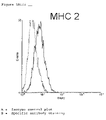
- the cell lines are further shown to posses significant levels of surface MHC-1 expression in addition to several other immune functional proteins not normally attributed to tumour cell lines, namely MHC-2, ICAM, and CD40 ligand.
- the two clones show differing morphology and also a differing pattern of gene expression with Onycap23 showing a distinct osteomimetic phenotype compared to Oncap1.
- a second embodiment of this invention is the use of any one of the cell lines in the formulation of a vaccine for the treatment of prostate cancer with or without a vaccine adjuvant which may include Il-2, IL-12, interferon gamma, BCG, tetanus toxoid or Mycobacterium Vaccae.
- the vaccine may be used as an adjuvant therapy in combination with other treatment modalities such as radiotherapy, or surgery or chemotherapy where the vaccine is used to treat or resolve minimal residual disease.
- a further aspect of the invention is the use of a combination of the cell lines in the formulation of a vaccine for the treatment of prostate cancer with or without a vaccine adjuvant which may include Il-2, IL-12, interferon gamma, BCG, tetanus toxoid or Mycobacterium Vaccae.
- the vaccine may be used as an adjuvant therapy in combination with other treatment modalities such as radiotherapy, surgery or chemotherapy where the vaccine is used to treat or resolve minimal residual disease
- a further aspect of the invention is the use of a combination of any of the cells with other prostate cell lines available from ATCC, ECACC or other laboratories and cell banks in the formulation of a vaccine for the treatment of prostate cancer with or without a vaccine adjuvant which may include IL-2, IL-12, interferon gamma, BCG, tetanus toxoid, Mycobacterium Vaccae or another suitable adjuvant or immunomodulator known in the art.
- the vaccine may be used as an adjuvant therapy in combination with other treatment modalities such as radiotherapy, surgery or chemotherapy where the vaccine is used to treat or resolve minimal residual disease or for the treatment of the disease at any stage with or without co-treatment of various types.
- a further embodiment of this invention are expression libraries derived from the cell lines which can be used in screening experiments to discover tumour associated or specific antigens for use as vaccine or immunotherapies and diagnostics.
- Expression cloning is now technically simple utilising commercially available kits such as the epitope tagged bacterial expression kits from InvitrogenTM and RocheTM or mammalian expression kits from StatageneTM.
- the various clones can be probed with anti-serum from vaccinated or non vaccinated patients using a SEREX type approach.
- mammalian cell expression clones can be used as targets for use with cytotoxic T-cells (CTL) from vaccinated or unvaccinated animals or patients to identify potential T cell antigens.
- CTL cytotoxic T-cells
- the cell lines may be used in specific proliferation experiments utilising whole blood to determine the precursor frequency of T cells that recognise antigens derived from the cell lines in both vaccinated and non-vaccinated patients.
- lysates of these cell lines are good at stimulating T cell proliferation in a high percentage of non-vaccinated patients indicating that there may be many shared antigens between these novel cell lines and early stage tumours in situ. Lysates of the cells may also be used to pulse antigen presenting cells or other cells expressing MHC-1 to enable these cells to be used as potential targets in CTL assays.
- Further embodiments of this invention relate to use of the cell lines grown in nude mice for drug screening, use of the cell lines in genomic screens for drug target identification and identification of antigens which may be used in diagnostic assays to screen for early phase detection of prostate cancer.
- FIG. 1 Morphology of Onycap 1 ( ⁇ 200 FIG. 1A ) and Onycap 23 ( ⁇ 200 FIG. 1B )
- FIG. 2 FACS Analysis of Cytokeratin Expression by Onycap1 and Oncap23
- FIG. 3 FACS Analysis of Known Immunological Surface Markers Onycap1 ( FIG. 3A ) and Oncap23 ( FIG. 23 )
- FIG. 4 Proliferation Response of Early Stage Prostate Cancer Patients to Lysates of Onycap1 and Oncap23
- ascitic fluid 3L was drawn from a patient with known metastatic prostate disease.
- the ascitic fluid was centrifuged at 1000 ⁇ g for 15 minutes and then resuspended in KSFM media supplemented with 25 g/ml bovine pituitary extract, 5 ng/ml of epidermal growth factor, 2 mM L-glutamine, 10 mM HEPES buffer and 5% foetal calf serum (FCS) (hereinafter called “modified KSFM”).
- FCS foetal calf serum
- a T175 flask of the ascitic outgrowth was trypsinised and plated out onto 96 well tissue culture plates at varying dilutions calculated to give 1, 10 and 100 cells per well. After a period of 14-21 days growth clones were picked from the 1 cell/well plate where there were less than 20 colonies per plate visible. Identified clones were trypsinised and plated into T25 flasks for expansion, after some 14-21 days growth the clones were further expanded to T75 and thereafter T175 flasks. The expanded clones were trypsinised and reformulated in freezing mixture comprising KSFM containing 10% v/v FCS and 20% v/v DMSO and then stored in 1 ⁇ 10 6 cell aliquots in liquid nitrogen.
- FIG. 1 A small epithelial morphology is shown in FIG. 1A and is representative of clone Onycap1.
- FIG. 1B A second unique morphology shown by clone Oncap23 is shown in FIG. 1B whereby the cells show dendrite-like processes emanating from a central cellular body similar to the so-called neuroendocrine phenotype (Chung T D K and Spiotto M T 2000 The Prostate 42 p186-195) which can be elicited by growth in IL6 containing medium.
- Onycap1 and Oncap23 clones have been analysed by flow cytometry using anti-cytokeratin 8, anti-cytokeratin 18 and desmin antibodies.
- Cytokeratin 8 and 18 are characteristic and considered true markers of prostatic epithelia.
- FIG. 2 shows examples of the flow cytometry analysis of the clones with CY8, CY18 and desmin antibodies compared with isotype control. In all cases the clones stained for these characteristic prostate epithelial markers proving their prostatic origin consistent with the medical condition of the patient.
- FIG. 3 shows representative flow cytometry data for Onycap1 and Oncap23. Unusually for prostate metastatic cell lines these clones show significant levels of MHC-I along with several other important markers which are important in cell based tumour immunotherapeutics, such as CD86 and CD40.
- MHC-I The presence of significant MHC-I levels is important since its cell surface presence will be important in eliciting an allogenic response when these cells are used as an whole cell allogeneic vaccine. There is also the possibility that with surface MHC-I expression these cells may also present directly to T cells, small peptide antigens restricted by the MPA clones MHC-I. In addition once the T cell receptor is engaged the presence of co-stimulatory molecules on the MPA clones may also elicit a significant proliferative response since both T receptor and co-stimulatory signals are present on the MPA cells.
- a double extraction was performed using TRI REAGENT (Sigma #T9424).
- the reagent was added directly to the washed cell pellets and samples were allowed to stand for 5 minutes before the addition of chloroform. Samples were vortexed and allowed to stand for a further 10 minutes at room temperature then centrifuged at 12,000 ⁇ g for 15 minutes.
- RNA precipitated with isopropanol RNA pellets were washed with 75% ethanol, dried and re-suspended in TE buffer.
- RNA sample was treated with Deoxyribonuclease I (Life Technologies #18068-015) to ensure there was no contamination with genomic DNA. Reactions were incubated for 15 minutes at room temperature then the DNase was inactivated by the addition of 25 mM EDTA and heating to 65° C. for 10 minutes.
- Reverse transcription was performed using the 1st strand cDNA synthesis kit for RT-PCR (AMV) from Boehringer Mannheim (#1 483 188). Reactions were incubated at 25° C. for 10 minutes then at 42° C. for 1 hour. The AMV enzyme was denatured by heating to 99° C. for 5 minutes then the reaction was cooled to 4° C.
- AMV 1st strand cDNA synthesis kit for RT-PCR
- PCR primers listed were selected from the literature to cover known prostate proteins, antigens and markers of invasion and metastasis.
- the cDNA extracted from the clones was probed with the primers and products of the reaction run out on high resolution agarose gels to check for products of the expected sizes. The results are tabulated in Table 1 for clones Onycap1 and Oncap23.
- Onycap1 and Oncap23 were seeded at 1 ⁇ 10 6 cells into T175 culture flasks. Cultures were maintained for 4-5 days in modified KSFM. Cells were harvested by trypsinisation from the surface of the plastic and washed in Hanks balanced salt solution before RNA extraction.
- a double extraction was performed using TRI REAGENT (Sigma #T9424).
- the reagent was added directly to the washed cell pellets and samples were allowed to stand for 5 minutes before the addition of chloroform. Samples were vortexed and allowed to stand for a further 10 minutes at room temperature then centrifuged at 12,000 ⁇ g for 15 minutes.
- RNA precipitated with isopropanol RNA pellets were washed with 75% ethanol, dried and re-suspended in TE buffer.
- RNA sample was treated with Deoxyribonuclease I (Life Technologies #18068-015) to ensure there was no contamination with genomic DNA. Reactions were incubated for 15 minutes at room temperature then the DNase inactivated by the addition of 25 mM EDTA and heating to 65° C. for 10 minutes.
- Labelled cDNA was used to probe a Clontech AtlasTM array (human cancer array II) following the manufacturers protocol for hybridisation and washing. Images were recorded utilising a phosphor imager and recorded files analysed with the Clontech Atlas ImageTM software.
- Clones were grown in T75 flasks in modified KSFM. Once the cells have reached confluency they were harvested by trypsinisation and washed in Hanks balanced salt solution and then pelleted by centrifugation.
- the pelleted cell mass was taken through four freeze thaw cycles in a minimal volume of Hanks balanced salt solution at a cell concentration of 2 ⁇ 10 6 cells/ml.
- the resulting cell supernatent was aliquoted into 25 ul aliquots and stored at ⁇ 70° C. until required.
- FIG. 4 The results for a series of proliferation assays are shown in FIG. 4 where a proliferation index for either CD4 or CD8 positive T-cells are plotted against Onycap1 and Onycap 23 cell lysates, the proliferation index being derived by dividing the percentage of T-cells proliferating by the no-lysate control.
- a proliferation index above 1 indicates significant T cell proliferation in response to the cell lysate used to stimulate.
- Results are shown for six early stage prostate patient sera 1, 2, 3, 4, 5 and 6 each of which was stimulated with Onycap1 or Oncap23 cell lysate.
- the results show that the Onycap cell lines posses antigens which differentially stimulate T cells from a variety of patients. Its is unlikely that a response is due to an mixed lymphocyte reaction since both cell lines posses the same haplotype and yet for each of the patients sera there are examples of one cell line stimulating a response where the other cell line does not.
Abstract
Description
| Reagents | Catalogue # | Supplier |
| RPMI medium | Life Technologies, | |
| Paisley Scotland. | ||
| BrdU | Sigma Chemical Co, | |
| Poole, Dorset. | ||
| PharMlyse | 35221E | Pharmingen, Oxford UK |
| Cytofix/Cytoperm | 2090KZ | ″ |
| Perm/Wash buffer (×10) | 2091KZ | ″ |
| FITC Anti-BrdU/Dnase | 340649 | Becton Dickinson |
| PerCP Anti-CD3 | 347344 | ″ |
| Pe Anti-CD4 | 30155X | Pharmingen |
| Pe Anti-CD8 | 30325X | ″ |
| FITC mu-IgG1 | 349041 | Becton Dickinson |
| PerCP IgG1 | 349044 | ″ |
| PE IgG1 | 340013 | ″ |
Method
- 1) Dilute 1 ml blood with 9 ml RPMI+2 mM L-glutamine+penicillin/streptomycin antibiotics+50 μM 2-mercaptoethanol. Do not add serum. Leave overnight at 37° C.
- 2) On following morning, aliquot 450 μl of diluted blood into wells of a 48-well plate and add 50 μl of stimulator lysate. The lysate is made by freeze-thawing tumour cells (2×10⁶ cell equivalents/ml)×3 in liquid nitrogen and then storing aliquots frozen until required.
- 3) Culture cells at 37° C. for 5 days
- 4) On the evening of
day 5 add 50 μl BrdU @ 30 μg/ml - 5)
Aliquot 100 μl of each sample into a 96-well round-bottomed plate. - 6) Spin plate and discard supernatant
- 7) Lyse red cells using 100 μl PharMlyse™ for 5 minutes at room temperature
- 8) Wash ×2 with 50 μl of Cytofix™
- 9) Spin and remove supernatant by flicking
- 10) Permeabilise with 100 μl Perm/Wash™ for 10 mins at RT
- 11) Add 30 μl of antibody mix comprising antibodies at correct dilution made up to volume with Perm/Wash™
- 12) Incubate for 30 mins in the dark at room temperature.
- 13) Wash ×1 and resuspend in 100
μl 2% paraformaldehyde - 14) Add this to 400 μl FACSFlow™ in cluster tubes ready for analysis
- 15) Analyse on FACScan™, storing 3000 gated CD3 events.
| 6-well plate for stimulation |
| Nil | Con | 15 | Ln | | Pn | ||
| PBL |
| 1 | ||||||
| |
||||||
| |
||||||
| |
||||||
| |
||||||
| |
||||||
| 96-well plate for |
| PBL |
| 1 | | | | | |
| Nil | 15 D | Nil | 15 D | Nil | 15 D | Nil | 15 D | Nil | 15 D | Nil | 15 D |
| A | A | A | A | A | A | ||||||
| Nil | 15 E | Nil | 15 E | Nil | 15 E | Nil | 15 E | Nil | 15 E | Nil | 15 E |
| D | D | D | D | D | D | ||||||
| Nil | Ln D | Nil | Ln D | Nil | Ln D | Nil | Ln D | Nil | Ln D | Nil | Ln D |
| E | E | E | E | E | E | ||||||
| Con | Ln E | Con | Ln E | Con | Ln E | Con | Ln E | Con | Ln E | Con | Ln E |
| D | D | D | D | D | D | ||||||
| Con | Du D | Con | Du D | Con | Du D | Con | Du D | Con | Du D | Con | Du D |
| E | E | E | E | E | E | ||||||
| Du E | Du E | Du E | Du E | Du E | Du E | ||||||
| Pn D | Pn D | Pn D | Pn D | Pu D | Pn D | ||||||
| Pn E | Pn E | Pn E | Pn E | Pn E | Pn E | ||||||
| Legend: | |||||||||||
| A: FITC mu-IgGl (5 μl) + PE IgGl (5 μl) + PerCP IgGl (5 μl) + 15 μl Perm/Wash ™ | |||||||||||
| D: FITC Anti-BrdU/Dnase (5 μl) + Pe Anti-CD4 (5 μl) + PerCP anti-CD3 (5 μl) + 15 μl Perm/Wash ™ | |||||||||||
| E: FITC Anti-BrdU/Dnase (5 μl) + Pe Anti-CD8 (5 μl) + PerCP Anti-CD3 (5 μl) + 15 μl Perm/Wash ™ | |||||||||||
| 15: NIH1542-CP3TX (an immortalised line derived from primary prostate cancers by Dr. Suzanne Topalian at the NIH) | |||||||||||
| Ln: LnCap (ATCC Number: CRL-1740) | |||||||||||
| D: Du145 (ATCC Number: HTB-81) | |||||||||||
| Pn: Pnt2 (ECACC Ref No: 95012613) | |||||||||||
| Con: ConA lectin (positive control) | |||||||||||
| Nil: No stimulation | |||||||||||
Results
| TABLE 1 |
| PCR Expression Analysis on Onycap1 and Oncap23 |
| Prostate | ||||
| Antigen | Onycap1 | Oncap23 | ||
| PSA | − | − | ||
| PAP | − | − | ||
| PSM | − | − | ||
| Androgen | − | − | ||
| recptr. | ||||
| EGF receptor | − | − | ||
| IGF-II | − | − | ||
| HGF receptor | + | + | ||
| uPA | + | + | ||
| PCTA-1 | + | + | ||
| PSCA | + | + | ||
| GRP receptor | − | + | ||
| uPA receptor | + | + | ||
| Hyaluronic acid | − | − | ||
| receptor | ||||
| MMP-9 | − | − | ||
| Vimentin | + | + | ||
| PAGE-1 | − | − | ||
| E-cadherin | − | − | ||
| TGFα | + | + | ||
| KAI1 | − | + | ||
| Heparanase | + | + | ||
| TABLE 2 |
| Gene Expression Analysis for Onycap1 and Oncap23 |
| Spot Intensity |
| Onycap1 | Oncap23 | Gene product |
| 2748 | 10332 | CD81 antigen; 26-kDa cell |
| surface protein TAPA-1 | ||
| 2260 | 9368 | leukemia virus receptor 1 (GLVR1) |
| 0 | 7652 | lysosome-associated membrane glycoprotein 2 |
| precursor (LAMP2); CD107B antigen | ||
| 0 | 5184 | lysosome membrane protein II (LIMP II); |
| 0 | 12796 | annexin V; lipo- |
| cortin V; endo- | ||
| nexin II; | ||
| 0 | 5688 | LGALS3, MAC2 (Galectin-3, MAC-2 antigen, |
| 0 | 6528 | lymphocyte function-associated antigen 3 |
| precursor (LFA3); CD58 antigen | ||
| 0 | 28900 | neprilysin; neutral endopeptidase (NEP); |
| enkephalinase (EPN); | ||
| 0 | 7264 | zinc finger protein |
| 248 | 2676 | 5′-TG-3′ INTERACTING FACTOR |
| (HOMEOBOXPROTEIN TGIF) | ||
| 0 | 5032 | HOMEOBOX PROTEIN SIX1 |
| 2504 | 4888 | SOX-9 PROTEIN |
| 1440 | 3128 | ZINC FINGER PROTEIN SLUG |
| 0 | 6052 | ENX-1 PUTATIVE TRANSCRIPTIONAL |
| REGULATOR | ||
| 804 | 7100 | LIM AND SH3 DOMAIN PROTEIN LASP-1 |
| (MLN 50) | ||
| 388 | 4708 | MEL-18 RING-FINGER PROTEIN |
| 8716 | 17132 | NAT1 TRANSLATION REPRESSOR |
| 0 | 6312 | SAP18 (Sin3 associated polypeptide P18) |
| 0 | 6984 | preliminary PRT 640 AA RNA polymerase |
| II elongation factor ELL2 | ||
| 1932 | 6228 | HLA-B-associated transcript 2; large proline- |
| rich protein BAT2 | ||
| 11008 | 2132 | complement 3(C3) |
| 2076 | 8868 | LANP CEREBELLAR LEUCINE RICH ACIDIC |
| NUCLEAR PROTEIN | ||
| 184 | 3164 | phospholipid transfer protein precursor (PTLP); |
| lipid transfer protein II | ||
| 3016 | 12244 | dek protein |
| 0 | 5452 | CYSTEINE-RICH PROTEIN 2 (CRP2) (ESP1 |
| PROTEIN) | ||
| 3748 | 8480 | RNA-binding protein EWS |
| 6200 | 15836 | RNA-binding protein fus/tls |
| 12996 | 35868 | nucleolar phosphoprotein B23; nucleophosmin |
| (NPM); numatrin | ||
| 4532 | 12992 | platelet-derived growth factor (PDGF) receptor |
| beta-like tumor suppressor (PRLTS) | ||
| 1680 | 5664 | retinoic acid receptor alpha |
| 1744 | 15192 | ras-related protein R-ras2; ras-like protein TC21; |
| teratocarcinoma oncogene | ||
| 0 | 5676 | clathrin assembly protein lymphoid myeloid |
| leukemia (CALM) | ||
| 27752 | 40916 | MT1H (Methallothionein-IH, Metallothinein-0, |
| MT-0) + Metallothionein isoform 1L (MT-1L) | ||
| 9124 | 26864 | voltage-dependent anion-selective channel |
| protein 1 (VDAC1); | ||
| 244 | 6964 | ras-related protein RAB-5C |
| 0 | 5584 | vesicle-membrane fusion protein SNAP23A |
| 3976 | 16024 | annexin IV (ANX4); lipocortin I; calpactin II; |
| chromobindin 9; phospholipase A2 inhibitoiy | ||
| protein | ||
| 13640 | 34420 | annexin I (ANX1) |
| 11428 | 27236 | annexin II (ANX2); lipocortin II; calpactin I |
| heavy subunit; chromobindin 8; protein I; | ||
| 0 | 7944 | ER lumen protein retaining receptor 2; KDEL |
| (SEQ ID NO: 1) receptor 2; ERD 22 | ||
| 0 | 5136 | protein SEC23 homolog isoform A (SEC23A) |
| 2896 | 11292 | coatomer beta′ subunit; beta′-coat |
| protein; beta′-COP; p102 | ||
| 0 | 5472 | coatomer delta subunit; delta-coat protein; |
| delta-COP; archain (ARCN1) | ||
| 0 | 6448 | cation-dependent mannose-6-phosphate |
| receptor precursor | ||
| 0 | 7516 | syntaxin 7 (STX7) |
| 3464 | 8192 | ras-related protein RAB-11B; YPT3 |
| 5740 | 9960 | ras-related protein RAB-7 |
| 3852 | 14512 | ras-related protein RAB-1A; YPTL- |
| related protein | ||
| 640 | 4004 | calcium-binding protein p22; calcium- |
| binding protein CHP | ||
| 0 | 9184 | clathrin coat assembly protein AP17; plasma |
| membrane adaptor AP-2 17-kDa protein; | ||
| 8496 | 19328 | clathrin coat assembly protein AP50; plasma |
| membrane adaptor AP-2 50-kDa protein; | ||
| 0 | 5908 | cytosolic acetoacetyl-coenzyme A thiolase |
| 312 | 7616 | beta-D-galactosidase precursor; lactase; acid |
| beta-galactosidase; GLB 1 | ||
| 1212 | 7572 | 6-PHOSPHOFRUCTOKINASE, |
| 3492 | 7320 | pyruvate kinase M2 isozyme (PKM2) |
| 708 | 6620 | succinyl-CoA:3-ketoacid-coenzyme A transferase |
| 240 | 5856 | alcohol dehydrogenase 5 chi polypeptide |
| 0 | 5188 | NADH-cytochroine B5 reductase |
| 0 | 7152 | mitochondrial enoyl-CoA hydratase short |
| subunit 1 | ||
| 0 | 5356 | 3-hydroxy-3-methylglutaryl-coenzyme A |
| reductase (HMG-CoA reductase; HMGCR) | ||
| 0 | 5212 | lysosomal acid lipase/cholesteryl ester |
| hydrolase precursor (LAL); | ||
| 0 | 8096 | phosphatidylethanolainine-binding protein (PBP); |
| neuropolypeptide H3 | ||
| 0 | 6420 | annexin III (ANX3); lipocortin III; placental |
| anticoagulant protein III) (PAP-III); | ||
| 432 | 5024 | farnesyl pyrophosphate synthetase |
| 448 | 5276 | H105E3 protein |
| 0 | 5084 | dihydrofolate reductase (DHFR) |
| 1976 | 8296 | thymidylate synthase (TYMS; TS) |
| 324 | 7856 | glutamate dehydrogenase 1 precursor (GDH; |
| GLUD1) | ||
| 2580 | 5504 | very-long-chain-specific acyl-CoA dehydrogenase |
| precursor (VLCAD) | ||
| 24144 | 34792 | femtin heavy chain (FTH1); FTHL6 |
| 44632 | 5996 | placental type 1 alkaline phosphatase precursor |
| (PLAP-1) | ||
| 3732 | 8760 | homolog of yeast D2-isopentenylpyrophosphate |
| isomerase (IPP isomerase) | ||
| 364 | 6132 | ornithine decarboxylase (ODC1) |
| 14304 | 24032 | protein disulfide isomerase |
| 0 | 5416 | cyclophilin 3 protein (CYP3); |
| 14884 | 31528 | 60S ribosomal protein L22 (RPL22); |
| 0 | 8068 | TIA-1 related protein; nucleolysin TIAR |
| 0 | 10204 | astrocytic phosphoprotein PEA-15 |
| 0 | 7020 | cyr61 protein precursor; GIG1 protein; |
| insulin-like growth factor-binding | ||
| protein 10 (IGFBP10) | ||
| 0 | 5668 | granulins precursor (GRN); acrogranin |
| 7288 | 16696 | natriuretic peptide precursor B |
| 932 | 3548 | SEMAPHORIN V |
| 660 | 8248 | signal transducing adaptor molecule (STAM) |
| 2096 | 4344 | beta-adaptin 1; plasma membrane adaptor |
| HA2/AP2 adaptin beta subunit; | ||
| 128 | 5132 | RIBOSOMAL PROTEIN S6 KINASE (EC |
| 2.7.1.-)(S6K) (P70-56K). | ||
| 2796 | 13364 | casein kinase I alpha isoform (CKI-alpha); |
| CK1; CSNK1A | ||
| 11716 | 21668 | casein kinase I delta isoform (CKI-delta); |
| CSNK1D | ||
| 812 | 6076 | casein kinase II beta subunit (CK II; CSNK2B; |
| CK2N); phosvitin | ||
| 2204 | 7884 | MAP kinase-activated protein kinase 2 |
| (MAPKAP kinase 2; MAPKAPK-2) | ||
| 0 | 5592 | protein phosphatase PP2A 65-kDa regulatory |
| subunit alpha isoform; | ||
| 0 | 5292 | protein phosphatase PP2A 55-kDa regulatory |
| subunit neuronal isoform; | ||
| 420 | 5744 | dual-specificity protein phosphatase 5; |
| dual-specificity protein phosphatase | ||
| HVH3 | ||
| 4328 | 10112 | dual-specificity protein phosphatase 7; dual- |
| specificity protein phosphatase PYST2 | ||
| 1484 | 5124 | serine/threonine protein phosphatase 2B |
| catalytic subunit alpha isoform; | ||
| 972 | 7260 | serine/threonine protein phosphatase 5 (PP5); |
| protein phosphatase T (PPT) | ||
| 0 | 6016 | protein-tyrosine phosphatase 1B (PTP-1B) |
| 1584 | 6508 | protein-tyrosine phosphatase G1 (PTP-G1) |
| 6480 | 14912 | serine/threonine protein phosphatase PPl-alpha 1 |
| catalytic subunit (PP-1A) | ||
| 952 | 6852 | guanine nucleotide-binding protein G(K) alpha 3 |
| subunit (GNA3) | ||
| 4104 | 12748 | GUANINE NUCLEOTIDE-BINDING |
| PROTEIN G(I), | ||
| 0 | 8304 | ras-related protein RAP-1B; GTP-binding |
| protein SMG p21B | ||
| 0 | 6180 | calpactin I light chain |
| 8284 | 15236 | ras GTPase-activating-like protein IQGAP1; |
| p19; KIAA0051 | ||
| 856 | 6416 | calmodulin |
| 1556 | 9264 | calmodulin |
| 440 | 4164 | ANNEXIN XI (CALCYCLIN-ASSOCIATED |
| ANNEXIN 50) (CAP-50) (56 KD AUTO- | ||
| ANTIGEN). | ||
| 2756 | 8752 | calgizzarin; S100C protein; MLN70 |
| 3768 | 11592 | sorcin 22-kDa protein (SRI); CP-22 |
| 472 | 6296 | calcium signal-modulating cyclophilin ligand |
| (CAML) | ||
| 5352 | 16056 | 14-3-3 protein beta/alpha; protein kinase C |
| inhibitor protein-1 (KCIP-1); protein 1054 | ||
| 9088 | 20912 | 14-3-3 PROTEIN EPSILON (MITO- |
| CHONDRIAL IMPORT STIMULATION | ||
| FACTOR L SUBUNIT) | ||
| 4576 | 11468 | 14-3-3 PROTEIN ZETA/DELTA (PROTEIN |
| KINASE C INHIBITOR PROTEIN-1) | ||
| (KCIP-1) | ||
| 1688 | 8056 | phosphatidylinositol transfer protein |
| alpha isoform (PI-TP-alpha; | ||
| 12604 | 26120 | guanine nucleotide-binding protein beta subunit- |
| like protein 12; | ||
| 532 | 3800 | lysosomal protective protein precursor; cathepsin |
| A; carboxypeptidase C; PPGB | ||
| 9808 | 23748 | calcium-dependent protease small (regulatory) |
| subunit; calpain; | ||
| 1036 | 8220 | HUNTINGTIN INTERACTING PROTEIN |
| (HIP2) | ||
| 0 | 5844 | lipoprotein-associated coagulation inhibitor |
| 504 | 4120 | protein-tyrosine phosphatase kappa precursor |
| (R-PTP-kappa; PTPRK; PTPK) | ||
| 4740 | 14824 | PUTATIVE RECEPTOR PROTEIN (PM1) |
| 40 | 3116 | orphan receptor TR4 |
| 168 | 2980 | dynactin 150-kDa isoform; 150-kDa dynein- |
| associated polypeptide (DAP-150; | ||
| 13744 | 33716 | COFILIN |
| 4044 | 12584 | alpha-actinin 1 cytoskeletal isoform; |
| F-actin cross linking protein | ||
| 852 | 5288 | drebrinE |
| 1684 | 11556 | major prion protein precursor (PRP); PRP27-30; |
| PRP33-35C; ASCR | ||
| 820 | 4628 | DXS6673E protein; X-linked mental retardation |
| candidate gene | ||
Claims (16)
Applications Claiming Priority (5)
| Application Number | Priority Date | Filing Date | Title |
|---|---|---|---|
| GB0008032A GB0008032D0 (en) | 2000-04-01 | 2000-04-01 | New prostate cell lines |
| GB0008032.5 | 2000-04-01 | ||
| GB0024237.0 | 2000-10-03 | ||
| GB0024237A GB0024237D0 (en) | 2000-10-03 | 2000-10-03 | New prostate cell lines |
| PCT/GB2001/001437 WO2001075073A2 (en) | 2000-04-01 | 2001-03-30 | Prostate cell lines and their use |
Publications (2)
| Publication Number | Publication Date |
|---|---|
| US20030185808A1 US20030185808A1 (en) | 2003-10-02 |
| US7303911B2 true US7303911B2 (en) | 2007-12-04 |
Family
ID=26244016
Family Applications (1)
| Application Number | Title | Priority Date | Filing Date |
|---|---|---|---|
| US10/240,523 Expired - Fee Related US7303911B2 (en) | 2000-04-01 | 2001-03-30 | Prostate cell lines |
Country Status (9)
| Country | Link |
|---|---|
| US (1) | US7303911B2 (en) |
| EP (1) | EP1272617B1 (en) |
| AT (1) | ATE507285T1 (en) |
| AU (1) | AU2001244341A1 (en) |
| CA (1) | CA2404388C (en) |
| DE (1) | DE60144515D1 (en) |
| DK (1) | DK1272617T3 (en) |
| HK (1) | HK1052531A1 (en) |
| WO (1) | WO2001075073A2 (en) |
Families Citing this family (4)
| Publication number | Priority date | Publication date | Assignee | Title |
|---|---|---|---|---|
| US20050019336A1 (en) * | 2003-07-23 | 2005-01-27 | Dalgleish Angus George | Human prostate cell lines in cancer treatment |
| WO2005078124A2 (en) * | 2004-02-16 | 2005-08-25 | Proteosys Ag | Diagnostic marker for cancer |
| DE102004038076A1 (en) * | 2004-02-16 | 2005-09-01 | Proteosys Ag | Use of annexin A3, or other proteins, as diagnostic markers for cancer, especially of the prostate, also use of modulators of these markers for treating cancer |
| TW202134430A (en) | 2019-12-03 | 2021-09-16 | 美商紐沃進公司 | Tumor cell vaccines |
Citations (8)
| Publication number | Priority date | Publication date | Assignee | Title |
|---|---|---|---|---|
| WO1997018296A1 (en) | 1995-11-15 | 1997-05-22 | University Of Utah | Novel method of culturing human epithelial cells for the identification of cancer therapeutics and diagnostics |
| WO1997024132A1 (en) | 1995-12-28 | 1997-07-10 | The Johns Hopkins University School Of Medicine | Allogeneic paracrine cytokine tumor vaccines |
| WO1997028255A1 (en) | 1996-02-02 | 1997-08-07 | The Government Of The United States Of America, Represented By The Secretary, Department Of Health And Human Services | Immortal human prostate epithelial cell lines and clones and their applications in the research and therapy of prostate cancer |
| WO1998016238A2 (en) | 1996-10-11 | 1998-04-23 | The Regents Of The University Of California | Cancer immunotherapy using tumor cells combined with mixed lymphocytes |
| WO1999019462A1 (en) | 1997-10-10 | 1999-04-22 | The Regents Of The University Of California | Enhanced immunogenic cell populations prepared using h2 receptor antagonists |
| WO2000004918A2 (en) | 1998-07-21 | 2000-02-03 | Kerkmann Tucek Aida | Agents for the immunotherapy of tumoral diseases |
| WO2000033869A2 (en) | 1998-12-10 | 2000-06-15 | Onyvax Limited | Use of human prostrate cell lines in cancer treatment |
| WO2000071155A2 (en) | 1999-05-21 | 2000-11-30 | Onyvax Limited | New vaccine formulations - 2 |
Family Cites Families (2)
| Publication number | Priority date | Publication date | Assignee | Title |
|---|---|---|---|---|
| US5804443A (en) * | 1997-02-28 | 1998-09-08 | Bernofsky; Carl | Human monocytic leukemia cell line |
| GB9827103D0 (en) * | 1998-12-10 | 1999-02-03 | Onyvax Ltd | New cancer treatments |
-
2001
- 2001-03-30 DE DE60144515T patent/DE60144515D1/en not_active Expired - Lifetime
- 2001-03-30 CA CA2404388A patent/CA2404388C/en not_active Expired - Fee Related
- 2001-03-30 EP EP01917257A patent/EP1272617B1/en not_active Expired - Lifetime
- 2001-03-30 US US10/240,523 patent/US7303911B2/en not_active Expired - Fee Related
- 2001-03-30 AT AT01917257T patent/ATE507285T1/en not_active IP Right Cessation
- 2001-03-30 AU AU2001244341A patent/AU2001244341A1/en not_active Abandoned
- 2001-03-30 DK DK01917257.6T patent/DK1272617T3/en active
- 2001-03-30 WO PCT/GB2001/001437 patent/WO2001075073A2/en active Application Filing
-
2003
- 2003-07-07 HK HK03104794.7A patent/HK1052531A1/en not_active IP Right Cessation
Patent Citations (8)
| Publication number | Priority date | Publication date | Assignee | Title |
|---|---|---|---|---|
| WO1997018296A1 (en) | 1995-11-15 | 1997-05-22 | University Of Utah | Novel method of culturing human epithelial cells for the identification of cancer therapeutics and diagnostics |
| WO1997024132A1 (en) | 1995-12-28 | 1997-07-10 | The Johns Hopkins University School Of Medicine | Allogeneic paracrine cytokine tumor vaccines |
| WO1997028255A1 (en) | 1996-02-02 | 1997-08-07 | The Government Of The United States Of America, Represented By The Secretary, Department Of Health And Human Services | Immortal human prostate epithelial cell lines and clones and their applications in the research and therapy of prostate cancer |
| WO1998016238A2 (en) | 1996-10-11 | 1998-04-23 | The Regents Of The University Of California | Cancer immunotherapy using tumor cells combined with mixed lymphocytes |
| WO1999019462A1 (en) | 1997-10-10 | 1999-04-22 | The Regents Of The University Of California | Enhanced immunogenic cell populations prepared using h2 receptor antagonists |
| WO2000004918A2 (en) | 1998-07-21 | 2000-02-03 | Kerkmann Tucek Aida | Agents for the immunotherapy of tumoral diseases |
| WO2000033869A2 (en) | 1998-12-10 | 2000-06-15 | Onyvax Limited | Use of human prostrate cell lines in cancer treatment |
| WO2000071155A2 (en) | 1999-05-21 | 2000-11-30 | Onyvax Limited | New vaccine formulations - 2 |
Non-Patent Citations (15)
| Title |
|---|
| Ablin, J. Cancer Res. Clin. Oncol. 123: 583-594 (1997). |
| Bellone et al. . (Immunology Today, v20 (10), 1999, pp. 457-461. * |
| Berthon et al., Intl. Journal of Oncology 6:333-343 (1995). |
| Brenner et al., Journal of Urology, pp. 1575-1579 (1995) (abstract only). |
| Dermer (Bio/Technology, 1994, 12:320). * |
| Freshney (Culture of Animal Cells, A Manual of Basic Technique, Alan R. Liss, Inc., 1983, New York, p. 4. * |
| Freshney, "Culture of Animal Cells", Chapter 23, p. 314 (1987). |
| Gorelik et al., Journal of Supramolecular Structure 12: 385-402 (1979) (abstract only). |
| Gura (Science, v278, 1997, pp. 1041-1042). * |
| Marble, Cancer Weekly Plus, p. 4(2) (1997). |
| Moran, American Medical News 42, 39, 23 (1999). |
| Olumi et al., Cancer Research 58: 4525-4530 (1998). |
| Tjoa et al., The Prostate 27: 63-69 (1995). |
| Weiner L.M., Seminars Oncology, vol. 26, No. 4, Suppl 12, pp. 41-50, 1999. * |
| Zhau et al., Proc. Natl. Acad. Sci. USA 93: 15152-15157 (1996). |
Also Published As
| Publication number | Publication date |
|---|---|
| HK1052531A1 (en) | 2003-09-19 |
| ATE507285T1 (en) | 2011-05-15 |
| US20030185808A1 (en) | 2003-10-02 |
| WO2001075073A3 (en) | 2002-02-14 |
| DK1272617T3 (en) | 2011-08-22 |
| EP1272617B1 (en) | 2011-04-27 |
| WO2001075073A2 (en) | 2001-10-11 |
| EP1272617A2 (en) | 2003-01-08 |
| DE60144515D1 (en) | 2011-06-09 |
| AU2001244341A1 (en) | 2001-10-15 |
| CA2404388C (en) | 2010-06-15 |
| CA2404388A1 (en) | 2001-10-11 |
Similar Documents
| Publication | Publication Date | Title |
|---|---|---|
| US7723107B2 (en) | Pharmaceutical composition for inducing an immune response in a human or animal | |
| Palucka et al. | Dendritic cells loaded with killed allogeneic melanoma cells can induce objective clinical responses and MART-1 specific CD8+ T-cell immunity | |
| Panelli et al. | Expansion of tumor-T cell pairs from fine needle aspirates of melanoma metastases | |
| CA2354046C (en) | New cancer treatments | |
| WO2000025813A1 (en) | CANCER IMMUNOTHERAPHY AND DIAGNOSIS USING UNIVERSAL TUMOR ASSOCIATED ANTIGENS, INCLUDING hTERT | |
| Ramanathan et al. | Development and clinical evaluation of dendritic cell vaccines for HPV related cervical cancer-a feasibility study | |
| JP2010523141A (en) | Cell lines, compositions containing them for the treatment of melanoma, compositions manufacturing procedures and methods of treatment | |
| US7303911B2 (en) | Prostate cell lines | |
| JP4949554B2 (en) | New treatment for cancer | |
| Saleh et al. | Melanoma immunotherapy: past, present, and future | |
| WO2007018047A1 (en) | Squamous cell carcinoma antigen-derived peptide binding to hla-a24 molecule | |
| US11260118B2 (en) | Immunogenic peptide composition | |
| ES2367006T3 (en) | PROSTATE CELL PHONES AND ITS USE. | |
| EP1178822B1 (en) | Whole cell cancer vaccine comprising co-cultured malignant and non-malignant cells | |
| JP2005170799A (en) | Hla (human leukocyte antigen)-a24 binding peptide of enhancer of zeste homolog 2 | |
| Saleh et al. | Vaccine immunotherapy for a deadly disease like malignant melanoma: where do we stand? | |
| Mohanakumar et al. | Definition of the T Cell-Mediated Immune Response to Mammaglobin, a Novel Breast Cancer-Associated Protein | |
| Kwak et al. | The shared tumor-associated antigen cytochrome P450 1B1 is recognized | |
| CLONES | Genes Coding for Antigens Recognized on Human Tumors by Autologous Cytolytic T Lymphocytes zyxwvutsrqpon | |
| Jonasch | Melanoma vaccination: state-of-the-art and experimental approaches | |
| NZ527763A (en) | Allogeneic immunotherapeutic agent comprising three human prostate tumour cell lines derived from three different primary tumours useful in treating prostate cancer |
Legal Events
| Date | Code | Title | Description |
|---|---|---|---|
| AS | Assignment |
Owner name: ONYVAX LIMITED, UNITED KINGDOM Free format text: ASSIGNMENT OF ASSIGNORS INTEREST;ASSIGNORS:THRAVES, PETER;SUTTON, ANDREW;REEL/FRAME:013628/0154;SIGNING DATES FROM 20021106 TO 20021117 |
|
| STCF | Information on status: patent grant |
Free format text: PATENTED CASE |
|
| FPAY | Fee payment |
Year of fee payment: 4 |
|
| FPAY | Fee payment |
Year of fee payment: 8 |
|
| FEPP | Fee payment procedure |
Free format text: PAYER NUMBER DE-ASSIGNED (ORIGINAL EVENT CODE: RMPN); ENTITY STATUS OF PATENT OWNER: SMALL ENTITY Free format text: PAYOR NUMBER ASSIGNED (ORIGINAL EVENT CODE: ASPN); ENTITY STATUS OF PATENT OWNER: SMALL ENTITY |
|
| FEPP | Fee payment procedure |
Free format text: MAINTENANCE FEE REMINDER MAILED (ORIGINAL EVENT CODE: REM.); ENTITY STATUS OF PATENT OWNER: SMALL ENTITY |
|
| LAPS | Lapse for failure to pay maintenance fees |
Free format text: PATENT EXPIRED FOR FAILURE TO PAY MAINTENANCE FEES (ORIGINAL EVENT CODE: EXP.); ENTITY STATUS OF PATENT OWNER: SMALL ENTITY |
|
| STCH | Information on status: patent discontinuation |
Free format text: PATENT EXPIRED DUE TO NONPAYMENT OF MAINTENANCE FEES UNDER 37 CFR 1.362 |
|
| FP | Lapsed due to failure to pay maintenance fee |
Effective date: 20191204 |