WO2004085648A2 - Nogo receptor binding protein - Google Patents
Nogo receptor binding protein Download PDFInfo
- Publication number
- WO2004085648A2 WO2004085648A2 PCT/US2004/008323 US2004008323W WO2004085648A2 WO 2004085648 A2 WO2004085648 A2 WO 2004085648A2 US 2004008323 W US2004008323 W US 2004008323W WO 2004085648 A2 WO2004085648 A2 WO 2004085648A2
- Authority
- WO
- WIPO (PCT)
- Prior art keywords
- polypeptide
- domain
- moiety
- injury
- neuron
- Prior art date
Links
Classifications
-
- C—CHEMISTRY; METALLURGY
- C12—BIOCHEMISTRY; BEER; SPIRITS; WINE; VINEGAR; MICROBIOLOGY; ENZYMOLOGY; MUTATION OR GENETIC ENGINEERING
- C12N—MICROORGANISMS OR ENZYMES; COMPOSITIONS THEREOF; PROPAGATING, PRESERVING, OR MAINTAINING MICROORGANISMS; MUTATION OR GENETIC ENGINEERING; CULTURE MEDIA
- C12N15/00—Mutation or genetic engineering; DNA or RNA concerning genetic engineering, vectors, e.g. plasmids, or their isolation, preparation or purification; Use of hosts therefor
- C12N15/09—Recombinant DNA-technology
- C12N15/11—DNA or RNA fragments; Modified forms thereof; Non-coding nucleic acids having a biological activity
-
- G—PHYSICS
- G01—MEASURING; TESTING
- G01N—INVESTIGATING OR ANALYSING MATERIALS BY DETERMINING THEIR CHEMICAL OR PHYSICAL PROPERTIES
- G01N33/00—Investigating or analysing materials by specific methods not covered by groups G01N1/00 - G01N31/00
- G01N33/48—Biological material, e.g. blood, urine; Haemocytometers
- G01N33/50—Chemical analysis of biological material, e.g. blood, urine; Testing involving biospecific ligand binding methods; Immunological testing
- G01N33/53—Immunoassay; Biospecific binding assay; Materials therefor
- G01N33/569—Immunoassay; Biospecific binding assay; Materials therefor for microorganisms, e.g. protozoa, bacteria, viruses
- G01N33/56966—Animal cells
-
- A—HUMAN NECESSITIES
- A61—MEDICAL OR VETERINARY SCIENCE; HYGIENE
- A61P—SPECIFIC THERAPEUTIC ACTIVITY OF CHEMICAL COMPOUNDS OR MEDICINAL PREPARATIONS
- A61P3/00—Drugs for disorders of the metabolism
- A61P3/08—Drugs for disorders of the metabolism for glucose homeostasis
- A61P3/10—Drugs for disorders of the metabolism for glucose homeostasis for hyperglycaemia, e.g. antidiabetics
-
- A—HUMAN NECESSITIES
- A61—MEDICAL OR VETERINARY SCIENCE; HYGIENE
- A61K—PREPARATIONS FOR MEDICAL, DENTAL OR TOILETRY PURPOSES
- A61K38/00—Medicinal preparations containing peptides
- A61K38/16—Peptides having more than 20 amino acids; Gastrins; Somatostatins; Melanotropins; Derivatives thereof
- A61K38/17—Peptides having more than 20 amino acids; Gastrins; Somatostatins; Melanotropins; Derivatives thereof from animals; from humans
-
- A—HUMAN NECESSITIES
- A61—MEDICAL OR VETERINARY SCIENCE; HYGIENE
- A61P—SPECIFIC THERAPEUTIC ACTIVITY OF CHEMICAL COMPOUNDS OR MEDICINAL PREPARATIONS
- A61P21/00—Drugs for disorders of the muscular or neuromuscular system
- A61P21/02—Muscle relaxants, e.g. for tetanus or cramps
-
- A—HUMAN NECESSITIES
- A61—MEDICAL OR VETERINARY SCIENCE; HYGIENE
- A61P—SPECIFIC THERAPEUTIC ACTIVITY OF CHEMICAL COMPOUNDS OR MEDICINAL PREPARATIONS
- A61P25/00—Drugs for disorders of the nervous system
-
- A—HUMAN NECESSITIES
- A61—MEDICAL OR VETERINARY SCIENCE; HYGIENE
- A61P—SPECIFIC THERAPEUTIC ACTIVITY OF CHEMICAL COMPOUNDS OR MEDICINAL PREPARATIONS
- A61P25/00—Drugs for disorders of the nervous system
- A61P25/02—Drugs for disorders of the nervous system for peripheral neuropathies
-
- A—HUMAN NECESSITIES
- A61—MEDICAL OR VETERINARY SCIENCE; HYGIENE
- A61P—SPECIFIC THERAPEUTIC ACTIVITY OF CHEMICAL COMPOUNDS OR MEDICINAL PREPARATIONS
- A61P25/00—Drugs for disorders of the nervous system
- A61P25/14—Drugs for disorders of the nervous system for treating abnormal movements, e.g. chorea, dyskinesia
- A61P25/16—Anti-Parkinson drugs
-
- A—HUMAN NECESSITIES
- A61—MEDICAL OR VETERINARY SCIENCE; HYGIENE
- A61P—SPECIFIC THERAPEUTIC ACTIVITY OF CHEMICAL COMPOUNDS OR MEDICINAL PREPARATIONS
- A61P25/00—Drugs for disorders of the nervous system
- A61P25/28—Drugs for disorders of the nervous system for treating neurodegenerative disorders of the central nervous system, e.g. nootropic agents, cognition enhancers, drugs for treating Alzheimer's disease or other forms of dementia
-
- A—HUMAN NECESSITIES
- A61—MEDICAL OR VETERINARY SCIENCE; HYGIENE
- A61P—SPECIFIC THERAPEUTIC ACTIVITY OF CHEMICAL COMPOUNDS OR MEDICINAL PREPARATIONS
- A61P43/00—Drugs for specific purposes, not provided for in groups A61P1/00-A61P41/00
-
- A—HUMAN NECESSITIES
- A61—MEDICAL OR VETERINARY SCIENCE; HYGIENE
- A61P—SPECIFIC THERAPEUTIC ACTIVITY OF CHEMICAL COMPOUNDS OR MEDICINAL PREPARATIONS
- A61P9/00—Drugs for disorders of the cardiovascular system
-
- A—HUMAN NECESSITIES
- A61—MEDICAL OR VETERINARY SCIENCE; HYGIENE
- A61P—SPECIFIC THERAPEUTIC ACTIVITY OF CHEMICAL COMPOUNDS OR MEDICINAL PREPARATIONS
- A61P9/00—Drugs for disorders of the cardiovascular system
- A61P9/10—Drugs for disorders of the cardiovascular system for treating ischaemic or atherosclerotic diseases, e.g. antianginal drugs, coronary vasodilators, drugs for myocardial infarction, retinopathy, cerebrovascula insufficiency, renal arteriosclerosis
-
- C—CHEMISTRY; METALLURGY
- C07—ORGANIC CHEMISTRY
- C07K—PEPTIDES
- C07K14/00—Peptides having more than 20 amino acids; Gastrins; Somatostatins; Melanotropins; Derivatives thereof
- C07K14/435—Peptides having more than 20 amino acids; Gastrins; Somatostatins; Melanotropins; Derivatives thereof from animals; from humans
- C07K14/46—Peptides having more than 20 amino acids; Gastrins; Somatostatins; Melanotropins; Derivatives thereof from animals; from humans from vertebrates
- C07K14/47—Peptides having more than 20 amino acids; Gastrins; Somatostatins; Melanotropins; Derivatives thereof from animals; from humans from vertebrates from mammals
- C07K14/4701—Peptides having more than 20 amino acids; Gastrins; Somatostatins; Melanotropins; Derivatives thereof from animals; from humans from vertebrates from mammals not used
- C07K14/4702—Regulators; Modulating activity
-
- C—CHEMISTRY; METALLURGY
- C07—ORGANIC CHEMISTRY
- C07K—PEPTIDES
- C07K14/00—Peptides having more than 20 amino acids; Gastrins; Somatostatins; Melanotropins; Derivatives thereof
- C07K14/435—Peptides having more than 20 amino acids; Gastrins; Somatostatins; Melanotropins; Derivatives thereof from animals; from humans
- C07K14/705—Receptors; Cell surface antigens; Cell surface determinants
- C07K14/70571—Receptors; Cell surface antigens; Cell surface determinants for neuromediators, e.g. serotonin receptor, dopamine receptor
-
- C—CHEMISTRY; METALLURGY
- C07—ORGANIC CHEMISTRY
- C07K—PEPTIDES
- C07K16/00—Immunoglobulins [IGs], e.g. monoclonal or polyclonal antibodies
- C07K16/18—Immunoglobulins [IGs], e.g. monoclonal or polyclonal antibodies against material from animals or humans
-
- C—CHEMISTRY; METALLURGY
- C07—ORGANIC CHEMISTRY
- C07K—PEPTIDES
- C07K16/00—Immunoglobulins [IGs], e.g. monoclonal or polyclonal antibodies
- C07K16/18—Immunoglobulins [IGs], e.g. monoclonal or polyclonal antibodies against material from animals or humans
- C07K16/28—Immunoglobulins [IGs], e.g. monoclonal or polyclonal antibodies against material from animals or humans against receptors, cell surface antigens or cell surface determinants
- C07K16/2803—Immunoglobulins [IGs], e.g. monoclonal or polyclonal antibodies against material from animals or humans against receptors, cell surface antigens or cell surface determinants against the immunoglobulin superfamily
-
- C—CHEMISTRY; METALLURGY
- C12—BIOCHEMISTRY; BEER; SPIRITS; WINE; VINEGAR; MICROBIOLOGY; ENZYMOLOGY; MUTATION OR GENETIC ENGINEERING
- C12N—MICROORGANISMS OR ENZYMES; COMPOSITIONS THEREOF; PROPAGATING, PRESERVING, OR MAINTAINING MICROORGANISMS; MUTATION OR GENETIC ENGINEERING; CULTURE MEDIA
- C12N15/00—Mutation or genetic engineering; DNA or RNA concerning genetic engineering, vectors, e.g. plasmids, or their isolation, preparation or purification; Use of hosts therefor
- C12N15/09—Recombinant DNA-technology
- C12N15/11—DNA or RNA fragments; Modified forms thereof; Non-coding nucleic acids having a biological activity
- C12N15/62—DNA sequences coding for fusion proteins
-
- C—CHEMISTRY; METALLURGY
- C12—BIOCHEMISTRY; BEER; SPIRITS; WINE; VINEGAR; MICROBIOLOGY; ENZYMOLOGY; MUTATION OR GENETIC ENGINEERING
- C12N—MICROORGANISMS OR ENZYMES; COMPOSITIONS THEREOF; PROPAGATING, PRESERVING, OR MAINTAINING MICROORGANISMS; MUTATION OR GENETIC ENGINEERING; CULTURE MEDIA
- C12N15/00—Mutation or genetic engineering; DNA or RNA concerning genetic engineering, vectors, e.g. plasmids, or their isolation, preparation or purification; Use of hosts therefor
- C12N15/09—Recombinant DNA-technology
- C12N15/63—Introduction of foreign genetic material using vectors; Vectors; Use of hosts therefor; Regulation of expression
-
- C—CHEMISTRY; METALLURGY
- C12—BIOCHEMISTRY; BEER; SPIRITS; WINE; VINEGAR; MICROBIOLOGY; ENZYMOLOGY; MUTATION OR GENETIC ENGINEERING
- C12N—MICROORGANISMS OR ENZYMES; COMPOSITIONS THEREOF; PROPAGATING, PRESERVING, OR MAINTAINING MICROORGANISMS; MUTATION OR GENETIC ENGINEERING; CULTURE MEDIA
- C12N5/00—Undifferentiated human, animal or plant cells, e.g. cell lines; Tissues; Cultivation or maintenance thereof; Culture media therefor
- C12N5/06—Animal cells or tissues; Human cells or tissues
- C12N5/0602—Vertebrate cells
- C12N5/0618—Cells of the nervous system
- C12N5/0619—Neurons
-
- G—PHYSICS
- G01—MEASURING; TESTING
- G01N—INVESTIGATING OR ANALYSING MATERIALS BY DETERMINING THEIR CHEMICAL OR PHYSICAL PROPERTIES
- G01N33/00—Investigating or analysing materials by specific methods not covered by groups G01N1/00 - G01N31/00
- G01N33/48—Biological material, e.g. blood, urine; Haemocytometers
- G01N33/50—Chemical analysis of biological material, e.g. blood, urine; Testing involving biospecific ligand binding methods; Immunological testing
- G01N33/5005—Chemical analysis of biological material, e.g. blood, urine; Testing involving biospecific ligand binding methods; Immunological testing involving human or animal cells
- G01N33/5008—Chemical analysis of biological material, e.g. blood, urine; Testing involving biospecific ligand binding methods; Immunological testing involving human or animal cells for testing or evaluating the effect of chemical or biological compounds, e.g. drugs, cosmetics
- G01N33/5044—Chemical analysis of biological material, e.g. blood, urine; Testing involving biospecific ligand binding methods; Immunological testing involving human or animal cells for testing or evaluating the effect of chemical or biological compounds, e.g. drugs, cosmetics involving specific cell types
- G01N33/5058—Neurological cells
-
- G—PHYSICS
- G01—MEASURING; TESTING
- G01N—INVESTIGATING OR ANALYSING MATERIALS BY DETERMINING THEIR CHEMICAL OR PHYSICAL PROPERTIES
- G01N33/00—Investigating or analysing materials by specific methods not covered by groups G01N1/00 - G01N31/00
- G01N33/48—Biological material, e.g. blood, urine; Haemocytometers
- G01N33/50—Chemical analysis of biological material, e.g. blood, urine; Testing involving biospecific ligand binding methods; Immunological testing
- G01N33/68—Chemical analysis of biological material, e.g. blood, urine; Testing involving biospecific ligand binding methods; Immunological testing involving proteins, peptides or amino acids
- G01N33/6893—Chemical analysis of biological material, e.g. blood, urine; Testing involving biospecific ligand binding methods; Immunological testing involving proteins, peptides or amino acids related to diseases not provided for elsewhere
- G01N33/6896—Neurological disorders, e.g. Alzheimer's disease
-
- A—HUMAN NECESSITIES
- A61—MEDICAL OR VETERINARY SCIENCE; HYGIENE
- A61K—PREPARATIONS FOR MEDICAL, DENTAL OR TOILETRY PURPOSES
- A61K38/00—Medicinal preparations containing peptides
-
- C—CHEMISTRY; METALLURGY
- C07—ORGANIC CHEMISTRY
- C07K—PEPTIDES
- C07K2319/00—Fusion polypeptide
- C07K2319/30—Non-immunoglobulin-derived peptide or protein having an immunoglobulin constant or Fc region, or a fragment thereof, attached thereto
-
- C—CHEMISTRY; METALLURGY
- C12—BIOCHEMISTRY; BEER; SPIRITS; WINE; VINEGAR; MICROBIOLOGY; ENZYMOLOGY; MUTATION OR GENETIC ENGINEERING
- C12N—MICROORGANISMS OR ENZYMES; COMPOSITIONS THEREOF; PROPAGATING, PRESERVING, OR MAINTAINING MICROORGANISMS; MUTATION OR GENETIC ENGINEERING; CULTURE MEDIA
- C12N2500/00—Specific components of cell culture medium
- C12N2500/30—Organic components
- C12N2500/46—Amines, e.g. putrescine
Definitions
- This invention relates to neurology, neurobiology and molecular biology. More particularly, this invention relates to molecules and methods for treatment of neurological diseases, disorders and injuries such as spinal cord injury.
- Axons and dendrites extend from neurons.
- the distal tip of an extending axon or neurite includes a specialized region, known as the growth cone.
- Growth cones sense the local environment and guide axonal growth toward a neuron's target cell. Growth cones respond to environmental cues, for example, surface adhesiveness, growth factors, neurotransmitters and electric fields.
- the growth cones generally advance at a rate of one to two millimeters per day.
- the growth cone explores the area ahead of it and on either side, by means of elongations classified as lamellipodia and filopodia. When an elongation contacts an unfavorable surface, it withdraws. When an elongation contacts a favorable growth surface, it continues to extend and guides the growth cone in that direction.
- Nerve cell function is influenced by contact between neurons and other cells in their immediate environment (Rutishauser, et al, 1988, Physiol. Rev.
- These cells include specialized glial cells, oligodendrocytes in the central nervous system (CNS), and Schwann cells in the peripheral nervous system (PNS), which sheathe the neuronal axon with myelin (Lemke, 1992, in An Introduction to Molecular Neurobiology, Z. Hall, Ed., p. 281, Sinauer).
- CNS neurons have the inherent potential to regenerate after injury, but they are inhibited from doing so by inhibitory proteins present in myelin (Brittis et al., 2001, Neuron 30:11-14; Jones et al, 2002, J. Neurosci. 22:2792-2803; Grimpe et al, 2002, J. Neurosci.:22:3144-3160).
- myelin inhibitory proteins found on oligodendrocytes have been characterized.
- myelin inhibitory proteins include NogoA (Chen et al., Nature, 2000, 403, 434-439; Grandpre et al., Nature 2000, 403, 439- 444), myelin associated glycoprotein (MAG) (McKerracher et al., 1994, Neuron
- oligodendrocyte glycoprotein oligodendrocyte glycoprotein (OM-gp), Mikol et al., 1988, J. Cell. Biol.l06:1273-1279.
- OM-gp oligodendrocyte glycoprotein
- Each of these proteins has been separately shown to be a ligand for the neuronal NgRl (Wang et al, Nature 2002, 417, 941-944; Grandpre et al., Nature 2000, 403, 439- 444; Chen et al, Nature, 2000, 403, 434-439; Domeniconi et al., Neuron 2002, published online June 28, 2002).
- Nogo receptor- 1 is a GPI-anchored membrane protein that contains 8 leucine rich repeats (Fournier et al., 2001, Nature 409:341-346).
- inhibitory proteins e.g., NogoA, MAG and OM-gp
- the NgRl complex transduces signals that lead to growth cone collapse and inhibition of neurite outgrowth.
- Sp35 (our designation). Alternate designations for Sp35 include “LINGO” and "LINGO-1.”
- Our discoveries include the following. Sp35 binds to NgRl. Sp35 binds to itself in a homotypic interaction. An Sp35-Fc fusion protein induces or promotes fasciculation in granular neurons. An Sp35-Fc fusion protein promotes neuronal survival in both the rubro-spinal tract hemisection injury model and the optic nerve transection model. Sp35 retro virus-infected cortical primary cells, when delivered into spinal cord-injured rats, result in enhanced neuron survival, increased ⁇ III tubulin staining of axons, and increased myelin content.
- the invention features an isolated nucleic acid containing a nucleotide sequence encoding a polypeptide wherein: (a) the polypeptide includes (i) an Sp35 LRR domain, (ii) an Sp35 basic region C- terminal to the LRR domain, and (iii) an Sp35 immuno globulin (Ig) domain C- terminal to the basic region; and (b) the polypeptide lacks a transmembrane domain.
- the Sp35 LRR domain can contain a carboxy-terminal LRR (LRRCT), an amino-terminal LRR (LRRNT), or both.
- the encoded Sp35 polypeptide lacks the cytoplasmic domain.
- the encoded Sp35 polypeptide includes amino acid residues 34-532 of SEQ ID NO: 2 and lacks amino acid residues 533-614.
- the invention also mcludes a nucleic acid encoding a polypeptide wherein the polypeptide includes an Sp35 Ig domain and lacks an Sp35 LRR domain, an Sp35 basic region, a transmembrane domain, and a cytoplasmic domain.
- the invention also includes a nucleic acid encoding a polypeptide wherein the polypeptide includes an Sp35 LRR domain and lacks an Sp35 Ig domain, an Sp35 basic region, a transmembrane domain, and a cytoplasmic domain.
- the invention also includes a nucleic acid encoding a polypeptide lacking a functional cytoplasmic domain but including all the other Sp35 domains.
- the encoded polypeptide could include amino acids 1-576 of SEQ ID NO: 2 (prior to processing of the signal sequence).
- the encoded polypeptide is a fusion polypeptide containing a non-Sp35 moiety.
- the non-Sp35 moiety can be, for example, an Ig moiety, a serum albumin moiety, a targeting moiety, a reporter moiety, or a purification- facilitating moiety.
- a preferred non-Sp35 moiety is an Ig moiety, e.g., an Fc moiety.
- the nucleotide sequence can be operatively linked to an expression control sequence, for example, in an expression vector.
- the invention also includes a host cell transformed with a vector that expresses an Sp35 polypeptide of the invention.
- the invention also includes an Sp35 polypeptide encoded by any of the above-described nucleic acids.
- the invention also mcludes an Sp35 polypeptide conjugated to a polymer, e.g., a polyalkylene glycol, a sugar polymer, and a polypeptide.
- a preferred polymer is a polyalkylene glycol, e.g., polyethylene glycol (PEG).
- PEG polyethylene glycol
- the polypeptide can be conjugated to 1, 2, 3 or 4 polymers.
- the total molecular weight of the conjugated polymers is from 20,000 Da to 40,000 Da per Sp35 polypeptide.
- the invention also includes a method of inhibiting signal transd ction by NgRl .
- the method includes contacting the NgRl with an effective amount of an Sp35 polypeptide.
- Preferred polypeptides for use in the method include the following: (a) an Sp35 polypeptide, wherein: (a) the polypeptide includes (i) an Sp35 LRR domain, (ii) an Sp35 basic region C-terminal to the LRR domain, and (iii) an Sp35 immuno globulin (Ig) domain C-terminal to the basic region; and (b) the polypeptide lacks a transmembrane domain; and
- the invention also includes a method of decreasing inhibition of axonal growth of a central nervous system (CNS) neuron.
- the method includes contacting the neuron with an effective amount of a polypeptide such as an Sp35 polypeptide, an anti-Sp35 antibody, or an antigen-binding fragment of an anti-
- the invention also includes a method of inhibiting growth cone collapse of a CNS neuron.
- the method includes contacting the neuron with an effective amount of a polypeptide such as an Sp35 polypeptide, an anti-Sp35 antibody, or an antigen-binding fragment of an anti-Sp35 antibody.
- the invention also includes a method of treating a CNS disease, disorder or injury in a mammal.
- the method includes administering to the mammal a therapeutically effective amount of a polypeptide such as an Sp35 polypeptide, an anti-Sp35 antibody, or an antigen-binding fragment of an anti-Sp35 antibody.
- the CNS disease, disorder or injury is a spinal cord injury.
- the Sp35 polypeptide can be administered locally.
- the Sp 35 polypeptide is administered initially within 48 hours of a spinal cord injury.
- the therapeutically effective amount of the polypeptide preferably is from 10 ⁇ g/kg to 10 mg/kg.
- the invention also includes an ex vivo gene therapy method of treating a CNS disease, disorder or injury in a mammal.
- the method includes (a) providing a cultured host cell expressing a recombinant Sp35 polypeptide; and (b) introducing the host cell into the mammal at the site of the CNS disease, disorder or injury, e.g., spinal cord injury.
- the cultured host cell can be derived from the mammal to be treated.
- the recombinant Sp35 polypeptide can be a full-length Sp35 polypeptide.
- the invention also mcludes a method of promoting myelination at the site of the CNS disease, disorder or injury.
- the method includes contacting the site of the CNS disease, disorder or injury with an effective amount of an Sp35 polypeptide, e.g., a polypeptide containing an Sp35 LRR domain and lacking an Sp35 Ig domain, an Sp35 basic region, a transmembrane domain, and a cytoplasmic domain.
- an Sp35 polypeptide e.g., a polypeptide containing an Sp35 LRR domain and lacking an Sp35 Ig domain, an Sp35 basic region, a transmembrane domain, and a cytoplasmic domain.
- the invention also includes an in vivo gene therapy method of treating a CNS disease, disorder or injury by in vivo gene therapy.
- the method includes the steps of administering to a mammal, at or near the site of the disease, disorder or injury, a viral vector containing a nucleotide sequence that encodes an Sp35 polypeptide so that the Sp35 polypeptide is expressed from the nucleotide sequence in the mammal in an amount sufficient to reduce inhibition of axonal extension by neurons at or near the site of the injury.
- the viral vector can be, e.g., an adeno viral vector, a lentiviral vector, a baculoviral vector, an Epstein Barr viral vector, a papovaviral vector, a vaccinia viral vector, and a herpes simplex viral vector.
- the disease, disorder or injury can be, e.g., spinal cord injury or optic nerve injury.
- the viral vector can be administered by a route such as topical administration, intraocular administration, parenteral administration, intrathecal administration, subdural administration and subcutaneous administration.
- the invention also mcludes a method of promoting survival of a neuron at risk of dying. The method includes contacting the neuron with an effective amount of an Sp35 polypeptide.
- the Sp35 polypeptide can be a soluble form of Sp35, e.g., an Sp35-Fc fusion protein.
- the neuron can be in vitro or in vivo, e.g., in a mammal with a neurodegenerative disease disorder or injury, e.g., multiple sclerosis, ALS, Huntington's disease, Alzheimer's disease, Parkinson's disease, diabetic neuropathy, stroke, traumatic brain injuries and spinal cord injury, h some embodiments of the invention, the Sp35 polypeptide is administered indirectly by: (a) providing a cultured host cell expressing a recombinant Sp35 polypeptide; and (b) introducing the host cell into the mammal at the site of the neuron.
- the polypeptide is administered indirectly through in vivo gene therapy.
- the method includes administering, at or near the site of the neuron, a viral vector comprising a nucleotide sequence that encodes an Sp35 polypeptide so that the Sp35 polypeptide is expressed from the nucleotide sequence in the mammal in an amount sufficient to promote survival of the neuron.
- full length human Sp35 polypeptide means the polypeptide whose amino acid sequence is amino acids 34-614 of SEQ ID NO: 2.
- heterologous moiety means an amino acid sequence not present in a full-length Sp35 polypeptide.
- no go receptor- 1 means the polypeptide whose sequence is publicly available under Genbank accession no. AAG53612.
- Sp35 antagonist polypeptide means an Sp35 polypeptide that blocks, inhibits, or interferes with the biological activity of naturally-occurring S ⁇ 35.
- Sp35 basic region means the following amino acid motif:
- R R A R I R D R K (SEQ ID NO: 4)
- R R I R A R D R K (SEQ ID NO: 8)
- the top row of amino acids is the preferred Sp35 basic region sequence, with variants showing optional substitutions shown below (SEQ ID NOS: 5, 6, 7 and 8).
- Sp35 fusion protein means a fusion protein that mcludes an Sp35 moiety fused to a heterologous moiety.
- Sp35 Ig domain means amino acids 433-493 of SEQ ID NO:
- Sp35 LRR domain means a domain that includes 10 to 14 of the leucine rich repeat sequences, including the LRRNT and LRRCT, listed in Table 1, provided that up to five amino acid insertions, deletions, or conservative amino acid substitutions can appear within the aggregate 10-14 leucine rich repeats.
- Sp35 moiety means a biologically active fragment of a full-length S ⁇ 35 polypeptide.
- Sp35 polypeptide means an Sp35 moiety or a fusion protein that includes an Sp35 moiety.
- FIG. 1 is the nucleotide sequence of a full-length human Sp35 cDNA
- FIG. 2 is the amino acid sequence of a full-length human Sp35 polypeptide (SEQ ID NO: 2).
- FIG. 3 is a schematic illustration of the Sp35 domain structure and deletion mapping to identify Sp35 sequence(s) that bind to NgRl.
- FIG. 4 is a histogram summarizing data on SP35 binding to COS7 cells transfected with an expression vector encoding rat p75 or a vector control. After 48 hours, AP-SP35 or AP was incubated with the cells. Bound AP was detected using chromogenic AP detection reagent.
- FIG. 5 is a histogram summarizing data on binding of AP-Omgp, and AP- Nogo-66 to COS7 cells transfected with an expression vector encoding NgRl; NgRl and p75; NgRl, p75, and SP35, or a vector control.
- AP- Omgp, AP-Nogo-66 or AP was incubated with the cells. Bound AP was detected using chromogenic AP detection reagent.
- FIG. 6 is a histogram summarizing data on the relief of inhibitory activity of myelin inhibitors on neurite outgrowth in vitro. Neurite length was measured on postnatal day 7 rat cerebellar granular neurons expressing DN-Sp35, full-length Sp35, or controls cultured on i mobolized substrate Omgp, Myelin and Nogo-66.
- FIG. 7 is a histogram summarizing data on reversal of inhibitory activity of myelin inhibitors by SP35-Fc.
- Neurite length of postnatal day 7 rat cerebellar granular neurons (1000 neurons) cultured on immobilized substrate OMgp, Myelin or Nogo-66 in the presence or absence of SP35-Fc.
- SP35-Fc reduced the neurite outgrowth inhibition caused by Omgp, Nogo-66 and MAG.
- Neurite length was quantified from 1000 neurons per treatment group from two independent experiments (p ⁇ 0.01).
- FIG. 8 is a graph summarizing data from an experiment showing that intrathecal-administered Sp35-Fc improves functional recovery after dorsal hemisection in a rat.
- the locomoter BBB score measured as a function of time after dorsal hemisection in control (IgG) or Sp35-Fc-treated rats (8 animals per group). Treatment was initiated at the time of spinal cord injury.
- FIG. 9 is a graph showing individual animal BBB scores at week four in the experiment summarized in FIG. 8.
- Naturally occurring human Sp35 is a glycosylated CNS-specific protein containing 614 amino acids (FIG. 2; SEQ ID NO: 2).
- the human, full-length wild- type SP35 polypeptide contains an LRR domain consisting of 14 leucine-rich repeats (including N- and C-terminal caps), an Ig domain, a transmembrane region, and a cytoplasmic domain (FIG. 3).
- the cytoplasmic domain contains a canonical tyrosine phosphorylation site.
- the naturally occurring Sp35 protein contains a signal sequence, a short basic region between the LRRCT and Ig domain, and a transmembrane region between the Ig domain and the cytoplasmic domain (FIG. 3).
- the human Sp35 gene contains alternative translation start codons, so that six additional amino acids, i.e., MQVSKR (SEQ ID NO: 9) may or may not be present at the N-terminus of the Sp35 signal sequence.
- MQVSKR SEQ ID NO: 9
- Table 1 lists the Sp35 domains and other regions, according to amino acid residue number, based on the sequence in FIG. 2 (SEQ ID NO: 2).
- Sp35 biology has been studied in an experimental animal (rat) model.
- Expression of rat SP35 is localized to CNS neurons and brain oligodendrocytes, as determined by northern blot and immuno-histochemical staining.
- Rat Sp35 mRNA expression level is regulated developmentally, peaking shortly after birth, i.e., ca. postnatal day one.
- Sp35 is up-regulated at the injury site, as determined by RT-PCR.
- the inventors have discovered that full-length, wild-type Sp35 binds to NgRl.
- Soluble derivatives of Sp35 function as Sp35 antagonist polypeptides by binding to NgRl and blocking, inhibiting, or interfering with its function, thereby relieving the NgRl -mediated inhibition of axonal extension that normally takes place in CNS neurons.
- This is beneficial in situations where axonal extension or neurite sprouting is needed in the brain or spinal cord.
- Spinal cord injury including partial or complete crush or severance, exemplifies a situation in which axonal extension is needed, but is normally inhibited through operation of the Nogo pathway. Examples of diseases or disorders in which axonal extension and/or neurite sprouting in the brain would be beneficial include stroke, multiple sclerosis, and other neurodegenerative diseases or disorders.
- an Sp35 polypeptide or an Sp35 blocking antibody (or antigen-binding antibody fragment) can be administered directly as a preformed polypeptide, or indirectly through a nucleic acid vector, to antagonize NgRl function and permit beneficial axonal outgrowth.
- a soluble Sp35 antagonist polypeptide is administered in a treatment method that includes: (1) transforming or transfecting an implantable host cell with a nucleic acid, e.g., a vector, that expresses an Sp35 polypeptide; and (2) implanting the transformed host cell into a mammal, at the site of a disease, disorder or injury.
- the transformed host cell can be implanted at the site of a spinal cord injury.
- the implantable host cell is removed from a mammal, temporarily cultured, transformed or transfected with an isolated nucleic acid encoding a soluble Sp35 polypeptide, and implanted back into the same mammal from which it was removed.
- the cell can be, but is not required to be, removed from the same site at which it is implanted.
- Such embodiments sometimes known as ex vivo gene therapy, can provide a continuous supply of the Sp35 polypeptide, localized at the site of site of action, for a limited period of time.
- the invention provides oligopeptides useful as modulators of the Sp35 interaction with NgRl and Sp35 homotypic interactions.
- the oligopeptides include the following amino acid motif:
- Sp35 polypeptide e.g., an Sp35 antagonist polypeptide
- an Sp35 moiety is fused to a heterologous polypeptide moiety to form an Sp35 fusion protein.
- Sp35 fusion proteins can be used to accomplish various objectives, e.g., increased serum half- life, improved bioavailability, in vivo targeting to a specific organ or tissue type, improved recombinant expression efficiency, improved host cell secretion, ease of purification, and higher avidity.
- the heterologous moiety can be inert or biologically active.
- heterologous moieties to accomplish different objectives are known in the art.
- a chosen heterologous moiety can be preformed and chemically conjugated to the Sp35 moiety. In most cases, a chosen heterologous moiety will function similarly, whether fused or conjugated to the Sp35 moiety. Therefore, in the following discussion of heterologous amino acid sequences, unless otherwise noted, it is to be understood that the heterologous sequence can be joined to the Sp35 moiety in the form of a fusion protein or as a chemical conjugate.
- Pharmacologically active polypeptides such as Sp35 often exhibit rapid in vivo clearance, necessitating large doses to achieve therapeutically effective concentrations in the body.
- polypeptides smaller than about 60 kDa potentially undergo glomerular filtration, which sometimes leads to nephrotoxicity.
- Fusion or conjugation of relatively small polypeptides such as Sp35 fragments can be employed to reduce or avoid the risk of such nephrotoxicity.
- Various heterologous amino acid sequences, i.e., polypeptide moieties or "carriers,” for increasing the in vivo stability, i.e., serum half-life, of therapeutic polypeptides are known.
- HSA human serum albumin
- the C-terminus of the HSA is fused to the N-terminus of the Sp35 moiety.
- HSA is a naturally secreted protein
- the HSA signal sequence can be exploited to obtain secretion of the Sp35 fusion protein into the cell culture medium, when the fusion protein is produced in a eukaryotic, e.g., mammalian, expression system.
- Some embodiments of the invention employ an Sp35 polypeptide wherein an Sp35 moiety is fused to an Fc region, i.e., the C-terminal portion of an Ig heavy chain constant region.
- Fc region i.e., the C-terminal portion of an Ig heavy chain constant region.
- Potential advantages of an Sp35-Fc fusion include solubility, in vivo stability, and multivalency, e.g., dimerization.
- the Fc region used can be an IgA, IgD, or IgG Fc region (hinge-CH2-CH3). Alternatively, it can be an IgE or IgM Fc region (hinge-CH2-CH3-CH4).
- An IgG Fc region is preferred, e.g., an IgGl Fc region or IgG4 Fc region.
- the signal sequence is a polynucleotide that encodes an amino acid sequence that initiates transport of a protein across the membrane of the endoplasmic reticulum.
- Signal sequences useful for constructing an immunofusin include antibody light chain signal sequences, e.g., antibody 14.18 (Gillies et.al, 1989, J. hrimunol.
- antibody heavy chain signal sequences e.g., the MOPC141 antibody heavy chain signal sequence (Sakano et al, 1980, Nature 286:5774).
- other signal sequences can be used. See, for example, Watson, 1984, Nucleic Acids Research 12:5145).
- the signal peptide is usually cleaved in the lumen of the endoplasmic reticulum by signal peptidases. This results in the secretion of a immunofusin protein containing the Fc region and the Sp35 moiety.
- the DNA sequence encodes a proteolytic cleavage site between the secretion cassette and the Sp35 moiety.
- a cleavage site provides for the proteolytic cleavage of the encoded fusion protein, thus separating the Fc domain from the target protein.
- Useful proteolytic cleavage sites include amino acids sequences recognized by proteolytic enzymes such as trypsin, plasmin, thrombin, factor Xa, or enterokinase K.
- the secretion cassette can be incorporated into a replicable expression vector.
- Useful vectors include linear nucleic acids, plasmids, phagemids, cosmids and the like.
- An exemplary expression vector is pdC, in which the transcription of the immunofusin DNA is placed under the control of the enhancer and promoter of the human cytomegalo virus. See, e.g., Lo et al, 1991, Biochim. Biophys. Acta 1088:712; and Lo et al, 1998, Protein Engineering 11:495-500.
- An appropriate host cell can be transformed or transfected with a DNA that encodes an Sp35 polypeptide, and is used for the expression and secretion of the Sp35 polypeptide.
- Preferred host cells include immortal hybridoma cells, myeloma cells, 293 cells, Chinese hamster ovary (CHO) cells, Hela cells, and COS cells.
- Fc region preferably is deleted from the Fc region during the construction of the secretion cassette.
- Bip the binding site for the heavy chain binding protein, Bip (Hendershot et al, 1987, Immunol. Today 8:111-114), is deleted from the CH2 domain of the Fc region of IgE, such that this site does not interfere with the efficient secretion of the immunofusin.
- cysteine residues present in the Fc regions which are responsible for binding to the light chain of the immunoglobulin should be deleted or substituted with another amino acid, such that these cysteine residues do not interfere with the proper folding of the Fc region when it is produced as an immunofusin.
- Transmembrane domain sequences, such as those present in IgM, should be deleted.
- the IgGl Fc region is preferred.
- the Fc region of the other subclasses of immunoglobulin gamma (gamma-2, gamma-3 and gamma-4) can be used in the secretion cassette.
- the IgGl Fc region of immunoglobulin gamma- 1 is preferably used in the secretion cassette includes the hinge region (at least part), the CH2 region, and the CH3 region.
- the Fc region of immunoglobulin gamma- 1 is a CH2-deleted-Fc, which includes part of the hinge region and the CH3 region, but not the CH2 region.
- a CH2-deleted-Fc has been described by Gillies et al, 1990, Hum. Antibod. Hybridomas, 1:47.
- the Fc regions of IgA, IgD, IgE, or IgM are used.
- Sp35-Fc fusion proteins can be constructed in several different configurations.
- the C-terminus of the Sp35 moiety is fused directly to the N-terminus of the Fc moiety.
- a short polypeptide e.g., 2-10 amino acids
- Such a linker provides conformational flexibility, which may improve biological activity in some circumstances. If a sufficient portion of the hinge region is retained in the Fc moiety, the Sp35-Fc fusion will dimerize, thus forming a divalent molecule. A homogeneous population of monomeric Fc fusions will yield monospecific, bivalent dimers.
- a mixture of two monomeric Fc fusions each having a different specificity will yield bispecific, bivalent dimers.
- Any of a number of cross-linkers that contain a corresponding amino reactive group and thiol reactive group can be used to link Sp35 to serum albumin.
- linkers examples include amine reactive cross-linkers that insert a thiol reactive-maleimide, e.g., SMCC, AMAS, BMPS, MBS, EMCS, SMPB, SMPH, KMUS, and GMBS.
- Suitable linkers insert a thiol reactive-haloacetate group, e.g., SBAP, SIA, SIAB.
- Linkers that provide a protected or non-protected thiol for reaction with sulfhydryl groups to product a reducible linkage include SPDP, SMPT, SATA, and SATP. Such reagents are commercially available (e.g., Pierce Chemicals).
- Sp35-albumin fusions can be obtained using genetic engineering techniques, wherein the Sp35 moiety is fused to the serum albumin gene at its N-terminus, C-terminus, or both.
- Sp35 polypeptides can be fused to heterologous peptides to facilitate purification or identification of the Sp35 moiety.
- a histidine tag can be fused to an Sp35 polypeptide to facilitate purification using commercially available chromatography media.
- an Sp35 fusion construct is used to enhance the production of an Sp35 moiety in bacteria.
- a bacterial protein normally expressed and/or secreted at a high level is employed as the N-terminal fusion partner of an Sp35 polypeptide. See, e.g., Smith et al, 1988 Gene 67:31; Hopp et al, 1988, Biotechnology 6:1204; La Vallie et al, 1993, Biotechnology 11:187.
- a fusion construct includes an
- Sp35 moiety and a second human NgRl -binding moiety e.g., an oligodendrocyte- myelin glycoprotein (OMgp) moiety, a myelin associated glycoprotein (MAG) moiety, or Nogo66 moiety.
- OMgp oligodendrocyte- myelin glycoprotein
- MAG myelin associated glycoprotein
- Sp35 (aa 34-532) + IgGl Fc + OMgp (amino acid residues 25-400);
- Sp35 (aa 34-532) + HSA + OMgp (amino acid residues 25-400).
- the full-length MAG amino acid sequence is known in the art (Genbank accession no. A61084).
- Specific examples of Sp35-MAG fusions include the following: [0074] Sp35 (aa 34-532) + IgGl Fc + MAG (amino acid residues 12-500); and
- Sp35 (aa 34-532) + HSA + MAG (amino acid residues 12-500).
- NogoA The full-length Nogo amino acid sequence is known in the art (NogoA
- Sp35-Nogo fusions include the following:
- an Sp35 moiety By fusing an Sp35 moiety at the amino and carboxy termini of a suitable fusion partner, bivalent or tetravalent forms of an Sp35 polypeptide can be obtained.
- an Sp35 moiety can be fused to the amino and carboxy termini of an Ig moiety to produce a bivalent monomeric polypeptide containing two Sp35 moieties. Upon dimerization of two of these monomers, by virtue of the
- Ig moiety a tetravalent form of an Sp35 protein is obtained.
- Such multivalent forms can be used to achieve increased binding affinity for the target.
- Multivalent forms of Sp35 also can be obtained by placing Sp35 moieties in tandem to form concatamers, which can be employed alone or fused to a fusion partner such as Ig or HSA.
- Conjugated Polymers (other than polypeptides)
- Some embodiments of the invention involve an Sp35 polypeptide wherein one or more polymers are conjugated (covalently linked) to the Sp35 polypeptide.
- polymers suitable for such conjugation include polypeptides (discussed above), sugar polymers and polyalkylene glycol chains.
- a polymer is conjugated to the Sp35 polypeptide for the purpose of improving one or more of the following: solubility, stability, or bioavailabihty.
- a preferred class of polymer for conjugation to an Sp35 polypeptide is a polyalkylene glycol.
- Polyethylene glycol (PEG) is particularly preferred.
- PEG moieties e.g., 1, 2, 3, 4 or 5 PEG polymers, can be conjugated to each Sp35 polypeptide to increase serum half life, as compared to the Sp35 polypeptide alone.
- PEG moieties are non-antigenic and essentially biologically inert.
- PEG moieties used in the practice of the invention may be branched or unbranched.
- the number of PEG moieties attached to the Sp35 polypeptide and the molecular weight of the individual PEG chains can vary.
- the higher the molecular weight of the polymer the fewer polymer chains attached to the polypeptide.
- the total polymer mass attached to the Sp35 polypeptide is from 20 kDa to 40 kDa.
- the preferred molecular weight of the chain is 20-40 kDa. If two chains are attached, the preferred molecular weight of each chain is 10-20 kDa. If three chains are attached, the preferred molecular weight is 7-14 kDa.
- the polymer e.g., PEG
- the exposed reactive group(s) can be, for example, an N-terminal amino group or the epsilon amino group of an internal lysine residue, or both.
- An activated polymer can react and covalently link at any free amino group on the Sp35 polypeptide.
- Free carboxylic groups suitably activated carbonyl groups, hydroxyl, guanidyl, imidazole, oxidized carbohydrate moieties and mercapto groups of the Sp35 (if available) also can be used as reactive groups for polymer attachment.
- the ratio chosen represents a balance between maximizing the reaction while minimizing side reactions (often non-specific) that can impair the desired pharmacological activity of the Sp35 moiety.
- at least 50% of the biological activity (as demonstrated, e.g., in any of the assays described herein or known in the art) of the Sp35 polypeptide is retained, and most preferably nearly 100% is retained.
- the polymer can be conjugated to the Sp35 polypeptide using conventional chemistry.
- a polyalkylene glycol moiety can be coupled to a lysine epsilon amino group of the Sp35 polypeptide.
- Linkage to the lysine side chain can be performed with an N-hydroxylsuccinimide (NHS) active ester such as PEG succinimidyl succinate (SS-PEG) and succinimidyl propionate (SPA-PEG).
- NHS N-hydroxylsuccinimide
- SS-PEG PEG succinimidyl succinate
- SPA-PEG succinimidyl propionate
- Suitable polyalkylene glycol moieties include, e.g. t carboxymethyl-NHS, norleucine-NHS, SC-PEG, tresylate, aldehyde, epoxide, carbonylimidazole, and PNP carbonate. These reagents are commercially available.
- Additional amine reactive PEG linkers can be substituted for the succinimidyl moiety. These include, e.g., isothiocyanates, nitrophenylcarbonates, epoxides, and benzotriazole carbonates. Conditions preferably are chosen to maximize the selectivity and extent or reaction. Such optimization of reaction conditions is within ordinary skill in the art. [0088] PEGylation can be carried out by any of the PEGylation reactions known in the art. See, e.g., Focus on Growth Factors, 3: 4-10, 1992; published European patent applications EP 0 154 316 and EP 0 401 384.
- PEGylation may be carried out using an acylation reaction or an alkylation reaction with a reactive polyethylene glycol molecule (or an analogous reactive water-soluble polymer).
- PEGylation by acylation generally involves reacting an active ester derivative of polyethylene glycol. Any reactive PEG molecule can be employed in the PEGylation.
- a preferred activated PEG ester is PEG esterif ⁇ ed to N-hydroxysuccinimide (NHS).
- acylation includes without limitation the following types of linkages between the therapeutic protein and a water soluble polymer such as PEG: amide, carbamate, urethane, and the like. See, Bioconjugate Chem. 5: 133-140, 1994.
- Reaction parameters should be chosen to avoid temperature, solvent, and pH conditions that would damage or inactivate the Sp35 polypeptide.
- the connecting linkage is an amide.
- at least 95% of the resulting product is mono, di- or tri-PEGylated.
- some species with higher degrees of PEGylation may be formed in amounts depending on the specific reaction conditions used.
- purified PEGylated species are separated from the mixture, particularly unreacted species, by conventional purification methods, including, e.g., dialysis, salting-out, ultrafiltration, ion-exchange chromatography, gel filtration chromatography, and electrophoresis.
- PEGylation by alkylation generally involves reacting a terminal aldehyde derivative of PEG with Sp35 in the presence of a reducing agent.
- a reducing agent i.e., one can manipulate the reaction conditions to favor PEGylation substantially only at the N-terminal amino group of Sp35 (i.e., a mono-PEGylated protein).
- the PEG groups are preferably attached to the protein via a -CH2-NH- group. With particular reference to the -CH2- group, this type of linkage is known as an "alkyl" linkage.
- the polymer molecules used in both the acylation and alkylation approaches are selected from among water soluble polymers.
- the polymer selected should be modified to have a single reactive group, such as an active ester for acylation or an aldehyde for alkylation, preferably, so that the degree of polymerization may be controlled as provided for in the present methods.
- An exemplary reactive PEG aldehyde is polyethylene glycol propionaldehyde, which is water stable, or mono C1-C10 alkoxy or aryloxy derivatives thereof (see, U.S. Patent 5,252,714).
- the polymer may be branched or unbranched.
- the polymer(s) selected should have a single reactive ester group.
- Methods for preparing a PEGylated Sp35 generally includes the steps of (a) reacting a Sp35 protein or polypeptide with polyethylene glycol (such as a reactive ester or aldehyde derivative of PEG) under conditions whereby the molecule becomes attached to one or more PEG groups, and (b) obtaining the reaction product(s).
- polyethylene glycol such as a reactive ester or aldehyde derivative of PEG
- the optimal reaction conditions for the acylation reactions will be determined case by case based on known parameters and the desired result.
- Reductive alkylation to produce a substantially homogeneous population of mono- polymer/ Sp35 generally includes the steps of: (a) reacting a Sp35 protein or polypeptide with a reactive PEG molecule under reductive alkylation conditions, at a pH suitable to pen-nit selective modification of the N-terminal amino group of Sp35; and (b) obtaining the reaction product(s).
- the reductive alkylation reaction conditions are those that permit the selective attachment of the water soluble polymer moiety to the N-terminus of Sp35. Such reaction conditions generally provide for pKa differences between the lysine side chain amino groups and the N-terminal amino group.
- the preferred pH is in the range of 3-9, preferably 3-6.
- Sp35 polypeptides can include a tag, e.g., a moiety that can be subsequently released by proteolysis.
- the lysine moiety can be selectively modified by first reacting a His-tag modified with a low molecular weight linker such as Traut's reagent (Pierce) which will react with both the lysine and N-terminus, and then releasing the his tag.
- the polypeptide will then contain a free SH group that can be selectively modified with a PEG containing a thiol reactive head group such as a maleimide group, a vinylsulfone group, a haloacetate group, or a free or protected SH.
- Traut's reagent can be replaced with any linker that will set up a specific site for PEG attachment. For example, Traut's reagent can be replaced with SPDP,
- SMPT SATA
- SATP Pierce
- an amine reactive linker that inserts a maleimide (for example SMCC, AMAS, BMPS, MBS, EMCS, SMPB, SMPH, KMUS, or GMBS), a haloacetate group (SBAP, SLA, SLAB), or a vinylsulfone group and react the resulting product with a PEG that contains a free SH.
- a maleimide for example SMCC, AMAS, BMPS, MBS, EMCS, SMPB, SMPH, KMUS, or GMBS
- SBAP haloacetate group
- SLA SLAB
- vinylsulfone group react the resulting product with a PEG that contains a free SH.
- the polyalkylene glycol moiety is coupled to a cysteine group of the Sp35 polypeptide.
- Coupling can be effected using, e.g., a maleimide group, a vinylsulfone group, a haloacetate group, or a thiol group.
- the Sp35 polypeptide is conjugated to the polyethylene glycol moiety through a labile bond.
- the labile bond can be cleaved in, e.g. , biochemical hydrolysis, proteolysis, or sulfhydryl cleavage.
- the bond can be cleaved under in vivo (physiological) conditions.
- the reactions may take place by any suitable method used for reacting biologically active materials with inert polymers, preferably at about pH 5-8, e.g., pH 5, 6, 7, or 8, if the reactive groups are on the alpha amino group at the N- terminus.
- the process involves preparing an activated polymer and thereafter reacting the protein with the activated polymer to produce the soluble protein suitable for formulation.
- the invention provides vectors comprising the nucleic acids encoding Sp35 polypeptides.
- the choice of vector and expression control sequences to which the nucleic acids of this invention is operably linked depends on the functional properties desired, e.g., protein expression, and the host cell to be transformed.
- Expression control elements useful for regulating the expression of an operably linked coding sequence are known in the art. Examples include, but are not limited to, inducible promoters, constitutive promoters, secretion signals, and other regulatory elements. When an inducible promoter is used, it can be controlled, e.g., by a change in nutrient status, or a change in temperature, in the host cell medium.
- the vector can include a prokaryotic replicon, i. e. , a DNA sequence having the ability to direct autonomous replication and maintenance of the recombinant DNA molecule extra-chromosomally in a bacterial host cell.
- a prokaryotic replicon i. e. , a DNA sequence having the ability to direct autonomous replication and maintenance of the recombinant DNA molecule extra-chromosomally in a bacterial host cell.
- replicons are well known in the art.
- vectors that include a prokaryotic replicon may also include a gene whose expression confers a detectable marker such as a drug resistance. Examples of bacterial drug resistance genes are those that confer resistance to ampicillin or tetracycline.
- Vectors that include a prokaryotic replicon can also include a prokaryotic or bacteriophage promoter for directing expression of the coding gene sequences in a bacterial host cell.
- Promoter sequences compatible with bacterial hosts are typically provided in plasmid vectors containing convenient restriction sites for insertion of a DNA segment to be expressed. Examples of such plasmid vectors are pUC8, pUC9, pBR322 and pBR329 (BioRad), pPL and pKK223 (Pharmacia). Any suitable prokaryotic host can be used to express a recombinant DNA molecule encoding a protein of the invention.
- Eukaryotic cell expression vectors are known in the art and are commercially available. Typically, such vectors contain convenient restriction sites for insertion of the desired DNA segment. Exemplary vectors include pSVL and pKSV-10 (Pharmacia), pBPV-1, pML2d (International Biotechnologies), pTDTl (ATCC 31255), retroviral expression vector pMIG, adenovirus shuttle vector pDC315, and AAV vectors.
- Eukaryotic cell expression vectors may include a selectable marker, e.g., a drug resistance gene. The neomycin phosphotransferase (neo) gene (Southern et al, 1982, J. Mol. Anal. Genet.
- DNAs encoding partial or full-length light and heavy chains are inserted into expression vectors such as plasmids, retroviruses, cosmids, YACs, EBV-derived episomes, and the like.
- the expression vector and expression control sequences are chosen to be compatible with the expression host cell used.
- the antibody light chain gene and the antibody heavy chain gene can be inserted into separate vector. In some embodiments, both genes are inserted into the same expression vector.
- a convenient vector is one that encodes a functionally complete human
- C H or C immunoglobulin sequence Preferably, restriction sites engineered so that any V H or V sequence can be easily inserted and expressed.
- splicing usually occurs between the splice donor site in the inserted J region and the splice acceptor site preceding the human C region, and also at the splice regions that occur within the human C H exons. Polyadenylation and transcription termination occur at native chromosomal sites downstream of the coding regions.
- the recombinant expression vector can also encode a signal peptide that facilitates secretion of the antibody chain from a host cell.
- Preferred regulatory sequences for mammalian host cell expression include viral elements that direct high levels of protein expression in mammalian cells, such as promoters and enhancers derived from retroviral LTRs, cytomegalovirus (CMV) (such as the CMV promoter/enhancer), Simian Virus 40 (SV40) (such as the SV40 promoter/enhancer), adenovirus, (e.g., the adenovirus major late promoter (AdMLP)), polyoma and strong mammalian promoters such as native immunoglobulin and actin promoters.
- CMV cytomegalovirus
- SV40 Simian Virus 40
- AdMLP adenovirus major late promoter
- polyoma e.g., the adenovirus major late promoter (AdMLP)
- AdMLP adenovirus major late promoter
- polyoma such as native immunoglobulin and actin promoters.
- the recombinant expression vectors may carry sequences that regulate replication of the vector in host cells (e.g., origins of replication) and selectable marker genes.
- the selectable marker gene facilitates selection of host cells into which the vector has been introduced (see e.g., Axel U.S. Pat. Nos. 4,399,216; 4,634,665 and 5,179,017).
- the selectable marker gene confers resistance to drugs, such as G418, hygromycin or methotrexate, on a host cell into which the vector has been introduced.
- Preferred selectable marker genes include the dihydrofolate reductase (DHFR) gene (for use in dhfr " host cells with methotrexate selection/amplification) and the neo gene (for G418 selection).
- DHFR dihydrofolate reductase
- neo gene for G418 selection.
- Nucleic acid molecules encoding Sp35 polypeptides and anti-Sp35 antibodies, and vectors comprising these nucleic acid molecules can be used for transformation of a suitable host cell. Transformation can be by any suitable method.
- Methods for introduction of exogenous DNA into mammalian cells include dextran-mediated transfection, calcium phosphate precipitation, polybrene-mediated transfection, protoplast fusion, electroporation, encapsulation of the polynucleotide(s) in liposomes, and direct microinjection of the DNA into nuclei.
- nucleic acid molecules may be introduced into mammalian cells by viral vectors.
- Transformation of host cells can be accomplished by conventional methods suited to the vector and host cell employed. For transformation of prokaryotic host cells, electroporation and salt treatment methods can be employed (Cohen et al, 1972, Proc. Natl. Acad. Sci. USA 69:2110-2114).
- cationic lipid or salt treatment methods can be employed. See, e.g., Graham et al, 1973, Virology 52:456-467; Wigler et al, 1979, Proc. Natl. Acad. Sci. USA 76:1373-1376f.
- Mammalian cell lines available as hosts for expression are known in the art and include many immortalized cell lines available from the American Type Culture Collection (ATCC).
- CHO Chinese hamster ovary
- NSO Chinese hamster ovary
- SP2 cells
- HeLa HeLa cells
- BHK baby hamster kidney
- COS monkey kidney cells
- human hepatocellular carcinoma cells e.g., Hep G2
- A549 cells and a number of other cell lines.
- Glutaminase glutamine synthetase
- Host cells can be prokaryotic or eukaryotic.
- Preferred eukaryotic host cells include, but are not limited to, yeast and mammalian cells, e.g., Chinese hamster ovary (CHO) cells (ATCC Accession No. CCL61), NTH Swiss mouse embryo cells NLH-3T3 (ATCC Accession No. CRLl 658), and baby hamster kidney cells (BHK).
- Other useful eukaryotic host cells include insect cells and plant cells.
- Exemplary prokaryotic host cells are E. coli and Streptomyces.
- compositions containing Sp35 polypeptides, anti-Sp35 antibodies, or antigen binding fragments of anti-Sp35 antibodies may contain suitable pharmaceutically acceptable carriers.
- they may contain excipients and/or auxiliaries that facilitate processing of the active compounds into preparations designed for delivery to the site of action.
- Suitable formulations for parenteral administration include aqueous solutions of the active compounds in water-soluble form, for example, water-soluble salts.
- suspensions of the active compounds as appropriate oily injection suspensions may be administered.
- Suitable lipophilic solvents or vehicles include fatty oils, for example, sesame oil, or synthetic fatty acid esters, for example, ethyl oleate or triglycerides.
- Aqueous injection suspensions may contain substances that increase the viscosity of the suspension include, for example, sodium carboxymethyl cellulose, sorbitol and dextran.
- the suspension may also contain stabilizers.
- Liposomes also can be used to encapsulate the molecules of the invention for delivery into cells or interstitial spaces.
- Exemplary pharmaceutically acceptable carriers are physiologically compatible solvents, dispersion media, coatings, antibacterial and antifungal agents, isotonic and absorption delaying agents, water, saline, phosphate buffered saline, dextrose, glycerol, ethanol and the like.
- the composition comprises isotonic agents, for example, sugars, polyalcohols such as mannitol, sorbitol, or sodium chloride.
- the compositions comprise pharmaceutically acceptable substances such as wetting or minor amounts of auxiliary substances such as wetting or emulsifying agents, preservatives or buffers, which enhance the shelf life or effectiveness of the active ingredients.
- compositions of the invention may be in a variety of forms, including, for example, liquid (e.g., injectable and infusible solutions), dispersions, suspensions, semi-solid and solid dosage forms.
- liquid e.g., injectable and infusible solutions
- dispersions e.g., dispersions, suspensions, semi-solid and solid dosage forms.
- suspensions e.g., suspensions, semi-solid and solid dosage forms.
- solid dosage forms e.g., liquid (e.g., injectable and infusible solutions), dispersions, suspensions, semi-solid and solid dosage forms.
- the preferred form depends on the mode of administration and therapeutic application.
- composition can be formulated as a solution, micro emulsion, dispersion, liposome, or other ordered structure suitable to high drug concentration.
- Sterile injectable solutions can be prepared by incorporating the active ingredient in the required amount in an appropriate solvent with one or a combination of ingredients enumerated above, as required, followed by filtered sterilization.
- dispersions are prepared by incorporating the active ingredient into a sterile vehicle that contains a basic dispersion medium and the required other ingredients from those enumerated above.
- a sterile vehicle that contains a basic dispersion medium and the required other ingredients from those enumerated above.
- the preferred methods of preparation are vacuum drying and freeze-drying that yields a powder of the active ingredient plus any additional desired ingredient from a previously sterile-filtered solution.
- the proper fluidity of a solution can be maintained, for example, by the use of a coating such as lecithin, by the maintenance of the required particle size in the case of dispersion and by the use of surfactants.
- Prolonged absorption of injectable compositions can be brought about by including in the composition an agent that delays absorption, for example, monostearate salts and gelatin.
- the active ingredient can be formulated with a controlled-release formulation or device.
- formulations and devices include implants, transdermal patches, and microencapsulated delivery systems.
- Biodegradable, biocompatible polymers can be used, for example, ethylene vinyl acetate, polyanhydrides, polyglycolic acid, collagen, polyorthoesters, and polylactic acid. Methods for the preparation of such formulations and devices are known in the art. See e.g., Sustained and Controlled Release Drug Delivery Systems, J. R. Robinson, ed. Marcel Dekker, Inc., New York, 1978.
- Injectable depot formulations can be made by forming microencapsulated matrices of the drug in biodegradable polymers such as polylactide-polyglycolide. Depending on the ratio of drug to polymer, and the nature of the polymer employed, the rate of drug release can be controlled.
- biodegradable polymers are polyorthoesters and polyanhydrides.
- Depot injectable formulations also can be prepared by entrapping the drug in liposomes or microemulsions.
- Supplementary active compounds can be incorporated into the compositions, hi some embodiments, an Sp35 polypeptide, anti-Sp35 antibody or fragment thereof is co administered with an anti-NgRl antibody, or an antigen- binding fragments thereof, or soluble NgRl polypeptides or NgRl fusion protein.
- Dosage regimens may be adjusted to provide the optimum desired response. For example, a single bolus may be administered, several divided doses may be administered over time, or the dose may be proportionally reduced or increased as indicated by the exigencies of the therapeutic situation. It is advantageous to formulate parenteral compositions in dosage unit form for ease of administration and uniformity of dosage. See, e.g., Remington's Pharmaceutical Sciences (Mack Pub. Co, Easton, PA 1980).
- the liquid dosage form may contain inert ingredients such as water, ethyl alcohol, ethyl carbonate, ethyl acetate, benzyl alcohol, benzyl benzoate, propylene glycol, 1,3-butylene glycol, dimethylformamide, oils, glycerol, tetrahydrofurfuryl alcohol, polyethylene glycols, and fatty acid esters of sorbitan.
- inert ingredients such as water, ethyl alcohol, ethyl carbonate, ethyl acetate, benzyl alcohol, benzyl benzoate, propylene glycol, 1,3-butylene glycol, dimethylformamide, oils, glycerol, tetrahydrofurfuryl alcohol, polyethylene glycols, and fatty acid esters of sorbitan.
- An Sp35 polypeptide can be produced in vivo in a mammal, e.g., a human patient, using a gene therapy approach to treatment of a CNS disease, disorder or injury in which reducing inhibition of axonal extension would be therapeutically beneficial.
- these sequences are incorporated into a viral vector.
- Suitable viral vectors for such gene therapy include adenoviral vectors, lentiviral vectors, baculoviral vectors, Epstein Barr viral vectors, papovav ⁇ al vectors, vaccinia viral vectors, herpes simplex viral vectors, and adeno associated virus (AAV) vectors.
- the viral vector can be a replication-defective viral vector.
- a preferred adenoviral vector has a deletion in its El gene or E3 gene. When an adenoviral vector is used, preferably the mammal is not exposed to a nucleic acid encoding a selectable marker gene.
- Sp35 expression in human tissues was evaluated by Northern blot analysis. Multiple tissue blots containing 12 human major tissues or 14 human CNS tissues were hybridized overnight at 68°C with P 32 labeled Sp35 probe (nucleotides 150-450 of the S ⁇ 35 cDNA sequence). The blots were washed 3 times with 2x SSC, 0.5% SDS, then 3 times with 0.5x SSC, 0.1% SDS. The blot was then exposed to X-ray film and the mRNA levels visualized by autoradiography.
- Sp35 was highly expressed in the human brain but not in heart, skeletal muscle, colon, thymus, spleen, kidney, liver, small intestine, placenta, lung and peripheral blood leukocytes. Sp35 was expressed in all brain tissues tested, including tissues isolated from frontal cortex, posterior cortex, entorhinal cortex, hippocampus, olfactory bulb, striatum, thalamus, cerebellum, midbrains, pons, medulla and spinal cord. A gradient of gene expression along the rostral/cordal axis was observed for Sp35, with highest levels in the cortical cortex and lowest levels in the spinal cord.
- Rat brain tissue sections were stained with various agents, including anti-astrocyte marker GFAP, an antibody to an oligodendrocyte marker (O4), and an antibody to the neuronal marker Bill tubulin, all counterstained with an anti-Sp35 antibody. Oligodendrocytes and neurons were intensely stained with the anti-Sp35 antibody. No staining of astrocytes was observed.
- Example 2 Sp35-Fc Fusion Protein [0132] To study the biological function of Sp35, a construct was made fusing the extra-cellular portion of human Sp35 (residuse 1-531) to the hinge and Fc region of human IgGl . A partial coding sequence for human Sp35 was obtained by PCR from clone 227.2 (Incyte) using the forward primer 5'CAGCAGGTCGACGCGGC CGCATGCTGGCGGGGGGCGT3' (SEQ LD NO: 16) and reverse primer 5'CAGCAGGTCGACCTCGCCCGGCTGGTTGG3' (SEQ LD NO: 17).
- the blunt end PCR product was subcloned into the Srfl site of the PCR SCRIPT AMP vector (Stratagene) to create PCR SCRIPT AMP-sp35.
- a Sail fragment was isolated from PCR SCRIPT AMP-sp35 and subcloned into the PCRCAMP Ig vector (derivative of Stratagene vector PCR SCRIPT AMP wherein the Fc gamma sequence is subcloned as a Sall(5') to NotI(3') fragment), fusing the Sp35 signal sequence and ectodomain sequence (codons 1-531) in-frame with sequences encoding the hinge and Fc region of human Igl .
- Stable cell lines expressing Sp35-Fc fusion protein were generated by electroporation of CHO host cells DG44 with plasmid GT123. Transfected CHO cells were cultured in alpha minus MEM in the presence of 10% dialyzed serum and 4mM glutamine to select for nucleoside-independent growth. Fourteen days post-transfection, cells were fed fresh media. To screen for cells expressing Sp35- Fc, CHO cells were labeled with Phycoerythrin (PE)-labeled goat anti-human IgG (Jackson Labs) and subjected to high speed flow cytometry sorting in a FACS Mo- Flo (Cytomation). The cells that expressed the highest levels of Sp35-Ig were selected.
- PE Phycoerythrin
- Sp35-Fc fusion protein was purified from the clarified culture medium as follows: 9 ml of 1M HEPES pH 7.5 was added to 900 ml of conditioned medium. The medium was batch loaded for 3 hr at 4°C onto 3 ml of Protein A Sepharose (Pharmacia).
- the resin was collected in a 1.5 cm (LD.) column, and washed four times with 3 ml PBS, two times with 4 ml of PBS containing 800 mM NaCl, and then again with 3 mL of PBS.
- the Sp35-Fc was eluted from the column with 25 mM NaH 2 PO 4 pH 2.8, 100 mM NaCl in 1.5 mL fractions and neutralized by adding 75 ⁇ L of 0.5 M NaH 2 PO pH 8.6. Peak protein-containing fractions were identified by absorbence at 280 nm, pooled, and subjected to further purification on a 1 mL Protein A column. Prior to loading, NaCl was added to 600 mM and HEPES pH 7.5 to 50 mM.
- the column was washed twice with 600 ⁇ L of 10 mM HEPES pH 7.5, 1 M NaCl , and then with 1 mL PBS.
- Sp35-Fc was eluted from the column with 25 mM NaH 2 PO 4 pH 2.8, 100 mM NaCl, collecting 0.5 mL fractions, and neutralized by adding 25 ⁇ L of 0.5 M NaH 2 PO 4 pH 8.6. Peak protein-containing fractions were identified by absorbance at 280 nm and pooled. By reducing SDS-PAGE, the Sp35-Ig migrated as a single band (>95% pure) with an apparent mass of 90 kDa.
- the protein ran as a dimer with an approximate mass of 180 kDa.
- the purified Sp35-Fc was aliquoted and stored at -70°C.
- the Notl fragment of GT123 which contains Sp35 amino acids 1-531 and human IgGl Fc, was subcloned into the PV90 vector Notl site to create DB002.
- the protein was expressed in COS7 and CHO cells as a His-tagged-alkaline phosphatase (His-AP) fusion protein.
- the plasmid was constructed as follows: The extracellular domain of Sp35 (a.a. 34-532) was PCR amplified using primers (forward)
- the digested product was ligated into a Hind Ill-filled in/ EcoR I His-AP fragment from the His-AP-pcDNA 1.1 vector (Invitrogen).
- the His-AP -Sp35 fragment was digested with Hind III and Eco RI, filled in, then ligated into the Not I-filled site in vector pV90.
- the DNA sequence of the insert was confirmed by DNA sequencing. [0137] COS7 cells were split the day before transfection. His-AP-Sp35 vector DNA (8 ⁇ g) was used to transfect 5xl0 6 cells using lipofectamine (Invitrogen). The conditioned medium was harvested 48 hr post transfection.
- CHO host cells DG44 (2x10 ⁇ cells) were transfected with 100 ⁇ g of plasmid by electroporation. Cells were cultured in alpha minus MEM in the presence of 10% dialyzed serum and 4mM glutamine to select for nucleoside- independent growth. Fourteen days post transfection cells were fed fresh media in anticipation of screening by FACS Mo-Flo (Cytomation) sorting. Transfected CHO cells were labeled with the mouse monoclonal antibody 8B6 directed against human placental alkaline phosphatase (Sigma).
- His-AP-Sp35 was purified from the conditioned medium as follows: 400 mL of conditioned medium from CHO cells expressing His-AP-Sp35 was diluted with 400 mL of water. Triethanolamine pH 8.5 was added to 25 mM from a 0.5 M stock and the sample was batch loaded for 2 hours at 4°C onto 6 ml of Fractogel TMAE (EM Industries) anion exchange resin.
- the resin was collected in a 1.5 cm (LD.) column, and washed two times with 6 mL of 10 mM HEPES pH7.5, 50 mM NaCl.
- the AP-S ⁇ 35 was eluted from the column with 10 mM HEPES pH 7.5, 200 mM NaCl in 2 mL fractions. Peak fractions were identified by monitoring AP activity and by SDS-PAGE.
- the flow-through fraction from the TMAE column was further diluted with 300 ml of water and loaded in batch overnight at 4°C onto 6 ml of TMAE resin.
- the resin was collected and washed as described above and eluted with 10 mM HEPES pH 7.5, 150 mM NaCl.
- the Sp35 constructs were also engineered as Fc fusions.
- An Sp-35 LRR- Fc construct was generated by PCR using primers (forward) 5'CTTGACACG
- the transfected cells were washed once with HBH (Hank's balanced salt buffer, 1 mg/ml BSA, 20 mM HEPES, pH 7.0), and then incubated for 1.5 hr at 23°C with 4 ⁇ g/ml of the AP-Sp35 fusion protein in HBH.
- HBH Hydrogenous hydrogen-semiconductor
- the cells were washed with ice- cold HBH buffer 3 times for 3 min each, then fixed with 3.7% formaldehyde in 20 mM HEPES, pH 7.0, 150 mM NaCl for 15 min, and transferred back into HBH buffer. Endogenous heat-labile AP was heat-inactivated for 2 hours at 67°C.
- Bound AP-Sp35 was detected by incubation with nitro blue tetrazolium NBT (Roche). Ap-Sp35 bound to COS7 cells expressing human NgRl receptor, but not to control COS7 cells transfected with the vector alone. A punctate staining pattern for NgRl was observed, reflecting that only a fraction, probably 50% of the cells, were transfected with the NgRl .
- ELISA plates (Costar) were coated with 10 ⁇ g/ml soluble NgRl-Fc receptors (sNgR310-Fc containing rat NgRl peptide 35-310 fused to the hinge and Fc of rat IgGl and sNgR344-Fc containing rat NgRl peptide 35-344 fused to the rat IgGl) in 0. IM NaHCO 3 , pH 9.0 for 1 hr at 37°C. The plates were blocked and washed with 25mM Hepes, pH 7.0, 0.1% BSA, 0.1% ovalbumin, 0.1% non-fat dried milk and 0.001% NaN 3 .
- AP-Sp35 protein 4 ⁇ g/ml was added to the plate and incubated overnight at 4°C. The plates were then washed with 10 mM Tris pH 7.5, 150 mM NaCl and bound AP detected using 10 ⁇ g/ml of the chromogenic substrate 4-nitrophenyl phosphate diluted in 0.1 M glycine, 1 mM MgCl 2 , ImM ZnCl 2 pH 10.5. The OD 410 was determined in an ELISA reader (Molecular Devices) equipped with the Softmax program.
- 80% confluent COS7 cells grown on 100 mm tissue culture dishes, were transfected with plasmids encoding Sp35-hemagglutinin (Sp35-HA) and NgR-FLAG using Fugene 6 reagents (Roche) 48 hours after transfection, cells were harvested and lysed in 1ml lysis buffer (50 mM HEPES, pH 7.5, 150 mM NaCl, 1.5 mM MgCl 2 , 1 mM EGTA, 1% Triton X- 100 and 10% glycerol) at 4 C for 30 min.
- 1ml lysis buffer 50 mM HEPES, pH 7.5, 150 mM NaCl, 1.5 mM MgCl 2 , 1 mM EGTA, 1% Triton X- 100 and 10% glycerol
- the lysate was then centrifuged at 14,000 x g for 15 min, and the supernatants collected and incubated 4 ° C overnight with agitation, using anti-HA affinity matrix (Roche).
- the samples were washed 3 times with 1 ml of lysis buffer, then boiled for 3 minutes in Laemmli sample buffer, subjected to 4-15% SDS-PAGE, and analyzed by immuno-blotting with Anti- FLAG M2 antibody (Sigma).
- the anti-HA tag affinity resin collected a complex containing both Sp35-HA and FLAG-NgR, as is evident by the presence of FLAG.
- Sp35-HA was made as follows.
- the Sp35 signal sequence and extracellular domain (amino acids 1-531) was PCR amplified using primers 5 ⁇ TATTCTAGAATGCTGGCGGGGGGCGTGAG3' (SEQ LD NO: 24) and 5' ATATACTAGTGTCGTTGCCGCCCGCGTTGG3' (SEQ LD NO: 25) containing Xbal and Spel sites (underlined).
- the PCR product was digested by Xbal and Spel and inserted into the vector pCGCHA between the Xba I and Spe I sites.
- the sequence of the insert was confirmed by DNA sequencing.
- the FLAG NgRl construct was gift from Dr. Zhigang He (Nature, Vol 420, Nov 7, 2002).
- CGN cerebellum granular neurons
- 90% confluent postnatal day 8 CGN cells were grown on 100 mm tissue culture dishes. After 48 hours, the cells were washed once with HBH buffer, and then incubated with 4 ⁇ g/ml of AP-Sp35 in HBH buffer for 1.5 hour at 23°C. The cells were then washed with ice-cold HBH 3 times for 3 minutes each, then fixed with 3.7% formaldehyde in 20 mM HEPES, pH 7.0, and 150 mM NaCl for 15 min, and transferred back to HBH.
- Endogenous heat labile AP was heat-inactivated for 2 hours at 67°C. Bound AP-Sp35 was detected by incubation with nitro blue tetrazolium NBT (Roche). AP-Sp35 bound to postnatal day 8 cerebellum granular neurons, which express NgRl. The binding of AP-Sp35 to the neurons was inhibited by treating the CGN with PLPLC (5 units/ml) which cleaves most GPI anchored proteins from membrane surfaces. Since NgRl is a GPI-linked protein, this result further supports the notion that Sp35 is binding to NgRl on CGN cells.
- His-AP-Sp35b which contains the leucine rich repeat domain of Sp35 plus the basic region (a.a 34-432) was cloned from the His-AP-Sp35 (a.a. 34-532) vector by PCR. Primers used were 5'CCCAGCAGGTGTTTGTGGACGAGTG ATCTAGGGCCGCGGATCCCTG-3' (SEQ LD NO: 26) and 5'-CAGGGATCCG CGGCCCTAG ATCACTCGTCCACAAACACCTGCTGGG-3' (SEQ LD NO: 27).
- His-AP-Sp35d which encodes the Ig domain of Sp35 plus the basic region (a.a 417-531), was cloned from the His-AP- Sp35a (a.a. 37-531) vector by PCR.
- Primers used were 5'CGCCGCGCACCCGGGTGAATTCCGCGCCCGC ATCCGGGACCGC-3' (SEQ LD NO: 28) and 5'-GCGGTCCCGGATGCGGGCGC GGAATTCACCCGGGTGCGCGGCG-3' (SEQ LD NO: 29).
- His-AP-Sp35e which encodes only the Ig domain (a.a 425-531), was cloned from the His-AP- Sp35(aa 34-532) vector by PCR. Primers used were 5'-CGCCGCGCACCCGGGTGAATTCGCCCAGCAGGTGTTTGTGGAC-3' (SEQ ID NO: 30) and 5'-GTCCACAAACACCTGCTGGGCGAATTCACCCG GGTGCGCGGCG-3' (SEQ ID NO: 31).
- a commercial mutagenesis kit and protocol (Stratagene Quikchange) were used to mutate amino acid 456 (from arginine to glutamic acid) and amino acid 458 (from histidine to valine) of Sp35 in the vector His-AP-Sp35 (34-532).
- the primers used were 5'-CATCCTCTGGCTCTCACCCGAAAAGGTACTGG TCTCAGCCAAGAGC-3' (SEQ LD NO: 32) and 5'-GCTCTTGGCTGAGACCA
- His-AP-Sp35 deletion constructs (FIG. 3) were engineered in pV90 expression vectors and expressed in 293 cells. The conditioned medium was collected and the AP adducts purified by sequential chromatography steps on Fractogel TMAE resin and NiNTA agarose. The purified proteins were tested for binding to NgRl expressed on COS7 cells. The three constructs all bound wealdy to Sp35. These results indicated that the Sp35 LRR repeat 1-14 (amino acid 34 to 417) and the Ig domain of Sp35 (amino acid 425-531) both contribute to Sp35 binding to NgRl .
- the Ig domain showed higher affinity than the LRR domain.
- a structural model for the Ig domain of Sp35 was generated using the NCAM crystal structure as a framework (Rasmussen et al, 2000, Nat. Struct. Biol. 7:389-393). From this model, we observed a loop (residue numbers 454-458, amino acids: SPRKH; SEQ LD NO: 34) that might be involved in binding. To test this hypothesis, we engineered an Sp35 construct in which residues R at 456 and H at 458 were changed into E and V, respectively. When this construct was tested for NgRl binding, we observed a >10 fold drop in signal.
- Sp35-Fc induced postnatal cerebellum granular neurons to undergo fasciculation. Because the signaling molecule RhoA is known to be involved in fasciculation, we determined if Sp35-Fc can regulate RhoA functions in neurons.
- RhoA activation experiment as follows: 293 cells or COS7 cells were transfected with expression vectors containing combinations of RhoA, Sp35 or NgRl using Fugene 6 reagents (Roche).
- cells were serum starved overnight, then lysed in 50 mM Tris, pH 7.5, 1% Triton X-100, 0.5% sodium deoxycholate, 0.1% SDS, 500 mM NaCl, 10 mM MgCl 2 , plus a protease inhibitor cocktail.
- Cell lysates were clarified by centrifugation at 13,000xg at 4°C for 5 minutes, and 95% of the supernatants were incubated with 20 ⁇ g of an immobilized GST-Rho binding domain affinity matrix (Rhotekin beads, Upstate Biotechnology) at 4°C for 45 minutes.
- RhoA-GTP was washed 3 times with wash buffer (50 mM Tris, pH 7.5, 1% Triton X-100, 150 mM NaCl, 10 mM MgCl , with protease inhibitors).
- GTP-bound Rho was eluted from the beads by heating at 95°C for 5 min in SDS-PAGE sample buffer. Bound and total Rho proteins were detected by western blotting using a monoclonal antibody against RhoA (Santa Cruz). COS7 and HEK293 cells transfected with Sp35 induced RhoA activation, as evident by an increase in the amount of RhoA-GTP detected in the blot following transfection with the Sp35 gene.
- RhoA-GTP A further enhancement in RhoA-GTP was observed following treatment with Sp35-Fc. h contrast to the increase in RhoA-GTP following transfection with Sp35 alone, when cells were transfected with Sp35 and NgRl, RhoA was partially inactivated. Treatment of these cells with Sp35-Fc resulted in further inactivation of RhoA.
- Sp35-FLAG was made as follows. Sp35 gene extracellular domain (a.a.
- the Sp35 retrovirus construct was made as follows: The Sp35 gene was PCR amplified using primers 5'-GATTACTCGAGATGCTGGCGGGGGGCGT GAGG-3' (SEQ LD NO: 37), containing an Xhol site (underlined), and 5'CGCGGGAATTCTCATATCATCTTCATGTTGAACTTG-3' (SEQ LD NO: 38), containing an EcoRI site (underlined). The PCR product was digested with Xhol and EcoRI , then ligated into the Retrovirus vector pMIG (which contains IRES- GFP), which was previously cleaved with Xhol and EcoRI. The new vector was named pMMC078.
- a dorsal laminectomy at the spinal level L6 (and L7 if placement of an intrathecal catheter is necessary, see below) is performed, L6/L7and the adjacent spinous processes rigidly fixed in a spinal frame (David Kopf Instruments).
- a dorsal hemisection is performed at L6 with fine iridectomy scissors completely interrupting the main dorsomedial and the minor dorsolateral coticospinal tract (CST) components.
- CST dorsolateral coticospinal tract
- adenovirus expressing Sp35 or GFP or control virus (10 10 particles) are injected at the site of transection and regions immediately caudal and rostral to the injury site. A total of lO ⁇ l of Adv are injected at 5 different sites (4ul/site).
- a small hole is made into the dura of the spinal cord at 2mm caudal to the lesion L7 and an intrathecal catheter is inserted into the subarachnoid space at L7. The catheter is slowly and gently slid above the spinal cord about 1mm caudal to the lesion. The portion of the catheter lying outside the intrathecal space is firmly sutured in place to the surrounding tissue.
- a primed mini-osmotic pump (Alza corp.) containing the test material (Sp35 protein or control protein) is connected to the exposed end of the guide cannula and inserted into the subcutaneous space.
- the laminectomy sites are covered with a protective material such as Durafilm and the overlying muscle sutured with 4.0 chromic gut to protect the exposed spinal column.
- the skin is sutured and wiped with betadine solution.
- Tract tracing surgery occurs at the time of the surgery to induce spinal cord injury.
- the skin on the head is shaved and wiped with Betadine and 70% alcohol.
- the animal is placed in a stereotaxic frame.
- the scalp is incised longitudinally and the periosteum is scraped from the calvaria.
- a hole is drilled in the skull approximately 1-2 mm in diameter, and a glass microliter needle is inserted vertically into 8 locations in the motor cortex (coordinates are determined according to the rat brain atlas of Paxinos and Waston, 1997).
- Approximately 5 ⁇ l of tract tracer material e.g., Biotin dextran amine , 10,000M. Wt
- the hole in the scull cap is plugged with gel foam and the scalp stapled closed over the injury site. Animals are allowed to recover and receive post-operative care (described below).
- the tract tracer is carried by anterograde transport mechanisms down the cortical spinal tract towards the caudal end of the spinal cord and provides a means to quantify anatomical connectivity within the corticospinal tract.
- Example 12 Sp-35 Viral Vector Constructs [0169] A pMIG-derived Sp-35 viral vector was made as follows. The full-length
- S ⁇ 35 coding sequence was PCR amplified using primers 5 '-GATT ACTCGAGA TGCTGGCGGGGGGCGTGAGG-3' (SEQ LD NO: 37), containing an Xhol site, and 5' CGCGGGAATTCTCATATCATCTTCATGTTGAACTTG-3' (SEQ LD NO: 38), containing an EcoRI site.
- the PCR product was cut with Xhol and EcoRI , then ligated into the Retrovirus vector pMIG (Cheng et al, 1996, Nat. Biotechnol.145:576) which was cut with Xhol and EcoRI. This vector was designated pMMC078.
- pMMC078 All isolates of pMMC078 contained point mutations, so two isolates of pMMC078 were ligated together.
- Vector pMMC078.6 was cut with Xhol and Accl and pMMC078.7 was cut with Xhol and Accl. These two fragments were ligated to make the plasmid, pMMC089.
- a pMIG-derived Sp35-HA viral vector was made as follows. A fragment encoding Sp35 amino acids 326-614 in frame with the HA sequence was obtained by using PCR with primers 5'- GCCTTCCGCGGCCTCAACTACCTGCGCGTG CTC-3' (SEQ LD NO: 39), containing a SacII site, and 5'-CCGGAATTCTCA
- GGGCGCGTCGGC-3' SEQ LD NO: 40
- the longer primer includes the HA coding sequence (italics) after Sp35 codon 614 and before the EcoR I site.
- the PCR product was then cut with Sac II and EcoR I, and used to replace the Sac II-EcoR I fragment containing wild-type Sp35 codons
- Sp35-baculovirus HA vector was made as follows. The Sp35-HA coding sequence from S ⁇ 35-HA retroviral vector was cut out with Xho I and EcoR
- S ⁇ 35-adeno viral vector was made as follows. S ⁇ 35-LRES-GFP coding sequence from the S ⁇ 35-retroviral was cut out with Xho I-fill in and Nhe I, then cloned into the EcoRI -fill in/Nhe I sites of the Adenovirus shuttle vector pDC315, under minimal CMV promoter.
- Rats Female Long Evans rats are used in all studies. Rats are anaesthetized using Isoflurane and the T3/4 exposed and a dorsal hemi-laminectomy performed.
- the chemical demyelinating agent lysolecithin (3 ⁇ l of 1% lysolecithin in 0.9% saline), is then injected into the right side of dorsal columns of the spinal cord 0.5- lmm below the surface of the cord). Appropriate analgesic treatment is administered before and after surgery.
- the injection site is re-exposed (under isoflurane anesthesia, with appropriate analgesic treatment) and the following therapies injected into the injured spinal cord and an adenovirus vector encoding protein Sp35/control protein is injected into the injury site.
- 10 10 particles of adenovirus encoding Sp35 or GFP control in a volume of 10 ⁇ l will be injected into injured rat spinal cord in up to 5 different sites in and around the site of lysolecithin-induced demyelination. A volume of not greater than 2 ⁇ l is injected at each of the 5 injection sites.
- Murine and rat Sp35 DNA sequences were compared to find homologous regions to use for candidate shRNAs.
- CH324 was constructed by annealing oligonucleo tides LVl-035 and LV 1-036 and li gating to Hpal and Xhol digested pLL3.7.
- the oligonucleotides were purchased from MWG. The sequences are: LVl-035 (sense oligo) 5'TGATCGTCATCCTGCTAGACTTCAAGAGAGTCT AGCAGGATGACGATCTTTTTTC (SEQ LD NO: 41) LV1-036 (antisense oligo)
- Viruses were produced from the best candidate for use in rat neuronal cultures.
- the vector, additional methodology and virus production were as described in Rubinson et al. "A lentivirus-based system to functionally silence genes in primary mammalian cells, stem cells and transgenic mice by RNA interference.” Nat. Genet. 33, 401-6 (2003).
- Example 15 RhoA Activation
- COS7 cells co-expressing NgRl and SP35 showed no changes in RhoA/GTP levels in response to OMgp. This suggested that the SP35/NgRl complex is not sufficient for mediating signal transduction by a myelin inhibitor.
- binding of AP-SP35 to NgRl and p75 were measured by ELISA (FIG. 4).
- An anti-NgRl antibody immunoprecipitated a complex containing SP35 and p75.
- An ami SP35 antibody also immunoprecipitated a complex containing p75.
- the interaction and co-immunopreicipitation data provided evidence for a direct interaction between SP35, NgRland p75.
- Non-neuronal COS7 cells were engineered to express all three components. Using these cells, we showed that RhoA/GTP levels were up-regulated by OMgp. OMgp-Fc treatment increased
- RhoA/GTP levels in SP35/p75/NgRl co-expressing cells as compared to other combinations of these three components.
- the affinity of myelin inhibitors binding to NgRl were not affected by the presence of p75 or p75 and SP35.
- the combined results support a model whereby a ternary complex of NgRl, SP35 and p75 is required for RhoA regulation in the presence of NgRl ligands (FIG. 5).
- SP35 contains a cytoplasmic domain that has potential direct or indirect involvement in signaling.
- cytoplasmic domain truncation of SP35 amino acids 34 to 576 of SEQ LD NO: 2
- DN-SP35 for dominant negative SP35
- CG neurons with full-length SP35 or DN-SP35, and then assayed for response to the inhibitory myelin components (Omgp, myelin and Nogo66).
- Omgp, myelin and Nogo66 inhibitory myelin components
- DN-SP35 transfected cells failed to respond to the inhibitory myelin components and showed longer neurites than controls, hi contrast, cells transfected with the full length SP35 construct showed enhanced response to the inhibitory substrates, and had shorter neurites as compared to controls. This demonstrated that DN-SP35 acts as a competitor to attenuate neurite outgrowth inhibition caused by myelin components.
- exogenous, soluble SP35-Fc also would bind NgRl and block the action of inhibitory substrates. As shown in FIG. 7, SP35-Fc reduced the neurite outgrowth inhibition by Omgp, Nogo66 and MAG.
- Equal numbers of rat p6 cerebellar granule neurons was plated in each well of a 12-well cell culture plate in the presence or absence of 50 nM of sp35-Fc protein. These poly-D-lysine plates have been pre-coated [dried down] with 10 ⁇ g of CNS myelin, or 200ng of Nogo66, MAG and OMgp or control-Fa The neuronal cultures were maintained for 1-7 days at 37°C and 5%CO 2 . The neurons were healthy and grew well in the PBS control wells independent of sp35-Fc treatment with full neurite extension [determined by neuronal specific marker, Dili tubulin] as examined after 3 days.
- Sp35-Fc reduced neurite outgrowth inhibition caused by OMgp, Nogo-66 and MAG in vitro, we expected the molecule to promote functional recovery of CNS injuries in vivo.
- Sp35-Fc spinal chord hemisected rats, i.e., an animal model of acute CNS trauma.
- FIG. 8 and FIG. 9 Sp35-Fc treated rats demonstrated significantly improved functional recovery, compared to control rats treated with IgG.
- Spinal cord injury and behavioral analysis were performed as follows. All surgical procedures were performed in accordance with the guidelines of the Biogen Institutional Animal Use and Care Committee.
- mice Female Long Evans rats (190-210g, Charles River, Wilmington, MA) were anesthetized using 2.5mg/kg Midazolam, I.P. and 2-3% Fluothane in O 2 .
- a dorsal laminectomy was performed at spinal level T6 and T7.
- a dorsal hemisection was performed, completely interrupting the main dorsomedial and the minor dorsolateral corticospinal tract (CST) components.
- CST corticospinal tract
- an intrathecal catheter was inserted into the subarachnoid space at T7 and connected to a primed mini-osmotic pump (Alzet model 2004) inserted into the subcutaneous space.
- the laminectomy site was sutured and the skin wound stapled closed.
- Postoperative care included analgesia (Buprenorphine 0.05mg/kg s.c.) for 3 days and antibiotic treatment (Ampicillin lOOmg/kg s.c.twice daily) for 7 days after surgery. Bladders were expressed manually twice a day for the duration of the study (28 days) or until return of function (the time of which was noted).
- BBB Basso-Beattie-Bresnahan
- Example 18 Neuronal survival and axon regeneration in the rubro-spinal tract (RST) hemi-section injury model:
- animals Following spinal cord hemi-section, animals received a piece of gelfoam soaked with either lO ⁇ l of a 2 ⁇ g/ml solution of Sp35-Fc, or lO ⁇ l of a 2 ⁇ g/ml solution of human Ig, or lO ⁇ l PBS, placed on top of the lesion site.
- animals in each group were subdivided for axonal tracing and behavioral analysis.
- Fluoro-Gold (FG, 6% w/v, Fluorochrome) was used to label the RST neurons that had regenerated their axons across the injury scar and reentered the caudal spinal cord.
- a dorsal laminectomy was carried out and the T2 spinal segment was identified.
- FG at a volume of 0.5 ml was manually injected into the right T2 spinal cord using a Hamilton syringe.
- the brains and spinal cords were removed, postfixed with paraformaldehyde overnight, and then placed in 30% phosphate-buffered sucrose. Brain and spinal cord tissue were cut into 30 mm sections on a cryostat and mounted onto gelatin- coated slides. The number of FG-labeled RST neurons on the lesion side was expressed as a percentage of the total number of FG-labeled neurons on the contra- lateral intact side. This percentage among groups was compared statistically using one-way ANOVA followed by a Tukey-Kramer multiple comparisons test. As shown in Table 3, Sp35-Fc at 2 ⁇ g/ml promoted the survival of rubro-spinal tract (RST) neurons.,
- the vertical exploration behavior was expressed in terms of (1) percentage use of left (unimpaired) forelimb relative to the total number of impaired, unimpaired, and both limb use; (2) percentage use of right (impaired) forelimb relative to the total number of impaired, unimpaired, and both limb use; and (3) percentage use of both forelimbs relative to the total number of impaired, unimpaired, and both limb use.
- the differences between groups were tested by one-way ANOVA followed by Bonferroni post hoc analysis.
- Sp35-Fc treated animals showed significantly improved front limb movement: 30% usage for both forelimbs in Sp35-1-Fc treated animals versus 10% usage for both forelimbs in control-Fc or PBS-treated animals; 55% left (unimpaired) limb usage versus 80% usage in control-Fc or PBS-treated animals; and 29% right (impaired) limb usage versus approximately 15% in control-Fc or PBS treated animals.
- Example 19 Sp35-Fc promotes retinal ganglion cell (RGC) survival in the optic nerve transection model [0190]
- RRC retinal ganglion cell
- the percentage of surviving RGCs resulting from each treatment was expressed by comparing the number of surviving RGCs in the injured eyes with their contra-lateral eyes. All data were expressed as mean ⁇ SEM. Statistical significance was evaluated by one way ANOVA, followed by a Tukey-Kramer post hoc test. Differences were considered significant for p ⁇ 0.05. Sp35-Fc treated animals showed significant neuronal survival (83%) when compared to control-Fc or PBS treated animals, which each only showed approximately 50% neuronal survival.
Abstract
Description

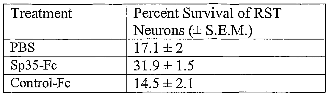
Claims
Priority Applications (26)
| Application Number | Priority Date | Filing Date | Title |
|---|---|---|---|
| MXPA05009913A MXPA05009913A (en) | 2003-03-19 | 2004-03-17 | Nogo receptor binding protein. |
| SI200431552T SI1606409T1 (en) | 2003-03-19 | 2004-03-17 | Nogo receptor binding protein |
| KR1020057017379A KR101106441B1 (en) | 2003-03-19 | 2004-03-17 | Nogo receptor binding protein |
| CN200480013836.XA CN1798840B (en) | 2003-03-19 | 2004-03-17 | Nogo receptor binding protein |
| US10/553,685 US7785829B2 (en) | 2003-03-19 | 2004-03-17 | Nogo receptor binding protein |
| UAA200509809A UA87106C2 (en) | 2003-03-19 | 2004-03-17 | Nogo receptor binding protein |
| EP04757823A EP1606409B1 (en) | 2003-03-19 | 2004-03-17 | Nogo receptor binding protein |
| AU2004223464A AU2004223464C1 (en) | 2003-03-19 | 2004-03-17 | NOGO receptor binding protein |
| JP2006507330A JP5341311B2 (en) | 2003-03-19 | 2004-03-17 | NOGO receptor binding protein |
| CA2519227A CA2519227C (en) | 2003-03-19 | 2004-03-17 | Nogo receptor binding protein |
| DK04757823.2T DK1606409T3 (en) | 2003-03-19 | 2004-03-17 | Nogo receptor binding protein |
| AT04757823T ATE479754T1 (en) | 2003-03-19 | 2004-03-17 | NOGO RECEPTOR BINDING PROTEIN |
| DE602004028916T DE602004028916D1 (en) | 2003-03-19 | 2004-03-17 | NOGO RECEPTOR BINDING PROTEIN |
| PL04757823T PL1606409T3 (en) | 2003-03-19 | 2004-03-17 | Nogo receptor binding protein |
| BRPI0408501-9A BRPI0408501A (en) | 2003-03-19 | 2004-03-17 | nogo receptor binding protein |
| EA200501480A EA010055B1 (en) | 2003-03-19 | 2004-03-17 | Isolated nucleic acid encoding sp35 polypeptide, sp35 polypeptide and methods of use nucleic acid and polypeptide |
| YU20050709A RS54160B1 (en) | 2003-03-19 | 2004-03-17 | Nogo receptor binding protein |
| IL170721A IL170721A (en) | 2003-03-19 | 2005-09-07 | Antibodies that decrease inhibition of axonal growth of a cns neuron and uses thereof |
| IS8016A IS2786B (en) | 2003-03-19 | 2005-09-08 | Protein binding to Nogo receptors |
| NO20054836A NO336530B1 (en) | 2003-03-19 | 2005-10-19 | Nogo reseptorbindingsprotein |
| HK06105710.2A HK1085764A1 (en) | 2003-03-19 | 2006-05-17 | Nogo receptor binding protein nogo |
| US12/698,935 US8153580B2 (en) | 2003-03-19 | 2010-02-02 | Nogo receptor binding protein |
| AU2010202636A AU2010202636A1 (en) | 2003-03-19 | 2010-06-24 | NOGO Receptor Binding Protein |
| US13/414,222 US8765662B2 (en) | 2003-03-19 | 2012-03-07 | NOGO receptor binding protein |
| US13/827,771 US8932821B2 (en) | 2003-03-19 | 2013-03-14 | NOGO receptor binding protein |
| US14/557,653 US20150177240A1 (en) | 2003-03-19 | 2014-12-02 | Nogo Receptor Binding Protein |
Applications Claiming Priority (6)
| Application Number | Priority Date | Filing Date | Title |
|---|---|---|---|
| US45575603P | 2003-03-19 | 2003-03-19 | |
| US60/455,756 | 2003-03-19 | ||
| US48024103P | 2003-06-20 | 2003-06-20 | |
| US60/480,241 | 2003-06-20 | ||
| US49205703P | 2003-08-01 | 2003-08-01 | |
| US60/492,057 | 2003-08-01 |
Related Child Applications (2)
| Application Number | Title | Priority Date | Filing Date |
|---|---|---|---|
| US10/553,685 A-371-Of-International US7785829B2 (en) | 2003-03-19 | 2004-03-17 | Nogo receptor binding protein |
| US12/698,935 Division US8153580B2 (en) | 2003-03-19 | 2010-02-02 | Nogo receptor binding protein |
Publications (2)
| Publication Number | Publication Date |
|---|---|
| WO2004085648A2 true WO2004085648A2 (en) | 2004-10-07 |
| WO2004085648A3 WO2004085648A3 (en) | 2004-11-18 |
Family
ID=33102167
Family Applications (1)
| Application Number | Title | Priority Date | Filing Date |
|---|---|---|---|
| PCT/US2004/008323 WO2004085648A2 (en) | 2003-03-19 | 2004-03-17 | Nogo receptor binding protein |
Country Status (27)
| Country | Link |
|---|---|
| US (5) | US7785829B2 (en) |
| EP (3) | EP1606409B1 (en) |
| JP (3) | JP5341311B2 (en) |
| KR (1) | KR101106441B1 (en) |
| AT (1) | ATE479754T1 (en) |
| AU (2) | AU2004223464C1 (en) |
| BR (1) | BRPI0408501A (en) |
| CA (1) | CA2519227C (en) |
| CY (2) | CY1111097T1 (en) |
| DE (1) | DE602004028916D1 (en) |
| DK (2) | DK1606409T3 (en) |
| EA (1) | EA010055B1 (en) |
| ES (1) | ES2537015T3 (en) |
| GE (1) | GEP20094629B (en) |
| HK (3) | HK1085764A1 (en) |
| HU (1) | HUE025347T2 (en) |
| IL (1) | IL170721A (en) |
| IS (1) | IS2786B (en) |
| MX (1) | MXPA05009913A (en) |
| NO (1) | NO336530B1 (en) |
| NZ (1) | NZ607886A (en) |
| PL (3) | PL378582A1 (en) |
| PT (2) | PT1606409E (en) |
| RS (1) | RS54160B1 (en) |
| SI (2) | SI1606409T1 (en) |
| UA (1) | UA87106C2 (en) |
| WO (1) | WO2004085648A2 (en) |
Cited By (22)
| Publication number | Priority date | Publication date | Assignee | Title |
|---|---|---|---|---|
| EP1681565A1 (en) * | 2005-01-14 | 2006-07-19 | Abbott GmbH & Co. KG | Cellular RhoGTPase activation assay |
| EP1776136A2 (en) * | 2004-06-24 | 2007-04-25 | Biogen Idec MA Inc. | Treatment of conditions involving demyelination |
| WO2007056161A1 (en) * | 2005-11-04 | 2007-05-18 | Biogen Idec Ma Inc. | Methods for promoting neurite outgrowth and survival of dopaminergic neurons |
| WO2007064882A2 (en) | 2005-12-02 | 2007-06-07 | Biogen Idec Ma Inc. | Treatment of conditions involving demyelination |
| JP2009502123A (en) * | 2005-07-08 | 2009-01-29 | バイオジェン・アイデック・エムエイ・インコーポレイテッド | Sp35 antibody and use thereof |
| JP2009513666A (en) * | 2005-10-27 | 2009-04-02 | バイオジェン・アイデック・エムエイ・インコーポレイテッド | Oligodendrocyte-myelin glycoprotein composition and method of use thereof |
| EP2068866A2 (en) * | 2006-07-24 | 2009-06-17 | Biogen Idec MA Inc. | Methods for promoting myelination, neuronal survival and oligodendrocyte differentiation via administration of sp35 or trka antagonists |
| JP2010515456A (en) * | 2007-01-09 | 2010-05-13 | バイオジェン・アイデック・エムエイ・インコーポレイテッド | SP35 antibody and use thereof |
| US7785829B2 (en) | 2003-03-19 | 2010-08-31 | Biogen Idec Ma, Inc. | Nogo receptor binding protein |
| EP2329714A1 (en) | 2004-08-03 | 2011-06-08 | Biogen Idec MA Inc. | Influence of TAJ in the neuronal functions |
| US8058406B2 (en) | 2008-07-09 | 2011-11-15 | Biogen Idec Ma Inc. | Composition comprising antibodies to LINGO or fragments thereof |
| EP2395022A1 (en) | 2006-11-17 | 2011-12-14 | Novartis AG | Lingo binding molecules and pharmaceutical use thereof |
| US8128926B2 (en) | 2007-01-09 | 2012-03-06 | Biogen Idec Ma Inc. | Sp35 antibodies and uses thereof |
| WO2014058875A2 (en) | 2012-10-09 | 2014-04-17 | Biogen Idec Ma Inc. | Combination therapies and uses for treatment of demyelinating disorders |
| WO2016197009A1 (en) | 2015-06-05 | 2016-12-08 | Vertex Pharmaceuticals Incorporated | Triazoles for the treatment of demyelinating diseases |
| US9796780B2 (en) | 2012-05-14 | 2017-10-24 | Biogen Ma Inc. | LINGO-2 antagonists for treatment of conditions involving motor neurons |
| WO2018013714A1 (en) | 2016-07-13 | 2018-01-18 | Biogen Ma Inc. | Dosage regimens of lingo-1 antagonists and uses for treatment of demyelinating disorders |
| WO2018106641A1 (en) | 2016-12-06 | 2018-06-14 | Vertex Pharmaceuticals Incorporated | Pyrazoles for the treatment of demyelinating diseases |
| WO2018106646A1 (en) | 2016-12-06 | 2018-06-14 | Vertex Pharmaceuticals Incorporated | Aminotriazoles for the treatment of demyelinating diseases |
| WO2018106643A1 (en) | 2016-12-06 | 2018-06-14 | Vertex Pharmaceuticals Incorporated | Heterocyclic azoles for the treatment of demyelinating diseases |
| US10435467B2 (en) | 2015-01-08 | 2019-10-08 | Biogen Ma Inc. | LINGO-1 antagonists and uses for treatment of demyelinating disorders |
| WO2020185750A1 (en) | 2019-03-11 | 2020-09-17 | Biogen Ma Inc. | Pharmaceutical compositions containing anti-lingo-1 antibodies |
Families Citing this family (4)
| Publication number | Priority date | Publication date | Assignee | Title |
|---|---|---|---|---|
| WO2009048605A1 (en) * | 2007-10-11 | 2009-04-16 | Biogen Idec Ma Inc. | Methods for treating pressure induced optic neuropathy, preventing neuronal degeneration and promoting neuronal cell, survival via administration of lingo-1 antagonists and trkb agonists |
| AU2008325107B2 (en) * | 2007-11-08 | 2015-04-23 | Biogen Ma Inc. | Use of LINGO-4 antagonists in the treatment of conditions involving demyelination |
| US20130231464A1 (en) * | 2010-04-28 | 2013-09-05 | Oncolmmune, Inc. | Methods of use of soluble cd24 for therapy of rheumatoid arthritis |
| JP2012048656A (en) * | 2010-08-30 | 2012-03-08 | Canon Inc | Image processing apparatus, and image processing method |
Family Cites Families (219)
| Publication number | Priority date | Publication date | Assignee | Title |
|---|---|---|---|---|
| US4444887A (en) * | 1979-12-10 | 1984-04-24 | Sloan-Kettering Institute | Process for making human antibody producing B-lymphocytes |
| US5179017A (en) | 1980-02-25 | 1993-01-12 | The Trustees Of Columbia University In The City Of New York | Processes for inserting DNA into eucaryotic cells and for producing proteinaceous materials |
| US4634665A (en) * | 1980-02-25 | 1987-01-06 | The Trustees Of Columbia University In The City Of New York | Processes for inserting DNA into eucaryotic cells and for producing proteinaceous materials |
| US4399216A (en) | 1980-02-25 | 1983-08-16 | The Trustees Of Columbia University | Processes for inserting DNA into eucaryotic cells and for producing proteinaceous materials |
| IE52535B1 (en) | 1981-02-16 | 1987-12-09 | Ici Plc | Continuous release pharmaceutical compositions |
| US4716111A (en) * | 1982-08-11 | 1987-12-29 | Trustees Of Boston University | Process for producing human antibodies |
| US4510245A (en) | 1982-11-18 | 1985-04-09 | Chiron Corporation | Adenovirus promoter system |
| GB8308235D0 (en) * | 1983-03-25 | 1983-05-05 | Celltech Ltd | Polypeptides |
| US4816567A (en) | 1983-04-08 | 1989-03-28 | Genentech, Inc. | Recombinant immunoglobin preparations |
| US6054561A (en) * | 1984-02-08 | 2000-04-25 | Chiron Corporation | Antigen-binding sites of antibody molecules specific for cancer antigens |
| DE3572982D1 (en) | 1984-03-06 | 1989-10-19 | Takeda Chemical Industries Ltd | Chemically modified lymphokine and production thereof |
| US4694778A (en) * | 1984-05-04 | 1987-09-22 | Anicon, Inc. | Chemical vapor deposition wafer boat |
| US5807715A (en) * | 1984-08-27 | 1998-09-15 | The Board Of Trustees Of The Leland Stanford Junior University | Methods and transformed mammalian lymphocyte cells for producing functional antigen-binding protein including chimeric immunoglobulin |
| US5168062A (en) | 1985-01-30 | 1992-12-01 | University Of Iowa Research Foundation | Transfer vectors and microorganisms containing human cytomegalovirus immediate-early promoter-regulatory DNA sequence |
| DE3668186D1 (en) | 1985-04-01 | 1990-02-15 | Celltech Ltd | TRANSFORMED MYELOMA CELL LINE AND METHOD USING THE SAME FOR EXPRESSING A GENE ENCODING AN EUKARYONTIC POLYPEPTIDE. |
| US4968615A (en) | 1985-12-18 | 1990-11-06 | Ciba-Geigy Corporation | Deoxyribonucleic acid segment from a virus |
| GB8601597D0 (en) | 1986-01-23 | 1986-02-26 | Wilson R H | Nucleotide sequences |
| US5225539A (en) * | 1986-03-27 | 1993-07-06 | Medical Research Council | Recombinant altered antibodies and methods of making altered antibodies |
| GB8607679D0 (en) | 1986-03-27 | 1986-04-30 | Winter G P | Recombinant dna product |
| US4902505A (en) | 1986-07-30 | 1990-02-20 | Alkermes | Chimeric peptides for neuropeptide delivery through the blood-brain barrier |
| US4946778A (en) * | 1987-09-21 | 1990-08-07 | Genex Corporation | Single polypeptide chain binding molecules |
| US5116742A (en) | 1986-12-03 | 1992-05-26 | University Patents, Inc. | RNA ribozyme restriction endoribonucleases and methods |
| US5258498A (en) * | 1987-05-21 | 1993-11-02 | Creative Biomolecules, Inc. | Polypeptide linkers for production of biosynthetic proteins |
| US4904582A (en) | 1987-06-11 | 1990-02-27 | Synthetic Genetics | Novel amphiphilic nucleic acid conjugates |
| US5892019A (en) * | 1987-07-15 | 1999-04-06 | The United States Of America, As Represented By The Department Of Health And Human Services | Production of a single-gene-encoded immunoglobulin |
| GB8717430D0 (en) | 1987-07-23 | 1987-08-26 | Celltech Ltd | Recombinant dna product |
| US5336603A (en) | 1987-10-02 | 1994-08-09 | Genentech, Inc. | CD4 adheson variants |
| WO1989006692A1 (en) * | 1988-01-12 | 1989-07-27 | Genentech, Inc. | Method of treating tumor cells by inhibiting growth factor receptor function |
| GB8809129D0 (en) | 1988-04-18 | 1988-05-18 | Celltech Ltd | Recombinant dna methods vectors and host cells |
| DE68921982D1 (en) | 1988-06-14 | 1995-05-04 | Cetus Oncology Corp | COUPLING AGENTS AND STERICALLY DISABLED CONJUGATES THEREOF. |
| US5223409A (en) * | 1988-09-02 | 1993-06-29 | Protein Engineering Corp. | Directed evolution of novel binding proteins |
| EP1892296A1 (en) | 1988-09-02 | 2008-02-27 | Dyax Corporation | Generation and selection of recombinant varied binding proteins |
| AU634186B2 (en) | 1988-11-11 | 1993-02-18 | Medical Research Council | Single domain ligands, receptors comprising said ligands, methods for their production, and use of said ligands and receptors |
| JP2989002B2 (en) | 1988-12-22 | 1999-12-13 | キリン―アムジエン・インコーポレーテツド | Chemically modified granulocyte colony stimulating factor |
| US5530101A (en) * | 1988-12-28 | 1996-06-25 | Protein Design Labs, Inc. | Humanized immunoglobulins |
| US5225538A (en) * | 1989-02-23 | 1993-07-06 | Genentech, Inc. | Lymphocyte homing receptor/immunoglobulin fusion proteins |
| ZA902949B (en) | 1989-05-05 | 1992-02-26 | Res Dev Foundation | A novel antibody delivery system for biological response modifiers |
| DK0474727T3 (en) * | 1989-05-19 | 1998-01-12 | Genentech Inc | HER2 extracellular domain |
| US5413923A (en) * | 1989-07-25 | 1995-05-09 | Cell Genesys, Inc. | Homologous recombination for universal donor cells and chimeric mammalian hosts |
| US5180820A (en) | 1989-08-30 | 1993-01-19 | Barde Yves Alain | Brain-derived neurotrophic factor |
| US5605690A (en) | 1989-09-05 | 1997-02-25 | Immunex Corporation | Methods of lowering active TNF-α levels in mammals using tumor necrosis factor receptor |
| GB8928874D0 (en) | 1989-12-21 | 1990-02-28 | Celltech Ltd | Humanised antibodies |
| US5780225A (en) * | 1990-01-12 | 1998-07-14 | Stratagene | Method for generating libaries of antibody genes comprising amplification of diverse antibody DNAs and methods for using these libraries for the production of diverse antigen combining molecules |
| WO1991010737A1 (en) | 1990-01-11 | 1991-07-25 | Molecular Affinities Corporation | Production of antibodies using gene libraries |
| US6150584A (en) * | 1990-01-12 | 2000-11-21 | Abgenix, Inc. | Human antibodies derived from immunized xenomice |
| DE69133566T2 (en) | 1990-01-12 | 2007-12-06 | Amgen Fremont Inc. | Formation of xenogenic antibodies |
| US6075181A (en) * | 1990-01-12 | 2000-06-13 | Abgenix, Inc. | Human antibodies derived from immunized xenomice |
| US5314995A (en) * | 1990-01-22 | 1994-05-24 | Oncogen | Therapeutic interleukin-2-antibody based fusion proteins |
| EP0521985B1 (en) | 1990-03-20 | 1997-09-24 | The Trustees Of Columbia University In The City Of New York | Chimeric antibodies with receptor binding ligands in place of their constant region |
| US5427908A (en) * | 1990-05-01 | 1995-06-27 | Affymax Technologies N.V. | Recombinant library screening methods |
| US5219837A (en) * | 1990-06-21 | 1993-06-15 | Trustees Of The University Of Pennsylvania | Method of stimulating myelination of cells |
| GB9015198D0 (en) | 1990-07-10 | 1990-08-29 | Brien Caroline J O | Binding substance |
| US5532351A (en) | 1990-07-12 | 1996-07-02 | Arch Development Corporation | Nucleic acid sequences encoding OMGP |
| US5661016A (en) | 1990-08-29 | 1997-08-26 | Genpharm International Inc. | Transgenic non-human animals capable of producing heterologous antibodies of various isotypes |
| US5625126A (en) * | 1990-08-29 | 1997-04-29 | Genpharm International, Inc. | Transgenic non-human animals for producing heterologous antibodies |
| US5545806A (en) * | 1990-08-29 | 1996-08-13 | Genpharm International, Inc. | Ransgenic non-human animals for producing heterologous antibodies |
| US5633425A (en) * | 1990-08-29 | 1997-05-27 | Genpharm International, Inc. | Transgenic non-human animals capable of producing heterologous antibodies |
| US5814318A (en) * | 1990-08-29 | 1998-09-29 | Genpharm International Inc. | Transgenic non-human animals for producing heterologous antibodies |
| JP2938569B2 (en) * | 1990-08-29 | 1999-08-23 | ジェンファーム インターナショナル,インコーポレイティド | Method for producing xenogeneic immunoglobulin and transgenic mouse |
| US5698426A (en) * | 1990-09-28 | 1997-12-16 | Ixsys, Incorporated | Surface expression libraries of heteromeric receptors |
| ATE218889T1 (en) | 1990-11-09 | 2002-06-15 | Stephen D Gillies | CYTOKINE IMMUNOCONJUGATES |
| US5252714A (en) | 1990-11-28 | 1993-10-12 | The University Of Alabama In Huntsville | Preparation and use of polyethylene glycol propionaldehyde |
| WO1992009690A2 (en) * | 1990-12-03 | 1992-06-11 | Genentech, Inc. | Enrichment method for variant proteins with altered binding properties |
| JP3672306B2 (en) | 1991-04-10 | 2005-07-20 | ザ スクリップス リサーチ インスティテュート | Heterodimeric receptor library using phagemids |
| EP0519596B1 (en) | 1991-05-17 | 2005-02-23 | Merck & Co. Inc. | A method for reducing the immunogenicity of antibody variable domains |
| CA2110799A1 (en) | 1991-06-14 | 1992-12-23 | Arnold H. Horwitz | Microbially-produced antibody fragments and their conjugates |
| US5756096A (en) * | 1991-07-25 | 1998-05-26 | Idec Pharmaceuticals Corporation | Recombinant antibodies for human therapy |
| IE922437A1 (en) * | 1991-07-25 | 1993-01-27 | Idec Pharma Corp | Recombinant antibodies for human therapy |
| US5565332A (en) * | 1991-09-23 | 1996-10-15 | Medical Research Council | Production of chimeric antibodies - a combinatorial approach |
| DK1024191T3 (en) | 1991-12-02 | 2008-12-08 | Medical Res Council | Preparation of autoantibodies displayed on phage surfaces from antibody segment libraries |
| ATE244763T1 (en) * | 1992-02-11 | 2003-07-15 | Cell Genesys Inc | ACHIEVEMENT OF HOMOZYGOTE THROUGH TARGETED GENETIC EVENTS |
| US5733743A (en) * | 1992-03-24 | 1998-03-31 | Cambridge Antibody Technology Limited | Methods for producing members of specific binding pairs |
| US5639641A (en) | 1992-09-09 | 1997-06-17 | Immunogen Inc. | Resurfacing of rodent antibodies |
| EP0627932B1 (en) * | 1992-11-04 | 2002-05-08 | City Of Hope | Antibody construct |
| ES2088838T3 (en) * | 1992-11-13 | 2004-01-01 | Idec Pharmaceuticals Corporation | FULLY ALTERED KOZAK CONSENSUS SEQUENCES INTENDED FOR EXPRESSION IN THE MAMMALS. |
| US5468872A (en) | 1993-09-16 | 1995-11-21 | Cephalon, Inc. | K-252a functional derivatives potentiate neurotrophin-3 for the treatment of neurological disorders |
| ATE163231T1 (en) * | 1993-11-23 | 1998-02-15 | Genentech Inc | KINASE RECEPTOR ACTIVATION TEST |
| US5753225A (en) | 1993-12-03 | 1998-05-19 | The Regents Of The University Of California | Antibodies that mimic actions of neurotrophins |
| JPH09506262A (en) | 1993-12-08 | 1997-06-24 | ジェンザイム・コーポレイション | Method for producing specific antibody |
| DK1231268T3 (en) | 1994-01-31 | 2005-11-21 | Univ Boston | Polyclonal antibody libraries |
| GB9402331D0 (en) | 1994-02-07 | 1994-03-30 | Univ Mcgill | Nerve growth factor structural analogs and their uses |
| US5877016A (en) | 1994-03-18 | 1999-03-02 | Genentech, Inc. | Human trk receptors and neurotrophic factor inhibitors |
| DE4447588C2 (en) * | 1994-05-03 | 1997-11-20 | Omer Osama Dr Dr Med | Treatment of herpes and hepatitis virus infections and bronchitis |
| US5516637A (en) * | 1994-06-10 | 1996-05-14 | Dade International Inc. | Method involving display of protein binding pairs on the surface of bacterial pili and bacteriophage |
| WO2001004311A1 (en) | 1999-07-07 | 2001-01-18 | Genentech, Inc. | Secreted and transmembrane polypeptides and nucleic acids encoding the same |
| ES2171559T3 (en) | 1994-10-25 | 2002-09-16 | Toyama Chemical Co Ltd | AGENT TO POTENTIATE THE ACTIVITY OF THE NERVOUS GROWTH FACTOR CONTAINING A 1,2-ETHANODIOL DERIVATIVE OR A SALT OF THE SAME. |
| US5770577A (en) | 1994-11-14 | 1998-06-23 | Amgen Inc. | BDNF and NT-3 polypeptides selectively linked to polyethylene glycol |
| US6130364A (en) * | 1995-03-29 | 2000-10-10 | Abgenix, Inc. | Production of antibodies using Cre-mediated site-specific recombination |
| US6280964B1 (en) * | 1995-04-14 | 2001-08-28 | The Regents Of The University Of California | Binding sites for phosphotyrosine binding domains |
| EP0822830B1 (en) | 1995-04-27 | 2008-04-02 | Amgen Fremont Inc. | Human anti-IL-8 antibodies, derived from immunized xenomice |
| WO1996034096A1 (en) | 1995-04-28 | 1996-10-31 | Abgenix, Inc. | Human antibodies derived from immunized xenomice |
| US5811524A (en) * | 1995-06-07 | 1998-09-22 | Idec Pharmaceuticals Corporation | Neutralizing high affinity human monoclonal antibodies specific to RSV F-protein and methods for their manufacture and therapeutic use thereof |
| EP0873363B1 (en) | 1995-06-14 | 2010-10-06 | The Regents of The University of California | High affinity human antibodies to tumor antigens |
| US5707829A (en) * | 1995-08-11 | 1998-01-13 | Genetics Institute, Inc. | DNA sequences and secreted proteins encoded thereby |
| IT235676Y1 (en) | 1995-07-27 | 2000-07-12 | Felice Gaudioso | SET-UP FOR PLAYING THE GAME WORMHOLE ACTION MATCH. |
| GB9525180D0 (en) | 1995-12-08 | 1996-02-07 | Univ Mcgill | Design of hormone-like antibodies with agonistic and antagonistic fuctions |
| JP2978435B2 (en) * | 1996-01-24 | 1999-11-15 | チッソ株式会社 | Method for producing acryloxypropyl silane |
| US6455277B1 (en) | 1996-04-22 | 2002-09-24 | Amgen Inc. | Polynucleotides encoding human glial cell line-derived neurotrophic factor receptor polypeptides |
| JP2000501416A (en) | 1996-04-26 | 2000-02-08 | ユニバーシティー オブ オタワ | Methods of treatment and drug screening to treat and prevent neuronal disease |
| US5611016A (en) * | 1996-06-07 | 1997-03-11 | Lucent Technologies Inc. | Dispersion-balanced optical cable |
| PT912184E (en) | 1996-06-25 | 2002-12-31 | Cephalon Inc | USE OF A K-252A DERIVATIVE FOR THE TREATMENT OF CENTRAL OR PERIPHERAL NERVOUS SIETMA DISEASES AND CITOQUINE OVER-PRODUCTION |
| ATE486937T1 (en) * | 1996-07-12 | 2010-11-15 | Genentech Inc | CHIMERIC HETEROMULTIMERIC ADHESINS |
| NZ333650A (en) | 1996-07-19 | 2000-02-28 | Amgen Inc | Analogs of cationic proteins like NT-3 (neurotrophin-3) and BDNF (brain derived neurotrophic factor) that are circulated more readily due to a lower isoelectric point and a lower protein charge relative to the native protein |
| WO1998011243A2 (en) | 1996-09-11 | 1998-03-19 | The General Hospital Corporation | Expression of an exogenous gene in a mammalian cell by use of a non-mammalian dna virus having an altered coat protein |
| IL128941A (en) | 1996-09-13 | 2007-05-15 | Advanced Medicine Res Inst | Ophthalmic composition containing a neurotrophic factor for treating an optic nerve functional disorder |
| US6420140B1 (en) * | 1996-10-11 | 2002-07-16 | Abgenix, Inc. | Production of multimeric protein by cell fusion method |
| US5916771A (en) | 1996-10-11 | 1999-06-29 | Abgenix, Inc. | Production of a multimeric protein by cell fusion method |
| US20070213290A1 (en) | 1996-10-17 | 2007-09-13 | Kingsman Alan J | Neurite regeneration |
| US6593290B1 (en) | 1996-11-01 | 2003-07-15 | Genentech, Inc. | Treatment of inner ear hair cells |
| US6156728A (en) | 1996-11-01 | 2000-12-05 | Genentech, Inc. | Treatment of inner ear hair cells |
| ES2301183T3 (en) | 1996-12-03 | 2008-06-16 | Amgen Fremont Inc. | COMPLETELY HUMAN ANTIBODY THAT JOINS THE EGFR RECEIVER. |
| ES2242997T3 (en) * | 1997-03-14 | 2005-11-16 | Biogen Idec Inc. | METHOD FOR INTEGRATING GENES IN SPECIFIC SITES IN MAMMER CELLS BY HOMOLOGICAL RECOMBINATION AND VECTORS TO MAKE THE SAME. |
| US20020137890A1 (en) * | 1997-03-31 | 2002-09-26 | Genentech, Inc. | Secreted and transmembrane polypeptides and nucleic acids encoding the same |
| SI0970126T1 (en) | 1997-04-14 | 2001-08-31 | Micromet Ag | Novel method for the production of antihuman antigen receptors and uses thereof |
| US6235883B1 (en) | 1997-05-05 | 2001-05-22 | Abgenix, Inc. | Human monoclonal antibodies to epidermal growth factor receptor |
| JP2002512624A (en) | 1997-05-21 | 2002-04-23 | バイオベーション リミテッド | Method for producing non-immunogenic protein |
| JP2001512003A (en) | 1997-08-04 | 2001-08-21 | ミレニアム ファーマシューティカルズ インク. | Tango-78, Tango-79 and Tango-81 polypeptides, nucleic acid molecules encoding Tango-78, Tango-79 and Tango-81 and uses thereof |
| US20020112251A1 (en) * | 1997-08-04 | 2002-08-15 | Mccarthy Sean A. | Novel genes encoding proteins having prognostic, diagnostic, preventive, therapeutic and other uses |
| IL134968A0 (en) | 1997-09-17 | 2001-05-20 | Genentech Inc | Secreted and transmembrane polypeptides and nucleic acids encoding the same |
| US6974689B1 (en) * | 1997-09-18 | 2005-12-13 | Genentech, Inc. | Nucleic acid encoding PRO211 polypeptides |
| EP1205546B1 (en) * | 1997-10-24 | 2005-11-23 | Genentech, Inc. | Polypeptide and nucleic acids encoding the same |
| FR2776661B1 (en) | 1998-03-26 | 2002-04-05 | Centre Nat Rech Scient | IMMUNOREACTIVE POLYPEPTIDE OF THE NGF TRKA RECEPTOR AND USES |
| US7282482B2 (en) | 1998-04-08 | 2007-10-16 | The Regents Of The University Of California | NGF for the prevention of demyelination in the nervous system |
| CA2343577A1 (en) | 1998-09-16 | 2000-03-23 | Genentech, Inc. | Secreted and transmembrane polypeptides and nucleic acids encoding the same |
| WO2000031235A2 (en) | 1998-11-06 | 2000-06-02 | Schwab Martin E | Nucleotide and protein sequences of nogo genes and methods based thereon |
| WO2000034317A2 (en) | 1998-12-08 | 2000-06-15 | Biovation Limited | Method for reducing immunogenicity of proteins |
| WO2000058473A2 (en) | 1999-03-31 | 2000-10-05 | Curagen Corporation | Nucleic acids including open reading frames encoding polypeptides; 'orfx' |
| US7034132B2 (en) | 2001-06-04 | 2006-04-25 | Anderson David W | Therapeutic polypeptides, nucleic acids encoding same, and methods of use |
| US20040067490A1 (en) | 2001-09-07 | 2004-04-08 | Mei Zhong | Therapeutic polypeptides, nucleic acids encoding same, and methods of use |
| US6949245B1 (en) * | 1999-06-25 | 2005-09-27 | Genentech, Inc. | Humanized anti-ErbB2 antibodies and treatment with anti-ErbB2 antibodies |
| EP1189931B1 (en) * | 1999-07-02 | 2006-06-21 | Genentech, Inc. | Peptide compounds that bind her2 |
| EP1074617A3 (en) | 1999-07-29 | 2004-04-21 | Research Association for Biotechnology | Primers for synthesising full-length cDNA and their use |
| WO2001009318A1 (en) * | 1999-07-29 | 2001-02-08 | Helix Research Institute | Liver cancer-associated genes |
| WO2001012662A2 (en) * | 1999-08-17 | 2001-02-22 | Incyte Genomics, Inc. | Membrane associated proteins |
| US6461414B1 (en) | 1999-10-29 | 2002-10-08 | Baker Hughes Incorporated | Foam monitoring and control system |
| EP1690872A3 (en) | 1999-12-01 | 2006-08-23 | Genentech, Inc. | Composition and methods for the diagnosis of tumours |
| US6680209B1 (en) | 1999-12-06 | 2004-01-20 | Biosite, Incorporated | Human antibodies as diagnostic reagents |
| US20020012965A1 (en) | 2000-01-12 | 2002-01-31 | Strittmatter Stephen M. | Nogo receptor-mediated blockade of axonal growth |
| US7119165B2 (en) | 2000-01-12 | 2006-10-10 | Yale University | Nogo receptor-mediated blockade of axonal growth |
| ATE340571T1 (en) | 2000-01-18 | 2006-10-15 | Univ Mcgill | PHARMACEUTICAL PREPARATIONS CONTAINING BETA-TURN PEPTIDOMIMMETIC CYCLIC SUBSTANCES |
| US20040132022A1 (en) | 2000-01-25 | 2004-07-08 | Boyle Bryan J. | Methods and materials relating to soluble transforming growth factor alpha-like polypeptides and polynucleotides |
| WO2001055317A2 (en) | 2000-01-31 | 2001-08-02 | Human Genome Sciences, Inc. | Nucleic acids, proteins, and antibodies |
| WO2001059063A2 (en) | 2000-01-31 | 2001-08-16 | Human Genome Sciences, Inc. | Nucleic acids, proteins, and antibodies |
| WO2001055312A2 (en) * | 2000-01-31 | 2001-08-02 | Human Genome Sciences, Inc. | Nucleic acids, proteins, and antibodies |
| EP1254269A4 (en) | 2000-02-03 | 2004-10-27 | Nuvelo Inc | Methods and materials relating to neuronal guidance molecule-like (ngm-like) polypeptides and polynucleotides |
| AU2001234178A1 (en) | 2000-02-29 | 2001-09-12 | Ltt Institute Co., Ltd. | Modified bdnf |
| WO2001077342A1 (en) * | 2000-04-11 | 2001-10-18 | Genentech, Inc. | Multivalent antibodies and uses therefor |
| WO2002000677A1 (en) * | 2000-06-07 | 2002-01-03 | Human Genome Sciences, Inc. | Nucleic acids, proteins, and antibodies |
| US6430914B1 (en) | 2000-06-29 | 2002-08-13 | Foster Wheeler Energy Corporation | Combined cycle power generation plant and method of operating such a plant |
| US20040010118A1 (en) | 2000-08-16 | 2004-01-15 | Zerhusen Bryan D. | Novel proteins and nucleic acids encoding same |
| BR0113820A (en) | 2000-09-13 | 2003-06-24 | Smithkline Beecham Corp | Compounds |
| JP2004531203A (en) | 2000-10-05 | 2004-10-14 | キュラジェン コーポレイション | Human proteins, polynucleotides encoding them, and methods of using them |
| AU2002211539B2 (en) * | 2000-10-06 | 2007-01-25 | Biogen Idec Ma Inc. | Nogo receptor homologs |
| AU2001297872B2 (en) * | 2000-11-17 | 2006-11-09 | University Of Rochester | In vitro methods of producing and identifying immunoglobulin molecules in eukaryotic cells |
| US20030216558A1 (en) | 2000-12-22 | 2003-11-20 | Morris David W. | Novel compositions and methods for cancer |
| WO2002068579A2 (en) | 2001-01-10 | 2002-09-06 | Pe Corporation (Ny) | Kits, such as nucleic acid arrays, comprising a majority of human exons or transcripts, for detecting expression and other uses thereof |
| CN1494553A (en) | 2001-01-29 | 2004-05-05 | IDECҩ�﹫˾ | Modified antibodies and method of use |
| AU2002327164A1 (en) | 2001-01-29 | 2002-12-09 | Idec Pharmaceuticals Corporation | Engineered tetravalent antibodies and methods of use |
| WO2003008583A2 (en) | 2001-03-02 | 2003-01-30 | Sagres Discovery | Novel compositions and methods for cancer |
| FI111522B (en) * | 2001-05-07 | 2003-08-15 | Marioff Corp Oy | Fire fighting equipment and source of fire fighting equipment |
| EP1401858A4 (en) | 2001-06-04 | 2005-12-21 | Curagen Corp | Therapeutic polypeptides, nucleic acids encoding same, and methods of use |
| US20030162734A1 (en) | 2001-06-28 | 2003-08-28 | Miller Carol A. | Modulation of DENN-MADD expression and interactions for treating neurological disorders |
| US7223558B2 (en) | 2001-07-11 | 2007-05-29 | Bristol-Myers Squibb Company | Polynucleotides encoding three novel human cell surface proteins with leucine rich repeats and immunologobulin folds, BGS2, 3, and 4 and variants thereof |
| US20030226155A1 (en) | 2001-08-30 | 2003-12-04 | Biorexis Pharmaceutical Corporation | Modified transferrin-antibody fusion proteins |
| US20040142325A1 (en) * | 2001-09-14 | 2004-07-22 | Liat Mintz | Methods and systems for annotating biomolecular sequences |
| US20050123990A1 (en) | 2001-09-26 | 2005-06-09 | Incyte Corporation | Membrane associated proteins |
| WO2003061559A2 (en) | 2001-10-12 | 2003-07-31 | University Of Vermont And State Agricultural College | Binding peptides specific for the extracellular domain of erbb2 and uses therefor |
| AU2002337916A1 (en) | 2001-10-22 | 2003-05-06 | Exelixis, Inc. | Modifier of the p53 pathway and methods of use |
| US7309485B2 (en) | 2001-12-03 | 2007-12-18 | Children's Medical Center Corporation | Reducing myelin-mediated inhibition of axon regeneration |
| EP1504099A4 (en) | 2001-12-10 | 2006-05-10 | Nuvelo Inc | Novel nucleic acids and polypeptides |
| AU2003216482A1 (en) | 2002-03-01 | 2003-09-16 | Exelixis, Inc. | MSRAs AS MODIFIERS OF THE p53 PATHWAY AND METHODS OF USE |
| US7718776B2 (en) | 2002-04-05 | 2010-05-18 | Amgen Inc. | Human anti-OPGL neutralizing antibodies as selective OPGL pathway inhibitors |
| KR20050062525A (en) | 2002-08-10 | 2005-06-23 | 예일 유니버시티 | Nogo receptor antagonists |
| NZ593428A (en) | 2002-09-06 | 2013-01-25 | Amgen Inc | Therapeutic human anti-il-1r1 monoclonal antibody |
| US6919426B2 (en) | 2002-09-19 | 2005-07-19 | Amgen Inc. | Peptides and related molecules that modulate nerve growth factor activity |
| US7816497B2 (en) | 2002-10-30 | 2010-10-19 | University Of Kentucky | Compositions and methods for inhibiting drusen complement components C3a and C5a for the treatment of age-related macular degeneration |
| US20040186044A1 (en) | 2002-11-06 | 2004-09-23 | Cosgaya Jose Miguel | Modulation of myelination by interaction with P75 and TRK receptors |
| US7388079B2 (en) | 2002-11-27 | 2008-06-17 | The Regents Of The University Of California | Delivery of pharmaceutical agents via the human insulin receptor |
| SI1606409T1 (en) | 2003-03-19 | 2011-01-31 | Biogen Idec Inc | Nogo receptor binding protein |
| CA2535007A1 (en) | 2003-08-07 | 2005-02-24 | Biogen Idec Ma Inc. | Nogo receptor antagonists |
| CA2536408A1 (en) | 2003-08-22 | 2005-03-03 | Biogen Idec Ma Inc. | Improved antibodies having altered effector function and methods for making the same |
| US7205387B2 (en) | 2003-08-28 | 2007-04-17 | Agency For Science, Technology And Research | Recombinant polypeptide useful for neurotrophin receptor mediated gene delivery and as neurotrophin agonist |
| WO2005021579A2 (en) | 2003-08-28 | 2005-03-10 | Biorexis Pharmaceutical Corporation | Epo mimetic peptides and fusion proteins |
| US7288638B2 (en) | 2003-10-10 | 2007-10-30 | Bristol-Myers Squibb Company | Fully human antibodies against human 4-1BB |
| ES2378767T3 (en) | 2003-12-23 | 2012-04-17 | Crucell Holland B.V. | Human binding molecule against CD1a |
| CA2555340A1 (en) | 2004-02-12 | 2005-09-01 | Lexicon Genetics Incorporated | Gene disruptions, compositions and methods relating thereto |
| US7973139B2 (en) * | 2004-03-26 | 2011-07-05 | Human Genome Sciences, Inc. | Antibodies against nogo receptor |
| US20050214288A1 (en) * | 2004-03-26 | 2005-09-29 | Human Genome Sciences, Inc. | Antibodies against Nogo receptor |
| ATE540696T1 (en) * | 2004-04-08 | 2012-01-15 | David B Agus | ERBB2 ANTAGONISTS FOR TUMOR PAIN THERAPY |
| EP1776136B1 (en) | 2004-06-24 | 2012-10-03 | Biogen Idec MA Inc. | Treatment of conditions involving demyelination |
| US20060009288A1 (en) | 2004-07-07 | 2006-01-12 | Devos John A | Conveying information to an interrogator using resonant and parasitic radio frequency circuits |
| CN101014245A (en) | 2004-08-03 | 2007-08-08 | 比奥根艾迪克Ma公司 | Taj in neuronal function |
| JO3000B1 (en) * | 2004-10-20 | 2016-09-05 | Genentech Inc | Antibody Formulations. |
| AR054260A1 (en) | 2005-04-26 | 2007-06-13 | Rinat Neuroscience Corp | METHODS OF TREATMENT OF DISEASES OF THE LOWER MOTOR NEURONE AND COMPOSITIONS USED IN THE SAME |
| JP2008540339A (en) | 2005-04-29 | 2008-11-20 | ワイス | Nogo receptor functional motif and peptidomimetics related thereto, and methods of using them |
| WO2006136006A1 (en) | 2005-05-16 | 2006-12-28 | Mcgill University | Lgi, lingo and p75ntr family members: novel modulators of neuronal growth |
| EP1891114A2 (en) | 2005-06-06 | 2008-02-27 | Wyeth | Anti-trkb monoclonal antibodies and uses thereof |
| EP1893192A4 (en) | 2005-06-08 | 2010-03-31 | Univ North Carolina | Methods of facilitating neural cell survival using non-peptide and peptide bdnf neurotrophin mimetics |
| AU2005332949A1 (en) | 2005-06-16 | 2006-12-21 | University Technologies International Inc. | A treatment for short bowel syndrome |
| EP2478917A1 (en) | 2005-07-08 | 2012-07-25 | Biogen Idec MA Inc. | SP35 antibodies and uses thereof |
| US7612181B2 (en) * | 2005-08-19 | 2009-11-03 | Abbott Laboratories | Dual variable domain immunoglobulin and uses thereof |
| EP1940460A4 (en) | 2005-10-27 | 2009-08-12 | Biogen Idec Inc | Oligodendrocyte-myelin glycoprotein compositions and methods of use thereof |
| US20090246189A1 (en) | 2005-11-04 | 2009-10-01 | Biogen Idec Ma Inc. And Mclean Hospital | Methods for Promoting Neurite Outgrowth and Survival of Dopaminergic Neurons |
| US20090175872A1 (en) | 2005-12-02 | 2009-07-09 | Biogen Idec Ma Inc. | Treatment of Conditions Involving Demyelination |
| WO2007092777A2 (en) | 2006-02-02 | 2007-08-16 | Wyeth | Cloning, characterization, and application of tnfrsf19 in neurological disorders |
| US7693698B2 (en) * | 2006-02-03 | 2010-04-06 | Wyeth Llc | Method for identifying or designing a candidate agent that interacts with LINGO-1 polypeptide using a LINGO-1 three-dimensional structure |
| WO2007098283A2 (en) | 2006-02-27 | 2007-08-30 | Biogen Idec Ma Inc. | Use of antagonists of maif receptor complex molecules and neurotrophic factors for treatment of neurological diseases and disorders |
| JP2009544703A (en) | 2006-07-24 | 2009-12-17 | バイオジェン・アイデック・エムエイ・インコーポレイテッド | Method for promoting myelination, neuronal survival and oligodendrocyte differentiation by administration of an Sp35 or TrkA antagonist |
| US8299221B2 (en) | 2006-11-17 | 2012-10-30 | Novartis Ag | LINGO binding molecules and pharmaceutical use thereof |
| US8128926B2 (en) | 2007-01-09 | 2012-03-06 | Biogen Idec Ma Inc. | Sp35 antibodies and uses thereof |
| GEP20125693B (en) | 2007-01-09 | 2012-11-26 | Biogen Idec Inc | Sp35 antibodies and usage thereof |
| WO2009048605A1 (en) | 2007-10-11 | 2009-04-16 | Biogen Idec Ma Inc. | Methods for treating pressure induced optic neuropathy, preventing neuronal degeneration and promoting neuronal cell, survival via administration of lingo-1 antagonists and trkb agonists |
| AU2008325107B2 (en) | 2007-11-08 | 2015-04-23 | Biogen Ma Inc. | Use of LINGO-4 antagonists in the treatment of conditions involving demyelination |
| DK2307042T3 (en) | 2008-06-25 | 2014-05-12 | Lundbeck & Co As H | MODULE TRPV: VPS10P DOMAIN RECEPTOR SYSTEM FOR THE TREATMENT OF PAIN |
| US20110152173A1 (en) | 2008-07-02 | 2011-06-23 | Emergent Product Development Seattle ,LLC | TNF-a ANTAGONIST MULTI-TARGET BINDING PROTEINS |
| JP2011527572A (en) | 2008-07-09 | 2011-11-04 | バイオジェン・アイデック・エムエイ・インコーポレイテッド | Composition comprising a LINGO antibody or fragment |
| GB2512153B (en) | 2008-11-17 | 2014-11-12 | Veracyte Inc | Methods and compositions of molecular profiling for disease diagnostics |
| FR2958293B1 (en) | 2010-04-01 | 2014-09-26 | Centre Nat Rech Scient | TOOLS FOR THE IDENTIFICATION OF LINGO-1, LINGO-2, LINGO-3 AND LINGO-4 LIGANDS, AND USES THEREOF |
| JP2015518829A (en) | 2012-05-14 | 2015-07-06 | バイオジェン・エムエイ・インコーポレイテッドBiogen MA Inc. | LINGO-2 antagonist for treatment of conditions involving motor neurons |
-
2004
- 2004-03-17 SI SI200431552T patent/SI1606409T1/en unknown
- 2004-03-17 GE GEAP20049025A patent/GEP20094629B/en unknown
- 2004-03-17 PL PL378582A patent/PL378582A1/en not_active Application Discontinuation
- 2004-03-17 NZ NZ607886A patent/NZ607886A/en not_active IP Right Cessation
- 2004-03-17 WO PCT/US2004/008323 patent/WO2004085648A2/en active Application Filing
- 2004-03-17 CA CA2519227A patent/CA2519227C/en not_active Expired - Fee Related
- 2004-03-17 PL PL10171533T patent/PL2248899T3/en unknown
- 2004-03-17 HU HUE10171533A patent/HUE025347T2/en unknown
- 2004-03-17 AT AT04757823T patent/ATE479754T1/en active
- 2004-03-17 KR KR1020057017379A patent/KR101106441B1/en not_active IP Right Cessation
- 2004-03-17 EP EP04757823A patent/EP1606409B1/en not_active Expired - Lifetime
- 2004-03-17 US US10/553,685 patent/US7785829B2/en active Active
- 2004-03-17 PL PL04757823T patent/PL1606409T3/en unknown
- 2004-03-17 DK DK04757823.2T patent/DK1606409T3/en active
- 2004-03-17 JP JP2006507330A patent/JP5341311B2/en not_active Expired - Fee Related
- 2004-03-17 UA UAA200509809A patent/UA87106C2/en unknown
- 2004-03-17 DK DK10171533.2T patent/DK2248899T3/en active
- 2004-03-17 BR BRPI0408501-9A patent/BRPI0408501A/en not_active IP Right Cessation
- 2004-03-17 SI SI200432246T patent/SI2248899T1/en unknown
- 2004-03-17 PT PT04757823T patent/PT1606409E/en unknown
- 2004-03-17 ES ES10171533.2T patent/ES2537015T3/en not_active Expired - Lifetime
- 2004-03-17 RS YU20050709A patent/RS54160B1/en unknown
- 2004-03-17 EA EA200501480A patent/EA010055B1/en not_active IP Right Cessation
- 2004-03-17 EP EP10171533.2A patent/EP2248899B8/en not_active Expired - Lifetime
- 2004-03-17 AU AU2004223464A patent/AU2004223464C1/en not_active Ceased
- 2004-03-17 EP EP15160028.5A patent/EP2902491A1/en not_active Withdrawn
- 2004-03-17 DE DE602004028916T patent/DE602004028916D1/en not_active Expired - Lifetime
- 2004-03-17 PT PT101715332T patent/PT2248899E/en unknown
- 2004-03-17 MX MXPA05009913A patent/MXPA05009913A/en active IP Right Grant
-
2005
- 2005-09-07 IL IL170721A patent/IL170721A/en not_active IP Right Cessation
- 2005-09-08 IS IS8016A patent/IS2786B/en unknown
- 2005-10-19 NO NO20054836A patent/NO336530B1/en not_active IP Right Cessation
-
2006
- 2006-05-17 HK HK06105710.2A patent/HK1085764A1/en not_active IP Right Cessation
- 2006-05-17 HK HK11100406.5A patent/HK1146297A1/en not_active IP Right Cessation
-
2010
- 2010-02-02 US US12/698,935 patent/US8153580B2/en active Active
- 2010-02-18 JP JP2010034094A patent/JP2010154861A/en active Pending
- 2010-06-24 AU AU2010202636A patent/AU2010202636A1/en not_active Abandoned
- 2010-11-30 CY CY20101101094T patent/CY1111097T1/en unknown
-
2012
- 2012-03-07 US US13/414,222 patent/US8765662B2/en not_active Expired - Lifetime
-
2013
- 2013-03-14 US US13/827,771 patent/US8932821B2/en not_active Expired - Lifetime
- 2013-04-16 JP JP2013085754A patent/JP2013138688A/en not_active Withdrawn
-
2014
- 2014-12-02 US US14/557,653 patent/US20150177240A1/en not_active Abandoned
-
2015
- 2015-07-29 CY CY20151100669T patent/CY1116863T1/en unknown
- 2015-12-11 HK HK15112253.0A patent/HK1211618A1/en unknown
Non-Patent Citations (3)
| Title |
|---|
| "Sustained and Controlled Release Drug Delivery Systems", 1978, MARCEL DEKKER, INC. |
| HENDERSHOT ET AL., IMMUNOL. TODAY, vol. 8, 1987, pages 111 - 114 |
| RUTISHAUSER ET AL., PHYSIOL. REV., vol. 68, 1988, pages 819 |
Cited By (57)
| Publication number | Priority date | Publication date | Assignee | Title |
|---|---|---|---|---|
| US8932821B2 (en) | 2003-03-19 | 2015-01-13 | Biogen Idec Ma Inc. | NOGO receptor binding protein |
| US8765662B2 (en) | 2003-03-19 | 2014-07-01 | Biogen Idec Ma Inc. | NOGO receptor binding protein |
| US7785829B2 (en) | 2003-03-19 | 2010-08-31 | Biogen Idec Ma, Inc. | Nogo receptor binding protein |
| EP1776136A4 (en) * | 2004-06-24 | 2009-12-09 | Biogen Idec Inc | Treatment of conditions involving demyelination |
| EP1776136A2 (en) * | 2004-06-24 | 2007-04-25 | Biogen Idec MA Inc. | Treatment of conditions involving demyelination |
| US9068992B2 (en) | 2004-06-24 | 2015-06-30 | Biogen Ma Inc. | Screening methods for identifying Sp35 antagonists |
| US8486893B2 (en) | 2004-06-24 | 2013-07-16 | Biogen Idec Ma Inc. | Treatment of conditions involving demyelination |
| EP2474317A1 (en) * | 2004-06-24 | 2012-07-11 | Biogen Idec MA Inc. | Treatment of conditions involving demyelination |
| EP2329714A1 (en) | 2004-08-03 | 2011-06-08 | Biogen Idec MA Inc. | Influence of TAJ in the neuronal functions |
| EP1681565A1 (en) * | 2005-01-14 | 2006-07-19 | Abbott GmbH & Co. KG | Cellular RhoGTPase activation assay |
| EP2238986A2 (en) | 2005-07-08 | 2010-10-13 | Biogen Idec MA Inc. | Sp35 antibodies and uses thereof |
| JP2009502123A (en) * | 2005-07-08 | 2009-01-29 | バイオジェン・アイデック・エムエイ・インコーポレイテッド | Sp35 antibody and use thereof |
| JP2015180688A (en) * | 2005-07-08 | 2015-10-15 | バイオジェン・エムエイ・インコーポレイテッドBiogen MA Inc. | Sp35 antibodies and uses thereof |
| US9066984B2 (en) | 2005-07-08 | 2015-06-30 | Biogen Ma Inc. | Sp35 antibodies and uses thereof |
| US8551476B2 (en) * | 2005-07-08 | 2013-10-08 | Biogen Idec Ma Inc. | SP35 antibodies and uses thereof |
| AU2006269458B2 (en) * | 2005-07-08 | 2012-11-08 | Biogen Ma Inc. | Sp35 antibodies and uses thereof |
| EP2238986A3 (en) * | 2005-07-08 | 2010-11-03 | Biogen Idec MA Inc. | Sp35 antibodies and uses thereof |
| EP2478917A1 (en) | 2005-07-08 | 2012-07-25 | Biogen Idec MA Inc. | SP35 antibodies and uses thereof |
| WO2007008547A3 (en) * | 2005-07-08 | 2009-03-26 | Biogen Idec Inc | Sp35 antibodies and uses thereof |
| EP2394661A1 (en) | 2005-07-08 | 2011-12-14 | Biogen Idec MA Inc. | Sp35 antibodies and uses thereof |
| JP2009513666A (en) * | 2005-10-27 | 2009-04-02 | バイオジェン・アイデック・エムエイ・インコーポレイテッド | Oligodendrocyte-myelin glycoprotein composition and method of use thereof |
| JP2013166795A (en) * | 2005-11-04 | 2013-08-29 | Biogen Idec Ma Inc | Method for promoting neurite outgrowth and survival of dopaminergic neuron |
| EP2510934A1 (en) | 2005-11-04 | 2012-10-17 | Biogen Idec MA Inc. | Methods for promoting neurite outgrowth and survival of dopaminergic neurons |
| JP2009517340A (en) * | 2005-11-04 | 2009-04-30 | バイオジェン・アイデック・エムエイ・インコーポレイテッド | Methods for promoting neurite outgrowth and survival of dopaminergic neurons |
| WO2007056161A1 (en) * | 2005-11-04 | 2007-05-18 | Biogen Idec Ma Inc. | Methods for promoting neurite outgrowth and survival of dopaminergic neurons |
| AU2006311828B2 (en) * | 2005-11-04 | 2013-07-11 | Biogen Ma Inc. | Methods for promoting neurite outgrowth and survival of dopaminergic neurons |
| WO2007064882A2 (en) | 2005-12-02 | 2007-06-07 | Biogen Idec Ma Inc. | Treatment of conditions involving demyelination |
| AU2006320479B2 (en) * | 2005-12-02 | 2012-11-08 | Biogen Ma Inc. | Treatment of conditions involving demyelination |
| EP2543384A3 (en) * | 2005-12-02 | 2013-04-10 | Biogen Idec MA Inc. | Treatment of conditions involving demyelination |
| JP2009518299A (en) * | 2005-12-02 | 2009-05-07 | バイオジェン・アイデック・エムエイ・インコーポレイテッド | Treatment of conditions involving demyelination |
| WO2007064882A3 (en) * | 2005-12-02 | 2007-11-22 | Biogen Idec Inc | Treatment of conditions involving demyelination |
| EP2068866A2 (en) * | 2006-07-24 | 2009-06-17 | Biogen Idec MA Inc. | Methods for promoting myelination, neuronal survival and oligodendrocyte differentiation via administration of sp35 or trka antagonists |
| EP2068866A4 (en) * | 2006-07-24 | 2010-04-07 | Biogen Idec Inc | Methods for promoting myelination, neuronal survival and oligodendrocyte differentiation via administration of sp35 or trka antagonists |
| US8642040B2 (en) | 2006-07-24 | 2014-02-04 | Biogen Idec Ma Inc. | Methods for promoting myelination, neuronal survival and oligodendrocyte differentiation via administration of Sp35 or TrkA antagonists |
| JP2013056888A (en) * | 2006-11-17 | 2013-03-28 | Novartis Ag | Lingo binding molecule and pharmaceutical use thereof |
| EP2395022A1 (en) | 2006-11-17 | 2011-12-14 | Novartis AG | Lingo binding molecules and pharmaceutical use thereof |
| US8299221B2 (en) | 2006-11-17 | 2012-10-30 | Novartis Ag | LINGO binding molecules and pharmaceutical use thereof |
| JP2014155501A (en) * | 2007-01-09 | 2014-08-28 | Biogen Idec Ma Inc | Sp35 antibodies and uses thereof |
| US8128926B2 (en) | 2007-01-09 | 2012-03-06 | Biogen Idec Ma Inc. | Sp35 antibodies and uses thereof |
| EP2740744A2 (en) | 2007-01-09 | 2014-06-11 | Biogen Idec MA Inc. | SP35 antibodies and uses thereof |
| JP2013135695A (en) * | 2007-01-09 | 2013-07-11 | Biogen Idec Ma Inc | Sp35 ANTIBODY AND USE THEREOF |
| JP2010515456A (en) * | 2007-01-09 | 2010-05-13 | バイオジェン・アイデック・エムエイ・インコーポレイテッド | SP35 antibody and use thereof |
| US8609407B2 (en) | 2007-01-09 | 2013-12-17 | Biogen Idec Ma Inc. | Sp35 antibodies and uses thereof |
| US8425910B2 (en) | 2008-07-09 | 2013-04-23 | Biogen Idec Ma Inc. | Composition comprising antibodies to LINGO or fragments thereof |
| US8058406B2 (en) | 2008-07-09 | 2011-11-15 | Biogen Idec Ma Inc. | Composition comprising antibodies to LINGO or fragments thereof |
| EP2982695A1 (en) | 2008-07-09 | 2016-02-10 | Biogen MA Inc. | Compositions comprising antibodies to lingo or fragments thereof |
| US9745375B2 (en) | 2008-07-09 | 2017-08-29 | Biogen Ma Inc. | Compositions comprising antibodies to LINGO or fragments thereof |
| US9796780B2 (en) | 2012-05-14 | 2017-10-24 | Biogen Ma Inc. | LINGO-2 antagonists for treatment of conditions involving motor neurons |
| WO2014058875A2 (en) | 2012-10-09 | 2014-04-17 | Biogen Idec Ma Inc. | Combination therapies and uses for treatment of demyelinating disorders |
| EP3750560A2 (en) | 2012-10-09 | 2020-12-16 | Biogen MA Inc. | Combination therapies and uses for treatment of demyelinating disorders |
| US10435467B2 (en) | 2015-01-08 | 2019-10-08 | Biogen Ma Inc. | LINGO-1 antagonists and uses for treatment of demyelinating disorders |
| WO2016197009A1 (en) | 2015-06-05 | 2016-12-08 | Vertex Pharmaceuticals Incorporated | Triazoles for the treatment of demyelinating diseases |
| WO2018013714A1 (en) | 2016-07-13 | 2018-01-18 | Biogen Ma Inc. | Dosage regimens of lingo-1 antagonists and uses for treatment of demyelinating disorders |
| WO2018106641A1 (en) | 2016-12-06 | 2018-06-14 | Vertex Pharmaceuticals Incorporated | Pyrazoles for the treatment of demyelinating diseases |
| WO2018106646A1 (en) | 2016-12-06 | 2018-06-14 | Vertex Pharmaceuticals Incorporated | Aminotriazoles for the treatment of demyelinating diseases |
| WO2018106643A1 (en) | 2016-12-06 | 2018-06-14 | Vertex Pharmaceuticals Incorporated | Heterocyclic azoles for the treatment of demyelinating diseases |
| WO2020185750A1 (en) | 2019-03-11 | 2020-09-17 | Biogen Ma Inc. | Pharmaceutical compositions containing anti-lingo-1 antibodies |
Also Published As
Similar Documents
| Publication | Publication Date | Title |
|---|---|---|
| US8932821B2 (en) | NOGO receptor binding protein | |
| JP2021080290A (en) | ACTIVIN-ActRII ANTAGONISTS AND USES THEREOF FOR INCREASING RED BLOOD CELL LEVELS | |
| US20060241284A1 (en) | Transmembrane protein amigo and uses thereof | |
| ZA200507501B (en) | Nogo receptor binding protein | |
| NZ575052A (en) | NOGO Receptor Binding Protein | |
| US7893032B2 (en) | NgR variants and compositions thereof for suppressing axonal growth inhibition | |
| US20080241168A1 (en) | Transmembrane protein amigo and uses thereof | |
| EP1805209A2 (en) | Nogo-a polypeptide fragments, variant nogo receptor-1 polypeptides, and uses thereof | |
| YANAN | Investigations on the tissue distribution, localization and functions of brain-enriched leucine-rich repeats (LRR) containing proteins AMIGO AND NgR2 |
Legal Events
| Date | Code | Title | Description |
|---|---|---|---|
| WWE | Wipo information: entry into national phase |
Ref document number: P-2005/0709 Country of ref document: YU |
|
| AK | Designated states |
Kind code of ref document: A2 Designated state(s): AE AG AL AM AT AU AZ BA BB BG BR BW BY BZ CA CH CN CO CR CU CZ DE DK DM DZ EC EE EG ES FI GB GD GE GH GM HR HU ID IL IN IS JP KE KG KP KR KZ LC LK LR LS LT LU LV MA MD MG MK MN MW MX MZ NA NI NO NZ OM PG PH PL PT RO RU SC SD SE SG SK SL SY TJ TM TN TR TT TZ UA UG US UZ VC VN YU ZA ZM ZW |
|
| AL | Designated countries for regional patents |
Kind code of ref document: A2 Designated state(s): BW GH GM KE LS MW MZ SD SL SZ TZ UG ZM ZW AM AZ BY KG KZ MD RU TJ TM AT BE BG CH CY CZ DE DK EE ES FI FR GB GR HU IE IT LU MC NL PL PT RO SE SI SK TR BF BJ CF CG CI CM GA GN GQ GW ML MR NE SN TD TG |
|
| 121 | Ep: the epo has been informed by wipo that ep was designated in this application | ||
| WWE | Wipo information: entry into national phase |
Ref document number: 170721 Country of ref document: IL |
|
| WWE | Wipo information: entry into national phase |
Ref document number: 2519227 Country of ref document: CA Ref document number: 4139/DELNP/2005 Country of ref document: IN |
|
| WWE | Wipo information: entry into national phase |
Ref document number: PA/a/2005/009913 Country of ref document: MX |
|
| WWE | Wipo information: entry into national phase |
Ref document number: 2005/07501 Country of ref document: ZA Ref document number: 1020057017379 Country of ref document: KR Ref document number: 200507501 Country of ref document: ZA Ref document number: 2006507330 Country of ref document: JP Ref document number: 1-2005-501678 Country of ref document: PH |
|
| WWE | Wipo information: entry into national phase |
Ref document number: 378582 Country of ref document: PL |
|
| WWE | Wipo information: entry into national phase |
Ref document number: 2004223464 Country of ref document: AU |
|
| WWE | Wipo information: entry into national phase |
Ref document number: 2004757823 Country of ref document: EP Ref document number: 543053 Country of ref document: NZ |
|
| WWE | Wipo information: entry into national phase |
Ref document number: 200501480 Country of ref document: EA |
|
| WWE | Wipo information: entry into national phase |
Ref document number: 9025 Country of ref document: GE |
|
| ENP | Entry into the national phase |
Ref document number: 2004223464 Country of ref document: AU Date of ref document: 20040317 Kind code of ref document: A |
|
| WWP | Wipo information: published in national office |
Ref document number: 2004223464 Country of ref document: AU |
|
| WWE | Wipo information: entry into national phase |
Ref document number: 2004813836X Country of ref document: CN |
|
| WWP | Wipo information: published in national office |
Ref document number: 1020057017379 Country of ref document: KR |
|
| WWP | Wipo information: published in national office |
Ref document number: 2004757823 Country of ref document: EP |
|
| ENP | Entry into the national phase |
Ref document number: PI0408501 Country of ref document: BR |
|
| WWE | Wipo information: entry into national phase |
Ref document number: 2007059793 Country of ref document: US Ref document number: 10553685 Country of ref document: US |
|
| WWP | Wipo information: published in national office |
Ref document number: 10553685 Country of ref document: US |