WO2006126177A2 - 2-indolyl imidazo[4,5-d]phenanthroline derivatives and their use in the treatment of cancer - Google Patents
2-indolyl imidazo[4,5-d]phenanthroline derivatives and their use in the treatment of cancer Download PDFInfo
- Publication number
- WO2006126177A2 WO2006126177A2 PCT/IB2006/051675 IB2006051675W WO2006126177A2 WO 2006126177 A2 WO2006126177 A2 WO 2006126177A2 IB 2006051675 W IB2006051675 W IB 2006051675W WO 2006126177 A2 WO2006126177 A2 WO 2006126177A2
- Authority
- WO
- WIPO (PCT)
- Prior art keywords
- alkyl
- substituted
- compound
- hydrogen
- aryl
- Prior art date
Links
- 0 CC(C(*)CC1)=C(C=O)c2c1c(*)c(*)c(*)c2* Chemical compound CC(C(*)CC1)=C(C=O)c2c1c(*)c(*)c(*)c2* 0.000 description 4
- KCALAFIVPCAXJI-UHFFFAOYSA-N O=C(c1cccnc1-c1ncccc11)C1=O Chemical compound O=C(c1cccnc1-c1ncccc11)C1=O KCALAFIVPCAXJI-UHFFFAOYSA-N 0.000 description 2
- VPCBJXAUQDGCPX-UHFFFAOYSA-N Brc(cc1)cc2c1[nH]cc2-c1nc2c(cccn3)c3c3ncccc3c2[nH]1 Chemical compound Brc(cc1)cc2c1[nH]cc2-c1nc2c(cccn3)c3c3ncccc3c2[nH]1 VPCBJXAUQDGCPX-UHFFFAOYSA-N 0.000 description 1
- QNAGQMJHELNVDZ-UHFFFAOYSA-N C(C(C1)C23C(C4)CC1C2)C34c([nH]c1ccccc11)c1-c1nc2c(cccn3)c3c3ncccc3c2[nH]1 Chemical compound C(C(C1)C23C(C4)CC1C2)C34c([nH]c1ccccc11)c1-c1nc2c(cccn3)c3c3ncccc3c2[nH]1 QNAGQMJHELNVDZ-UHFFFAOYSA-N 0.000 description 1
- YODGYXZBJPEUHD-UHFFFAOYSA-N C(CC1)CC1c([nH]c1c2cccc1)c2-c1nc2c(cccn3)c3c3ncccc3c2[nH]1 Chemical compound C(CC1)CC1c([nH]c1c2cccc1)c2-c1nc2c(cccn3)c3c3ncccc3c2[nH]1 YODGYXZBJPEUHD-UHFFFAOYSA-N 0.000 description 1
- JIBAKFHMORXCCH-UHFFFAOYSA-N C(CC1)CCC1c([nH]c1ccccc11)c1-c1nc2c(cccn3)c3c3ncccc3c2[nH]1 Chemical compound C(CC1)CCC1c([nH]c1ccccc11)c1-c1nc2c(cccn3)c3c3ncccc3c2[nH]1 JIBAKFHMORXCCH-UHFFFAOYSA-N 0.000 description 1
- NFOAPDRDILGXGH-UHFFFAOYSA-N CC(C)(C)c([nH]c(c1c2)c(C)cc2Cl)c1-c1nc2c(cccn3)c3c3ncccc3c2[nH]1 Chemical compound CC(C)(C)c([nH]c(c1c2)c(C)cc2Cl)c1-c1nc2c(cccn3)c3c3ncccc3c2[nH]1 NFOAPDRDILGXGH-UHFFFAOYSA-N 0.000 description 1
- WDKLWHUKGMVKKH-UHFFFAOYSA-N CC(C)(C)c([nH]c(c1c2)ccc2Br)c1-c1nc2c(cccn3)c3c3ncccc3c2[nH]1 Chemical compound CC(C)(C)c([nH]c(c1c2)ccc2Br)c1-c1nc2c(cccn3)c3c3ncccc3c2[nH]1 WDKLWHUKGMVKKH-UHFFFAOYSA-N 0.000 description 1
- VTSMKUVUUQPHNH-UHFFFAOYSA-N CC(C)(C)c([nH]c(cc1)c2cc1-c1ccccc1)c2-c1nc2c(cccn3)c3c3ncccc3c2[nH]1 Chemical compound CC(C)(C)c([nH]c(cc1)c2cc1-c1ccccc1)c2-c1nc2c(cccn3)c3c3ncccc3c2[nH]1 VTSMKUVUUQPHNH-UHFFFAOYSA-N 0.000 description 1
- YGWGWPJHTCHATC-UHFFFAOYSA-N CC(C)(C)c([nH]c(cc1)c2cc1C#N)c2-c1nc2c(cccn3)c3c3ncccc3c2[nH]1 Chemical compound CC(C)(C)c([nH]c(cc1)c2cc1C#N)c2-c1nc2c(cccn3)c3c3ncccc3c2[nH]1 YGWGWPJHTCHATC-UHFFFAOYSA-N 0.000 description 1
- ZNDAYIMAOITTCE-UHFFFAOYSA-N CC(C)(C)c([nH]c(cc1)c2cc1S(C(O)O)N(C)C)c2-c1nc2c(cccn3)c3c3ncccc3c2[nH]1 Chemical compound CC(C)(C)c([nH]c(cc1)c2cc1S(C(O)O)N(C)C)c2-c1nc2c(cccn3)c3c3ncccc3c2[nH]1 ZNDAYIMAOITTCE-UHFFFAOYSA-N 0.000 description 1
- YEUYZFIBEYECTB-UHFFFAOYSA-N CC(C)(C)c([nH]c1c(C)ccc(Cl)c11)c1-c1nc2c(cccn3)c3c3ncccc3c2[nH]1 Chemical compound CC(C)(C)c([nH]c1c(C)ccc(Cl)c11)c1-c1nc2c(cccn3)c3c3ncccc3c2[nH]1 YEUYZFIBEYECTB-UHFFFAOYSA-N 0.000 description 1
- VFPYJMOIFGWEIH-UHFFFAOYSA-N CC(C)(C)c([nH]c1c(C)ccc(F)c11)c1-c1nc2c(cccn3)c3c3ncccc3c2[nH]1 Chemical compound CC(C)(C)c([nH]c1c(C)ccc(F)c11)c1-c1nc2c(cccn3)c3c3ncccc3c2[nH]1 VFPYJMOIFGWEIH-UHFFFAOYSA-N 0.000 description 1
- DLRVHNRVOXWMTQ-UHFFFAOYSA-N CC(C)(C)c([nH]c1c2cccc1F)c2-c1nc2c(cccn3)c3c3ncccc3c2[nH]1 Chemical compound CC(C)(C)c([nH]c1c2cccc1F)c2-c1nc2c(cccn3)c3c3ncccc3c2[nH]1 DLRVHNRVOXWMTQ-UHFFFAOYSA-N 0.000 description 1
- NUMPMWDDSSEQCG-UHFFFAOYSA-N CC(C)(C)c(cc1)cc2c1[nH]c(-c1ccccc1C)c2-c1nc(c2c(c3c4cccn3)nccc2)c4[nH]1 Chemical compound CC(C)(C)c(cc1)cc2c1[nH]c(-c1ccccc1C)c2-c1nc(c2c(c3c4cccn3)nccc2)c4[nH]1 NUMPMWDDSSEQCG-UHFFFAOYSA-N 0.000 description 1
- SDHMUXKRISOVGZ-UHFFFAOYSA-N CC(C)(C)c(cc1)cc2c1[nH]c(C1CCCC1)c2-c1nc2c(cccn3)c3c3ncccc3c2[nH]1 Chemical compound CC(C)(C)c(cc1)cc2c1[nH]c(C1CCCC1)c2-c1nc2c(cccn3)c3c3ncccc3c2[nH]1 SDHMUXKRISOVGZ-UHFFFAOYSA-N 0.000 description 1
- JEEVWYHCAVVWAL-UHFFFAOYSA-N CC(C)c([nH]c(c1c2)ccc2F)c1-c1nc2c(cccn3)c3c3ncccc3c2[nH]1 Chemical compound CC(C)c([nH]c(c1c2)ccc2F)c1-c1nc2c(cccn3)c3c3ncccc3c2[nH]1 JEEVWYHCAVVWAL-UHFFFAOYSA-N 0.000 description 1
- RUYUSDACCDJMRS-UHFFFAOYSA-N CC(C)c([nH]c1ccccc11)c1-c1nc2c(cccn3)c3c3ncccc3c2[nH]1 Chemical compound CC(C)c([nH]c1ccccc11)c1-c1nc2c(cccn3)c3c3ncccc3c2[nH]1 RUYUSDACCDJMRS-UHFFFAOYSA-N 0.000 description 1
- PHTAGEUPNCWFSK-UHFFFAOYSA-N CCc(cc1)cc2c1[nH]c(C(C)(C)C)c2-c1nc2c(cccn3)c3c3ncccc3c2[nH]1 Chemical compound CCc(cc1)cc2c1[nH]c(C(C)(C)C)c2-c1nc2c(cccn3)c3c3ncccc3c2[nH]1 PHTAGEUPNCWFSK-UHFFFAOYSA-N 0.000 description 1
- UJWHTXXYFLTZBA-UHFFFAOYSA-N CN(C)S(c(cc1)cc2c1[nH]c(Cl)c2-c1nc2c(cccn3)c3c3ncccc3c2[nH]1)(O)O Chemical compound CN(C)S(c(cc1)cc2c1[nH]c(Cl)c2-c1nc2c(cccn3)c3c3ncccc3c2[nH]1)(O)O UJWHTXXYFLTZBA-UHFFFAOYSA-N 0.000 description 1
- WUXFNPVMTNFVLH-UHFFFAOYSA-N C[n](c1ccccc11)c(-c2ccccc2)c1-c1nc(c2c(c3c4cccn3)nccc2)c4[nH]1 Chemical compound C[n](c1ccccc11)c(-c2ccccc2)c1-c1nc(c2c(c3c4cccn3)nccc2)c4[nH]1 WUXFNPVMTNFVLH-UHFFFAOYSA-N 0.000 description 1
- NIRXBXIPHUTNNI-UHFFFAOYSA-N Cc([nH]c(cc1)c2cc1F)c2-c1nc2c(cccn3)c3c3ncccc3c2[nH]1 Chemical compound Cc([nH]c(cc1)c2cc1F)c2-c1nc2c(cccn3)c3c3ncccc3c2[nH]1 NIRXBXIPHUTNNI-UHFFFAOYSA-N 0.000 description 1
- NCOWUBYRGKTOGS-UHFFFAOYSA-N Cc([nH]c(cc1)c2cc1O)c2-c1nc2c(cccn3)c3c3ncccc3c2[nH]1 Chemical compound Cc([nH]c(cc1)c2cc1O)c2-c1nc2c(cccn3)c3c3ncccc3c2[nH]1 NCOWUBYRGKTOGS-UHFFFAOYSA-N 0.000 description 1
- DBPUQMTVLYKCLT-UHFFFAOYSA-N Cc([nH]c(cc1)c2cc1OC)c2-c1nc(c2c(c3c4cccn3)nccc2)c4[nH]1 Chemical compound Cc([nH]c(cc1)c2cc1OC)c2-c1nc(c2c(c3c4cccn3)nccc2)c4[nH]1 DBPUQMTVLYKCLT-UHFFFAOYSA-N 0.000 description 1
- QBOHAINJUORADM-UHFFFAOYSA-N Cc([n](Cc1ccccc1)c(c1c2)ccc2F)c1-c1nc2c(cccn3)c3c3ncccc3c2[nH]1 Chemical compound Cc([n](Cc1ccccc1)c(c1c2)ccc2F)c1-c1nc2c(cccn3)c3c3ncccc3c2[nH]1 QBOHAINJUORADM-UHFFFAOYSA-N 0.000 description 1
- RBLXCKOMTHYYPE-UHFFFAOYSA-N Cc(cc1)cc2c1[nH]c(C(C1)(C3)C4(C5)C1CC5CC3C4)c2-c1nc2c(cccn3)c3c3ncccc3c2[nH]1 Chemical compound Cc(cc1)cc2c1[nH]c(C(C1)(C3)C4(C5)C1CC5CC3C4)c2-c1nc2c(cccn3)c3c3ncccc3c2[nH]1 RBLXCKOMTHYYPE-UHFFFAOYSA-N 0.000 description 1
- KKCOTSFOLXYPDV-UHFFFAOYSA-N Cc1c(c(-c2nc3c(cccn4)c4c4ncccc4c3[nH]2)c(C(C2)(C3)C4(C5)C2CC5CC3C4)[nH]2)c2c(C)cc1 Chemical compound Cc1c(c(-c2nc3c(cccn4)c4c4ncccc4c3[nH]2)c(C(C2)(C3)C4(C5)C2CC5CC3C4)[nH]2)c2c(C)cc1 KKCOTSFOLXYPDV-UHFFFAOYSA-N 0.000 description 1
- FQHUZFGCZVOYGG-UHFFFAOYSA-N OC(C[n]1c2ccccc2c(-c2nc(c3c(c4c5cccn4)nccc3)c5[nH]2)c1)=O Chemical compound OC(C[n]1c2ccccc2c(-c2nc(c3c(c4c5cccn4)nccc3)c5[nH]2)c1)=O FQHUZFGCZVOYGG-UHFFFAOYSA-N 0.000 description 1
- IMISREKWWOFDEG-UHFFFAOYSA-N OS(c(cc1)cc2c1[nH]c(Cl)c2-c1nc2c(cccn3)c3c3ncccc3c2[nH]1)(N1CCOCC1)O Chemical compound OS(c(cc1)cc2c1[nH]c(Cl)c2-c1nc2c(cccn3)c3c3ncccc3c2[nH]1)(N1CCOCC1)O IMISREKWWOFDEG-UHFFFAOYSA-N 0.000 description 1
- WAPLJTPWONPSSY-UHFFFAOYSA-N c([nH]c1c2cccc1)c2-c1nc2c(cccn3)c3c3ncccc3c2[nH]1 Chemical compound c([nH]c1c2cccc1)c2-c1nc2c(cccn3)c3c3ncccc3c2[nH]1 WAPLJTPWONPSSY-UHFFFAOYSA-N 0.000 description 1
Classifications
-
- C—CHEMISTRY; METALLURGY
- C07—ORGANIC CHEMISTRY
- C07D—HETEROCYCLIC COMPOUNDS
- C07D471/00—Heterocyclic compounds containing nitrogen atoms as the only ring hetero atoms in the condensed system, at least one ring being a six-membered ring with one nitrogen atom, not provided for by groups C07D451/00 - C07D463/00
- C07D471/12—Heterocyclic compounds containing nitrogen atoms as the only ring hetero atoms in the condensed system, at least one ring being a six-membered ring with one nitrogen atom, not provided for by groups C07D451/00 - C07D463/00 in which the condensed system contains three hetero rings
- C07D471/14—Ortho-condensed systems
-
- A—HUMAN NECESSITIES
- A61—MEDICAL OR VETERINARY SCIENCE; HYGIENE
- A61P—SPECIFIC THERAPEUTIC ACTIVITY OF CHEMICAL COMPOUNDS OR MEDICINAL PREPARATIONS
- A61P35/00—Antineoplastic agents
Definitions
- the present invention relates to the field of cancer therapies and in particular to the use of 2-indolyl imidazo[4,5-d]phenanthroline derivatives in the treatment of cancer.
- ribonucleotide reductase is an iron-containing protein that is essential for the conversion of ribonucleotides into deoxyribonucleotides for DNA synthesis and thus, a target for anti-cancer therapies.
- Many iron chelators are powerful inhibitors of RR due to their ability to bind iron (Richardson, D. R. (2002) Crit Rev Oncol Hematol 42(3): 267-81.).
- the iron chelator desferoxamine (DFO), which has been clinically approved for the treatment of iron overload diseases including ⁇ -thalassemia (Buss, J. L., B. T. Greene, J. Turner, F. M. Torti and S. V. Torti (2004) Curr Top Med Chem 4(15): 1623-35), has also been shown to be an inhibitor of RR. Moreover, some aggressive tumours have been shown to be sensitive to iron chelation by DFO. The use of DFO, however, is costly, requires long subcutaneous administration and the compound exhibits a short half-life.
- DFO iron chelator desferoxamine
- Triapine currently in phase II
- 311, tachpyridine tachpyridine
- O-Trensox tachpyridine
- Triapine may, however, have limited usefulness as an anticancer therapy as it exhibits low solubility in water.
- U.S. Patent No. 6,589,966 describes a novel family of metal chelators characterized as hexadentate chemical compounds that bind iron and that have antiproliferative activity against tumour cells.
- U.S. Patent Application No. 2002/0119955 describes additional compounds based on 3-AP (structurally related to Triapine) that may exhibit adequate therapeutic utility in the treatment of neoplasia, including cancer.
- the chelation of zinc may also be an important but relatively unexplored determinant of the biological effects of iron chelators.
- the iron chelator tachpyridine which is under preclinical investigation as a potential anti-cancer agent, chelates zinc in addition to iron, and this may play a role in its cytotoxicity (Zhao, R., et al. (2004) Biochem Pharmacol 67(9): 1677-88.).
- Zinc has catalytic and structural roles in hundreds of zinc-dependent enzymes and zinc-finger motifs of proteins involved in DNA-protein or protein-protein interaction. Consequently, deficiency as well as overload of zinc causes a wide variety of alterations in mammalian metabolism.
- bleomycins are a family of glycopeptide antibiotics with anti-tumour activity. They are used clinically in combination chemotherapy against lymphomas, squamous cell carcinomas and germ cell tumours. They contain a DNA binding domain and a metal binding domain, which binds Fe(II) or Cu(I). The presence of oxygen and a reductant leads to DNA cleavage through the formation of radical intermediates (Chen, J. and J. Stubbe (2005) Nat Rev Cancer 5(2): 102-12).
- the iron chelator Triapine which inhibits RR by chelating iron, may also damage RR and other vital molecules by the generation of free radicals upon formation of the iron complex (Chaston, T. B., et al. (2003) Clin Cancer Res 9(1): 402-14).
- the use of bleomycin conjugates for targeting a compound to a body tumour are described in U.S. Patent No. 4,758,421.
- the cytotoxicity of the metal chelator 1,10-phenanthroline (OP) has been attributed to its ability to function as both chelator and chelate type. As a chelator, it has been shown to combine with zinc or iron and thus inhibit enzymes that require zinc or iron for activity.
- chelate complexes of 1,10-phenanthroline with divalent metal ions are reported to be cytotoxic (Shulman, A. and G. A. Laycock (1977) Chem Biol Interact 16(1): 89-99.) and the copper-chelate promotes the degradation of DNA (Downey, K. M., B. G. Que and A. G. So (1980). Biochem Biophys Res Commun 93(1): 264-70).
- Copper-OP complexes are frequently used as chemical nucleases, and high- specificity DNA cleavage agents have been generated by attachment to sequence specific DNA binding proteins (Pan, C. Q., R. Landgraf and D. S. Sigman (1994) MoI Microbiol 12(3): 335-42.).
- OP is also used widely as an inhibitor of matrix metalloproteases (Springman, E. B., et al. (1995) Biochemistry 34(48): 15713-20) and has been shown to inhibit the synthesis of glycophosphatidylinositol (GPI) anchors (Mann, K. J. and D. Sevlever (2001) Biochemistry 40(5): 1205-13) through chelation of zinc.
- GPI glycophosphatidylinositol
- KLF Kr ⁇ ppel-like factor
- KLF4 has also been suggested to be a candidate tumour suppressor gene at chromosomal location 10pl5, with frequent mutations observed in prostate adenocarcinoma.
- KLF6 was also shown to transactivate WAFl, which encodes a cyclin-dependent kinase inhibitor of the cell cycle via a p53 -independent pathway (Narla G, et al. (2001) Science, 294: 2563-6).
- KLF4 may have a tumour suppressor effect.
- the level of KLF4 mRNA is reduced compared to normal matched tissues (Dang et al. (2000), supra), and re-expression of KLF4 in a colorectal cancer cell line results in diminished tumourigenicity (Dang, D. T., et al. (2003) Oncogene 22(22): 3424-30).
- a similar down-regulation and growth suppressive effect of KLF4 has also been described in bladder cancer (Ohnishi, S., et al.
- KLF4 has been considered as a marker of an aggressive phenotype in early-stage infiltrating ductal breast carcinoma (Pandya, A. Y., et al. (2004). Clin Cancer Res 10(8): 2709- 19.).
- KLF4 likely plays a tumour suppressor role in gastrointestinal cancers and leukemia, the role of KLF4 in the development of other types of cancers is still not clear.
- KLF4 The expression of KLF4 is negatively regulated by zinc.
- MTF-I metal transcription factor- 1
- MTF-I is a zinc-sensory transcriptional activator with six zinc-fingers, which binds to metal- responsive elements (MREs) of target genes, and the promoter of KLF4 also has 3 MREs.
- MTF-I is usually up-regulated in zinc deficient cells and increased expression of MTF-I has been observed in zinc-deficient HT- 29 cells (Kindermann et al. 2004, supra). Therefore, zinc responsiveness of KLF4 in HT-29 is mediated at least in part by MTF-I (Kindermann et al. 2005, supra).
- the expression of KLF4 is primarily associated with a terminally differentiated state of epithelial cells in organs such as gut, skin and thymus (Kaczynski et al. 2003, supra).
- 1,10-phenanthroline is a well known metal chelator. Recent studies have investigated derivatives of 1,10-phenanthroline and their ability to chelate various metals. For example, Chao et al., have synthesized l,3-bis([l,10]) phenanthroline-[5,6-d]imidazol-2-yl)benzene (mbpibH2) and its (bpy)2Ru 2+ complexes and studied their electrochemical and spectroscopic properties (Polyhedron, 2000, 1975-1983).
- Liu et al prepared ruthenium complexes with 2-(2- hydroxyphenyl)imidazo[4,5-f][l,10]phenanthroline (HPIP) and studied the binding behaviour of these complexes towards calf thymus DNA (JBIC, 2000, 5, 119-128).
- JBIC 2-(2- hydroxyphenyl)imidazo[4,5-f][l,10]phenanthroline
- Xu et al. have described the synthesis of 2-(4-methylphenyl)imidazol[4,5- fj 1,10-phenanthroline and its Ru(II) complexes and binding of the prepared complexes to calf thymus DNA (New J. Chem., 2003, 27, 1255-1263).
- PCT/IB04/052433 (WO 2005/047266) describes a broad class of 2,4,5-trisubstituted imidazole compounds, including some 1,10- phenanthroline substituted compounds, and their use in the treatment of cancer.
- An object of the present invention is to provide 2-indolyl imidazo[4,5-d]phenanthroline derivatives and uses thereof in the treatment of cancer.
- Rl, R2, R3, R4, R6 and R7 are independently selected from hydrogen, halogen, hydroxyl, thiol, lower alkyl, substituted lower alkyl, lower alkenyl, substituted lower alkenyl, lower alkynyl, substituted lower alkynyl, alkoxy, alkylthio, acyl, aryloxy, amino, amido, carboxyl, aryl, substituted aryl, heterocycle, substituted heterocycle, heteroalkyl, substituted heteroalkyl, heteroaryl, substituted heteroaryl, cycloalkyl, substituted cycloalkyl, nitro, or cyano or -S(O) 1-2 R wherein R is alkyl, substituted alkyl, aryl, substituted aryl, heterocycle, heteroaryl, substituted heterocycle, or substituted heteroaryl;
- R5 is H, alkyl, substituted alkyl, alkenyl, substituted alkenyl, alkynyl, substituted alkynyl, aryl, substituted aryl, heteroaryl, substituted heteroaryl, acyl, -CH 2 -aryl, -CH 2 - heteroaryl.
- a use of a compound having structural formula (I), or a salt thereof, in inducing apoptosis in a cancer cell in inducing apoptosis in a cancer cell.
- a use of a compound having structural formula (I), or a salt thereof, in chelating transition metal ions in a cell in accordance with another aspect of the present invention.
- Rl, R2, R3, R4 are independently hydrogen; halogen; C 1 -C 4 alkyl; C 1 -C 4 alkoxy; or C 6 -C 14 aryl;
- R5 is hydrogen; C 1 -C 4 alkyl; C 1 -C 4 alkyl substituted with C 6 -C 14 aryl; or C 4 -C 6 cycloalkyl;
- R6 is hydrogen; halogen; C 1 -C 4 alkyl; C 1 -C 4 alkyl substituted with Cs-C 6 heterocycloalkyl wherein the heteroatom is N; C 6 -C 14 aryl; C 6 -C 14 aryl substituted with C 1 -C 4 alkyl or halo; Cs-C 6 cycloalkyl; Cs-C 6 heterocycloalkyl; or polycycloalkyl.
- Rl, R2, R3, R4 are independently hydrogen; halogen; C 1 -C 4 alkyl; C 1 -C 4 alkoxy; or phenyl;
- R5 is hydrogen; C 1 -C 4 alkyl; C 1 -C 4 alkyl substituted with phenyl; or cyclopentyl;
- R6 is hydrogen; halogen; C 1 -C 4 alkyl; C 1 -C 4 alkyl substituted with Cs-C 6 heterocycloalkyl wherein the heteroatom is N; phenyl; phenyl substituted with C 1 -C 4 alkyl or halo; C5 -C 6 cycloalkyl; Cs-C 6 heterocycloalkyl; or adamantane; and
- R7 is H.
- Figure 1 presents the average and mean GI 50 values for proliferation of compound 2 and compound 3 in a number of cancer cell lines in vitro.
- Figure 2 presents the effect of compound 3 on proliferation of a number of cancer cell lines in vitro.
- Figure 3 depicts the effect of metal ions on the ability of compound 3 and compound 13 to inhibit growth of HT-29 cells in vitro.
- Figure 4 depicts the effect of copper ions on the ability of compound 3 to inhibit the growth of HT-29 cells in vitro.
- Figure 5 depicts the effect of metal ions on the ability of compound 3 to inhibit the growth of HT-29 cells in vitro.
- A Effect of zinc ions
- B Effect of copper ions
- C Effect of iron (II) ions
- D Effect of iron (III) ions
- E Effect of magnesium ions
- F Effect of calcium ions.
- Figure 6 depicts the effect of copper or zinc ions on the ability of compound 5 (A) and compound 7 (B) to inhibit growth of HT-29 cells in vitro.
- Figure 7 depicts the effect of compound 3 on expression of a metallothionein gene mRNA in HT-29 cells in vitro.
- Figure 8 depicts the effect of compound 3 on expression of KLF4 mRNA in HT-29 cells in vitro.
- Figure 9 depicts the effect of compound 3 on expression of KLF4 protein in HT- 29 cells in vitro.
- Figure 10 depicts the effect of compound 3 on expression of p21 mRNA in HT-29 cells in vitro.
- Figure 11 depicts the ability of compound 3 to block cell cycle progression in HT-29 cells (A) and CCRF-CEM cells (B) in vitro.
- Figure 12 presents the ability of compound 3 to induce apoptosis in CCRF-CEM cells in vitro.
- Figure 13 depicts the ability of compound 3 to decrease tumour size (A) and tumour weight (B) in a colon adenocarcinoma xenograft model.
- Figure 14 depicts the ability of compound 3 to decrease tumour size (A) and tumour weight (B) in a large-cell lung carcinoma xenograft model.
- Figure 15 depicts the ability of compounds 3, 5, and 7 to decrease tumour size (A) and tumour weight (B), in a colon adenocarcinoma xenograft model.
- Figure 16 depicts the ability of compounds 3, 5, and 7 to decrease tumour size (A) and tumour weight (B) in a lung carcinoma xenograft model.
- Figure 17 depicts the effect of compound 3 on KLF4 mRNA levels in vivo in tumours from a colon adenocarcinoma xenograft model.
- Figure 18 depicts the effect of compound 3 on p21 mRNA levels in vivo in tumours from a colon adenocarcinoma xenograft model.
- Figure 19 depicts the effect of compound 3 on Cyclin Dl mRNA levels in vivo, in tumours from a colon adenocarcinoma xenograft model (A).
- Figure 20 depicts a comparison of the effect of compound 3 on KLF4, p21, and Cyclin Dl mRNA levels in a colon adenocarcinoma xenograft model.
- Figure 21 depicts the sub-cellular localization of compound 3 in HT- 29 cells in vitro.
- (A) Cells treated with compound 3 for 5 minutes;
- differential interference contrast images were overlaid with fluorescent images.
- B) and (C) are the same images.
- Figure 22 depicts the ability of compounds 3 and 13 to cleave DNA (A) in the absence of metal ions; (B) in the presence of copper, zinc or iron (II) ions; and (C) in the presence of varying amounts of copper.
- Figure 23 depicts the ability of compounds 3, 5, 7, 9, 12 and 13 to cleave DNA.
- Figure 25 depicts the ability of compounds 3 and 7 to inhibit tumour growth in a non- small cell lung carcinoma xenograft model (A) and in a colon adenocarcinoma xenograft model (B), the ability of compound 7 to inhibit tumour growth in a non-small cell lung carcinoma xenograft model (C) and the ability of compounds 7, 63, 64, 69, 72, 73, 74, 18 and 78 to inhibit tumour cell growth in a non-small cell lung carcinoma xenograft model (D).
- Figure 26 depicts the effect of metal ion supplements on compound 3-mediated cell growth inhibition, Zn +2 , Cu +2 and Fe +2 (A) and Fe +3 , Ca +2 and Mg +2 (B).
- Figure 27 depicts cell cycle analysis in HT- 29 cells treated with compound 3 (A) and compound 7 (B).
- Figure 28 depicts the metal chelation property of compound 3 and compound 7 in vitro in the presence of ZnCl 2 (A), CuCl 2 (B) and FeCl 2 (C).
- Figure 29 depicts the changes in expression of metal- sensitive genes zinc- sensitive gene metallothionein IA (A), copper- sensitive copper transporter 1 (B) and iron- sensitive transferrin receptor- 1 (C) in HT-29 cells treated with compound 3, compound 7 or respective metal- specific chelators.
- Figure 30 depicts MTF-I binding DNA sequences in Cyclin Dl gene promoter (A) and expression levels of MTF-I and Cyclin Dl in HT- 29 colon cancer xennograft tissue treated with compound 3 (B) and compound 7 (C).
- Figure 31 depicts MTF-I (A) and Cyclin Dl (B) expression, measured by RT-PCR, after compound 3 treatment in HT-29 cells, and MTF-I expression (C) and Cyclin Dl expression (D) after MTF-I gene knock-down by siRNA.
- Figure 32 depicts transcription factor binding sites on KLF4 gene promoter (A) and DNA binding activities of SpI (B) and KLF4 (C) in HT-29 cells treated with compound 3.
- Figure 33 depicts transcription factor binding sites on Cyclin Dl gene promoter (A) and in vivo KLF4 and SpI binding to Cyclin Dl promoter after compound 3 treatment in HT-29 cells by chromatin immuno-precipitation assay (B).
- Figure 34 depicts the effect of compound 3 and knock-down of KLF4 gene by siRNA in HT-29 cells on KLF4 gene expression measured by RT-PCR (A) and on cell proliferation (B).
- Figure 35 depicts the ability of compounds 7, 41, 42, 50, 52, 53, 54, 55 and 4 to inhibit tumour cell growth in a large cell lung carcinoma xenograft model.
- Figure 36 depicts the ability of compound 3 to chelate zinc ions in vitro, using zinc- sensitive dye Zinquin.
- Figure 37 depicts ability of compound 3 to chelate zinc ions in vitro in HT-29 cells preloaded with ZnCl 2 (A) or without preloaded ZnCl 2 (B) (endogenous zinc ions).
- Figure 38 depicts the effect of compound 3 on expression of metal- sensitive gene zinc- sensitive gene metallothionein IA in HT-29 cells in vitro over time (A) and following treatment with zinc supplement (B).
- Figure 39 depicts the effect of compound 3 on expression of metal-sensitive tumour suppressor KLF4 in HT-29 cells in vitro over time (A) and following treatment with zinc supplement (B).
- Figure 40 depicts the effect of compound 3 on expression of zinc-sensitive metal- responsive element (MRE)-binding transcription factor 1 (MTF-I) in HT- 29 cells in vitro over time.
- MRE zinc-sensitive metal- responsive element
- Figure 41 the effect of MTF-I gene knock-down by siRNA treatment in HT- 29 cells in vitro on expression of MTF-I, KLF5 and KLF4 (A) and the effect of compound 3 treatment on expression of KLF5 in HT-29 cells in vitro over time (B).
- Figure 42 depicts the effect of compound 3 on expression of cell-cycle regulatory protein p21 in HT-29 cells in vitro over time, mRNA levels (A) and protein levels (B).
- Figure 43 depicts the effect of compound 3 on expression of Cyclin Dl in HT-29 cells in vitro over time mRNA levels (A) and protein levels (B).
- Figure 44 depicts the effect of compound 3 on expression of a tumour suppressor gene, early growh response protein (EGR-I) in HT-29 cells in vitro over time (A) and following treatment with zinc supplement (B).
- EGR-I early growh response protein
- Figure 45 depicts the effect of compound 3 and compound 7 on gene expression levels of MTlA, MTF-I, KLF4, KLF5, p21, Cyclin Dl and Erg-1 in HT-29 cells in vitro.
- Figure 46 depicts the in vivo effect of compound 3 on gene expression levels of KLF4 and Cyclin Dl in HT-29 colon cancer xenograft tissue from mice.
- Figure 47 depicts the ability of compound 3 to chelate zinc from the zinc- storage protein metallothionein 1 (MT-I) in vitro.
- Figure 48 depicts inactivation of DNA-binding activity of zinc- activated MTF-I after treatment with compound 3 in vitro (A) and inactivation of DNA-binding activity of MTF-I by compound 3 in HT-29 cells in vitro (B).
- Figure 49 depicts the ability of compound 3 to decrease the DNA-binding activity of MTF-I in HT-29 cells to Cyclin Dl promoter region by chromatin immuno- precipitation assay (ChIP).
- Figure 50 depicts the effect of compound 3 and zinc supplement on gene expression of Cyclin Dl in HT- 29 cells in vitro (A) and the effect of compound 3 on protein levels of Cyclin Dl and other Cyclins (B).
- Figure 51 depicts the effect of compound 3 and zinc supplement on expression of MTF- 1 (A), effect of zinc supplement on expression of Cyclin Dl (B) and effect of zinc supplement and MTF-I gene knock-down by siRNA on expression of Cyclin Dl (C) in HT-29 cells in vitro.
- Figure 52 depicts the effect of compound 3 and MTF-I gene knock-down by siRNA on expression of KLF4 in HT-29 cells in vitro.
- Figure 53 depicts comparison of gene expression levels of KLF4, KLF2 and KLF6 in H-460 cancer cells in vitro (A) and the effect of compound 3 and compound 7 on expression of KLF4, KLF2 and KLF6 in H-460 cancer cells in vitro (B).
- Figure 54 depicts the ability of compound 3 to decrease tumour size (A) in a colon adenocarcinoma xenograft model and the effect on MTF-I, Cyclin Dl and KLF4 mRNA levels in vivo in tumours from a colon adenocarcinoma xenograft model (B).
- Figure 55 depicts the effect of compound 3, compound 7 and zinc supplement on mRNA levels in HT-29 cells in vitro for MTlA (A), MTF-I (B), Cyclin Dl (C) and KLF4 (D).
- Figure 56 depicts the metal chelation property of compound 3 and compound 64 in vitro in the presence of ZnCl 2 (A), CuCl 2 (B) and FeCl 2 (C).
- Figure 57 depicts the effect of compound 3, compound 64 and zinc supplement on mRNA levels in HT-29 cells in vitro for MTlA (A), MTF-I (B), Cyclin Dl (C) and KLF4 (D).
- the present invention relates to 2-indolyl imidazo[4,5-d]phenanthroline compounds of Formula I.
- the compounds of Formula I as demonstrated herein, are capable of intracellular chelation of transition metals and of exerting antiproliferative effects in one or more cancer cells, that are cytostatic and/or cytotoxic. Accordingly, one embodiment of the present invention provides for the use of compounds of Formula I for inhibiting proliferation of cancer cells. The present invention further provides for methods and uses of compounds of Formula I in the treatment of cancer.
- the compounds of Formula I are capable of inducing apoptosis in cancer cells and thus can exert a cytotoxic effect. Accordingly, the present invention further provides for methods and uses of the compounds of Formula I for the induction of apoptosis in cancer cells. In another embodiment, the use of compounds of Formula I for the induction of apoptosis for the treatment of various leukemias is provided.
- compounds of Formula I are capable of selectively inhibiting the proliferation of one or more of prostate cancer cells, colon cancer cells, non-small cell lung cancer cells and leukemia cells.
- the present invention provides for the use of compounds of Formula I for selectively inhibiting the proliferation of one or more of prostate cancer cells, colon cancer cells, non-small cell lung cancer cells and leukemia cells.
- the capability of the compounds of Formula I to selectively inhibit the proliferation of one or more of prostate cancer cells, colon cancer cells, non-small cell lung cancer cells and leukemia cells further provides for methods and uses of the compounds of Formula I to treat a cancer selected from the group of prostate cancer, colon cancer, non-small cell lung cancer and leukemia.
- the compounds of Formula I are capable of chelating transition metal ions in a cellular environment. Accordingly, the present invention further provides for methods and uses of the compounds of Formula I for chelating transition metal ions in vivo or in vitro.
- the compounds of Formula I are capable of altering expression of genes that are regulated by transition metals in cancer cells through chelation of transition metals.
- the compounds of Formula I are capable of increasing the expression of a transition metal-regulated tumour suppressor gene in cancer cells.
- tumour suppressor genes are often associated with the regulation of cell proliferation and thus, by increasing the expression of a transition metal-regulated tumour suppressor gene, which functions to regulate cell proliferation, the compounds of Formula I are capable of inhibiting the proliferation of cancer cells.
- the compounds of Formula I are capable of increasing the expression of the zinc-regulated tumour suppressor, KLF4 and thus are useful in inhibiting the proliferation of cancer cells in which KLF4 acts as a tumour suppressor, including, but not limited to, bladder cancer, cancers of the gastrointestinal tract and various leukemias.
- the present invention further provides for metal chelate complexes of compounds of Formula I, for example copper complexes of compounds of Formula I and the use of these complexes in the treatment of cancer.
- the present invention also contemplates application of the compounds of Formula I therapeutically in the treatment of diseases or disorders in which there is a need to chelate transition metals, as well as in various non-therapeutic situations in which chelation of transition metals is required.
- “Selective inhibition” as used herein with reference to the anti-cancer activity of the compounds of Formula I, in one embodiment of the invention, can be determined using a panel of cancer cell lines comprising at least 50 of the 60 cancer cells lines used in the NCI/NIH Developmental Therapeutics Program in vitro screen (shown in Table 1 below), wherein the panel comprises both of the listed prostate cancer cell lines and at least 4 cell lines from each of the other listed cancers.
- a compound is said to show selective inhibition of a selected cancer ⁇ i.e.
- prostate cancer colon cancer, non-small cell lung cancer and/or leukemia
- the compound inhibits the proliferation of the cell lines from the selected cancer with an average GI 50 at least 10% lower than the average GI50 for inhibition of cell lines from each of breast cancer, CNS cancer, melanoma, ovarian cancer and renal cancer.
- e term own-regu ate as use ere n w t respect to express on o a trans t on- metal tumour suppressor gene in cancer cells means that the gene is not over-expressed in the cancer cells, i.e., that the cells exhibit a reduced level or substantially the same level of expression of the gene compared to normal cells.
- colon cancer cells exhibit reduced expression of the KLF4 gene compared to normal colon cells
- prostate cancer cells exhibit the same level of KLF4 expression when compared to normal prostate cancer cells, but the level of expression is lower than in breast cancer cells, which over-express KLF4.
- halogen refers to fluorine, bromine, chlorine, and iodine atoms.
- hydroxyl refers to the group -OH.
- thiol or “mercapto” refers to the group -SH, and -S(O) 0 - 2 .
- lower alkyl refers to a straight chain or branched, or cyclic, alkyl group of one to eight carbon atoms.
- substituted lower alkyl refers to lower alkyl as just described including one or more groups such as hydroxyl, thiol, alkylthiol, halogen, alkoxy, amino, amido, carboxyl, cycloalkyl, substituted cycloalkyl, heterocycle, substituted heterocycle, cycloheteroalkyl, substituted cycloheteroalkyl, acyl, aryl, substituted aryl, aryloxy, hetaryl, substituted hetaryl, aralkyl, heteroaralkyl, alkyl alkenyl, alkyl alkynyl, alkyl cycloalkyl, alkyl cycloheteroalkyl, nitro, cyano. These groups may be attached to any carbon atom of the lower alkyl moiety.
- lower alkenyl refers to a straight chain or branched hydrocarbon of two to eight carbon atoms having at least one carbon to carbon double bond.
- substituted lower alkenyl refers to lower alkenyl as just described including one or more groups such as hydroxyl, thiol, alkylthiol, halogen, alkoxy, amino, amido, carboxyl, cycloalkyl, substituted cycloalkyl, heterocycle, substituted heterocycle, cycloheteroalkyl, substituted cycloheteroalkyl, acyl, aryl, substituted aryl, aryloxy, hetaryl, substituted hetaryl, aralkyl, heteroaralkyl, alkyl, alkenyl, alkynyl, alkyl alkenyl, alkyl alkynyl, alkyl cycloalkyl, alkyl cycloheteroalkyl, nitro, cyano. These groups may be attached to any carbon atom to produce a stable compound.
- lower alkynyl refers to a straight chain or
- substituted lower alkynyl refers to lower alkynyl as just described including one or more groups such as hydroxyl, thiol, alkylthiol, halogen, alkoxy, amino, amido, carboxyl, cycloalkyl, substituted cycloalkyl, heterocycle, substituted heterocycle, cycloheteroalkyl, substituted cycloheteroalkyl, acyl, aryl, substituted aryl, aryloxy, hetaryl, substituted hetaryl, aralkyl, heteroaralkyl, alkyl, alkenyl, alkynyl, alkyl alkenyl, alkyl alkynyl, alkyl cycloalkyl, alkyl cycloheteroalkyl, nitro, cyano. These groups may be attached to any carbon atom to produce a stable compound.
- alkoxy refers to the group -OR, where R is lower alkyl, substituted lower alkyl, acyl, aryl, substituted aryl, aralkyl, substituted aralkyl, heteroalkyl, heteroarylalkyl, cycloalkyl, substituted cycloalkyl, cycloheteroalkyl, or substituted cycloheteroalkyl as defined below.
- acyl refers to groups -C(O)R, where R is hydrogen, lower alkyl, substituted lower alkyl, aryl, substituted aryl.
- aryloxy refers to groups -OAr, where Ar is an aryl, substituted aryl, heteroaryl, or substituted heteroaryl group as defined below.
- amino refers to the group NRR', where R and R' may independently be hydrogen, lower alkyl, substituted lower alkyl, aryl, substituted aryl, hetaryl, cycloalkyl, substituted cycloalkyl, or substituted hetaryl as defined below or acyl.
- amido refers to the group -C(O)NRR', where R and R' may independently be hydrogen, lower alkyl, substituted lower alkyl, cycloalkyl, substituted cycloalkyl, aryl, substituted aryl, hetaryl, substituted hetaryl as defined below.
- carboxyl refers to the group -C(O)OR, where R may independently be hydrogen, lower alkyl, substituted lower alkyl, aryl, substituted aryl, hetaryl, substituted hetaryl and the like as defined.
- aryl refers to an aromatic carbocyclic group having at least one aromatic ring (e.g., phenyl or biphenyl) or multiple condensed rings in which at least one ring is aromatic, (e.g., 1,2,3,4-tetrahydronaphthyl, naphthyl, anthryl, or phenanthryl, 9-fluorenyl etc.).
- heterocycle refers to a saturated, unsaturated, or aromatic carbocyclic group having a single ring (e.g., morpholino, pyridyl or furyl) or multiple condensed rings (e.g., naphthpyridyl, quinoxalyl, quinolinyl, indolizinyl, indanyl or benzo[b]thienyl) and having at least one hetero atom, such as N, O or S, within the ring.
- a single ring e.g., morpholino, pyridyl or furyl
- multiple condensed rings e.g., naphthpyridyl, quinoxalyl, quinolinyl, indolizinyl, indanyl or benzo[b]thienyl
- hetero atom such as N, O or S
- substituted heterocycle refers to heterocycle optionally substituted with, halogen, hydroxyl, thiol, lower alkyl, substituted lower alkyl, trifluoromethyl, lower alkenyl, substituted lower alkenyl, lower alkynyl, substituted lower alkynyl, alkylalkenyl, alkyl alkynyl, alkoxy, alkylthio, acyl, aryloxy, amino, amido, carboxyl, aryl, substituted aryl, heterocycle, substituted heterocycle, heteroaryl, substituted heteroaryl, heteroalkyl, substituted heteroalkyl, cycloalkyl, substituted cycloalkyl, alkylcycloalkyl, alkylcycloheteroalkyl, nitro, sulfamido or cyano and the like.
- heteroaryl or “hetaryl” refer to a heterocycle in which at least one heterocyclic ring
- substituted heteroaryl refers to a heterocycle optionally mono or poly substituted with one or more functional groups, e.g., halogen, hydroxyl, thiol, lower alkyl, substituted lower alkyl, trifluoromethyl, lower alkenyl, substituted lower alkenyl, lower alkynyl, substituted lower alkynyl, alkylalkenyl, alkyl alkynyl, alkoxy, alkylthio, acyl, aryloxy, amino, amido, carboxyl, aryl, substituted aryl, heterocycle, substituted heterocycle, heteroaryl, substituted heteroaryl, heteroalkyl, substituted heteroalkyl, cycloalkyl, substituted cycloalkyl, alkylcycloalkyl, alkylcycloheteroalkyl, nitro, sulf amido or cyano and the like.
- functional groups e.g., halogen, hydroxyl, thiol
- cycloalkyl refers to a cyclic or polycyclic alkyl group containing 3 to 15 carbon.
- these may be multiple condensed rings in which one of the distal rings may be aromatic (e.g. tetrahydronaphthalene, etc.).
- substituted cycloalkyl refers to a cycloalkyl group comprising one or more substituents with, e.g halogen, hydroxyl, thiol, lower alkyl, substituted lower alkyl, trifluoromethyl, lower alkenyl, substituted lower alkenyl, lower alkynyl, substituted lower alkynyl, alkylalkenyl, alkyl alkynyl, alkoxy, alkylthio, acyl, aryloxy, amino, amido, carboxyl, aryl, substituted aryl, heterocycle, heteroaryl, substituted heterocycle, heteroalkyl, cycloalkyl, substituted cycloalkyl, alkylcycloalkyl, alkylcycloheteroalkyl, nitro, sulfamido or cyano and the like.
- therapy and treatment refer to an intervention performed with the intention of alleviating the symptoms associated with, preventing the development of, or altering the pathology of a disease, disorder or condition.
- therapy and treatment are used in the broadest sense, and include the prevention (prophylaxis), moderation, management, reduction, or curing of a disease, disorder or condition at various stages. Prevention or reduction of the progression of a disease, disorder or condition are encompassed by these terms. Also encompassed by these terms is an intervention resulting in an alteration of physiology and/or biochemistry of a living subject.
- Those in need of therapy/treatment include those already having the disease, disorder or condition as well as those prone to, or at risk of developing, the disease, disorder or condition and those in whom the disease, disorder or condition is to be prevented.
- the therapeutic application of compounds of the invention therefore, refers to a therapy or treatment, as defined herein.
- subject or “patient,” as used herein, refer to an animal in need of treatment, including humans and other mammals.
- Administration of the compounds of the invention "in combination with" one or more further therapeutic agents is intended to include simultaneous (concurrent) administration and consecutive administration. Consecutive administration is intended to encompass various orders of administration of the therapeutic agent(s) and the compound(s) of the invention to the subject.
- adjuvant therapy refers to a treatment that is added to increase the effectiveness of a primary treatment.
- adjuvant therapy usually refers to chemotherapy, hormonal therapy or radiation therapy after surgery (primary therapy) to increase the likelihood of killing all cancer cells.
- neoadjuvant therapy refers to a treatment given before the primary treatment.
- examples of neoadjuvant therapy include chemotherapy, radiation therapy, and hormone therapy.
- the term "about” refers to a +/-10% variation from the nominal value. It is to be understood that such a variation is always included in any given value provided herein, whether or not it is specifically referred to.
- the present invention provides compounds of the general formula (I):
- Rl, R2, R3, R4, R6 and R7 are independently selected from hydrogen, halogen, hydroxyl, thiol, lower alkyl, substituted lower alkyl, lower alkenyl, substituted lower alkenyl, lower alkynyl, substituted lower alkynyl, alkoxy, alkylthio, acyl, aryloxy, amino, amido, carboxyl, aryl, substituted aryl, heterocycle, substituted heterocycle, heteroalkyl, substituted heteroalkyl, heteroaryl, substituted heteroaryl, cycloalkyl, substituted cycloalkyl, nitro, or cyano or -S(O) 1-2 R wherein R is alkyl, substituted alkyl, aryl, substituted aryl, heterocycle, heteroaryl, substituted heterocycle, or substituted heteroaryl;
- R5 is H, alkyl, substituted alkyl, alkenyl, substituted alkenyl, alkynyl, substituted alkynyl, aryl, substituted aryl, heteroaryl, substituted heteroaryl, acyl, -CH 2 -aryl, -CH 2 - heteroaryl.
- Rl, R3, R4, R5 and R7 are H, and R2 and R6 are as defined above.
- the compounds of Formula I are those, wherein:
- Rl, R2, R3, R4 are independently hydrogen; halogen; C 1 -C 4 alkyl; C 1 -C 4 alkoxy; or C 6 - C 14 aryl;
- R5 is hydrogen; C 1 -C 4 alkyl; C 1 -C 4 alkyl substituted with C 6 -C 14 aryl; or C 4 -C 6 cycloalkyl;
- R6 is hydrogen; halogen; C 1 -C 4 alkyl; C 1 -C 4 alkyl substituted with Cs-C 6 heterocycloalkyl wherein the heteroatom is N; C 6 -C 14 aryl; C 6 -C 14 aryl substituted with C 1 -C 4 alkyl or halo; Cs-C 6 cycloalkyl; Cs-C 6 heterocycloalkyl; or polycycloalkyl.
- the compounds of Formula I are those, wherein:
- Rl, R2, R3, R4 are independently hydrogen; halogen; C 1 -C 4 alkyl; C 1 -C 4 alkoxy; or phenyl;
- R5 is hydrogen; C 1 -C 4 alkyl; C 1 -C 4 alkyl substituted with phenyl; or cyclopentyl;
- R6 is hydrogen; halogen; C 1 -C 4 alkyl; C 1 -C 4 alkyl substituted with Cs-C 6 heterocycloalkyl wherein the heteroatom is N; phenyl; phenyl substituted with C 1 -C 4 alkyl or halo; Cs -C 6 cycloalkyl; Cs-C 6 heterocycloalkyl; or adamantane; and
- R7 is H.
- the compounds of Formula I are those, wherein:
- Rl, R2, R3, R4 are independently hydrogen; halogen; or C 1 -C 4 alkyl;
- R5 is hydrogen; C 1 -C 4 alkyl; C 1 -C 4 alkyl substituted with phenyl; or C 4 -C 6 cycloalkyl;
- R6 is C 1 -C 4 alkyl; C 1 -C 4 alkyl substituted with Cs-C 6 heterocycloalkyl wherein the heteroatom is N; Cs -C 6 cycloalkyl; adamantane; phenyl; or phenyl substituted with C 1 - C 4 alkyl or halo; and
- R7 is H.
- the compounds of Formula I are those wherein:
- Rl, R2, R3, R4 are independently hydrogen; halogen; or C 1 -C 4 alkyl;
- R5 is hydrogen; C 1 -C 4 alkyl; or C 4 -C 6 cycloalkyl;
- R6 is C 1 -C 4 alkyl; adamantane; phenyl; or phenyl substituted with C 1 -C 4 alkyl or halo;
- R7 is H.
- the compounds of Formula I are those wherein:
- Rl, R2, R3, R4 are independently hydrogen; halogen; or C 1 -C 4 alkyl;
- R5 is hydrogen
- R6 is C 1 -C 4 alkyl or adamantane
- R7 is H.
- the compounds of Formula I are those wherein:
- Rl, R2, R3, R4 are independently hydrogen; halogen; or C 1 -C 4 alkyl;
- R5 is hydrogen
- R6 is C 1 -C 4 alkyl
- R7 is H.
- the compounds of Formula I are those wherein:
- Rl, R2, R3, R4 are independently hydrogen; C 1 -C 4 alkyl; or C 1 -C 4 alkoxy;
- R5 is C 1 -C 4 alkyl, or C 4 -C 6 cycloalkyl
- R6 is hydrogen; C 1 -C 4 alkyl; adamantane; phenyl; or phenyl substituted with C 1 -C 4 alkyl or halo; and
- R7 is H.
- the compounds of Formula I are those wherein:
- Rl, R2, R3, R4 are independently hydrogen; C 1 -C 4 alkyl; or C 1 -C 4 alkoxy;
- R5 is C 1 -C 4 alkyl; C 1 -C 4 alkyl substituted with phenyl; or C 4 -C 6 cycloalkyl;
- R6 is hydrogen; C 1 -C 4 alkyl; or C 5 -C 6 heterocycloalkyl; and
- R7 is H.
- the present invention includes various salts of the compounds defined by Formula I, including pharmaceutically acceptable salts.
- Compounds according to the present invention can possess a sufficiently acidic, a sufficiently basic, or both functional groups, and accordingly react with a number of organic and inorganic bases, and organic and inorganic acids, to form pharmaceutically acceptable salts.
- pharmaceutically acceptable salt refers to a salt of a compound of Formula I, which is substantially non-toxic to living organisms.
- Typical pharmaceutically acceptable salts include those salts prepared by reaction of the compound of the present invention with a pharmaceutically acceptable mineral or organic acid or an organic or inorganic base. Such salts are known as acid addition and base addition salts.
- Acids commonly employed to form acid addition salts are inorganic acids such as hydrochloric acid, hydrobromic acid, hydroiodic acid, sulphuric acid, phosphoric acid, and the like, and organic acids such as /?-toluenesulphonic acid, methanesulphonic acid, oxalic acid, /?-bromophenylsulphonic acid, carbonic acid, succinic acid, citric acid, benzoic acid, acetic acid, and the like.
- inorganic acids such as hydrochloric acid, hydrobromic acid, hydroiodic acid, sulphuric acid, phosphoric acid, and the like
- organic acids such as /?-toluenesulphonic acid, methanesulphonic acid, oxalic acid, /?-bromophenylsulphonic acid, carbonic acid, succinic acid, citric acid, benzoic acid, acetic acid, and the like.
- salts examples include the sulphate, pyrosulphate, bisulphate, sulphite, phosphate, monohydrogenphosphate, dihydrogenphosphate, metaphosphate, pyrophosphate, bromide, iodide, acetate, propionate, decanoate, caprylate, acrylate, formate, hydrochloride, dihydrochloride, isobutyrate, caproate, heptanoate, propiolate, oxalate, malonate, succinate, suberate, sebacate, fumarate, maleate, butyne-l,4-dioate, hexyne- 1,6-dioate, benzoate, chlorobenzoate, methylbenzoate, hydroxybenzoate, methoxybenzoate, phthalate, xylenesulphonate, phenylacetate, phenylpropionate, phenylbutyrate, citrate, lactate, gamm
- Salts of amine groups may also comprise quarternary ammonium salts in which the amino nitrogen carries a suitable organic group such as an alkyl, lower alkenyl, substituted lower alkenyl, lower alkynyl, substituted lower alkynyl, or aralkyl moiety.
- Base addition salts include those derived from inorganic bases, such as ammonium or alkali or alkaline earth metal hydroxides, carbonates, bicarbonates, and the like.
- Bases useful in preparing the salts of this invention thus include sodium hydroxide, potassium hydroxide, ammonium hydroxide, potassium carbonate, sodium carbonate, sodium bicarbonate, potassium bicarbonate, calcium hydroxide, calcium carbonate, and the like.
- the present invention further encompasses the pharmaceutically acceptable solvates of a compound of Formula I.
- Many of the compounds of Formula I can combine with solvents such as water, methanol, ethanol and acetonitrile to form pharmaceutically acceptable solvates such as the corresponding hydrate, methanolate, ethanolate and acetonitrilate.
- the compounds of the present invention may have multiple asymmetric (chiral) centres. As a consequence of these chiral centres, the compounds of the present invention occur as racemates, mixtures of enantiomers and as individual enantiomers, as well as diastereomers and mixtures of diastereomers. All asymmetric forms, individual isomers and combinations thereof, are within the scope of the present invention.
- compounds of the present invention can be prepared by a number of standard techniques.
- Compounds of Formula I can be prepared by several general synthetic methods, for example, as described by Grimmett, (Grimmett, M.R., Comprehensive Heterocyclic Chemistry: The Structure, Reaction, Synthesis and Uses of Heterocyclic Compounds, A. R. Katrizky and C. W. Rees, eds., Vol. 5, Pergamon Press. Oxford, 1984, pp. 457-498; Grimmett, M. R., Imidazole and Benzimidazole Synthesis, Academic Press, San Diego CA, 1997).
- compounds of Formula I are prepared via solution or solid phase synthesis, by reacting a dione of Formula II with the aldehyde (III) in the presence of ammonium acetate in acetic acid (see, for example, Krieg et ah, Naturforsch. 1967, 22b, 132; Sarshar et al., Tetrahedron Lett. 1996, 37, 835-838; Bian et al, Synthetic communications 2003, 33, 3477-3482; Hong Xu et al., J. Chem. Soc, Dalton Trans., 2003, 11, 2260-2268; Hong Xu et al., Inorg. Chem. Commun., 2003, 6, 766-768; Bian et al., Polyhedron 2002, 21, 313-319; Chao et al., J. Chem. Soc, Dalton Trans., 2001, 12, 1920-1926.
- the compounds of Formula (II) and (III) are either commercially available or may be prepared using standard procedures known to a person skilled in the relevant art.
- Compounds of Formula (II) can be prepared by several general synthetic methods, for example, as described by: Fischer et. al (J. Am. Chem. Soc. 1961, 83, 4208-4210);
- the separation and purification of the products (1) is generally based on their property to form water-soluble salts.
- the reaction media is diluted with water, the impurities are extracted from the obtained solution with a nonpolar solvent, the aqueous layer is basified and the separated imidazo[4,5-d]phenanthroline (1) is filtered and recrystallized from a suitable solvent.
- compounds of Formula I contemplated for use in the methods of the present invention are capable of chelating transition metals and of inhibiting the proliferation of cancer cells.
- the compounds of Formula I are capable of increasing the expression of a transition metal-regulated tumour suppressor gene, such as KLF4, in cancer cells.
- the compounds of Formula I induce apoptosis in cancer cells and exert a cytotoxic effect on cancer cells.
- a candidate compound of Formula I to chelate transition metals and inhibit neoplastic cell proliferation in vitro and in vivo can be tested using standard techniques known in the art.
- the ability of the compounds to increase the expression of a tumour suppressor gene and/or induce apoptosis in cancer cells can be tested using standard techniques.
- Exemplary methods of testing candidate compounds of Formula I are provided below and in the Examples included herein. One skilled in the art will understand that other methods of testing the compounds are known in the art and are also suitable for testing candidate compounds.
- Compounds of Formula I that demonstrate inhibitory activity may be further tested in vitro and/or in vivo in combination with various known chemotherapeutics to evaluate their potential use in combination therapies.
- Metal chelate complexes of compounds of Formula I are also contemplated by the present invention. Such chelate complexes can also be tested using the methods described below.
- the compounds of Formula I are capable of chelating transition metal ions in a cellular environment.
- compounds of Formula I are capable of chelating iron, zinc, copper, ruthenium and cobalt ions.
- compounds of Formula I are capable of chelating first-row transition metal ions.
- compounds of Formula I are capable of chelating iron, zinc and copper ions.
- compounds of Formula I are capable of chelating zinc ions.
- compounds of Formula I are capable of chelating copper ions.
- the metal chelating properties of the compounds of Formula I can be determined using various techniques known in the art, including but not limited to X-ray crystallography, NMR spectroscopy, fluorescence spectroscopy, atomic absorption spectroscopy (AAS), electron paramagnetic resonance spectroscopy, high-performance liquid chromatography (HPLC), combined liquid chromatography/mass spectrometry, and potentiometric titrations.
- PAA peracetic acid
- metal ions such as manganese, iron or copper.
- the efficiency of a chelating compound to stabilize PAA solutions in the presence of metal ions can be assessed to determine the metal chelating ability of the compound.
- a water solution containing metal ions can be prepared and the appropriate amount of a candidate compound of Formula I added to the solution, followed by pH and temperature adjustment.
- the stability of PAA solutions containing the compounds of Formula I can then be followed by iodometric titration.
- Other tests to determine metal chelation ability can include, for example, measurement of non-chelated metal ions in a solution containing metal ions and a chelating compound, with a metal ion selective electrode.
- a solution containing a metal is titrated with a solution containing a chelating compound followed by measurement with a metal selective electrode to determine the amount of non- chelated metal ions, thereby providing an indication of the metal chelating ability of the compounds of Formula I.
- the metal-chelation property of the compounds of Formula I can also be assessed spectrophotometrically utilizing the in vitro 4-(2-pyridylazo) resorcinol (PAR) metal binding assay.
- PAR is a commercially available dye that behaves as a terdentate or a bidentate ligand to form soluble or insoluble coloured complexes with cations of Mg, Al, Ca, Sr, Ba, Sc, Ti, V, Cr, Mn, Fe, Co, Ni, Cu, Zn, Ga, Y, Zr, Nb, Ru, Rh, Pd, Ag, Cd, In, Sn, Hf, Ta, Os, Ir, Pt, Au, Hg, Tl, Pb, Bi, Th, U, Np and the lanthanides at specific pH values, with a maximal absorbance around 500 nm.
- the resulting PAR-metal ion complexes that form in the presence of metal ions can be measured spectrophotometrically at about 500 nm and a comparison can be made between PAR- metal ion complexes that form in samples containing compounds of Formula I and control samples, for example in samples containing a known chelator and/or samples not containing the compounds of Formula I but containing a control vehicle.
- the ability of the compounds of Formula I to chelate transition metals in a cellular environment can be assessed utilizing methods well known in the art, for example, by measuring the chelatable pools of intracellular zinc, iron or copper in the presence and absence of a candidate compound using specific fluorescent indicators, such as zinquin or Phen Green SK (see, for example, Zalewski et al, Biochem J. 1993, 296 (Pt 2), 403- 8; and Petrat et al, Biol Chem. 2002, 383(3-4), 489-502).
- specific fluorescent indicators such as Zalewski et al, Biochem J. 1993, 296 (Pt 2), 403- 8; and Petrat et al, Biol Chem. 2002, 383(3-4), 489-502).
- the metal chelating ability of the compounds of Formula I in a cellular environment can be assessed in various biological samples such as cellular extracts, isolated cells, tissues, or body fluids, by measuring the chelatable pools of intracellular metal ions utilizing methods such as high-performance liquid chromatography (HPLC), spectrophotometrical detection, electron spin resonance (ESR) or atomic absorption spectroscopy, as described, for example, by Gower et al, Anal. Biochem. 1989, 180, 126-130; Ollinger and Roberg, J. Biol. Chem. 1997, 272, 23707-23711; Flatmark and Tangeras, Proteins of Iron Metabolism, Brown, Aisen, Fielding, and Crichton, eds.
- HPLC high-performance liquid chromatography
- ESR electron spin resonance
- atomic absorption spectroscopy as described, for example, by Gower et al, Anal. Biochem. 1989, 180, 126-130; Ollinger and Roberg, J. Biol.
- the metal chelating ability of the compounds of Formula I in a cellular environment can also be assessed indirectly by assessing cell growth, changes in expression of genes associated with metal regulation, cytoxicity, enzyme assays, or other measurable endpoints that are known in the art, in the presence of the candidate compound and varying concentrations of metal ions.
- the metal chelation ability of the compounds of Formula I can be assessed in cultured cells or in whole animals by manipulating cellular metal pools and measuring changes in cell growth in the presence and absence of the candidate compound.
- changes in gene expression of known metal-regulated genes or metal regulatory proteins such as metallothionein, or metal transcription factor- 1
- metal-regulated genes or metal regulatory proteins such as metallothionein, or metal transcription factor- 1
- MT metal transcription factor
- MREs metal response elements
- iron regulatory proteins IRP
- ferritin ferritin
- transferrin receptor transferrin receptor
- zinc transporter protein- 1 which is regulated by zinc levels
- Candidate compounds of Formula I can be assayed initially in vitro for their ability to inhibit proliferation of cancer cells using standard techniques.
- a candidate compound of Formula I to inhibit proliferation of cancer cells can be tested as follows. Cells of a specific test cancer cell line are grown to a suitable density (e.g. approximately 1 x 10 4 ) and various concentrations of the candidate compound are added. After an appropriate incubation time (typically between about 48 to 74 hours), cell survival is assessed, for example, by assaying for tetrazolium salt (or modified tetrazolium salt) cleavage, or by using the resazurin reduction test (see Fields & Lancaster (1993) Am. Biotechnol. Lab. 11:48-50; O'Brien et al, (2000) Eur J. Biochem. 267:5421-5426 and U.S. Patent No.
- metabolic activity can also be used to assess the effect of candidate compounds on cell proliferation, given that proliferating cells tend to be metabolically more active than resting cells.
- Candidate compounds can also be tested in vitro for their ability to inhibit anchorage- independent growth of tumour cells.
- Anchorage-independent growth is known in the art to be a good indicator of tumourigenicity.
- anchorage-independent growth is assessed by plating cells from an appropriate cancer cell-line onto soft agar and determining the number of colonies formed after an appropriate incubation period. Growth of cells treated with the candidate compound can then be compared with that of cells treated with an appropriate control (as described above).
- a wide variety of cancer cell lines suitable for testing the compounds of Formula I are available commercially, for example the American Type Culture Collection (ATCC; Manassas, VA) currently supplies over 700 different human cancer cell lines and the DCTD Tumor Depository (NCI at Frederick, Frederick, MD) supplies a variety of mammalian cell lines, including the human cancer cell lines used in the NCI/NIH screen.
- ATCC American Type Culture Collection
- VA Manassas, VA
- NCI at Frederick, Frederick, MD
- mammalian cell lines including the human cancer cell lines used in the NCI/NIH screen.
- suitable human cancer cell-lines against which the compounds of Formula I can be tested include, but are not limited to, bladder cancer cell lines HT- 1376, HT- 1197, and Hs 195.T; colon and colorectal adenocarcinoma and carcinoma cell lines such as CaCo, COLO320, HCT-116, LoVo, NCI-H498, NCI-H548 and SNU-C2B; duodenal cancer cell line HuTu 80; gastric adenocarcinoma and carcinoma cell lines Hs 740.T, AGS, Hs 746T, NCI-N87, NCI-SNU-I and RF-48; large cell lung cancer cell lines NCI- H661 and NCI-H1581; prostate adenocarcinoma and carcinoma cell lines MDA PCa 2b, LNCaP-FGC and 22RvI; Burkitts lymphoma (Non-Hodgkin's) cell lines raji, Namalwa and HS Sultan; his
- the differential neoplastic selectivity of the candidate compounds of Formula I can also be tested, i.e. the ability of the compound to demonstrate some level of selective action toward neoplastic (or cancer) cells in comparison to normal proliferating cells.
- An exemplary method of assessing the differential sensitivity between normal and cancer cells for a compound has been described by Vassilev et al. (Anti-Cancer Drug Design (2001) 16:7). This method involves the comparison of IC 9 0 values, i.e. the molar concentration of a test compound required to cause 90% growth inhibition of exponentially growing cells.
- the IC 90 values for candidate compounds can be evaluated in various cancer cell lines (such as those outlined above) and normal cells (such as HUVEC and/or WI38 cells) and compared.
- IC 9 0 values can be measured using a variety of standard techniques as known in the art. Cancer cell selectivity is calculated as a ratio between the average IC 90 for all normal cell lines and the average IC 9 0 for all cancer cell lines. Compounds with an IC 9 0 ratio (normal/cancer) of >4 are considered to be selective for cancer cells (L.T. Vassilev et al., Anti-cancer Drug Design, 2001, 16: 7-17).
- compounds of Formula I selectively inhibit the proliferation of one or more of leukemia cells, prostate cancer cells, non- small cell lung cancer cells and colon cancer cells.
- Selectivity of the candidate compounds is assessed using human cancer cell-lines used in the NCI/NIH Therapeutic Drug Program in vitro screen. The cancer cell lines used in this screen are listed in Table 1 supra.
- a compound shows selective inhibition of the selected cancer (i.e. prostate cancer, colon cancer, non-small lung cancer and/or leukemia) when the compound inhibits the proliferation of the cell lines from the selected cancer with an average GI50 at least 10% lower than the average GI50 for inhibition of cell lines from each of breast cancer, CNS cancer, melanoma, ovarian cancer and renal cancer.
- the average GI 50 for prostate cancer cells, colon cancer cells, non-small lung cancer cells and/or leukemia cells is at least 15% lower than the average GI50 for inhibition of cell lines from each of breast cancer, CNS cancer, melanoma, ovarian cancer and renal cancer.
- the average GI 50 for prostate cancer cells, colon cancer cells, non-small cell lung cancer cells and/or leukemia cells is at least 20% lower than the average GI 50 for inhibition of cell lines from each of breast cancer, CNS cancer, melanoma, ovarian cancer and renal cancer.
- GI 50 is a renaming of the IC 50 value (the concentration that causes 50% growth inhibition) that emphasizes the correction in the calculation of the GI 50 for the cell count at time zero.
- the GI 50 is thus the concentration of a candidate compound where:
- the optical density of the test well after a 48-h period of exposure to test drug is T
- the optical density at time zero is TO
- the control optical density is C
- the following methodology can be used to assess the GI50 of a candidate compound of Formula I in the selected cancer cell lines. Briefly, cell suspensions are diluted (5000-40,000 cells per well) and 100 ⁇ L of the diluted cell suspension is added into 96-well microtiter plates. Inoculates are allowed a preincubation period of 24h at 37 0 C for stabilization. Dilutions at twice the intended test concentration of the candidate compound are added at time zero in 100 ⁇ L aliquots to the microtiter plate wells. Test compounds are generally evaluated at five 10-fold dilutions. In routine testing, the highest well concentration is usually 10E-4 M, but this may be adjusted depending on the compound being tested.
- the cells are incubated with the test compound for 48 h in 5% CO 2 atmosphere and 100% humidity.
- the cells are then assayed by using the sulforhodamine B assay (see, for example, Skehan, P., et al. (1990) JNCI, J. Natl. Cancer Inst. 82, 1107-1112; and Chen, S. F., et al. (1990) P roc. Am. Assoc. Cancer Res. 31, A2644) employing a plate reader which can be used to read the optical densities.
- compounds of Formula I increase the expression of a transition metal regulated tumour suppressor gene in cancer cells.
- the compounds of Formula I increase expression of the tumour suppressor gene in cancer cells in which the expression of the tumour suppressor gene is down-regulated. Increased or up-regulated expression of a transition metal regulated tumour suppressor gene in cancer cells can be determined as a percentage increase in expression of the gene in treated cells versus untreated cells.
- the compounds of Formula I increase the expression of a transition metal regulated tumour suppressor gene by about 10%.
- the compounds of Formula I increase the expression of a transition metal regulated tumour suppressor gene by about 20%.
- the compounds of Formula I can increase the expression of a transition metal regulated tumour suppressor gene by about 25%, 50%, 75% or 100%.
- the increase or up-regulation of expression of a transition metal regulated tumour suppressor gene in cancer cells can also be determined as a "fold" increase or up- regulation of expression of the gene in cancer cells, in which gene expression in untreated cancer cells is presented as "1" and respective "fold” increase in gene expression in treated cancer cells is presented relative to respective gene expression in untreated cancer cells.
- the compounds of Formula I are capable of increasing or up-regulating expression of a transition metal regulated tumour suppressor gene by about 1.5-fold. In another embodiment, compounds of Formula I are capable of increasing the expression of a transition metal regulated tumour suppressor gene by about 2.0-fold.
- the transition metal regulated tumour suppressor gene is KLF4.
- the ability of candidate compounds to modulate the expression of tumour suppressor genes, such as KLF-4, can be assessed by measuring changes in the levels of the KLF4 mRNA or protein. Methods of performing these assays are known in the art.
- the candidate compound can be introduced into a selected cancer cell line and the amount of mRNA transcribed from the tumour suppressor gene of interest can be measured by standard techniques such as Northern blot analysis, RT-PCR, and the like.
- the amount of tumour suppressor protein produced by the cell can be measured by standard techniques such as Western blot analysis.
- the amount of mRNA or protein produced in a cell treated with the candidate compound can then be compared with the amount produced in control cells and will provide an indication of how successfully the compound has inhibited expression of the tumour suppressor gene.
- Suitable control cells include, for example, untreated cells and/or cells treated with a control compound.
- the candidate compounds can be screened for their ability to increase gene expression in a selected cancer cell line using standard methods for screening expression of multiple genes ("expression profiling").
- expression profiling Such methods are well known in the art and include, for example, microarray analysis, such as high density microarray assays containing 10-fold more (for example, 19,000) human genes to identify suitable functional clusters of genes whose expression is affected by the compound.
- microarrays of short DNA sequences or oligonucleotides.
- Methods of constructing microarrays are well known in the art [see, for example, Ausubel, et ah, Current Protocols in Molecular Biology, John Wiley & Sons, Inc, NY. (1989 and updates)].
- microarrays can be custom made, for example, to include sequences corresponding to known tumour suppressor genes.
- RNA is isolated from cells treated with the candidate compound and from control cells. If necessary, the RNA can be amplified by conventional techniques to ensure a sufficient quantity for analysis.
- RNA is then hybridised to the microarray under suitable conditions and a routine analysis of the microarray by commercially available scanners and software is conducted to identify genes whose expression is altered in the treated cells relative to the control cells. Suitable hybridization conditions can readily be determined by one skilled in the art using standard techniques. Following the identification of such other genes, mRNA quantitation and respective protein levels can also be evaluated to determine the extent of the effect of the compound on the genes under investigation.
- compounds of Formula I are capable of inducing apoptosis in cancer cells.
- Methods of assessing the ability of candidate compounds to induce apoptosis are known in the art (see, for example, Current Protocols in Cell Biology, 2000 and updates, K. Morgan, ed., J.
- the effect of compounds of Formula I on apoptosis can be assessed by incubating cells with the candidate compound for a period of time, followed by cytometric analysis using the annexinV-FITC-propidium iodide method. Entry into apoptosis leads to the translocation of phosphatidylserine from the inner leaflet to the extracellular side of the plasma membrane.
- Annexin V a protein that binds with high affinity to phosphatidylserine, can be used to detect this apoptosis-induced membrane alteration.
- the DNA binding dye, propidium iodide (PI) readily enters and stains nonviable cells, but cannot cross the membrane of viable cells.
- Assays to investigate potential mechanisms of action of the compounds may be conducted if desired in order to provide information useful in determining what aspects of tumour growth the compounds affect. This type of information may help to determine cancer types that will benefit from treatment with the compounds. Examples of such assays include, but are not limited to, cell-cycle analysis (for example, employing flow cytometry techniques), anti-angiogenesis assays (for example, various Matrigel assays, including cord formation and Matrigel plug assays) and immunohistochemical analysis. C. In Vivo Testing
- the ability of the candidate compounds to inhibit tumour growth, proliferation and/or metastasis in vivo can be determined in an appropriate animal model using standard techniques known in the art (see, for example, Enna, et al, Current Protocols in Pharmacology, J. Wiley & Sons, Inc., New York, NY). Exemplary protocols are provided below and in the Examples.
- the in vivo activity of candidate compounds can also be tested using the Hollow Fiber Assay (Hollingshead, M., et al, (1995) Life Sciences 57:131-141; and Decker et al, Eur. J. of Cancer 40: 821-826 (2004)).
- the Hollow Fiber Assay (Hollingshead, M., et al, (1995) Life Sciences 57:131-141; and Decker et al, Eur. J. of Cancer 40: 821-826 (2004)).
- cells growing in hollow fibers polyvinylidine fluoride, PVDF
- a standard panel of 12 tumour cell lines can be used for the hollow fiber screening of candidate compounds which have shown activity in vitro.
- These cell lines may include NCI-H23, NCI-H522, MDA-MB -231, MDA-MB-435, SW-620, COLO 205, LOX-IMVI, UACC-62, OVCAR-3, OVCAR-5, U251 and SF- 295.
- alternate lines such as those described in the above in vitro section can be used for specialized testing of compounds.
- the cell lines are cultivated according to standard protocols, and fibers are prepared by flushing cells into the PVDF fibers and sealing them at 2 cm intervals. The samples generated from these seals are placed into tissue culture medium and incubated at 37°C in 5% CO 2 for 24 to 48 hours prior to implantation.
- tumour lines A total of 3 different tumour lines are prepared for each experiment so that each mouse receives 3 intraperitoneal implants (1 of each tumour line) and 3 subcutaneous implants (1 of each tumour line).
- samples of each tumour cell line preparation are quantitated for viable cell mass by, for example, a stable endpoint MTT assay, so that the time zero cell mass is known.
- Mice are treated with experimental agents starting on day 3 or 4 following fiber implantation and continuing daily for 4 days. Each agent is administered by intraperitoneal injection at 2 dose levels.
- the fibers are collected from the mice on the day following the fourth compound treatment and subjected to the stable endpoint MTT assay.
- the optical density of each sample is determined spectrophotometrically at 540 nm and the mean of each treatment group is calculated.
- the percent net growth for each cell line in each treatment group is calculated and compared to the percent net growth in the vehicle treated controls. A 50% or greater reduction in percent net growth in the treated samples compared to the vehicle control samples is considered a positive result.
- Each positive result is given a score of 2 and all of the scores are totaled for a given compound.
- the maximum possible score for an agent is 96 (12 cell lines X 2 sites X 2 dose levels X 2 [score]).
- a candidate compound that is screened initially in the hollow fiber assay may subsequently be tested in a xenograft model if it has a combined ip + sc score of 20 or greater, a sc score of 8 or greater, or produces cell kill of any cell line at either dose level evaluated.
- This scoring system has been validated by DCTDC statisticians in CTEP to represent a level of detection expected to score current "standard" agents as active.
- Xenograft models in which a human tumour has been implanted into an animal, are a standard model in which to assess the anti-cancer activity of a candidate compound.
- xenograft models of human cancer include, but are not limited to, human solid tumour xenografts, implanted by sub-cutaneous injection or implantation and used in tumour growth assays; human solid tumour isografts, implanted by fat pad injection and used in tumour growth assays; human solid tumour orthotopic xenografts, implanted directly into the relevant tissue and used in tumour growth assays; disseminated disease models for solid tumours or for leukemias, via intravenous injection, used in survival assays; experimental models of lymphoma and leukaemia in mice, used in survival assays, and experimental models of lung metastasis in mice.
- the xenograft models can further comprise transplanted human peripheral blood leukocytes, which allow for evaluation of the anti-cancer immune response.
- the implanted or transplanted human tumour cells can be primary tumour cells or tumour cells derived from a cell line.
- Non-obese diabetic/severe combined immunodeficient mice represent another host animal that can be used in various xenograft transplantation models, for example, for the engraftment of hematological cancer cells, such as leukemia and/or lymphoma cells.
- murine cancer models can be used for screening anti-tumour compounds.
- appropriate murine cancer models include, but are not limited to, implantation models in which murine cancer cells are implanted by intravenous, subcutaneous, fat pad or orthotopic injection; murine metastasis models; transgenic mouse models; and knockout mouse models. The effect of the candidate compound can also be assessed on spontaneous tumours in normal mice.
- the candidate compounds can be tested in vivo on solid tumours using mice that are subcutaneously grafted or injected with 30 to 60 mg of a tumour fragment, or an appropriate number of tumour cells (e.g. about 10 to 10 ) on day 0.
- the animals bearing tumours are mixed before being subjected to the various treatments and controls.
- tumours are allowed to develop to the desired size, animals having insufficiently developed tumours being eliminated.
- the selected animals are distributed at random to undergo the treatments and controls. Animals not bearing tumours may also be subjected to the same treatments as the tumour-bearing animals in order to be able to dissociate the toxic effect from the specific effect on the tumour.
- Chemotherapy generally begins from 3 to 22 days after grafting, depending on the type of tumour, and the animals are observed every day.
- Candidate compounds can be administered to the animals, for example, by bolus infusion.
- the different animal groups are weighed about 3 or 4 times a week until the maximum weight loss is attained, after which the groups are weighed at least once a week until the end of the trial.
- tumours are measured about 2 or 3 times a week until the tumour reaches a predetermined size and/or weight, or until a pre-determined time period has passed, or until the animal dies (if this occurs before the tumour reaches the pre-determined size/weight).
- the animals are then sacrificed and the tissue histology, size and/or proliferation of the tumour assessed.
- the effect of the candidate compounds on drug-resistant tumours can be assessed in vivo by utilising a drug- or multidrug-resistant cancer cell in the xenograft experiments.
- the animals are grafted or injected with a particular number of cells, and the anti-tumour activity is determined by the increase in the survival time of the treated mice relative to the controls.
- Assessing disease burden in leukemia xenograft models can also be performed by measuring various indicators of leukemia, such as cell surface markers or expression of leukemia specific genes, using flow cytometry or polymerase chain reaction (PCR) from serial blood samples.
- tumour cells are typically treated with the compound ex vivo and then injected into a suitable test animal. The spread of the tumour cells from the site of injection is then monitored over a suitable period of time.
- test animals would be treated with both the chemotherapeutic agent and the candidate compound of Formula I.
- Control animals could include animals treated with the chemotherapeutic alone, animals treated with the candidate compound alone and/or untreated animals.
- In vivo toxic effects of the compounds of Formula I can be evaluated by standard techniques, for example, by measuring their effect on animal body weight during treatment and by performing haematological profiles and liver enzyme analysis after the animal has been sacrificed (survival assays).
- Table 2 Examples of in vivo models of human cancer
- the effect of the compound of Formula I on the expression level of a transition metal regulated tumour suppressor gene can be assessed in the tumour from the test animals by measuring, for example, changes in the tumour suppressor mRNA or protein levels. Methods of carrying out these assays are known in the art as described above. If desired, one or more Standard immunohistochemical tests may also be conducted on tissues isolated from the test animals in order to determine the effects of the compound on tumour growth, differentiation, apoptosis and/or angiogenesis.
- Such tests include, but are not limited to, the use of specific antibodies (for example, antibodies against Ki-67 to assess proliferation, CD31 to assess angiogenesis, NKl.1 as an indication of the presence of NK cells, F4/80 as an indication of the presence of macrophages) and TUNEL assays to determine apoptosis.
- specific antibodies for example, antibodies against Ki-67 to assess proliferation, CD31 to assess angiogenesis, NKl.1 as an indication of the presence of NK cells, F4/80 as an indication of the presence of macrophages
- TUNEL assays to determine apoptosis.
- the compounds of Formula I can also be submitted to toxicity testing if desired. Toxicity tests for potential drugs are well-known in the art (see, for example, Hayes, A.W., ed., (1994), Principles and Methods of Toxicology, 3 rd ed., Raven Press, NY; Maines, M., ed., Current Protocols in Toxicology, John Wiley & Sons, Inc., NY).
- In vitro acute toxicity testing of a compound of Formula I can be performed using mammalian cell lines (see, for example, Ekwall, B., Ann. N.Y. Acad. ScL, (1983) 407:64-77). Selection of an appropriate cell line is dependent on the potential application of the candidate compound and can be readily determined by one skilled in the art. For example, these tests include the treatment of human primary fibroblasts in vitro with the compounds of Formula I in the presence of a commercial carrier. Cells are then tested at different time points following treatment for their viability using a standard viability assay, such as the trypan-blue exclusion assay, XTT or MTT assays.
- a standard viability assay such as the trypan-blue exclusion assay, XTT or MTT assays.
- Cells can also be assayed for their ability to synthesize DNA, for example, using a thymidine incorporation assay, and for changes in cell cycle dynamics, for example, using a standard cell sorting assay in conjunction with a fluorocytometer cell sorter (FACS).
- FACS fluorocytometer cell sorter
- In vivo toxicity testing can be performed by standard methodology, for example, by injecting varying concentrations of the candidate compound into an appropriate animal model.
- the compound can be injected once, or administration can be repeated over several days.
- the toxic effects of the compound can be evaluated over an appropriate time period by monitoring the mortality, changes in behavior, appearance, and body weight of the animals. After the completion of the period of assessment, the animals can be sacrificed and the appearance and weight of the relevant organs determined. If necessary, additional assessments of, for example, hematological profiles, histology, and liver enzyme analysis may be performed.
- An indication of the toxicity of a compound can also be obtained during the in vivo anti-cancer testing of the compound.
- the genotoxicity of compounds of Formula I can be assessed in vitro if necessary using standard techniques such as the Ames Assay to screen for mutagenic activity, the mouse lymphoma assay to determine the ability of a test article to induce gene mutation in a mammalian cell line, in vitro chromosomal aberration assays using, for example, Chinese hamster ovary cells (CHO) to determine any DNA rearrangements or damage induced by the test article.
- Other assays include the sister chromatid assay, which determines any exchange between the arms of a chromosome induced by the test article and in vitro mouse micronucleus assays to determine any damage to chromosomes or to the mitotic spindle.
- the genotoxicity of compounds of Formula I can also be assessed in vivo if necessary using the in vivo sister chromatid exchange assay, in vivo micronucleus assay, or the in vivo chromosomal abberation assay. Protocols for these and other standard assays are known in the art, for example, see OECD Guidelines for the Testing of Chemicals and protocols developed by the ISO.
- Compounds of Formula I can be used to treat, stabilize or prevent cancer in a subject.
- the compounds may exert either a cytotoxic or cytostatic effect resulting in a reduction in the size of a tumour, the slowing or prevention of an increase in the size of a tumour, an increase in the disease-free survival time between the disappearance or removal of a tumour and its reappearance, prevention of an initial or subsequent occurrence of a tumour (e.g. metastasis), an increase in the time to progression, reduction of one or more adverse symptom associated with a tumour, or an increase in the overall survival time of a subject having cancer.
- the compounds of Formula I exert a cytotoxic effect on cancer cells through induction of apoptosis in the cancer cells.
- exemplary tumours include, but are not limited to, haematologic neoplasms, including leukaemias, myelomas and lymphomas; carcinomas, including adenocarcinomas and squamous cell carcinomas; melanomas and sarcomas.
- Solid tumours Carcinomas and sarcomas are also frequently referred to as "solid tumours.”
- solid tumours include, but are not limited to, cancer of the brain, breast, cervix, colon, head and neck, kidney, lung, ovary, pancreas, prostate, stomach and uterus, non-small cell lung cancer and colorectal cancer.
- Various forms of lymphoma also may result in the formation of a solid tumour and, therefore, are also often considered to be solid tumours.
- One embodiment of the present invention provides for the use of the compounds of Formula I in the treatment of one or more of prostate cancer, non-small cell lung cancer, colon cancer and leukemia.
- the compounds of Formula I can be used in the treatment of a cancer for increasing expression of a transition metal-regulated tumour suppressor gene in cancer cells.
- the compounds of Formula I can be used to treat, stabilize or prevent a cancer in which the metal-regulated tumour suppressor gene KLF4 functions as a tumour suppressor, including cancers of the gastrointestinal (GI) tract, bladder cancer and leukemia.
- Cancers of the GI tract include gastric (stomach) cancers, oesophageal cancers, small intestine cancers, duodenal cancers, colon cancers, colorectal cancers, rectal cancers and anal cancers.
- the present invention also provides for the use of a compound of Formula I to increase expression of a transition metal-regulated tumour suppressor gene in cancer cells.
- the invention provides for the use of a compound of Formula I to increase expression of the KLF4 tumour suppressor gene in cancer cells.
- the cancers which can be treated in accordance with one embodiment of the present invention thus include, but are not limited to, leukaemias; adenocarcinomas and carcinomas, including squamous cell carcinomas.
- Carcinomas are also frequently referred to as "solid tumours," as described above, and examples of commonly occurring solid tumours that can be treated in accordance with the present invention include, but are not limited to, anal cancer, bladder cancer, colon cancer, colorectal cancer, duodenal cancer, gastric (stomach) cancer, lung (non-small cell) cancer, oesophageal cancer, prostate cancer, rectal cancer and small intestine cancer.
- one embodiment of the present invention provides for the use of a compound of Formula I in the treatment of a cancer selected from the group of leukemia, bladder cancer, lung (non-small cell) cancer, prostate cancer and a cancer of the GI tract, wherein cancers of the GI tract include, but are not limited to, anal cancer, colon cancer, colorectal cancer, duodenal cancer, gastric (stomach) cancer, oesophageal cancer, rectal cancer and small intestine cancer.
- leukaemia refers broadly to progressive, malignant diseases of the blood- forming organs. Leukaemia is typically characterized by a distorted proliferation and development of leukocytes and their precursors in the blood and bone marrow but can also refer to malignant diseases of other blood cells such as erythroleukaemia, which affects immature red blood cells. Leukaemia is generally clinically classified on the basis of (1) the duration and character of the disease - acute or chronic; (2) the type of cell involved - myeloid (myelogenous), lymphoid (lymphogenous) or monocytic, and (3) the increase or non-increase in the number of abnormal cells in the blood - leukaemic or aleukaemic (subleukaemic).
- Leukaemia includes, for example, acute nonlymphocytic leukaemia, chronic lymphocytic leukaemia, acute granulocytic leukaemia, chronic granulocytic leukaemia, acute promyelocytic leukaemia, adult T- cell leukaemia, aleukaemic leukaemia, aleukocythemic leukaemia, basophylic leukaemia, blast cell leukaemia, bovine leukaemia, chronic myelocytic leukaemia, leukaemia cutis, embryonal leukaemia, eosinophilic leukaemia, Gross' leukaemia, hairy-cell leukaemia, hemoblastic leukaemia, hemocytoblastic leukaemia, histiocytic leukaemia, stem cell leukaemia, acute monocytic leukaemia, leukopenic leukaemia, lymphatic leukaemia, lymphoblastic leukaemia, lymphocytic leuk
- carcinoma refers to a malignant new growth made up of epithelial cells tending to infiltrate the surrounding tissues and give rise to metastases.
- the term “carcinoma” also encompasses adenocarcinomas.
- Adenocarcinomas are carcinomas that originate in cells that make organs which have glandular (secretory) properties or that originate in cells that line hollow viscera, such as the gastrointestinal tract or bronchial epithelia, and include adenocarcinomas of the lung and prostate.
- the compounds according to Formula I can be used to treat various stages and grades of cancer cell, tumour and/or cancer development and progression.
- the present invention contemplates the use of the combinations in the treatment of early stage cancers including early neoplasias that may be small, slow growing, localized and/or nonaggressive, for example, with the intent of curing the disease or causing regression of the cancer, as well as in the treatment of intermediate stage and in the treatment of late stage cancers including advanced and/or metastatic and/or aggressive neoplasias, for example, to slow the progression of the disease, to reduce metastasis or to increase the survival of the patient.
- the combinations may be used in the treatment of low grade cancers, intermediate grade cancers and or high grade cancers.
- the present invention also contemplates that the compounds can be used in the treatment of indolent cancers, recurrent cancers including locally recurrent, distantly recurrent and/or refractory cancers (i.e. cancers that have not responded to treatment), metastatic cancers, locally advanced cancers and aggressive cancers.
- an "advanced” cancer includes locally advanced cancer and metastatic cancer and refers to overt disease in a patient, wherein such overt disease is not amenable to cure by local modalities of treatment, such as surgery or radiotherapy.
- metal cancer refers to cancer that has spread from one part of the body to another. Advanced cancers may also be unresectable, that is, they have spread to surrounding tissue and cannot be surgically removed.
- Aggressive cancer refers to a rapidly growing cancer.
- aggressive cancer will refer to an advanced cancer that has relapsed within approximately the earlier two- thirds of the spectrum of relapse times for a given cancer, whereas for other types of cancer, such as small cell lung carcinoma (SCLC) nearly all cases present rapidly growing cancers which are considered to be aggressive.
- SCLC small cell lung carcinoma
- the compounds may also be used to treat drug resistant cancers, including multidrug resistant tumours.
- drug resistant cancers including multidrug resistant tumours.
- the resistance of cancer cells to chemotherapy is one of the central problems in the management of cancer.
- Certain cancers such as prostate, can be treated by hormone therapy, i.e. with hormones or anti-hormone drugs that slow or stop the growth of certain cancers by blocking the body's natural hormones. Such cancers may develop resistance, or be intrinsically resistant, to hormone therapy.
- the present invention further contemplates the use of the compounds in the treatment of such "hormone -resistant” or “hormone- refractory” cancers.
- the present invention also contemplates the use of the compounds as "sensitizing agents," which selectively inhibit the growth of cancer cells.
- the compound alone does not have a cytotoxic effect on the cancer cell, but provides a means of weakening the cancer cells, and better facilitate the benefit obtained from the application of conventional anti-cancer therapeutics, or to otherwise potentiate said therapeutics.
- the present invention contemplates the administration to a subject of a therapeutically effective amount of one or more compounds together with one or more anti-cancer therapeutics.
- the compound(s) can be administered before, during or after treatment with the anti-cancer therapeutic.
- An "anti-cancer therapeutic” is a compound, composition or treatment that prevents or delays the growth and/or metastasis of cancer cells.
- Such anti-cancer therapeutics include, but are not limited to, chemotherapeutic drug treatment, radiation, gene therapy, hormonal manipulation, immunotherapy and antisense oligonucleotide therapy.
- chemotherapeutic drugs include, but are not limited to, hydroxyurea, busulphan, cisplatin, carboplatin, chlorambucil, melphalan, cyclophosphamide, Ifosphamide, danorubicin, doxorubicin, epirubicin, mitoxantrone, vincristine, vinblastine, Navelbine® (vinorelbine), etoposide, teniposide, paclitaxel, docetaxel, gemcitabine, cytosine, arabinoside, bleomycin, neocarcinostatin, suramin, taxol, mitomycin C and the like.
- the compounds of the invention are also suitable for use with standard combination therapies employing two or more chemotherapeutic agents. It is to be understood that anti-cancer therapeutics for use in the present invention also include novel compounds or treatments developed in the future.
- the compounds of Formula I are also suitable for use in a variety of applications in which chelation of a transition metal is desired, for example, in therapeutic applications.
- Metal chelation is relevant to the treatment of metal poisoning, metal toxicity, excess metals in the body such as iron overload associated with genetic disorders and/or transfusion-dependent anemias, microorganism infection, immune- mediated diseases or disorders, skin diseases or disorders, neurological diseases or disorders, cardiovascular diseases or disorders, aging related diseases or disorders, or genetic diseases or disorders.
- the compounds of Formula I can be used in therapeutic applications, for example, for chelating metal ions in vivo, for the treatment of diseases or disorders other than cancer.
- the compounds of Formula I can also be used in products or processes in which chelation of a transition metal is desired, for example, for preventing or controlling scaling, chemical degradation, discoloration, precipitation, microbial growth, emulsion instability, rancidity and/or other problems associated with unwanted metal ions such as off-odors, off-flavors, clouding, loss of clarity, deterioration of texture, crystal formation, viscosity shifts, oxidation.
- the use of chelators in these situations can help to improve product quality, consumer appeal, shelf-life, or value, improve processing efficiency, decrease equipment downtime, or reduce processing costs.
- the present invention contemplates use of the compounds of Formula I for chelating metal ions in various applications including but not limited to food and beverage products, cleaning products, personal care products, pharmaceuticals, diagnostic applications, pulp and paper applications, water treatment applications, metalworking applications, textile applications, agriculture products and applications, rubber processing applications, photography applications, printing ink, oilfield applications, mining applications, gas sweetening, building applications, electronic applications, or scale removal and prevention.
- the compounds of the present invention are typically formulated prior to administration.
- the present invention thus provides pharmaceutical compositions comprising one or more compounds of Formula I and a pharmaceutically acceptable carrier, diluent, or excipient.
- the pharmaceutical compositions are prepared by known procedures using well-known and readily available ingredients.
- Pharmaceutical compositions comprising one or more compounds of Formula I in combination with one or more known cancer chemotherapeutics are also contemplated by the present invention.
- Compounds of the general Formula I or pharmaceutical compositions comprising the compounds may be administered orally, topically, parenterally, by inhalation or spray, or rectally in dosage unit formulations containing conventional non-toxic pharmaceutically acceptable carriers, adjuvants and vehicles.
- the active compound is incorporated into an acceptable vehicle to form a composition for topical administration to the affected area, such as hydrophobic or hydrophilic creams or lotions, or into a form suitable for oral, rectal or parenteral administration, such as syrups, elixirs, tablets, troches, lozenges, hard or soft capsules, pills, suppositories, oily or aqueous suspensions, dispersible powders or granules, emulsions, injectables, or solutions.
- parenteral as used herein includes subcutaneous injections, intradermal, intra-articular, intravenous, intramuscular, intravascular, intrasternal, intrathecal injection or infusion techniques.
- a vehicle such as an artificial membrane vesicle (including a liposome, lipid micelle and the like), microparticle or microcapsule.
- compositions intended for oral use may be prepared in either solid or fluid unit dosage forms.
- Fluid unit dosage form can be prepared according to procedures known in the art for the manufacture of pharmaceutical compositions and such compositions may contain one or more agents selected from the group consisting of sweetening agents, flavouring agents, colouring agents and preserving agents in order to provide pharmaceutically elegant and palatable preparations.
- An elixir is prepared by using a hydroalcoholic (e.g., ethanol) vehicle with suitable sweeteners such as sugar and saccharin, together with an aromatic flavoring agent.
- Suspensions can be prepared with an aqueous vehicle with the aid of a suspending agent such as acacia, tragacanth, methylcellulose and the like.
- Solid formulations such as tablets contain the active ingredient in admixture with nontoxic pharmaceutically acceptable excipients that are suitable for the manufacture of tablets.
- excipients may be for example, inert diluents, such as calcium carbonate, sodium carbonate, lactose, calcium phosphate or sodium phosphate: granulating and disintegrating agents for example, corn starch, or alginic acid: binding agents, for example starch, gelatin or acacia, and lubricating agents, for example magnesium stearate, stearic acid or talc and other conventional ingredients such as dicalcium phosphate, magnesium aluminum silicate, calcium sulfate, starch, lactose, methylcellulose, and functionally similar materials.
- inert diluents such as calcium carbonate, sodium carbonate, lactose, calcium phosphate or sodium phosphate
- granulating and disintegrating agents for example, corn starch, or alginic acid: binding agents, for example starch, gelatin
- the tablets may be uncoated or they may be coated by known techniques to delay disintegration and absorption in the gastrointestinal tract and thereby provide a sustained action over a longer period.
- a time delay material such as glyceryl monostearate or glyceryl distearate may be employed.
- Formulations for oral use may also be presented as hard gelatin capsules wherein the active ingredient is mixed with an inert solid diluent, for example, calcium carbonate, calcium phosphate or kaolin, or as soft gelatin capsules wherein the active ingredient is mixed with water or an oil medium, for example peanut oil, liquid paraffin or olive oil.
- Soft gelatin capsules are prepared by machine encapsulation of a slurry of the compound with an acceptable vegetable oil, light liquid petrolatum or other inert oil.
- Aqueous suspensions contain active materials in admixture with excipients suitable for the manufacture of aqueous suspensions.
- excipients are suspending agents, for example sodium carboxylmethylcellulose, methyl cellulose, hydropropylmethylcellulose, sodium alginate, polyvinylpyrrolidone, gum tragacanth and gum acacia: dispersing or wetting agents may be a naturally-occurring phosphatide, for example, lecithin, or condensation products of an alkylene oxide with fatty acids, for example polyoxyethylene stearate, or condensation products of ethylene oxide with long chain aliphatic alcohols, for example hepta-decaethyleneoxycetanol, or condensation products of ethylene oxide with partial esters derived from fatty acids and a hexitol such as polyoxyethylene sorbitol monooleate, or condensation products of ethylene oxide with partial esters derived from fatty acids and hexitol anhydrides, for example polyethylene sorbitan monoo
- the aqueous suspensions may also contain one or more preservatives, for example ethyl, or n-propyl- / ⁇ -hydroxy benzoate, one or more colouring agents, one or more flavouring agents or one or more sweetening agents, such as sucrose or saccharin.
- preservatives for example ethyl, or n-propyl- / ⁇ -hydroxy benzoate, one or more colouring agents, one or more flavouring agents or one or more sweetening agents, such as sucrose or saccharin.
- Oily suspensions may be formulated by suspending the active ingredients in a vegetable oil, for example peanut oil, olive oil, sesame oil or coconut oil, or in a mineral oil such as liquid paraffin.
- the oily suspensions may contain a thickening agent, for example beeswax, hard paraffin or cetyl alcohol. Sweetening agents such as those set forth above, and flavouring agents may be added to provide palatable oral preparations.
- compositions may be preserved by the addition of an anti-oxidant such as ascorbic acid.
- Dispersible powders and granules suitable for preparation of an aqueous suspension by the addition of water provide the active ingredient in admixture with a dispersing or wetting agent, suspending agent and one or more preservatives.
- a dispersing or wetting agent e.g., kaolin, kaolin, kaolin, kaolin, kaolin, kaolin, kaolin, kaolin, kaolin, kaolin, kaolin, kaolin, kaolin, kaolin, kaolin, kaolin, sorbitol, sorbitol, sorbitol, sorbitol, sorbitol, sorbitol, sorbitol, sorbitol, sorbitol, mannitol, mannitol, mannitol, mannitol, mannitol, mannitol, mannitol, mannitol, mannitol, mannitol,
- compositions of the invention may also be in the form of oil-in-water emulsions.
- the oil phase may be a vegetable oil, for example olive oil or peanut oil, or a mineral oil, for example liquid paraffin or mixtures of these.
- Suitable emulsifying agents may be naturally-occurring gums, for example gum acacia or gum tragacanth, naturally-occurring phosphatides, for example soy bean, lecithin, and esters or partial esters derived from fatty acids and hexitol, anhydrides, for example sorbitan monooleate, and condensation products of the said partial esters with ethylene oxide, for example polyoxyethylene sorbitan monooleate.
- the emulsions may also contain sweetening and flavoring agents.
- the pharmaceutical compositions may be in the form of a sterile injectable aqueous or oleaginous suspension.
- This suspension may be formulated according to known art using those suitable dispersing or wetting agents and suspending agents that have been mentioned above.
- the sterile injectable preparation may also be a sterile injectable solution or a suspension in a non-toxic parentally acceptable diluent or solvent, for example as a solution in 1,3-butanediol.
- the acceptable vehicles and solvents that may be employed are water, Ringer's solution and isotonic sodium chloride solution.
- sterile, fixed oils are conventionally employed as a solvent or suspending medium.
- any bland fixed oil may be employed including synthetic mono- or diglycerides.
- fatty acids such as oleic acid find use in the preparation of injectables.
- Adjuvants such as local anaesthetics, preservatives and buffering agents can also be included in the injectable solution or suspension.
- the compound(s) of the general Formula I may be administered, together or separately, in the form of suppositories for rectal administration of the drug.
- These compositions can be prepared by mixing the drug with a suitable non-irritating excipient which is solid at ordinary temperatures but liquid at the rectal temperature and will therefore melt in the rectum to release the drug.
- a suitable non-irritating excipient which is solid at ordinary temperatures but liquid at the rectal temperature and will therefore melt in the rectum to release the drug.
- Such materials include cocoa butter and polyethylene glycols.
- Other pharmaceutical compositions and methods of preparing pharmaceutical compositions are known in the art and are described, for example, in "Remington: The Science and Practice of Pharmacy” (formerly “Remingtons Pharmaceutical Sciences”); Gennaro, A., Lippincott, Williams & Wilkins, Philidelphia, PA (2000).
- Compounds of Formula I may be administered to a subject by a variety of routes depending on the cancer to be treated, for example, the compounds may be administered orally, topically, parenterally, by inhalation or spray, or rectally in dosage unit formulations.
- the compounds are administered systemically to a subject, for example, by bolus injection or infusion into a subject's bloodstream or by oral administration.
- the compounds can be administered prior to, or after, administration of the chemotherapeutic agents, or they can be administered concomitantly.
- the one or more chemotherapeutic may also be administered systemically, for example, by bolus injection, infusion, or oral administration.
- the compounds of Formula I may be used as part of a neo-adjuvant therapy (to primary therapy), or as part of an adjuvant therapy regimen.
- the present invention contemplates the use of the compounds of Formula I at various stages in tumour development and progression, including in the treatment of advanced and/or aggressive neoplasias (i.e. overt disease in a subject that is not amenable to cure by local modalities of treatment, such as surgery or radiotherapy), metastatic disease, locally advanced disease and/or refractory tumours (i.e. a cancer or tumour that has not responded to treatment).
- Primary therapy refers to a first line of treatment upon the initial diagnosis of cancer in a subject.
- exemplary primary therapies may involve surgery, a wide range of chemotherapies and radiotherapy.
- adjuvant therapy refers to a therapy that follows a primary therapy and that is administered to subjects at risk of relapsing. Adjuvant systemic therapy is usually begun soon after primary therapy to delay recurrence, prolong survival or cure a subject. It is contemplated that the compounds of the invention can be used alone or in combination with one or more other chemotherapeutic agents as part of a primary therapy or an adjuvant therapy.
- Combinations of the compounds of Formula I and Standard chemotherapeutics may act to improve the efficacy of the chemotherapeutic and, therefore, can be used to improve standard cancer therapies.
- This application can be important in the treatment of drug-resistant cancers which are not responsive to standard treatment.
- Drug-resistant cancers can arise, for example, from heterogeneity of tumour cell populations, alterations in response to chemotherapy and increased malignant potential. Such changes are often more pronounced at advanced stages of disease.
- the dosage to be administered is not subject to defined limits, but it will usually be an effective amount. It will usually be the equivalent, on a molar basis of the pharmacologically active free form produced from a dosage formulation upon the metabolic release of the active free drug to achieve its desired pharmacological and physiological effects.
- the compositions may be formulated in a unit dosage form.
- unit dosage form refers to physically discrete units suitable as unitary dosages for human subjects and other mammals, each unit containing a predetermined quantity of active material calculated to produce the desired therapeutic effect, in association with a suitable pharmaceutical excipient. Examples of ranges for the compound(s) in each dosage unit are from about 0.05 to about 100 mg, or more usually, from about 1.0 to about 50 mg.
- Daily dosages of the compounds of the present invention will typically fall within the range of about 0.01 to about 100 mg/kg of body weight, in single or divided dose.
- the actual amount of the compound(s) to be administered will be determined by a physician, in the light of the relevant circumstances, including the condition to be treated, the chosen route of administration, the actual compound administered, the age, weight, and response of the individual patient, and the severity of the patient's symptoms.
- the above dosage range is given by way of example only and is not intended to limit the scope of the invention in any way. In some instances dosage levels below the lower limit of the aforesaid range may be more than adequate, while in other cases still larger doses may be employed without causing harmful side effects, for example, by first dividing the larger dose into several smaller doses for administration throughout the day.
- Phase I trials are used to determine the best mode of administration (for example, by pill or by injection), the frequency of administration, and the toxicity for the compound.
- Phase I studies frequently include laboratory tests, such as blood tests and biopsies, to evaluate the effects of the compound of Formula I in the body of the patient.
- a Phase I trial a small group of cancer patients are treated with a specific dose of the compound of Formula I. During the trial, the dose is typically increased group by group in order to determine the maximum tolerated dose (MTD) and the dose-limiting toxicities (DLT) associated with the compound. This process determines an appropriate dose to use in a subsequent Phase II trial.
- MTD maximum tolerated dose
- DLT dose-limiting toxicities
- Phase II trial can be conducted to further evaluate the effectiveness and safety of the compounds according to the present invention.
- these compounds are administered to groups of patients with either one specific type of cancer or with related cancers, using the dosage found to be effective in Phase I trials.
- Phase III trials focus on determining how a compound compares to the standard, or most widely accepted, treatment.
- patients are randomly assigned to one of two or more "arms".
- one arm will receive the standard treatment (control group) and the other arm will receive treatment with a compound according to the present invention (investigational group).
- Phase IV trials are used to further evaluate the long-term safety and effectiveness of a compound.
- Phase IV trials are less common than Phase I, II and III trials and will take place after the compound has been approved for standard use.
- Participant eligibility criteria can range from general (for example, age, sex, type of cancer) to specific (for example, type and number of prior treatments, tumour characteristics, blood cell counts, organ function). Eligibility criteria may also vary with trial phase. For example, in Phase I and II trials, the criteria often exclude patients who may be at risk from the investigational treatment because of abnormal organ function or other factors. In Phase II and III trials additional criteria are often included regarding disease type and stage, and number and type of prior treatments.
- Phase I cancer trials usually comprise 15 to 30 participants for whom other treatment options have not been effective.
- Phase II trials typically comprise up to 100 participants who have already received chemotherapy, surgery, or radiation treatment, but for whom the treatment has not been effective. Participation in Phase II trials is often restricted based on the previous treatment received.
- Phase III trials usually comprise hundreds to thousands of participants. This large number of participants is necessary in order to determine whether there are true differences between the effectiveness of the compounds according to the present invention and the standard treatment.
- Phase III may comprise patients ranging from those newly diagnosed with cancer to those with extensive disease in order to cover the disease continuum.
- clinical trials should be designed to be as inclusive as possible without making the study population too diverse to determine whether the treatment might be as effective on a more narrowly defined population.
- the more diverse the population included in the trial the more applicable the results could be to the general population, particularly in Phase III trials. Selection of appropriate participants in each phase of clinical trial is considered to be within the ordinary skills of a worker in the art.
- ECOG PS Eastern Cooperative Oncology Group
- ECOG PS O no functional impairment
- ECOG PS 1 and 2 indicating that the patients have progressively greater functional impairment but are still ambulatory
- ECOG PS 3 and 4 indicating progressive disablement and lack of mobility.
- MQOL McGiIl Quality of Life Questionnaire
- SDS Symptom Distress Scale
- Patients can also be classified according to the type and/or stage of their disease and/or by tumour size.
- samples such as blood or urine
- samples can be taken at regular intervals up until about 72 hours after the start of infusion.
- samples are taken at O, 0.33, 0.67, 1, 1.25, 1.5, 2, 4, 6, 8, 12, 24, 48 and 72 hours after the start of each infusion of compound.
- samples can be placed on dry ice after collection and subsequently transported to a freezer to be stored at -70 °C until analysis can be conducted.
- Samples can be prepared for analysis using standard techniques known in the art and the amount of compound present can be determined, for example, by high-performance liquid chromatography (HPLC).
- HPLC high-performance liquid chromatography
- Pharmacokinetic data can be generated and analyzed in collaboration with an expert clinical pharmacologist and used to determine, for example, clearance, half-life and maximum plasma concentration.
- the endpoint of a clinical trial is a measurable outcome that indicates the effectiveness of a compound under evaluation.
- the endpoint is established prior to the commencement of the trial and will vary depending on the type and phase of the clinical trial.
- Examples of endpoints include, for example, tumour response rate - the proportion of trial participants whose tumour was reduced in size by a specific amount, usually described as a percentage; disease-free survival - the amount of time a participant survives without cancer occurring or recurring, usually measured in months; overall survival - the amount of time a participant lives, typically measured from the beginning of the clinical trial until the time of death.
- disease stabilization the proportion of trial participants whose disease has stabilized, for example, whose tumour(s) has ceased to grow and/or metastasize, can be used as an endpoint.
- Other endpoints include toxicity and quality of life.
- Tumour response rate is a typical endpoint in Phase II trials. However, even if a treatment reduces the size of a participant's tumour and lengthens the period of disease- free survival, it may not lengthen overall survival. In such a case, side effects and failure to extend overall survival might outweigh the benefit of longer disease-free survival. Alternatively, the participant's improved quality of life during the tumour-free interval might outweigh other factors. Thus, because tumour response rates are often temporary and may not translate into long-term survival benefits for the participant, response rate is a reasonable measure of a treatment's effectiveness in a Phase II trial, whereas participant survival and quality of life are typically used as endpoints in a Phase III trial.
- the present invention additionally provides for therapeutic kits containing one or more compounds of Formula I.
- the therapeutic kits are for use in the treatment of cancer.
- the contents of the kit can be lyophilized and the kit can additionally contain a suitable solvent for reconstitution of the lyophilized components.
- Individual components of the kit would be packaged in separate containers and, associated with such containers, can be a notice in the form prescribed by a governmental agency regulating the manufacture, use or sale of pharmaceuticals or biological products, which notice reflects approval by the agency of manufacture, for use or sale for human or animal administration.
- the liquid solution can be an aqueous solution, for example a sterile aqueous solution.
- the compounds may be formulated into a pharmaceutically acceptable syringeable composition.
- the container means may itself be an inhalant, syringe, pipette, eye dropper, or other such like apparatus, from which the formulation may be applied to an infected area of the subject, such as the lungs, injected into an subject, or even applied to and mixed with the other components of the kit.
- kits or packs comprising one or more compound of the present invention in combination with one or more standard chemotherapeutic for combination therapy applications are also contemplated by the present invention.
- the compounds of Formula I are capable of chelating transition metal ions.
- the present invention thus additionally provides for kits containing one or more compounds of Formula I for chelation of transition metal ions.
- Phenylhydrazine (4.32 g) was added to a solution of 4-acetylpyridine (4.76 g) in 30 ml of abs. EtOH. 3 Drops of AcOH were added and the solution was stirred at room temp. An exothermical reaction occurred and a white precipitate started appearing. The mixture was refluxed for 1 hr. After cooling, the separated precipitates of 4- acetylpyridine phenylhydrazone were filtered, dried. Yield 6.04 g.
- EXAMPLE 12 IN VITRO ANTIPROLIFERATIVE ACTIVITY OF COMPOUND 3 IN HUMAN CANCER CELL LINES
- Compounds 2 and 3 were evaluated for their antiproliferative effects in a panel of 60 human cancer cell lines as part of the in vitro anticancer screening services provided by the DTP (Developmental Therapeutics Program) of the US National Cancer Institute (NCI). These compounds exhibited antiproliferative activity against most human tumour cell lines including NSCLC, leukemia, colon cancer, prostate cancer, melanoma, ovarian cancer, renal cancer, CNS cancer, and breast cancer, with an average GI 50 (growth inhibition by 50%) value of less than 2 ⁇ M ( Figure 1).
- One of the compounds of Formula I, compound 3 showed inhibitory activities with GI 50 average values from 0.61 ⁇ M to 12.3 ⁇ M.
- Compound 3 was an extremely potent, but selective inhibitor of colon, leukemia, non-small cell lung cancer, and prostate cell lines ( Figure 2).
- the leukemia cell lines MOLT-4 and CCRF-CEM were the most inhibited, with GI 50 values of 16 nM and 30 nM respectively.
- Three leukemia cell lines (MOLT-4, K-562, and CCRF-CEM) had GI 50 values in the nanomolar range.
- the fourth leukemia cell line which had a GI50 value of 2.88 ⁇ M is, in fact, a myeloma cell line, and may be expected to function differently than the other leukemia cell lines.
- EXAMPLE 13 IN VITRO ANTIPROLIFERATIVE ACTIVITY OF COMPOUNDS OF FORMULA I IN HT-29 COLON CARCINOMA CELLS
- HT-29 colon carcinoma cells used in this example and subsequent examples were maintained as a monolayer in a growth medium; McCoy's 5A modified medium (Sigma, St. Louis, MO), supplemented with 2 mM L-glutamine (Gibco, Grand Island, NY), 10% fetal bovine serum (FBS) (Multicell, WISENT Inc. St-Bruno, QC), antibiotic- antimycotic (Multicell), at 37°C in a 5% CO 2 - humidified incubator. Cells were transferred onto 150mm tissue culture plates and grown until sub-confluency (70-80%) prior to their use.
- McCoy's 5A modified medium Sigma, St. Louis, MO
- FBS fetal bovine serum
- Multicell antibiotic- antimycotic
- the in vitro antiproliferative activity of compounds was evaluated by incubating the cells with varying concentrations of the compounds as shown in Table 1 for 6-7 days.
- the efficacy of these compounds in this cell proliferation assay was measured based in the ability of live cells to reduce the tetrazolium salt XTT to orange colored compounds of formazan (XTT cell proliferation kit II, Roche Applied Science, Montreal, QC). Results of this experiment are shown in Table 3.
- EXAMPLE 14 DETERMINATION OF THE ROLE OF METAL CHELATION IN THE ABILITY OF COMPOUND 3 TO INHIBIT THE GROWTH OF HT-29 COLON CARCINOMA CELLS
- exogenous metals ZnCl 2 , FeCl 2 .4H 2 O, FeCl 3 .4H 2 O, MgCl 2 , at 100 ⁇ M
- ZnCl 2 , FeCl 2 .4H 2 O, FeCl 3 .4H 2 O, MgCl 2 at 100 ⁇ M
- compound 13 (see structure below) is a less potent compound than compound 3 and is closely related structurally to compound 3, but lacks the essential chelating-nitrogens in the phenanthroline ring. Growth inhibition by compound 13 is predicted to occur through a different mechanism than compound 3.
- EXAMPLE 15 EFFECT OF COPPER ON THE ABILITY OF VARIOUS CONCENTRATIONS OF COMPOUND 3 TO INHIBIT GROWTH OF HT-29 COLON CARCINOMA CELLS
- HT-29 cells were incubated with up to 5 ⁇ M of compound 3, in the presence or absence of 25 ⁇ M CuSO4.5H 2 0 for 5 days.
- addition of 25 ⁇ M exogenous copper to HT-29 cells impaired the growth inhibitory activities of compound 3 on HT-29 cells, but only at low concentrations of compound 3 (less than 2 ⁇ M).
- EXAMPLE 16 EFFECT OF EXOGENOUS METALS ON THE ABILITY OF COMPOUND 3 TO INHIBIT GROWTH OF HT-29 COLON CANCER CELLS IN VITRO
- the medium was removed and replaced with a total volume of lOO ⁇ l growth medium containing concentrations of metal ions as shown in Figure 5 with compound 3 or 0.1% DMSO vehicle control. After incubation of the cells at 37°C for 5 days, cell viability was quantitated using the XTT (sodium 3'- [l-(phenylamino-carbonyl)-3,4-tetrazolium ⁇ -bis (4-methoxy-6-nitro) benzene sulfonic acid hydrate) colorimetric assay (Roche Applied Science, Penzberg, Germany).
- XTT sodium 3'- [l-(phenylamino-carbonyl)-3,4-tetrazolium ⁇ -bis (4-methoxy-6-nitro) benzene sulfonic acid hydrate
- XTT labeling reagent (1 mg/mL) was mixed with electron-coupling reagent, following the manufacturer's instructions, and 50 ⁇ l of the mixture was added directly to the cells cultured in lOO ⁇ l growth medium. The plates were further incubated at 37°C for 4 hours and the absorbance of each well was measured by a multi-well spectrophotometer (Bio-Tek Instruments Inc.) at 460nm. The data were adjusted relative to the blank, and were expressed as % of cell growth compared to the DMSO control.
- EXAMPLE 17 EFFECT OF EXOGENOUS METALS ON THE ABILITY OF COMPOUNDS 5 AND 7 TO INHIBIT GROWTH OF HT-29 COLON CANCER CELLS IN VITRO
- HT-29 cells were treated with various concentrations compounds 5 and 7, plus or minus copper (25 ⁇ M CuSO 4 .5H 2 0) or zinc (100 ⁇ M of ZnCl 2 ), for 5 days.
- Cell viability was determined by XTT assay.
- the effects of copper and zinc on growth inhibition of HT-29 cells are shown in Figure 6A for compound 5 and in Figure 6B for compound 7.
- zinc impaired the growth inhibitory activities of compounds 5 and 7, while copper impaired the activity only at low concentrations of compounds 5 and 7.
- the IC 90 values for proliferation for compounds 3 and 9 were measured in normal cell lines and cancer cell lines.
- the normal cells lines used were PrEC (prostate), HMEC (breast), and WD 8 (lung fibroblast) cell lines and the cancer cells lines were DU145 (prostate), MDA-MB-435 (breast), HT- 29 (colon).
- IC 90 values were also determined for the controls doxorubicin and etoposide.
- the cells were cultured as generally described in Example 13.
- Various concentrations of the compounds were incubated with the cells for 6 to 7 days, and the number of proliferating cells was measured using the XTT assay. Cancer cell selectivity was calculated as a ratio between the average IC 90 for all normal and the average IC 90 for all cancer cell lines. Both compounds 3 and 9 had an IC 90 ratio (normal/cancer) of >4, indicating that these compounds were selective for cancer cells.
- EXAMPLE 19 ABILITY OF COMPOUND 3 TO INHIBIT EXPRESSION OF THE METALLOTHIONEIN IA GENE IN CANCER CELLS
- HT-29 cells 1 X 10 6 cells in 10 mL volume of the growth medium were seeded in 100mm dishes and incubated overnight at 37°C. The medium was removed and replaced with the growth medium containing 35 ⁇ M ZnCl 2 , 1 ⁇ M or 4 ⁇ M TPEN (NNNN-tetrakis(2-pyridyl- methyl)ethylenediamine), or 1 ⁇ M or 4 ⁇ M compound 3, or 0.1% DMSO vehicle control.
- Realtime PCR was performed in ABI Prism 7000 Sequence Detection System (Applied Biosystems Inc., ABI, Foster City, CA), using 5 ⁇ L of cDNA synthesized by above- mentioned procedure and respective TaqMan ® Gene Expression Assays (ABI) by following ABI TaqMan ® Universal PCR master mix protocol.
- Figure 7 shows that the addition of zinc (35 ⁇ M) to HT-29 cells increased the expression of the MTlA gene, while addition of the specific zinc chelator TPEN, decreased the expression of MTlA.
- Compound 3 decreased the expression of MTlA by more than 50%, indicating that compound 3 was able to decrease the amount of labile intracellular zinc. Therefore, compound 3 functions as a chelator of zinc in cells.
- EXAMPLE 20 IN VITRO ABILITY OF COMPOUND 3 TO MODULATE EXPRESSION OF KLF4 mRNA IN HT-29 CELLS
- the ability of compounds of Formula I to modulate the mRNA level of a transition metal-regulated tumour suppressor gene was determined in vitro as follows. Compound 3 was tested to determine its ability to increase the expression of zinc- regulated tumour suppressor KLF4 mRNA in colon carcinoma HT-29 cells. The expression of KLF4 has previously been shown to be induced by changes in intracellular zinc concentration. HT-29 cells were incubated for 16 hours with DMSO as a vehicle control, 35 ⁇ M ZnCl 2 , 1 ⁇ M or 4 ⁇ M of compound 3, or 1 ⁇ M or 4 ⁇ M TPEN for 16 hours and mRNA was extracted from the cells as described in Example 19.
- KLF4 mRNA The expression of KLF4 mRNA was analyzed by real-time polymerase chain reaction using the comparative CT method also as described in Example 19. Compound 3 induced the expression of KLF4 mRNA (approximately 8-fold) in HT- 29 cells compared with HT- 29 cells treated with vehicle alone (See Figure 8).
- EXAMPLE 21 IN VITRO ABILITY OF COMPOUND 3 TO MODULATE EXPRESSION OF KLF4 PROTEIN IN HT-29 CELLS
- HT-29 cells were treated with vehicle control (DMSO), TPEN (4 ⁇ M), ZnCl 2 (100 ⁇ M), or compound 3 (2.5, 5, 7.5 and 10 ⁇ M) for 16 hours. Cells were lysed and subjected to Western blot with anti-KLF4 antibodies, or GAPDH antibodies to ensure equal protein loading. As shown in Figure 9, compound 3 was able to increase the level of KLF4 protein in HT-29 cells after 16 hours of treatment.
- vehicle control DMSO
- TPEN 4 ⁇ M
- ZnCl 2 100 ⁇ M
- compound 3 2.5, 5, 7.5 and 10 ⁇ M
- EXAMPLE 22 EFFECT OF COMPOUND 3 ON EXPRESSION OF THE CYCLIN DEPENDENT KINASE INHIBITOR P21
- the effect of compounds of Formula I on the expression of genes regulated by KLF4 was determined as follows. Compound 3 was tested to determine its ability to increase the expression of the cell cycle regulator p21 (cyclin-dependent kinase inhibitor) in HT- 29 colon carcinoma cells. The tumour suppressor KLF4 has been shown to mediate cell cycle arrest through suppression of p21.
- HT-29 cells were incubated with DMSO (vehicle control), 35 ⁇ M ZnCl 2 , 1 ⁇ M or 4 ⁇ M TPEN, or 0.5 ⁇ M, 1 ⁇ M, 4 ⁇ M, or 7.5 ⁇ M compound 3 for 16 hours, and mRNA was extracted from the cells.
- the expression of p21 mRNA was analyzed by real-time polymerase chain reaction using the comparative CT method. This assay was carried out using methods described in Example 19. Compound 3 induced the expression of p21 in HT-29 cells by 30- to 60- fold compared with HT-29 cells treated with vehicle alone (See Figure 10).
- HT- 29 cells were maintained as described in Example 13.
- CCRF-CEM cells used in this example and subsequent examples were maintained as suspension cells in a growth medium; RPMI- 1640 medium (Sigma, St. Louis, MO), supplemented with 2 mM L-glutamine (Gibco, Grand Island, NY), 10% fetal bovine serum (FBS) (Multicell, WISENT Inc. St-Bruno, QC), antibiotic-antimycotic (Multicell), at 37°C in a 5% CO 2 -humidified incubator.
- the assay was carried out as follows. For HT- 29 cells, 1 X 10 6 cells in 10 mL volume of the growth medium were seeded in 100mm dishes. For CCRF-CEM cells, 3 X 10 6 cells in 10 mL volume of the growth medium were seeded in 25cm 2 flask. After 16 hours, the growth medium containing concentrations of compound 3 as shown in Figure 11 (0.5 ⁇ M, 1 ⁇ M, or 5 ⁇ M, for HT-29 cells, and 0.25 ⁇ M, 0.5 ⁇ M, or 1 ⁇ M for CCRF-CEM cells) or 0.1% DMSO vehicle control was added. After 24 hours, HT-29 cells were detached by trypsination.
- Both cell types were respectively collected by centrifugation at 2000 rpm for 4 min, washed once with PBS and fixed in 70% ethanol at -20 0 C for at least 4 hrs.
- the fixed cells were centrifuged at 1500 rpm for 3 min, washed once with cold PBS containing 2% FBS, treated with 3 mg/ml ribonuclease (Sigma) and 50 ⁇ g/ml propidium iodide (Sigma) for 30 min at 37°C.
- the fluorescence of stained cells was analyzed using FACScan flow cytometer and the Cell Quest program (BD Biosciences, San Jose, CA). Data were evaluated using Modfit software (Verity software House, Topsham, ME). Values were determined by gate analysis of flow cytometric plots.
- Figure HA and HB are presented as a percentage of the total cell population, after eliminating doublets.
- EXAMPLE 24 IN VITRO ABILITY OF COMPOUND 3 TO INDUCE APOPTOSIS IN CCRF-CEM AND MOLT4 LEUKEMIA CELLS The ability of compound 3 to alter the population of cells at different stages of apoptosis was measured by determining the effect of compound 3 on populations of CCRF-CEM leukemia cells treated with vehicle (DMSO), 0.1 ⁇ M, 0.25 ⁇ M, 0.5 ⁇ M, or 1 ⁇ M of compound 3 for 24 hours. CCRF-CEM cells were seeded and treated with the above concentrations of compound 3 or 0.1% DMSO vehicle control as described in Example 23. After 24 hours, the cells were collected by centrifugation and re- suspended in growth media to approximately 1 X 10 6 cells/ml.
- vehicle DMSO
- the cells were stained for annexin V binding and PI staining using the annexin V-FITC apoptosis detection kit (OncogeneTM research products, MA) and following the manufacturer's RAPID Annexin V Binding protocol.
- the fluorescence of stained cells was analyzed using FACScan flow cytometer and the Cell Quest program (BD Biosciences, San Jose, CA). Data were evaluated using Modfit software (Verity software House, Topsham, ME) to generate quadrant statistics.
- Cells stained with Annexin V only are considered to be in early apoptosis, whereas cells stained with both Annexin V and propidium iodide are considered to be in late apoptosis.
- Cells stained with propidium iodide only are non-viable, whereas no staining indicates viable cells.
- MOLT-4 leukemia cells were treated similarly.
- the fibers were collected 6-8 days post-implantation and the viability of the cells is evaluated by the MTT method; the agent was considered to have an effect if there is a 50% or greater reduction in growth compared with controls.
- CD-I female nude mice (7 mice per treatment group, 6-7 weeks) were injected intraperitoneally with human colon adenocarcinoma cells HT-29 cells (3 x 10 6 cells in 0.1 ml PBS).
- the size of the tumours was measured over the course of the experiment using calipers, and the weight of the tumours was measured after the animals were sacrificed.
- Compound 3 was able to inhibit tumour growth, as measured by tumour size and weight, compared to vehicle-treated control animals (See Figure 13A and 13B).
- EXAMPLE 27 IN VIVO EFFICACY OF COMPOUND 3 IN A LARGE-CELL LUNG CARCINOMA XENOGRAFT MODEL
- Compound 3 was tested as a lipid-based formulation having the following composition: distearoylphosphatidylethanolamine-polyethylene glycol 2000 (DSPE-PEG/ 5% mole) and egg phosphatidylcoline (ePC/ 95% mole).
- the formulation was prepared as follows: Stock solutions of compound 3 and lipid (ePC and DSPE- PEG) were prepared in DMF.
- CD-I female nude mice (6-7 weeks, 7 mice per treatment group) were injected intrperitoneally with human lung NCI-H460 cells (3 x 10 6 cells in 0.1 ml PBS) subcutaneously.
- Mice were treated with 80 mg/kg/d for the first week followed by 40 mg/kg/d until the end of the experiment.
- the size of the tumours was measured over the course of the experiment using calipers, and the weight of the tumours was measured after the animals were sacrificed.
- Compound 3 was able to inhibit large-cell lung tumour growth, as measured by tumour size and weight, compared to vehicle-treated control animals (See Figure 14A and 14B).
- EXAMPLE 28 IN VIVO EFFICACY OF COMPOUNDS 3, 5, AND 7 IN A COLON CARCINOMA XENOGRAFT MODEL
- administered Lv. administered Lv.
- water-based formulations administered Lp.
- vehicle controls included Lutrol control (administered Lp.), lipid micelle control (administered
- Lutrol formulations contained 15% Lutrol and 10% DMSO.
- the composition of the lipid-based formulations and preparation of same was as described in Example 27 above.
- EXAMPLE 30 ABILITY OF COMPOUND 3 TO MODULATE EXPRESSION OF KLF4 IN VIVO
- KLF4 mRNA The expression of KLF4 mRNA was analyzed by real-time polymerase chain reaction using the comparative CT method as described in Example 19.
- Compound 3 induced the expression of KLF4 in tumour xenografts from all mice treated with compound 3 by approximately 1.5 -fold compared to tumour xenografts treated with vehicle alone ( Figure 17).
- EXAMPLE 31 ABILITY OF COMPOUND 3 TO MODULATE EXPRESSION P21 IN VIVO
- EXAMPLE 32 ABILITY OF COMPOUND 3 TO MODULATE EXPRESSION OF CYCLIN Dl IN VIVO
- the ability of compound 3 to modulate expression of Cyclin Dl was determined using the HT-29 colon tumour xenograft model as described in Example 26.
- the expression of Cyclin Dl mRNA was analyzed by real-time polymerase chain reaction as described in Example 30.
- Compound 3 consistently reduced the expression of Cyclin Dl in tumour xenografts from mice treated with compound 3 compared to tumour xenografts treated with vehicle alone ( Figure 19).
- the results of this experiment combined with the results described for the effect of compound 3 in vivo on KLF4 and P21 expression as described in Examples 30 and 31 are shown in Figure 20.
- Groups of 3 ICR female mice were injected with one intra-peritoneal (Lp.) injection of 250 ⁇ l of each compound at 4.0 mg/ml twice per day (100 mg/ kg) for 1 week (Group I); or with one intra-peritoneal (Lp.) injection of 250 ⁇ l of each compound at 2.0 mg/ml twice per day (50 mg/ kg) for 1 week (Group II), or with one intra-peritoneal (Lp.) injection of 250 ⁇ l of lutrol vehicle control twice per day for 1 week (Control Group).
- the compounds to be tested were prepared to provide enough for one week at concentrations of 4.0 mg/ml and 2.0 mg/ml. Briefly, 50 mg of each compound was dissolved in 1.25 ml of 100 % DMSO and diluted with 6.25 ml of Lutrol (30 % in water) and 5 ml water, to prepare a solution of 4mg/ml compound in 15% Lutrol in water. Next, 4 ml of each prepared solution was further diluted with 4 ml of 15% Lutrol in water to provide the 2.0 mg/ml solution of each compound. For the vehicle control solution, 8 ml of 15% Lutrol in water was prepared. Compounds at concentrations of 4.0 mg/ml and 2.0 mg/ml and the vehicle control were administered as described above. Toxicity was evaluated based on mortality, changes in behaviour, appearance and weight and is shown in Table 6. Table 6: Sub-Acute Toxicity Testing
- EXAMPLE 34 IN VITRO SUB-CELLULAR LOCALIZATION OF COMPOUND 3 IN HT-29 CELLS
- the subcellular localization of compounds of Formula I in cancer cells was determined as follows. Since compound 3 is intrinsically fluorescent, it was possible to examine its subcellular localization in cancer cells by fluorescent microscopy in HT-29 colon carcinoma cells. Cells were treated with 10 or 25 ⁇ M compound 3 for 5 minutes ( Figure 21A) or 4 hours ( Figure 21B and 21C), washed once in PBS, fixed in 3.7% formaldehyde/PBS for 10 minutes, washed again three times, and mounted with Cytoseal. Images were obtained with a Zeiss laser scanning fluorescent microscope with an excitation filter range of 360-370 nm.
- Figures 21A-C The results are shown in Figures 21A-C.
- Figures 21 A and 21B differential interference contrast images were overlaid with fluorescent images.
- Figures 21B and 21C are the same images.
- Compound 3 entered the cell within 5 minutes of treatment, and was evenly distributed throughout the nucleus and cytoplasm, but was excluded from the plasma membrane of HT-29 cells ( Figure 21A). By 4 hours of treatment, compound 3 was localized predominantly around the outside of the nucleus (perinuclear region), but a portion was still located within the nucleus ( Figure 21B and 21C).
- EXAMPLE 35 ABILITY OF COMPOUNDS OF FORMULA I TO CLEAVE DNA IN VITRO
- Compound 3 was tested for its ability to cleave plasmid DNA in vitro in the presence of copper and the reducing agent ascorbic acid. Experiments were performed in a total of 20 ⁇ l, containing 10 mM NaH 2 PO 4 /Na 2 HPO 4 buffer (pH 6.7), 1 ⁇ g of supercoiled plasmid DNA, 100 ⁇ M ascorbic acid, 25 ⁇ M drug compound (or vehicle), and 10 ⁇ M of CuSO 4 .5H 2 0. The reaction mixture was incubated at 37°C for 30 minutes, and 1 ⁇ l of 0.1 M EDTA was added to terminate the reaction.
- DNA loading buffer was added, and reactions were run at 80V for 80 minutes on a 1% agarose gel containing ethidium bromide. Both compound 3 and the positive control 1,10-phenanthroline (OP) were able to cleave plasmid DNA, converting the supercoiled and open circular forms into low molecular weight fragments of DNA. The non-chelating compound 13, and the vehicle control, DMSO, were unable to cleave DNA ( Figure 22A). Neither compound 3 nor OP was able to cleave DNA in the absence of copper or reducing agent. To determine whether ions other than copper could suffice, DNA cleavage reactions were also carried out in the presence of zinc or iron.
- EXAMPLE 36 IN VITRO ANTIPROLIFERATIVE ACTIVITY OF COMPOUNDS OF FORMULA I IN HT-29 COLON CARCINOMA CELLS
- Metal transcription factor- 1 is a constitutively expressed transcription factor, which regulates target genes by binding to metal-responsive elements, upon activation by zinc. Activation of MTF-I in hypoxic microenvironment of rapidly proliferating tumor cells, following hypoxia-induced oxidative stress and release of zinc from intracellular stores, is associated with tumor development and progression.
- Kr ⁇ ppel- like factor 4 is a member of Sp/KLF family transcription factors, which regulates target genes by binding to GC -box and CACCC box DNA sequences. KLF4 is a negative regulator of cell growth by mechanisms such as activation of p21 wafl/cipl and suppression of Cyclin Dl expression by counteracting a positive regulator SpI binding to Cyclin Dl promoter.
- KLF4 Tumor suppressor role of KLF4 has been recognized in T-cell leukemia, gastrointestinal, bladder and prostate cancers.
- Compounds of Formula I were selected from more than 3000 imidazole-phenanthroline structures by ligand-based structural design, as shown in Figure 24.
- Example 12 cell growth inhibition by the parental compound
- Example 25 cell growth inhibition by compound 3 was screened in vivo by National Cancer Institute Hollow Fiber assay. A total score of 20 or greater with at least subcutaneous score of 8 or cell killing of any cell line is considered as a significantly active anti-cancer compound. Compound 3 exhibited a total score of 32 with subcutaneous score of 10 and positive cell killing as shown in Table 5 supra. Tumor cell growth inhibition is observed in vitro in cancer cell proliferation assays and in vivo in Hollow Fiber assays and mouse tumor xenograft models, by compounds of Formula I in different cancer cell types.
- EXAMPLE 38 IN VITRO ABILITY OF COMPOUNDS OF FORMULA I TO MODULATE CELL GROWTH INHIBITION AND KLF4 GENE EXPRESSION IN HUMAN CANCER CELLS LINES
- Cell growth inhibition IC50 ( ⁇ M) by compounds of Formula I was tested on various cancer cell types by XTT cell proliferation assay. Low or sub-micromolar IC 50 was observed in different types of cancer as shown in Table 8. The growth inhibitory effects of the compounds of Formula I as shown below for the melanoma cell line SK-MEL-2 were reproducible by Lorus Therapeutics Inc.
- IC 50 values for compounds of Formula I in the breast cancer cell line MDA-MB- 435 were between 0.2 ⁇ M and 0.6 ⁇ M as shown below in Table 8.
- KLF4 Gene expression levels of KLF4, induced by compounds of Formula I, were also examined on various cancer cell types by RT-PCR. Fold changes in KLF4 in compound-treated cells were expressed relative to KLF4 expression in vehicle control- treated cells of respective cell type as "1". Increased KLF4 expression was observed in different types of cancer, also shown in Table 8. Gene expression studies have revealed that the pattern of gene expression for the breast cancer cell line MDA-MB -435 more closely resembles that of melanoma cell lines than of other breast tumor lines (Ross et al. (2000) Nat Genet 24(3): 227-233).
- xenografts of MDA-MB-435 implanted into mammary fat pads of female SCID mice have shown immunohistochemical staining consistent with melanocytic origin (Ellison G, Klinowska T, Westwood RF, Docter E, French T, Fox JC. (2002) MoI Pathol. 55(5): 294-299).
- the breast cancer cell line MDA-MB-435 may be expected to function differently than the other breast cancer cell lines.
- Table 8 Cell Growth Inhibition IC 50 ( ⁇ M) and Modulation of KLF4 Gene Expression by Compounds of Formula I in Different Cancer Cell Types
- EXAMPLE 40 EFFECT OF EXOGENOUS METALS ON THE ABILITY OF COMPOUND 3 TO INHIBIT GROWTH OF HT-29 CELLS IN VITRO
- Zinc-depletion-induced cell growth inhibition is shown in Figure 26. Effect of metal ion supplements on compound 3-mediated cell growth inhibition of HT-29 cells was examined by XTT cell proliferation assay. Cell growth inhibition was completely reversed by zinc only as shown in Figure 26 A and 26B.
- EXAMPLE 41 IN VITRO ABILITY OF COMPOUND 3 AND COMPOUND 7 TO BLOCK CELL CYCLE PROGRESSION IN HT-29 CELLS Cell cycle analysis was assessed by flow cytometry in HT-29 cells treated with compound 3 as shown in Figure 27 A and compound 7 as shown in Figure 27B. Cell cycle arrest at Gl/S phase was observed after the treatment.
- EXAMPLE 43 IN VITRO ABILITY OF COMPOUND 3 TO MODULATE EXPRESSION OF METAL-SENSITIVE GENES IN HT-29 CELLS
- EXAMPLE 44 ABILITY OF COMPOUNDS OF FORMULA I TO MODULATE EXPRESSION OF MTF-I AND CYCLIN Dl IN VIVO
- Cyclin Dl expression in HT- 29 colon cancer xenograft tissues is shown in Figure 30.
- expression levels of MTF-I and Cyclin Dl in xenograft tissues were examined by RT-PCR, using tissue RNA extract. Decreased MTF-I expression was correlated with decreased Cyclin Dl expression with compound 3 as shown in Figure 30B and compound 7 as shown in Figure 30C.
- EXAMPLE 45 ABILITY OF COMPOUND 3 TO MODULATE EXPRESSION OF MTF-I AND CYCLIN Dl IN VITRO
- MTF-I Expression and activity of MTF-I is decreased by the compounds of Formula I. Down- regulation of MTF-I is correlated with decreased Cyclin Dl expression.
- EXAMPLE 46 ABILITY OF COMPOUND 3 TO MODULATE KLF4 BINDING ACTIVITY IN VITRO
- MTF-I expression leading to induction of tumor suppressor KLF4 is shown in Figure 32.
- MTF-I binding may favor increased binding of other KLF4 inducers; SpI and KLF4.
- ESA electrophoretic mobility shift assay
- Electrophoretic mobility shift assay was performed as follows. Nuclear extracts from HT-29 cells (10 7 cells in 100 mm culture dish in each experimental group) were prepared using nuclear extraction kit (Panomics, Redwood City, CA).
- MTF-I activity favors induction of KLF4 by the positive regulators; SpI and KLF4 auto-regulation.
- EXAMPLE 47 ABILITY OF COMPOUND 3 TO MODULATE KLF4 BINDING TO CYCLIN Dl PROMOTER IN VITRO
- Chromatin immunoprecipitation was performed as follows. Cell lysates from HT-29 cells (5.5 X 10 5 cells in three 15 cm-culture plates in each experimental group) were prepared at the end of the indicated experiments, and ChIP assays were performed using anti- KLF4 and anti-Spl antibodies (Santa Cruz Biotechnology Inc.) and ChIP- ITTM kit (Active Motif, Carlsbad, CA) by following the manufacturer's instruction.
- EXAMPLE 48 IN VITRO ABILITY OF COMPOUND 3 TO INHBIT CELL GROWTH AND MODULATE EXPRESSION OF KLF4 IN HT-29 CELLS
- KLF4 gene expression measured by RT-PCR, showed effective knock-down of KLF4 gene by siRNA (Figure 34A).
- Cell proliferation measured by XTT assay showed the loss of compound 3-mediated cell growth inhibition after knock-down of KLF4 gene by siRNA ( Figure 34B).
- siRNA transfection was performed as follows. Pre-designed MTF-I siRNA (ID #115734) (Ambion, Austin, TX) was used to knockdown endogenous MTF-I mRNA. Similarly, pre-designed KLF4 siRNA (ID #115492) (Ambion, Austin, TX) was used to knockdown endogenous KLF4 mRNA. A nonspecific, double-stranded RNA
- HT-29 cells (3 X 10 5 cells in 35 mm-culture dishes) were transfected with indicated concentrations of siRNA or scrambled RNA control using LipofectamineTM
- EXAMPLE 49 EFFECT OF COMPOUNDS OF FORMULA I ON THE GROWTH OF HUMAN LARGE CELL LUNG CARCINOMA (H460) IN CD-I NUDE MICE
- Zinquin fluorescence upon zinc binding was measured in a Fluoroskan Ascent luminescence spectrofluorometer, at 364 nm excitation and 485 nm emission wavelengths.
- EXAMPLE 51 CHELATION OF INTRACELLULAR ZINC ION IN HT-29 CELLS IN VITRO AFTER TREATMENT WITH COMPOUNDS OF FORMULA I
- HT-29 cells with or without pre-loaded with 35 ⁇ M ZnCl 2 were treated with compound 3, a known zinc chelator TPEN or vehicle control (DMSO), and intracellular free zinc was determined by measuring Zinquin fluorescence as described in the preceding Example ( Figure 37).
- HT-29 cells (4x10 5 cells/group) were pre-treated with 35 ⁇ M ZnCl 2 for 20 min, followed by addition of indicated concentrations of compound 3 or TPEN and 10 ⁇ M Zinquin for 30 min at 37°C, and the fluorescence count was measured (Figure 37A).
- EXAMPLE 52 EFFECT OF COMPOUNDS OF FORMULA I ON ZINC CHELATION AND EXPRESSION OF METALLOTHIONEIN IA GENE IN HT-29 CELLS IN VITRO
- HT-29 cells were treated with 1 ⁇ M compound 3 for the indicated time and MTlA gene expression was measured as follows. Total RNA was extracted using TRIZOL method and gene expression level was determined by quantitative reverse-transcription polymerase chain reaction (RT-PCR). MTlA gene expression was normalized with ⁇ - actin gene expression in the same sample. Fold change in MTlA was expressed relative to MTlA level of DMSO control at respective time points. The decrease in MTlA gene expression was obvious after 8 hr treatment as shown in Figure 38 A.
- HT-29 cells were treated with 1 ⁇ M compound 3, 35 ⁇ M ZnCl 2 or compound 3 and ZnCl 2 together for 16 hr ( Figure 38B).
- MTlA gene expression was determined by RT-PCR as described above.
- MTlA gene expression was elevated in response to increased intracellular zinc ions in ZnCl 2 -treated cells.
- the decrease in MTlA expression in response to intracellular zinc depletion, following compound 3 treatment, was reversed by addition of zinc supplement (Figure 38B).
- EXAMPLE 53 EFFECT OF COMPOUNDS OF FORMULA I ON ZINC CHELATION AND EXPRESSION OF KRUPPEL-LIKE FACTOR 4 (KLF4) GENE IN HT-29 CELLS IN VITRO
- HT-29 cells were treated with 1 ⁇ M compound 3 for the indicated time ( Figure 39A) or HT-29 cells were treated with 1 ⁇ M compound 3, 35 ⁇ M ZnCl 2 or compund 3 and ZnCl 2 together for 16 hr ( Figure 39B). Total RNA was extracted and gene expression level was determined by RT-PCR.
- EXAMPLE 54 EFFECT OF COMPOUNDS OF FORMULA I ON GENE EXPRESSION AND DNA BINDING ACTIVITY OF ZINC-SENSITIVE METAL-RESPONSIVE ELEMENT (MRE)-BINDING TRANSCRIPTION FACTOR 1 (MTF l) IN HT-29 CELLS IN VITRO
- MTF-I nuclear translocation and DNA- binding activity was determined in nuclear extract of HT-29 cells treated with compound 3 for 4 hr, by electrophoretic mobility shift assay (EMSA) ( Figure 40B) as follows.
- HT-29 cells were treated with DMSO, 35 ⁇ M ZnCl 2 (positive control) and 1 ⁇ M compound 3 for 4 hr.
- Nuclear protein was extracted and incubated with biotin- labeled MTF-I binding oligonucleotide probes. The retarded mobility of labeled probe upon MTF-I binding was observed as a band shift (Figure 40B).
- EXAMPLE 55 IN VITRO ABILITY OF COMPOUND 3 TO MODULATE GENE EXPRESSION OF KLF4-OPPONENT; KRUPPEL-LIKE FACTOR 5 (KLF5) IN HT-29 CELLS
- KLF 4 and 5 are two closely related members of KLF family transcription factors but KLF5 stimulates cell proliferation while KLF4 inhibits cell growth (Ghaleb et al., (2005) Cell Res. 15(2): 92-96). KLF 4 and 5 bind to similar GC-rich DNA consensus sequence but exhibit opposing transcriptional activities. KLF4 can auto- activate its own gene by binding to GC-rich region in KLF4 promoter while KLF5 inhibits KLF4 transcription by binding to the same DNA element (Dang et al. , (2002) Nucleic Acids Res. 30(13):2736-2741).
- MTF-I mRNA was knocked-down using MTF-I -targeted siRNA and gene expressions of MTF-I, KLF4 and KLF5 were measured by RT-PCR as follows.
- HT-29 cells were transfected with 100 nM MTF-I siRNA for 6 hr, using Lipofectamine 2000 reagent. After replacement of the transfection medium with normal growth medium and incubation for 18 hr, total RNA was extracted and gene expression level was determined by RT-PCR. Following inhibition of MTF-I translation by using MTF-I gene-specific siRNA, decreased expression of KLF5 gene was observed while KLF4 expression was increased (Figure 41A).
- KLF5 gene expression after time-course treatment with compound 3 was also determined as follows.
- HT-29 cells were treated with 1 ⁇ M compound 3 for indicated time ( Figure 41B).
- Total RNA was extracted and gene expression level was determined by RT-PCR. Consistent with the above result, the decrease in KLF5 gene expression was also detected in HT-29 cells after 4 hr treatment with 1 ⁇ M compound 3 ( Figure 41B).
- EXAMPLE 56 IN VITRO ABILITY OF COMPOUND 3 TO MODULATE CELL-CYCLE REGULATORY PROTEIN P21 GENE AND PROTEIN EXPRESSION IN HT-29 CELLS
- p21 gene and proteins were examined following treatment with compound 3.
- Gene expression of p21 in HT-29 cells treated with 1 ⁇ M compound 3 was determined at the indicated times as follows.
- Total RNA was extracted and gene expression level was determined by RT-PCR ( Figure 42A).
- the level of p21 protein in total cell lysate was measured by ELISA ( Figure 42B) as follows.
- HT-29 cells were treated with 1 ⁇ M compound 3 with and without 20 ⁇ M MG- 132 (proteasome inhibitor) for indicated time.
- EXAMPLE 57 IN VITRO ABILITY OF COMPOUND 3 TO MODULATE EXPRESSION OF CELL-CYCLE REGULATORY PROTEIN CYCLIN Dl
- HT- 29 cells were treated with 1 ⁇ M compound 3 for indicated time.
- Total RNA was extracted and gene expression level was determined by RT-PCR.
- Cyclin Dl protein expression in HT-29 cell lysate was determined by SDS-PAGE followed by Western blotting.
- EXAMPLE 58 IN VITRO ABILITY OF COMPOUND 3 TO MODULATE EXPRESSION OF THE TUMOR-SUPPRESSOR GENE, EARLY GROWTH RESPONSE PROTEIN -1 (EGR-I)
- Egr-1 also is a transcription factor with zinc finger DNA-binding domains, which recognizes GC-rich sequences in the regulatory promoter region of the targeted gene (Al-Sarraj et al, (2005) J. Cell Biochem. 94(1): 153-167).
- Changes in Egr-1 gene expression were detected in compound 3-treated HT-29 cells by RT-PCR ( Figure 44A) as follows. HT-29 cells were treated with 1 ⁇ M compound 3 for the indicated times. Total RNA was extracted and gene expression level was determined by RT-PCR. To determine the effects of zinc supplement, HT-29 cells were treated with 1 ⁇ M compound 3, 35 ⁇ M ZnCl 2 or compound 3 and ZnCl 2 together for 8 hr. Egr-1 gene expression was determined by RT-PCR ( Figure 44B).
- EXAMPLE 60 ABILITY OF COMPOUND 3 TO MODULATE GENE EXPRESSION OF KLF4 AND CYCLIN Dl IN HT-29 COLON CANCER XENOGRAFT MODEL
- KLF4 and Cyclin Dl gene expression in HT-29 xenograft tissues after 14 days treatment with compound 3 was measured as follows.
- Total RNA was extracted and gene expression level was determined by RT-PCR.
- KLF4 and Cyclin Dl gene expressions were normalized with ⁇ -actin gene expression in the same sample. Fold changes in KLF4 and Cyclin Dl were expressed relative to the average of respective gene expression from 6 control mice treated with Lutrol.
- Intracellular zinc exists as a labile pool, loosely bound to storage proteins metallothioneins (MT), which store up to 7 or 8 zinc per molecule.
- the labile zinc pool donates zinc to enzymes or transcription factors, which require reversible binding with zinc for their full activity (Tapiero and Tew (2003) Biomed Pharmacother 57(9): 399- 411).
- the efficiency of compound 3 to remove zinc from MT was investigated.
- MT-I isolated from rabbit liver, containing mainly MT-Ia and MT-2e with ⁇ 7 zinc per molecule in >95% of MT was purchased from Alexis Biochemicals (Lausen, Switzerland).
- the content of zinc in MT was measured in vitro using 4-(2-pyridylazo) resorcinol (PAR) colorimetric zinc assay as described in the preceding Examples.
- PAR 4-(2-pyridylazo) resorcinol
- the change in absorbance of PAR after PAR- zinc ion complex formation was measured at 500 nm (Dinkova-Kostova et al, (2005) Biochemistry 44(18): 6889-6899).
- MT-I 25 ⁇ M in 100 ml volume of 0.2M Tris-HCl, pH 7.5 was added to 96-well plate.
- EXAMPLE 62 IN VITRO ABILITY OF COMPOUND 3 TO EFFECT MTF l DNA BINDING ACTIVITY
- MRE zinc-finger-containing transcription factor metal-responsive element binding transcription factor
- HT-29 cells were treated with 35 ⁇ M ZnCl 2 for 4 hr and the nuclear extract from zinc- treated cells (zinc- activated MTF-I) was treated with compound 3 in vitro and DNA- binding activity of MTF-I to MRE sequence was assessed by electrophoretic mobility shift assay (EMSA) (Panomics, Redwood City, CA). Inactivation of MTF-I DNA- binding by compound 3 in vitro is shown in Figure 48A.
- ESA electrophoretic mobility shift assay
- Cyclin Dl, HT-29 cells were treated with compound 3 for 16 hr and MTF-I binding to Cyclin Dl promoter was assessed by chromatin immuno-precipitation assay (ChIP) (Active Motif, Carlsbad, CA).
- ChIP chromatin immuno-precipitation assay
- the cells were fixed with 1% formaldehyde to cross-link transcriptions factors and their target chromatin. Chromatin complexes were sheared and MTF-I -bound chromatin was pulled down by using antibody to MTF-I (Santacruz Biotechnology Inc.).
- EXAMPLE 64 EFFECT OF COMPOUNDS OF FORMULA I ON ZINC CHELATION AND EXPRESSION OF CYCLIN Dl GENE IN HT-29 CELLS IN VITRO
- Cyclin Dl the key cell cycle regulator of Gl/S phase progression, Cyclin Dl
- RT-PCR the key cell cycle regulator of Gl/S phase progression
- Cyclin Dl expression was significantly decreased after treatment of HT-29 cells with I DM compound 3 for 16 hr and which was reversed in the presence of 25 DM ZnCl 2 , confirming that decreased Cyclin Dl expression was a consequence of zinc depletion (Figure 50A).
- Decreased protein expression of Cyclin Dl was also confirmed by SDS-PAGE followed by Western blotting ( Figure 50B).
- measurement of the expression of other types of cyclin identified decreased expression of Cyclin E ( Figure 50B).
- EXAMPLE 65 EFFECT OF COMPOUNDS OF FORMULA I ON GENE EXPRESSION OF MTF-I IN HT-29 CELLS IN VITRO
- HT-29 cells were treated with ZnCl 2 and expression of Cyclin Dl gene was measured by RT-PCR. Fold change in gene expression was presented relative to the expression of Cyclin Dl gene in control cells. Increased expression of Cyclin Dl gene was observed after treatment of the cells with zinc, as an activator of MTF-I ( Figure 51B).
- EXAMPLE 66 IN VITRO ABILITY OF COMPOUND 3 TO MODULATE EXPRESSION OF KLF4 IN HT-29 CELLS WITH SIRNA-MEDIATED MTF-I KNOCK-DOWN
- KLF4 gene expression in response to compound 3 treatment was measured by RT-PCR in HT-29 cells transfected with MTF-I siRNA. Fold change in KLF4 gene expression was presented relative to KLF4 expression in non-specific siRNA-transfected cells treated with vehicle control. Basal expression of KLF4 gene was significantly reduced after MTF-I gene knock-down, indicating constitutive MTF-I -dependent KLF4 gene transcription ( Figure 52). However, KLF4 gene expression was still increased following compound 3 treatment, despite the knock-down of MTF-I gene by siRNA ( Figure 52), suggesting MTF-I independent activation of KLF4 gene upon compound 3 treatment.
- EXAMPLE 67 IN VITRO EFFECT OF COMPOUNDS OF FORMULA I ON EXPRESSION OF KLF4, KLF2 AND KLF6 GENES IN H-460 NON-SMALL CELL LUNG CANCER CELLS
- KLF2 lung- KLF
- KLF6 KLF6
- EXAMPLE 68 IN VIVO EFFICACY OF COMPOUND 3 AND MODULATION OF EXPRESSION OF MTF l, CYCLIN Dl AND KLF4 IN A COLON CANCER XENOGRAFT MODEL
- the tumor regression efficacy of compound 3 was assessed in compound 3-treated HT- 29-transplanted athymic mice ( Figure 54A), and correlated with gene expressions of MTF-I, KLF4 and Cyclin Dl in HT-29 xenograft tissues ( Figure 54B).
- CD-I athymic nude mice (4 per group) were injected subcutaneously with HT-29 cells (3xlO 6 cells in 0.1 mL PBS). At 5 days after tumor cell inoculation, the mice were intra-peritoneally injected with 200 ⁇ L vehicle control or 100 mg/kg compound 3, for 5 days followed by 10 days interval, for 2 cycles (day 5 to 9 as a first cycle injection and day 20 to 24 as a second cycle injection).
- the tumor size was measured during the course of treatment using calipers (Figure 54A).
- the mice were sacrificed at 34 days after tumor cell inoculation.
- the tumor tissues were excised, frozen immediately and stored at -80 0 C until RNA extraction and gene expression was analyzed by RT-PCR ( Figure 54B).
- Fold change in gene expression was presented relative to the average of 4 control mice injected with lutrol vehicle control.
- EXAMPLE 69 IN VITRO ABILITY OF COMPOUND 3 AND COMPOUND 7 TO MODULATE EXPRESSION OF MTlA, MTF l, CYCLIN Dl AND KLF4 IN HT-29 CELLS
- the color development of PAR-metal ion complex was measured by a multi-well spectrophotometer at 500 nm.
- EXAMPLE 71 IN VITRO ABILITY OF COMPOUND 64 TO MODULATE GENE EXPRESSION OF MTlA, MTF-I, CYCLIN Dl AND KLF4 IN HT-29 CELLS
Abstract
Description


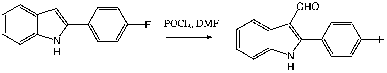


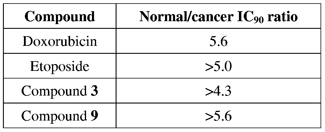


Claims


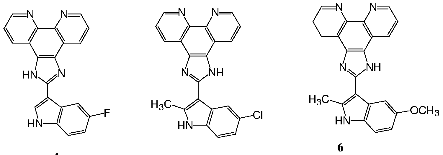


Priority Applications (7)
| Application Number | Priority Date | Filing Date | Title |
|---|---|---|---|
| ES06756007.8T ES2473597T3 (en) | 2005-05-25 | 2006-05-25 | Derivatives of 2-indolyl imidazo [4,5-d] phenanthroline and its use in cancer treatment |
| JP2008513005A JP5334575B2 (en) | 2005-05-25 | 2006-05-25 | 2-Indolylimidazo [4,5-d] phenanthroline derivatives and their use in cancer treatment |
| CA2611032A CA2611032C (en) | 2005-05-25 | 2006-05-25 | 2-indolyl imidazo[4,5-d]phenanthroline derivatives and their use in the treatment of cancer |
| CN2006800233772A CN101248072B (en) | 2005-05-25 | 2006-05-25 | 2-indolyl imidazo[4,5-d]phenanthroline derivatives and their use in the treatment of cancer |
| US11/915,257 US8148392B2 (en) | 2005-05-25 | 2006-05-25 | 2-indolyl imidazo [4,5-d] phenanthroline derivatives and their use in the treatment of cancer |
| EP06756007.8A EP1915374B1 (en) | 2005-05-25 | 2006-05-25 | 2-indolyl imidazo[4,5-d]phenanthroline derivatives and their use in the treatment of cancer |
| AU2006250809A AU2006250809B2 (en) | 2005-05-25 | 2006-05-25 | 2-indolyl imidazo(4,5-D)phenanthroline derivatives and their use in the treatment of cancer |
Applications Claiming Priority (6)
| Application Number | Priority Date | Filing Date | Title |
|---|---|---|---|
| US68416205P | 2005-05-25 | 2005-05-25 | |
| US60/684,162 | 2005-05-25 | ||
| US71055105P | 2005-08-22 | 2005-08-22 | |
| US60/710,551 | 2005-08-22 | ||
| US78752606P | 2006-03-31 | 2006-03-31 | |
| US60/787,526 | 2006-03-31 |
Publications (2)
| Publication Number | Publication Date |
|---|---|
| WO2006126177A2 true WO2006126177A2 (en) | 2006-11-30 |
| WO2006126177A3 WO2006126177A3 (en) | 2007-03-29 |
Family
ID=37452431
Family Applications (1)
| Application Number | Title | Priority Date | Filing Date |
|---|---|---|---|
| PCT/IB2006/051675 WO2006126177A2 (en) | 2005-05-25 | 2006-05-25 | 2-indolyl imidazo[4,5-d]phenanthroline derivatives and their use in the treatment of cancer |
Country Status (7)
| Country | Link |
|---|---|
| US (1) | US8148392B2 (en) |
| EP (1) | EP1915374B1 (en) |
| CN (1) | CN101248072B (en) |
| AU (1) | AU2006250809B2 (en) |
| CA (1) | CA2611032C (en) |
| ES (1) | ES2473597T3 (en) |
| WO (1) | WO2006126177A2 (en) |
Cited By (8)
| Publication number | Priority date | Publication date | Assignee | Title |
|---|---|---|---|---|
| WO2010102393A1 (en) * | 2009-03-12 | 2010-09-16 | Lorus Therapeutics Inc. | 2-indolyl imidazo [4,5-d]phenanthroline derivatives and their use to inhibit angiogenesis |
| US8148392B2 (en) | 2005-05-25 | 2012-04-03 | Lorus Therapeutics Inc. | 2-indolyl imidazo [4,5-d] phenanthroline derivatives and their use in the treatment of cancer |
| WO2014153464A2 (en) * | 2013-03-20 | 2014-09-25 | Lorus Therapeutics Inc. | 2-substituted imidazo[4,5-d]phenanthroline derivatives and their use in the treatment of cancer |
| WO2015051302A1 (en) | 2013-10-04 | 2015-04-09 | Aptose Biosciences Inc. | Compositions and methods for treating cancers |
| US10080739B2 (en) | 2003-11-14 | 2018-09-25 | Aptose Biosciences Inc. | Aryl imidazoles and their use as anti-cancer agents |
| JP2021501203A (en) * | 2017-10-30 | 2021-01-14 | アプトース バイオサイエンシズ インコーポレイテッド | Arylimidazole for cancer treatment |
| CN113461684A (en) * | 2021-07-20 | 2021-10-01 | 浙江工业大学 | PPAR-gamma ligand-4, 5-diazafluorene-rhodanine conjugate, preparation method and anti-tumor application thereof |
| EP3917397A4 (en) * | 2019-01-28 | 2022-10-26 | Board of Regents, The University of Texas System | Metal chelator combination therapy for the treatment of cancer |
Families Citing this family (9)
| Publication number | Priority date | Publication date | Assignee | Title |
|---|---|---|---|---|
| CN101899006B (en) * | 2002-08-19 | 2013-11-06 | 劳洛斯治疗公司 | 2,4,5-trisubstituted imidazoles and their use as anti-microbial agents |
| CN101735217B (en) * | 2009-12-15 | 2012-09-26 | 广东药学院 | Application of imidazole [4,5-f][1,10] phenanthroline and derivative thereof to preparation of antineoplastic drug |
| CN106459007B (en) | 2014-06-17 | 2020-02-28 | 韩国化学研究院 | Pyrimidine-2, 4-diamine derivatives and pharmaceutical compositions for anticancer containing the same as the active ingredient |
| CN104611429B (en) * | 2015-01-21 | 2017-02-22 | 湖州市中心医院 | Application of miR-10b (micro ribonucleic acid-10b) gene to regulation of gastric cancer gene expression |
| EP3265059A4 (en) | 2015-03-03 | 2018-08-29 | Cureport Inc. | Combination liposomal pharmaceutical formulations |
| US10736845B2 (en) | 2015-03-03 | 2020-08-11 | Cureport Inc. | Dual loaded liposomal pharmaceutical formulations |
| CN106565713B (en) * | 2016-10-10 | 2019-01-15 | 浙江工业大学 | 2 '-pyrazoles -1H- imidazoles [4,5-f] [1,10] Féraud quinoline derivants and its preparation method and application |
| WO2021247717A1 (en) * | 2020-06-02 | 2021-12-09 | Aptose Biosciences Inc. | 2-indolyl imidazo [4,5-d] phenanthroline polymorphs and compositions regarding the same |
| CN112957357B (en) * | 2021-03-01 | 2022-05-27 | 中国医科大学附属第一医院 | Target KLF4 ubiquitination small molecule inhibitor and application thereof |
Citations (5)
| Publication number | Priority date | Publication date | Assignee | Title |
|---|---|---|---|---|
| US4758421A (en) | 1985-03-15 | 1988-07-19 | The Board Of Trustees Of The Leland Stanford Junior University | Bleomycin conjugates and method |
| US5501959A (en) | 1989-01-17 | 1996-03-26 | Alamar Biosciences Laboratory, Inc. | Antibiotic and cytotoxic drug susceptibility assays using resazurin and poising agents |
| US20020119955A1 (en) | 2000-10-13 | 2002-08-29 | Doyle Terrence W. | Modified prodrug forms of AP/AMP |
| US6589966B1 (en) | 1998-03-18 | 2003-07-08 | Wake Forest University Health Sciences | Cytotoxic metal chelators and methods for making and using same |
| WO2005047266A1 (en) | 2003-11-14 | 2005-05-26 | Lorus Therapeutics Inc. | Aryl imidazoles and their use as anti-cancer agents |
Family Cites Families (77)
| Publication number | Priority date | Publication date | Assignee | Title |
|---|---|---|---|---|
| BE585555A (en) * | 1960-03-31 | |||
| US3297710A (en) * | 1963-04-09 | 1967-01-10 | Du Pont | Oxoarylidene-imidazoles |
| US3714181A (en) * | 1970-12-31 | 1973-01-30 | American Cyanamid Co | 2-aryl-5,10-diphenylphenanthro(9,10-d)azoles |
| US4089747A (en) * | 1976-08-09 | 1978-05-16 | Eastman Kodak Company | Compositions for the detection of hydrogen peroxide |
| DE3141063A1 (en) * | 1981-10-13 | 1983-04-28 | Schering Ag, 1000 Berlin Und 4619 Bergkamen | NEW IMIDAZOLE DERIVATIVES, METHOD FOR THE PRODUCTION THEREOF AND PHARMACEUTICAL PREPARATIONS CONTAINING THEM |
| US4423046A (en) * | 1982-04-05 | 1983-12-27 | Sterling Drug Inc. | Antibacterial and antiprotozoal 1-methyl-5-nitro-2-(2-phenylvinyl)imidazoles |
| DE3422175A1 (en) | 1984-06-04 | 1985-12-19 | Julius Dr. 8000 München Kern | Process for adding ammonia to exhaust gases or air containing acidic pollutants |
| JPS614960A (en) * | 1984-06-19 | 1986-01-10 | Fuji Photo Film Co Ltd | Analyzing reagent, analyzing method and multilayered chemical analyzing element |
| IT1214597B (en) * | 1985-04-15 | 1990-01-18 | Rorer Italiana Spa | DERIVATIVES OF IMIDAZOLE ANTIMICOTIC ADAPTITY, PROCEDURE AND INTERMEDIATES FOR THEIR PREPARATION AND COMPOSITIONS CONTAINING THEM. |
| US4902705A (en) * | 1985-12-12 | 1990-02-20 | Ube Industries, Ltd. | Imidazole derivatives, an antibacterial and antifungal agent comprising said derivatives, and a process for the production of said imidazole derivatives |
| US5024935A (en) * | 1987-12-18 | 1991-06-18 | Eastman Kodak Company | Dye-providing composition, diagnostic test kit and their use in method for ligand determination using peroxidase labeled-receptor |
| US5047318A (en) * | 1988-06-13 | 1991-09-10 | Eastman Kodak Company | Imidazole leuco dye composition containing 4'-hydroxyacetanilide, diagnostic kit and method using same |
| US5514550A (en) * | 1989-02-03 | 1996-05-07 | Johnson & Johnson Clinical Diagnostics, Inc. | Nucleic acid test article and its use to detect a predetermined nucleic acid |
| EP0462226B1 (en) * | 1989-03-08 | 1995-05-10 | Rocky Research | Method and apparatus for achieving high reaction rates in solid-gas reactor systems |
| US5441716A (en) | 1989-03-08 | 1995-08-15 | Rocky Research | Method and apparatus for achieving high reaction rates |
| JPH02258017A (en) | 1989-03-31 | 1990-10-18 | Babcock Hitachi Kk | Denitrifying apparatus for waste gas using solid reducing agent |
| US4970226A (en) * | 1989-10-03 | 1990-11-13 | Harbor Branch Oceanographic Institution, Inc. | Bis-indole imidazole compounds which are useful antitumor and antimicrobial agents |
| US5161389A (en) * | 1990-11-13 | 1992-11-10 | Rocky Research | Appliance for rapid sorption cooling and freezing |
| MX9300141A (en) * | 1992-01-13 | 1994-07-29 | Smithkline Beecham Corp | NOVEL IMIDAZOLE COMPOUNDS, PROCEDURE FOR THE PREPARATION AND PHARMACEUTICAL COMPOSITIONS CONTAINING IT. |
| US5656644A (en) * | 1994-07-20 | 1997-08-12 | Smithkline Beecham Corporation | Pyridyl imidazoles |
| US5916891A (en) * | 1992-01-13 | 1999-06-29 | Smithkline Beecham Corporation | Pyrimidinyl imidazoles |
| WO1995003297A1 (en) | 1993-07-21 | 1995-02-02 | Smithkline Beecham Corporation | Imidazoles for treating cytokine mediated disease |
| US5496702A (en) * | 1994-09-01 | 1996-03-05 | Johnson & Johnson Clinical Diagnostics, Inc. | Immunoassay elements having stable leuco dye coatings |
| ES2208698T3 (en) | 1994-12-13 | 2004-06-16 | F. Hoffmann-La Roche Ag | IMIDAZOL DERIVATIVES |
| TW424172B (en) * | 1995-04-19 | 2001-03-01 | Hitachi Chemical Co Ltd | Photosensitive resin composition and photosensitive element using the same |
| US5700826A (en) * | 1995-06-07 | 1997-12-23 | Ontogen Corporation | 1,2,4,5-tetra substituted imidazoles as modulators of multi-drug resistance |
| US5753687A (en) * | 1995-06-19 | 1998-05-19 | Ontogen Corporation | Modulators of proteins with phosphotryrosine recognition units |
| WO1997036587A1 (en) | 1996-04-03 | 1997-10-09 | Merck & Co., Inc. | A method of treating cancer |
| US5693589A (en) * | 1996-05-08 | 1997-12-02 | Eastman Kodak Company | Thermal imaging recording element |
| EP0812829A1 (en) | 1996-06-14 | 1997-12-17 | Ontogen Corporation | Substituted imidazoles as modulators of multi-drug resistance |
| IT240917Y1 (en) | 1996-10-01 | 2001-04-11 | Stefcom Spa | SHOE WITH UPPER HAVING PORTION FOR BREATHING THE INTERIOR OF THE SHOE |
| EP0946518A1 (en) | 1996-12-16 | 1999-10-06 | Ontogen Corporation | Modulators of proteins with phosphotyrosine recognition units |
| WO1998027108A2 (en) | 1996-12-16 | 1998-06-25 | Fujisawa Pharmaceutical Co., Ltd. | New amide compounds and their use as nitric oxide synthase inhibitors |
| US5945418A (en) * | 1996-12-18 | 1999-08-31 | Vertex Pharmaceuticals Incorporated | Inhibitors of p38 |
| US5809775A (en) * | 1997-04-02 | 1998-09-22 | Clean Diesel Technologies, Inc. | Reducing NOx emissions from an engine by selective catalytic reduction utilizing solid reagents |
| KR100568438B1 (en) * | 1997-04-24 | 2006-04-07 | 오르토-맥네일 파마슈티칼, 인코퍼레이티드 | Substituted imidazoles useful in the treatment of inflammatory diseases, a process for the preparation thereof and pharmaceutical compositions containing the same |
| JPH10319584A (en) * | 1997-05-21 | 1998-12-04 | Brother Ind Ltd | Photosensitive recording material using microcapsules |
| EP0996443A1 (en) | 1997-07-03 | 2000-05-03 | Neurogen Corporation | Certain diarylimidazole derivatives; a new class of npy specific ligands |
| DE19728343C5 (en) | 1997-07-03 | 2013-02-21 | Robert Bosch Gmbh | Process and apparatus for selective catalytic NOx reduction |
| US5840721A (en) | 1997-07-09 | 1998-11-24 | Ontogen Corporation | Imidazole derivatives as MDR modulators |
| WO1999007701A1 (en) | 1997-08-05 | 1999-02-18 | Sugen, Inc. | Tricyclic quinoxaline derivatives as protein tyrosine kinase inhibitors |
| GB9717804D0 (en) * | 1997-08-22 | 1997-10-29 | Zeneca Ltd | Chemical compounds |
| JPH11199582A (en) | 1998-01-05 | 1999-07-27 | Sagami Chem Res Center | 4(5)-(3-indolyl) imidazole derivative |
| WO2000034255A1 (en) | 1998-12-04 | 2000-06-15 | Ontogen Corporation | Substituted thiazoles for treatment of human diseases involving modulators of p-, l- and e- selectin |
| JP2000273088A (en) | 1999-01-18 | 2000-10-03 | Nippon Soda Co Ltd | Triphenylimidazole |
| JP2000281588A (en) | 1999-03-30 | 2000-10-10 | Sankyo Co Ltd | Prophylactic or therapeutic agent for cancer and method for screening same |
| DE19920936A1 (en) | 1999-05-07 | 2000-11-09 | Basf Ag | Heterocyclically substituted benzimidazoles, their preparation and use |
| AU5886500A (en) | 1999-06-23 | 2001-01-09 | Sepracor, Inc. | Indolyl-benzimidazole antibacterials, and methods of use thereof |
| US6266955B1 (en) * | 1999-08-20 | 2001-07-31 | Caterpillar Inc. | Diagnostic system for an emissions control on an engine |
| CN1088713C (en) | 1999-09-24 | 2002-08-07 | 中国科学院化学研究所 | Ruthenium (II) polypyridine match for two-stage pH sensing and its preparing process |
| CN1087744C (en) | 1999-09-24 | 2002-07-17 | 中国科学院化学研究所 | Ruthenium (II) polypyridine match and its preparing process |
| JP2003511396A (en) | 1999-10-08 | 2003-03-25 | オントジエン・コーポレイシヨン | How to enhance chemotherapy |
| US7115645B2 (en) * | 2000-01-14 | 2006-10-03 | Schering Aktiengesellschaft | 1,2 diarylbenzimidazoles and their pharmaceutical use |
| WO2002024680A1 (en) | 2000-09-21 | 2002-03-28 | Smithkline Beecham P.L.C. | Imidazole derivatives as raf kinase inhibitors |
| EP1341768A1 (en) | 2000-12-07 | 2003-09-10 | AstraZeneca AB | Therapeutic benzimidazole compounds |
| BR0207957A (en) | 2001-03-09 | 2004-02-25 | Pfizer Prod Inc | Benzimidazole Anti-Inflammatory Compounds |
| JP2002275458A (en) | 2001-03-21 | 2002-09-25 | Fukuoka Prefecture | Photochromic amorphous material and high density optical recording medium |
| CN100540538C (en) * | 2001-04-16 | 2009-09-16 | 田边三菱制药株式会社 | The high conductance calcium-activated k channel is opened agent |
| US6916440B2 (en) | 2001-05-31 | 2005-07-12 | 3M Innovative Properties Company | Processes and apparatus for making transversely drawn films with substantially uniaxial character |
| JP3759882B2 (en) | 2001-06-01 | 2006-03-29 | 株式会社日立製作所 | Pump device |
| DE10134775A1 (en) | 2001-07-06 | 2003-01-30 | Schering Ag | 1-alkyl-2.aryl-benzimidazole derivatives, their use for the manufacture of medicaments and pharmaceutical preparations containing these derivatives |
| AU2002355732B2 (en) * | 2001-08-01 | 2006-11-09 | Merck Sharp & Dohme Corp. | Benzimidazo[4,5-f]isoquinolinone derivatives |
| DE60221866T2 (en) | 2001-10-19 | 2008-05-29 | Ortho-Mcneil Pharmaceutical, Inc. | 2-PHENYL BENZIMIDAZOLE AND IMIDAZO-i4,5ö-PYRIDINE AS CDS1 / CHK2 INHIBITORS AND ADJUVANTIES IN CHEMOTHERAPY OR RADIATION THERAPY FOR THE TREATMENT OF CANCER |
| AU2003215112A1 (en) | 2002-02-07 | 2003-09-02 | Axys Pharmaceuticals | Novel bicyclic hydroxamates as inhibitors of histone deacetylase |
| US6790852B2 (en) | 2002-04-18 | 2004-09-14 | Hoffmann-La Roche Inc. | 2-(2,6-dichlorophenyl)-diarylimidazoles |
| WO2004000857A1 (en) | 2002-06-19 | 2003-12-31 | Kurita Water Industries Ltd. | Method of storing hydrogen, hydrogen inclusion compound and process for producing the same |
| EP1532118A2 (en) | 2002-07-05 | 2005-05-25 | Axxima Pharmaceuticals Aktiengesellschaft | Imidazole compounds for the treatment of hepatitis c virus infections |
| CN101899006B (en) * | 2002-08-19 | 2013-11-06 | 劳洛斯治疗公司 | 2,4,5-trisubstituted imidazoles and their use as anti-microbial agents |
| DE10251472A1 (en) | 2002-11-06 | 2004-05-19 | Robert Bosch Gmbh | Reduction of automotive nitrous oxide emissions during cold start comprises supplementary injection of ammonia from holding reservoir |
| CA2425797C (en) * | 2003-04-17 | 2013-10-15 | Xerox Corporation | Organic light emitting devices |
| US7989089B2 (en) * | 2003-04-17 | 2011-08-02 | Lg Display Co., Ltd. | Organic luminescent compounds and methods of making and using same |
| DE10323591A1 (en) | 2003-05-16 | 2004-12-02 | E.G.O. Elektro-Gerätebau GmbH | Device for producing a gas from a sublimation material, preferably ammonia from ammonium carbamate, comprises a heating arrangement assigned to sublimation material in a container |
| US20050282285A1 (en) * | 2004-06-21 | 2005-12-22 | Eaton Corporation | Strategy for controlling NOx emissions and ammonia slip in an SCR system using a nonselective NOx/NH3 |
| KR20070057143A (en) | 2004-08-03 | 2007-06-04 | 아미넥스 에이/에스 | A solid ammonia storage and delivery material |
| AU2006209949B2 (en) | 2005-02-03 | 2011-09-29 | Amminex Emissions Technology A/S | High density storage of ammonia |
| US8148392B2 (en) | 2005-05-25 | 2012-04-03 | Lorus Therapeutics Inc. | 2-indolyl imidazo [4,5-d] phenanthroline derivatives and their use in the treatment of cancer |
| EP1901831B1 (en) | 2005-06-29 | 2012-12-19 | Amminex A/S | Method and device for safe and controlled delivery of ammonia from a solid ammonia storage medium |
-
2006
- 2006-05-25 US US11/915,257 patent/US8148392B2/en active Active
- 2006-05-25 ES ES06756007.8T patent/ES2473597T3/en active Active
- 2006-05-25 AU AU2006250809A patent/AU2006250809B2/en not_active Ceased
- 2006-05-25 WO PCT/IB2006/051675 patent/WO2006126177A2/en active Application Filing
- 2006-05-25 CA CA2611032A patent/CA2611032C/en not_active Expired - Fee Related
- 2006-05-25 CN CN2006800233772A patent/CN101248072B/en not_active Expired - Fee Related
- 2006-05-25 EP EP06756007.8A patent/EP1915374B1/en not_active Not-in-force
Patent Citations (5)
| Publication number | Priority date | Publication date | Assignee | Title |
|---|---|---|---|---|
| US4758421A (en) | 1985-03-15 | 1988-07-19 | The Board Of Trustees Of The Leland Stanford Junior University | Bleomycin conjugates and method |
| US5501959A (en) | 1989-01-17 | 1996-03-26 | Alamar Biosciences Laboratory, Inc. | Antibiotic and cytotoxic drug susceptibility assays using resazurin and poising agents |
| US6589966B1 (en) | 1998-03-18 | 2003-07-08 | Wake Forest University Health Sciences | Cytotoxic metal chelators and methods for making and using same |
| US20020119955A1 (en) | 2000-10-13 | 2002-08-29 | Doyle Terrence W. | Modified prodrug forms of AP/AMP |
| WO2005047266A1 (en) | 2003-11-14 | 2005-05-26 | Lorus Therapeutics Inc. | Aryl imidazoles and their use as anti-cancer agents |
Non-Patent Citations (92)
| Title |
|---|
| ARMESTO, SYNTHESIS, 1988, pages 799 - 801 |
| AUSUBEL ET AL.: "Current Protocols in Molecular Biology", 1989, JOHN WILEY & SONS, INC |
| BIAN ET AL., POLYHEDRON, vol. 21, 2002, pages 313 - 319 |
| BIAN ET AL., SYNTHETIC COMMUNICATIONS, vol. 33, 2003, pages 3477 - 3482 |
| BOYD, M. R. ET AL.: "Cytotoxic Anticancer Drugs: Models and Concepts for Drug Discovery and Development", 1992, KLUWER ACADEMIC |
| BUSS, J. L.; B. T. GREENE; J. TURNER; F. M. TORTI; S. V. TORTI, CUIT TOP MED CHEM, vol. 4, no. 15, 2004, pages 1623 - 35 |
| CAIRO ET AL., J. BIOL. CHEM., vol. 270, 1995, pages 700 - 703 |
| CAMMACK; COOPER, METHODS ENZYMOL., vol. 227, 1993, pages 353 - 384 |
| CHAO ET AL., J. CHEM. SOC., DALTON TRANS., vol. 12, 2001, pages 1920 - 1926 |
| CHASTON, T. B. ET AL., CLIN CANCER RES, vol. 9, no. 1, 2003, pages 402 - 14 |
| CHEN C ET AL., ONCOGENE, vol. 21, 2002, pages 6567 - 72 |
| CHEN, J.; J. STUBBE, NAT REV CANCER, vol. 5, no. 2, 2005, pages 102 - 12 |
| CHEN, S. F. ET AL., PROC. AM. ASSOC. CANCER RES., vol. 31, 1990, pages A2644 |
| CHEN, X. ET AL., J BIOL CHEM, vol. 276, no. 32, 2001, pages 30423 - 8 |
| CHI, SYNTH. COMM., vol. 24, no. 15, 1994, pages 2119 - 2122 |
| COHEN ET AL., PALLIATIVE MEDICINE, vol. 9, 1995, pages 207 - 219 |
| DANG DT ET AL., FEBS LETT, vol. 476, 2000, pages 203 - 7 |
| DANG ET AL., NUCLEIC ACIDS RES., vol. 30, no. 13, 2002, pages 2736 - 2741 |
| DANG, D. T. ET AL., ONCOGENE, vol. 22, no. 22, 2003, pages 3424 - 30 |
| DECKER ET AL., EUR. J. OF CANCER, vol. 40, 2004, pages 821 - 826 |
| DINKOVA-KOSTOVA ET AL., BIOCHEMISTRY, vol. 44, no. 18, 2005, pages 6889 - 6899 |
| DOWNEY, K. M.; B. G. QUE; A. G. SO, BIOCHEM BIOPHYS RES COMMUN, vol. 93, no. 1, 1980, pages 264 - 70 |
| EISENSTEIN; BLEMINGS, J. NUTR., vol. 128, no. 12, 1998, pages 2295 - 2295 |
| EKWALL, B., ANN. N.Y. ACAD. SCI., vol. 407, 1983, pages 64 - 77 |
| ELLISON G; KLINOWSKA T; WESTWOOD RF; DOCTER E; FRENCH T; FOX JC., MOL PATHOL., vol. 55, no. 5, 2002, pages 294 - 299 |
| ENNA ET AL.: "Current Protocols in Pharmacology", J. WILEY & SONS, INC. |
| FIELDS; LANCASTER, AM. BIOTECHNOL. LAB., vol. 11, 1993, pages 48 - 50 |
| FISCHER, J. AM. CHEM. SOC., vol. 83, 1961, pages 4208 - 4210 |
| FLATMARK; TANGERAS: "Proteins of Iron Metabolism", 1976, GRUNE & STRATTON, pages: 349 - 358 |
| FOSTER, K. W. ET AL., CANCER RES, vol. 60, no. 22, 2000, pages 6488 - 95 |
| FOSTER, K. W. ET AL., CELL GROWTH DIFFER, vol. 10, no. 6, 1999, pages 423 - 34 |
| GENNARO, A.: "Remington: The Science and Practice of Pharmacy", 2000, LIPPINCOTT, WILLIAMS & WILKINS |
| GHALEB ET AL., CELL RES., vol. 15, no. 2, 2005, pages 92 - 96 |
| GHALEB, A. M. ET AL., CELL RES, vol. 15, no. 2, 2005, pages 92 - 6 |
| GOWER ET AL., ANAL. BIOCHEM., vol. 180, 1989, pages 126 - 130 |
| GRIMMETT, M. R.: "Imidazole and Benzimidazole Synthesis", 1997, ACADEMIC PRESS |
| GRIMMETT, M.R.: "Comprehensive Heterocyclic Chemistry: The Structure, Reaction, Synthesis and Uses of Heterocyclic Compounds", vol. 5, 1984, PERGAMON PRESS, pages: 457 - 498 |
| GUIJARRO ET AL., J. AM. CHEM. SOC., vol. 121, 1999, pages 4155 - 4157 |
| HAYES, A.W.,: "Principles and Methods of Toxicology, 3rd ed.,", 1994, RAVEN PRESS |
| HIORT, J. AM. CHEM SOC., vol. 115, 1993, pages 3448 - 3454 |
| HOLLINGSHEAD, M. ET AL., LIFE SCIENCES, vol. 57, 1995, pages 131 - 141 |
| HONG XU ET AL., INORG. CHEM. COMMUN., vol. 6, 2003, pages 766 - 768 |
| HONG XU ET AL., J. CHEM. SOC., DALTON TRANS., vol. 11, 2003, pages 2260 - 2268 |
| JBIC, vol. 5, 2000, pages 119 - 128 |
| K. MORGAN,: "Current Protocols in Cell Biology", 2000, J. WILEY & SONS |
| KACZYNSKI, J.; T. COOK; R. URRUTIA, GENOME BIOL, vol. 4, no. 2, 2003, pages 206 |
| KINDERMANN ET AL., J. NUTR., vol. 134, no. 1, 2004, pages 57 - 62 |
| KINDERMANN, B.; F. DORING; J. BUDCZIES; H. DANIEL, BIOCHEM CELL BIOL, vol. 83, no. 2, 2005, pages 221 - 9 |
| KINDERMANN, B.; F. DORING; M. PFAFFL; H. DANIEL, J NUTR, vol. 134, no. 1, 2004, pages 57 - 62 |
| KITANO ET AL., EURO. J. CLIN. INVESTG., vol. 21, 1991, pages 53 - 58 |
| KOZLOV ET AL., FREE RADIC. BIOL. MED., vol. 13, 1992, pages 9 - 16 |
| KRIEG ET AL., NATURFORSCH, vol. 22B, 1967, pages 132 |
| L.T. VASSILEV ET AL., ANTI-CANCER DRUG DESIGN, vol. 16, 2001, pages 7 - 17 |
| LANGMADE ET AL., J. BIOL. CHEM., vol. 275, no. 44, 2000, pages 34803 - 24809 |
| LICHTLEN; SCHAFFNER, BIOESSAYS, vol. 23, no. 11, 2001, pages 1010 - 1017 |
| LIU, POLYHEDRON, 2000, pages 1975 - 1983 |
| MAINES, M.,: "Current Protocols in Toxicology", JOHN WILEY & SONS, INC. |
| MANN, K. J.; D. SEVLEVER, BIOCHEMISTRY, vol. 40, no. 5, 2001, pages 1205 - 13 |
| MARTINI ET AL., J. NUTR., vol. 132, no. 4, 2002, pages 693 - 696 |
| MCCABE, M. J., JR.; S. A. JIANG; S. ORRENIUS, LAB INVEST, vol. 69, no. 1, 1993, pages 101 - 10 |
| MCCORKLE; YOUNG, CANCER NURSING, vol. 1, 1978, pages 373 - 378 |
| MONKS, A. ET AL., JNCI, J. NATL. CANCER INST., vol. 83, 1991, pages 757 - 766,11-34 |
| NARLA G ET AL., SCIENCE, vol. 294, 2001, pages 2563 - 6 |
| NEW J. CHEM., vol. 27, 2003, pages 1255 - 1263 |
| NIELSEN ET AL., INT. J.BIOCHEM., vol. 25, 1993, pages 223 - 232 |
| O'BRIEN ET AL., EUR J. BIOCHEM., vol. 267, 2000, pages 5421 - 5426 |
| OHNISHI, S. ET AL., BIOCHEM BIOPHYS RES COMMUN, vol. 308, no. 2, 2003, pages 251 - 6 |
| OLLINGER; ROBERG, J. BIOL. CHEM., vol. 272, 1997, pages 23707 - 23711 |
| PAN, C. Q.; R. LANDGRAF; D. S. SIGMAN, MOL MICROBIOL, vol. 12, no. 3, 1994, pages 335 - 42 |
| PANDYA, A. Y. ET AL., CLIN CANCER RES, vol. 10, no. 8, 2004, pages 2709 - 19 |
| PETRAT ET AL., BIOL CHEM., vol. 383, no. 3-4, 2002, pages 489 - 502 |
| RICHARDSON, D. R., CRIT REV ONCOL HEMATOL, vol. 42, no. 3, 2002, pages 267 - 81 |
| ROSS ET AL., NAT GENET, vol. 24, no. 3, 2000, pages 227 - 233 |
| RUBINSTEIN ET AL., J. NATL. CANCER INST., vol. 82, 1990, pages 113 - 118 |
| SARSHAR ET AL., TETRAHEDRON LETT., vol. 37, 1996, pages 835 - 838 |
| SHULMAN, A.; G. A. LAYCOCK, CHEM BIOL INTERACT, vol. 16, no. 1, 1977, pages 89 - 99 |
| SIGMAN, D. S. ET AL., J BIOL CHEM, vol. 254, no. 24, 1979, pages 12269 - 72 |
| SKEHAN, P. ET AL., JNCI, J. NATL. CANCER INST., vol. 82, 1990, pages 1107 - 1112 |
| SPRINGMAN, E. B. ET AL., BIOCHEMISTRY, vol. 34, no. 48, 1995, pages 15713 - 20 |
| SUBRAMANIAM M ET AL., J CELL BIOCHEM, vol. 68, 1998, pages 226 - 36 |
| SYNTHESIS, vol. 8, 1985, pages 641 - 645 |
| TETRAHEDRON LETTERS, vol. 45, no. 33, 2004, pages 6361 - 6363 |
| VASSILEV ET AL., ANTI-CANCER DRUG DESIGN, vol. 16, no. 7, 2001 |
| VILSMEIER, CHEM. BER., vol. 91, 1958, pages 850 - 861 |
| WANG, N. ET AL., WORLD J GASTROENTEROL, vol. 8, no. 6, 2002, pages 966 - 70 |
| WEI, D. ET AL., CANCER RES, vol. 65, no. 7, 2005, pages 2746 - 54 |
| WEST ET AL., J. INVESTIGATIVE DENN., vol. 99, 1992, pages 95 - 100 |
| YAMADA, BULL. SOC CHEM. JPN., vol. 63, no. 9, 1990, pages 2710 - 2712 |
| YASUNAGA, J. ET AL., CANCER RES, vol. 64, no. 17, 2004, pages 6002 - 9 |
| YEGOROV ET AL., FREE RADIC. BIOL. MED., vol. 15, 1993, pages 565 - 574 |
| ZALEWSKI ET AL., BIOCHEM J., vol. 296, 1993, pages 403 - 8 |
| ZHAO, R., BIOCHEM PHARMACOL, vol. 67, no. 9, 2004, pages 1677 - 88 |
Cited By (15)
| Publication number | Priority date | Publication date | Assignee | Title |
|---|---|---|---|---|
| US10080739B2 (en) | 2003-11-14 | 2018-09-25 | Aptose Biosciences Inc. | Aryl imidazoles and their use as anti-cancer agents |
| US8148392B2 (en) | 2005-05-25 | 2012-04-03 | Lorus Therapeutics Inc. | 2-indolyl imidazo [4,5-d] phenanthroline derivatives and their use in the treatment of cancer |
| WO2010102393A1 (en) * | 2009-03-12 | 2010-09-16 | Lorus Therapeutics Inc. | 2-indolyl imidazo [4,5-d]phenanthroline derivatives and their use to inhibit angiogenesis |
| WO2014153464A2 (en) * | 2013-03-20 | 2014-09-25 | Lorus Therapeutics Inc. | 2-substituted imidazo[4,5-d]phenanthroline derivatives and their use in the treatment of cancer |
| WO2014153464A3 (en) * | 2013-03-20 | 2014-11-27 | Lorus Therapeutics Inc. | 2-substituted imidazo[4,5-d]phenanthroline derivatives and their use in the treatment of cancer |
| US9309247B2 (en) | 2013-03-20 | 2016-04-12 | Lorus Therapeutics Inc. | 2-substituted imidazo[4,5-D]phenanthroline derivatives and their use in the treatment of cancer |
| EP3052102A4 (en) * | 2013-10-04 | 2017-05-31 | Aptose Biosciences Inc. | Compositions and methods for treating cancers |
| WO2015051302A1 (en) | 2013-10-04 | 2015-04-09 | Aptose Biosciences Inc. | Compositions and methods for treating cancers |
| EP3650023A1 (en) * | 2013-10-04 | 2020-05-13 | Aptose Biosciences Inc. | Compositions for treating cancers |
| US11104957B2 (en) | 2013-10-04 | 2021-08-31 | Aptose Biosciences, Inc. | Compositions and methods for treating cancers |
| JP2021501203A (en) * | 2017-10-30 | 2021-01-14 | アプトース バイオサイエンシズ インコーポレイテッド | Arylimidazole for cancer treatment |
| US11149047B2 (en) | 2017-10-30 | 2021-10-19 | Aptose Biosciences, Inc. | Aryl imidazoles for treatment of cancer |
| EP3917397A4 (en) * | 2019-01-28 | 2022-10-26 | Board of Regents, The University of Texas System | Metal chelator combination therapy for the treatment of cancer |
| CN113461684A (en) * | 2021-07-20 | 2021-10-01 | 浙江工业大学 | PPAR-gamma ligand-4, 5-diazafluorene-rhodanine conjugate, preparation method and anti-tumor application thereof |
| CN113461684B (en) * | 2021-07-20 | 2022-11-11 | 浙江工业大学 | PPAR-gamma ligand-4, 5-diazafluorene-rhodanine conjugate, preparation method and anti-tumor application thereof |
Also Published As
| Publication number | Publication date |
|---|---|
| US8148392B2 (en) | 2012-04-03 |
| ES2473597T3 (en) | 2014-07-07 |
| EP1915374B1 (en) | 2014-04-02 |
| CA2611032A1 (en) | 2006-11-30 |
| CN101248072B (en) | 2013-01-23 |
| AU2006250809A1 (en) | 2006-11-30 |
| AU2006250809B2 (en) | 2011-05-12 |
| WO2006126177A3 (en) | 2007-03-29 |
| US20100168417A1 (en) | 2010-07-01 |
| CN101248072A (en) | 2008-08-20 |
| CA2611032C (en) | 2012-01-17 |
| EP1915374A2 (en) | 2008-04-30 |
Similar Documents
| Publication | Publication Date | Title |
|---|---|---|
| CA2611032C (en) | 2-indolyl imidazo[4,5-d]phenanthroline derivatives and their use in the treatment of cancer | |
| JP5095216B2 (en) | Arylimidazoles and their use as anticancer agents | |
| EP2310014B1 (en) | Tricyclic compounds having antimitotic and/or antitumor activity and methods of use thereof | |
| JP2002533429A (en) | Ortho-diphenol compounds, methods for inhibiting PARP and pharmaceutical compositions | |
| CN101389629A (en) | Indolopyridines as EG5 kinesin modulators | |
| Driowya et al. | Synthesis of triazoloquinazolinone based compounds as tubulin polymerization inhibitors and vascular disrupting agents | |
| JP5334575B2 (en) | 2-Indolylimidazo [4,5-d] phenanthroline derivatives and their use in cancer treatment | |
| CN115197225B (en) | Five-membered heterocyclic quinazolinone compound and preparation method thereof | |
| Mylonas et al. | Synthesis and antitumor activity of new thiosemicarbazones of 2‐acetylimidazo [4, 5‐b] pyridine | |
| CN112010789A (en) | Vinyl sulfonamide or vinyl amide compounds, and preparation method and application thereof | |
| EP4230204A1 (en) | Thiobenzimidazole derivative or pharmaceutically acceptable salt thereof and use thereof | |
| KR20230106583A (en) | Synthesis of novel imipridone derivatives and evaluation of their anticancer activity | |
| CN111116572B (en) | Oxadiazole derivative, preparation method and application thereof | |
| US9309247B2 (en) | 2-substituted imidazo[4,5-D]phenanthroline derivatives and their use in the treatment of cancer | |
| EP3484892B1 (en) | 1,2,4-triazolo-[3,4-b]-1,3,4-thiadiazole derivatives | |
| Anastassova et al. | SYNTHESIS OF NEW TRIAZOLE AND THIADIAZOLE DERIVATIVES OF THE N, N'-DISUBSTITUTED BENZIMIDAZOLE-2-THIONE AND EVALUATION OF THEIR ANTITUMOR POTENTIAL. | |
| CN110066274B (en) | Pyridine-2-formic acid derivative and preparation method and application thereof | |
| CN117800979A (en) | Tetrahydrobenzofuran [2,3-c ] pyridine kinase inhibitor and preparation method and application thereof | |
| PL236095B1 (en) | Triazolo-quinoline derivatives of cinchona-tree alkaloids, method for obtaining them and their application | |
| CN110713487A (en) | Novel heterocyclic aromatic hydrazone derivative, pharmaceutically acceptable salt thereof, preparation method and application thereof | |
| JP2009519954A (en) | Dimers and adducts of 6-[(substituted) phenyl] triazolopyrimidines useful as anticancer agents | |
| CA2226310A1 (en) | New pyrido-thiopyranoindazoles with anti-tumor activity | |
| WO2008018778A1 (en) | New heterocyclic compounds containing nitrogen atoms or pharmaceutically acceptable salts thereof, process for the preparation thereof and pharmaceutical composition comprising the same for treatment of cancer |
Legal Events
| Date | Code | Title | Description |
|---|---|---|---|
| WWE | Wipo information: entry into national phase |
Ref document number: 2006250809 Country of ref document: AU |
|
| WWE | Wipo information: entry into national phase |
Ref document number: 2008513005 Country of ref document: JP Ref document number: 2611032 Country of ref document: CA |
|
| NENP | Non-entry into the national phase |
Ref country code: DE |
|
| WWW | Wipo information: withdrawn in national office |
Ref document number: DE |
|
| WWE | Wipo information: entry into national phase |
Ref document number: 2006756007 Country of ref document: EP |
|
| WWP | Wipo information: published in national office |
Ref document number: 2006250809 Country of ref document: AU |
|
| NENP | Non-entry into the national phase |
Ref country code: RU |
|
| WWW | Wipo information: withdrawn in national office |
Ref document number: RU |
|
| WWE | Wipo information: entry into national phase |
Ref document number: 200680023377.2 Country of ref document: CN |
|
| WWP | Wipo information: published in national office |
Ref document number: 2006756007 Country of ref document: EP |
|
| WWE | Wipo information: entry into national phase |
Ref document number: 11915257 Country of ref document: US |