WO2008048982A2 - Method for obtaining spatial images through mri and processing the resulting spatial images and product - Google Patents
Method for obtaining spatial images through mri and processing the resulting spatial images and product Download PDFInfo
- Publication number
- WO2008048982A2 WO2008048982A2 PCT/US2007/081580 US2007081580W WO2008048982A2 WO 2008048982 A2 WO2008048982 A2 WO 2008048982A2 US 2007081580 W US2007081580 W US 2007081580W WO 2008048982 A2 WO2008048982 A2 WO 2008048982A2
- Authority
- WO
- WIPO (PCT)
- Prior art keywords
- pixel
- function
- images
- intensity
- parameters
- Prior art date
Links
- 238000000034 method Methods 0.000 title claims abstract description 58
- 238000012545 processing Methods 0.000 title claims abstract description 15
- 230000004931 aggregating effect Effects 0.000 claims abstract description 6
- 239000013598 vector Substances 0.000 claims description 71
- 239000002872 contrast media Substances 0.000 claims description 33
- 230000002776 aggregation Effects 0.000 claims description 22
- 238000004220 aggregation Methods 0.000 claims description 22
- 239000011159 matrix material Substances 0.000 claims description 20
- 230000008569 process Effects 0.000 claims description 14
- 238000012935 Averaging Methods 0.000 claims description 10
- 238000013507 mapping Methods 0.000 claims description 8
- XLYOFNOQVPJJNP-UHFFFAOYSA-N water Substances O XLYOFNOQVPJJNP-UHFFFAOYSA-N 0.000 claims description 8
- 238000012546 transfer Methods 0.000 claims description 7
- 230000008859 change Effects 0.000 claims description 5
- 238000001454 recorded image Methods 0.000 claims description 3
- 230000006870 function Effects 0.000 description 38
- 238000002595 magnetic resonance imaging Methods 0.000 description 16
- 238000004891 communication Methods 0.000 description 13
- 238000004590 computer program Methods 0.000 description 12
- 210000001519 tissue Anatomy 0.000 description 7
- 206010028980 Neoplasm Diseases 0.000 description 5
- 230000006399 behavior Effects 0.000 description 5
- 201000011510 cancer Diseases 0.000 description 5
- 230000003287 optical effect Effects 0.000 description 3
- 210000004291 uterus Anatomy 0.000 description 3
- 102000020897 Formins Human genes 0.000 description 2
- 108091022623 Formins Proteins 0.000 description 2
- 210000001015 abdomen Anatomy 0.000 description 2
- 239000000872 buffer Substances 0.000 description 2
- 238000001514 detection method Methods 0.000 description 2
- 238000010586 diagram Methods 0.000 description 2
- 230000006872 improvement Effects 0.000 description 2
- 238000012986 modification Methods 0.000 description 2
- 230000004048 modification Effects 0.000 description 2
- 210000003205 muscle Anatomy 0.000 description 2
- 210000000689 upper leg Anatomy 0.000 description 2
- 210000003932 urinary bladder Anatomy 0.000 description 2
- 206010006187 Breast cancer Diseases 0.000 description 1
- 208000026310 Breast neoplasm Diseases 0.000 description 1
- 241001465754 Metazoa Species 0.000 description 1
- 210000000481 breast Anatomy 0.000 description 1
- 230000001413 cellular effect Effects 0.000 description 1
- 238000006243 chemical reaction Methods 0.000 description 1
- 230000000694 effects Effects 0.000 description 1
- 238000005516 engineering process Methods 0.000 description 1
- 239000000835 fiber Substances 0.000 description 1
- 238000009499 grossing Methods 0.000 description 1
- 238000010348 incorporation Methods 0.000 description 1
- 230000010365 information processing Effects 0.000 description 1
- 239000000463 material Substances 0.000 description 1
- 238000005259 measurement Methods 0.000 description 1
- 238000012805 post-processing Methods 0.000 description 1
- 238000007639 printing Methods 0.000 description 1
- 238000000264 spin echo pulse sequence Methods 0.000 description 1
- 230000002123 temporal effect Effects 0.000 description 1
Classifications
-
- G—PHYSICS
- G01—MEASURING; TESTING
- G01R—MEASURING ELECTRIC VARIABLES; MEASURING MAGNETIC VARIABLES
- G01R33/00—Arrangements or instruments for measuring magnetic variables
- G01R33/20—Arrangements or instruments for measuring magnetic variables involving magnetic resonance
- G01R33/44—Arrangements or instruments for measuring magnetic variables involving magnetic resonance using nuclear magnetic resonance [NMR]
- G01R33/48—NMR imaging systems
- G01R33/54—Signal processing systems, e.g. using pulse sequences ; Generation or control of pulse sequences; Operator console
- G01R33/56—Image enhancement or correction, e.g. subtraction or averaging techniques, e.g. improvement of signal-to-noise ratio and resolution
- G01R33/563—Image enhancement or correction, e.g. subtraction or averaging techniques, e.g. improvement of signal-to-noise ratio and resolution of moving material, e.g. flow contrast angiography
- G01R33/56366—Perfusion imaging
-
- G—PHYSICS
- G06—COMPUTING; CALCULATING OR COUNTING
- G06T—IMAGE DATA PROCESSING OR GENERATION, IN GENERAL
- G06T7/00—Image analysis
- G06T7/10—Segmentation; Edge detection
- G06T7/11—Region-based segmentation
-
- G—PHYSICS
- G06—COMPUTING; CALCULATING OR COUNTING
- G06T—IMAGE DATA PROCESSING OR GENERATION, IN GENERAL
- G06T7/00—Image analysis
- G06T7/10—Segmentation; Edge detection
- G06T7/187—Segmentation; Edge detection involving region growing; involving region merging; involving connected component labelling
-
- G—PHYSICS
- G01—MEASURING; TESTING
- G01R—MEASURING ELECTRIC VARIABLES; MEASURING MAGNETIC VARIABLES
- G01R33/00—Arrangements or instruments for measuring magnetic variables
- G01R33/20—Arrangements or instruments for measuring magnetic variables involving magnetic resonance
- G01R33/44—Arrangements or instruments for measuring magnetic variables involving magnetic resonance using nuclear magnetic resonance [NMR]
- G01R33/48—NMR imaging systems
- G01R33/54—Signal processing systems, e.g. using pulse sequences ; Generation or control of pulse sequences; Operator console
- G01R33/56—Image enhancement or correction, e.g. subtraction or averaging techniques, e.g. improvement of signal-to-noise ratio and resolution
- G01R33/5601—Image enhancement or correction, e.g. subtraction or averaging techniques, e.g. improvement of signal-to-noise ratio and resolution involving use of a contrast agent for contrast manipulation, e.g. a paramagnetic, super-paramagnetic, ferromagnetic or hyperpolarised contrast agent
-
- G—PHYSICS
- G01—MEASURING; TESTING
- G01R—MEASURING ELECTRIC VARIABLES; MEASURING MAGNETIC VARIABLES
- G01R33/00—Arrangements or instruments for measuring magnetic variables
- G01R33/20—Arrangements or instruments for measuring magnetic variables involving magnetic resonance
- G01R33/44—Arrangements or instruments for measuring magnetic variables involving magnetic resonance using nuclear magnetic resonance [NMR]
- G01R33/48—NMR imaging systems
- G01R33/54—Signal processing systems, e.g. using pulse sequences ; Generation or control of pulse sequences; Operator console
- G01R33/56—Image enhancement or correction, e.g. subtraction or averaging techniques, e.g. improvement of signal-to-noise ratio and resolution
- G01R33/5608—Data processing and visualization specially adapted for MR, e.g. for feature analysis and pattern recognition on the basis of measured MR data, segmentation of measured MR data, edge contour detection on the basis of measured MR data, for enhancing measured MR data in terms of signal-to-noise ratio by means of noise filtering or apodization, for enhancing measured MR data in terms of resolution by means for deblurring, windowing, zero filling, or generation of gray-scaled images, colour-coded images or images displaying vectors instead of pixels
-
- G—PHYSICS
- G06—COMPUTING; CALCULATING OR COUNTING
- G06T—IMAGE DATA PROCESSING OR GENERATION, IN GENERAL
- G06T2207/00—Indexing scheme for image analysis or image enhancement
- G06T2207/10—Image acquisition modality
- G06T2207/10016—Video; Image sequence
-
- G—PHYSICS
- G06—COMPUTING; CALCULATING OR COUNTING
- G06T—IMAGE DATA PROCESSING OR GENERATION, IN GENERAL
- G06T2207/00—Indexing scheme for image analysis or image enhancement
- G06T2207/10—Image acquisition modality
- G06T2207/10072—Tomographic images
- G06T2207/10088—Magnetic resonance imaging [MRI]
Definitions
- the present invention relates to a method for obtaining spatial images through magnetic resonance imaging and processing the resultant spatial images and a product resulting from the application of the method, and more particularly a method for diagnosing cancer through MRI and aggregating the resulting MRI images according to a novel and specialized algorithm and a product comprised of computer readable recorded programs for carrying out the method.
- the present invention has for its principal object an improved method for obtaining and processing of spatial images through magnetic resonance imaging whereby the obtained images provide an improvement in order to obtain better detection of cancer in human tissue, and to a computer readable medium containing programs for carrying out the method.
- the present invention achieves the foregoing is accomplished by a novel processing of the spatial images obtained by magnetic resonance of human tissue.
- the new processing technique includes a novel algorithm that aggregates similar dynamic behavior in a set of spatial images.
- the invention is accomplished by a method comprising the steps of recording a set of MRI images of a tissue pixel by pixel that evolve with time in a specific manner described by a function; processing the recorded MRI images by aggregating the pixels thereof according to a preselected aspect of time behavior; best fitting the aggregated pixels to a predetermined pixel function to obtain the parameters of the function; and presenting the parameters visually.
- visually presented is meant that the parameters are displayed by being sent immediately to a monitor, are printed immediately by being sent to a printer, or are stored in memory by being sent to a store or memory for future display or printing.
- the function is intensity of the pixels; the function is
- I SE (TR) I(O)[l-exp(-TR/T 1 )]
- I SE is the intensity in a pixel for a certain TR
- Ti the second free parameter, is the longitudinal relaxation time of the water in a pixel.
- the inventive method can take the form of a method comprising the steps of injecting a contrast agent into a patient; recording a set of time-spaced MRI pixel-by-pixel images of a preselected area of tissue of the patient using a gradient echo sequence; performing aggregation based on pixel intensity on the recorded images; best-fitting the aggregated intensities to a preselected intensity function to yield maps of specific parameters of the tissue; and presenting the maps visually.
- the specific parameters are transcapillary transfer constant and extravascular extracellular volume fraction.
- I (t) is intensity at time t after administration of the contrast agent
- 1(0) is intensity pre-contrast
- TR is the repetition time of the sequence
- ⁇ is the flip angle of the pulse
- Ti 0 is the longitudinal relaxation time pre-contrast
- Ri is the relaxivity of the contrast agent, namely, the change in the relaxation of water when the concentration of the contrast agent increases by 1 millimolar
- C t (t) is the concentration of the contrast agent which is given by
- D is the contrast agent dose (known)
- a ls mi and a 2 , m 2 are the corresponding amplitude and decay constants of the contrast agent in the body plasma which can be determined independently
- K and v e are the transcapillary transfer constant and the extravascular extracellular volume fraction, respectively.
- the invention can also be a method comprising the steps of: a.
- creating a dataset by reading a set of N images of size n*n that specify x, y location; b. converting the dataset into a 3D matrix of n*n*N where N is the number of discrete samples of a function (creating a vector); c. determining the similarities for the vector of each pixel with x,y coordinates in the images to the vectors of each one of its 4 surrounding pixels according to a predefined norm; d. finding the most similar vector in the surrounding neighbors for each pixel x,y in the image; e. averaging the two similar vectors or employing another mathematical manipulation; f. creating a new n*n*N matrix that includes the similar vectors; g. repeating steps d, e and f q times and stopping according to convergence criterion; h. performing an automatic fitting process, for each pixel x,y, using the final vectors to find the function parameters; and i. presenting the parameters visually.
- the above method can include the further step of mapping the function parameters; and displaying the mapped function parameters.
- the invention may also be a method comprising the steps of: a. creating a dataset by reading N images of size n*n*m that specify x, y, z location; b. converting the dataset into a 4D matrix of n*n*m*N where N is the number of discrete samples of a function creating a vector; c. determining for the vector of each pixel with x,y,z coordinates in the images similarities to the vectors of each one of its 6 surrounding pixels according to a predefined norm; d. finding for each pixel x,y,z in the image the most similar vector in the surrounding neighbors; e. averaging the two similar vectors or employ another mathematical manipulation; f.
- the foregoing method can include the step of mapping the function parameters.
- the invention further contemplates a computer readable medium containing executable program instructions for recording a set of MRI images of a tissue pixel by pixel that evolve with time in a specific manner described by a function; processing the recorded MRI images by aggregating the pixels thereof according to a preselected aspect of time behavior; and best fitting the aggregated pixels to a predetermined pixel function to obtain the parameters of the function; and presenting the parameters visually.
- the function is intensity of the pixels.
- I SE (TR) I(O)[l-exp(-TR/T 1 )]
- I SE is the intensity in a pixel for a certain TR
- Ti the second free parameter, is the longitudinal relaxation time of the water in a pixel.
- the invention further contemplates a computer readable medium containing executable program instructions for recording a set of time-spaced MRI pixel-by-pixel images of a preselected area of tissue of a patient injected with a contrast agent using a gradient echo sequence; performing aggregation based on pixel intensity on the recorded images; best- fitting the aggregated intensities to a preselected intensity function to yield maps of specific parameters of the tissue; and presenting the maps visually.
- the specific parameters are transcapillary transfer constant and extravascular extracellular volume fraction.
- the intensity function is expressed as
- [I(t) -1(0)]/ 1(0) [l-exp(-TR/Tio)cos ⁇ ](l-exp ⁇ -TR[l/Ti O +RiC(t))] ⁇ / ⁇ l-exp[-TR(l/Tio+RiC,(t))]cos ⁇ [l-exp(-TR/Ti 0 )] -1.
- I (t) is intensity at time t after administration of the contrast agent
- 1(0) is intensity pre-contrast
- TR is the repetition time of the sequence
- ⁇ is the flip angle of the pulse
- Tio is the longitudinal relaxation time pre-contrast
- Ri is the relaxivity of the contrast agent, namely, the change in the relaxation of water when the concentration of the contrast agent increases by 1 millimolar
- C t (t) is the concentration of the contrast agent which is given by
- D is the contrast agent dose (known)
- a ls mi and a 2 , m 2 are the corresponding amplitude and decay constants of the contrast agent in the body plasma which can be determined independently
- K and v e are the transcapillary transfer constant and the extravascular extracellular volume fraction, respectively.
- the invention further contemplates a computer readable medium containing executable program instructions for a. creating a dataset by reading a set of N images of size n*n that specify x, y location; b. converting the dataset into a 3D matrix of n*n*N where N is the number of discrete samples of a function (creating a vector); c. determining the similarities for the vector of each pixel with x,y coordinates in the images to the vectors of each one of its 4 surrounding pixels according to a predefined norm; d. finding the most similar vector in the surrounding neighbors for each pixel x,y in the image; e. averaging the two similar vectors or employing another mathematical manipulation; f.
- the instructions can include mapping the function parameters.
- a still further invention contemplated is a computer readable medium containing executable program instructions for a. creating a dataset by reading N images of size n*n*m that specify x, y, z location; b. converting the dataset into a 4D matrix of n*n*m*N where N is the number of discrete samples of a function creating a vector; c. determining for the vector of each pixel with x,y,z coordinates in the images similarities to the vectors of each one of its 6 surrounding pixels according to a predefined norm; d. finding for each pixel x,y,z in the image the most similar vector in the surrounding neighbors; e. averaging the two similar vectors or employ another mathematical manipulation; f.
- This medium can include instructions for mapping the function parameters.
- Figs. Ia and Ib show Tl maps of a rat abdomen including the bladder, uterus and thigh muscle.
- Fig. Ia shows the map (T 1 , pixel by pixel) before aggregation and
- Fig. Ib shows the map (T 1 , aggregation method) after aggregation.
- Ti is shown in ms at the right of Figs. Ia and Ib.
- Fig. 2 shows in six views, Figs. 2A to 2F, the fitting of dynamic contrast enhanced magnetic resonance images to Equation (2) (after incorporating Eq. (3)) using a known non-linear least square fitting program.
- the three left side views, Figs. 2A, 2C and 2E, show the image mapped pixel by pixel, v e , pixel by pixel; K, pixel by pixel; and R 2 , goodness of fit of pixel by pixel, respectively.
- the three right side views, Figs. 2B, 2D and 2F show the images mapped after aggregation, v e , after aggregation; K, after aggregation; and R 2 , goodness of fit after aggregation, respectively.
- the color chart for min "1 is shown to the right of the pairs of views.
- Fig. 3 and Fig. 4 respectively, show the programs (flow charts) for a 2D n*n image size and N time points and a 3D n*n*m image with N time points.
- FIG. 5 is a block diagram of a computer system useful for implementing an embodiment of the present invention.
- the novel processing can be applied to both 2D and 3D images obtained by MRI of animal and human tissue.
- the algorithm, forming the basis of the new method is applied to either 2D or 3D images that evolve with time in a specific manner, and are described by a function.
- the principal purpose of the novel method is to aggregate similar time behavior.
- the method of the present invention one obtains an increased ability for a better fitting to the function that describes the temporal behavior.
- the invention has advantages in cases where the signal intensity-to-noise ratio of the images is low.
- averaging intensity may reduce the spatial resolution, depending on how many pixels were averaged.
- the uterine tissue of a rat was imaged with a spin echo sequence having the same echo time (TE) and with increasing repetition times (TR).
- TE echo time
- TR repetition times
- I SE is the intensity in a pixel for a certain TR
- T 1 the second free parameter
- a set of MRI images with varying repetition times, TR, is first recorded or stored in electronic form, and, then, the images are electronically processed by the method of the invention, by performing on them an aggregation process with n iterations.
- the final step of the method is to fit the resultant intensities to the function expressed in equation (1), with the results as shown in Figure 1.
- Figures Ia and Ib show Tl maps of a rat abdomen including the bladder, uterus and thigh muscle.
- Fig. Ia shows the map (T 1 , pixel by pixel) before aggregation and
- Fig. Ib shows the map (T 1 , aggregation method) after aggregation.
- Ti is shown in ms at the right of Figs. Ia and Ib.
- I (t) intensity at time t after administration of the contrast agent
- 1(0) intensity pre-contrast
- TR is the repetition time of the sequence
- a is the flip angle of the pulse
- Tio is the longitudinal relaxation time pre-contrast
- Ri is the relaxivity of the contrast agent, namely, the change in the relaxation of water when the concentration of the contrast agent increases by 1 millimolar
- C t (t) is the concentration of the contrast agent which is given by equation (3) as follows:
- D is the contrast agent dose (known)
- a ls mi and a 2 , m 2 are the corresponding amplitude and decay constants of the contrast agent in the body plasma which can be determined independently
- K and v e are the transcapillary transfer constant and the extravascular extracellular volume fraction respectively.
- Figure 2 shows in six views, 2A to 2F, the fitting of the dynamic contrast enhanced magnetic resonance images to Equation (2) (after incorporating Eq. (3)) using a known non-linear least square fitting program.
- the three left side views, 2A, 2C and 2E, show the image mapped pixel by pixel, v e , pixel by pixel; K, pixel by pixel; and R 2 , goodness of fit of pixel by pixel, respectively.
- the three right side views, 2B, 2D and 2F show the images mapped after aggregation, v e , after aggregation; K, after aggregation; and R 2 , goodness of fit after aggregation, respectively.
- the color chart for min "1 is shown to the right of the pairs of views.
- the purpose of the novel algorithm is to improve post processing fitting of 2D or 3D spatial data evolving with time (3D or 4D analysis) by applying an aggregation process using the time dependence as the criterion for aggregation.
- the input for the method of processing is a set of N images each of size n*n or n*n*m. For each x,y or x,y,z location in the N images there is a vector of discrete samples of some function.
- the output of the method of processing is a set of N images of size n*n or n*n*m after the aggregation process.
- the programs (flow charts) for a 2D n*n image size and N time points and a 3D n*n*m image with N time points are shown in Fig. 3 and Fig. 4, respectively.
- the first step 100 is to read N images of size n*n that specify x, y location.
- the next step 102 is to convert the dataset into a 3D matrix of n*n*N where N is the number of discrete samples of a function (creating a vector).
- the next step 106 is to calculate — for the vector of each pixel with x,y coordinates in the images - similarities to the vectors of each one of its 4 surrounding pixels according to predefined norm.
- the next step 108 is to find the most similar vector in the surrounding neighbors, then, average the two similar vectors (or employ another mathematical manipulation), and then to create a new n*n*N matrix and with that include the similar vectors. Then the step 104 is carried out by repeating q times and stopping according to convergence criterion. The next step 110 performs an automatic fitting process, for each pixel x,y, and then the step of using the final vectors to find the function parameters. Finally maps are displayed or printed out or saved to memory.
- the first step 120 is to read N images of size n*n*m that specify x, y, z location.
- the next step 122 is to convert the dataset into a 4D matrix of n*n*m*N where N is the number of discrete samples of a function creating a vector.
- the next step 126 is to find —for each pixel x,y,z in the image - the most similar vector in the surrounding neighbors, then carry out the step of averaging the two similar vectors (or employ another mathematical manipulation), and then the step of creating a new n*n*m*N matrix and with that include the similar vectors.
- the next step 128 is to repeat q times and stop according to convergence criterion.
- the next step 130 performs an automatic fitting process, for each pixel x,y,z and the step of using the final vectors to find the function parameters. Finally maps are displayed or printed out or saved to memory.
- the present invention can be realized in hardware, software, or a combination of hardware and software.
- a method and system according to a preferred embodiment of the present invention can be realized in a centralized fashion with an MRI machine connected to one computer system, or in a distributed fashion where different elements are spread across several interconnected computer systems. Any kind of computer system - or other apparatus adapted for carrying out the methods described herein - is suited.
- a typical combination of hardware and software could be a general-purpose computer system with a computer program that, when being loaded and executed, controls the computer system such that it carries out the methods described herein.
- An embodiment of the present invention can also be embedded in a computer program product, which comprises all the features enabling the implementation of the methods described herein, and which - when loaded in a computer system - is able to carry out these methods.
- Computer program means or computer program in the present context mean any expression, in any language, code or notation, of a set of instructions intended to cause a system having an information processing capability to perform a particular function either directly or after either or both of the following a) conversion to another language, code or, notation; and b) reproduction in a different material form.
- a computer system may include, inter alia, one or more computers and at least a computer readable medium, allowing a computer system, to read data, instructions, messages or message packets, and other computer readable information from the computer readable medium.
- the computer readable medium may include nonvolatile memory, such as ROM, Flash memory, Disk drive memory, CD-ROM, and other permanent storage. Additionally, a computer readable medium may include, for example, volatile storage such as RAM, buffers, cache memory, and network circuits. Furthermore, the computer readable medium may comprise computer readable information in a transitory state medium such as a network link and/or a network interface, including a wired network or a wireless network that allow a computer system to read such computer readable information.
- FIG. 5 is a block diagram of a computer system useful for implementing an embodiment of the present invention.
- the computer system includes one or more processors, such as processor 1304.
- the processor 1304 is connected to a communication infrastructure 1302 (e.g., a communications bus, cross-over bar, or network).
- a communication infrastructure 1302 e.g., a communications bus, cross-over bar, or network.
- the computer system can include a display interface 1308 that forwards graphics, text, and other data from the communication infrastructure 1302 (or from a frame buffer not shown) for display on the display unit 1310, or alternatively the data can be printed out on a printer or saved to memory.
- the computer system also includes a main memory 1306, preferably random access memory (RAM), and may also include a secondary memory 1312.
- the secondary memory 1312 may include, for example, a hard disk drive 1314 and/or a removable storage drive 1316, representing a floppy disk drive, a magnetic tape drive, an optical disk drive, and more.
- the removable storage drive 1316 reads from and/or writes to a removable storage unit 1318 in a manner well known to those having ordinary skill in the art.
- Removable storage unit 1318 represents a floppy disk, magnetic tape, optical disk, and more which is read by and written to by removable storage drive 1316.
- the removable storage unit 1318 includes a computer usable storage medium having stored therein computer software and/or data.
- the secondary memory 1312 may include other similar means for allowing computer programs or other instructions to be loaded into the computer system. Such means may include, for example, a removable storage unit 1322 and an interface 1320.
- Examples of such may include a program cartridge and cartridge interface (such as that found in video game devices), a removable memory chip (such as an EPROM, or PROM) and associated socket, and other removable storage units 1322 and interfaces 1320 which allow software and data to be transferred from the removable storage unit 1322 to the computer system.
- the computer system may also include a communications interface 1324.
- Communications interface 1324 allows software and data to be transferred between the computer system and external devices.
- Examples of communications interface 1324 may include a modem, a network interface (such as an Ethernet card), a communications port, a PCMCIA slot and card, and more Software and data transferred via communications interface 1324 are in the form of signals which may be, for example, electronic, electromagnetic, optical, or other signals capable of being received by communications interface 1324.
- This channel 1326 carries signals and may be implemented using wire or cable, fiber optics, a phone line, a cellular phone link, an RF link, and/or other communications channels.
- the terms "computer program medium,” “computer usable medium,” and “computer readable medium” are used to generally refer to media such as main memory 1306 and secondary memory 1312, removable storage drive 1316, a hard disk installed in hard disk drive 1314, and signals. These computer program products are means for providing software to the computer system.
- the computer readable medium allows the computer system to read data, instructions, messages or message packets, and other computer readable information from the computer readable medium.
- the computer readable medium may include non- volatile memory, such as Floppy, ROM, Flash memory, Disk drive memory, CD-ROM, and other permanent storage. It is useful, for example, for transporting information, such as data and computer instructions, between computer systems.
- the computer readable medium may comprise computer readable information in a transitory state medium such as a network link and/or a network interface, including a wired network or a wireless network that allow a computer to read such computer readable information.
- Computer programs are stored in main memory 1306 and/or secondary memory 1312. Computer programs may also be received via communications interface 1324. Such computer programs, when executed, enable the computer system to perform the features of the present invention as discussed herein. In particular, the computer programs, when executed, enable the processor 1304 to perform the features of the computer system. Accordingly, such computer programs represent controllers of the computer system.
Abstract
A method for recording a set of MRI images of a tissue pixel by pixel that evolve with time in a specific manner described by a function; processing the recorded MRI images by aggregating the pixels thereof according to a preselected aspect of time behavior; best fitting the aggregated pixels to a predetermined pixel function to obtain the parameters of the function; and presenting the parameters visually. A computer readable medium containing executable program instructions for carrying out the method.
Description
METHOD FOR OBTAINING SPATIAL IMAGES THROUGH MRI AND PROCESSING THE RESULTING SPATIAL IMAGES AND PRODUCT
Background of the Invention
The present invention relates to a method for obtaining spatial images through magnetic resonance imaging and processing the resultant spatial images and a product resulting from the application of the method, and more particularly a method for diagnosing cancer through MRI and aggregating the resulting MRI images according to a novel and specialized algorithm and a product comprised of computer readable recorded programs for carrying out the method.
Prior Art
The art is always looking for improvements to existing technology in order to obtain better and more precise results. This is true with respect to the field of magnetic resonance imaging, known as MRI, particularly regarding the detection of cancer in human tissue.
Summary of the Invention
Accordingly, the present invention has for its principal object an improved method for obtaining and processing of spatial images through magnetic resonance imaging whereby the obtained images provide an improvement in order to obtain better detection of cancer in human tissue, and to a computer readable medium containing programs for carrying out the method.
The manner by which the present invention achieves the foregoing is accomplished by a novel processing of the spatial images obtained by magnetic resonance of human tissue. In more detail, the new processing technique includes a novel algorithm that aggregates similar dynamic behavior in a set of spatial images.
The invention is accomplished by a method comprising the steps of recording a set of MRI images of a tissue pixel by pixel that evolve with time in a specific manner described by a function; processing the recorded MRI images by aggregating the pixels thereof according to a preselected aspect of time behavior; best fitting the aggregated pixels to a predetermined pixel function to obtain the parameters of the
function; and presenting the parameters visually. By visually presented is meant that the parameters are displayed by being sent immediately to a monitor, are printed immediately by being sent to a printer, or are stored in memory by being sent to a store or memory for future display or printing.
The function is intensity of the pixels; the function is
ISE (TR) = I(O)[l-exp(-TR/T1)] where ISE is the intensity in a pixel for a certain TR, 1(0) is a free parameter reaching the intensity for TR = infinity, and Ti, the second free parameter, is the longitudinal relaxation time of the water in a pixel.
The inventive method can take the form of a method comprising the steps of injecting a contrast agent into a patient; recording a set of time-spaced MRI pixel-by-pixel images of a preselected area of tissue of the patient using a gradient echo sequence; performing aggregation based on pixel intensity on the recorded images; best-fitting the aggregated intensities to a preselected intensity function to yield maps of specific parameters of the tissue; and presenting the maps visually.
The specific parameters are transcapillary transfer constant and extravascular extracellular volume fraction. The intensity function is expressed as [I(t) -1(0)]/ 1(0) = [l-exp(-TR/T10)cos α](l-exp{-TR[l/T10+RiC,(t))]}/
{l-exp[-TR(l/Tio+RiC,(t))]cos α}[l-exp(-TR/Ti0)] -1 where I (t) is intensity at time t after administration of the contrast agent, 1(0) is intensity pre-contrast, TR is the repetition time of the sequence, α is the flip angle of the pulse, Ti0 is the longitudinal relaxation time pre-contrast, Ri is the relaxivity of the contrast agent, namely, the change in the relaxation of water when the concentration of the contrast agent increases by 1 millimolar, and Ct(t) is the concentration of the contrast agent which is given by
Ct(t) = D[biexp(-mit)+b2exp(-m2t)+b3exp(-A:t/ve), bi =
 b2 = K.Α2I {Kl vs-m2); and b3 = -(bi+b2) where D is the contrast agent dose (known), als mi and a2, m2, are the corresponding amplitude and decay constants of the contrast agent in the body plasma which can be determined independently, and K and ve are the transcapillary transfer constant and the extravascular extracellular volume fraction, respectively.
The invention can also be a method comprising the steps of: a. creating a dataset by reading a set of N images of size n*n that specify x, y location; b. converting the dataset into a 3D matrix of n*n*N where N is the number of discrete samples of a function (creating a vector); c. determining the similarities for the vector of each pixel with x,y coordinates in the images to the vectors of each one of its 4 surrounding pixels according to a predefined norm; d. finding the most similar vector in the surrounding neighbors for each pixel x,y in the image; e. averaging the two similar vectors or employing another mathematical manipulation; f. creating a new n*n*N matrix that includes the similar vectors; g. repeating steps d, e and f q times and stopping according to convergence criterion; h. performing an automatic fitting process, for each pixel x,y, using the final vectors to find the function parameters; and i. presenting the parameters visually.
b2 = K.Α2I {Kl vs-m2); and b3 = -(bi+b2) where D is the contrast agent dose (known), als mi and a2, m2, are the corresponding amplitude and decay constants of the contrast agent in the body plasma which can be determined independently, and K and ve are the transcapillary transfer constant and the extravascular extracellular volume fraction, respectively.
The invention can also be a method comprising the steps of: a. creating a dataset by reading a set of N images of size n*n that specify x, y location; b. converting the dataset into a 3D matrix of n*n*N where N is the number of discrete samples of a function (creating a vector); c. determining the similarities for the vector of each pixel with x,y coordinates in the images to the vectors of each one of its 4 surrounding pixels according to a predefined norm; d. finding the most similar vector in the surrounding neighbors for each pixel x,y in the image; e. averaging the two similar vectors or employing another mathematical manipulation; f. creating a new n*n*N matrix that includes the similar vectors; g. repeating steps d, e and f q times and stopping according to convergence criterion; h. performing an automatic fitting process, for each pixel x,y, using the final vectors to find the function parameters; and i. presenting the parameters visually.
The above method can include the further step of mapping the function parameters; and displaying the mapped function parameters.
The invention may also be a method comprising the steps of: a. creating a dataset by reading N images of size n*n*m that specify x, y, z location; b. converting the dataset into a 4D matrix of n*n*m*N where N is the number of discrete samples of a function creating a vector; c. determining for the vector of each pixel with x,y,z coordinates in the images similarities to the vectors of each one of its 6 surrounding pixels according to a predefined norm; d. finding for each pixel x,y,z in the image the most similar vector in the surrounding neighbors; e. averaging the two similar vectors or employ another mathematical manipulation; f. creating a new n*n*m*N matrix with that include the similar vectors; g. repeating steps c, d, e and f q times and stopping according to convergence criterion; h. performing an automatic fitting process, for each pixel x,y,z using the final vectors to find the function parameters; and i. presenting visually the function parameters.
The foregoing method can include the step of mapping the function parameters.
The invention further contemplates a computer readable medium containing executable program instructions for recording a set of MRI images of a tissue pixel by pixel that evolve with time in a specific manner described by a function; processing
the recorded MRI images by aggregating the pixels thereof according to a preselected aspect of time behavior; and best fitting the aggregated pixels to a predetermined pixel function to obtain the parameters of the function; and presenting the parameters visually. The function is intensity of the pixels.
Further, the function is
ISE (TR) = I(O)[l-exp(-TR/T1)] where ISE is the intensity in a pixel for a certain TR, 1(0) is a free parameter reaching the intensity for TR = infinity, and Ti, the second free parameter, is the longitudinal relaxation time of the water in a pixel.
The invention further contemplates a computer readable medium containing executable program instructions for recording a set of time-spaced MRI pixel-by-pixel images of a preselected area of tissue of a patient injected with a contrast agent using a gradient echo sequence; performing aggregation based on pixel intensity on the recorded images; best- fitting the aggregated intensities to a preselected intensity function to yield maps of specific parameters of the tissue; and presenting the maps visually. The specific parameters are transcapillary transfer constant and extravascular extracellular volume fraction.
The intensity function is expressed as
[I(t) -1(0)]/ 1(0) = [l-exp(-TR/Tio)cos α](l-exp{-TR[l/TiO+RiC(t))]}/ {l-exp[-TR(l/Tio+RiC,(t))]cos α}[l-exp(-TR/Ti0)] -1. where I (t) is intensity at time t after administration of the contrast agent, 1(0) is intensity pre-contrast, TR is the repetition time of the sequence, α is the flip angle of the pulse, Tio is the longitudinal relaxation time pre-contrast, Ri is the relaxivity of the contrast agent, namely, the change in the relaxation of water when the concentration of the contrast agent increases by 1 millimolar, and Ct(t) is the concentration of the contrast agent which is given by
Ct(t) = D[biexp(-mit)+b2exp(-m2t)+b3exp(-A:t/ve), bi =
 b2 = K.Α2I {Kl vs-m2); and b3 = -(bi+b2) where D is the contrast agent dose (known), als mi and a2, m2, are the corresponding amplitude and decay constants of the contrast agent in the body plasma which can be
determined independently, and K and ve are the transcapillary transfer constant and the extravascular extracellular volume fraction, respectively.
b2 = K.Α2I {Kl vs-m2); and b3 = -(bi+b2) where D is the contrast agent dose (known), als mi and a2, m2, are the corresponding amplitude and decay constants of the contrast agent in the body plasma which can be
determined independently, and K and ve are the transcapillary transfer constant and the extravascular extracellular volume fraction, respectively.
The invention further contemplates a computer readable medium containing executable program instructions for a. creating a dataset by reading a set of N images of size n*n that specify x, y location; b. converting the dataset into a 3D matrix of n*n*N where N is the number of discrete samples of a function (creating a vector); c. determining the similarities for the vector of each pixel with x,y coordinates in the images to the vectors of each one of its 4 surrounding pixels according to a predefined norm; d. finding the most similar vector in the surrounding neighbors for each pixel x,y in the image; e. averaging the two similar vectors or employing another mathematical manipulation; f. creating a new n*n*N matrix that includes the similar vectors; g. repeating steps d, e and f q times and stopping according to convergence criterion; and h. performing an automatic fitting process, for each pixel x,y, using the final vectors to find the function parameters. The instructions can include mapping the function parameters.
A still further invention contemplated is a computer readable medium containing executable program instructions for a. creating a dataset by reading N images of size n*n*m that specify x, y, z location; b. converting the dataset into a 4D matrix of n*n*m*N where N is the number of discrete samples of a function creating a vector; c. determining for the vector of each pixel with x,y,z coordinates in the images similarities to the vectors of each one of its 6 surrounding pixels according to a predefined norm; d. finding for each pixel x,y,z in the image the most similar vector in the surrounding neighbors; e. averaging the two similar vectors or employ another mathematical manipulation; f. creating a new n*n*m*N matrix with that include the similar vectors; g. repeating steps c, d, e and f q times and stopping according to convergence criterion; and h. performing an automatic fitting process, for each pixel x,y,z using the final vectors to find the function parameters. This medium can include instructions for mapping the function parameters.
Further objects and advantages of the present invention will become apparent from the following detailed description of preferred embodiments of the invention when taken in conjunction with the appended drawings.
BRIEF DESCRIPTION OF THE DRAWINGS
Figs. Ia and Ib show Tl maps of a rat abdomen including the bladder, uterus and thigh muscle. Fig. Ia shows the map (T1, pixel by pixel) before aggregation and Fig. Ib shows the map (T1, aggregation method) after aggregation. Ti is shown in ms at the right of Figs. Ia and Ib.
Fig. 2 shows in six views, Figs. 2A to 2F, the fitting of dynamic contrast enhanced magnetic resonance images to Equation (2) (after incorporating Eq. (3)) using a known non-linear least square fitting program. The three left side views, Figs. 2A, 2C and 2E, show the image mapped pixel by pixel, ve, pixel by pixel; K, pixel by pixel; and R2, goodness of fit of pixel by pixel, respectively. The three right side views, Figs. 2B, 2D and 2F show the images mapped after aggregation, ve, after aggregation; K, after aggregation; and R2, goodness of fit after aggregation, respectively. The color chart for min"1 is shown to the right of the pairs of views.
Fig. 3 and Fig. 4, respectively, show the programs (flow charts) for a 2D n*n image size and N time points and a 3D n*n*m image with N time points.
FIG. 5 is a block diagram of a computer system useful for implementing an embodiment of the present invention.
DETAILED DESCRIPTION OF PREFERRED EMBODIMENTS OF THE INVENTION
The novel processing can be applied to both 2D and 3D images obtained by MRI of animal and human tissue. The algorithm, forming the basis of the new method, is applied to either 2D or 3D images that evolve with time in a specific manner, and are described by a function. The principal purpose of the novel method is to aggregate similar time behavior. By practice of the method of the present invention, one obtains an increased ability for a better fitting to the function that describes the temporal behavior. The invention has advantages in cases where the signal intensity-to-noise ratio of the images is low. By practice of the invention, it is possible to average the intensity of nearby pixels, which has the effect of increasing the signal-to-noise ratio and improving the fitting. However, averaging intensity may reduce the spatial resolution, depending on how many pixels were averaged. Alternatively, it is possible
to find all the pixels in an image or within a region of interest in an image that demonstrate a similar time dependency, "aggregate" them and obtain their average time dependency and then, further fit to the function. This alternative retains the original spatial resolution but gains in signal to noise ratio.
The present invention will be described in detail in the following. The practicality of the method of the invention will be demonstrated by showing how the novel algorithm is used in processing Tl measurements of a rat uterus and in dynamic contrast enhanced studies of breast cancer.
In the first example of the present invention, the uterine tissue of a rat was imaged with a spin echo sequence having the same echo time (TE) and with increasing repetition times (TR). The function (of interest) describing the intensity in each pixel of the images as a function of the repetition time is given by equation (1) as follows:
(1) IsE (TR) = I(O)[l-exp(-TR/Ti)]
where ISE is the intensity in a pixel for a certain TR, 1(0) is a free parameter reaching the intensity for TR = infinity, and T1, the second free parameter, is the longitudinal relaxation time of the water in a pixel.
A set of MRI images with varying repetition times, TR, is first recorded or stored in electronic form, and, then, the images are electronically processed by the method of the invention, by performing on them an aggregation process with n iterations. The final step of the method is to fit the resultant intensities to the function expressed in equation (1), with the results as shown in Figure 1.
Figures Ia and Ib show Tl maps of a rat abdomen including the bladder, uterus and thigh muscle. Fig. Ia shows the map (T1, pixel by pixel) before aggregation and Fig. Ib shows the map (T1, aggregation method) after aggregation. Ti is shown in ms at the right of Figs. Ia and Ib.
In the second example, the breasts of patients with cancer were imaged by MRI before and several times after injecting a contrast agent using a gradient echo
sequence. The signal enhancement per pixel that evolved in time (neglecting the contribution Of T2) is given by equation (2) as follows:
(2) [I(t) -1(0)]/ 1(0) = [l-exp(-TR/Tio)cosα](l-exp{-TR[l/Tio+RiC,(t))]}/
{l-exp[-TR(l/Tio+RiC,(t))]cosα}[l-exp(-TR/Tio)] -l
where I (t) is intensity at time t after administration of the contrast agent, 1(0) is intensity pre-contrast, TR is the repetition time of the sequence, a is the flip angle of the pulse, Tio is the longitudinal relaxation time pre-contrast, Ri is the relaxivity of the contrast agent, namely, the change in the relaxation of water when the concentration of the contrast agent increases by 1 millimolar, and Ct(t) is the concentration of the contrast agent which is given by equation (3) as follows:
(3) Ct(t) = D[biexp(-mit)+b2exp(-m2t)+b3exp(-A:t/ve), bi = K&il(Klvc-mi); b2 = Ka2/(K/ve-m2); and b3 = -(bi+b2)
where D is the contrast agent dose (known), als mi and a2, m2, are the corresponding amplitude and decay constants of the contrast agent in the body plasma which can be determined independently, and K and ve are the transcapillary transfer constant and the extravascular extracellular volume fraction respectively.
By incorporating Eq. (3) into Eq. (2), and fitting the enhancement using a known nonlinear best fit algorithm, the incorporation yields the two parameters K and ve. In the example described, a set of pre- and post- contrast enhanced MRI images were recorded and stored electronically at various times. Then, the method of the invention continued with the steps of performed an aggregation process with n iterations on the stored and recorded images. The resultant aggregated intensities were then fitted to equation (2) to yield maps of AT and ve, the specific parameters of the cancer.
Figure 2 shows in six views, 2A to 2F, the fitting of the dynamic contrast enhanced magnetic resonance images to Equation (2) (after incorporating Eq. (3)) using a known non-linear least square fitting program. The three left side views, 2A, 2C and 2E, show the image mapped pixel by pixel, ve, pixel by pixel; K, pixel by pixel; and
R2, goodness of fit of pixel by pixel, respectively. The three right side views, 2B, 2D and 2F show the images mapped after aggregation, ve, after aggregation; K, after aggregation; and R2, goodness of fit after aggregation, respectively. The color chart for min"1 is shown to the right of the pairs of views. Note the dramatic increase in the goodness of the fitting (R2) after aggregation, and also, the "smoothing" of the maps. The purpose of the novel algorithm is to improve post processing fitting of 2D or 3D spatial data evolving with time (3D or 4D analysis) by applying an aggregation process using the time dependence as the criterion for aggregation.
The input for the method of processing is a set of N images each of size n*n or n*n*m. For each x,y or x,y,z location in the N images there is a vector of discrete samples of some function. The output of the method of processing is a set of N images of size n*n or n*n*m after the aggregation process. The programs (flow charts) for a 2D n*n image size and N time points and a 3D n*n*m image with N time points are shown in Fig. 3 and Fig. 4, respectively.
For a 2D n*n image size and N time points, see Fig. 3, the first step 100 is to read N images of size n*n that specify x, y location. The next step 102 is to convert the dataset into a 3D matrix of n*n*N where N is the number of discrete samples of a function (creating a vector). The next step 106 is to calculate — for the vector of each pixel with x,y coordinates in the images - similarities to the vectors of each one of its 4 surrounding pixels according to predefined norm. Then, for each pixel x,y in the image, the next step 108 is to find the most similar vector in the surrounding neighbors, then, average the two similar vectors (or employ another mathematical manipulation), and then to create a new n*n*N matrix and with that include the similar vectors. Then the step 104 is carried out by repeating q times and stopping according to convergence criterion. The next step 110 performs an automatic fitting process, for each pixel x,y, and then the step of using the final vectors to find the function parameters. Finally maps are displayed or printed out or saved to memory.
For a 3D n*n*m image with N time points, see Fig. 4, the first step 120 is to read N images of size n*n*m that specify x, y, z location. The next step 122 is to convert the dataset into a 4D matrix of n*n*m*N where N is the number of discrete samples of a function creating a vector. Then the step 124 of calculating — for the vector of each pixel with x,y,z
coordinates in the images - similarities to the vectors of each one of its 6 surrounding pixels according to predefined norm. The next step 126 is to find —for each pixel x,y,z in the image - the most similar vector in the surrounding neighbors, then carry out the step of averaging the two similar vectors (or employ another mathematical manipulation), and then the step of creating a new n*n*m*N matrix and with that include the similar vectors. The next step 128 is to repeat q times and stop according to convergence criterion. The next step 130 performs an automatic fitting process, for each pixel x,y,z and the step of using the final vectors to find the function parameters. Finally maps are displayed or printed out or saved to memory.
The present invention can be realized in hardware, software, or a combination of hardware and software. A method and system according to a preferred embodiment of the present invention can be realized in a centralized fashion with an MRI machine connected to one computer system, or in a distributed fashion where different elements are spread across several interconnected computer systems. Any kind of computer system - or other apparatus adapted for carrying out the methods described herein - is suited. A typical combination of hardware and software could be a general-purpose computer system with a computer program that, when being loaded and executed, controls the computer system such that it carries out the methods described herein.
An embodiment of the present invention can also be embedded in a computer program product, which comprises all the features enabling the implementation of the methods described herein, and which - when loaded in a computer system - is able to carry out these methods. Computer program means or computer program in the present context mean any expression, in any language, code or notation, of a set of instructions intended to cause a system having an information processing capability to perform a particular function either directly or after either or both of the following a) conversion to another language, code or, notation; and b) reproduction in a different material form.
A computer system may include, inter alia, one or more computers and at least a computer readable medium, allowing a computer system, to read data, instructions, messages or message packets, and other computer readable information from the
computer readable medium. The computer readable medium may include nonvolatile memory, such as ROM, Flash memory, Disk drive memory, CD-ROM, and other permanent storage. Additionally, a computer readable medium may include, for example, volatile storage such as RAM, buffers, cache memory, and network circuits. Furthermore, the computer readable medium may comprise computer readable information in a transitory state medium such as a network link and/or a network interface, including a wired network or a wireless network that allow a computer system to read such computer readable information.
FIG. 5 is a block diagram of a computer system useful for implementing an embodiment of the present invention. The computer system includes one or more processors, such as processor 1304. The processor 1304 is connected to a communication infrastructure 1302 (e.g., a communications bus, cross-over bar, or network). Various software embodiments are described in terms of this exemplary computer system. After reading this description, it will become apparent to a person of ordinary skill in the relevant art(s) how to implement the invention using other computer systems and/or computer architectures.
The computer system can include a display interface 1308 that forwards graphics, text, and other data from the communication infrastructure 1302 (or from a frame buffer not shown) for display on the display unit 1310, or alternatively the data can be printed out on a printer or saved to memory. The computer system also includes a main memory 1306, preferably random access memory (RAM), and may also include a secondary memory 1312. The secondary memory 1312 may include, for example, a hard disk drive 1314 and/or a removable storage drive 1316, representing a floppy disk drive, a magnetic tape drive, an optical disk drive, and more. The removable storage drive 1316 reads from and/or writes to a removable storage unit 1318 in a manner well known to those having ordinary skill in the art. Removable storage unit 1318 represents a floppy disk, magnetic tape, optical disk, and more which is read by and written to by removable storage drive 1316. As will be appreciated, the removable storage unit 1318 includes a computer usable storage medium having stored therein computer software and/or data.
In alternative embodiments, the secondary memory 1312 may include other similar means for allowing computer programs or other instructions to be loaded into the computer system. Such means may include, for example, a removable storage unit 1322 and an interface 1320. Examples of such may include a program cartridge and cartridge interface (such as that found in video game devices), a removable memory chip (such as an EPROM, or PROM) and associated socket, and other removable storage units 1322 and interfaces 1320 which allow software and data to be transferred from the removable storage unit 1322 to the computer system. The computer system may also include a communications interface 1324. Communications interface 1324 allows software and data to be transferred between the computer system and external devices. Examples of communications interface 1324 may include a modem, a network interface (such as an Ethernet card), a communications port, a PCMCIA slot and card, and more Software and data transferred via communications interface 1324 are in the form of signals which may be, for example, electronic, electromagnetic, optical, or other signals capable of being received by communications interface 1324. These signals are provided to communications interface 1324 via a communications path (i.e., channel) 1326. This channel 1326 carries signals and may be implemented using wire or cable, fiber optics, a phone line, a cellular phone link, an RF link, and/or other communications channels.
In this document, the terms "computer program medium," "computer usable medium," and "computer readable medium" are used to generally refer to media such as main memory 1306 and secondary memory 1312, removable storage drive 1316, a hard disk installed in hard disk drive 1314, and signals. These computer program products are means for providing software to the computer system. The computer readable medium allows the computer system to read data, instructions, messages or message packets, and other computer readable information from the computer readable medium. The computer readable medium, for example, may include non- volatile memory, such as Floppy, ROM, Flash memory, Disk drive memory, CD-ROM, and other permanent storage. It is useful, for example, for transporting information, such as data and computer instructions, between computer systems. Furthermore, the computer readable medium may comprise computer readable information in a transitory state medium such as a network link and/or a network interface, including a
wired network or a wireless network that allow a computer to read such computer readable information.
Computer programs (also called computer control logic) are stored in main memory 1306 and/or secondary memory 1312. Computer programs may also be received via communications interface 1324. Such computer programs, when executed, enable the computer system to perform the features of the present invention as discussed herein. In particular, the computer programs, when executed, enable the processor 1304 to perform the features of the computer system. Accordingly, such computer programs represent controllers of the computer system.
Although the method and computer readable medium have been described in terms of specific preferred embodiments, nevertheless changes and modifications are possible without departing from the teachings herein. Such changes and modifications as will be readily evident to those skilled in the art are deemed to fall within the purview of the appended claims.
Claims
1. A method comprising the steps of a. recording a set of MRI images of a tissue pixel by pixel that evolve with time in a specific manner described by a function; b. processing the recorded MRI images by aggregating the pixels thereof according to a preselected aspect of time behavior; and c. best fitting the aggregated pixels to a predetermined pixel function to obtain the parameters of the function; and d. presenting the parameters visually.
2. The method of claim 1 wherein the function is intensity of the pixels.
3. The method of claim 2 wherein the function is
ISE (TR) = I(O)[l-exp(-TR/T1)] where ISE is the intensity in a pixel for a certain TR, 1(0) is a free parameter reaching the intensity for TR = infinity, and Ti, the second free parameter, is the longitudinal relaxation time of the water in a pixel.
4. A method comprising the steps of a. injecting a contrast agent into a patient; b. recording a set of time-spaced MRI pixel-by-pixel images of a preselected area of tissue of the patient using a gradient echo sequence; c. performing aggregation based on pixel intensity on the recorded images; d. best-fitting the aggregated intensities to a preselected intensity function to yield maps of specific parameters of the tissue; and e. presenting the maps visually.
5. The method of claim 4 wherein the specific parameters are transcapillary transfer constant and extravascular extracellular volume fraction.
6. The method of claim 4 wherein the intensity function is
[I(t) -1(0)]/ 1(0) = [l-exp(-TR/Tio)cos α](l-exp{-TR[l/TiO+RiC(t))]}/ {l-exp[-TR(l/Tio+RiC,(t))]cos α}[l-exp(-TR/T10)] -1 where I (t) is intensity at time t after administration of the contrast agent, 1(0) is intensity pre-contrast, TR is the repetition time of the sequence, α is the flip angle of the pulse, T1O is the longitudinal relaxation time pre-contrast, Ri is the relaxivity of the contrast agent, namely, the change in the relaxation of water when the concentration of the contrast agent increases by 1 millimolar, and Ct(t) is the concentration of the contrast agent which is given by
Ct(t) = D[biexp(-mit)+b2exp(-m2t)+b3exp(-A:t/ve), bi = 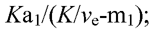 b2 = Ka2/(K/ve-m2); and b3 = -(bi+b2) where D is the contrast agent dose (known), als mi and a2, m2, are the corresponding amplitude and decay constants of the contrast agent in the body plasma which can be determined independently, and K and ve are the transcapillary transfer constant and the extravascular extracellular volume fraction, respectively.
7. Method comprising the steps of: a. creating a dataset by reading a set of N images of size n*n that specify x, y location; b. converting the dataset into a 3D matrix of n*n*N where N is the number of discrete samples of a function (creating a vector); c. determining the similarities for the vector of each pixel with x,y coordinates in the images to the vectors of each one of its 4 surrounding pixels according to a predefined norm; d. finding the most similar vector in the surrounding neighbors for each pixel x,y in the image, e. averaging the two similar vectors or employing another mathematical manipulation; f. creating a new n*n*N matrix that includes the similar vectors; g. repeating steps d, e and f q times and stopping according to convergence criterion; h. performing an automatic fitting process, for each pixel x,y, using the final vectors to find the function parameters; and presenting the parameters visually.
8. Method according to claim 7 including the further step of mapping the function parameters; and displaying the mapped function parameters.
9. Method comprising the steps of: a. creating a dataset by reading N images of size n*n*m that specify x, y, z location; b. converting the dataset into a 4D matrix of n*n*m*N where N is the number of discrete samples of a function creating a vector; c. determining for the vector of each pixel with x,y,z coordinates in the images similarities to the vectors of each one of its 6 surrounding pixels according to a predefined norm; d. finding for each pixel x,y,z in the image the most similar vector in the surrounding neighbors; e. averaging the two similar vectors or employ another mathematical manipulation; f. creating a new n*n*m*N matrix with that include the similar vectors; g. repeating steps c, d, e and f q times and stopping according to convergence criterion; h. performing an automatic fitting process, for each pixel x,y,z using the final vectors to find the function parameters; and i. presenting visually the function parameters.
10. Method according to claim 9 including the step of mapping the function parameters.
11. A computer readable medium containing executable program instructions for recording a set of MRI images of a tissue pixel by pixel that evolve with time in a specific manner described by a function; processing the recorded MRI images by aggregating the pixels thereof according to a preselected aspect of time behavior; and best fitting the aggregated pixels to a predetermined pixel function to obtain the parameters of the function; and presenting the parameters visually.
12. A computer readable medium according to claim 11 wherein the function is intensity of the pixels.
13. A computer readable medium according to claim 12 wherein the function is ISE (TR) = I(O)[l-exp(-TR/T1)] where ISE is the intensity in a pixel for a certain TR, 1(0) is a free parameter reaching the intensity for TR = infinity, and T1, the second free parameter, is the longitudinal relaxation time of the water in a pixel.
14. A computer readable medium containing executable program instructions for recording a set of time-spaced MRI pixel-by-pixel images of a preselected area of tissue of a patient injected with a contrast agent using a gradient echo sequence; performing aggregation based on pixel intensity on the recorded images; best- fitting the aggregated intensities to a preselected intensity function to yield maps of specific parameters of the tissue; and presenting the maps visually.
15. A computer readable medium according to claim 14 wherein the specific parameters are transcapillary transfer constant and extravascular extracellular volume fraction.
16. A computer readable medium according to claim 14 wherein the intensity function is
[I(t) -1(0)]/ 1(0) = [l-exp(-TR/T10)cos α](l-exp{-TR[l/T10+RiC,(t))]}/ {l-exp[-TR(l/Tio+RiC,(t))]cos α}[l-exp(-TR/Ti0)] -1. where I (t) is intensity at time t after administration of the contrast agent, 1(0) is intensity pre-contrast, TR is the repetition time of the sequence, α is the flip angle of the pulse, Tio is the longitudinal relaxation time pre-contrast, Ri is the relaxivity of the contrast agent, namely, the change in the relaxation of water when the concentration of the contrast agent increases by 1 millimolar, and Ct(t) is the concentration of the contrast agent which is given by
Ct(t) = D[biexp(-mit)+b2exp(-m2t)+b3exp(-A:t/ve), bi = Ka1I[KIv^m1); b2 = Ka2/(K/vβ-m2); and b3 = -(bi+b2) where D is the contrast agent dose (known), als mi and a2, m2, are the corresponding amplitude and decay constants of the contrast agent in the body plasma which can be determined independently, and K and ve are the transcapillary transfer constant and the extravascular extracellular volume fraction, respectively.
17. A computer readable medium containing executable program instructions for a. creating a dataset by reading a set of N images of size n*n that specify x, y location; b. converting the dataset into a 3D matrix of n*n*N where N is the number of discrete samples of a function (creating a vector); c. determining the similarities for the vector of each pixel with x,y coordinates in the images to the vectors of each one of its 4 surrounding pixels according to a predefined norm; d. finding the most similar vector in the surrounding neighbors for each pixel x,y in the image; e. averaging the two similar vectors or employing another mathematical manipulation; f. creating a new n*n*N matrix that includes the similar vectors; g. repeating steps d, e and f q times and stopping according to convergence criterion; and h. performing an automatic fitting process, for each pixel x,y, using the final vectors to find the function parameters.
18. A computer readable medium according to claim 17 including instructions for mapping the function parameters.
19. A computer readable medium containing executable program instructions for a. creating a dataset by reading N images of size n*n*m that specify x, y, z location; b. converting the dataset into a 4D matrix of n*n*m*N where N is the number of discrete samples of a function creating a vector; c. determining for the vector of each pixel with x,y,z coordinates in the images similarities to the vectors of each one of its 6 surrounding pixels according to a predefined norm; d. finding for each pixel x,y,z in the image the most similar vector in the surrounding neighbors; e. averaging the two similar vectors or employ another mathematical manipulation; f. creating a new n*n*m*N matrix with that include the similar vectors; g. repeating steps c, d, e and f q times and stopping according to convergence criterion; and h. performing an automatic fitting process, for each pixel x,y,z using the final vectors to find the function parameters.
20. Method according to claim 9 including instructions for mapping the function parameters.
Priority Applications (3)
| Application Number | Priority Date | Filing Date | Title |
|---|---|---|---|
| EP07844336.3A EP2074555A4 (en) | 2006-10-17 | 2007-10-17 | Method for obtaining spatial images through mri and processing the resulting spatial images and product |
| US12/443,943 US8175366B2 (en) | 2006-10-17 | 2007-10-17 | Method for obtaining spatial images through MRI and processing the resulting spatial images and product |
| IL197410A IL197410A (en) | 2006-10-17 | 2009-03-05 | Method for obtaining spatial images through mri and processing the resulting spatial images and product |
Applications Claiming Priority (2)
| Application Number | Priority Date | Filing Date | Title |
|---|---|---|---|
| US82970506P | 2006-10-17 | 2006-10-17 | |
| US60/829,705 | 2006-10-17 |
Publications (2)
| Publication Number | Publication Date |
|---|---|
| WO2008048982A2 true WO2008048982A2 (en) | 2008-04-24 |
| WO2008048982A3 WO2008048982A3 (en) | 2008-06-26 |
Family
ID=39314803
Family Applications (1)
| Application Number | Title | Priority Date | Filing Date |
|---|---|---|---|
| PCT/US2007/081580 WO2008048982A2 (en) | 2006-10-17 | 2007-10-17 | Method for obtaining spatial images through mri and processing the resulting spatial images and product |
Country Status (4)
| Country | Link |
|---|---|
| US (1) | US8175366B2 (en) |
| EP (1) | EP2074555A4 (en) |
| IL (1) | IL197410A (en) |
| WO (1) | WO2008048982A2 (en) |
Families Citing this family (1)
| Publication number | Priority date | Publication date | Assignee | Title |
|---|---|---|---|---|
| WO2013138794A1 (en) | 2012-03-16 | 2013-09-19 | Hologic, Inc. | Mri display output reflecting contrast agent concentration as a function of time |
Family Cites Families (10)
| Publication number | Priority date | Publication date | Assignee | Title |
|---|---|---|---|---|
| US6353803B1 (en) * | 1996-01-18 | 2002-03-05 | Yeda Research And Development Co., Ltd. At The Welzmann Institute Of Science | Apparatus for monitoring a system in which a fluid flows |
| US6553327B2 (en) | 1998-09-16 | 2003-04-22 | Yeda Research & Development Co., Ltd. | Apparatus for monitoring a system with time in space and method therefor |
| US6157192A (en) | 1998-04-14 | 2000-12-05 | Wisconsin Alumni Research Foundation | Recovery of signal void arising from field inhomogeneities in echo planar imaging |
| US6681132B1 (en) * | 1999-05-13 | 2004-01-20 | The Trustees Of Columbia University In The City Of New York | Sodium magnetic reasonance imaging used in diagnosing tumors and assessing response to treatment |
| US6724923B2 (en) * | 2001-04-13 | 2004-04-20 | Ge Medical Systems Global Technology Co., Llc | Automatic coil selection of multi-receiver MR data using fast prescan data analysis |
| US6909914B2 (en) * | 2003-06-13 | 2005-06-21 | Esaote, S.P.A. | Method for generating time independent images of moving objects |
| EP1997068B1 (en) * | 2004-08-17 | 2018-12-26 | Alan Penn & Associates, Inc. | Method and system for discriminating image representations of classes of objects |
| US7505037B2 (en) | 2004-10-02 | 2009-03-17 | Accuray, Inc. | Direct volume rendering of 4D deformable volume images |
| US7599542B2 (en) * | 2005-04-08 | 2009-10-06 | John Philip Brockway | System and method for detection and display of diseases and abnormalities using confidence imaging |
| US7799530B2 (en) * | 2005-09-23 | 2010-09-21 | Celera Corporation | Genetic polymorphisms associated with cardiovascular disorders and drug response, methods of detection and uses thereof |
-
2007
- 2007-10-17 WO PCT/US2007/081580 patent/WO2008048982A2/en active Application Filing
- 2007-10-17 US US12/443,943 patent/US8175366B2/en not_active Expired - Fee Related
- 2007-10-17 EP EP07844336.3A patent/EP2074555A4/en not_active Withdrawn
-
2009
- 2009-03-05 IL IL197410A patent/IL197410A/en not_active IP Right Cessation
Non-Patent Citations (1)
| Title |
|---|
| See references of EP2074555A4 * |
Also Published As
| Publication number | Publication date |
|---|---|
| EP2074555A2 (en) | 2009-07-01 |
| IL197410A0 (en) | 2009-12-24 |
| US8175366B2 (en) | 2012-05-08 |
| IL197410A (en) | 2013-03-24 |
| WO2008048982A3 (en) | 2008-06-26 |
| US20100092058A1 (en) | 2010-04-15 |
| EP2074555A4 (en) | 2013-10-16 |
Similar Documents
| Publication | Publication Date | Title |
|---|---|---|
| Ning et al. | A joint compressed-sensing and super-resolution approach for very high-resolution diffusion imaging | |
| Zhao et al. | A deep learning based anti-aliasing self super-resolution algorithm for MRI | |
| US11747424B2 (en) | Magnetic resonance imaging apparatus, image processing apparatus, and image processing method | |
| Jog et al. | Improving magnetic resonance resolution with supervised learning | |
| US11741580B2 (en) | Machine learning processing of contiguous slice image data | |
| EP1728213A1 (en) | Method and apparatus for identifying pathology in brain images | |
| EP3201643B1 (en) | Magnetic resonance imaging with enhanced bone visualization | |
| JP4090671B2 (en) | Image processing method, image processing apparatus, and image photographing apparatus | |
| JP5591687B2 (en) | Image processing method | |
| CN114402215A (en) | Generating MRT images of liver | |
| Do et al. | 7T MRI super-resolution with Generative Adversarial Network | |
| Xu et al. | STRESS: Super-resolution for dynamic fetal MRI using self-supervised learning | |
| JP6647836B2 (en) | Magnetic resonance imaging apparatus, image processing apparatus, and image processing method | |
| CN111681297B (en) | Image reconstruction method, computer device, and storage medium | |
| CN111044958B (en) | Tissue classification method, device, storage medium and magnetic resonance imaging system | |
| US8175366B2 (en) | Method for obtaining spatial images through MRI and processing the resulting spatial images and product | |
| CN111275783A (en) | Phase unwrapping method and device for magnetic resonance image and magnetic resonance imaging system | |
| US20220218222A1 (en) | Reconstruction augmentation by constraining with intensity gradients in mri | |
| Segers et al. | Discrete tomography in MRI: a simulation study | |
| CN112890798B (en) | Neural network training method, magnetic resonance imaging method, device, equipment and medium | |
| CN112782629B (en) | Magnetic resonance scanning control method, storage medium and system | |
| Gulzar et al. | Optical Flow Video Frame Interpolation Based MRI Super-Resolution | |
| Salvador et al. | A simple and fast technique for on-line fMRI data analysis | |
| CN115251883A (en) | Medical image acquisition apparatus and method | |
| Weisenseel et al. | A variational approach to multi-modality subsurface data inversion and fusion based on shared image structure |
Legal Events
| Date | Code | Title | Description |
|---|---|---|---|
| 121 | Ep: the epo has been informed by wipo that ep was designated in this application |
Ref document number: 07844336 Country of ref document: EP Kind code of ref document: A2 |
|
| WWE | Wipo information: entry into national phase |
Ref document number: 12443943 Country of ref document: US |
|
| WWE | Wipo information: entry into national phase |
Ref document number: 2007844336 Country of ref document: EP |
|
| NENP | Non-entry into the national phase |
Ref country code: DE |