WO2016134251A1 - Homogenous immunoassay with compensation for background signal - Google Patents
Homogenous immunoassay with compensation for background signal Download PDFInfo
- Publication number
- WO2016134251A1 WO2016134251A1 PCT/US2016/018667 US2016018667W WO2016134251A1 WO 2016134251 A1 WO2016134251 A1 WO 2016134251A1 US 2016018667 W US2016018667 W US 2016018667W WO 2016134251 A1 WO2016134251 A1 WO 2016134251A1
- Authority
- WO
- WIPO (PCT)
- Prior art keywords
- sample
- sdma
- calibrator
- analyte
- conjugate
- Prior art date
Links
Classifications
-
- G—PHYSICS
- G01—MEASURING; TESTING
- G01N—INVESTIGATING OR ANALYSING MATERIALS BY DETERMINING THEIR CHEMICAL OR PHYSICAL PROPERTIES
- G01N33/00—Investigating or analysing materials by specific methods not covered by groups G01N1/00 - G01N31/00
- G01N33/48—Biological material, e.g. blood, urine; Haemocytometers
- G01N33/50—Chemical analysis of biological material, e.g. blood, urine; Testing involving biospecific ligand binding methods; Immunological testing
- G01N33/53—Immunoassay; Biospecific binding assay; Materials therefor
- G01N33/5308—Immunoassay; Biospecific binding assay; Materials therefor for analytes not provided for elsewhere, e.g. nucleic acids, uric acid, worms, mites
-
- C—CHEMISTRY; METALLURGY
- C12—BIOCHEMISTRY; BEER; SPIRITS; WINE; VINEGAR; MICROBIOLOGY; ENZYMOLOGY; MUTATION OR GENETIC ENGINEERING
- C12N—MICROORGANISMS OR ENZYMES; COMPOSITIONS THEREOF; PROPAGATING, PRESERVING, OR MAINTAINING MICROORGANISMS; MUTATION OR GENETIC ENGINEERING; CULTURE MEDIA
- C12N9/00—Enzymes; Proenzymes; Compositions thereof; Processes for preparing, activating, inhibiting, separating or purifying enzymes
- C12N9/0004—Oxidoreductases (1.)
- C12N9/0006—Oxidoreductases (1.) acting on CH-OH groups as donors (1.1)
-
- G—PHYSICS
- G01—MEASURING; TESTING
- G01N—INVESTIGATING OR ANALYSING MATERIALS BY DETERMINING THEIR CHEMICAL OR PHYSICAL PROPERTIES
- G01N33/00—Investigating or analysing materials by specific methods not covered by groups G01N1/00 - G01N31/00
- G01N33/48—Biological material, e.g. blood, urine; Haemocytometers
- G01N33/50—Chemical analysis of biological material, e.g. blood, urine; Testing involving biospecific ligand binding methods; Immunological testing
- G01N33/53—Immunoassay; Biospecific binding assay; Materials therefor
- G01N33/536—Immunoassay; Biospecific binding assay; Materials therefor with immune complex formed in liquid phase
- G01N33/542—Immunoassay; Biospecific binding assay; Materials therefor with immune complex formed in liquid phase with steric inhibition or signal modification, e.g. fluorescent quenching
-
- G—PHYSICS
- G01—MEASURING; TESTING
- G01N—INVESTIGATING OR ANALYSING MATERIALS BY DETERMINING THEIR CHEMICAL OR PHYSICAL PROPERTIES
- G01N33/00—Investigating or analysing materials by specific methods not covered by groups G01N1/00 - G01N31/00
- G01N33/48—Biological material, e.g. blood, urine; Haemocytometers
- G01N33/50—Chemical analysis of biological material, e.g. blood, urine; Testing involving biospecific ligand binding methods; Immunological testing
- G01N33/68—Chemical analysis of biological material, e.g. blood, urine; Testing involving biospecific ligand binding methods; Immunological testing involving proteins, peptides or amino acids
- G01N33/6803—General methods of protein analysis not limited to specific proteins or families of proteins
- G01N33/6806—Determination of free amino acids
- G01N33/6812—Assays for specific amino acids
-
- C—CHEMISTRY; METALLURGY
- C12—BIOCHEMISTRY; BEER; SPIRITS; WINE; VINEGAR; MICROBIOLOGY; ENZYMOLOGY; MUTATION OR GENETIC ENGINEERING
- C12Y—ENZYMES
- C12Y101/00—Oxidoreductases acting on the CH-OH group of donors (1.1)
- C12Y101/01—Oxidoreductases acting on the CH-OH group of donors (1.1) with NAD+ or NADP+ as acceptor (1.1.1)
- C12Y101/01049—Glucose-6-phosphate dehydrogenase (1.1.1.49)
-
- G—PHYSICS
- G01—MEASURING; TESTING
- G01N—INVESTIGATING OR ANALYSING MATERIALS BY DETERMINING THEIR CHEMICAL OR PHYSICAL PROPERTIES
- G01N2333/00—Assays involving biological materials from specific organisms or of a specific nature
- G01N2333/90—Enzymes; Proenzymes
- G01N2333/902—Oxidoreductases (1.)
-
- G—PHYSICS
- G01—MEASURING; TESTING
- G01N—INVESTIGATING OR ANALYSING MATERIALS BY DETERMINING THEIR CHEMICAL OR PHYSICAL PROPERTIES
- G01N2800/00—Detection or diagnosis of diseases
- G01N2800/34—Genitourinary disorders
- G01N2800/347—Renal failures; Glomerular diseases; Tubulointerstitial diseases, e.g. nephritic syndrome, glomerulonephritis; Renovascular diseases, e.g. renal artery occlusion, nephropathy
Definitions
- the disclosure generally relates to the homogeneous immunoassays that allow for compensation of background signals inherent in samples and reagents.
- the disclosure generally relates to the homogeneous immunoassays that allow for compensation of background signals inherent in samples and reagents.
- the disclosure is directed to the use of homogeneous immunoassays for the detection of the presence or amount of symmetrical dimethyl arginine (SDMA) in biological samples.
- SDMA symmetrical dimethyl arginine
- SDMA has been identified in biological samples as a marker for the assessment of, for example, renal function, cardiovascular function, and SLE. SDMA is typically present in biological samples in a relatively low concentration compared to analytes for drugs of abuse.
- the disclosure is directed to a conjugates of symmetrical dimethyl arginine (SDMA), for example conjugates of SDMA and an enzyme.
- SDMA symmetrical dimethyl arginine
- the conjugate is SDMA conjugated to glucose-6-phosphate dehydrogenase (G6PDH).
- G6PDH glucose-6-phosphate dehydrogenase
- the SDMA and the enzyme are conjugated through a 5 to 15 atom linker.
- the SDMA may be enzyme through a linker having a length of about 5-15 Angstroms.
- Example conjugates of the disclosure include the following:
- the disclosure is directed to composition including conjugates of the disclosure and an antibody specific for free SDMA.
- the antibody specific for free SDMA may have reactivity for asymmetrical dimethylarginine (ADMA) of less than 25% of its reactivity for free SDMA.
- the antibody may have no or substantially no cross-reactivity with one or more compounds selected from the group consisting of asymmetrical dimethylarginine (ADMA), L-arginine, and N-methylarginine.
- kits that include a conjugate of the disclosure and an antibody specific for SDMA.
- kits of the disclosure include reagents for conducting an assay on a sample containing an analyte.
- the kits may include the following components
- first set of reagents for conducting a first assay including:
- a first reagent including an anti-analyte antibody and a signal producing substrate for an enzyme
- a second reagent including a conjugate of the analyte and the enzyme ii. a second reagent including a conjugate of the analyte and the enzyme
- a second set of reagents for conducting a second assay including i. a third reagent including the substrate.
- the second set of reagents may further include a fourth reagent including at least one of a diluent and a buffer.
- the fourth reagent may also further include the conjugate or the enzyme.
- at least one of the reagents in the kits includes an inhibitor for an enzyme other than the enzyme of the conjugate.
- the concentration of the conjugate in the first reagent is about 5 to about 150 times more than the concentration of the conjugate in the fourth reagent.
- the kits many also include a standard including a known amount of the analyte diluted in a sample solution that has been stripped of the analyte.
- the sample solution that has been stripped of the analyte may be stripped serum, stripped plasma, or a pretreated sample.
- the first reagent and/or the second reagent may further includes an electron mediator and a dye
- the third reagent and/or the fourth reagent may further include the mediator and the dye, wherein the mediator accepts an electron from the substrate and transfers it to the dye.
- reaction mixtures that include the components of the kits of the disclosure and a sample suspected of including SDMA.
- the disclosure is directed to method for determining the presence or amount of free SDMA in a sample.
- the method includes contacting the sample with an anti-SDMA antibody specific for free SDMA, the conjugate of claim 1, and a substrate including Nicotinamide Adenine Dinucleotide (NAD), measuring the conversion of NAD to NADH, and determining the presence or amount of SDMA in the sample based upon the conversion of NAD to NADH.
- NAD Nicotinamide Adenine Dinucleotide
- the measuring the conversion of NAD to NADH may include measuring the rate of conversion of NAD to NADH.
- the determining the presence or amount of SDMA in the sample based upon the conversion of NAD to NADH may include comparing the rate of the conversion of NAD to NADH to a standard curve, or the measuring the conversion of NAD to NADH may include measuring an amount of conversion of NAD to NADH.
- the determining the presence or amount of SDMA in the sample based upon the conversion of NAD to NADH may also include comparing the amount of the conversion of NAD to NADH to a standard curve.
- the methods include the following steps:
- a sample first reaction mixture by contacting the sample with an anti-analyte antibody, a conjugate including the analyte and an enzyme, and a substrate that produces a signal when in contact with the enzyme, and
- step (c) subtracting an amount of signal from step (b) from an amount of signal from step (a) to provide a net signal
- the methods of the disclosure may further include the following steps:
- calibrator first reaction mixtures by individually contacting each calibrator of a set of calibrators including known amounts of the analyte with an anti-analyte antibody, a conjugate including the analyte and an enzyme, and a substrate that produces a signal when in contact with the enzyme, and
- step (c) subtracting an amount of signal from step (a) from an amount of signal from step (b) to provide a net signal for each of the calibrators, (d) using the net signal from two or more of the calibrators to generate a
- the methods may include the following steps:
- a sample first reaction mixture by contacting the sample with an anti-analyte antibody, a conjugate including the analyte and an enzyme, and a substrate that produces a signal when in contact with the enzyme, and
- step (c) subtracting the normalized rate of step (b)(iii) from the normalized rate of step (a)(iii) to provide a final reaction rate for the sample containing the analyte
- the methods of the disclosure may also include the following steps:
- a calibrator first reaction sequence including: i. forming calibrator first reaction mixtures by individually contacting each calibrator of a set of calibrators including known amounts of the analyte with an anti-analyte antibody, a conjugate including the analyte and an enzyme, and a substrate that produces a signal when in contact with the enzyme, and
- step (c) subtracting the normalized rates of step (b)(iii) from the normalized rates of step (a)(iii) to provide a final reaction rate for each of the calibrators
- the sample and calibrator second reaction mixtures may include any one or more of a diluent, a buffer, and an anti-analyte antibody.
- the accounting for background associated with the sample first reaction mixture and/or the calibrator first reaction mixture may include subtracting the background from each of a plurality of signal measurements associated with the determination of a reaction rate for each sample and/or calibrator first reaction mixtures.
- the accounting for background associated with the sample and/or calibrator second reaction mixtures includes subtracting the background from each of a plurality of signal measurements associated with the determination of a reaction rate for the sample second reaction mixture and/or each calibrator second reaction mixture.
- the sample may be a biological sample, such as serum, plasma, urine or cerebral-spinal fluid.
- the calibrator includes the analyte diluted in plasma or serum, including for example a stripped serum or plasma.
- the calibrator may be a pretreated sample.
- sample and/or calibrator second reaction mixtures may further include the conjugate.
- the conjugate in the sample or calibrator first reactions mixture may be present at about 5 to about 150 times more than the concentration of the conjugate in the sample or calibrator second reaction mixtures.
- any of the reaction mixtures may include an inhibitor for an enzyme other than the enzyme of the conjugate.
- any of the reaction mixtures may further include an electron mediator and dye, wherein the mediator accepts an electron from the substrate and transfers it to the dye.
- the disclosure is directed to a method for determining chronic kidney disease in an animal.
- the method includes determining the presence or amount of SDMA in a biological sample from an animal according to methods of the disclosure, and determining chronic kidney disease in the animal based upon the presence amount of SDMA in the sample.
- Figure 1 shows the results of the assay of the disclosure on human serum samples from a normal population and patients suffering from Chronic Kidney Disease (CKD).
- CKD Chronic Kidney Disease
- Figure 2 shows a schematic representation of a procedure for conjugating SDMA to G6PDH using SIA to activate the G6PDH.
- Figure 3 shows a schematic representation of a procedure for conjugating SDMA to G6PDH using SBAP to activate the G6PDH.
- Figure 4 shows a schematic representation of a procedure for conjugating SDMA to G6PDH using SMCC to activate the G6PDH.
- Figure 5 is a schematic representation of a mediator-dye reaction mechanism wherein a mediator passes an electron to a dye in order to reduce the dye and shift the absorbance of the dye.
- Figure 6 shows a calibration curve for SDMA spiked into human sera and analyzed according to the method of the disclosure.
- Figure 7 shows calibration curves for SDMA spiked in human serum and analyzed according to a fixed calculation method and the rate calculation method of the disclosure.
- Figure 8 shows the results of an assay for known concentrations of SDMA conducted according to the disclosure without subtracting for background signal.
- Figures 9, 10 and 11 show the results of an SDMA assay according to the disclosure using canine (Fig. 9), feline (Fig. 10), and equine (Fig. 11) stripped serum calibrators.
- Figure 12 shows the results of an SDMA assay according to the disclosure using a buffer-based calibrator.
- SDMA is symmetrical dimethylarginine.
- the structure of SDMA is:
- free SDMA refers to SDMA that is not part of a polypeptide chain, including salts of SDMA.
- analyte analog generally refers to a modified form of the analyte which can compete with the analyte for a receptor, the modification providing a means to join the analyte to another moiety, such as a label or solid support.
- the analyte analog can bind to an antibody in a manner similar to the analyte.
- antibody generally refers to a glycoprotein produced by B lymphocyte cells in response to exposure to an antigen and binds specifically to that antigen.
- antibody is used in its broadest sense and specifically covers monoclonal antibodies (including full length monoclonal antibodies), polyclonal antibodies, multispecific antibodies (e.g., bispecific antibodies), and antibody fragments so long as they exhibit the desired biological activity.
- anti-SDMA antibody fragment and/or “anti-SDMA antibody variant” and the like include any protein or peptide containing molecule that includes at least a portion of an immunoglobulin molecule, such as, but not limited to, one complementarity determining region (CDR) of a heavy chain or light chain constant region, a framework region, or any portion thereof.
- CDR complementarity determining region
- antibody fragment refers to a portion of a full length antibody, generally the antigen binding or variable domain thereof.
- antibody fragments may include Fab, Fab', F(ab') 2 , and Fv fragments; diabodies; linear antibodies; single-chain antibody molecules; and multispecific antibodies from antibody fragments.
- antigen generally refers to a substance that is capable, under appropriate conditions, of reacting with an antibody specific for the antigen.
- analyte generally refers to the substance, or set of substances in a sample that are detected and/or measured.
- biological sample generally refers to a sample of tissue or fluid from a human or animal including, but not limited to whole blood, plasma, serum, spinal fluid such as cerebral-spinal fluid; lymph fluid, abdominal fluid (ascites), the external sections of skin, respiratory, intestinal and genitourinary tracts, tears, saliva, urine, blood cells, tumors, organs, tissue, and sample of in vitro cell culture constituents.
- cross-reactivity generally refers to the ability of an individual antigen binding site of an antibody to react with more than one antigenic determinant or the ability of a population of antibody molecules to react with more than one antigen. In general, cross reactions arise because (i) the cross reacting antigen shares an epitope in common with the immunizing antigen or (ii) it has an epitope which is structurally similar to one on the immunizing antigen (multispecificity).
- label refers to a detectable compound or composition that can be conjugated directly or indirectly ⁇ e.g., via covalent or non-covalent means, alone or
- an enzymatic label may catalyze chemical alteration of a substrate compound or composition which is detectable.
- the enzymes employed in the current disclosure could be, but are not limited to: alkaline phosphatase (AP); glucose-6-phosphate dehydrogenase ("G6PDH”); Beta Galactosidase (B GAL); and horse radish peroxidase (HRP), malate dehydrogenase (MDH).
- G6PDH glucose-6-phosphate dehydrogenase
- the utilization of a label produces a signal that may be detected by means such as detection of electromagnetic radiation or direct visualization, and that can optionally be measured.
- the term "monoclonal antibody,” as used herein generally refers to an antibody obtained from a population of substantially homogeneous antibodies, i.e., the individual antibodies including the population are identical. Monoclonal antibodies are highly specific, being directed against a single antigenic site. In contrast to polyclonal antibody preparations, which typically include different antibodies directed against different epitopes, each monoclonal antibody is directed against a single epitope on the antigen.
- the modifier “monoclonal” merely refers to the character of the antibody and is not to be construed as requiring production of the antibody by any particular method.
- monoclonal antibodies may be made by hybridoma methodologies, or may be made by recombinant DNA methods, or may be isolated from phage antibody libraries using known techniques.
- polypeptide generally refers to a molecule having a sequence of amino acids linked by peptide bonds. This term includes proteins, fusion proteins, oligopeptides, cyclic peptides, and polypeptide derivatives. Antibodies and antibody derivatives are discussed above in a separate section, but antibodies and antibody derivatives are, for purposes of the disclosure, treated as a subclass of the polypeptides and polypeptide derivatives.
- Receptor refers to any compound or composition capable of recognizing a particular spatial and polar organization of a molecule, e.g., epitopic or determinant site.
- Illustrative receptors include antibodies, Fab fragments, and the like.
- Binding specificity refers to the substantial recognition of a first molecule for a second molecule, for example an analyte, such as SDMA, and a polyclonal or monoclonal antibody, or an antibody fragment (e.g. a Fv, single chain Fv, Fab', or F(ab')2 fragment) specific for the analyte.
- analyte such as SDMA
- a polyclonal or monoclonal antibody or an antibody fragment (e.g. a Fv, single chain Fv, Fab', or F(ab')2 fragment) specific for the analyte.
- an antibody fragment e.g. a Fv, single chain Fv, Fab', or F(ab')2 fragment
- specificity generally refers to the ability of an individual antibody combining site to react with only one antigenic determinant or the ability of a population of antibody molecules to react with only one antigen. In general, there is a high degree of specificity in ana
- Antibodies can distinguish differences in (i) the primary structure of an analyte, (ii) isomeric forms of an analyte, and (iii) if applicable, secondary and tertiary structure of an analyte. Antibody-analyte reactions that exhibit high specificity exhibit low cross reactivity.
- substantially bind refers to an amount of specific binding or recognizing between molecules in an assay mixture under particular assay conditions.
- substantial binding relates to the difference between a first molecule's incapability of binding or recognizing a second molecule, and the first molecules capability of binding or recognizing a third molecule, such that the difference is sufficient to allow a meaningful assay to be conducted distinguishing specific binding under a particular set of assay conditions, which includes the relative concentrations of the molecules and the time and temperature of an incubation.
- one molecule is substantially incapable of binding or recognizing another molecule in a cross-reactivity sense where the first molecule exhibits a reactivity for a second molecule that is less than 25%, for instance less than 20%, less than 15%), less than 10%>, less than 5% or less than 1%> of the reactivity exhibited toward a third molecule under a particular set of assay conditions.
- Specific binding can be tested using a number of widely known methods, e.g., an immunohistochemical assay, an enzyme-linked immunosorbent assay (ELISA), a radioimmunoassay (RIA), or a western blot assay.
- salt means a salt formed between an acid and a basic functional group of a compound.
- Illustrative salts include, but are not limited, to sulfate, citrate, acetate, oxalate, chloride, bromide, iodide, nitrate, bisulfate, phosphate, acid phosphate, isonicotinate, lactate, salicylate, acid citrate, tartrate, oleate, tannate, pantothenate, bitartrate, ascorbate, succinate, maleate, gentisinate, fumarate, gluconate, glucaronate, saccharate, formate, benzoate, glutamate, methanesulfonate, ethanesulfonate, benzenesulfonate, p-toluenesulfonate, and pamoate (i.e., l, l '-methylene-bis-(2-
- salt also refers to a salt formed between a compound having an acidic functional group, such as a carboxylic acid functional group, and an inorganic or organic base.
- Suitable bases include, but are not limited to, hydroxides of alkali metals such as sodium, potassium, and lithium;
- hydroxides of alkaline earth metal such as calcium and magnesium; hydroxides of other metals, such as aluminum and zinc; ammonia, and organic amines, such as unsubstituted or hydroxy- substituted mono-, di-, or trialkylamines; dicyclohexylamine; tributyl amine; pyridine; N-methyl, N-ethylamine; diethylamine; triethylamine; mono-, bis-, or tris-(2-hydroxy-lower alkyl amines), such as mono-, bis-, or tris-(2-hydroxyethyl)amine, 2-hydroxy-tert-butylamine, or tris- (hydroxymethyl)methylamine, N, N,-di-lower alkyl-N-(hydroxy lower alkyl)-amines, such as N,N,-dimethyl-N-(2-hydroxyethyl)amine, or tri-(2-hydroxyethyl)amine; N-methyl-D-glucamine; and amino
- sample components other than the analyte may react with each other or with components of the reagents for the assays. Such reactions can lead to interference with the accuracy of the detection method.
- Biological samples are notorious for including many components that can create background noise in assays.
- Samples may be analyzed using a modified assay based upon the EMIT ®
- EMIT® assay Enzyme Multiplied Immunoassay Technique homogeneous immunoassay system.
- a sample containing the analyte is contacted with an anti-analyte antibody, a conjugate of the analyte and an enzyme, and a substrate that produces a signal when in contact with the enzyme. Binding of the antibody to the conjugate inhibits or reduces enzyme activity.
- the sample analyte competes with the conjugated analyte for binding to the antibody, which results in the generation of more signal from the enzyme/substrate.
- no analyte is present, more binding can occur between antibody and conjugate to limit or prevent signal generation. Therefore, more signal is generated when more analyte is present.
- Kinetic assays can use the rate of signal generation as an indicator of the presence or amount of analyte in a sample
- the traditional EMIT® assay format is conducted in a first reaction sequence for both the analyte and the calibrators.
- this first reaction sequence may be identified herein as the "Color Assay.”
- a second and separate assay is conducted in a second reaction sequence for the sample and calibrators.
- the second reaction sequence may be identified herein as the "Blank Assay. "
- the reagents of the Blank Assay do not contain anti-analyte antibody and typically does not contain conjugate. Therefore, the Blank Assay provides a sample-dependent background signal.
- disclosure is directed to the use of the signal in two example methods for determining the concentration of an analyte.
- the example methods are referred to herein as the "Rate” method and the “Fixed” method.
- the difference between an amount of signal or signals in the Color and Blank assays is used.
- signal is measured as absorbance at a wavelength specific for an enzyme/substrate system as is well known in the art. For instance, measurement of absorbance at 340 nm for a
- G6PDH/NAD enzyme/substrate system will provide a value for the relative amount of conversion of NAD to NADH in the presence of G6PDH.
- the value can be used to provide a net signal (for the Fixed method) or a reaction rate (for the Rate method) reflecting the conversion of the substrate by the enzyme.
- the enzyme substrate reaction can be used in conjunction with an electron carrier that mediates electron transfer between the substrate and various electron acceptors (e.g., a dye) that will allow for measurement of the reaction rate at a different wavelength than that of the substrate.
- a net signal is calculated.
- the net signal is based upon the difference in signal (e.g., absorbance) between the Color and Blank assays at a predetermined end point, which may or may not be the completion of the reaction by exhaustion of all substrate.
- the difference in the amount of signal between a predetermined starting point Tl) (which may be at the completion of the combination all reagents or another predetermined time thereafter) and the predetermined end point (T2) can be determined for both the Color and Blank assays can be determined.
- the net signal can be compared to a calibration curve to determine the amount of analyte in the sample.
- the measurements may be taken after the reagents have had a sufficient to time to react.
- the single measurement in each assay may be taken at about 30 second to about 10 minutes following the combination of all of the reagents. More particularly, the single measurement may be taken at one of 30, 60, 90, 120, 150, 180, 210, 240, 270, 300, 330, 360, 390, 420, 450, 480, 510, 540, 570 and 600 seconds following the combination of all of the reagents.
- the first measurement (Tl) is taken after about 15 second to two minutes from the beginning of assay (combination of sample and all reagents). This time can be adjusted based upon the concentration of reagents and the expected concentration of the sample. In particular, Tl may be 15, 30, 45, 60, 75, 90, 105 or 120 seconds following the combination of all the reagents.
- the second measurement (T2) can be taken from 15 seconds to several minutes following Tl . For example, T2 may be for example, 15 seconds, 18 seconds, 30 seconds, 45 second, or 1, 2, 3, 4, or 5 minutes, after the first measurement (Tl).
- the signal from the Blank Assay is subtracted from the signal of the Color assay at defined intervals to provide a reaction rate.
- the reaction rates of calibrators are used to provide a calibration curve that can be used to determine the amount of analyte in the sample by comparing the curve to the reaction rate of the assay on the sample to the calibration curve.
- the reaction rate can be determined by measuring signal (e.g. absorbance) at a plurality of time points during the enzyme mediated reaction. Determination of the time and interval of signal measurement are within the skill in the art taking into
- a rate can be determined by measuring absorbance beginning about 2-10 minutes after the combination of sample (or calibrator) and all reagents at room temperature and measured every 5-60 seconds for an additional 1-15 minutes.
- Reaction rate can be expressed as the change in absorbance over time. For example, absorbance can be measured starting at about 1, 2, 3, 4, 5, 6, 7, 8, 9, or 10 minutes after combining the sample (or calibrator) and all the reagents.
- Absorbance is typically measured at intervals of 5, 10, 15, 20, 25, 30, 35, 40, 45, 50, 55 or 60 seconds for about 2, 3, 4, 5, 6, 7, 8, 9, 10, 11, 12, 13, 14 or 15 minutes. Each of these times can be extended or shortened, depending on reaction conditions, analyte, and reagents.
- background from sample first reaction mixture can be accounted for in calculation of the net signal or reaction rate to provide a normalized signal.
- the background absorbance is measured from a calibrator matrix used in place of the sample.
- the absorbance of the calibrator matrix is measured initially upon the combination of all of the reagents with the matrix, or at a finite time thereafter.
- the calibrator matrix may be a calibrator or control mixture lacking any analyte.
- a calibrator matrix lacking analyte may be the sample that has been stripped of the analyte by dialysis or by another pretreatment process as further described herein.
- stripped or endogenous species specific or non-specific serum, plasma, or other biological fluid may be used as the calibrator matrix.
- the calibrator matrix may be water or a diluent containing protein and/or other compositions (e.g., BSA in PBS). The calibrator matrix is used in place of the sample in both the Color Assay and the Blank Assay to provide a background signal for both Assays.
- the background is then subtracted from the net signal in the Fixed method (or measurements at Tl and T2) or from each measurement used to determine the sample reaction rate in the Rate method.
- the absorbance of the calibrator matrix is subtracted from each absorbance measurement that is used to determine the reaction rate for the sample (i.e., change in absorbance over time).
- a background rate is determined in a manner similar to the sample reaction rate. The background rate is then subtracted from sample reaction rate to provide a normalized reaction rate for the first sample reaction mixture.
- the disclosure is directed to methods for conducting an assay for an analyte.
- the method includes conducting the Color Assay on the sample (sample first reaction sequence) by forming a sample first reaction mixture including the sample, an anti-analyte antibody, a conjugate including the analyte and an enzyme, and a substrate that produces a signal when in contact with the enzyme.
- the sample first reaction mixture is formed by first contacting the sample with the antibody and the substrate, and then in a second step adding the conjugate.
- the sample may be first combined with the antibody and the conjugate, and the substrate added in a second step.
- the enzyme and conjugate are kept separate until the reaction sequence is ready to begin.
- Contacting as used here is used in its broadest aspect to refer to combining reagents in any order unless otherwise specified herein.
- sample second reaction sequence or "Blank Assay”
- the reagents of the sample second reaction sequence do not contain anti-analyte antibody.
- the reagents of the Blank Assay do not contain conjugate, but described herein are embodiments where a small amount of conjugate or enzyme is added.
- a sample second reaction mixture is formed by contacting the sample with the substrate and, in some instances, the conjugate but no anti-analyte antibody. Signal is measured from the sample second reaction mixture.
- the net signal or reaction rate using the background from the calibrator matrix is determined in a manner identical to the sample first reaction sequence to provide a normalized net signal or a normalized reaction rate for the sample second reaction sequence.
- the normalized rate of sample second reaction sequence is subtracted from the normalized reaction rate of the sample first reaction sequence.
- the final reaction rate can be used to determine the presence or amount of the analyte in the sample. Typically, this is done by comparing the final reaction rate for the sample to a standard curve, which can be prepared according to methods known in the art.
- the Color and Blank assays for the calibrators can be conducted in manner similar to the Color and Blank assays for the sample. Accordingly, a series of calibrator first reaction sequences (Color Assays) is conducted by forming calibrator first reaction mixtures by individually contacting each calibrator of a set of calibrators including known amounts of the analyte with an anti-analyte antibody, a conjugate including the analyte and an enzyme, and a substrate that produces a signal when in contact with the enzyme.
- a series of calibrator first reaction sequences is conducted by forming calibrator first reaction mixtures by individually contacting each calibrator of a set of calibrators including known amounts of the analyte with an anti-analyte antibody, a conjugate including the analyte and an enzyme, and a substrate that produces a signal when in contact with the enzyme.
- Signal is measured from each of the calibrator first reaction mixtures and a normalized net signal (Fixed method) or normalized reaction rate (Rate method) for each of the calibrator first reaction mixtures is generated by accounting for background associated with each of the calibrator first reaction mixtures in the manner described above for the sample first reaction mixtures.
- One of the calibrators may be the calibrator matrix (which contains no analyte) that is used to determine background.
- calibrator second reaction sequence are performed by forming calibrator second reaction mixtures by contacting each calibrator of the set of calibrators with the substrate in the absence of anti-analyte antibody and, typically, conjugate.
- conjugate may also be present, usually in a small amount as described further herein.
- Signal is measured for each of the calibrator second reaction mixtures, and the net signal or reaction rate for each mixture is normalized by accounting for background associated with the calibrator second reaction mixtures in the manner described above.
- the normalized net signal from the Blank assay is subtracted from a normalized net signal from the Color assay.
- a final reaction rate for each of the calibrators is determined by subtracting the normalized rate from the calibrators in the calibrator second reaction sequences from the normalized rate for each of the calibrators in the calibrator first reaction sequences.
- a normalized standard curve based upon the final reaction rate for each of the calibrators can then be prepared and used to determine the amount of the analyte in the sample by comparing the normalized reaction rate for the sample to the normalized standard curve.
- the reagents associated with the Color Assay and Blank are identical to the reagents associated with the Color Assay and Blank
- Assays are shown in Table 1.
- the reagents are combined in the appropriate diluent and/or buffers.
- Blank Assay reagent 2 (R2) does not contain the conjugate, although it may be present as further described herein to ensure that the reaction has the proper volume of buffer.
- Blank Assay Rl may have extra volume in the event that Blank Assay R2 is not used.
- the reagents for the Color Assay are identical to the reagents for the Blank Assay except for the presence or absence of the antibody and conjugate. When R2 does not contain any conjugate in the Blank Assay, it still contains the diluent, buffer and other components of R2 for the Color Assay.
- Blank Assay Rl may also contain the anti-analyte antibody.
- Calibrators and test samples in the Color Assay can commonly be expected to produce an increase in absorbance due to the analyte-enzyme conjugate that is added to the reaction mixture.
- Many samples also produce a positive reaction when tested in the Blank Assay because endogenous enzymes reactive with endogenous substrates or with the substrate of Rl will produce an increase in absorbance even without conjugate present.
- samples and calibrators are diluted to the extent that a usually small background signal is weakened further such that the reaction mixtures will produce little to no change in absorbance when tested in the Blank Assay. Because the change of absorbance may fall below the sensitivity range of a detector, it can lead to calibration curve failure on automated analyzers.
- the concentration of enzyme or conjugate in R2 is typically kept low to prevent unnecessary noise in the assay for the calibrator matrix.
- the enzyme is G6PDH
- about 1-15 ng/mL of the enzyme can be added to the Blank Assay.
- about 1, 2, 3, 4, 5, 6, 7, 8, 9, 10, 11, 12, 13, 14 or 15 ng/ml may be added.
- the amount of the conjugate in R2 depends on the size of the analyte, as larger analytes will require greater quantities of conjugate to provide the same amount of enzyme.
- the concentration of the enzyme (either alone or conjugated) in R2 for the Color Assay is about 10 to about 150 times more than the concentration of the enzyme in R2 for the Blank Assay.
- the concentration of the conjugate in the Color Assay is 10, 20, 30, 40, 50, 60, 70, 80, 90, 100, 110, 120, 130, 140 and 150 times more than the concentration of the enzyme in the reagents for the Blank Assay.
- the activity of the enzyme is typically less than that of the unconjugated enzyme.
- the activity of the conjugated enzyme varies widely from the activity of the unconjugated enzyme, for example the activity of the conjugate enzyme is 10%, 20%, 30%, 40%, 50%, 60%, 70%, 80% or 90% of the unconjugated enzyme. Therefore, when unconjugated enzyme is used in the R2 for the Blank assay, it can be used in a small amount than if it were conjugate and still provide the same activity as the conjugated enzyme.
- the assay reagents can include stabilizers of assay reagents.
- the signal producing enzyme is glucose-6-phosphate dehydrogenase (G6PDH), glucose-6-phosphate (G6P), or NAD, can be added to conjugate containing reagent to stabilize the enzyme-analyte conjugate.
- Serum contains several enzymes that can contribute to background. Therefore, the addition of specific enzyme inhibitors to the reagent diluents can further improve assay accuracy. The inhibitors help eliminate a portion of the interfering noise in the color and blank assays. Accordingly, in various aspects, the reaction components can include inhibitors of endogenous sample enzymes.
- sodium oxamate is a known inhibitor of lactate dehydrogenase (LDH), which is an enzyme of NAD and/or NADP.
- Sodium oxamate prevents background signal associated with endogenous sample LDH by preventing LDH from turning over NAD in the sample or that may be part of the enzyme-substrate system associated with the assay (e.g., G6PDH/NAD). Over thirty serum enzyme and enzyme inhibitor combinations are known and inhibitors are available for many of them.
- the calibrators include known quantities of the analyte (or none for the calibrator matrix) in dionized water or saline to provide the calibration matrix that is used in place of the sample.
- stripped serum is used instead of water or saline.
- the dialyzed serum is natural, species specific or non-specific serum or plasma that has been stripped the analyte in, for example, a dialysis process. Numerous other sample pretreatment processes are known to remove or inactivate the analyte.
- the solutions are spiked with known quantities of the analyte to provide a series of calibrators over the expected range of concentration of the analyte in the sample.
- the calibration solutions is a protein based solution such as 1-7% BSA in PBS. In this embodiment, it may be required to calibrate the BSA calibrators to stripped and spiked serum calibrators. .
- the Blank Assay is intended to compensate for endogenous proteins, enzymes or other large molecule components that contribute to background signal
- water or saline are more suited for non-biological samples, although sufficient specificity may be obtained with the use of water or saline as a calibrator solution for biological samples in many instances.
- the use of water or saline buffer for the calibrator matrix in automated analyzers may have the tendency to create an error message for the calibrator matrix because the reaction rate associated with the use of buffer can result in significantly increased reaction rates compared to serum. In this situation, the subtraction of the absorbance value of the calibration matrix could result in a negative rate. While these rates can be normalized against serum-based calibrator matrices, automated analyzers will typically create an error message in this situation.
- the analyte is SDMA and the enzyme-conjugate system is G6PDH/NAD.
- an analog of SDMA is conjugated to G6PDH and used as the conjugate in the Color and Blanks Assays in order to determine the presence or amount of SDMA in serum or plasma samples from animals such as humans, cats and dogs.
- calibrators are prepared by combining known amounts of SDMA with in the calibrator matrix (stripped serum or plasma). Color and Blank assays are conducted as described above using the following reagents in exemplary amounts as described in Table 3 :
- the anti-SDMA antibody can have a concentration in Rl of the Color
- the G6P and NAD may have a concentration of about 8-75 mM, more particularly about 10-65 mM, or about 20-55 mM, and more particular about 8, 10, 15, 20, 25, 30, 35, 40 ,45 50, 55, 60, 65, 70 mM.
- the SDMA-G6PHD conjugate may have a concentration of about 0.25 to about 0.75 ⁇ g/mL, more particularly about 0.25, 0.30, 0.35, 0.40, 0.45, 0.50, 0.55, 0.65, 0.70 or 0.75 ⁇ g/mL.
- the concentration of the conjugate may be about 5-30 ng/mL, more particularly about 5, 10, 15, 20, 25 or 30 ng/mL.
- G6P is present in the R2 reagents to stabilize the enzyme.
- NAD could be used.
- the stabilizing substrate is optional.
- Reagents may be combined with the sample and calibrators at the following volumes for the Color and Blank Assays, although volumes may be adjusted to accommodate different analytes and reaction conditions:
- the reaction mixture contains 10 ⁇ of sample, 40 ⁇ of Rl and 125 ⁇ . of R2.
- a calibration curve can be prepared and used to determine the presence or amount of SDMA in serum or plasma samples.
- Figure 1 shows the results using the Rate method for the determination of SDMA in normal and chronic kidney disease human serum samples with a Beckman AU680 ® clinical chemistry analyzer. Results show accurate determination of SDMA concentration when compared to gold-standard LC-MS values. Human serum (unstripped) was used as the calibrator matrix.
- the disclosure is directed to a method of determining chronic kidney disease in animals, including humans, and, for example, domestic, farm, and zoo animals. The method includes determining SDMA concentration in a biological sample from an animal subject according to a method of the disclosure and comparing concentration to a standard curve or other model that the amount of SDMA in samples from the same species as the subject in healthy subjects and subject suffering from chronic kidney disease.
- kits containing reagents for determining the presence or amount of analytes in samples.
- the kit can include a first set of reagents for conducting the Color Assay, such as a first reagent including an anti- analyte antibody and a signal producing substrate for an enzyme, and a second reagent including a conjugate of the analyte and the enzyme.
- the kit may also include a second set of reagents that includes a third reagent containing the substrate.
- the second set of reagents may also include a fourth reagent containing the buffer/diluent, and optionally the conjugate.
- the purpose of the fourth reagent containing only buffer/diluent is to ensure that the reaction associated with the second set of reagents is conducted at the proper volume.
- the volume of the third reagent should be adjusted accordingly.
- either the third or fourth reagent may contain the anti-analyte antibody.
- the concentrations and amounts of the components in each reagent can be as described above for Rl and R2 in each of the Color and Blank Assays.
- the concentration of the conjugate in the first reagent is about 5 to about 150 times more than the concentration of the conjugate in the fourth reagent.
- at least one of the reagents in the kit includes an inhibitor for an enzyme other than the enzyme of the conjugate.
- the kit may also include a standard or set of standards (calibrators) that include a known amount of the analyte diluted an appropriate diluent, such as water, saline, or stripped or processed serum or plasma (e.g., a pretreated sample).
- an exemplary kit includes in the first reagent an anti-
- the second reagent includes a conjugate of an SDMA analog and G6PDH as a stabilizer for the enzyme.
- the third reagent includes the SDMA-G6PDH conjugate and, optionally a fourth reagent.
- the fourth reagent may contain only diluent or buffer, or it may contain the conjugate.
- the disclosure is directed to a reaction mixture that includes reagents from the kits and a sample suspected of containing SDMA.
- the reaction mixture may include the sample or calibrator, the anti-SDMA antibody, the SDMA-G6PDH conjugate, NAD, and G6P.
- a number of enzyme/substrates systems can be used in place of G6PDH/NAD.
- malate dehydrogenase is an enzyme catalyzes the oxidation of malate to oxaloacetate using the reduction of NAD + to NADH similar to G6PDH.
- many enzyme mutants are known which enhance the signal or stability of the enzymes. See, e.g., U.S. Patent 6,455,288.
- Analyte-enzyme conjugates can be prepared by a variety of known methods.
- Linker length can have an effect on the ability of the antibody to inhibit enzyme activity and to therefore affect assay sensitivity.
- linkers typically of about 5- 15 atoms, or 2-20 Angstroms may be used.
- the conjugation of enzymes to analytes is within the skill in the art for many analytes that can be detected according to the disclosure.
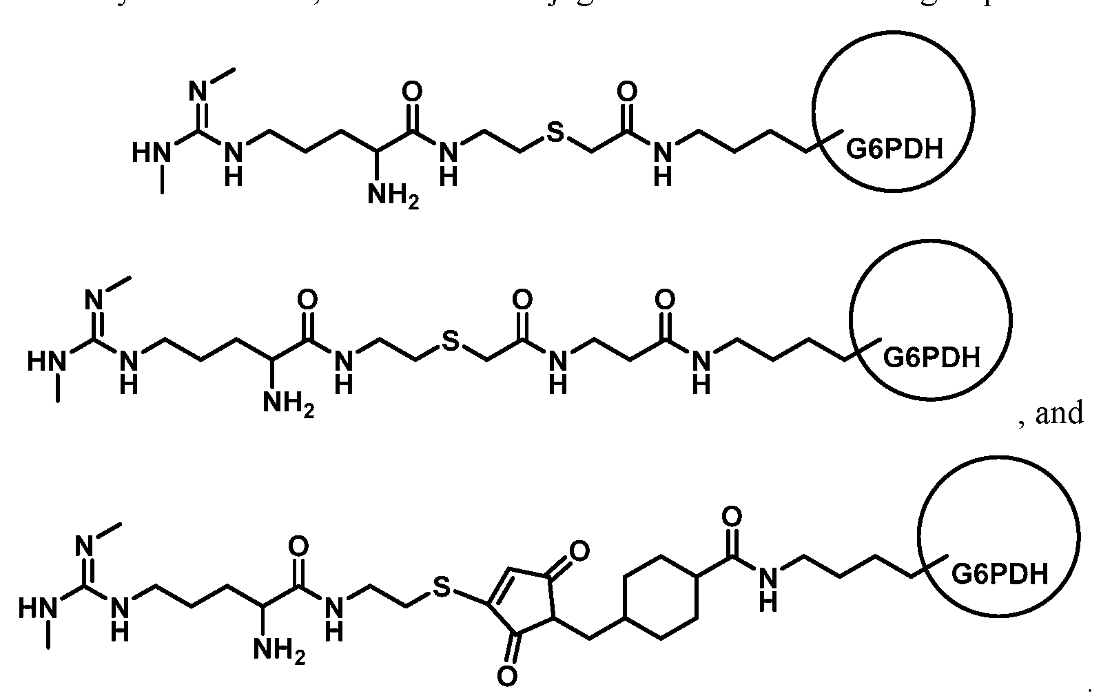
- Figure 2 shows the conjugation of SDMA analog of Formula 1
- G6PDH glucose-6-phosphate dehydrogenase
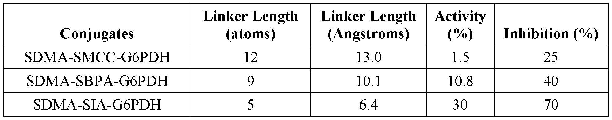
- the G6PDH is activated with succinimidyl iodoacetate (SIA) prior to conjugation.
- SIA succinimidyl iodoacetate
- the activation with SIA results in a five atom linker between SDMA and the enzyme (-C-C-S-C-C(O)-), which does not include the non- native nitrogen on the SDMA which results from replacement of oxygen with nitrogen during the derivitization of SDMA.
- Figure 3 shows conjugation of the SDMA analog of Formula 1 to G6PDH using
- Figure 4 shows conjugation of the SDMA analog of Formula 1 to G6PDH using sulfosuccinimidyl-4-(N-maleimidomethyl)cyclohexane-l-carboxylate (SMCC) which results in a twelve atom linker.
- SMCC sulfosuccinimidyl-4-(N-maleimidomethyl)cyclohexane-l-carboxylate
- Linker length can be adjusted by modifying G6PDH with other reagents or using other SDMA analogs in the conjugation reaction to provide linker lengths of, for example, about 5 to about 15 atoms, in particular about 5, 6, 7, 8, 9, 10, 1 1 , 12, 13, 14 or 15 atoms.
- the disclosure is directed to a conjugate of SDMA and an enzyme through a linker of 5-15 atoms, which include only those atom in directly in the chain of the linker through the shortest route excluding any atoms of side chains, substituents, or rings in the linker molecule that are not part of the shortest route between the SDMA and enzyme.
- Linker lengths can range from about 2 Angstroms to about 20 Angstroms, in particular about 5 to about 15 Angstroms, more particularly about 6-10 Angstroms.
- SDMA is conjugated to G6PDH in the presence of G6PDH substrates glucose-6-phosphate (G6P) and/or nicotinamide adenine dinucleotide (NADH).
- G6PDH substrates glucose-6-phosphate (G6P) and/or nicotinamide adenine dinucleotide (NADH).
- G6P glucose-6-phosphate
- NADH nicotinamide adenine dinucleotide
- Optimal conjugate-enzyme activity and inhibition of that activity by the antibody can be obtained by adjusting the ratios of enzyme, substrates and SDMA.
- x and y are integers ranging from 1 to 5.
- Formulas A, B and C provide an available thiol that can react with a conjugation target that includes an appropriate "thiol-reactive site," i.e., a site that will react with a thiol group.
- thiol-reactive site i.e., a site that will react with a thiol group.
- maleimides, alkyl and aryl halides, and alpha-haloacyls are illustrative thiol -reactive sites that can react with thiols to form thio-ethers.
- pyridyl disulfides can react with thiols to form mixed disulfides.
- G6PDH activated with SIA, SBAP, or SMCC provide an appropriate thiol reactive site.
- the SDMA analog has the following formula (Formula 1):
- Formula 1 may be prepared from SDMA (commercially available from EMD).
- (1) can be linked to a cysteamine-4-methoxy trityl resin (EMD Chemicals, Inc., Gibbstown, NJ) by contacting the (Boc 3 )-SDMA (1) with the resin in the presence of 2-(lH-7-azabenzotriazol-l- yl)-l, l,3,3-tetramethyl uranium hexafluorophosphate methanamininium (HATU) and N,N- diisopropylethylamine (DIPEA) in dimethyl formamide (DMF) to provide resin bound (Boc 3 )- SDMA cystamide (2).
- the BOC protecting groups on the resin bound (Boc 3 )-SDMA cystamide EMD Chemicals, Inc., Gibbstown, NJ
- a color changing dye and an electron mediator can allow absorbance to be measured at an absorbance other than 340 nm.
- the dye is 3-(4,5- dimethylthiazol-2-yl)-2,5-diphenyltetrazolium bromide (MTT) and the mediator is 1-methoxy phenazine methosulfate (PMS).
- MTT 3-(4,5- dimethylthiazol-2-yl)-2,5-diphenyltetrazolium bromide
- PMS 1-methoxy phenazine methosulfate
- the PMS picks up an electron from NADP and transfers it to the MTT, which reduces the MTT to provide for absorbance at approximately 650 nm.
- Anti-SDMA antibodies may be polyclonal or monoclonal as described in U.S.
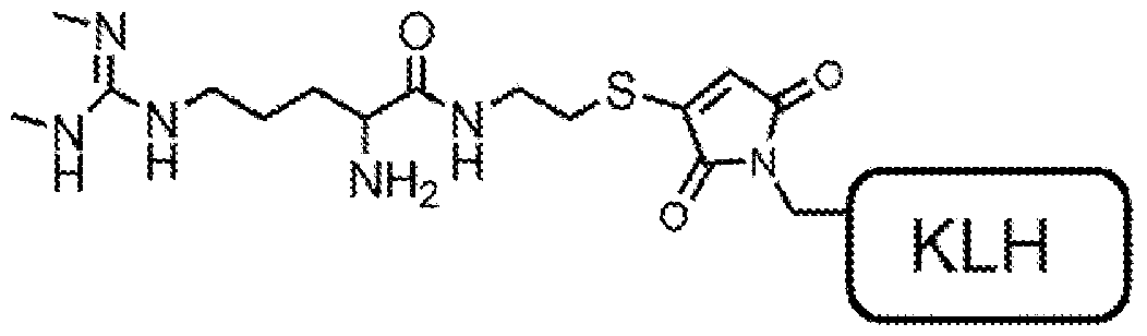
- the antibody is a monoclonal antibody raised against a SDMA-KLH conjugate having the following structure:
- the anti-SDMA antibodies used in the modified assay of the disclosure can have high specificity for SDMA and no or substantially no cross-reactivity with one or more compounds selected from the group consisting of asymmetrical dimethylarginine (ADMA), L-arginine, and N-methylarginine.
- ADMA asymmetrical dimethylarginine
- the anti-SDMA antibody exhibit a reactivity for a ADMA, L-arginine, and N-methylarginine that is less than 25%, less than 20%, less than 15%, less than 10%, less than 5%, or less than 1% of the reactivity exhibited towards SDMA under a particular set of assay conditions.
- the method includes contacting the sample with an anti-SDMA antibody, a conjugate including SDMA and G6PDH, and a substrate(s) including NAD and G6P.
- an anti-SDMA antibody As described herein the rate of conversion of NAD to NADH, is measured and compared to a standard curve to determine the presence or amount of SDMA in the sample.
- the SDMA is conjugated to the G6PDH with a 5-15 atom linker (2-10 Angstroms) as described herein.
- the anti-SDMA antibody may be monoclonal antibody that has no reactivity or substantially no reactivity for ADMA, L-arginine, and N-methylarginine
- the disclosure is directed to a method for determining
- SDMA in a sample that includes derivatizing free SDMA and using an antibody directed to the derivative.
- U.S. Patent Publication 2004/0214252 which is incorporated by reference herein in its entirety, describes a method for modification of the guanidino nitrogens of SDMA and antibodies to the modified SDMA.
- the SDMA in a sample is modified prior to determination in the method according to the disclosure herein.
- the anti-SDMA antibody should bind to both the modified SDMA and the SDMA of the conjugate, which may also be modified, with sufficient affinity to provide a suitable assay.
- Untreated commercial canine serum 500 mL was loaded to a two foot
- SNAKESKINTM Dialysis tube (3.5 K MWCO, 35 mm Dry I.D.)(Thermo Scientific) and dialyzed against PBS buffer (20 L) with 20 g carbon powder at 4 °C for at least six hours. The process was repeated three times by changing buffer and carbon.
- SDMA concentration in the serum was measured by LC/MS before and after dialysis. In the serum before dialysis, SDMA was 9.89 ⁇ g/dL. After dialysis, SDMA was 0.02 ⁇ dL.
- SDMA.HC1 (ChemBio) was dissolved in deionized water to a final concentration of SDMA of 1000 200 ⁇ of the SDMA aqueous solution was added into 10 ml of the stripped serum for final stock solution having an SDMA concentration of 20 ⁇ g/ml. The solution was stored at -80 °C. 280 ⁇ of the SDMA stock solution was transferred into 10 ml of stripped serum to prepare standard SDMA at 56 ⁇ g/dL. Other SDMA standards were prepared by serially diluting the stock solution in stripped serum to provide solutions of 28.0, 14.0, 7.0, and 3.75 ⁇ /dL. The standards may be validated by LC/MS.
- Diluent Formulations Rl and R2 contained the components identified in Table 4.
- Sodium oxamate is an optional component as described elsewhere herein. Table 4
- the solution was mixed well using a stir bar on a slow speed. Once powder is fully dissolved, the pH was adjusted to 8.0 with 10N NaOH or 3N HCl as needed. The solution was added to a graduated cylinder and brought to a final volume of 1 L using deionized water. After mixing well, the solution was gently mixed well, and filtered through a 0.2 ⁇ cellulose nitride filter unit and stored at 4 °C prior to use.
- the Rl Reagent and Rl Blank contained NAD (35 mM), G6P (56 mM) in Rl diluent.
- the Rl Reagent also included antibody (10.5 ⁇ g/ml).
- G6P is a stabilizer for the conjugate and is optional.
- the diluent and other materials were brought to room temperature before preparing reagents.
- a 2X substrate working solution 100 ml was prepared by adding 4.644g of NAD, and 3.160g of Sodium Glucose-6-Phosphate (G6P) to the Rl diluent.
- the powders were fully dissolved by gentle mixing and the pH was adjusted to 7.0 with 10M NaOH or 3 M HC1.
- the solution was added to a graduated cylinder and additional Rl Diluent was added to give a final volume of 100 mL.
- the solution was mixed well by gently rotating on a roller and filtered through a 0.2 ⁇ cellulose nitride filter unit
- 4X antibody working solution 25 ml was prepared by pre-diluting antibody stock (6.6 mg/ml) into Rl Diluent 1 : 10 fold to give an antibody solution of 660 ⁇ g/ml. 1.59 ml of pre-diluted antibody solution (660 ⁇ g/ml) was added into 23.41ml of Rl Diluent to prepare antibody working solution (4X) at concentration 42 ⁇ g/ml.
- the Rl Reagent was prepared by mixing 40 ml of substrate working solution
- Rl Blank was prepared by mixing 40 ml of substrate working solution (2X) and
- the R2 Reagent contained the SDMA-G6PDH conjugate (0.42 ⁇ g/ml) in R2
- the R2 Blank contained the SDMA-G6PDH conjugate (0.014 ⁇ g/mL) in R2 Diluent.
- Cysteamine 4-methoxytrityl resin Loading 0.7 mmol/g resin and 200-400 mesh copolymer matrix (Novabiochem).
- Fmoc-SDMA(Boc)20Na (0.9 g, 1.4 mmol, 3.0 equivalents), HATU (0.54 g, 1.4 mmol, 3.0 equivalents), diisopropyl ethylamine (0.4 mL, 2.3 mmol, 5.0 equivalents) and anhydrous DMF (18 mL).
- the mixture was capped and inverted at room temperature for 16 hours.
- the liquid was removed from the vial using a glass pipet.
- the resin was then washed with DMF (18 mL, 4 times) then methanol (18 mL, 4 times).
- the resin was inverted in 20% piperidine in DMF at room temperature for 15 minutes (18 mL, 3 times).
- the resin was then washed with DMF (18 mL, 4 times) then methanol (18 mL, 4 times) and dried on the lyophilizer for 1 hour.
- the yellow/light pink resin can then be stored at 4 °C or cleaved to give SDMA-SH.
- SDMA-SH (Formula 1) was confirmed by LC/MS and NMR.
- Conjugates of SDMA and G6PDH were prepared by conjugating the SDMA analog SDMA-SH with G6PDH activated with either SIA, SBPA or SMCC in the presence of NAD and G6P.
- the length of the linker between the SDMA and G6PDH could be varied by selecting the reagent used for activation as shown in Figs. 2, 3and 4.
- G6PDH dehydrogenase
- the dialysis buffer was changed to MES (4L, 25mM, pH 8.0) and the solution was dialyzed for 3 hours at 4 °C. 12.5ml of the enzyme solution was removed from the dialysis cassette and 0.32ml MES buffer (1M, pH8.0) and 0.32 ml EDTA ( 0.2M, pH8.0) was added to bring the final concentration of the solution to 50mM MES and 5mM EDTA. If necessary, if the enzyme solution is less than 12.5ml, the volumes of MES and EDTA may be adjusted accordingly. The solution was degassed with argon for 5 minutes.
- SBPA 50 mg was dissolved in 0.5 ml DMSO (100 mg/ml). 0.025 ml SBAP was added to the enzyme solution, mixed well through vortexing (5 seconds), covered in aluminum foil, and rotated at room temperature for 2 hours. The solution was transferred to a G2 Slide-A- Lyzer Dialysis Cassette and dialyzed for five hours against PBS buffer (2 L) at 4 °C in the dark. The buffer was changed to fresh PBS (2 L), and the solution was dialyzed at 4 °C overnight in the dark.
- the dialysis buffer was changed to MES (2L, 25mM, pH 8.0) and the solution was dialyzed for 3 hours at 4 °C. 2.5ml of enzyme solution was removed from the dialysis cassette and 0.060 ml MES buffer (1M, pH8.0) and 0.025ml EDTA (0.5M, pH8.0) was added to bring the final concentration of the solution to 50mM MES and 5mM EDTA.
- Enzyme Preactivation with SMCC To 2 mg of G6PDH in 1 ml MES buffer
- SMCC Sulfosuccinimidyl-4-(N-maleimidomethyl)cyclohexane-l-carboxylate
- the buffer was changed to fresh PBS (2 L), and the solution was dialyzed the solution at 4 °C overnight in the dark. Change the dialysis buffer to MES (2L, 25mM, pH 8.0) for 3 hours at 4 °C. 2.5ml of enzyme solution was removed from the dialysis cassette and 0.060 ml MES buffer (1M, pH8.0) and 0.025ml EDTA (0.5M, pH8.0) was added to bring the final concentration of the solution to 50mM MES and 5mM EDTA.
- SH (Formula 1) was added to the preactivated enzyme solutions in the following amounts: 0.35 ml (100 mg/ml) for SIA-activated G6PDH and 0.065ml (lOOmg/ml) for SBAP or SMCC- activated G6PDH.
- the solutions were mixed well, and the mixture was rotated for 36 hours at 4°C.
- the reaction mixture was dialyzed (3 or 4 cycles) using a G2 Slide-A-Lyzer Dialysis Cassette (Thermo Scientific), against PBS (2 or 4 L) at 4 °C.
- the SDMA-enzyme conjugate solution was equilibrated by dialyzing against Tris HC1 buffer (25mM, pH 8.0) for 4 hours at 4°C.
- the solution was filtered using a 0.45 ⁇ centrifugal filter (1500*g for 10 minutes).
- Table 5 shows the linker length and activity of each of the conjugates and ability of the antibody to inhibit G6PDH of the conjugates.
- Linker length does not include the non- native nitrogen in the SDMA derivative of Formula 1.
- Preactivation of G6PDH in the presence of SIA resulted in better enzyme activity than conjugates prepared with SBPA or SMCC.
- the antibody is a monoclonal antibody raised against a SDMA-KLH conjugate having the following structure:
- the modified assay of the disclosure was carried out for SDMA in canine, feline and human samples.
- Table 6 shows the results of the SDMA assays for feline and canine serum using the Color Assay alone ("without subtraction") and when the Blank Assay rate is subtracted from the Color Assay rate ("with subtraction”).
- Sample Bias reflects the difference between the LC/MS result and the result using the above procedure.
- Table 7 shows a calibration curves prepared with and without subtraction using known amounts of SDMA in stripped canine serum. Similar curves were prepared for the other species.
- Table 8 shows the values for the calibration curve show in Figure 6 using unstripped human sera spiked with SDMA using the Rate assay procedure described above (i.e., Color and Blank Assays with subtraction).
- Example 9 Use of Enzyme Inhibitors [00157] Lactate dehydrogenase inhibitor sodium oxamate was added to the blank reagents diluent and reagents as described above in Example 3. Use of the inhibitor can improve the accurate of the assay as shown in Table 9.
- the standard curve for canine serum was prepared with and without sodium oxamate as shown in in Table 10.
- a similar curve can be prepared for other species.
- Example 10 Testing Human Serum Samples with Stripped and Unstripped
- the anti-SDMA mAb was prepared as in Example 7 and used at 1.5pg/mL in- assay concentration
- SDMA-G6PDH was prepared as in Example 6 and used at 0.3pg/mL in-assay concentration
- Calibrators were prepared with charcoal stripped human serum with the following
- SDMA concentrations (pg/dL): 0.0, 4.7, 15.0, 29.0, 59.0 and 111.0 determined by LC/MS.
- the Beckman instrument was set to measure a change in absorbance at 340 nm between one minute and three minutes after the start of the reaction.
- An SDMA assay was calibrated using buffer-based calibrators (0, 6, 11, 24, 46, and 95 ug/dL SDMA in PBS buffer with 1% BSA) and used to test unstripped human serum standards to determine recovery.
- Table 13 shows the net change in absorbance for the calibrators in the Fixed reaction method:
- Table 14 shows the measurement of SDMA in a sample using a standard curve generated from the calibrators shown in Table 13.
- Example 12 Analysis of Calibrator Matrices from Different Species
- Example 14 SDMA assay without conjugate added to blank reagents.
- any numerical values recited herein include all values from the lower value to the upper value in increments of one unit provided that there is a separation of at least two units between any lower value and any higher value. As an example, if it is stated that the
- concentration of a component or value of a process variable such as, for example, size, angle size, pressure, time and the like, is, for example, from 1 to 90, specifically from 20 to 80, more specifically from 30 to 70, it is intended that values such as 15 to 85, 22 to 68, 43 to 51, 30 to 32, etc. are expressly enumerated in this specification. For values which are less than one, one unit is considered to be 0.0001, 0.001, 0.01 or 0.1 as appropriate. These are only examples of what is specifically intended and all possible combinations of numerical values between the lowest value and the highest value enumerated are to be considered to be expressly stated in this application in a similar manner.
Landscapes
- Health & Medical Sciences (AREA)
- Life Sciences & Earth Sciences (AREA)
- Engineering & Computer Science (AREA)
- Chemical & Material Sciences (AREA)
- Immunology (AREA)
- Molecular Biology (AREA)
- Biomedical Technology (AREA)
- Urology & Nephrology (AREA)
- Hematology (AREA)
- General Health & Medical Sciences (AREA)
- Biochemistry (AREA)
- Medicinal Chemistry (AREA)
- Biotechnology (AREA)
- Microbiology (AREA)
- Physics & Mathematics (AREA)
- Bioinformatics & Cheminformatics (AREA)
- Analytical Chemistry (AREA)
- Cell Biology (AREA)
- General Physics & Mathematics (AREA)
- Pathology (AREA)
- Food Science & Technology (AREA)
- Organic Chemistry (AREA)
- Zoology (AREA)
- Wood Science & Technology (AREA)
- Genetics & Genomics (AREA)
- General Engineering & Computer Science (AREA)
- Tropical Medicine & Parasitology (AREA)
- Proteomics, Peptides & Aminoacids (AREA)
- Biophysics (AREA)
- Bioinformatics & Computational Biology (AREA)
- Measuring Or Testing Involving Enzymes Or Micro-Organisms (AREA)
- Investigating Or Analysing Materials By The Use Of Chemical Reactions (AREA)
- Medicines Containing Antibodies Or Antigens For Use As Internal Diagnostic Agents (AREA)
- Steroid Compounds (AREA)
- Apparatus Associated With Microorganisms And Enzymes (AREA)
- Enzymes And Modification Thereof (AREA)
- Peptides Or Proteins (AREA)
Abstract
Description


Claims


Priority Applications (14)
| Application Number | Priority Date | Filing Date | Title |
|---|---|---|---|
| FIEP16753142.5T FI3259593T3 (en) | 2015-02-20 | 2016-02-19 | Homogenous immunoassay with compensation for background signal |
| MX2017010505A MX2017010505A (en) | 2015-02-20 | 2016-02-19 | Homogenous immunoassay with compensation for background signal. |
| BR112017017719A BR112017017719A2 (en) | 2015-02-20 | 2016-02-19 | homogeneous immunoassay with antecedent signal compensation |
| JP2017543909A JP6896637B2 (en) | 2015-02-20 | 2016-02-19 | Homogeneous immunoassay that corrects background signals |
| KR1020237002450A KR20230018536A (en) | 2015-02-20 | 2016-02-19 | Homogenous immunoassay with compensation for background signal |
| KR1020177026206A KR102492243B1 (en) | 2015-02-20 | 2016-02-19 | Homogeneous immunoassay with compensation for background signal |
| ES16753142T ES2936294T3 (en) | 2015-02-20 | 2016-02-19 | Homogeneous immunoassay with compensation for background signal |
| EP22199062.5A EP4170346A1 (en) | 2015-02-20 | 2016-02-19 | Homogenous immunoassay with compensation for background signal |
| EP16753142.5A EP3259593B1 (en) | 2015-02-20 | 2016-02-19 | Homogenous immunoassay with compensation for background signal |
| CN201680011235.8A CN107250795B (en) | 2015-02-20 | 2016-02-19 | Homogeneous immunoassay with compensation for background signal |
| PL16753142.5T PL3259593T3 (en) | 2015-02-20 | 2016-02-19 | Homogenous immunoassay with compensation for background signal |
| CA2977062A CA2977062A1 (en) | 2015-02-20 | 2016-02-19 | Homogenous immunoassay with compensation for background signal |
| AU2016219847A AU2016219847B2 (en) | 2015-02-20 | 2016-02-19 | Homogenous immunoassay with compensation for background signal |
| AU2022204015A AU2022204015A1 (en) | 2015-02-20 | 2022-06-09 | Homogenous immunoassay with compensation for background signal |
Applications Claiming Priority (2)
| Application Number | Priority Date | Filing Date | Title |
|---|---|---|---|
| US201562118832P | 2015-02-20 | 2015-02-20 | |
| US62/118,832 | 2015-02-20 |
Publications (1)
| Publication Number | Publication Date |
|---|---|
| WO2016134251A1 true WO2016134251A1 (en) | 2016-08-25 |
Family
ID=56689082
Family Applications (1)
| Application Number | Title | Priority Date | Filing Date |
|---|---|---|---|
| PCT/US2016/018667 WO2016134251A1 (en) | 2015-02-20 | 2016-02-19 | Homogenous immunoassay with compensation for background signal |
Country Status (16)
| Country | Link |
|---|---|
| US (2) | US10775365B2 (en) |
| EP (2) | EP3259593B1 (en) |
| JP (2) | JP6896637B2 (en) |
| KR (2) | KR20230018536A (en) |
| CN (2) | CN107250795B (en) |
| AR (1) | AR103745A1 (en) |
| AU (2) | AU2016219847B2 (en) |
| BR (1) | BR112017017719A2 (en) |
| CA (1) | CA2977062A1 (en) |
| ES (1) | ES2936294T3 (en) |
| FI (1) | FI3259593T3 (en) |
| HU (1) | HUE060884T2 (en) |
| MX (1) | MX2017010505A (en) |
| PL (1) | PL3259593T3 (en) |
| TW (1) | TWI721966B (en) |
| WO (1) | WO2016134251A1 (en) |
Cited By (3)
| Publication number | Priority date | Publication date | Assignee | Title |
|---|---|---|---|---|
| JP2021500551A (en) * | 2017-10-19 | 2021-01-07 | アイデックス ラボラトリーズ インコーポレイテッドIDEXX Laboratories, Inc. | Detection of symmetric dimethylarginine |
| EP3259593B1 (en) | 2015-02-20 | 2022-10-05 | IDEXX Laboratories, Inc. | Homogenous immunoassay with compensation for background signal |
| EP3948280A4 (en) * | 2019-04-03 | 2022-12-28 | ARK Diagnostics, Inc. | Antibodies to symmetrically dimethylated arginine analytes and use thereof |
Citations (4)
| Publication number | Priority date | Publication date | Assignee | Title |
|---|---|---|---|---|
| US6455288B1 (en) * | 1993-04-08 | 2002-09-24 | Dade Behring Marburg Gmbh | Homogeneous immunoassays using mutant glucose-6-phosphate dehydrogenases |
| US6706742B2 (en) * | 2001-05-15 | 2004-03-16 | Les Laboratories Servier | Alpha-amino-acid compounds |
| US20040214252A1 (en) * | 2002-11-15 | 2004-10-28 | Lin Ken Y. | Methods for detecting asymmetric dimethylarginine in a biological sample |
| US20130280740A1 (en) * | 2008-08-07 | 2013-10-24 | Idexx Laboratories, Inc. | Methods for Detecting Symmetrical Dimethylarginine |
Family Cites Families (62)
| Publication number | Priority date | Publication date | Assignee | Title |
|---|---|---|---|---|
| US3875011A (en) | 1972-11-06 | 1975-04-01 | Syva Co | Enzyme immunoassays with glucose-6-phosphate dehydrogenase |
| US4341866A (en) * | 1980-06-02 | 1982-07-27 | Syva Company | Antienzyme termination in enzyme immunoassays |
| DE3134787A1 (en) | 1981-09-02 | 1983-03-10 | Boehringer Mannheim Gmbh, 6800 Mannheim | CREATININE ANTIBODIES |
| IT1199088B (en) | 1984-03-09 | 1988-12-30 | Miles Italiana | SPECIFIC BOND TEST BY USING ANTI-G6PDH AS A MARKER |
| US4818703A (en) | 1985-10-23 | 1989-04-04 | Pizzolante John M | Stabilized alkaline picrate reagent for jaffe creatinine determination |
| US6461825B1 (en) * | 1987-09-30 | 2002-10-08 | Sanofi (Societe Anonyme) | Immunometric assay kit and method applicable to whole cells |
| EP0356160A3 (en) | 1988-08-24 | 1991-09-11 | The Board Of Trustees Of The Leland Stanford Junior University | Capillary device |
| EP0399127A1 (en) * | 1989-05-23 | 1990-11-28 | Pharmacia ENI Diagnostics Inc. | Homogeneous immunochemical method for determining haptens by means of ion selective electrodes |
| US5726010A (en) | 1991-07-31 | 1998-03-10 | Idexx Laboratories, Inc. | Reversible flow chromatographic binding assay |
| US5972703A (en) * | 1994-08-12 | 1999-10-26 | The Regents Of The University Of Michigan | Bone precursor cells: compositions and methods |
| US5804452A (en) | 1995-04-27 | 1998-09-08 | Quidel Corporation | One step urine creatinine assays |
| AU7269098A (en) | 1997-05-01 | 1998-11-24 | Cooke Pharma, Inc. | Cardiovascular disease risk assessment |
| US6736957B1 (en) | 1997-10-16 | 2004-05-18 | Abbott Laboratories | Biosensor electrode mediators for regeneration of cofactors and process for using |
| US6358699B1 (en) | 1999-02-05 | 2002-03-19 | Cooke Pharma | Assay for asymmetrical NG, NG dimethyl-l-arginine |
| AU2452301A (en) | 1999-12-22 | 2001-07-03 | Research Foundation Of The State University Of New York, The | Protein methylarginine-specific antibodies |
| EP1299398A1 (en) | 2000-07-06 | 2003-04-09 | Fal Diagnostics | Methods and kits for the detection of arginine compounds |
| DE60133558D1 (en) | 2000-07-12 | 2008-05-21 | Werner Naser | DIRECTLY DETERMINING THE QUOTIUM OF ANALYTIC TO REFERENCE MOLECULES |
| DE10040904A1 (en) | 2000-08-18 | 2002-02-28 | Boeger Rainer H | Methods and means to demonstrate the likelihood of future vascular disease progression |
| US6601006B2 (en) * | 2000-12-22 | 2003-07-29 | Idexx Laboratories, Inc. | Methods for the calibration of analyte assays |
| US7241856B2 (en) | 2003-06-02 | 2007-07-10 | Pentron Clinical Technologies Llc | Dental resins, dental composite materials, and method of manufacture thereof |
| JP4214271B2 (en) | 2002-10-15 | 2009-01-28 | アークレイ株式会社 | Test piece for measuring creatinine |
| US7501053B2 (en) | 2002-10-23 | 2009-03-10 | Abbott Laboratories | Biosensor having improved hematocrit and oxygen biases |
| US20050148029A1 (en) | 2003-09-29 | 2005-07-07 | Biosite, Inc. | Methods and compositions for determining treatment regimens in systemic inflammatory response syndromes |
| US6991911B2 (en) | 2003-12-15 | 2006-01-31 | Dade Behring Inc. | Assay for entactogens |
| US7813880B2 (en) | 2004-03-25 | 2010-10-12 | University Of Maryland, Baltimore | Apparatus and method for providing optimal concentrations for medication infusions |
| EP1733233B1 (en) | 2004-03-30 | 2012-12-12 | GE Healthcare Bio-Sciences Corp. | Lateral flow format, materials and methods |
| US7220842B2 (en) | 2004-04-05 | 2007-05-22 | Dade Behring Inc. | Immunoassays for buprenorphine and norbuprenorphine |
| US20060046273A1 (en) * | 2004-08-27 | 2006-03-02 | Lin-Zhi International Inc. | Homogeneous enzyme immunoassay for oral fluid |
| EP1666884B1 (en) | 2004-11-04 | 2009-04-01 | GermedIQ Forschungs- und Entwicklungsgesellschaft mbH | Method for determination of arginine, methylated arginines and derivatives thereof |
| CA2495138C (en) * | 2005-01-20 | 2012-10-23 | Alison Jane Basile | Multiplexed analysis for determining a serodiagnosis of viral infection |
| WO2006078813A2 (en) | 2005-01-21 | 2006-07-27 | Biosite Incorporated | Arginine analogs, and methods for their synthesis and use |
| US7723127B2 (en) * | 2005-03-03 | 2010-05-25 | Novx Systems Inc. | Immunoassay with extended dynamic range |
| AU2006239315B2 (en) | 2005-04-28 | 2012-03-01 | Ventana Medical Systems, Inc. | Enzymes conjugated to antiobodies via a PEG heterobifuctional linker |
| EP1946121A2 (en) | 2005-10-27 | 2008-07-23 | Yale University, Inc. | Urinary proteomic biomarker patterns in preeclampsia |
| DE102005060057A1 (en) | 2005-12-15 | 2007-06-28 | Kellner, Karl-Heinz, Dr. | Sandwich immunoassay for detecting small haptens, useful, e.g. for determining asymmetrical dimethylarginine, uses a single antibody and excess of detection-derivatization agent |
| JP2007176872A (en) | 2005-12-28 | 2007-07-12 | Sentan Seimei Kagaku Kenkyusho:Kk | Antibody recognizing asymmetric dimethylarginine, method for producing the same, and method for detecting modified amino acid-containing protein after translation |
| EP1996923B1 (en) * | 2006-03-02 | 2012-06-27 | PerkinElmer Health Sciences, Inc. | Methods for distinguishing isomers using mass spectrometry |
| WO2008154457A1 (en) | 2007-06-08 | 2008-12-18 | Ark Diagnosties, Inc. | Topiramate immunoassays |
| FR2919063B1 (en) | 2007-07-19 | 2009-10-02 | Biomerieux Sa | METHOD OF DETERMINING LEUCOCYTE ELASTASE INHIBITOR FOR IN VITRO DIAGNOSIS OF COLORECTAL CANCER. |
| ES2547307T3 (en) | 2007-12-05 | 2015-10-05 | King's College London | Procedure to help diagnose a disorder |
| EP2349335B1 (en) | 2008-10-24 | 2013-08-07 | ARK Diagnostics, Inc. | Levetiracetam immunoassays |
| EP2438441B1 (en) * | 2009-06-02 | 2014-05-21 | BIOCRATES Life Sciences AG | New biomarkers for assessing kidney diseases |
| CN101587118B (en) | 2009-07-03 | 2012-09-12 | 长春迪瑞医疗科技股份有限公司 | Creatinine urine test paper and preparing method thereof |
| CN101598727B (en) | 2009-07-09 | 2012-10-10 | 上海科华生物工程股份有限公司 | Dry chemistry test paper for quantitative determination of urea content in human blood |
| CN101865911A (en) | 2010-03-16 | 2010-10-20 | 苏州市玮琪生物科技有限公司 | Urine creatine quantitative test card |
| US8329424B2 (en) * | 2010-06-25 | 2012-12-11 | Siemens Healthcare Diagnostics | Reduction in false results in assay measurements |
| DE102011055265A1 (en) | 2010-11-17 | 2012-05-24 | Karl-Heinz Kellner | Automated immunoassay for biogenic amines |
| JP2012112785A (en) | 2010-11-24 | 2012-06-14 | Tohoku Univ | Evaluation method of dialysis membrane |
| CN102154444B (en) * | 2010-12-30 | 2013-09-25 | 北京九强生物技术股份有限公司 | Method for measuring concentration of asymmetric dimethylarginine and diagnostic reagent kit |
| US8394647B2 (en) | 2011-02-17 | 2013-03-12 | Siemens Healthcare Diagnostics Inc. | Reducing non-covalently bound polysaccharide on supports |
| CN102628868B (en) | 2011-12-30 | 2014-09-17 | 北京九强生物技术股份有限公司 | Latex enhanced immunoturbidimetry kit for detection of asymmetric dimethylarginine content |
| JP6373191B2 (en) | 2012-01-10 | 2018-08-15 | アイデックス ラボラトリーズ インコーポレイテッドIDEXX Laboratories, Inc. | Immunoassay test slides |
| WO2013124478A1 (en) * | 2012-02-24 | 2013-08-29 | Universität des Saarlandes | Symmetric dimethylarginine (sdma) modifies high density lipoprotein (hdl) to induce endothelial dysfunction |
| US9074482B2 (en) * | 2012-04-24 | 2015-07-07 | United Technologies Corporation | Airfoil support method and apparatus |
| US20140038203A1 (en) | 2012-07-09 | 2014-02-06 | Musc Foundation For Research Development | Methods for detecting or predicting kidney disease |
| ES2822907T3 (en) * | 2012-08-21 | 2021-05-05 | Janssen Pharmaceutica Nv | Quetiapine Haptens for Use in Immunoassays |
| EP2945942B1 (en) | 2013-01-18 | 2018-05-09 | ARK Diagnostics, Inc. | Voriconazole immunoassays |
| US9920136B2 (en) | 2013-02-13 | 2018-03-20 | Ark Diagnostics, Inc. | Posaconazole immunoassays |
| RS59287B1 (en) | 2013-09-05 | 2019-10-31 | Idexx Lab Inc | Methods for detecting renal disease |
| EP3083657B1 (en) | 2013-12-17 | 2022-01-26 | Siemens Healthcare Diagnostics Inc. | Preparation of multi-hapten mutant g6pdh conjugates and their use for detection of multiple analytes |
| KR20230018536A (en) | 2015-02-20 | 2023-02-07 | 아이덱스 래보러토리즈, 인코포레이티드 | Homogenous immunoassay with compensation for background signal |
| CN111246918A (en) * | 2017-10-19 | 2020-06-05 | 艾德克斯实验室公司 | Detection of symmetrical dimethylarginine |
-
2016
- 2016-02-19 KR KR1020237002450A patent/KR20230018536A/en not_active Application Discontinuation
- 2016-02-19 PL PL16753142.5T patent/PL3259593T3/en unknown
- 2016-02-19 EP EP16753142.5A patent/EP3259593B1/en active Active
- 2016-02-19 KR KR1020177026206A patent/KR102492243B1/en active IP Right Grant
- 2016-02-19 EP EP22199062.5A patent/EP4170346A1/en active Pending
- 2016-02-19 US US15/048,209 patent/US10775365B2/en active Active
- 2016-02-19 CN CN201680011235.8A patent/CN107250795B/en active Active
- 2016-02-19 JP JP2017543909A patent/JP6896637B2/en active Active
- 2016-02-19 BR BR112017017719A patent/BR112017017719A2/en unknown
- 2016-02-19 ES ES16753142T patent/ES2936294T3/en active Active
- 2016-02-19 MX MX2017010505A patent/MX2017010505A/en unknown
- 2016-02-19 WO PCT/US2016/018667 patent/WO2016134251A1/en active Application Filing
- 2016-02-19 HU HUE16753142A patent/HUE060884T2/en unknown
- 2016-02-19 FI FIEP16753142.5T patent/FI3259593T3/en active
- 2016-02-19 CN CN202110789639.2A patent/CN113699125A/en active Pending
- 2016-02-19 CA CA2977062A patent/CA2977062A1/en active Pending
- 2016-02-19 AU AU2016219847A patent/AU2016219847B2/en active Active
- 2016-02-22 TW TW105105218A patent/TWI721966B/en active
- 2016-02-23 AR ARP160100454A patent/AR103745A1/en unknown
-
2020
- 2020-07-08 US US16/924,081 patent/US11913942B2/en active Active
-
2021
- 2021-06-09 JP JP2021096446A patent/JP7459018B2/en active Active
-
2022
- 2022-06-09 AU AU2022204015A patent/AU2022204015A1/en active Pending
Patent Citations (4)
| Publication number | Priority date | Publication date | Assignee | Title |
|---|---|---|---|---|
| US6455288B1 (en) * | 1993-04-08 | 2002-09-24 | Dade Behring Marburg Gmbh | Homogeneous immunoassays using mutant glucose-6-phosphate dehydrogenases |
| US6706742B2 (en) * | 2001-05-15 | 2004-03-16 | Les Laboratories Servier | Alpha-amino-acid compounds |
| US20040214252A1 (en) * | 2002-11-15 | 2004-10-28 | Lin Ken Y. | Methods for detecting asymmetric dimethylarginine in a biological sample |
| US20130280740A1 (en) * | 2008-08-07 | 2013-10-24 | Idexx Laboratories, Inc. | Methods for Detecting Symmetrical Dimethylarginine |
Cited By (6)
| Publication number | Priority date | Publication date | Assignee | Title |
|---|---|---|---|---|
| EP3259593B1 (en) | 2015-02-20 | 2022-10-05 | IDEXX Laboratories, Inc. | Homogenous immunoassay with compensation for background signal |
| JP2021500551A (en) * | 2017-10-19 | 2021-01-07 | アイデックス ラボラトリーズ インコーポレイテッドIDEXX Laboratories, Inc. | Detection of symmetric dimethylarginine |
| EP3697499A4 (en) * | 2017-10-19 | 2021-07-21 | IDEXX Laboratories, Inc. | Detection of symmetrical dimethylarginine |
| US11422136B2 (en) | 2017-10-19 | 2022-08-23 | Idexx Laboratories, Inc. | Detection of symmetrical dimethylarginine |
| JP7398366B2 (en) | 2017-10-19 | 2023-12-14 | アイデックス ラボラトリーズ インコーポレイテッド | Detection of symmetrical dimethylarginine |
| EP3948280A4 (en) * | 2019-04-03 | 2022-12-28 | ARK Diagnostics, Inc. | Antibodies to symmetrically dimethylated arginine analytes and use thereof |
Also Published As
| Publication number | Publication date |
|---|---|
| JP2018507408A (en) | 2018-03-15 |
| KR102492243B1 (en) | 2023-01-25 |
| US11913942B2 (en) | 2024-02-27 |
| EP3259593B1 (en) | 2022-10-05 |
| US20210018496A1 (en) | 2021-01-21 |
| JP2021156896A (en) | 2021-10-07 |
| MX2017010505A (en) | 2017-11-13 |
| CN113699125A (en) | 2021-11-26 |
| JP6896637B2 (en) | 2021-06-30 |
| US20160245801A1 (en) | 2016-08-25 |
| AU2016219847A1 (en) | 2017-08-24 |
| EP3259593A1 (en) | 2017-12-27 |
| PL3259593T3 (en) | 2023-02-20 |
| JP7459018B2 (en) | 2024-04-01 |
| HUE060884T2 (en) | 2023-04-28 |
| CA2977062A1 (en) | 2016-08-25 |
| FI3259593T3 (en) | 2023-01-13 |
| ES2936294T3 (en) | 2023-03-15 |
| US10775365B2 (en) | 2020-09-15 |
| TW201643428A (en) | 2016-12-16 |
| AR103745A1 (en) | 2017-05-31 |
| KR20230018536A (en) | 2023-02-07 |
| AU2016219847B2 (en) | 2022-03-10 |
| TWI721966B (en) | 2021-03-21 |
| AU2022204015A1 (en) | 2022-06-30 |
| KR20170117574A (en) | 2017-10-23 |
| EP4170346A1 (en) | 2023-04-26 |
| CN107250795B (en) | 2021-07-30 |
| CN107250795A (en) | 2017-10-13 |
| BR112017017719A2 (en) | 2018-04-10 |
| EP3259593A4 (en) | 2018-08-08 |
Similar Documents
| Publication | Publication Date | Title |
|---|---|---|
| US20220221446A1 (en) | Methods for Detecting Symmetrical Dimethylarginine | |
| US20220357331A1 (en) | Levetiracetam Immunoassays | |
| JP7459018B2 (en) | Homogeneous immunoassay with background signal correction | |
| AU2020256440B2 (en) | Methods for detecting renal disease |
Legal Events
| Date | Code | Title | Description |
|---|---|---|---|
| 121 | Ep: the epo has been informed by wipo that ep was designated in this application |
Ref document number: 16753142 Country of ref document: EP Kind code of ref document: A1 |
|
| REEP | Request for entry into the european phase |
Ref document number: 2016753142 Country of ref document: EP |
|
| WWE | Wipo information: entry into national phase |
Ref document number: MX/A/2017/010505 Country of ref document: MX |
|
| ENP | Entry into the national phase |
Ref document number: 2977062 Country of ref document: CA |
|
| ENP | Entry into the national phase |
Ref document number: 2017543909 Country of ref document: JP Kind code of ref document: A |
|
| NENP | Non-entry into the national phase |
Ref country code: DE |
|
| ENP | Entry into the national phase |
Ref document number: 2016219847 Country of ref document: AU Date of ref document: 20160219 Kind code of ref document: A |
|
| REG | Reference to national code |
Ref country code: BR Ref legal event code: B01A Ref document number: 112017017719 Country of ref document: BR |
|
| ENP | Entry into the national phase |
Ref document number: 20177026206 Country of ref document: KR Kind code of ref document: A |
|
| ENP | Entry into the national phase |
Ref document number: 112017017719 Country of ref document: BR Kind code of ref document: A2 Effective date: 20170818 |